The “Teen Brain” research: an introduction and implications for practitioners

Welcome message from author
This document is posted to help you gain knowledge. Please leave a comment to let me know what you think about it! Share it to your friends and learn new things together.
Transcript

The “Teen Brain” research: an
introduction and implications
for practitioners
Professor Howard Sercombe, Division of Community Education,
Strathclyde University, Glasgow
Professor Tomas Paus, Brain and Body Centre, University of
Nottingham, Nottingham
August 2007
AbstractThis paper discusses advances in developmental cognitive neuroscience over the last decade,
outlining its major developments, the way that these perspectives are reshaping our ideas
about human development (especially youth/adolescence) and considering implications for
practice for people working with young people. The authors also discuss the implications of
the research for the concept of youth itself, arguing that youth can no longer be seen as
separate from adulthood. Youth is the emergence of adulthood. The social environment,
including policy settings, will significantly determine the shape of adulthood as it emerges.
Throughout the last five years, a swathe of writing in the
academic and popular press has been talking about “the teen
brain” (Strauch 2004; Wallis, Dell et al. 2004 ; Epstein 2007).
A new generation of tools and techniques has not only allowed
scientists to see the internal structure of the brain in
exquisite detail while the person is alive, but to study brain
function when they are awake and working. These studies have
indicated some differences between young people and other adult
members of the population: but what do these observed differences
in structure and function mean for our understanding of young
people?
The purpose of this paper is to sketch out what the research on
young people’s brains has found so far (at least the major
milestones), to contemplate what the implications might be for an
understanding of young people, and how this theory might be used
in practice. It will be followed in a second article by a more
critical look at the limitations of the research versus the
strength of the claims that are sometimes made for its
conclusions, and the way the data is being interpreted in the
scientific literature and the popular press.
The technology
At the forefront of the new brain research has been an
exponential improvement in the tools available to study the
brain. Twenty years ago, most of what was known about the brain
came from brain injury, dead people, or EEG (electro-
encephalogram) exams that recorded brain impulses across a
handful of electrodes placed on the scalp. There wasn’t much
capacity to visualise brain structure and function while a
subject was alive, let alone awake and functioning. In the last
15 years, a range of new techniques have allowed us to see what
is happening to the brain structurally, over time, and
functionally, in real-time, while the brain is actually working.
These include:
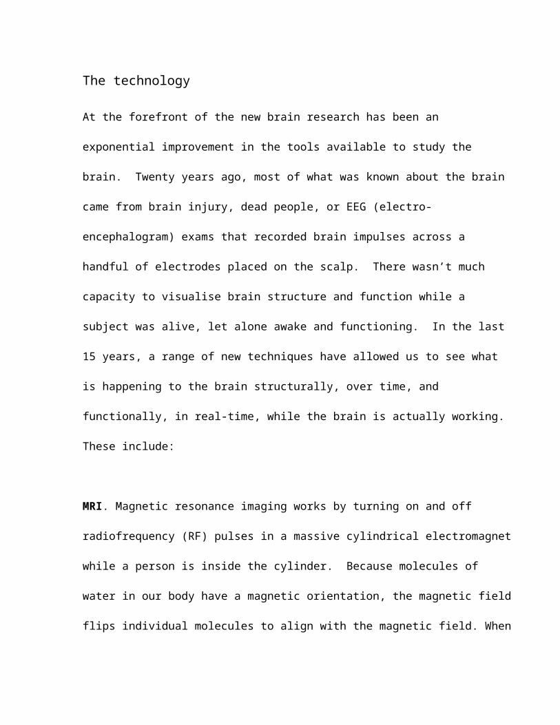
MRI. Magnetic resonance imaging works by turning on and off
radiofrequency (RF) pulses in a massive cylindrical electromagnet
while a person is inside the cylinder. Because molecules of
water in our body have a magnetic orientation, the magnetic field
flips individual molecules to align with the magnetic field. When
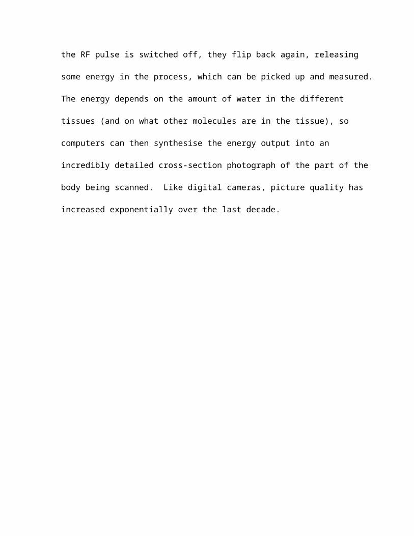
the RF pulse is switched off, they flip back again, releasing
some energy in the process, which can be picked up and measured.
The energy depends on the amount of water in the different
tissues (and on what other molecules are in the tissue), so
computers can then synthesise the energy output into an
incredibly detailed cross-section photograph of the part of the
body being scanned. Like digital cameras, picture quality has
increased exponentially over the last decade.
Improvement of MR images of brain structure in the last 30years.
From left to right: An image of a dead brain obtained on a 0.1 T scannerin Nottingham in 1978 (courtesy of Prof. Peter Morris, Sir Peter Mansfield MR Centre); An average of 27 images obtained by scanning the same individual repeatedly on a 1.5T scanner in 1995 (courtesy of Prof.
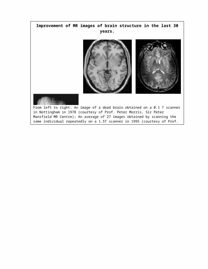
Principles of fMRI
Courtesy of Prof. Bruce Pike, Montreal Neurological Institute
Functional MRI (fMRI). Functional MRI works a little
differently, though still using the MRI scanner. Brains get
their energy from the oxidation of sugars in the bloodstream. If
a part of the brain is active, blood containing oxygen will flow
to that part of the brain, and as the oxygen is used, the oxygen
in the blood in that part of the brain will drop. A MRI scanner
will pick that up, giving a clear picture of the parts of the
brain that are active and which are not: all while the person is
awake and active, albeit lying quite still in the scanner. A
person can be shown pictures or videos, and asked to think about

certain things or do mental tasks, and we can see what parts of
the brain are working while they do that.
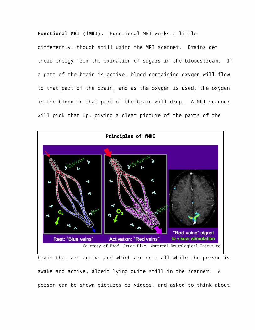
PET (Positron Emission Tomography). PET works by injecting a
radio-isotope substance into the body. The isotope gives off
positrons (a sub-atomic particle) in such a way that the isotope
can be located precisely at that moment. Positrons interact with
electrons and this can be picked up in scanning devices,
effectively giving an internal map of the body in the area
connected to the site of injection. There are risks, however,
because the isotope is radioactive. It can’t be used on small
children.
Principles of PET
Courtesy of Dr. Ernst Meyer, Montreal Neurological Institute
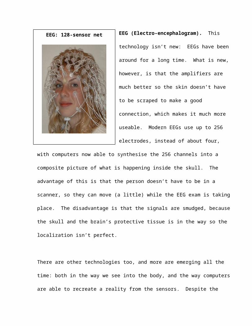
EEG: 128-sensor net EEG (Electro-encephalogram). This
technology isn’t new: EEGs have been
around for a long time. What is new,
however, is that the amplifiers are
much better so the skin doesn’t have
to be scraped to make a good
connection, which makes it much more
useable. Modern EEGs use up to 256
electrodes, instead of about four,
with computers now able to synthesise the 256 channels into a
composite picture of what is happening inside the skull. The
advantage of this is that the person doesn’t have to be in a
scanner, so they can move (a little) while the EEG exam is taking
place. The disadvantage is that the signals are smudged, because
the skull and the brain’s protective tissue is in the way so the
localization isn’t perfect.
There are other technologies too, and more are emerging all the
time: both in the way we see into the body, and the way computers
are able to recreate a reality from the sensors. Despite the
advances, however, the technology is expensive, and experiments
have to be painstakingly carried out. A lot of work (and money)
goes into finding each fragment of new data. There are, however,
some interesting and exciting findings.
Structure and function
For a long time in the human sciences (especially psychology),
debate has raged about the extent to which human behaviour is
determined by genes, and to what extent by experience and
environment – the so-called “nature-nurture debate”. Research
has pushed the pendulum this way or that over the last century,
but without resolution. One of the most important findings of
brain science research is that experience actually creates physical
structures in the brain.
The theory isn’t new: Donald Hebb wrote in 1949 that
When one cell repeatedly assists in firing another, the axon of the
first cell develops synaptic knobs (or enlarges them if they already
exist) in contact with the soma of the second cell. …The general
idea is an old one, that any two cells or systems of cells that are
repeatedly active at the same time will tend to become 'associated',
so that activity in one facilitates activity in the other.
(Hebb 1949: 63, 70)
Or, put more simply, “neurons that fire together, wire together”.
Brains are formed of a massive number of networks of neurons:
spindly, branching cells looking like plant roots that transmit
electrical currents along their length, like circuits in a
computer. With every experience you have, something happens to a
neuron somewhere. It might be a new connection, or an extension
of an existing connection, or a new branch. Making a new
connection may take some time, and there are an almost unlimited
number of ways to make the connection in a way that works. The
brain cannot develop without this kind of experience. It is as
useless as a computer without software.
While the theory is not new, the scanners are now so good that we
can see the differences in brain structure and function that
result from different experiences. Like hardware and software,
both genes and experience are important. In a recent special
issue of Human Brain Mapping (Glahn, Paus et al. 2007), a number
of articles reported that when you study twins, looking at the
amount of grey matter in different parts of the brain, you find
that their brains really are more similar than those of unrelated
individuals. This is true for adults, children and young people.
When the genes are different, brain structure is different
(Pezawas, Verchinski et al. 2004; Pezawas, Meyer-Lindenberg et
al. 2005).
On the other side, several studies have confirmed that when a
particular neural circuit is engaged repeatedly, it leads to
changes in brain structure. This has been tried across
populations as diverse as musicians (Sluming, Barrick et al.
2002; Gaser and Schlaug 2003); London taxi drivers (Maguire,
Gadian et al. 2000) and people who are bilingual (Mechelli,
Crinion et al. 2004). You can actually see, using a scanner, the
bit of their brains that is different. In one experiment, a
group of students were taught to juggle, and were set on a
program of juggling practice over some months. Brain scans done
after the practice, and compared with the initial scans, showed
that the part of their brains associated with tracking moving
objects had physically grown. Over the next few months, once the
juggling had stopped, it shrank somewhat, though not back to what
it was (Draganski, Gaser et al. 2004). To add complexity, it is
clear that genes can be switched on and off according to age, or
environmental stimulus. You might have the gene, but it won’t do
anything until your fifteenth birthday, and only then if the
conditions are right.
Overall, there is an increasing body of evidence that challenges
the simple, one-way view that genes directly influence the brain
and, in turn, the individual’s behaviour, or that experience or
socialization directly determines behaviour, without the
influence of genetic programming. It is difficult to work with
the way that all these factors influence each other. It isn’t
possible to hold one still while you see what happens with the
other one, because the first one doesn’t hold still. What we do
know is that the human brain as a structure is highly “plastic”:
it is flexible, responsive, and by no means determined at birth.
There are a number of important implications of this discovery.
The first is that the nature-nurture debate is obsolete. Neither
genes nor experience determine behaviour. Both do, in a complex
dance which includes the person’s own brain as a structure. It
makes no more sense to talk about which is determining behaviour
than it does to talk about whether it is Torvill or Dean1 who is
doing the dancing, or to talk about a coin only having one side.
Neither variable is independent.
The second results from the fact that the process of circuit-
building is not linear throughout life. There is a massive
proliferation of synapses, for example, in the first two years of
life, and another just before puberty. Between 10 and 12 years
the volume of grey matter in the frontal and parietal lobes
peaks, and then decreases slightly. In the temporal lobes the
peak occurs around 16 years (Giedd, Blumenthal et al. 1999a). If
the environment is poor, cruel, or chaotic during these periods,
that may determine many of the circuits that are laid down, if
not the way they are laid down. This is already having an impact
1 Jayne Torvill and Christopher Dean were the ice dancers who scored a perfect score at the 1984 Winter Olympics.
on policy around the care and education of infants. The same
attention is not yet being paid to school-age children and young
people. If you want good circuits to use as an adult, you need
good things in your environment when you are a kid.
Notwithstanding this, no matter how poor, cruel or chaotic the
environment, some good things happen to children – some good
experiences, some good relationships. Some people seem to be
able to foreground these experiences, regardless of how few they
have had. On the other hand, some people seem to foreground bad
experiences, no matter how privileged, kind and ordered their
environment has been. This can change. It is obvious that the
more poverty, cruelty or chaos you have had while the circuits
are being built, the more difficult it is to foreground the
helpful ones. One of the things we do in practice is to try to
help young people foreground what is helpful in their experience,
and sideline the circuits that make them smaller and meaner.
“Pruning” and the changing balance of grey and white
matter
Over the past 15 years, magnetic resonance imaging (MRI) has
provided new opportunities to assess brain development in large
numbers of healthy children and adolescents. Sophisticated image-
analysis tools allow investigators to identify and measure
various structural features from MR images of the living brain
(Fig. 1). It is now clear from a number of studies that the
human brain continues to change during adolescence (reviewed by
Paus 2005; Blakemore and Choudhury 2006; Lenroot and Giedd 2006).
One of the key changes is in the balance between grey matter and
white matter. Using a computer analogy, the grey matter is the
circuits and processors. The white matter is the wires between
them, with insulation wrapped around them. It is called white
matter because a major component is myelin, a white fatty
substance that insulates the wires, making the transfer of
electrical pulses, or “messages”, faster and more efficient.

Another useful analogy is that of roads. If you look at aerial
photos of the Australian outback, you will see roads everywhere:
little dirt roads running to mine diggings or shacks or forgotten
places that didn’t exist any more; graded roads to sheep stations
or water tanks or places people still lived in; gravel roads
between small towns; bitumen roads between bigger towns or places
of wealth and importance. Back at the turn of last century, in
the gold rushes, if a person wanted to go somewhere, they pointed
their cart or wheelbarrow in the direction they thought they
wanted to go and off they went. Others might follow their track,
and as they did, a road formed. Or they might go another way
that they thought was quicker or easier, and make another road.
Over time, if the road was used a lot, the Shire might grade it
and lay gravel, and eventually bitumen. Once the bitumen was
down, everyone went along the bitumen, and the little dirt roads
got overgrown. They rarely completely disappeared because it was
so dry, but going along them was hard work and they mostly ended
nowhere. Myelin is the bitumen of the brain.

Childhood is a process of creating little dirt roads all over the
place, learning so fast, learning or inventing a hundred ways to
do things, and learning a hundred things to do every day.
Children’s grey matter is just blossoming. In adults, it is much
harder to see all these little dirt roads. Instead, there is a
network of bitumen highways: serious, efficient, fast. All the
roads that don’t go anywhere, or aren’t the fastest or safest
ways to get there, have been left to grow over.
Now, one has to be careful of analogies. The fact is that the
connections between nerves are as important as the nerves
themselves in the transmission of information. It’s like every
road in the analogy above is a toll road. The toll stations vary
a lot in efficiency: some have electronic readers that
automatically deduct a bank account and mean that you don’t have
to slow down at all, others have a single toll lane with a grumpy
old attendant who never has the right change. There can be a
highly efficient motorway with an inefficient toll station and
the whole circuit will still be inefficient. (Though even this
analogy has to be taken with a grain of salt.)
Although the timing is different for boys and girls, and
different in different parts of the brain, the amount of grey
matter appears to reach a limit in the teenage years. After
that, there appears to be a decrease in grey matter (the total
number of circuits), and an increase in white matter (the
myelin). In the frontal, parietal and temporal lobes, this change
appears to start around puberty in the sensori-motor areas and
spreads forward over the frontal cortex and then back, first over
the parietal cortex and then the temporal (Sowell, Thompson et
al. 2001; Gogtay, Giedd et al. 2004). The change comes to the
dorsolateral prefrontal cortex and the posterior part of the
superior temporal gyrus last of all (Gogtay et al. 2004), as late
as the early twenties. The same process can be observed by
measuring the thickness of the cortex (Shaw, Greenstein et al.
2006).
A common way to describe this change is “synaptic pruning”
(Thompson, Giedd et al. 2000). In this metaphor, unwanted
circuits are pruned away, leaving the circuits that are most
efficient or most useful for survival. We aren’t sure that
“pruning” is the best way to see it, however. The amount of grey
matter might not actually decrease: it might just be that the
signal from grey matter is “diluted” by the white matter and so
appears as a reduction in volume in the scans (Sowell, Thompson
et al. 2001; Paus 2005). What is certain, though, is that the
amount of white matter increases significantly in the teenage
years. There is a serious road-building program going on,
starting with the areas that are more fundamental to survival,
and moving on to areas that are more concerned with conscious
thought. The process is generally called myelination.
Implications of the myelination process
We assume that the human organism will make decisions about which
circuits will be confirmed and which ones bypassed according to
the imperatives of its environment. These are, presumably, part
of a survival process. The environment in which young people
live while these decisions are made is critical in determining
the mind-set of the adult. If young people live in an
environment of suspicion and repression, the circuits that are
confirmed during the teenage years will be those that are most
appropriate to survival in such an environment. If this was to
be taken seriously, it is doubtful that what passes for youth
policy would have quite the shape that it does at the moment.
At the level of practice, our work is often about helping young
people find other ways to do things. They may have myelinised a
circuit in a context where their life was full of threat and
violence, and where there were few real options. Now, later,
they are in a place where the hyper-alertness and instant
defensive reactions appropriate to that kind of life are no
longer necessary – and, indeed, threaten their survival in the
present. In this very simplified model that we are using, change
can often be the struggle to find the way onto a little overgrown
dirt track that will enable the person to deal with situations in
a way that are happier and more successful. It isn’t easy: the
bitumen is always easier to find and quicker and smoother to
travel down. But in time, with practice and hard work, the dirt
becomes a graded road, the graded road becomes gravel, the gravel
becomes bitumen, and the old bitumen road becomes broken up and
potholed.
In counselling situations, helping young people connect with the
relationships or experiences in their past that worked and
nourished them can help them find a different way of being in the
present. So asking questions like “So who liked you as you were
growing up? What teachers respected you? What was that like?” or
“When have you been at your best with this stuff? When has it
worked? What was going on for you then?” or “How would you like
to be? What are you like when you are at your best?” can help
young people find the beginning of the little dirt track and move
off the bitumen.
In practice, this approach is very useful in working with young
people, especially young men, around a range of issues including
violence and drug use. The mechanics of how the brain works
often makes real sense to them, helping them understand why they
react the way they do, and empowering them to take charge of the
way they want their brain to work.
It can also help inform the logic of our activity work with young
people. Young people from impoverished backgrounds often have a
limited range of experiences, and their environment can be highly
conservative in its own way. New experiences can force the
development of different connections and new circuits, creating
opportunities for young people to do things a little differently
and see other possibilities while still respecting the integrity
of their lives and the choices they make. We have called this
methodology “ecological shock”, and have used experiences ranging
from travel to light aircraft joyrides to a dress-up dinner at a
fancy restaurant. Often, you can see the new connections happen
before your eyes, as a young person suddenly “gets it” or “the
penny drops”.
Different locations for processing information
Functional MRI (fMRI) provides yet another avenue for exploring
brain-behaviour relationships in the maturing human brain.
Functional MRI allows researchers to see what parts of the brain
“light up” when subjects are asked to respond to different
situations or perform different kinds of mental activity. It is
difficult work: it can be hard to pinpoint exactly what is being
measured. For example, whether the person is actually thinking
about the activity or something else. Or whether a difference in
the way that the brain works is about age, or something else like
intelligence or performance. Some of these things can be
controlled for, some can’t, and some the researcher may not have
thought of yet. But the possibility of seeing how the brain is
working while tasks are being performed is groundbreaking.
An influential study of this kind was published by Deborah
Yurgelin-Todd and her colleagues in 1999. In this fMRI study,
the researchers mapped what parts of the brain were active when a
research group of teenagers (mostly around 13 years old) were
shown pictures of faces expressing different emotions. They
found that while older adults used their prefrontal cortex while
they identified the emotion expressed in the photograph, the
younger research subjects used the amygdala, part of the more
fundamental limbic structure of the brain responsible for
emotional reactions (Baird, Gruber et al. 1999). The
interpretation was that this was because the prefrontal cortex
wasn’t effectively wired up to the amygdala yet, though this has
not been confirmed. There are also some initial indications that
young people use their brains differently from other adults. The
evidence does seem to building that the process of referring
information to the frontal cortex is less immediate for young
people than for adults.
As we noted above, the pre-frontal cortex is associated with a
number of mental functions, including decision making, working
memory and the suppression of alternative programs interfering
with planned actions (Duncan & Owen 2000, Miller & Cohen 2001,
Petrides 2005, Paus 2001). The Yurgelin-Todd study has been
interpreted as indicating that young people’s responses are more
“primitive” than those of other adults, that they are much more
likely to react out of their gut reactions, and are less able to
think about the consequences of their action. A more careful
interpretation might be that referring a decision to formal
thinking processes is not necessarily so automatic or
streamlined, and may be slower, in young people than for those
who are older.
During adolescence (and the rest of life for that matter), high
demands are placed not only on the brain’s executive systems –
the systems that coordinate action - but also on the interplay
between cognitive (thinking) and affective (feeling) processes.
Such cognition-emotion interactions are particularly crucial in
the context of peer-peer interactions and the processing of
verbal and non-verbal cues. It is likely that the interplay of
thinking and feeling is particularly important in social
situations in which the right balance must be struck between
peer-based influences and the individual’s own goals. Peer
influence was the basis of a recent fMRI study undertaken by
Tomas’ team in the Brain and Body Institute.
In this study the team wanted to see which neural systems – if
any – are engaged in children or adolescents who differ in their
resistance to peer influences. We asked this question by
examining neural activity across different areas of the brain.
Whether or not an adolescent follows the goals set by peers or
those set by himself/herself might depend on the interplay
between three neural systems in particular, namely the fronto-
parietal network (which deals with bottom-up imitation of
actions), the superior temporal sulcus (STS) network (which sorts
out social cues) and the prefrontal cortex (PFC) network (which
directs top-down regulation of actions).
To answer the question, a group of 10-year olds were asked to
fill out a questionnaire developed by psychologists Laurence
Steinberg and Kathryn Monahan (2007) to study resistance to peer
pressure as children move into and through adolescence. The
Resistance-to-Peer-Influence (RPI) scale, a self-report
questionnaire, is designed to elicit attitudes to peer influence
but minimize subjects answering with the “right” (socially
desirable) response. Then they were asked to watch brief video-
clips containing face or hand/arm actions, executed in calm or
angry ways, while measuring changes in fMRI signal. The team
found that the children with high RPI scores showed stronger
inter-regional correlations in brain activity across the three
networks mentioned above while watching angry hand-actions
(Grosbras et al. 2007; Fig. 4). The pattern of inter-regional
correlations identified by this method included both (i) regions
involved in action observation: the fronto-parietal as well as
temporo-occipital systems and (ii) regions in the prefrontal
cortex.
What the scans showed was that a number of prefrontal regions
showed coordinated changes in the fMRI signal that correlated with
those in the other two neural systems involved in action
observation. Typically, the prefrontal cortex is engaged when the
subject performs an explicit task requiring, for example,
manipulation of information in working memory, inhibition of
imminent action and/or suppression of interference, or planning
and decision-making (Petrides, 2005). These children were not
asked to do anything that required that. We think that the
brains of the children who scored high on resistance to peer
influence engaged “executive” processes automatically when
challenged with relatively complex and socially relevant stimuli.
Now. We know that experience creates structures in the brain.
The experiment has found out that children who score high on
these tests are better at coordinating different areas of their
brain when they process information with interpersonal
implications. Does this function also produce corresponding
structures in the brain? The team examined this possibility in a
large sample of healthy adolescents (n=295, 12 to 18 years of
age) and found that inter-regional correlations in cortical thickness
in the same cortical regions revealed by the above fMRI study
were higher in adolescents who scored high on the RPI test vs.
those who scored low (Paus et al. 2007, Fig. 5). Based on these
results, they suggested that individuals with certain personality
and cognitive characteristics, compatible with high resistance to
peer influences, are more likely to engage relevant neural
networks whenever challenged with relatively complex and socially
relevant stimuli. These networks include cortical regions
activated during action observation and cognitive/executive
control. Over time, such a coordinated functional engagement is
likely to shape these regions so that they become structurally
alike.
There are risks in the interpretation of these data. Brain
research, and fMRI in particular, is vulnerable to over-
simplification, over-interpretation, and the confirmation of
prior prejudice. Especially in media reports, huge claims can be
made about differences in human capacity based on pretty tenuous,
and often small, observations of differences in brain activity or
structure in different populations. As a rule, if people are
making strong claims about “this is why adolescents do x”, treat
with caution. We just aren’t that far along yet.
The implications of brain research for our concept of
youth/adolescence
The question of what youth/adolescence is exactly is still hotly
disputed (Sercombe 1996; Bessant, Sercombe et al. 1998; Epstein
2007). On one hand there is the claim that it is nothing but a
social construction, with no material difference between
teenagers and other adults, and any observed differences a
product of the streaming of young people into age cohorts, age
specific institutions, social exclusion and repressive social
treatment. Development over the lifespan is a continuous
unfolding, shaped by the learning process. On the other is the
dominant view that youth/adolescence is a biologically
determined, discrete stage of the life span, qualitatively
different from childhood and adulthood, and characterized by
turbulence, storm and stress. Both of these views are now, we
believe, obsolete.
The brain is in a process of continuous development through the
lifespan, in a constant dance between the influence of biological
factors and the physical and social environment, and involving
the person’s own agency. However, the process is not linear:
there are surges of growth and change in different parts of the
brain, and in different processes within it, at different times.
The timing of these is a function of the interplay between the
environment and the genetic program. And likely, in ways we do
not yet understand, also the person’s own agency.
The brains of young people are not radically different from other
adults in structure: there is no great difference in capacity
between young people and other adults. There is a difference,
however, in the degree of myelination, which makes brains more
reliable and efficient in their reactions and responses but less
flexible and less available for new learning. The major brain
development in the teenage years is the ramping up of the process
of myelination, which then levels off to some degree in the mid-
twenties.
The primary difference between a teenager’s brain and an older
person’s brain then is not a difference in capacity but in the selection
of capacities: that is, which of the brain’s capacities are to be
foregrounded and used and which are to be sidelined and fall into
disuse. This is an active process, in which young people are
consciously or unconsciously selecting preferred pathways for
action and response, confirming favoured templates for life from
the smorgasbord of ways of being generated through the process of
childhood. It happens according to the survival and other
interests of the individual in their social context.

Young people are not passive victims of brains that are out of
control. They are active agents in the design of an adulthood
that meets their needs and enables them to survive within their
environment and make sense of their experience. Youth is not
separate from adulthood. It is the becoming of adulthood. There
is no “next stage” of adulthood, which is qualitatively different
from being a young person, and adulthood is not itself a
destination. You don’t learn what you need for adulthood by
being excluded from it until you can demonstrate that you have
got the right circuits. A smart society would engage young
people progressively in adult processes, as they demonstrate
their readiness. Our society does this a little. But mostly, we
exclude young people until a certain arbitrary age is reached,
and then bestow the right to participate - mostly without
guidance and support. It should be no surprise that it doesn’t
work too well. Faced with this, however, we respond to this
failure usually by increasing the age at which responsibility
will be granted. Folly, as Barbara Tuchman tells us, is the
pursuit of a failing strategy by prescribing ever-increasing
amounts of the same (Tuchman 1984).
The research isn’t always interpreted this way, though
influential work by Robert Epstein, among others, is pushing
strongly in this direction (Epstein 2007). The research tends to
be dominated, not surprisingly, by the century-old dominant view
of adolescence, which is the “stage of life characterized by
turbulence” view. Experiments are designed within this frame,
and written up and publicised accordingly, with the media often
taking what are already stigmatizing interpretations and pushing
them further for mass titillation. In the next article, we will
focus more on problems and dangers in the teen brain research,
and the assumptions underlying its interpretations.
References:
Baird, A. A., S. A. Gruber, et al. (1999). "Functional magnetic resonance imaging of facial affect recognition in children and adolescents." Journal of the American Academy of Child & Adolescent Psychiatry 38: 195-9.
Bessant, J., H. Sercombe, et al. (1998). Youth studies: an Australian perspective. Melbourne, Addison Wesley Longman.
Blakemore, S. J. and S. Choudhury (2006). "Development of the adolescent brain: implications for executive domain and social cognition." Journal of Child Psychology and Psychiatry 47: 296-312
Draganski, B., C. Gaser, et al. (2004). "Neuroplasticity: changesin grey matter induced by training." Nature 427: 311-2.
Epstein, R. (2007). The case against adolescence: rediscovering the adult in every teen Sanger, California, Quill Driver Books.
Epstein, R. (2007). "The myth of the teen brain." Scientific American Mind: 57-63.
Gaser, C. and G. Schlaug (2003). "Brain structures differ betweenmusicians and non-musicians." Journal of Neuroscience 23: 9240-5.
Giedd, J. N., J. Blumenthal, et al. (1999a). "Brain development during childhood and adolescence: a longitudinal MRI study." Nature Neuroscience 2.
Glahn, D. C., T. Paus, et al. (2007). "Imaging genomics: mapping the influence of genetics on brain structure and function." HumanBrain Mapping 28: 461-3.
Gogtay, N., J. N. Giedd, et al. (2004). "Dynamic mapping of humancortical development during childhood through early adulthood." Proceedings of the National Academy of Sciences USA 101: 8174-9
Hebb, D. O. (1949). The organization of behavior. New York, Wiley.
Lenroot, R. K. and J. N. Giedd (2006). "Brain development in children and adolescents: Insights from anatomical magnetic resonance imaging." Neuroscience and Biobehavioural Reviews 30: 718-729.
Maguire, E. A., D. G. Gadian, et al. (2000). "Navigation-related structural change in the hippocampi of taxi drivers." Proceedingsof the National Academy of Sciences USA 97: 4398-403.
Mechelli, A., J. T. Crinion, et al. (2004). "Neurolinguistics: structural plasticity in the bilingual brain." Nature 431: 757.
Paus, T. (2005). "Mapping brain maturation and cognitive development during adolescence." Trends in Cognitive Science 9: 60-8.
Pezawas, L., A. Meyer-Lindenberg, et al. (2005). "5-HTTLPR polymorphism impacts human cingulate-amygdala interactions: a genetic susceptibility mechanism for depression." Nature Neuroscience 8: 828-34.
Pezawas, L., B. A. Verchinski, et al. (2004). "The brain-derived neurotrophic factor val66met polymorphism and variation in human cortical morphology." Journal of Neuroscience 24: 10099-102.
Sercombe, H. (1996). Naming youth: the construction of the youth category. Philosophy, Politics and Sociology. Perth, Murdoch University. Doctor of Philosophy.

Shaw, P., D. Greenstein, et al. (2006). " Intellectual ability and cortical development in children and adolescents." Nature 440: 676-9.
Sluming, V., T. Barrick, et al. (2002). "Voxel-based morphometry reveals increased gray matter density in Broca's area in male symphony orchestra musicians." Neuroimage 17: 1613-22.
Sowell, E. R., P. M. Thompson, et al. (2001). "Mapping continued brain growth and gray matter density reduction in dorsal frontal cortex: Inverse relationships during post-adolescent brain maturation." J Neurosci 21: 8819-29.
Strauch, B. (2004). The Primal Teen: What the New Discoveries about the Teenage Brain Tell Us about Our Kids New York, Doubleday.
Thompson, P. M., J. N. Giedd, et al. (2000). "Growth patterns in the developing brain detected by using continuum mechanical tensor maps." Nature 404: 190-193.
Tuchman, B. (1984). The march of folly: from Troy to Vietnam New York, Random House.
Wallis, C., K. Dell, et al. (2004 ). "Secrets of the Teen Brain."TIME Magazine.
Related Documents


