The Sterol Methyltransferases SMT1, SMT2, and SMT3 Influence Arabidopsis Development through Nonbrassinosteroid Products 1[W][OA] Francine Carland, Shozo Fujioka, and Timothy Nelson* Department of Molecular, Cellular, and Developmental Biology, Yale University, New Haven, Connecticut 06520 (F.C., T.N.); and RIKEN Advanced Science Institute, Wako-shi, Saitama 351–0198, Japan (S.F.) Plant sterols are structural components of cell membranes that provide rigidity, permeability, and regional identity to membranes. Sterols are also the precursors to the brassinosteroid signaling molecules. Evidence is accumulating that specific sterols have roles in pattern formation during development. COTYLEDON VASCULAR PATTERNING1 (CVP1) encodesC-24 STEROL METHYLTRANSFERASE2 (SMT2), one of three SMTs in Arabidopsis (Arabidopsis thaliana). SMT2 and SMT3, which also encodes a C-24 SMT, catalyze the reaction that distinguishes the synthesis of structural sterols from signaling brassinosteroid derivatives and are highly regulated. The deficiency of SMT2 in the cvp1 mutant results in moderate developmental defects, including aberrant cotyledon vein patterning, serrated floral organs, and reduced stature, but plants are viable, suggesting that SMT3 activity can substitute for the loss of SMT2. To test the distinct developmental roles of SMT2 and SMT3, we identified a transcript null smt3 mutant. Although smt3 single mutants appear wild type, cvp1 smt3 double mutants show enhanced defects relative to cvp1 mutants, such as discontinuous cotyledon vein pattern, and produce novel phenotypes, including defective root growth, loss of apical dominance, sterility, and homeotic floral transformations. These phenotypes are correlated with major alterations in the profiles of specific sterols but without significant alterations to brassinosteroid profiles. The alterations to sterol profiles in cvp1 mutants affect auxin response, demonstrated by weak auxin insensitivity, enhanced axr1 auxin resistance, ectopically expressed DR5:b-glucuronidase in developing embryos, and defective response to auxin-inhibited PIN2-green fluorescent protein endocytosis. We discuss the developmental roles of sterols implied by these results. Sterols are integral components of the cell mem- brane and serve to provide stability to the phospho- lipid bilayer. In addition to this structural role, in higher plants, animals, and insects, sterols are precur- sors of the steroid hormones. There are over 100 types of sterols identified in plant species, but the most abundant are sitosterol, stigmasterol, and campesterol (Guo et al., 1995; Hartmann, 1998). The diversity in plants is in marked contrast to the single sterol accu- mulated in fungi and animals, ergosterol and choles- terol, respectively (Benveniste, 2004). The balance of phytosterols with varied properties has been postu- lated to provide membrane integrity under the broad range of temperatures to which plants are exposed (Beck et al., 2007). Sterols are isoprenoid derivatives with a four-ring steroid nucleus (Edwards and Ericsson, 1999). A dis- tinguishing feature of cholesterol and the major phy- tosterols is the number of carbons at the carbon 24 (C-24) position (Benveniste, 1986). Phytosterols have one to two additional carbons due to alkylation events catalyzed by S-adenosyl-L-Met-dependent C-24 STE- ROL METHYLTRANSFERASEs (SMTs; Fig. 1). SMT1 catalyzes the initial step in sterol biosynthesis by a single methyl addition at C-24 (C 1 addition). SMT2 and SMT3 can each add a second methyl group (C 2 addition), thus completing an ethyl side chain addi- tion on C-24 (Husselstein et al., 1996; Bouvier-Nave ´ et al., 1997; Schaller et al., 1998; Nes et al., 1999). In plants, cycloartenol is the primary starting compound in sterol biosynthesis and is the substrate of SMT1, although there is some promiscuity in substrate spec- ificity among the SMTs (Diener et al., 2000). Arabidop- sis (Arabidopsis thaliana) SMT2 and SMT3 can promote both C 1 and C 2 additions in a yeast ergosterol6 mutant blocked at SMT1 and SMT2, and SMT1 can perform both methyl additions in bacteria (Husselstein et al., 1996; Bouvier-Nave ´ et al., 1997). Recently, a second sterol pathway was identified that utilizes lanosterol rather than cycloartenol in its initial step. This lano- sterol pathway is a minor branch, estimated to contrib- ute a small fraction (1.5%) of total sitosterol (Ohyama et al., 2009). Redundancy among enzymes and path- 1 This work was supported by the National Science Foundation (grant nos. IOS–0718881 and IBN–0416731 to T.N.) and by the Ministry of Education, Culture, Sports, Science and Technology of Japan (Grant-in-Aid for Scientific Research B no. 19380069 to S.F.). * Corresponding author; e-mail [email protected]. The author responsible for distribution of materials integral to the findings presented in this article in accordance with the policy described in the Instructions for Authors (www.plantphysiol.org) is: Timothy Nelson ([email protected]). [W] The online version of this article contains Web-only data. [OA] Open Access articles can be viewed online without a sub- scription. www.plantphysiol.org/cgi/doi/10.1104/pp.109.152587 Plant Physiology Ò , June 2010, Vol. 153, pp. 741–756, www.plantphysiol.org Ó 2010 American Society of Plant Biologists 741 Downloaded from https://academic.oup.com/plphys/article/153/2/741/6109622 by guest on 24 November 2021

Welcome message from author
This document is posted to help you gain knowledge. Please leave a comment to let me know what you think about it! Share it to your friends and learn new things together.
Transcript
The Sterol Methyltransferases SMT1, SMT2, and SMT3Influence Arabidopsis Development throughNonbrassinosteroid Products1[W][OA]
Francine Carland, Shozo Fujioka, and Timothy Nelson*
Department of Molecular, Cellular, and Developmental Biology, Yale University, New Haven, Connecticut06520 (F.C., T.N.); and RIKEN Advanced Science Institute, Wako-shi, Saitama 351–0198, Japan (S.F.)
Plant sterols are structural components of cell membranes that provide rigidity, permeability, and regional identity tomembranes. Sterols are also the precursors to the brassinosteroid signaling molecules. Evidence is accumulating that specificsterols have roles in pattern formation during development. COTYLEDON VASCULAR PATTERNING1 (CVP1) encodes C-24STEROL METHYLTRANSFERASE2 (SMT2), one of three SMTs in Arabidopsis (Arabidopsis thaliana). SMT2 and SMT3, whichalso encodes a C-24 SMT, catalyze the reaction that distinguishes the synthesis of structural sterols from signalingbrassinosteroid derivatives and are highly regulated. The deficiency of SMT2 in the cvp1 mutant results in moderatedevelopmental defects, including aberrant cotyledon vein patterning, serrated floral organs, and reduced stature, but plantsare viable, suggesting that SMT3 activity can substitute for the loss of SMT2. To test the distinct developmental roles of SMT2and SMT3, we identified a transcript null smt3 mutant. Although smt3 single mutants appear wild type, cvp1 smt3 doublemutants show enhanced defects relative to cvp1 mutants, such as discontinuous cotyledon vein pattern, and produce novelphenotypes, including defective root growth, loss of apical dominance, sterility, and homeotic floral transformations. Thesephenotypes are correlated with major alterations in the profiles of specific sterols but without significant alterations tobrassinosteroid profiles. The alterations to sterol profiles in cvp1 mutants affect auxin response, demonstrated by weak auxininsensitivity, enhanced axr1 auxin resistance, ectopically expressed DR5:b-glucuronidase in developing embryos, and defectiveresponse to auxin-inhibited PIN2-green fluorescent protein endocytosis. We discuss the developmental roles of sterols impliedby these results.
Sterols are integral components of the cell mem-brane and serve to provide stability to the phospho-lipid bilayer. In addition to this structural role, inhigher plants, animals, and insects, sterols are precur-sors of the steroid hormones. There are over 100 typesof sterols identified in plant species, but the mostabundant are sitosterol, stigmasterol, and campesterol(Guo et al., 1995; Hartmann, 1998). The diversity inplants is in marked contrast to the single sterol accu-mulated in fungi and animals, ergosterol and choles-terol, respectively (Benveniste, 2004). The balance ofphytosterols with varied properties has been postu-lated to provide membrane integrity under the broadrange of temperatures to which plants are exposed(Beck et al., 2007).
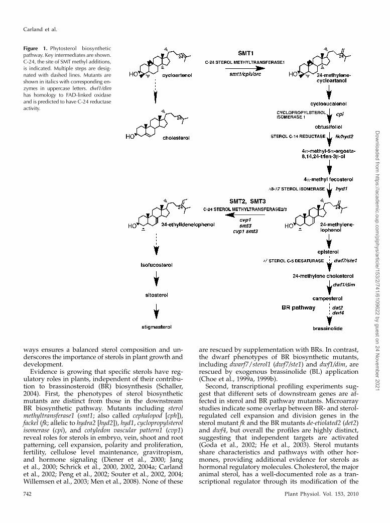
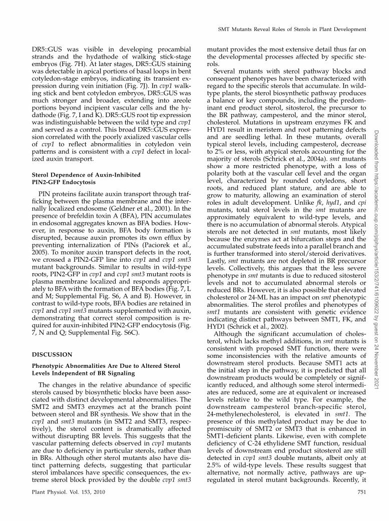
Sterols are isoprenoid derivatives with a four-ringsteroid nucleus (Edwards and Ericsson, 1999). A dis-tinguishing feature of cholesterol and the major phy-tosterols is the number of carbons at the carbon 24(C-24) position (Benveniste, 1986). Phytosterols haveone to two additional carbons due to alkylation eventscatalyzed by S-adenosyl-L-Met-dependent C-24 STE-ROL METHYLTRANSFERASEs (SMTs; Fig. 1). SMT1catalyzes the initial step in sterol biosynthesis by asingle methyl addition at C-24 (C1 addition). SMT2and SMT3 can each add a second methyl group (C2addition), thus completing an ethyl side chain addi-tion on C-24 (Husselstein et al., 1996; Bouvier-Naveet al., 1997; Schaller et al., 1998; Nes et al., 1999). Inplants, cycloartenol is the primary starting compoundin sterol biosynthesis and is the substrate of SMT1,although there is some promiscuity in substrate spec-ificity among the SMTs (Diener et al., 2000). Arabidop-sis (Arabidopsis thaliana) SMT2 and SMT3 can promoteboth C1 and C2 additions in a yeast ergosterol6 mutantblocked at SMT1 and SMT2, and SMT1 can performboth methyl additions in bacteria (Husselstein et al.,1996; Bouvier-Nave et al., 1997). Recently, a secondsterol pathway was identified that utilizes lanosterolrather than cycloartenol in its initial step. This lano-sterol pathway is a minor branch, estimated to contrib-ute a small fraction (1.5%) of total sitosterol (Ohyamaet al., 2009). Redundancy among enzymes and path-
1 This work was supported by the National Science Foundation(grant nos. IOS–0718881 and IBN–0416731 to T.N.) and by theMinistry of Education, Culture, Sports, Science and Technology ofJapan (Grant-in-Aid for Scientific Research B no. 19380069 to S.F.).
* Corresponding author; e-mail [email protected] author responsible for distribution of materials integral to the
findings presented in this article in accordance with the policydescribed in the Instructions for Authors (www.plantphysiol.org) is:Timothy Nelson ([email protected]).
[W] The online version of this article contains Web-only data.[OA] Open Access articles can be viewed online without a sub-
scription.www.plantphysiol.org/cgi/doi/10.1104/pp.109.152587
Plant Physiology�, June 2010, Vol. 153, pp. 741–756, www.plantphysiol.org � 2010 American Society of Plant Biologists 741
Dow
nloaded from https://academ
ic.oup.com/plphys/article/153/2/741/6109622 by guest on 24 N
ovember 2021
ways ensures a balanced sterol composition and un-derscores the importance of sterols in plant growth anddevelopment.
Evidence is growing that specific sterols have reg-ulatory roles in plants, independent of their contribu-tion to brassinosteroid (BR) biosynthesis (Schaller,2004). First, the phenotypes of sterol biosyntheticmutants are distinct from those in the downstreamBR biosynthetic pathway. Mutants including sterolmethyltransferase1 (smt1; also called cephalopod [cph]),fackel (fk; allelic to hydra2 [hyd2]), hyd1, cyclopropylsterolisomerase (cpi), and cotyledon vascular pattern1 (cvp1)reveal roles for sterols in embryo, vein, shoot and rootpatterning, cell expansion, polarity and proliferation,fertility, cellulose level maintenance, gravitropism,and hormone signaling (Diener et al., 2000; Janget al., 2000; Schrick et al., 2000, 2002, 2004a; Carlandet al., 2002; Peng et al., 2002; Souter et al., 2002, 2004;Willemsen et al., 2003; Men et al., 2008). None of these
are rescued by supplementation with BRs. In contrast,the dwarf phenotypes of BR biosynthetic mutants,including dwarf7/sterol1 (dwf7/ste1) and dwf1/dim, arerescued by exogenous brassinolide (BL) application(Choe et al., 1999a, 1999b).
Second, transcriptional profiling experiments sug-gest that different sets of downstream genes are af-fected in sterol and BR pathway mutants. Microarraystudies indicate some overlap between BR- and sterol-regulated cell expansion and division genes in thesterol mutant fk and the BR mutants de-etiolated2 (det2)and dwf4, but overall the profiles are highly distinct,suggesting that independent targets are activated(Goda et al., 2002; He et al., 2003). Sterol mutantsshare characteristics and pathways with other hor-mones, providing additional evidence for sterols ashormonal regulatory molecules. Cholesterol, the majoranimal sterol, has a well-documented role as a tran-scriptional regulator through its modification of the
Figure 1. Phytosterol biosyntheticpathway. Key intermediates are shown.C-24, the site of SMT methyl additions,is indicated. Multiple steps are desig-nated with dashed lines. Mutants areshown in italics with corresponding en-zymes in uppercase letters. dwf1/dimhas homology to FAD-linked oxidaseand is predicted to have C-24 reductaseactivity.
Carland et al.
742 Plant Physiol. Vol. 153, 2010
Dow
nloaded from https://academ
ic.oup.com/plphys/article/153/2/741/6109622 by guest on 24 N
ovember 2021

Hedgehog (Hh) ligand for the cell surface receptorPatched (Ptc; Incardona and Eaton, 2000). The Hhsignal transduction pathway activates target genes incell expansion and proliferation during embryonicand adult plant development (Duman-Scheel et al.,2002).Third, sterol mutants influence membrane structure
and traffic. The sterol biosynthetic mutants smt1orc,fk/hyd2, cpi, and cvp1 exhibit misdistribution of thepolarly localized PIN protein, an efflux transporter ofauxin, suggesting a sterol requirement at the level ofPIN endocytosis (Simons and Ikonen, 1997; Souteret al., 2002; Willemsen et al., 2003; Men et al., 2008; Panet al., 2009). In animals, cholesterol influences the polartrafficking of proteins through its ability to interactwith sphingolipids in specialized membrane micro-domains or lipid rafts (Simons and Ikonen, 1997).These membrane compartments serve to concentrateassociated proteins for enhanced interaction and, thus,more efficient cellular processes. Likewise, sterol reg-ulation in plants may not be solely at the transcrip-tional level.Fourth, the sterol biosynthetic genes are expressed
in regions of active cell division and expansion. In-deed, sitosterol, stigmasterol, and some abnormal fksterols up-regulate characteristic cell expansion andproliferation genes (He et al., 2003). Sterol balance isaffected in all mutants, but not always as predictedbased on a simple linear pathway. For example, com-promised SMT1 activity does not completely restrictfurther sterol transformations, as some downstreamsterol levels remain unaffected (Diener et al., 2000).Consistent with this result, the smt1 mutant pheno-type, particularly at the adult stage, is relatively mildcompared with other sterol biosynthetic mutants, eventhough smt1 acts at the initial step. The downstreambut more severe sterol mutants fk, hyd1, and cpi displaya more drastic reduction in sterol levels, accumulateabnormal sterols, and show decreased BR precursorlevels (Souter et al., 2002; Schrick et al., 2004a;Men et al.,2008). Furthermore, genetic data indicate a functionfor HYD1 and FK independent from SMT1 and sug-gest that the sterol biosynthetic pathway is morecomplex than previously believed (Schrick et al., 2002).In this study, we provide further evidence that
specific sterols influence numerous processes in plantdevelopment, independent of BR action. We previ-ously reported on CVP1 as encoding SMT2, a branchpoint enzyme functioning to balance sterol and BRlevels (Carland et al., 2002). Although SMT2 is ex-pressed in regions of rapid cell division and cellexpansion throughout development, the phenotypicabnormalities of cvp1 mutants are predominantly re-stricted to a cotyledon vein pattern defect and do notshare the gross embryo defects of smt1, cpi, fk, andhyd1. The close relative, SMT3, has a similar expressionprofile. Since overexpression of SMT3 can complementcvp1 mutants, we reasoned that the mild cvp1 pheno-type may be due to genetic redundancy with smt3.Here, we assign function in plant development to the
third SMT and report causal effects of depleting C-24ethylidene SMTs both at the phenotypic and sterollevels. Our results reveal some novel roles for sterols,particularly in flower development. A complete BRprofile shows that BR levels are unaffected in the cvp1mutant alleles, indicating that the phenotypic abnor-malities are BR independent.
RESULTS
Identification of the smt3 Mutant
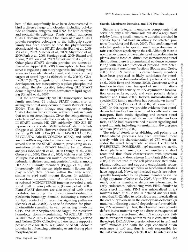
SMT2 and SMT3 genes are highly homologous(83% identity) and encode highly similar sterol24-carbon methyltransferases (Diener et al., 2000;Carland et al., 2002). The overexpression of SMT3complements the cotyledon venation pattern defectsof cvp1 (SMT2) mutants. In addition, antisense smt3plants did not display a phenotype. These resultssuggest that SMT3 and SMT2 are functionally redun-dant. To investigate the effects of complete deficiencyin C-24 ethylidene SMTs, we identified a T-DNAinsertion in the coding region of SMT3 from theSALK SIGNAL collection (Fig. 2A) and combined itwith the cvp1mutant allele of SMT2. The ArabidopsisSMTs are members of the methyltransferase super-family, with the signature methyltransferase domainand an S-adenosyl-L-Met-binding region. S-Adenosyl-L-Met provides the methyl group for transfer to the
Figure 2. Identification of an smt3mutant. A, Predicted structure of theSMT3 protein. The arrow denotes the T-DNA insertion in SALK SIGNALline 085292. Functional regions of the protein are indicated. SAMdenotes the S-adenosyl-L-Met-binding sites. The SMT C-terminal do-main is found in fungal and plant SMTs. B, RNA gel-blot analysis ofSMT transcript levels in the smt3 mutant. RNAwas extracted from 7-d-old seedlings. The filter was probed sequentially after complete proberemoval between hybridizations. WT, Wild type.
SMT Mutants Reveal Roles of Sterols in Plant Development
Plant Physiol. Vol. 153, 2010 743
Dow
nloaded from https://academ
ic.oup.com/plphys/article/153/2/741/6109622 by guest on 24 N
ovember 2021
SMT substrate, 24-methylenelophenol (24-ML). TheT-DNA insertion resides at amino acid 55 and ispredicted to eliminate expression of the 359-aminoacid SMT3 protein. RNA gel-blot analyses showed theabsence of an SMT3 transcript in smt3 mutants (Fig.2B). As has been demonstrated previously with cvp1and smt1 mutants, SMT1 and SMT2 transcript levelswere reduced in the smt3mutant, suggesting that SMTtranscription may be regulated through a sterol feed-back mechanism (Diener et al., 2000; Carland et al.,2002).
SMT3 Is Functionally Redundant with SMT2
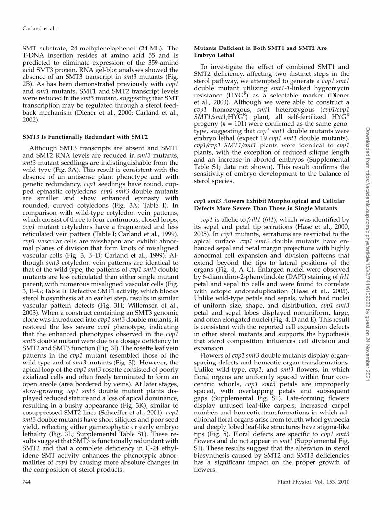
Although SMT3 transcripts are absent and SMT1and SMT2 RNA levels are reduced in smt3 mutants,smt3 mutant seedlings are indistinguishable from thewild type (Fig. 3A). This result is consistent with theabsence of an antisense plant phenotype and withgenetic redundancy. cvp1 seedlings have round, cup-ped epinastic cotyledons. cvp1 smt3 double mutantsare smaller and show enhanced epinasty withrounded, curved cotyledons (Fig. 3A; Table I). Incomparison with wild-type cotyledon vein patterns,which consist of three to four continuous, closed loops,cvp1 mutant cotyledons have a fragmented and lessreticulated vein pattern (Table I; Carland et al., 1999).cvp1 vascular cells are misshapen and exhibit abnor-mal planes of division that form knots of misalignedvascular cells (Fig. 3, B–D; Carland et al., 1999). Al-though smt3 cotyledon vein patterns are identical tothat of the wild type, the patterns of cvp1 smt3 doublemutants are less reticulated than either single mutantparent, with numerous misaligned vascular cells (Fig.3, E–G; Table I). Defective SMT1 activity, which blockssterol biosynthesis at an earlier step, results in similarvascular pattern defects (Fig. 3H; Willemsen et al.,2003). When a construct containing an SMT3 genomicclone was introduced into cvp1 smt3 double mutants, itrestored the less severe cvp1 phenotype, indicatingthat the enhanced phenotypes observed in the cvp1smt3 double mutant were due to a dosage deficiency inSMT2 and SMT3 function (Fig. 3I). The rosette leaf veinpatterns in the cvp1 mutant resembled those of thewild type and of smt3 mutants (Fig. 3J). However, theapical loop of the cvp1 smt3 rosette consisted of poorlyaxialized cells and often freely terminated to form anopen areole (area bordered by veins). At later stages,slow-growing cvp1 smt3 double mutant plants dis-played reduced stature and a loss of apical dominance,resulting in a bushy appearance (Fig. 3K), similar tocosuppressed SMT2 lines (Schaeffer et al., 2001). cvp1smt3 double mutants have short siliques and poor seedyield, reflecting either gametophytic or early embryolethality (Fig. 3L; Supplemental Table S1). These re-sults suggest that SMT3 is functionally redundant withSMT2 and that a complete deficiency in C-24 ethyl-idene SMT activity enhances the phenotypic abnor-malities of cvp1 by causing more absolute changes inthe composition of sterol products.
Mutants Deficient in Both SMT1 and SMT2 Are
Embryo Lethal
To investigate the effect of combined SMT1 andSMT2 deficiency, affecting two distinct steps in thesterol pathway, we attempted to generate a cvp1 smt1double mutant utilizing smt1-1-linked hygromycinresistance (HYGR) as a selectable marker (Dieneret al., 2000). Although we were able to construct acvp1 homozygous, smt1 heterozygous (cvp1/cvp1SMT1/smt1;HYGR) plant, all self-fertilized HYGR
progeny (n = 101) were confirmed as the same geno-type, suggesting that cvp1 smt1 double mutants wereembryo lethal (expect 19 cvp1 smt1 double mutants).cvp1/cvp1 SMT1/smt1 plants were identical to cvp1plants, with the exception of reduced silique lengthand an increase in aborted embryos (SupplementalTable S1; data not shown). This result confirms thesensitivity of embryo development to the balance ofsterol species.
cvp1 smt3 Flowers Exhibit Morphological and Cellular
Defects More Severe Than Those in Single Mutants
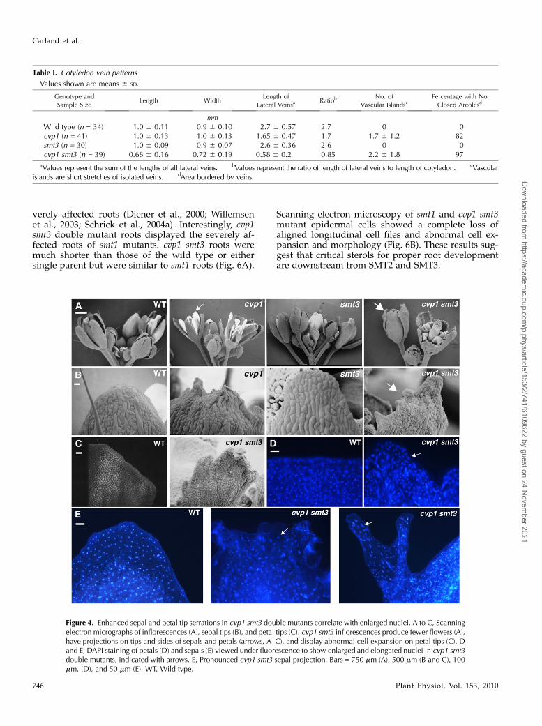
cvp1 is allelic to frill1 (frl1), which was identified byits sepal and petal tip serrations (Hase et al., 2000,2005). In cvp1 mutants, serrations are restricted to theapical surface. cvp1 smt3 double mutants have en-hanced sepal and petal margin projections with highlyabnormal cell expansion and division patterns thatextend beyond the tips to lateral positions of theorgans (Fig. 4, A–C). Enlarged nuclei were observedby 6-diamidino-2-phenylindole (DAPI) staining of frl1petal and sepal tip cells and were found to correlatewith ectopic endoreduplication (Hase et al., 2005).Unlike wild-type petals and sepals, which had nucleiof uniform size, shape, and distribution, cvp1 smt3petal and sepal lobes displayed nonuniform, large,and often elongated nuclei (Fig. 4, D and E). This resultis consistent with the reported cell expansion defectsin other sterol mutants and supports the hypothesisthat sterol composition influences cell division andexpansion.
Flowers of cvp1 smt3 double mutants display organ-spacing defects and homeotic organ transformations.Unlike wild-type, cvp1, and smt3 flowers, in whichfloral organs are uniformly spaced within four con-centric whorls, cvp1 smt3 petals are improperlyspaced, with overlapping petals and subsequentgaps (Supplemental Fig. S1). Late-forming flowersdisplay unfused leaf-like carpels, increased carpelnumber, and homeotic transformations in which ad-ditional floral organs arise from fourth whorl gynoeciaand deeply lobed leaf-like structures have stigma-liketips (Fig. 5). Floral defects are specific to cvp1 smt3flowers and do not appear in smt1 (Supplemental Fig.S1). These results suggest that the alteration in sterolbiosynthesis caused by SMT2 and SMT3 deficiencieshas a significant impact on the proper growth offlowers.
Carland et al.
744 Plant Physiol. Vol. 153, 2010
Dow
nloaded from https://academ
ic.oup.com/plphys/article/153/2/741/6109622 by guest on 24 N
ovember 2021
Root Defects in cvp1 smt3 Double Mutants ResembleThose in smt1
SMT2 is expressed in internal cells throughout thedeveloping embryo and has significant overlap with
SMT3 expression in both early and late stages of root
growth. Although cvp1 and smt3 mutant roots are
indistinguishable from those of the wild type, many
sterol biosynthetic mutants, including smt1, have se-
Figure 3. Developmental defects displayed in cvp1 smt3 double mutants. A, Seven-day-old seedlings. cvp1 smt3 doublemutants show enhanced epinasty. B to I, Cotyledon vein patterns of cleared specimens viewed under dark-field optics. D, Highermagnification of the boxed region in C. F, cvp1 smt3 double mutants have reduced vein length. G, Higher magnification of theboxed vascular island in F. H, smt1-1 to show similar vein pattern defects among smtmutants. I, cvp1 smt3with genomic clone ofSMT3 to show complementation of the cvp1 smt3 double mutant cotyledon vein pattern. J, First rosette leaf vein patterns. K,Adult plant phenotypes. The first four pots are 6-week-old plants. cvp1 plants are slightly dwarf. cvp1 smt3 plants show retardedgrowth and reduced plant height. The 3-month-old cvp1 smt3 plant on the far right shows excessive branching, resulting in abushy appearance. L, Representative siliques to show reduced length in cvp1 smt3 double mutants. Bars = 500 mm (A–C, E, andH–J), 50 mm (D and G), 250 mm (F), 3 cm (K), and 1 mm (L). WT, Wild type.
SMT Mutants Reveal Roles of Sterols in Plant Development
Plant Physiol. Vol. 153, 2010 745
Dow
nloaded from https://academ
ic.oup.com/plphys/article/153/2/741/6109622 by guest on 24 N
ovember 2021
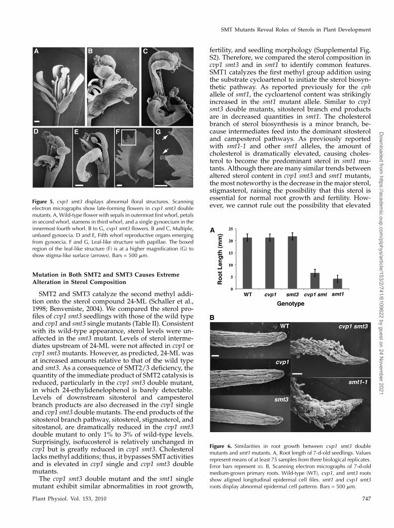
verely affected roots (Diener et al., 2000; Willemsenet al., 2003; Schrick et al., 2004a). Interestingly, cvp1smt3 double mutant roots displayed the severely af-fected roots of smt1 mutants. cvp1 smt3 roots weremuch shorter than those of the wild type or eithersingle parent but were similar to smt1 roots (Fig. 6A).
Scanning electron microscopy of smt1 and cvp1 smt3mutant epidermal cells showed a complete loss ofaligned longitudinal cell files and abnormal cell ex-pansion and morphology (Fig. 6B). These results sug-gest that critical sterols for proper root developmentare downstream from SMT2 and SMT3.
Table I. Cotyledon vein patterns
Values shown are means 6 SD.
Genotype and
Sample SizeLength Width
Length of
Lateral VeinsaRatiob No. of
Vascular IslandscPercentage with No
Closed Areolesd
mm
Wild type (n = 34) 1.0 6 0.11 0.9 6 0.10 2.7 6 0.57 2.7 0 0cvp1 (n = 41) 1.0 6 0.13 1.0 6 0.13 1.65 6 0.47 1.7 1.7 6 1.2 82smt3 (n = 30) 1.0 6 0.09 0.9 6 0.07 2.6 6 0.36 2.6 0 0cvp1 smt3 (n = 39) 0.68 6 0.16 0.72 6 0.19 0.58 6 0.2 0.85 2.2 6 1.8 97
aValues represent the sum of the lengths of all lateral veins. bValues represent the ratio of length of lateral veins to length of cotyledon. cVascularislands are short stretches of isolated veins. dArea bordered by veins.
Figure 4. Enhanced sepal and petal tip serrations in cvp1 smt3 double mutants correlate with enlarged nuclei. A to C, Scanningelectron micrographs of inflorescences (A), sepal tips (B), and petal tips (C). cvp1 smt3 inflorescences produce fewer flowers (A),have projections on tips and sides of sepals and petals (arrows, A–C), and display abnormal cell expansion on petal tips (C). Dand E, DAPI staining of petals (D) and sepals (E) viewed under fluorescence to show enlarged and elongated nuclei in cvp1 smt3double mutants, indicated with arrows. E, Pronounced cvp1 smt3 sepal projection. Bars = 750 mm (A), 500 mm (B and C), 100mm, (D), and 50 mm (E). WT, Wild type.
Carland et al.
746 Plant Physiol. Vol. 153, 2010
Dow
nloaded from https://academ
ic.oup.com/plphys/article/153/2/741/6109622 by guest on 24 N
ovember 2021
Mutation in Both SMT2 and SMT3 Causes ExtremeAlteration in Sterol Composition
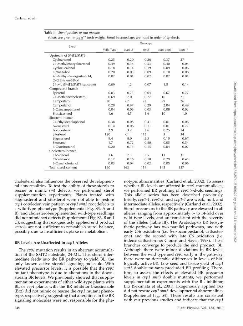
SMT2 and SMT3 catalyze the second methyl addi-tion onto the sterol compound 24-ML (Schaller et al.,1998; Benveniste, 2004). We compared the sterol pro-files of cvp1 smt3 seedlings with those of the wild typeand cvp1 and smt3 single mutants (Table II). Consistentwith its wild-type appearance, sterol levels were un-affected in the smt3 mutant. Levels of sterol interme-diates upstream of 24-ML were not affected in cvp1 orcvp1 smt3 mutants. However, as predicted, 24-ML wasat increased amounts relative to that of the wild typeand smt3. As a consequence of SMT2/3 deficiency, thequantity of the immediate product of SMT2 catalysis isreduced, particularly in the cvp1 smt3 double mutant,in which 24-ethylidenelophenol is barely detectable.Levels of downstream sitosterol and campesterolbranch products are also decreased in the cvp1 singleand cvp1 smt3 double mutants. The end products of thesitosterol branch pathway, sitosterol, stigmasterol, andsitostanol, are dramatically reduced in the cvp1 smt3double mutant to only 1% to 3% of wild-type levels.Surprisingly, isofucosterol is relatively unchanged incvp1 but is greatly reduced in cvp1 smt3. Cholesterollacks methyl additions; thus, it bypasses SMTactivitiesand is elevated in cvp1 single and cvp1 smt3 doublemutants.The cvp1 smt3 double mutant and the smt1 single
mutant exhibit similar abnormalities in root growth,
fertility, and seedling morphology (Supplemental Fig.S2). Therefore, we compared the sterol composition incvp1 smt3 and in smt1 to identify common features.SMT1 catalyzes the first methyl group addition usingthe substrate cycloartenol to initiate the sterol biosyn-thetic pathway. As reported previously for the cphallele of smt1, the cycloartenol content was strikinglyincreased in the smt1 mutant allele. Similar to cvp1smt3 double mutants, sitosterol branch end productsare in decreased quantities in smt1. The cholesterolbranch of sterol biosynthesis is a minor branch, be-cause intermediates feed into the dominant sitosteroland campesterol pathways. As previously reportedwith smt1-1 and other smt1 alleles, the amount ofcholesterol is dramatically elevated, causing choles-terol to become the predominant sterol in smt1 mu-tants. Although there are many similar trends betweenaltered sterol content in cvp1 smt3 and smt1 mutants,the most noteworthy is the decrease in the major sterol,stigmasterol, raising the possibility that this sterol isessential for normal root growth and fertility. How-ever, we cannot rule out the possibility that elevated
Figure 5. cvp1 smt3 displays abnormal floral structures. Scanningelectron micrographs show late-forming flowers in cvp1 smt3 doublemutants. A,Wild-type flower with sepals in outermost first whorl, petalsin second whorl, stamens in third whorl, and a single gynoecium in theinnermost fourth whorl. B to G, cvp1 smt3 flowers. B and C, Multiple,unfused gynoecia. D and E, Fifth whorl reproductive organs emergingfrom gynoecia. F and G, Leaf-like structure with papillae. The boxedregion of the leaf-like structure (F) is at a higher magnification (G) toshow stigma-like surface (arrows). Bars = 500 mm.
Figure 6. Similarities in root growth between cvp1 smt3 doublemutants and smt1 mutants. A, Root length of 7-d-old seedlings. Valuesrepresent means of at least 75 samples from three biological replicates.Error bars represent SD. B, Scanning electron micrographs of 7-d-oldmedium-grown primary roots. Wild-type (WT), cvp1, and smt3 rootsshow aligned longitudinal epidermal cell files. smt1 and cvp1 smt3roots display abnormal epidermal cell patterns. Bars = 500 mm.
SMT Mutants Reveal Roles of Sterols in Plant Development
Plant Physiol. Vol. 153, 2010 747
Dow
nloaded from https://academ
ic.oup.com/plphys/article/153/2/741/6109622 by guest on 24 N
ovember 2021
cholesterol also influences the observed developmen-tal abnormalities. To test the ability of these sterols torescue or mimic smt defects, we performed sterolsupplementation experiments. Plants treated withstigmasterol and sitosterol were not able to restorecvp1 cotyledon vein pattern or cvp1 smt3 root defects toa wild-type phenotype (Supplemental Fig. S3, A andB), and cholesterol-supplemented wild-type seedlingsdid not mimic smt defects (Supplemental Fig. S3, B andC), suggesting that exogenously applied end productsterols are not sufficient to reestablish sterol balance,possibly due to insufficient uptake or metabolism.
BR Levels Are Unaffected in cvp1 Alleles
The cvp1 mutation results in an aberrant accumula-tion of the SMT2 substrate, 24-ML. This sterol inter-mediate feeds into the BR pathway to yield BL, theonly known active steroid signaling molecule. Withelevated precursor levels, it is possible that the cvp1mutant phenotype is due to alterations in the down-stream BR levels. We previously showed that supple-mentation experiments of either wild-type plants withBL or cvp1 plants with the BR inhibitor brassinazole(Brz) did not mimic or rescue the cvp1 mutant pheno-type, respectively, suggesting that alterations in the BRsignaling molecules were not responsible for the phe-
notypic abnormalities (Carland et al., 2002). To assesswhether BL levels are affected in cvp1 mutant alleles,we performed BR profiling of cvp1 7-d-old seedlings.This allelic series has been described previously.Briefly, cvp1-1, cvp1-3, and cvp1-4 are weak, null, andintermediate alleles, respectively (Carland et al., 2002).Sterol precursors to the BR pathway are elevated in allalleles, ranging from approximately 3- to 14-fold overwild-type levels, and are consistent with the severityof the alleles (Table III). The Arabidopsis BR biosyn-thetic pathway has two parallel pathways, one withearly C-6 oxidation (i.e. 6-oxocampestanol, cathaster-one) and the second with late C6 oxidation (i.e.6-deoxocathasterone; Clouse and Sasse, 1998). Thesebranches converge to produce the end product, BL.Although there were minor deviations in BR levelsbetween the wild type and cvp1 early in the pathway,there were no detectable differences in levels of bio-logically active BR. Low seed and tissue yield of cvp1smt3 double mutants precluded BR profiling. There-fore, to assess the effects of elevated BR precursorlevels in cvp1 smt3 double mutants, we performedsupplementation experiments with the BL inhibitor,Brz (Sekimata et al., 2001). Exogenously applied Brzdid not rescue cvp1 smt3 developmental abnormalities(Supplemental Fig. S4). These results are consistentwith our previous studies and indicate that the cvp1
Table II. Sterol profiles of smt mutants
Values are given in mg g21 fresh weight. Sterol intermediates are listed in order of synthesis.
SterolGenotype
Wild Type cvp1-3 smt3 cvp1 smt3 smt1-1
Upstream of SMT2/SMT3Cycloartenol 0.25 0.20 0.26 0.37 2724-Methylenecycloartanol 0.49 0.34 0.53 0.40 0.04Cycloeucalenol 0.18 0.14 0.19 0.09 0.06Obtusifoliol 0.20 0.05 0.09 0.10 0.084a-Methyl-5a-ergosta-8,14,24(28)-trien-3b-ol
0.02 0.01 0.02 0.02 0.01
24-ML (SMT2/SMT3 substrate) 0.09 1.2 0.07 1.5 0.14Campesterol branch
Episterol 0.03 0.23 0.04 0.67 0.2724-Methlenecholesterol 0.69 7.0 0.77 16 21Campesterol 20 67 22 99 26Campestanol 0.29 0.97 0.29 2.04 0.496-Oxocampestanol 0.04 0.08 0.03 0.08 0.02Brassicasterol 1.6 4.5 1.6 10 1.0
Sitosterol branch24-Ethylidenelophenol 0.38 0.08 0.41 0.01 0.06Avenasterol 0.14 0.06 0.11 0.01 0.22Isofucosterol 2.9 3.7 2.6 0.25 14Sitosterol 120 61 113 3 34Stigmasterol 9.4 8.0 5.5 0.10 0.67Sitostanol 1.7 0.72 0.80 0.05 0.546-Oxositostanol 0.20 0.13 0.15 0.04 0.07
Cholesterol branchCholesterol 1.6 7.3 5.5 11 31Cholestanol 0.12 0.16 0.10 0.29 0.456-Oxocholestanol 0.03 0.04 0.02 0.05 0.06
Total sterol content 160 163 154 145 157
Carland et al.
748 Plant Physiol. Vol. 153, 2010
Dow
nloaded from https://academ
ic.oup.com/plphys/article/153/2/741/6109622 by guest on 24 N
ovember 2021
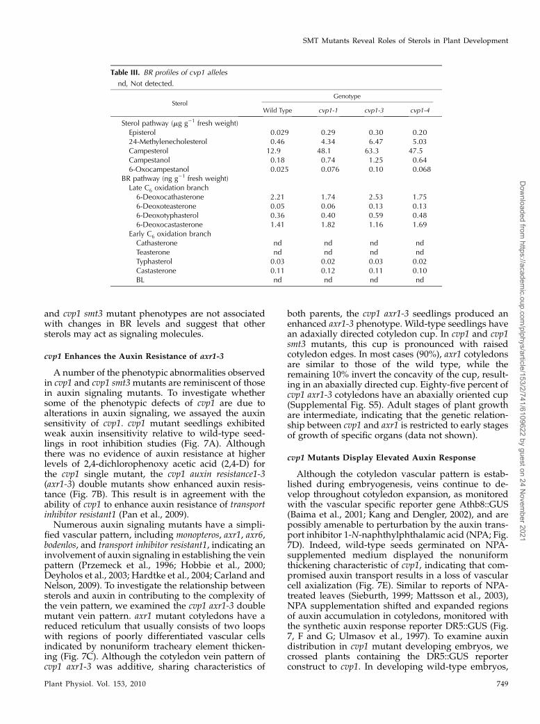
and cvp1 smt3 mutant phenotypes are not associatedwith changes in BR levels and suggest that othersterols may act as signaling molecules.
cvp1 Enhances the Auxin Resistance of axr1-3
A number of the phenotypic abnormalities observedin cvp1 and cvp1 smt3mutants are reminiscent of thosein auxin signaling mutants. To investigate whethersome of the phenotypic defects of cvp1 are due toalterations in auxin signaling, we assayed the auxinsensitivity of cvp1. cvp1 mutant seedlings exhibitedweak auxin insensitivity relative to wild-type seed-lings in root inhibition studies (Fig. 7A). Althoughthere was no evidence of auxin resistance at higherlevels of 2,4-dichlorophenoxy acetic acid (2,4-D) forthe cvp1 single mutant, the cvp1 auxin resistance1-3(axr1-3) double mutants show enhanced auxin resis-tance (Fig. 7B). This result is in agreement with theability of cvp1 to enhance auxin resistance of transportinhibitor resistant1 (Pan et al., 2009).Numerous auxin signaling mutants have a simpli-
fied vascular pattern, including monopteros, axr1, axr6,bodenlos, and transport inhibitor resistant1, indicating aninvolvement of auxin signaling in establishing the veinpattern (Przemeck et al., 1996; Hobbie et al., 2000;Deyholos et al., 2003; Hardtke et al., 2004; Carland andNelson, 2009). To investigate the relationship betweensterols and auxin in contributing to the complexity ofthe vein pattern, we examined the cvp1 axr1-3 doublemutant vein pattern. axr1 mutant cotyledons have areduced reticulum that usually consists of two loopswith regions of poorly differentiated vascular cellsindicated by nonuniform tracheary element thicken-ing (Fig. 7C). Although the cotyledon vein pattern ofcvp1 axr1-3 was additive, sharing characteristics of
both parents, the cvp1 axr1-3 seedlings produced anenhanced axr1-3 phenotype. Wild-type seedlings havean adaxially directed cotyledon cup. In cvp1 and cvp1smt3 mutants, this cup is pronounced with raisedcotyledon edges. In most cases (90%), axr1 cotyledonsare similar to those of the wild type, while theremaining 10% invert the concavity of the cup, result-ing in an abaxially directed cup. Eighty-five percent ofcvp1 axr1-3 cotyledons have an abaxially oriented cup(Supplemental Fig. S5). Adult stages of plant growthare intermediate, indicating that the genetic relation-ship between cvp1 and axr1 is restricted to early stagesof growth of specific organs (data not shown).
cvp1 Mutants Display Elevated Auxin Response
Although the cotyledon vascular pattern is estab-lished during embryogenesis, veins continue to de-velop throughout cotyledon expansion, as monitoredwith the vascular specific reporter gene Athb8::GUS(Baima et al., 2001; Kang and Dengler, 2002), and arepossibly amenable to perturbation by the auxin trans-port inhibitor 1-N-naphthylphthalamic acid (NPA; Fig.7D). Indeed, wild-type seeds germinated on NPA-supplemented medium displayed the nonuniformthickening characteristic of cvp1, indicating that com-promised auxin transport results in a loss of vascularcell axialization (Fig. 7E). Similar to reports of NPA-treated leaves (Sieburth, 1999; Mattsson et al., 2003),NPA supplementation shifted and expanded regionsof auxin accumulation in cotyledons, monitored withthe synthetic auxin response reporter DR5::GUS (Fig.7, F and G; Ulmasov et al., 1997). To examine auxindistribution in cvp1 mutant developing embryos, wecrossed plants containing the DR5::GUS reporterconstruct to cvp1. In developing wild-type embryos,
Table III. BR profiles of cvp1 alleles
nd, Not detected.
SterolGenotype
Wild Type cvp1-1 cvp1-3 cvp1-4
Sterol pathway (mg g21 fresh weight)Episterol 0.029 0.29 0.30 0.2024-Methylenecholesterol 0.46 4.34 6.47 5.03Campesterol 12.9 48.1 63.3 47.5Campestanol 0.18 0.74 1.25 0.646-Oxocampestanol 0.025 0.076 0.10 0.068
BR pathway (ng g21 fresh weight)Late C6 oxidation branch6-Deoxocathasterone 2.21 1.74 2.53 1.756-Deoxoteasterone 0.05 0.06 0.13 0.136-Deoxotyphasterol 0.36 0.40 0.59 0.486-Deoxocastasterone 1.41 1.82 1.16 1.69
Early C6 oxidation branchCathasterone nd nd nd ndTeasterone nd nd nd ndTyphasterol 0.03 0.02 0.03 0.02Castasterone 0.11 0.12 0.11 0.10BL nd nd nd nd
SMT Mutants Reveal Roles of Sterols in Plant Development
Plant Physiol. Vol. 153, 2010 749
Dow
nloaded from https://academ
ic.oup.com/plphys/article/153/2/741/6109622 by guest on 24 N
ovember 2021

Figure 7. Auxin response of cvp1 and cvp1 smt3 mutants. A, cvp1 mutants display weak auxin resistance. B, cvp1 enhancesauxin resistance of axr1-3. Root growth was measured 3 d after transfer of 4-d-old seedlings to MS medium or 2,4-D-containingMS medium. Root inhibition is the measurement of root growth on 2,4-D medium relative to that on medium without hormoneexpressed as a percentage. Values represent means of at least three experiments with measurements of approximately 40seedlings per experiment. Error bars indicate SD. C, Cotyledon vein patterns to demonstrate the additive phenotype in cvp1 axr1-3double mutants. The image at the far right is the boxed region in cvp1 axr1 at a higher magnification. D to G, Cotyledon veinpatterns of wild-type medium-grown plants without NPA (D and F) and with NPA (E and G). D, Athb8::GUS expression viewedunder dark-field optics to show that cotyledon veins are still developing and thus remain responsive to NPA-inducedperturbations. Pink and blue staining is weak and strong GUS staining, respectively. E, NPA-induced cotyledon vein patterndefects (arrow). F, DR5::GUS-stained 2-week-old cotyledon. DR5::GUS expression is no longer visible at this stage. G, EnhancedDR5:GUS expression in margins of NPA-grown cotyledons. H to K, Representative DR5::GUS staining in developing embryos toshow ectopic expression in cvp1mutants. H and I, Walking stick-stage embryos. J and K, Late bent cotyledon-stage embryos. L toQ, PIN2-GFP localization in wild-type (L–N) and cvp1 smt3 (O–Q) roots in response to BFA (M and P) or NAA + BFA (N and Q).Bars = 100 mm (C and D–I), 200 mm (J and K), and 25 mm (L–Q). WT, Wild type.
Carland et al.
750 Plant Physiol. Vol. 153, 2010
Dow
nloaded from https://academ
ic.oup.com/plphys/article/153/2/741/6109622 by guest on 24 N
ovember 2021
DR5::GUS was visible in developing procambialstrands and the hydathode of walking stick-stageembryos (Fig. 7H). At later stages, DR5::GUS stainingwas detectable in apical portions of basal loops in bentcotyledon-stage embryos, indicating its transient ex-pression during vein initiation (Fig. 7J). In cvp1 walk-ing stick and bent cotyledon embryos, DR5::GUS wasmuch stronger and broader, extending into areoleportions beyond incipient vascular cells and the hy-dathode (Fig. 7, I and K). DR5::GUS root tip expressionwas indistinguishable between the wild type and cvp1and served as a control. This broad DR5::GUS expres-sion correlated with the poorly axialized vascular cellsof cvp1 to reflect abnormalities in cotyledon veinpatterns and is consistent with a cvp1 defect in local-ized auxin transport.
Sterol Dependence of Auxin-InhibitedPIN2-GFP Endocytosis
PIN proteins facilitate auxin transport through traf-ficking between the plasma membrane and the inter-nally localized endosome (Geldner et al., 2001). In thepresence of brefeldin toxin A (BFA), PIN accumulatesin endosomal aggregates known as BFA bodies. How-ever, in response to auxin, BFA body formation isdisrupted, because auxin promotes its own efflux bypreventing internalization of PINs (Paciorek et al.,2005). To monitor auxin transport defects in the root,we crossed a PIN2-GFP line into cvp1 and cvp1 smt3mutant backgrounds. Similar to results in wild-typeroots, PIN2-GFP in cvp1 and cvp1 smt3 mutant roots isplasma membrane localized and responds appropri-ately to BFAwith the formation of BFA bodies (Fig. 7, Land M; Supplemental Fig. S6, A and B). However, incontrast to wild-type roots, BFA bodies are retained incvp1 and cvp1 smt3mutants supplemented with auxin,demonstrating that correct sterol composition is re-quired for auxin-inhibited PIN2-GFP endocytosis (Fig.7, N and Q; Supplemental Fig. S6C).
DISCUSSION
Phenotypic Abnormalities Are Due to Altered Sterol
Levels Independent of BR Signaling
The changes in the relative abundance of specificsterols caused by biosynthetic blocks have been asso-ciated with distinct developmental abnormalities. TheSMT2 and SMT3 enzymes act at the branch pointbetween sterol and BR synthesis. We show that in thecvp1 and smt3 mutants (in SMT2 and SMT3, respec-tively), the sterol content is dramatically affectedwithout disrupting BR levels. This suggests that thevascular patterning defects observed in cvp1 mutantsare due to deficiency in particular sterols, rather thanin BRs. Although other sterol mutants also have dis-tinct patterning defects, suggesting that particularsterol imbalances have specific consequences, the ex-treme sterol block provided by the double cvp1 smt3
mutant provides the most extensive detail thus far onthe developmental processes affected by specific ste-rols.
Several mutants with sterol pathway blocks andconsequent phenotypes have been characterized withregard to the specific sterols that accumulate. In wild-type plants, the sterol biosynthetic pathway producesa balance of key compounds, including the predom-inant end product sterol, sitosterol, the precursor tothe BR pathway, campesterol, and the minor sterol,cholesterol. Mutations in upstream enzymes FK andHYD1 result in meristem and root patterning defectsand are seedling lethal. In these mutants, overalltypical sterol levels, including campesterol, decreaseto 2% or less, with atypical sterols accounting for themajority of sterols (Schrick et al., 2004a). smt mutantsshow a more restricted phenotype, with a loss ofpolarity both at the vascular cell level and the organlevel, characterized by rounded cotyledons, shortroots, and reduced plant stature, and are able togrow to maturity, allowing an examination of sterolroles in adult development. Unlike fk, hyd1, and cpimutants, total sterol levels in the smt mutants areapproximately equivalent to wild-type levels, andthere is no accumulation of abnormal sterols. Atypicalsterols are not detected in smt mutants, most likelybecause the enzymes act at bifurcation steps and theaccumulated substrate feeds into a parallel branch andis further transformed into sterol/steroid derivatives.Lastly, smt mutants are not depleted in BR precursorlevels. Collectively, this argues that the less severephenotype in smt mutants is due to reduced sitosterollevels and not to accumulated abnormal sterols orreduced BRs. However, it is also possible that elevatedcholesterol or 24-ML has an impact on smt phenotypicabnormalities. The sterol profiles and phenotypes ofsmt1 mutants are consistent with genetic evidenceindicating distinct pathways between SMT1, FK, andHYD1 (Schrick et al., 2002).
Although the significant accumulation of choles-terol, which lacks methyl additions, in smt mutants isconsistent with proposed SMT function, there weresome inconsistencies with the relative amounts ofdownstream sterol products. Because SMT1 acts atthe initial step in the pathway, it is predicted that alldownstream products would be completely or signif-icantly reduced, and although some sterol intermedi-ates are reduced, some are at equivalent or increasedlevels relative to the wild type. For example, thedownstream campesterol branch-specific sterol,24-methylenecholesterol, is elevated in smt1. Thepresence of this methylated product may be due topromiscuity of SMT2 or SMT3 that is enhanced inSMT1-deficient plants. Likewise, even with completedeficiency of C-24 ethylidene SMT function, residuallevels of downstream end product sitosterol are stilldetected in cvp1 smt3 double mutants, albeit only at2.5% of wild-type levels. These results suggest thatalternative, not normally active, pathways are up-regulated in sterol mutant backgrounds. Recently, it
SMT Mutants Reveal Roles of Sterols in Plant Development
Plant Physiol. Vol. 153, 2010 751
Dow
nloaded from https://academ
ic.oup.com/plphys/article/153/2/741/6109622 by guest on 24 N
ovember 2021

was shown that the lanosterol pathway, the sterolbiosynthetic pathway in yeast and animals, exists inplants (Ohyama et al., 2009). In wild-type plants, it is aminor pathway, contributing to only 1.5% of totalsitosterol accumulation, but it could be exploited insmtmutant backgrounds and offers an explanation forthe presence of downstream sterols.
Sterol Requirements for Cell Expansion andCell Division
Sterols are abundantly synthesized during earlystages of seed development, coincident with a periodof intense cell division andmembrane synthesis (Herchiet al., 2009). Some of the cell expansion and divisiondefects in fk, smt1, hyd1, and cvp1 may be the result ofmembrane structural defects from deficiency in spe-cific sterols. Cell morphological defects in fk, smt1, andhyd1 were shown to be associated with cell wall gapsand aberrant cell wall thickenings with ectopic de-posits of lignin and callose (Schrick et al., 2004a). Thesterol biosynthetic genes are expressed in regions ofcell division and expansion, and SMT transcription isstrongly induced by the cell division promoter, thecytokinins (Diener et al., 2000; Jang et al., 2000; Schricket al., 2000; Carland et al., 2002; Souter et al., 2002). Inaddition, sterols are able to transcriptionally activategenes associated with cell expansion and proliferation(He et al., 2003). Lastly, specific sterol deficiencies areassociated with defects in cellulose synthesis and de-position (Schrick et al., 2004a). The sterol mutants smt1/cph, fk, and hyd1 exhibit a decrease specifically in bulkcellulose, without apparent effects on other cell wallcomponents such as pectin and sugars. This result isconsistent with the postulated role of the sterol conju-gate, sitosterol-b-glucoside, as a primer of cellulosesynthesis by providing Glc monomers to the develop-ing cellodextrin chain (Peng et al., 2002). Although tightassociation of sitosterol-b-glucoside with cellulose wasobserved, recent evidence suggests that this interactionmay not serve to prime biosynthesis. Mutations in theenzymes that catalyze the formation of sterol glucosideconjugates, UDP-Glc:sterol glucosyltransferase (i.e.ugt80A2 and ugt80B1mutants), do not disrupt cellulosebiosynthesis, nor does the single or double mutantcombination display decreased cellulose levels relativeto the wild type (DeBolt et al., 2009). Thus, if there is asitosterol role in priming cellulose synthesis, there mustbe alternative enzymes, such as glucosylceramide syn-thase, which has the ability to produce glycosylatedconjugates of sterols in vitro (Hillig et al., 2003). Alter-natively, residual sterol glucosyltransferase in theugt80A2 ugt80B1 double mutants may be enough tofacilitate the priming reaction.
The severe sterol block in the cvp1 smt3 doublemutant is associated with aberrant floral development.Although fk and hyd1 mutants are seedling lethal,precluding an examination of sterol-dependent floraldevelopment, null smt1 plants produce flowers andseeds. No floral defects are obvious in the smt1-1 allele
used in this study; however, smt1orc displays ruffledsepals and mild gynoecia fusion similar to cvp1(Willemsen et al., 2003). smt1orc is in the Utrecht back-ground, and like other sterol mutants, it displays anecotypic enhancement, suggesting that additional fac-tors may be involved. Ectopic endoreduplication isassociated with cvp1 petal and sepal tip cell enlarge-ment, suggesting that sterol-mediated cell cycle reg-ulation may be responsible (Hase et al., 2005).Cholesterol-dependent cell cycle progression throughHh signal transduction has been reported (Incardonaand Eaton, 2000). In this case, cholesterol covalentlybinds to the ligand, Hh. Modified Hh subsequentlyinteracts with its partner, Ptc, and activates transcrip-tion of the primary regulators of the cell cycle topromote cell growth. If depleted of cholesterol, thecells were arrested in the G2 phase of mitosis butresumed phase progression with the addition of cho-lesterol or a cell cycle protein. In insects, a rough eyephenotype, consisting of enlarged eye cells due toabnormal cell expansion and strikingly reminiscent ofcvp1 smt3 petal cells, is a direct consequence of defec-tive Hh signaling (Duman-Scheel et al., 2002). There-fore, it is conceivable that the cell expansion defects inthe smt mutants may be due to a sterol ligand and itsbinding partner that acts in cell cycle regulation anal-ogous to Hh/Ptc.
Mature ethylating SMT-depleted plants also in-duced misspecification of floral organ identity andclosely resembled strong ap2 mutants, characterizedby a loss of petals and transformation of sepals tocarpels (Bowman et al., 1991). AP2 and AP2-liketranscription factors are regulated by microRNA172(MiR172; Aukerman and Sakai, 2003). Overexpressionof MiR172 induced stigmatic papillae on cauline leafmargins, closely resembling cvp1 smt3 double mutantflowers. The means by which sterols influence floralpatterning are unknown, but the observations suggestthat sterols have roles in floral development andpossibly share common pathways/targets with micro-RNA-regulated transcription factors. Another connec-tion between sterols and microRNA-regulated geneexpression is discussed below in relation to STARTdomain proteins.
Sterols as Signaling Molecules
The phytosterol biosynthetic pathway leads to theproduction of the BRs, the only known steroid signal-ing molecules in plants (Belkhadir and Chory, 2006).Sterols themselves may act as signaling molecules inplants in a manner analogous to the action of choles-terol in mammalian systems. In these systems, thecandidate target-binding partners for cholesterol arethe steroidogenic acute regulatory protein-relatedlipid transfer (START) domain-containing proteins(Ponting and Aravind, 1999). These classic STARTdomain proteins were reassigned as members of thehelix-grip fold superfamily due to structural similarityin the ligand-binding pocket (Iyer et al., 2001). Mem-
Carland et al.
752 Plant Physiol. Vol. 153, 2010
Dow
nloaded from https://academ
ic.oup.com/plphys/article/153/2/741/6109622 by guest on 24 N
ovember 2021
bers of this superfamily have been demonstrated tobind a diverse range of molecules, including polyke-tide antibiotics, antigens, and RNA for both catalyticand noncatalytic activities. Plants contain numerousSTART domain proteins. One class of plant STARTprotein receptors belonging to the helix-grip super-family has been shown to bind the phytohormoneabscisic acid via the START domain (Fujii et al., 2009;Ma et al., 2009; Melcher et al., 2009; Miyazono et al.,2009; Park et al., 2009; Santiago et al., 2009; Sheard andZheng, 2009; Yin et al., 2009; Szostkiewicz et al., 2010).Other plant START domain proteins are homeodo-main-Leu zipper (HD ZIP) transcription factors thatserve to regulate plant morphogenesis, including mer-istem and vascular development, and thus are likelytargets of sterol ligands (Schrick et al., 2004b). GLA-BROUS2 (GL2), a regulator of trichome and root hairdevelopment, acts to negatively regulate phospholipidsignaling, thereby possibly integrating GL2 STARTdomain ligand binding with downstream lipid signal-ing (Ohashi et al., 2003).Of the approximately 90 HD transcription factor
family members, 21 include START domains in anarrangement that only occurs in plants (Schrick et al.,2004b). This tight linkage may suggest a uniqueSTART domain transcriptional activation role in plantsthat relies on sterol ligands. Given the vein patterningdefects in smt mutants, the vascularly expressed classIII START domain HD ZIP subfamily (HD ZIP III)represents candidate SMT-mediated sterol targets(Prigge et al., 2005). However, these HD ZIP proteins,including PHABULOSA (PHB), PHAVOLUTA (PHV),REVOLUTA, Athb15/CORONA (CRN), and Athb8,are microRNA regulated within an evolutionally con-served site in the START domain, precluding an ex-amination of sterol/START binding by mutationalanalysis (McConnell et al., 2001; Otsuga et al., 2001;Green et al., 2005; Kim et al., 2005; Melcher et al., 2009).Multiple loss-of-function mutant combinations revealredundant, distinct, and antagonistic functions amongHD ZIP III family members (Prigge et al., 2005).Interestingly, phb phv cna triple mutant flowers dis-play reproductive organs within the fifth whorl,similar to cvp1 smt3 mutant flowers. In addition,loss-of-function mutations in Athb-8 in a backgroundof reduced MONOPTEROS function revealed a rolefor Athb-8 in vein patterning (Donner et al., 2009).Plant START domains are also coupled with othermodules, including the phosphoinositide-bindingpleckstrin homology domain, indicating a functionfor lipid control of intracellular signaling pathways(Schrick et al., 2004b). A specific function for phos-phoinositide signaling in vein patterning involvingCVP2 and its phosphoinositide target, the pleckstrinhomology domain-containing VASCULAR NET-WORK3/SCARFACE, was recently reported (Carlandand Nelson, 2009). Collectively, these results support apossible role for sterol regulation of START domainproteins in influencing patterning events during plantmorphogenesis.
Sterols, Membrane Domains, and PIN Proteins
Sterols are integral membrane components thatserve not only a structural role but also a regulatoryrole by forming small membrane domains enriched inspecific lipids that have an affinity for selected pro-teins (Incardona and Eaton, 2000). This confinement ofselected proteins to specific small microdomains orrafts establishes a polarity to the cell. Although there isno direct evidence of the existence of microdomains inplants, due to technical difficulties in monitoring steroldistribution, there is circumstantial evidence accumu-lating with the identification of proteins from deter-gent-resistant membrane fractions (Boutte and Grebe,2009). The PIN auxin efflux carrier family membershave been proposed as likely candidates for sterol-enriched microdomain-localized proteins (Carlandet al., 2002; Betts and Moore, 2003). Consistent with afunction in directional transport of auxin, mutationsthat disrupt PIN activity or PIN asymmetric localiza-tion cause embryo, root, and vein polarity defects(Robert and Friml, 2009). Indeed, mislocalization ofspecific PIN family members has been shown in smtorc
and hyd1 roots (Souter et al., 2002; Willemsen et al.,2003). In this report, we provide evidence that sterol-dependent endocytosis is required for efficient auxintransport. Both auxin signaling and correct sterolcomposition are required for auxin-inhibited endocy-tosis, as BFA bodies are retained in auxin signaling andsterol mutants, including smt mutants, in the presenceof auxin (Pan et al., 2009).
The role of sterols in establishing cell polarity viaPIN-mediated processes has been examined moreclosely with the characterization of CPI, which en-codes the sterol biosynthetic enzyme CYCLOPRO-PYLESTEROL ISOMERASE1. cpi mutants are sterile,dwarf plants with small, compact rosettes and shortroots and thus share characteristics with upstreamsmt1 mutants and downstream br mutants (Men et al.,2008). CPI localized to the cell plate-associated endo-plasmic reticulum and supports endoplasmic reticu-lum as a site of sterol synthesis, as biochemical studieshave suggested. Newly synthesized sterols are subse-quently transported to the plasma membrane via theGolgi apparatus (Grebe et al., 2003). Upon BFA treat-ment, plasma membrane sterols are internalized toearly endosomes, colocalizing with PIN2. Similar toother sterol mutants, PIN2 was mislocalized in cpimutants (Men et al., 2008). A detailed analysis indi-cated that PIN2 failed to redistribute asymmetrically atthe end of cytokinesis in the endocytosis-defective cpimutants, indicating a sterol dependence for establish-ing PIN asymmetry. Thus, the observed vein polaritydefects in smt mutants may be a direct consequence ofa disruption in sterol-mediated PIN endocytosis. Fail-ure to transport auxin within veins is consistent withthe ectopic DR5::GUS expression observed in cvp1 andsmtorc mutants and with the cvp1 enhanced auxinresistance of axr1 and thus is likely responsible forthe smt vein patterning defects. It will be interesting to
SMT Mutants Reveal Roles of Sterols in Plant Development
Plant Physiol. Vol. 153, 2010 753
Dow
nloaded from https://academ
ic.oup.com/plphys/article/153/2/741/6109622 by guest on 24 N
ovember 2021

examine the sterol dependence of additional PINfamily members, particularly PIN5, which has beenshown to reside in the endoplasmic reticulum, the siteof sterol biosynthesis (Mravec et al., 2009). In sum-mary, studies with cvp1 and additional sterol mutantsindicate that sterols have multiple functions indepen-dent of BR signaling molecules. Future work will be tofurther elaborate on the mechanisms of SMT-derivedsterols.
MATERIALS AND METHODS
Plant Material, Growth Conditions, Mutant Selection,and Treatments
All genotypes were in Arabidopsis (Arabidopsis thaliana) ecotype Columbia
with the exception of smt1-1 (kindly provided by Dr. Gerald Fink), which had
been identified in an Ac-mutagenized Wassilewskija population. Therefore,
we crossed smt1-1 into the Columbia ecotype for at least five generations
before use in this study, utilizing the tightly linked hygromycin antibiotic
resistance marker to aid in selection. There were no differences in the smt1
phenotype between Columbia and Wassilewskija backgrounds. The cvp1-3
null allele was used throughout this study, except where noted otherwise. All
seeds were surface sterilized prior to growth on Murashige and Skoog (MS)
medium (MS salts [Sigma M0404], 1% Suc, and 0.75% agar, pH 5.7). Seven-
day-old seedlings were used for analysis or transplanted to soil (1.5:1 Farfard
premium fine soil:vermiculate) and grown under a 16-h-light cycle with 175
mmol m22 s21 illumination at 22�C with 50% relative humidity in environ-
mental chambers. Under these more recent growth conditions, the cvp1-3
allele is not as severe as was previously reported. The smt3 allele is a SALK
T-DNA insertion line (SALK_085292) and was obtained from the Arabidopsis
Biological Resource Center (ABRC). Sequencing of amplified PCR products
was used to verify the T-DNA insertion site (LBA1, 5#-GGTTCACG-
TAGTGGGCCATCG-3#; SMT3-5, 5#-CGTTGCAGAGAGAATCAAGTCC-3#).Homozygous smt3 mutants were selected by PCR using the primers SMT3-5
and SMT3-8 (5#-CACAAACCATTTTAGGTG-3#). cvp1 smt3 double mutants
were verified by sequence analysis and PCR for the cvp1-3 (F14MT1,
5#-GGTCTTCCTCACTCTTAACG-3#; F14MT2, 5#-GCACCGTCGAAACTGT-
TGTC-3#) and smt3 alleles, respectively. Plants homozygous for cvp1 and
heterozygous for smt1-1 were verified by hygromycin resistance and PCR (for
presence of the Ac element, AD333, 5#-CGGTTATACGATAACGGTCG-3#[Diener et al., 2000]; SMT1-1, 5#-CTCCGATTCATCTTTATCCTC-3#; to test for
homozygous smt1-1 line, SMT1-1 and SMT1-7, 5#-GGACCATCTCTCACTC-
ATC-3#). axr1-3 seedswere provided by theABRC. Tomake the cvp1 axr1-3double
mutant, 4-d-old F2 progeny were transferred to 0.1 mM 2,4-D-supplemented
mediumand grownvertically for 4 d.Auxin-resistant individualswere transferred
to soil and monitored for the cvp1 ruffled sepal and petal phenotype. These
putative cvp1 axr1-3 double mutants were verified by sequencing (AXR1-3F,
5#-GAGAATCCAGACACGTTG-3#; AXR1-3R, 5#-CGGCTCTGATACATT-
CAG-3#). For NPA (Sigma) studies, seeds were germinated on medium
supplemented with 20 mM BFA and grown for 2 weeks. For cvp1 smt3 rescue
experiments, medium was supplemented with the solvent (mock), 1 mM Brz
(kindly provided by Tadao Asami), or 1 mM sitosterol and 1 mM stigmasterol
(Sigma) dissolved in ethanol (10 mM stock). Higher concentrations of stigma-
sterol and sitosterol in the medium inhibited plant growth. For cvp1 rescue
experiments, plants were treated for the duration of embryo development
with 50 mM sitosterol and 50 mM stigmasterol. Wild-type seeds were germi-
nated and grown on 10 mM cholesterol-supplemented medium (Sigma; 10 mM
stock in ethanol). Root inhibition on 2,4-D (Sigma) was performed as
described (Carland and Nelson, 2009). Columbia seeds containing the
DR5::GUS construct were provided by Dr. Andrew Cary with permission
from Dr. Jane Murfett. Athb-8::GUS seeds were provided by Dr. Nancy
Dengler. PIN2-GFP was donated by Dr. Gloria Muday with permission from
Dr. Ben Scheres.
RNA Gel-Blot Analysis
Molecular biology techniques were derived from standard protocols. Total
RNA was extracted from 7-d-old seedlings using the Trizol reagent (Invitro-
gen). RNA (10 mg per lane) was loaded for RNA gel-blot analysis. To ensure
equal loading, ribosomal RNAwas detected after overnight transfer to a nylon
filter (Zetaprobe; Bio-Rad) using the methylene blue staining method (Herrin
and Schmidt, 1988). Full-length SMT1, SMT2, and SMT3 cDNA probes were
PCR amplified from constructs and labeled using the High Prime labeling kit
(Roche). Primers used to generate probes have been reported and are as
follows: for SMT1, SMT1-1 and 5#-GGGCATGTGCACATGATTCAG-3#; forSMT2, 5#-GGTCTTCCTCACTCTTAACG-3# and 5#-GTCAGTAGTGTTA-
CTCACACAGGC-3#; for SMT3, 5#-CAGAGTCGTGAACTTAACG-3# and
5#-CCAATAGAATTTCCCGGC-3#. Hybridizations were performed over-
night at 42�C in a formamide-based buffer and washed under stringent
conditions (Carland et al., 2002). Filters were stripped of the probe by
boiling in 0.13 SSC (15 mM sodium chloride, 1.5 mM trisodium citrate)/
0.05% SDS between hybridizations.
Microscopy
Vein patterns were imaged as described previously (Carland et al., 2002).
For quantitation of cotyledon size and vein patterns, cleared, mounted
specimens from 7-d-old seedlings were captured using Axiovision software
on a Zeiss Axiophot microscope and measured with ImageJ software. Plant
images were captured on a Zeiss dissecting microscope using Axiovision
software. Samples were prepared for scanning electronmicroscopy by fixation
in ethanol:acetic acid (3:1) overnight at 4�C and dehydrated in an ethanol
series to 100% ethanol. The remaining preparation and microscopy were
performed as reported (Carland and McHale, 1996). For DAPI staining,
flowers of similar developmental stages were cleared in 70% ethanol for 2 h at
room temperature. Samples were subsequently incubated in 1 mM DAPI in
70% ethanol for 1 h, rinsed three times with 0.53 MS, mounted in 0.53 MS/
10% glycerol, and viewed under fluorescence on a light compound micro-
scope (Zeiss Axiophot). GUS staining of embryos has been described in a
previous report (Carland and Nelson, 2004). For PIN2-GFP studies, roots of
9-d-old seedlings were treated with 50 mM BFA (Sigma) for 90 min or a 30-min
pretreatment of 10 mM naphthalene-1-acetic acid (NAA; Sigma) followed by
incubation with 10 mM NAA and 50 mM BFA for 90 min (Pan et al., 2009). PIN2-
GFP imaging was conducted on a Zeiss LSM 510 Meta NLO confocal
microscope (Carl Zeiss International).
Constructs and Plant Transformation
The SMT3 construct for complementation of the cvp1 smt3 double mutant
was generated by PCR amplification of the transcriptional unit (approxi-
mately 2.8 kb, containing a 1.5-kb promoter, an approximately 1-kb coding
region, and a 0.3-kb 3# untranslated region) of SMT3 from Columbia using the
primers SMT3-2 (5#-CGTGTGAGCAAATAGATCACG-3#) and SMT3-9
(5#-CTAATTTCCAGTGATCCATC-3#). The amplified product was cloned
into pCR2.1 TOPO vector (Invitrogen), sequenced to ensure that there were
no base pair changes, and subsequently subcloned into pCAMBIA 2300
using compatible restriction sites. The construct was electroporated into
Agrobacterium tumefaciens strain GV3101 and transformed into smt3 plants
using the floral dip method (Clough and Bent, 1998). Several transgenic
plants were selected on medium with 50 mg mL21 kanamycin and crossed to
cvp1-3 mutants. Kanamycin-resistant F1 progeny from independent trans-
formants were self-pollinated, and the resulting kanamycin-resistant F2
progeny were scored for cotyledon vein pattern, root, and petal defects.
There was no evidence of the cvp1 smt3 double mutant phenotype among
401 kanamycin-resistant progeny. A portion of kanamycin-resistant cvp1
plants were genotyped as cvp1 smt3.
Measurement of BR and Sterol Levels
One gram and 20 g of 7-d-old lyophilized seedling tissuewere used for sterol
and BR profiling, respectively. Purification and quantification of sterols and BRs
were carried out as described previously (Fujioka et al., 2002; He et al., 2003).
Sequence data from this article can be found in the GenBank/EMBL data
libraries under accession numbers NM_101884 (SMT2) andNM_106258 (SMT3).
Supplemental Data
The following materials are available in the online version of this article.
Supplemental Figure S1. smt mutant petal phenotypes.
Carland et al.
754 Plant Physiol. Vol. 153, 2010
Dow
nloaded from https://academ
ic.oup.com/plphys/article/153/2/741/6109622 by guest on 24 N
ovember 2021
Supplemental Figure S2. cvp1 smt3 double mutants share characteristics
with smt1 mutants in fertility and seedling morphology.
Supplemental Figure S3. Sterol supplementation does not rescue or mimic
smt defects.
Supplemental Figure S4. The BL inhibitor Brz does not rescue cvp1 smt3.
Supplemental Figure S5. cvp1 axr1-3 double mutants show reversal of
cotyledon concavity.
Supplemental Figure S6. Auxin inhibition of PIN2-GFP endocytosis
requires correct sterol balance.
Supplemental Table S1. Silique length of smt mutants.
ACKNOWLEDGMENTS
We thank the ABRC, Dr. Jane Murfett, Dr. Andrew Cary, Dr. Nancy
Dengler, Dr. Gerald Fink, Dr. Gloria Muday, and Dr. Ben Scheres for
providing seeds, Barry Piekos for advice with scanning electron microscopy,
Dr. Joseph Woelinski and Patrick Cournoyer for advice with confocal mi-
croscopy, Dr. Stephen Dellaporta for use of the lyophilizer, and Dr. Suguru
Takasuto for supplying deuterium-labeled internal standards for sterol and
BR analysis. We acknowledge Dr. Neeru Gandotra and Dr. S. Lori Tausta for
comments on the manuscript.
Received December 23, 2009; accepted April 19, 2010; published April 23,
2010.
LITERATURE CITED
Aukerman MJ, Sakai H (2003) Regulation of flowering time and floral
organ identity by a microRNA and its APETALA2-like target genes.
Plant Cell 15: 2730–2741
Baima S, Possenti M, Matteucci A, Wisman E, Altamura MM, Ruberti I,
Morelli G (2001) The Arabidopsis ATHB-8 HD-zip protein acts as a
differentiation-promoting transcription factor of the vascular meri-
stems. Plant Physiol 126: 643–655
Beck JG, Mathieu D, Loudet C, Buchoux S, Dufourc EJ (2007) Plant sterols
in “rafts”: a better way to regulate membrane thermal shocks. FASEB J
21: 1714–1723
Belkhadir Y, Chory J (2006) Brassinosteroid signaling: a paradigm for
steroid hormone signaling from the cell surface. Science 314: 1410–1411
Benveniste P (1986) Sterol biosynthesis. Annu Rev Plant Physiol 37:
275–308
Benveniste P (2004) Biosynthesis and accumulation of sterols. Annu Rev
Plant Biol 55: 429–457
Betts H, Moore I (2003) Plant cell polarity: the ins-and-outs of sterol
transport. Curr Biol 13: R781–R783
Boutte Y, Grebe M (2009) Cellular processes relying on sterol function in
plants. Curr Opin Plant Biol 12: 705–713
Bouvier-Nave P, Husselstein T, Desprez T, Benveniste P (1997) Identifi-
cation of cDNAs encoding sterol methyl-transferases involved in the
second methylation step of plant sterol biosynthesis. Eur J Biochem 246:
518–529
Bowman JL, Smyth DR, Meyerowitz EM (1991) Genetic interactions
among floral homeotic genes of Arabidopsis. Development 112: 1–20
Carland F, Nelson T (2009) CVP2- and CVL1-mediated phosphoinositide
signaling as a regulator of the ARF GAP SFC/VAN3 in establishment of
foliar vein patterns. Plant J 59: 895–907
Carland FM, Berg BL, FitzGerald JN, Jinamornphongs S, Nelson T, Keith
B (1999) Genetic regulation of vascular tissue patterning in Arabidopsis.
Plant Cell 11: 2123–2137
Carland FM, Fujioka S, Takatsuto S, Yoshida S, Nelson T (2002) The
identification of CVP1 reveals a role for sterols in vascular patterning.
Plant Cell 14: 2045–2058
Carland FM, McHale NA (1996) LOP1: a gene involved in auxin transport
and vascular patterning in Arabidopsis. Development 122: 1811–1819
Carland FM, Nelson T (2004) Cotyledon vascular pattern2-mediated inosi-
tol (1,4,5) triphosphate signal transduction is essential for closed vena-
tion patterns of Arabidopsis foliar organs. Plant Cell 16: 1263–1275
Choe S, Dilkes BP, Gregory BD, Ross AS, Yuan H, Noguchi T, Fujioka S,
Takatsuto S, Tanaka A, Yoshida S, et al (1999a) The Arabidopsis dwarf1
mutant is defective in the conversion of 24-methylenecholesterol to
campesterol in brassinosteroid biosynthesis. Plant Physiol 119: 897–907
Choe S, Noguchi T, Fujioka S, Takatsuto S, Tissier CP, Gregory BD, Ross
AS, Tanaka A, Yoshida S, Tax FE, et al (1999b) The Arabidopsis dwf7/ste1
mutant is defective in the delta7 sterol C-5 desaturation step leading to
brassinosteroid biosynthesis. Plant Cell 11: 207–221
Clough SJ, Bent AF (1998) Floral dip: a simplified method for Agro-
bacterium-mediated transformation of Arabidopsis thaliana. Plant J 16:
735–743
Clouse SD, Sasse JM (1998) Brassinosteroids: essential regulators of plant
growth and development. Annu Rev Plant Physiol Plant Mol Biol 49:
427–451
DeBolt S, Scheible WR, Schrick K, Auer M, Beisson F, Bischoff V,
Bouvier-Nave P, Carroll A, Hematy K, Li Y, et al (2009) Mutations in
UDP-glucose:sterol glucosyltransferase in Arabidopsis cause transpar-
ent testa phenotype and suberization defect in seeds. Plant Physiol 151:
78–87
Deyholos MK, Cavaness GF, Hall B, King E, Punwani J, Van Norman J,
Sieburth LE (2003) VARICOSE, a WD-domain protein, is required for
leaf blade development. Development 130: 6577–6588
Diener AC, Li H, Zhou W, Whoriskey WJ, Nes WD, Fink GR (2000) Sterol
methyltransferase 1 controls the level of cholesterol in plants. Plant Cell
12: 853–870
Donner TJ, Sherr I, Scarpella E (2009) Regulation of preprocambial cell
state acquisition by auxin signaling in Arabidopsis leaves. Development
136: 3235–3246
Duman-Scheel M, Weng L, Xin S, Du W (2002) Hedgehog regulates cell
growth and proliferation by inducing cyclin D and cyclin E. Nature 417:
299–304
Edwards PA, Ericsson J (1999) Sterols and isoprenoids: signaling molecules
derived from the cholesterol biosynthetic pathway. Annu Rev Biochem
68: 157–185
Fujii H, Chinnusamy V, Rodrigues A, Rubio S, Antoni R, Park SY, Cutler
SR, Sheen J, Rodriguez PL, Zhu JK (2009) In vitro reconstitution of an
abscisic acid signalling pathway. Nature 462: 660–664
Fujioka S, Takatsuto S, Yoshida S (2002) An early C-22 oxidation branch in
the brassinosteroid biosynthetic pathway. Plant Physiol 130: 930–939
Geldner N, Friml J, Stierhof YD, Jurgens G, Palme K (2001) Auxin
transport inhibitors block PIN1 cycling and vesicle trafficking. Nature
413: 425–428
Goda H, Shimada Y, Asami T, Fujioka S, Yoshida S (2002) Microarray
analysis of brassinosteroid-regulated genes in Arabidopsis. Plant
Physiol 130: 1319–1334
Grebe M, Xu J, Mobius W, Ueda T, Nakano A, Geuze HJ, Rook MB, Scheres
B (2003) Arabidopsis sterol endocytosis involves actin-mediated trafficking
via ARA6-positive early endosomes. Curr Biol 13: 1378–1387
Green KA, Prigge MJ, Katzman RB, Clark SE (2005) CORONA, a member
of the class III homeodomain leucine zipper gene family in Arabidopsis,
regulates stem cell specification and organogenesis. Plant Cell 17:
691–704
Guo DA, Venkatramesh M, Nes WD (1995) Developmental regulation of
sterol biosynthesis in Zea mays. Lipids 30: 203–219
Hardtke CS, Ckurshumova W, Vidaurre DP, Singh SA, Stamatiou G,
Tiwari SB, Hagen G, Guilfoyle TJ, Berleth T (2004) Overlapping and
non-redundant functions of the Arabidopsis auxin response factors
MONOPTEROS and NONPHOTOTROPIC HYPOCOTYL 4. Develop-
ment 131: 1089–1100
HartmannMA (1998) Plant sterols and the membrane environment. Trends
Plant Sci 3: 170–175
Hase Y, Fujioka S, Yoshida S, Sun G, Umeda M, Tanaka A (2005) Ectopic
endoreduplication caused by sterol alteration results in serrated petals
in Arabidopsis. J Exp Bot 56: 1263–1268
Hase Y, Tanaka A, Baba T, Watanabe H (2000) FRL1 is required for petal
and sepal development in Arabidopsis. Plant J 24: 21–32
He JX, Fujioka S, Li TC, Kang SG, Seto H, Takatsuto S, Yoshida S, Jang JC
(2003) Sterols regulate development and gene expression in Arabidop-
sis. Plant Physiol 131: 1258–1269
Herchi W, Harrabi S, Sebei K, Rochut S, Boukhchina S, Pepe C, Kallel H
(2009) Phytosterols accumulation in the seeds of Linum usitatissimum
L. Plant Physiol Biochem 47: 880–885
SMT Mutants Reveal Roles of Sterols in Plant Development
Plant Physiol. Vol. 153, 2010 755
Dow
nloaded from https://academ
ic.oup.com/plphys/article/153/2/741/6109622 by guest on 24 N
ovember 2021
Herrin DL, Schmidt GW (1988) Rapid, reversible staining of northern blots
prior to hybridization. Biotechniques 6: 196–197
Hillig I, Leipelt M, Ott C, Zahringer U, Warnecke D, Heinz E (2003)
Formation of glucosylceramide and sterol glucoside by a UDP-glucose-
dependent glucosylceramide synthase from cotton expressed in Pichia
pastoris. FEBS Lett 553: 365–369
Hobbie L, McGovern M, Hurwitz LR, Pierro A, Liu NY, Bandyopadhyay
A, Estelle M (2000) The axr6 mutants of Arabidopsis thaliana define a
gene involved in auxin response and early development. Development
127: 23–32
Husselstein T, Gachotte D, Desprez T, Bard M, Benveniste P (1996)
Transformation of Saccharomyces cerevisiae with a cDNA encoding a
sterol C-methyltransferase from Arabidopsis thaliana results in the
synthesis of 24-ethyl sterols. FEBS Lett 381: 87–92
Incardona JP, Eaton S (2000) Cholesterol in signal transduction. Curr Opin
Cell Biol 12: 193–203
Iyer LM, Koonin EV, Aravind L (2001) Adaptations of the helix-grip fold
for ligand binding and catalysis in the START domain superfamily.
Proteins 43: 134–144
Jang JC, Fujioka S, Tasaka M, Seto H, Takatsuto S, Ishii A, Aida M,
Yoshida S, Sheen J (2000) A critical role of sterols in embryonic
patterning and meristem programming revealed by the fackel mutants
of Arabidopsis thaliana. Genes Dev 14: 1485–1497
Kang J, Dengler N (2002) Cell cycling frequency and expression of the
homeobox gene ATHB-8 during leaf vein development in Arabidopsis.
Planta 216: 212–219
Kim J, Jung JH, Reyes JL, Kim YS, Kim SY, Chung KS, Kim JA, Lee M, Lee
Y, Narry Kim V, et al (2005) MicroRNA-directed cleavage of ATHB15
mRNA regulates vascular development in Arabidopsis inflorescence
stems. Plant J 42: 84–94
Ma Y, Szostkiewicz I, Korte A, Moes D, Yang Y, Christmann A, Grill E
(2009) Regulators of PP2C phosphatase activity function as abscisic acid
sensors. Science 324: 1064–1068
Mattsson J, Ckurshumova W, Berleth T (2003) Auxin signaling in Arabi-
dopsis leaf vascular development. Plant Physiol 131: 1327–1339
McConnell JR, Emery J, Eshed Y, Bao N, Bowman J, Barton MK (2001)
Role of PHABULOSA and PHAVOLUTA in determining radial pattern-
ing in shoots. Nature 411: 709–713
Melcher K, Ng LM, Zhou XE, Soon FF, Xu Y, Suino-Powell KM, Park SY,
Weiner JJ, Fujii H, Chinnusamy V, et al (2009) A gate-latch-lock
mechanism for hormone signalling by abscisic acid receptors. Nature
462: 602–608
Men S, Boutte Y, Ikeda Y, Li X, Palme K, Stierhof YD, Hartmann MA,
Moritz T, Grebe M (2008) Sterol-dependent endocytosis mediates post-
cytokinetic acquisition of PIN2 auxin efflux carrier polarity. Nat Cell
Biol 10: 237–244
Miyazono K, Miyakawa T, Sawano Y, Kubota K, Kang HJ, Asano A,
Miyauchi Y, Takahashi M, Zhi Y, Fujita Y, et al (2009) Structural basis of
abscisic acid signalling. Nature 462: 609–614
Mravec J, Skupa P, Bailly A, Hoyerova K, Krecek P, Bielach A, Petrasek J,
Zhang J, Gaykova V, Stierhof YD, et al (2009) Subcellular homeostasis
of phytohormone auxin is mediated by the ER-localized PIN5 trans-
porter. Nature 459: 1136–1140
NesWD,McCourt BS, Marshall JA, Ma J, Dennis AL, LopezM, Li H, He L
(1999) Site-directed mutagenesis of the sterol methyl transferase active
site from Saccharomyces cerevisiae results in formation of novel 24-
ethyl sterols. J Org Chem 64: 1535–1542
Ohashi Y, Oka A, Rodrigues-Pousada R, Possenti M, Ruberti I, Morelli G,
Aoyama T (2003) Modulation of phospholipid signaling by GLABRA2
in root-hair pattern formation. Science 300: 1427–1430
Ohyama K, Suzuki M, Kikuchi J, Saito K, Muranaka T (2009) Dual
biosynthetic pathways to phytosterol via cycloartenol and lanosterol in
Arabidopsis. Proc Natl Acad Sci USA 106: 725–730
Otsuga D, DeGuzman B, Prigge MJ, Drews GN, Clark SE (2001)
REVOLUTA regulates meristem initiation at lateral positions. Plant J
25: 223–236
Paciorek T, Zazimalova E, Ruthardt N, Petrasek J, Stierhof YD, Kleine-
Vehn J, Morris DA, Emans N, Jurgens G, Geldner N, et al (2005) Auxin
inhibits endocytosis and promotes its own efflux from cells. Nature 435:
1251–1256
Pan J, Fujioka S, Peng J, Chen J, Li G, Chen R (2009) The E3 ubiquitin
ligase SCFTIR1/AFB and membrane sterols play key roles in auxin
regulation of endocytosis, recycling, and plasma membrane accumula-
tion of the auxin efflux transporter PIN2 in Arabidopsis thaliana. Plant
Cell 21: 568–580
Park SY, Fung P, Nishimura N, Jensen DR, Fujii H, Zhao Y, Lumba S,
Santiago J, Rodrigues A, Chow TF, et al (2009) Abscisic acid inhibits
type 2C protein phosphatases via the PYR/PYL family of START
proteins. Science 324: 1068–1071
Peng L, Kawagoe Y, Hogan P, Delmer D (2002) Sitosterol-beta-glucoside as
primer for cellulose synthesis in plants. Science 295: 147–150
Ponting CP, Aravind L (1999) START: a lipid-binding domain in StAR, HD-
ZIP and signalling proteins. Trends Biochem Sci 24: 130–132
Prigge MJ, Otsuga D, Alonso JM, Ecker JR, Drews GN, Clark SE (2005)
Class III homeodomain-leucine zipper gene family members have
overlapping, antagonistic, and distinct roles in Arabidopsis develop-
ment. Plant Cell 17: 61–76
Przemeck GK, Mattsson J, Hardtke CS, Sung ZR, Berleth T (1996) Studies
on the role of the Arabidopsis gene MONOPTEROS in vascular devel-
opment and plant cell axialization. Planta 200: 229–237
Robert HS, Friml J (2009) Auxin and other signals on the move in plants.
Nat Chem Biol 5: 325–332
Santiago J, Dupeux F, Round A, Antoni R, Park SY, Jamin M, Cutler SR,
Rodriguez PL, Marquez JA (2009) The abscisic acid receptor PYR1 in
complex with abscisic acid. Nature 462: 665–668
Schaeffer A, Bronner R, Benveniste P, Schaller H (2001) The ratio of
campesterol to sitosterol that modulates growth in Arabidopsis is
controlled by STEROL METHYLTRANSFERASE 2;1. Plant J 25: 605–615
Schaller H (2004) New aspects of sterol biosynthesis in growth and
development of higher plants. Plant Physiol Biochem 42: 465–476
Schaller H, Bouvier-Nave P, Benveniste P (1998) Overexpression of an
Arabidopsis cDNA encoding a sterol-C24(1)-methyltransferase in to-
bacco modifies the ratio of 24-methyl cholesterol to sitosterol and is
associated with growth reduction. Plant Physiol 118: 461–469
Schrick K, Fujioka S, Takatsuto S, Stierhof YD, Stransky H, Yoshida S,
Jurgens G (2004a) A link between sterol biosynthesis, the cell wall, and
cellulose in Arabidopsis. Plant J 38: 227–243
Schrick K, Mayer U, Horrichs A, Kuhnt C, Bellini C, Dangl J, Schmidt J,
Jurgens G (2000) FACKEL is a sterol C-14 reductase required for
organized cell division and expansion in Arabidopsis embryogenesis.
Genes Dev 14: 1471–1484
Schrick K, Mayer U, Martin G, Bellini C, Kuhnt C, Schmidt J, Jurgens G
(2002) Interactions between sterol biosynthesis genes in embryonic
development of Arabidopsis. Plant J 31: 61–73
Schrick K, Nguyen D, Karlowski WM, Mayer KF (2004b) START lipid/
sterol-binding domains are amplified in plants and are predominantly
associated with homeodomain transcription factors. Genome Biol 5: R41
Sekimata K, Kimura T, Kaneko I, Nakano T, Yoneyama K, Takeuchi Y,
Yoshida S, Asami T (2001) A specific brassinosteroid biosynthesis inhibitor,
Brz2001: evaluation of its effects on Arabidopsis, cress, tobacco, and rice.
Planta 213: 716–721
Sheard LB, Zheng N (2009) Plant biology: signal advance for abscisic acid.
Nature 462: 575–576
Sieburth LE (1999) Auxin is required for leaf vein pattern in Arabidopsis.
Plant Physiol 121: 1179–1190
Simons K, Ikonen E (1997) Functional rafts in cell membranes. Nature 387:
569–572
Souter M, Topping J, Pullen M, Friml J, Palme K, Hackett R, Grierson D,
Lindsey K (2002) hydra mutants of Arabidopsis are defective in sterol
profiles and auxin and ethylene signaling. Plant Cell 14: 1017–1031
Souter MA, Pullen ML, Topping JF, Zhang X, Lindsey K (2004) Rescue of
defective auxin-mediated gene expression and root meristem function
by inhibition of ethylene signalling in sterol biosynthesis mutants of
Arabidopsis. Planta 219: 773–783
Szostkiewicz I, Richter K, Kepka M, Demmel S, Ma Y, Korte A, Assaad
FF, Christmann A, Grill E (2010) Closely related receptor complexes
differ in their ABA selectivity and sensitivity. Plant J 61: 25–35
Ulmasov T, Murfett J, Hagen G, Guilfoyle TJ (1997) Aux/IAA proteins
repress expression of reporter genes containing natural and highly
active synthetic auxin response elements. Plant Cell 9: 1963–1971
Willemsen V, Friml J, Grebe M, van den Toorn A, Palme K, Scheres B
(2003) Cell polarity and PIN protein positioning in Arabidopsis require
STEROL METHYLTRANSFERASE1 function. Plant Cell 15: 612–625
Yin P, Fan H, Hao Q, Yuan X, Wu D, Pang Y, Yan C, Li W, Wang J, Yan N
(2009) Structural insights into the mechanism of abscisic acid signaling
by PYL proteins. Nat Struct Mol Biol 16: 1230–1236
Carland et al.
756 Plant Physiol. Vol. 153, 2010
Dow
nloaded from https://academ
ic.oup.com/plphys/article/153/2/741/6109622 by guest on 24 N
ovember 2021
Related Documents