The Solution Structure of Bacillus anthracis Dihydrofolate Reductase Yields Insight into the Analysis of Structure–Activity Relationships for Novel Inhibitors ,†,‡ Jennifer M. Beierlein § , Lalit Deshmukh § , Kathleen M. Frey § , Olga Vinogradova * , and Amy C. Anderson * Department of Pharmaceutical Sciences, University of Connecticut, 69 North Eagleville Road, Storrs, Connecticut 06269 Abstract There is a significant need for new therapeutics to treat infections caused by the biodefense agent Bacillus anthracis. In pursuit of drug discovery against this organism, we have developed novel propargyl-linked inhibitors that target the essential enzyme dihydrofolate reductase (DHFR) from B. anthracis. Previously, we reported an initial series of these inhibitors and a high-resolution crystal structure of the ternary complex of the enzyme bound to its cofactor and one of the most potent inhibitors, UCP120B [Beierlein, J., Frey, K., Bolstad, D., Pelphrey, P., Joska, T., Smith, A., Priestley, N., Wright, D., and Anderson, A. (2008) J. Med. Chem. 51, 7532–7540]. Herein, we describe a three- dimensional solution structure of the ternary complex as determined by NMR. A comparison of this solution structure to the crystal structure reveals a general conservation of the DHFR fold and cofactor interactions as well as differences in the location of an active site helix and specific ligand interactions. In addition to data for the fully assigned ternary complex, data for the binary (enzyme– cofactor) complex were collected, providing chemical shift comparisons and revealing perturbations in residues that accommodate ligand binding. Dynamics of the protein, measured using 15 N T 1 and T 2 relaxation times and { 1 H}– 15 N heteronuclear NOEs, reveal residue flexibility at the active site that explains enzyme inhibition and structure–activity relationships for two different series of these propargyl-linked inhibitors. The information obtained from the solution structure regarding active site flexibility will be especially valuable in the design of inhibitors with increased potency. Bacillus anthracis, the causative agent of anthrax, is a well-known bioterrorism threat. The limited number of approved therapeutics exhibit serious drawbacks including indication spectrum, resistance, and expense, especially in a case of large-scale exposure. The development of stable, effective new therapeutics is certainly warranted. Dihydrofolate reductase (DHFR), 1 an essential enzyme in cellular metabolism, catalyzes the reduction of di-hydrofolate to form tetrahydrofolate using the cofactor NADPH. Over the past five decades, DHFR has been recognized as a validated drug target for both human ‡ Coordinates have been deposited in the Protein Data Bank with accession code 2KGK. † Funding provided by NIH Grant R01AI073375 (to A.C.A.). *Corresponding authors: Phone: (860) 486-2972 (O.V.); (860) 486-6145 (A.C.A.). Fax: (860) 486-6857. [email protected]; [email protected]. § These authors contributed equally. SUPPORTING INFORMATION AVAILABLE Figure S1 showing a superposition of the binary and ternary LcDHFR structures with structural differences noted and tables of chemical shift assignments for NADPH (Table S1) and UCP120B (Table S2) as well as a table of NOE signals involving helix B and the ligand (Table S3). This material is available free of charge via the Internet at http://pubs.acs.org. NIH Public Access Author Manuscript Biochemistry. Author manuscript; available in PMC 2010 May 19. Published in final edited form as: Biochemistry. 2009 May 19; 48(19): 4100–4108. doi:10.1021/bi802319w. NIH-PA Author Manuscript NIH-PA Author Manuscript NIH-PA Author Manuscript

Welcome message from author
This document is posted to help you gain knowledge. Please leave a comment to let me know what you think about it! Share it to your friends and learn new things together.
Transcript

The Solution Structure of Bacillus anthracis DihydrofolateReductase Yields Insight into the Analysis of Structure–ActivityRelationships for Novel Inhibitors,†,‡
Jennifer M. Beierlein§, Lalit Deshmukh§, Kathleen M. Frey§, Olga Vinogradova*, and Amy C.Anderson*Department of Pharmaceutical Sciences, University of Connecticut, 69 North Eagleville Road,Storrs, Connecticut 06269
AbstractThere is a significant need for new therapeutics to treat infections caused by the biodefense agentBacillus anthracis. In pursuit of drug discovery against this organism, we have developed novelpropargyl-linked inhibitors that target the essential enzyme dihydrofolate reductase (DHFR) fromB. anthracis. Previously, we reported an initial series of these inhibitors and a high-resolution crystalstructure of the ternary complex of the enzyme bound to its cofactor and one of the most potentinhibitors, UCP120B [Beierlein, J., Frey, K., Bolstad, D., Pelphrey, P., Joska, T., Smith, A., Priestley,N., Wright, D., and Anderson, A. (2008) J. Med. Chem. 51, 7532–7540]. Herein, we describe a three-dimensional solution structure of the ternary complex as determined by NMR. A comparison of thissolution structure to the crystal structure reveals a general conservation of the DHFR fold and cofactorinteractions as well as differences in the location of an active site helix and specific ligandinteractions. In addition to data for the fully assigned ternary complex, data for the binary (enzyme–cofactor) complex were collected, providing chemical shift comparisons and revealing perturbationsin residues that accommodate ligand binding. Dynamics of the protein, measured using 15N T1 andT2 relaxation times and {1H}–15N heteronuclear NOEs, reveal residue flexibility at the active sitethat explains enzyme inhibition and structure–activity relationships for two different series of thesepropargyl-linked inhibitors. The information obtained from the solution structure regarding activesite flexibility will be especially valuable in the design of inhibitors with increased potency.
Bacillus anthracis, the causative agent of anthrax, is a well-known bioterrorism threat. Thelimited number of approved therapeutics exhibit serious drawbacks including indicationspectrum, resistance, and expense, especially in a case of large-scale exposure. Thedevelopment of stable, effective new therapeutics is certainly warranted.
Dihydrofolate reductase (DHFR),1 an essential enzyme in cellular metabolism, catalyzes thereduction of di-hydrofolate to form tetrahydrofolate using the cofactor NADPH. Over the pastfive decades, DHFR has been recognized as a validated drug target for both human
‡Coordinates have been deposited in the Protein Data Bank with accession code 2KGK.†Funding provided by NIH Grant R01AI073375 (to A.C.A.).*Corresponding authors: Phone: (860) 486-2972 (O.V.); (860) 486-6145 (A.C.A.). Fax: (860) 486-6857. [email protected];[email protected].§These authors contributed equally.SUPPORTING INFORMATION AVAILABLEFigure S1 showing a superposition of the binary and ternary LcDHFR structures with structural differences noted and tables of chemicalshift assignments for NADPH (Table S1) and UCP120B (Table S2) as well as a table of NOE signals involving helix B and the ligand(Table S3). This material is available free of charge via the Internet at http://pubs.acs.org.
NIH Public AccessAuthor ManuscriptBiochemistry. Author manuscript; available in PMC 2010 May 19.
Published in final edited form as:Biochemistry. 2009 May 19; 48(19): 4100–4108. doi:10.1021/bi802319w.
NIH
-PA Author Manuscript
NIH
-PA Author Manuscript
NIH
-PA Author Manuscript

malignancies and infections by pathogenic organisms. Building on the interest in this enzyme,structures of DHFR from several species have been determined by both X-ray crystallographyand NMR methods. These structures include apo, binary (NADPH only), and ternary forms(NADPH and substrate or NADPH and inhibitor). Three complete ternary structures of DHFRdetermined using experimental NMR methods have been previously reported: Lactobacilluscasei DHFR (LcDHFR) bound to NADPH and trimethoprim ((1); PDB ID 1LUD), anantibacterial DHFR inhibitor, LcDHFR bound to NADPH and methotrexate ((2); PDB ID1AO8), and human DHFR (hDHFR) bound to NADPH and trimethoprim ((3); PDB ID 1YHO).In general, the solution structures are similar to the related X-ray crystal structures and yieldadditional details regarding the cooperativity of cofactor and ligand binding. Specifically, thesolution structures of LcDHFR and hDHFR bound to NADPH and trimethoprim reveal thatNADPH and trimethoprim are within an optimal distance for interaction. The hDHFR structurefurther identifies a hydrophobic interface formed by Trp 24 and Leu 22 that interacts with bothtrimethoprim and NADPH (3).
Previous NMR studies have also examined some of the conformational changes in active siteloops that occur upon ligand binding. The majority of chemical shift differences observedbetween the binary and ternary complexes of E. coli DHFR (EcDHFR) (4,5) are predictablylocated near the substrate and cofactor binding sites. Results from these studies showed thatthe enzyme predominantly populates two structural states in solution: one in which the loopcontaining residues Ala 9–Asn 23 (often called the Met 20 loop; this loop corresponds withresidues Asp 10–Arg 24 in BaDHFR) closes over the active site when NADPH is bound andone in which the loop occludes the site when only folate is bound.
We have initiated a program to develop novel inhibitors of DHFR from pathogenic organisms,including B. anthracis (6). Through this program, we screened a number of propargyl-basedDHFR inhibitors and identified 2,4-diamino-5-(3-(2,5-dimethoxyphenyl)prop-1-ynyl)-6-ethyl-pyrimidine (referred to here as UCP120B) as an efficient inhibitor of B. anthracis DHFR(BaDHFR). UCP120B is characterized by a 2,4-diaminopyrimidine ring with an ethylsubstitution at the C6 position, a propargylic linker, and a 2′,5′-dimethoxyphenyl ring (Figure1). It has a 50% inhibition concentration (IC50)of 0.89 μM for BaDHFR and modestly inhibitsthe growth of the bacteria. In order to design more potent and selective analogues of UCP120B,we previously determined a crystal structure of the complex of BaDHFR–NADPH andUCP120B (6). Herein, we describe a companion solution structure of the ternary complex thatyields informative results regarding residue flexibility. This complete solution structure ofBaDHFR represents the third species of DHFR to be determined by NMR methods. Overall,the solution structure resembles the crystal structure with a few notable exceptions, includingthe location of a helix that interacts with the ligand. Chemical shift differences that are evidentbetween the binary (BaDHFR–NADPH) and ternary (BaDHFR–NADPH–UCP120B)complexes correspond with those that have been previously identified in EcDHFR and indicatethat BaDHFR undergoes a concerted network of conformational changes to accommodateligand binding. In addition, the solution structure also provides details of residue flexibility inthe active site, explaining some of the structure–activity relationships evident in thebiochemical assays.
1Abbreviations: DHFR, dihydrofolate reductase; NMR, nuclear magnetic resonance; NADPH, nicotinamide adenine dinucleotidephosphate (reduced); LcDHFR, DHFR from Lactobacillus casei; hDHFR, human DHFR; EcDHFR, DHFR from Escherichia coli;BaDHFR, DHFR from Bacillus anthracis; UCP120B, 2,4-diamino-5-(3-(2,5-dimethoxyphenyl)prop-1-ynyl)-6-ethylpyrimidine; NOE,nuclear Overhauser effect; rmsd, root mean squared deviation; HSQC, heteronuclear single-quantum coherence.
Beierlein et al. Page 2
Biochemistry. Author manuscript; available in PMC 2010 May 19.
NIH
-PA Author Manuscript
NIH
-PA Author Manuscript
NIH
-PA Author Manuscript

MATERIALS AND METHODSProtein Expression, Labeling, and Purification
Cloning methods for the BaDHFR pET41 construct were previously reported (6). TheBaDHFR construct containing a C-terminal 8× histidine tag was used for NMR experimentsand transformed into Escherichia coli BL21(DE3) expression cells. Cells were grown in M9minimal media containing [13C]glucose and/or [15N]ammonium chloride (Cambridge IsotopeLaboratories) and induced at midlog phase with 1 mM IPTG. Protein expression was extendedfor an additional 6 h at 37 °C and harvested by centrifugation.
Half-liter pellets were chemically lysed with 1× Bugbuster (Novagen) and DNase and thenincubated for 20 min at room temperature. Supernatant was collected by high-speedcentrifugation and loaded onto a preequilibrated nickel affinity column. Size exclusionchromatography was used to ensure homogeneity and desalt the labeled protein into a finalbuffer of 20 mM TES, 50 mM KCl, 10 mM DTT, and 0.5 mM EDTA. Samples wereconcentrated to 0.25 mM and incubated with 2 mM NADPH (for binary complex samples) or2 mM NADPH and 2 mM UCP120B (for ternary complex samples) for 1 h. After incubation,ligand-bound 15N/13C-labeled protein was concentrated to 1 mM and used for NMR datacollection.
Enzyme Inhibition AssaysEnzyme activity assays were performed at 25 °C by monitoring the rate of enzyme-dependentNADPH oxidation at an absorbance of 340 nm over several minutes (6). Reactions wereperformed in a buffer containing 20 mM TES, pH 7.0, 50 mM KCl, 10 mM 2-mercaptoethanol,0.5 mM EDTA, and 1 mg/mL bovine serum albumin. All enzyme assays were performed witha single, limiting concentration of enzyme and saturating concentrations of NADPH anddihydrofolate. IC50 values were calculated as the average of three independent experiments.
Data CollectionAll heteronuclear NMR experiments were performed on 1 mM 15N and/or 13C samples ofBaDHFR (20 mM TES, 50 mM KCl, 10 mM DTT, 0.5 mM EDTA, 2 mM NADPH, and 2 mMUCP120B adjusted to pH 7.0 and 5% D2O) as described in Clore and Gronenborn (7) on VarianInova 500 MHz or 600 MHz spectrometers equipped with inverse triple-resonance cold probes.All of the spectra were recorded at 25 °C. The pulse sequences used to record 15N T1/T2 andsteady-state heteronuclear 1H–15N NOE values are from the last version of BioPack (Varian,Inc.). Relaxation data were collected on a Varian Inova 600 MHz spectrometer using thescheme adopted from Kay (8). 15N T1 values were measured from the spectra recorded with14 different durations of the delay T: T = 0, 60, 150, 250, 370, 530, 760, 1150, 1350, 1550,1750, 2500, 3000, and 4000 ms. T2 values were determined from spectra recorded with 8different durations of the delay T: T = 10, 30, 50, 70, 90, 110, 130, and 150 ms. The data wereprocessed and analyzed with CCPN (9). Steady-state heteronuclear 1H–15N NOE values weredetermined from spectra recorded in the presence and absence of a proton presaturation periodof 5s. In order to define exchanged and protected regions, the lyophilized DHFR ternarycomplex was dissolved in D2O, and a series of 2D 1H–15NHSQC spectra were collected,starting 20 min after the dissolution and continuing over a period of 13 days.
Data ProcessingThe resonance assignments of 15N/13 C-labeled BaDHFR were determined using standardtriple resonance experiments at 25 °C. Briefly, HNCACB and HNCA experiments were usedfor the backbone and HBHA(CBCACO)HN, and (H)CC(CO)HN and HCCH-TOCSY
Beierlein et al. Page 3
Biochemistry. Author manuscript; available in PMC 2010 May 19.
NIH
-PA Author Manuscript
NIH
-PA Author Manuscript
NIH
-PA Author Manuscript

experiments were used for side chain assignments. All spectra were processed with nmrPipe(10)and visualized with PIPP (10) and/or CARA (11).
Structure CalculationsDihedral angle restraints were obtained from TALOS (12). Initial protein structure calculationswere performed using CYANA 2.1 (13). Twenty best conformers with the lowest target energyfunctions were used for ternary complex calculations. BaDHFR– NADPH–UCP120B andBaDHFR–NADPH NOEs were identified from 3D 15N and 13C NOESY spectra. Twohydrogen bond restraints for the ternary complex were inferred from the crystal structure(6).The parameter and topology files for NADPH and UCP120B were generated using the DundeePRODRG2 server (14). Ternary complex calculations were performed using thesa_cross_tor.inp protocol from XPLOR-NIH (15). Fifteen lowest energy structures werefurther subjected to simulated annealing refinement (refine.inp) from XPLOR-NIH (15).Structures were validated by PSVS (http://psvs-1_3.nesg.org) and visualized by PyMol (16).
RESULTS AND DISCUSSIONDetermination of the Solution Structure
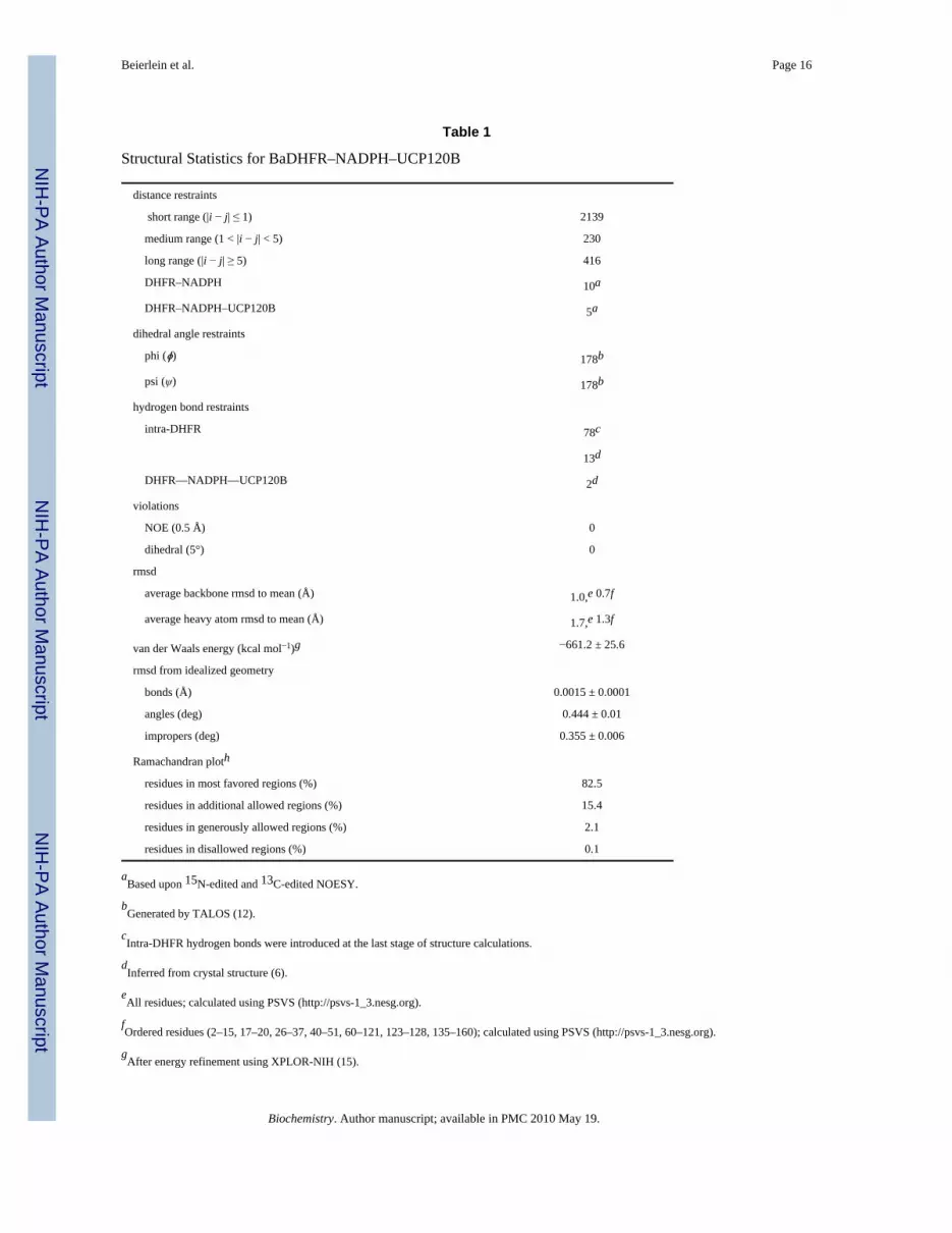
We determined the three-dimensional solution structure of BaDHFR by modern tripleresonance NMR methods as described in Materials and Methods. Twenty best conformers withlowest target energy functions, as defined by CYANA 2.1 (13), were used for the ternarycomplex calculations. In order to place the ligands, BaDHFR–NADPH–UCP120B andBaDHFR–NADPH NOEs were identified from 3D 15N and 13C NOESY experiments.Additionally, two conserved hydrogen bond restraints for the ternary complex, between theprotonated N1 and 2-amino group of the ligand and the conserved acidic residue Glu 28, wereinferred from the crystal structure (6). While these interactions were not observedexperimentally, their conservation in every DHFR structure with a 2,4-diaminopyrimidine ringjustifies their inclusion. Ternary complex calculations were performed, and 15 lowest energystructures were further subjected to simulated annealing refinement (Figure 2A). Statistics ofthe ensemble are presented in Table 1 and demonstrate the high quality of the structure.
Comparison of the Solution and Crystal Structures of BaDHFR–NADPH–UCP120BA direct comparison of the ternary solution (Figure 2) and crystal structures of BaDHFR (6),both bound to NADPH and UCP120B, reveals that the general fold of DHFR and the bindingof NADPH are conserved while there appear to be two different binding modes for UCP120B.Overall, the Cα atoms of the solution and crystal structures superimpose with an rmsd of 2.3Å (Figure 2B). The central β-sheets of both structures superimpose particularly well. Thecrystal structure assigns two short additional β-strands (residues 13–16 and 124–126) relativeto the solution structure. The most noticeable difference between the two structures is thetranslation of a helix (residues 25–37; often called helix B in DHFR nomenclature) near theactive site (Figure 2B). A survey of each of the models in the solution structure ensemblereveals that the N-terminus of this helix is displaced from the crystal structure by 1.2–3.6 Åand the C-terminus is displaced by 2.7–5.3 Å. The conformation of helix B in this solutionstructure is supported by 11 long-range NOE signals (a full list of the NOE signals in this regionis found in Supporting Information) arising from interactions between two residues in helix B(Pro 26 and Ser 27) and two residues in the remainder of the protein (Lys 148 and Asn 149).Additionally, an NOE is observed between the HD1 proton from Leu 29 and the C7 atom ofthe ligand.
NADPH is bound in the same extended conformation in the crystal and solution structures.The nicotinamide ring in the solution structure appears to be translated between 0.9 and 2.1 Å
Beierlein et al. Page 4
Biochemistry. Author manuscript; available in PMC 2010 May 19.
NIH
-PA Author Manuscript
NIH
-PA Author Manuscript
NIH
-PA Author Manuscript
away from the active site, relative to the molecule in the crystal structure. The adenine ring,on the surface of the protein, appears to be flexible.
The ligand, UCP120B, exhibits two different binding modes in the two structures (Figure 2B–D). In the crystal structure, the diaminopyrimidine ring forms hydrogen bonds with Glu 28 aswell as the backbone carbonyl atoms of Met 6 and Phe 96. The propargyl linker extends overthe phenyl ring of Phe 96, and the dimethoxyphenyl ring forms van der Waals interactions withIle 51, Leu 55, and Leu 21. In the solution structure, the hydrogen bonds between the protonatedN1 and 2-amino group of the ligand were constrained, shifting the pyrimidine ring toward helixB. A measured NOE signal between the C7 atom and Leu 29 provides experimental evidencefor the interaction of the pyrimidine ring and the displaced helix. The propargyl linker of theligand extends under Phe 96 (as opposed to “over” the residue in the crystal structure). NOEsignals between the two protons on the 4-amino group and Phe 96 provide experimentalevidence of the interaction. The dimethoxyphenyl ring generally superimposes well with thesame ring in the crystal structure and exhibits NOE signals with Ile 51. It also appears to forminteractions with the loop containing Pro 54-Leu 55-Pro 56, supported by an NOE signalbetween Leu 55 and the dimethoxyphenyl ring.
Previously published ternary solution structures with LcDHFR and hDHFR show agreementwith related crystal structures. Human DHFR TMP–NADPH solution and crystal structureshave a root mean squared deviation of 1.26 Å and validate that there are no significantconformational changes observed between the structures (3). Similar solution and crystalstructure comparisons have been observed in the ternary complex of LcDHFR (1). For hDHFRand LcDHFR, the position of helix B varies between the solution and crystal structures byapproximately 1–2.5 Å.
While the extended conformation of NADPH dominates in most DHFR structures, both humanand BaDHFR ensembles confirm an overall flexibility in the NADPH cofactor. Specifically,the pyrophosphate groups show the most flexibility and influence the distances between thenicotinamide ring and trimethoprim in hDHFR. The pyrophosphate is also variable in theBaDHFR structure, conferring flexibility to the deoxyribose sugar moiety.
Dynamics of BaDHFRIn order to better understand the backbone conformational flexibility of BaDHFR, we measuredvalues of the relaxation parameters, T1, T2, and heteronuclear 1H–15N NOEs for the ternarycomplex (Figure 3). The average value of T1 is 926 ms (with a standard deviation of 92); theaverage value of T2 is 47 ms (with a standard deviation of 9). This results in an average T1/T2 ratio of approximately 20 (with a standard deviation of 3) that can be used for a roughestimate, using the related spectral density function, of the overall correlation time of themolecule, τm (17). The average NOE value is 0.67 (with a standard deviation of 0.1). For mostof the residues, T1 and T2 values do not deviate significantly beyond experimental error fromthe average numbers. However, several regions (14–18, 65–73, 85–89, 124–133, 160–162)demonstrate some tendency to be involved in fast internal motion with larger T2 and smallerNOE values. These regions of fast internal motion correlate well with the general notion thatregions of secondary structure show lower mobility while turns, loops, and N-/C-terminiexhibit higher mobility. However, one amino acid, Val 32, located in the center of helix B,behaves very differently. The T1 and T2 values of Val 32 are 544 and 90 ms, respectively,resulting in a ratio of only 6. Its NOE value is also reduced to 0.25 as compared to an averageNOE value of 0.67 or compared to the NOE value of the neighboring Lys 33 (1.0). All of theselarge deviations suggest that Val 32 is involved in fast internal motion (17), which is a strikingfeature since this residue is making strong contacts with UCP120B. In addition to the motionof Val 32, the majority of the residues in helix B (residues 25–32) exhibit fast exchange inhydrogen/deuterium exchange experiments (see Figure 3). There are also several regions such
Beierlein et al. Page 5
Biochemistry. Author manuscript; available in PMC 2010 May 19.
NIH
-PA Author Manuscript
NIH
-PA Author Manuscript
NIH
-PA Author Manuscript
as residues 20–25 that lack backbone assignments due to an unfavorable conformationalexchange and residues 89–102 and 117–120 that show evidence of conformational exchangewith reduced T2 values.
Comparison between Binary and Ternary Enzyme ComplexesIn order to optimize conditions for data collection and to observe the effect of the ligand onthe complex, we acquired 15N HSQC spectra during a titration experiment in which the molarratios of protein to ligand included no ligands (apoprotein) (Figure 4a), the binary protein–NADPH complex, a 1:1 mix of protein–NADPH:ligand and a 1:4 mix of protein–NADPH:ligand (the last two represent ternary complexes) (Figure 4b). Both the binary andternary complexes were significantly more resolved than the apo form, reflecting results fromprevious experiments showing that the apoenzyme exists in at least two different isoforms(18–21). In addition, notable differences were observed between the 15N HSQC spectra. Usingthe complete assignments of the ternary HSQC and a superposition with the unassigned binaryform, we analyzed regions of the protein that significantly shifted upon ligand binding.
Examination of the shifted residues shows that the majority of these residues predictably lienear the active site. Figure 5 highlights the regions of the protein that shift between the twocomplexes. The acidic residue Glu 28 that makes conserved interactions to thediaminopyrimidine ring, along with the remainder of the residues in helix B, shows significantshift differences between the binary and ternary complexes. Another helix located near theNADPH cofactor containing residues Ile 95–Leu 104 and the β-strand containing F5–A8,which interacts with the ligand and NADPH, also show distinct shift differences. Interestingly,it appears that sections of the core canonical eight-stranded twisted β-sheet also undergo someslight to significant perturbations.
Movement of the β-sheet region in response to the addition of ligand has been observed inprevious NMR studies. In EcDHFR, differences between the NADPH binary complex and theternary complex with folate have been analyzed by NMR (4,5). The analysis reveals shifts inmany of the same regions as observed in BaDHFR, including Ala 8 in the first β-strand andhelix B containing the conserved acidic residue. Solution structures of the binary and ternaryLcDHFR enzyme complexes are also available (1,2,22,23), though no analysis of shiftdifferences between binary and ternary enzyme complexes has been previously reported.Comparison of the NADPH-bound binary complex (PDB ID 2HQP) and the trimethoprim –NADPH-bound ternary complex (PDB ID 1LUD (1)) shows the greatest structural differencesin regions already indicated by the EcDHFR NMR studies (4,5) and our own BaDHFR NMRstudies (Figure S1 in Supporting Information shows a superposition of the binary and ternaryLcDHFR structures and notes the structural differences). A superposition of the three structures(BaDHFR, EcDHFR, and LcDHFR) yields a structural alignment (Figure 6) and facilitates theinterpretation of residue characteristics at equivalent positions.
Analyses of these comparisons are useful to illustrate the network of intramolecular interactionsthat are involved in the conformational transitions between the binary and ternary enzymestates. Helix B containing the conserved acidic residue and several other residues critical toligand binding shows significant movement between the binary and ternary complexes. Thecore β-sheet region seems to shift upon ligand binding, ostensibly due to the fact that the initialβ-strand (comprising residues 4–9 in BaDHFR) at the N-terminus contains residues critical toboth ligand and cofactor binding. With this strand perturbed, the other strands must adjust tomaintain the intramolecular interactions required for the twisted β-sheet conformation. Alsoof interest is the movement observed in the helix containing residues 99–104 in BaDHFR. Theresidues in the loop directly before the helix, including Phe 96 in BaDHFR, play an importantrole in ligand binding, sometimes including the formation of a hydrogen bond between theligand and the protein backbone. This helix shows significant chemical shift displacement in
Beierlein et al. Page 6
Biochemistry. Author manuscript; available in PMC 2010 May 19.
NIH
-PA Author Manuscript
NIH
-PA Author Manuscript
NIH
-PA Author Manuscript
BaDHFR and some displacement in EcDHFR or LcDHFR. In EcDHFR and LcDHFR, thecorresponding residues to Phe 96 are smaller (Ile and Ala, respectively) and may not shift asdramatically to accommodate a ligand. It is also important to note that the ternary enzyme statesare bound to three different ligands: UCP120B (Ba), folate (Ec), and TMP (Lc). The ligandshave their own individual effects on enzyme conformation and account for some of thedifferences observed between species. Specifically in the 99–104 helix, UCP120B might playa role in the degree of shift since Phe 96 forms π-stacking interactions with the propargyl linker,which is unique to this inhibitor.
Structure–Activity Relationship Analysis of Previously Designed LigandsWe have previously designed, synthesized, and evaluated several inhibitors for many speciesof DHFR (6,24–26). However, none of the currently designed inhibitors has shown remarkablepotency against BaDHFR (Table 2). To date, the most active inhibitor (UCP120B), the sameligand bound in the crystal structure and bound to the solution structure reported here, has anIC50 of 0.89 μM. Features observed in the solution structure of BaDHFR could assist inexplaining the lack of potency in this series.
The first series of inhibitors contain the core scaffold of the propargyl-linked compounds withthe 3,4,5-tri-methoxyphenyl ring of TMP and a series of substituents at the bridge position ofthe propargyl linker (compounds 1–6 in Table 2). Enzyme inhibition assays reveal that theaddition of any group at the propargylic position lowers potency. The addition of a methylgroup or methoxy group in particular causes nearly a 10-fold loss in activity.
The solution structure shows that the phenyl ring of Phe 96 is positioned near this propargyllinker (see Figure 2C). Relaxation data for Phe 96 show that the residue is undergoingsignificant conformational exchange, even in the presence of saturating ligand concentrations.These data, in combination with the significant line broadening observed for Phe 96, suggestits flexibility. While the steric interference of Phe 96 was suspected based on the crystalstructure, the solution structure shows nicely how the flexibility of the phenyl ring may interferewith this linker region.
After the initial series of trimethoxy inhibitors were evaluated, a series of biphenyl compoundswere designed and synthesized (compounds 7–10 in Table 2). The rationale for this series wasto better occupy the hydrophobic pocket containing residues Leu 29, Ile 51, and Leu 55,normally occupied by the p-aminobenzoic acid moiety of the substrate. However, the expectedincrease in activity was not observed for these compounds.
An analysis of the solution structure of BaDHFR provides a possible explanation for the lackof improved affinity. The distal phenyl ring of the compound (relative to the pyrimidine) ispredicted to be located next to the loop containing residues 52–59 (Figure 2C). This loopcontains the flexible motif GRPLPGRR. Leu 55, a critical residue in this predicted biphenylbinding pocket, has a T2 value of 54.5 ms, slightly greater than the average T2 value (47 ms).Signals for the preceding residues in the loop (51 – 53) are broadened, and those for thefollowing residues (57 – 59) are not assigned, most likely reflecting an unfavorable exchangerate. Leu 55 also appears to be undergoing fast (20 min) hydrogen/ deuterium exchange (Figure3). Taken together, these results suggest significant flexibility in this loop region, which hasthe potential to cause steric interference with the biphenyl compounds. It is also possible thatthe phenyl ring simply does not create strong enough interactions to overcome the entropiccost that is incurred by ordering this loop region to prevent steric interference. The direct causeof the lower potency for the biphenyl compounds still needs further investigation; however,the information gained about this loop region from the solution structure will be very usefulin the design of future inhibitors. Some potential avenues of future investigation includeincreasing the size of substituents and varying the substitution pattern of substituents around
Beierlein et al. Page 7
Biochemistry. Author manuscript; available in PMC 2010 May 19.
NIH
-PA Author Manuscript
NIH
-PA Author Manuscript
NIH
-PA Author Manuscript

the aryl ring. Primarily, the ortho and meta positions have been investigated most extensively,and varying the substituents on the para position could prove fruitful.
CONCLUSIONSGiven the significance of identifying new therapeutics for infections caused by B. anthracis,we have begun a drug discovery program to identify inhibitors of the essential enzymedihydrofolate reductase (DHFR). In this work, we present a three-dimensional solutionstructure of DHFR from B. anthracis bound to its cofactor, NADPH, and UCP120B, the mostpotent of our inhibitors to date. The ternary solution structure is the third species of DHFR tobe fully determined by NMR methods and reveals the conserved DHFR fold with an eight-stranded β-sheet and four flanking α-helices. Differences with our previously determinedternary crystal structure are located primarily in the position of an α-helix at the active site andspecific interactions formed by UCP120B and the active site residues. Comparisons ofchemical shifts between the binary enzyme – NADPH complex and the ternary complex yieldinsight into the residues significantly affected by ligand binding. Specifically, Phe 96, whichinteracts with the propargyl group of UCP120B, displays significant chemical shift differencesbetween the binary and ternary complex and appears to be flexible in the active site.Significantly for our drug design effort, the solution structure reveals a possible basis forobserved structure–activity relationships. The flexibility of Phe 96 may interfere withpropargyl-substituted compounds, and the flexibility of Leu 55 in a loop at the active site mayaffect binding of a biphenyl series of these inhibitors. Understanding the flexibility of the activesite residues of BaDHFR will be important in designing more potent inhibitors of the enzyme.
Supplementary MaterialRefer to Web version on PubMed Central for supplementary material.
AcknowledgmentsWe thank Martha Morton for assistance with NADPH resonance assignments.
References1. Polshakov V, Smirnov E, Birdsall B, Kelly G, Feeney J. NMR-based solution structure of the complex
of Lactobacillus casei dihydrofolate reductase wtih trimethoprim and NADPH. J Biomol NMR2002;24:67–70. [PubMed: 12449420]
2. Gargaro A, Soteriou A, Frenkiel T, Bauer C, Birdsall B, Polshakov V, Barsukov I, Roberts G, FeeneyJ. The solution structure of the complex of Lactobacillus casei dihydrofolate reductase withmethotrexate. J Mol Biol 1998;277:119–134. [PubMed: 9514736]
3. Kovalevskaya N, Smurnyy Y, Polshakov V, Birdsall B, Bradbury A, Frenkiel T, Feeney J. Solutionstructure of human dihydrofolate reductase in its complex with trimethoprim and NADPH. J BiomolNMR 2005;33:69–72. [PubMed: 16222560]
4. Osborne M, Venkitakrishnan R, Dyson J, Wright P. Diagnostic chemical shift markers for loopconformation and substrate and cofactor binding in dihydrofolate reductase complexes. Protein Sci2003;12:2230–2238. [PubMed: 14500880]
5. Venkitakrishnan R, Zabrowski E, McElheny D, Benkovic S, Dyson J, Wright P. Conformationalchanges in the active site loops of dihydrofolate reductase during the catalytic cycle. Biochemistry2004;43:16046–16055. [PubMed: 15609999]
6. Beierlein J, Frey K, Bolstad D, Pelphrey P, Joska T, Smith A, Priestley N, Wright D, Anderson A.Synthetic and crystallographic studies of a new inhibitor series targeting Bacillus anthracisdihydrofolate reductase. J Med Chem 2008;51:7532–7540. [PubMed: 19007108]
7. Clore GM, Gronenborn AM. NMR structure determination of proteins and protein complexes largerthan 20 kDa. Curr Opin Chem Biol 1998;2:564–570. [PubMed: 9818180]
Beierlein et al. Page 8
Biochemistry. Author manuscript; available in PMC 2010 May 19.
NIH
-PA Author Manuscript
NIH
-PA Author Manuscript
NIH
-PA Author Manuscript

8. Farrow N, Muhandiram R, Singer A, Pascal S, Kay C, Gish G, Shoelson S, Pawson T, Forman-Kay J,Kay L. Backbone dynamics of a free and phosphopeptide-complexed Src homology 2 domain studiedby 15N NMR relaxation. Biochemistry 1994;33:5984–6003. [PubMed: 7514039]
9. Fogh R, Ionides J, Ulrich E, Boucher W, Vranken W, Linge J, Habeck M, Rieping W, Bhat T,Westbrook J, Henrick K, Gilliland G, Berman H, Thorton J, Nilges M, Markley J, Laue E. The CCPNproject: an interim report on a data model for the NMR community. Nat Struct Biol 2002;9:416–418.[PubMed: 12032555]
10. Delaglio F, Grzesiek S, Vuister GW, Zhu G, Pfeifer J, Bax A. NMRPipe: a multidimensional spectralprocessing system based on UNIX pipes. J Biomol NMR 1995;6:277–293. [PubMed: 8520220]
11. Keller, R. 2004. http://www.cara.nmr-software.org/downloads/3-85600-112-3.pdf12. Cornilescu G, Delaglio F, Bax A. Protein backbone angle restraints from searching a database for
chemical shift and sequence homology. J Biomol NMR 1999;13:289–302. [PubMed: 10212987]13. Guntert P. Automated NMR structure calculation with CYANA. Methods Mol Biol 2004;278:353–
378. [PubMed: 15318003]14. Schuettelkopf A, van Aalten D. PRODRG—a tool for high-throughput crystallography of protein-
ligand complexes. Acta Crystallogr 2004;D60:1355–1363.15. Schwieters C, Kuszewski J, Tjandra N, Clore G. The XPLOR-NIH NMR molecular structure
determination package. J Magn Reson 2003;160:66–74.16. DeLano, W. DeLano Scientific LLC. Palo Alto, CA: 2008.17. Clore G, Driscoll P, Wingfield P, Gronenborn A. Analysis of the backbone dynamics of interleukin-1
beta using two-dimensional inverse detected heteronuclear 15N-1H NMR spectroscopy.Biochemistry 1990;29:7387–7401. [PubMed: 2223770]
18. Epstein D, Benkovic S, Wright P. Dynamics of the dihydrofolate reductase-folate complex: catalyticsites and regions known to undergo conformational change exhibit diverse dynamical features.Biochemistry 1995;34:11037–11048. [PubMed: 7669761]
19. Falzone C, Wright P, Benkovic S. Evidence for two interconverting protein isomers in themethotrexate complex of dihydrofolate reductase from Escherichia coli. Biochemistry1991;30:2184–2191. [PubMed: 1998678]
20. Osborne M, Schnell J, Benkovic S, Dyson J, Wright P. Backbone dynamics in dihydrofolate reductasecomplexes: role of loop flexibility in catalytic mechanism. Biochemistry 2001;40:9846–9859.[PubMed: 11502178]
21. Schnell J, Dyson J, Wright P. Structure, dynamics and catalytic function of dihydrofolate reductase.Annu Rev Biophys Biomol Struct 2004;33:119–140. [PubMed: 15139807]
22. Polshakov V, Birdsall B, Feeney J. Effects of co-operative ligand binding on protein amide NHhydrogen exchange. J Mol Biol 2006;356:886–903. [PubMed: 16405904]
23. Polshakov V, Birdsall B, Frenkiel T, Gargaro A, Feeney J. Structure and dynamics in solution of thecomplex of Lactobacillus casei dihydrofolate reductase with the new lipophilic antifolate drugtrimetrexate. Protein Sci 1999;8:467–481. [PubMed: 10091649]
24. Bolstad D, Bolstad E, Frey K, Wright D, Anderson A. A structure-based approach to the developmentof potent and selective inhibitors of dihydrofolate reductase from Cryptosporidium. J Med Chem2008;51:6839–6852. [PubMed: 18834108]
25. Liu J, Bolstad D, Smith A, Priestley N, Wright D, Anderson A. Structure-guided development ofefficacious antifungal agents targeting Candida glabrata dihydrofolate reductase. Chem Biol2008;15:990–996. [PubMed: 18804036]
26. Pelphrey P, Popov V, Joska T, Beierlein J, Bolstad E, Fillingham Y, Wright D, Anderson A. Highlyefficient ligands for DHFR from Cryptosporidium hominis and Toxoplasma gondii inspired bystructural analysis. J Med Chem 2007;50:940–950. [PubMed: 17269758]
Beierlein et al. Page 9
Biochemistry. Author manuscript; available in PMC 2010 May 19.
NIH
-PA Author Manuscript
NIH
-PA Author Manuscript
NIH
-PA Author Manuscript
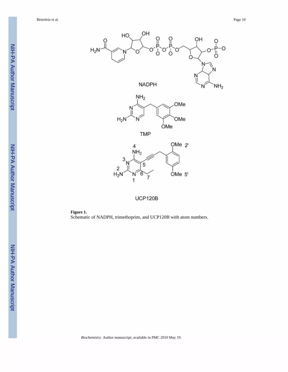
Figure 1.Schematic of NADPH, trimethoprim, and UCP120B with atom numbers.
Beierlein et al. Page 10
Biochemistry. Author manuscript; available in PMC 2010 May 19.
NIH
-PA Author Manuscript
NIH
-PA Author Manuscript
NIH
-PA Author Manuscript

Figure 2.(A) Superposition of the members of the solution structure ensemble. (B) Superposition of thesolution (dark blue) and crystal (cyan) structures of BaDHFR bound to ligands. Helix B(including the N-and C-termini of the helix) is labeled. (C) Superposition of the models ofactive site residues in the solution structure. Residues in the active site are shown as lines;residues within the 53–56 loop are colored orange; ligands are shown in stick form. (D) Activesite residues and ligands in the crystal structure.
Beierlein et al. Page 11
Biochemistry. Author manuscript; available in PMC 2010 May 19.
NIH
-PA Author Manuscript
NIH
-PA Author Manuscript
NIH
-PA Author Manuscript

Figure 3.Relaxation data plotted for the 137 measured amide residues. Labeled secondary structureelements are shown above each plot and colored according to the rate of hydrogen/deuteriumexchange (red = fast exchange in 20 min, yellow = intermediate exchange in 1 day, and blue= protected). (A) T1 values, (B) T2 values, and (C) 1H–15N NOE values.
Beierlein et al. Page 12
Biochemistry. Author manuscript; available in PMC 2010 May 19.
NIH
-PA Author Manuscript
NIH
-PA Author Manuscript
NIH
-PA Author Manuscript
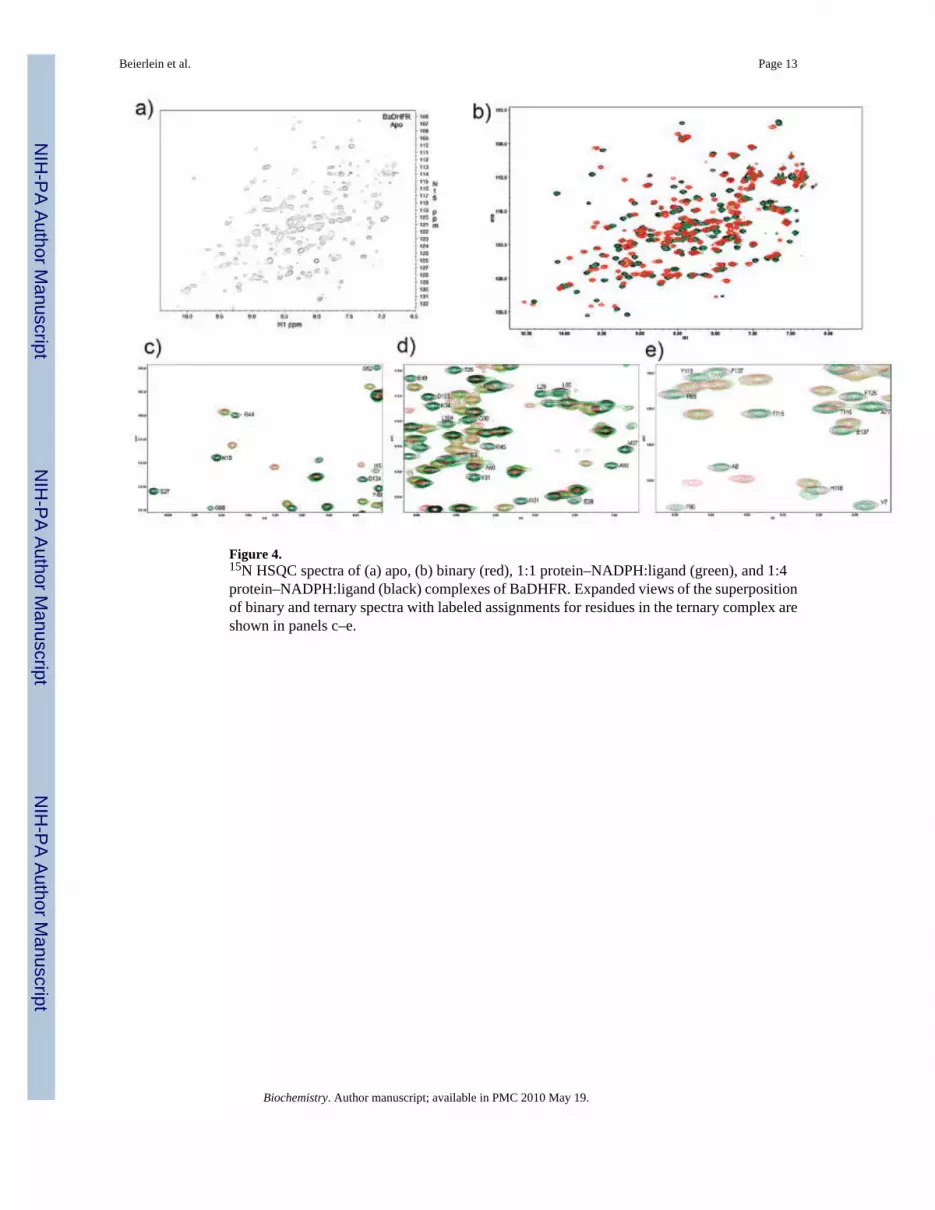
Figure 4.15N HSQC spectra of (a) apo, (b) binary (red), 1:1 protein–NADPH:ligand (green), and 1:4protein–NADPH:ligand (black) complexes of BaDHFR. Expanded views of the superpositionof binary and ternary spectra with labeled assignments for residues in the ternary complex areshown in panels c–e.
Beierlein et al. Page 13
Biochemistry. Author manuscript; available in PMC 2010 May 19.
NIH
-PA Author Manuscript
NIH
-PA Author Manuscript
NIH
-PA Author Manuscript
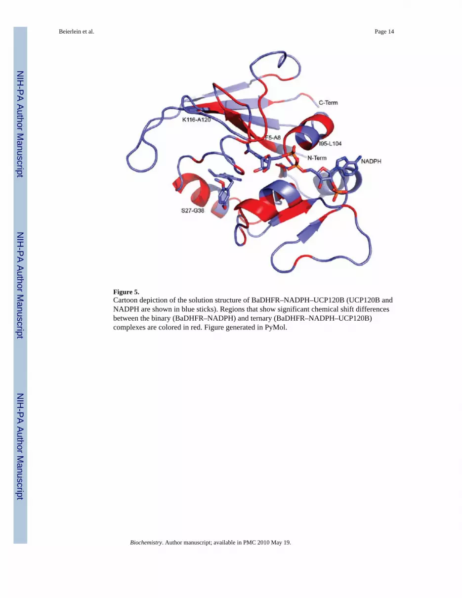
Figure 5.Cartoon depiction of the solution structure of BaDHFR–NADPH–UCP120B (UCP120B andNADPH are shown in blue sticks). Regions that show significant chemical shift differencesbetween the binary (BaDHFR–NADPH) and ternary (BaDHFR–NADPH–UCP120B)complexes are colored in red. Figure generated in PyMol.
Beierlein et al. Page 14
Biochemistry. Author manuscript; available in PMC 2010 May 19.
NIH
-PA Author Manuscript
NIH
-PA Author Manuscript
NIH
-PA Author Manuscript
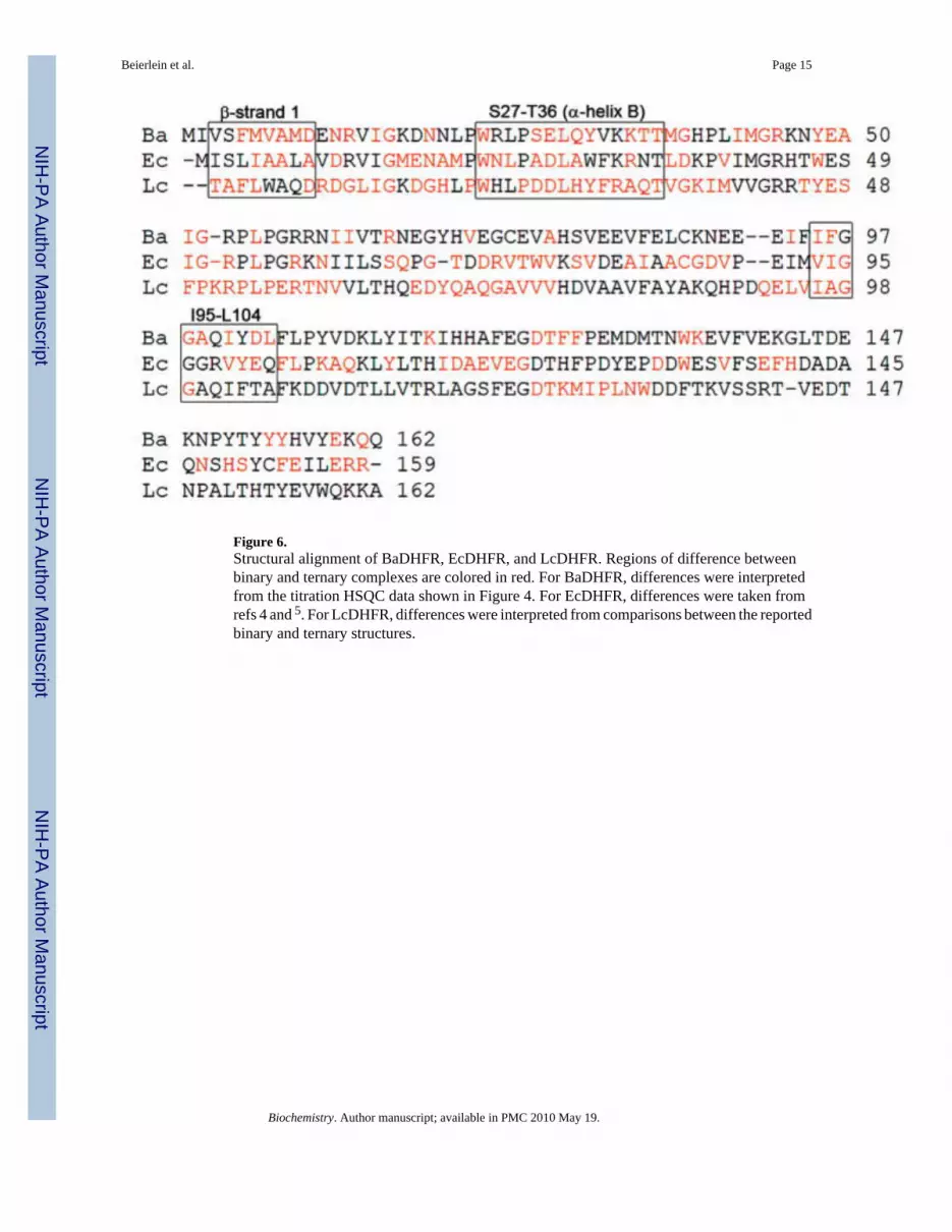
Figure 6.Structural alignment of BaDHFR, EcDHFR, and LcDHFR. Regions of difference betweenbinary and ternary complexes are colored in red. For BaDHFR, differences were interpretedfrom the titration HSQC data shown in Figure 4. For EcDHFR, differences were taken fromrefs 4 and 5. For LcDHFR, differences were interpreted from comparisons between the reportedbinary and ternary structures.
Beierlein et al. Page 15
Biochemistry. Author manuscript; available in PMC 2010 May 19.
NIH
-PA Author Manuscript
NIH
-PA Author Manuscript
NIH
-PA Author Manuscript
NIH
-PA Author Manuscript
NIH
-PA Author Manuscript
NIH
-PA Author Manuscript
Beierlein et al. Page 16
Table 1
Structural Statistics for BaDHFR–NADPH–UCP120B
distance restraints
short range (|i − j| ≤ 1) 2139
medium range (1 < |i − j| < 5) 230
long range (|i − j| ≥ 5) 416
DHFR–NADPH 10a
DHFR–NADPH–UCP120B 5a
dihedral angle restraints
phi (ϕ) 178b
psi (ψ) 178b
hydrogen bond restraints
intra-DHFR 78c
13d
DHFR—NADPH—UCP120B 2d
violations
NOE (0.5 Å) 0
dihedral (5°) 0
rmsd
average backbone rmsd to mean (Å) 1.0,e 0.7f
average heavy atom rmsd to mean (Å) 1.7,e 1.3f
van der Waals energy (kcal mol−1)g −661.2 ± 25.6
rmsd from idealized geometry
bonds (Å) 0.0015 ± 0.0001
angles (deg) 0.444 ± 0.01
impropers (deg) 0.355 ± 0.006
Ramachandran ploth
residues in most favored regions (%) 82.5
residues in additional allowed regions (%) 15.4
residues in generously allowed regions (%) 2.1
residues in disallowed regions (%) 0.1
aBased upon 15N-edited and 13C-edited NOESY.
bGenerated by TALOS (12).
cIntra-DHFR hydrogen bonds were introduced at the last stage of structure calculations.
dInferred from crystal structure (6).
eAll residues; calculated using PSVS (http://psvs-1_3.nesg.org).
fOrdered residues (2–15, 17–20, 26–37, 40–51, 60–121, 123–128, 135–160); calculated using PSVS (http://psvs-1_3.nesg.org).
gAfter energy refinement using XPLOR-NIH (15).
Biochemistry. Author manuscript; available in PMC 2010 May 19.
NIH
-PA Author Manuscript
NIH
-PA Author Manuscript
NIH
-PA Author Manuscript
Beierlein et al. Page 17
hCalculated using PSVS (http://psvs-1_3.nesg.org).
Biochemistry. Author manuscript; available in PMC 2010 May 19.

NIH
-PA Author Manuscript
NIH
-PA Author Manuscript
NIH
-PA Author Manuscript
Beierlein et al. Page 18
Table 2
IC50 Values for the Trimethoxyphenyl and Biphenyl Inhibitor Seriesa
Compound No. Inhibitor IC50 (μM)
1 3.7 ± 0.2
2 30.3 ± 0.4
3 14.5 ± 0.5
Biochemistry. Author manuscript; available in PMC 2010 May 19.

NIH
-PA Author Manuscript
NIH
-PA Author Manuscript
NIH
-PA Author Manuscript
Beierlein et al. Page 19
Compound No. Inhibitor IC50 (μM)
4 29.1 ± 0.06
5 10.5 ± 0.5
6 12.7 ± 1.0
7 7.9 ± 0.8
Biochemistry. Author manuscript; available in PMC 2010 May 19.

NIH
-PA Author Manuscript
NIH
-PA Author Manuscript
NIH
-PA Author Manuscript
Beierlein et al. Page 20
Compound No. Inhibitor IC50 (μM)
8 5.5 ± 1.0
9 17.3 ± 1.4
10 49.1 ± 1.0
aIC50 values for compounds 1–4 in Table 2 were also previously reported in ref 6 and are included here for comparison.
Biochemistry. Author manuscript; available in PMC 2010 May 19.
Related Documents


![Bacillus anthracis - As Biological WeaponsBacillus anthracis - as biological weapons :JOLN (Bacillus anthracis) ± MDNREUR ELRORJLF]QD miotr Daniszewski Department of Invertebrate](https://static.cupdf.com/doc/110x72/613e1f0259df642846165479/bacillus-anthracis-as-biological-weapons-bacillus-anthracis-as-biological-weapons.jpg)