Bosma et al. Fluids Barriers CNS (2018) 15:24 https://doi.org/10.1186/s12987-018-0109-2 REVIEW The role of plasmalemma vesicle-associated protein in pathological breakdown of blood–brain and blood–retinal barriers: potential novel therapeutic target for cerebral edema and diabetic macular edema Esmeralda K. Bosma 1 , Cornelis J. F. van Noorden 1,2 , Reinier O. Schlingemann 1,3 and Ingeborg Klaassen 1,4* Abstract Breakdown of the blood–brain barrier (BBB) or inner blood–retinal barrier (BRB), induced by pathologically elevated levels of vascular endothelial growth factor (VEGF) or other mediators, can lead to vasogenic edema and significant clinical problems such as neuronal morbidity and mortality, or vision loss. Restoration of the barrier function with corticosteroids in the brain, or by blocking VEGF in the eye are currently the predominant treatment options for brain edema and diabetic macular edema, respectively. However, corticosteroids have side effects, and VEGF has important neuroprotective, vascular protective and wound healing functions, implying that long-term anti-VEGF therapy may also induce adverse effects. We postulate that targeting downstream effector proteins of VEGF and other media- tors that are directly involved in the regulation of BBB and BRB integrity provide more attractive and safer treatment options for vasogenic cerebral edema and diabetic macular edema. The endothelial cell-specific protein plasma- lemma vesicle-associated protein (PLVAP), a protein associated with trans-endothelial transport, emerges as candi- date for this approach. PLVAP is expressed in a subset of endothelial cells throughout the body where it forms the diaphragms of caveolae, fenestrae and trans-endothelial channels. However, PLVAP expression in brain and eye barrier endothelia only occurs in pathological conditions associated with a compromised barrier function such as cancer, ischemic stroke and diabetic retinopathy. Here, we discuss the current understanding of PLVAP as a structural compo- nent of endothelial cells and regulator of vascular permeability in health and central nervous system disease. Besides providing a perspective on PLVAP identification, structure and function, and the regulatory processes involved, we also explore its potential as a novel therapeutic target for vasogenic cerebral edema and retinal macular edema. Keywords: Plasmalemma vesicle-associated protein, Blood–brain barrier, Blood–retinal barrier, Cerebral edema, Diabetic macular edema © The Author(s) 2018. This article is distributed under the terms of the Creative Commons Attribution 4.0 International License (http://creativecommons.org/licenses/by/4.0/), which permits unrestricted use, distribution, and reproduction in any medium, provided you give appropriate credit to the original author(s) and the source, provide a link to the Creative Commons license, and indicate if changes were made. The Creative Commons Public Domain Dedication waiver (http://creativecommons.org/ publicdomain/zero/1.0/) applies to the data made available in this article, unless otherwise stated. Open Access Fluids and Barriers of the CNS *Correspondence: [email protected] 4 Ocular Angiogenesis Group, Department of Medical Biology, Amsterdam UMC, University of Amsterdam, Meibergdreef 15, Room L3-154, 1105 AZ Amsterdam, The Netherlands Full list of author information is available at the end of the article

Welcome message from author
This document is posted to help you gain knowledge. Please leave a comment to let me know what you think about it! Share it to your friends and learn new things together.
Transcript

Bosma et al. Fluids Barriers CNS (2018) 15:24 https://doi.org/10.1186/s12987-018-0109-2
REVIEW
The role of plasmalemma vesicle-associated protein in pathological breakdown of blood–brain and blood–retinal barriers: potential novel therapeutic target for cerebral edema and diabetic macular edemaEsmeralda K. Bosma1, Cornelis J. F. van Noorden1,2, Reinier O. Schlingemann1,3 and Ingeborg Klaassen1,4*
Abstract
Breakdown of the blood–brain barrier (BBB) or inner blood–retinal barrier (BRB), induced by pathologically elevated levels of vascular endothelial growth factor (VEGF) or other mediators, can lead to vasogenic edema and significant clinical problems such as neuronal morbidity and mortality, or vision loss. Restoration of the barrier function with corticosteroids in the brain, or by blocking VEGF in the eye are currently the predominant treatment options for brain edema and diabetic macular edema, respectively. However, corticosteroids have side effects, and VEGF has important neuroprotective, vascular protective and wound healing functions, implying that long-term anti-VEGF therapy may also induce adverse effects. We postulate that targeting downstream effector proteins of VEGF and other media-tors that are directly involved in the regulation of BBB and BRB integrity provide more attractive and safer treatment options for vasogenic cerebral edema and diabetic macular edema. The endothelial cell-specific protein plasma-lemma vesicle-associated protein (PLVAP), a protein associated with trans-endothelial transport, emerges as candi-date for this approach. PLVAP is expressed in a subset of endothelial cells throughout the body where it forms the diaphragms of caveolae, fenestrae and trans-endothelial channels. However, PLVAP expression in brain and eye barrier endothelia only occurs in pathological conditions associated with a compromised barrier function such as cancer, ischemic stroke and diabetic retinopathy. Here, we discuss the current understanding of PLVAP as a structural compo-nent of endothelial cells and regulator of vascular permeability in health and central nervous system disease. Besides providing a perspective on PLVAP identification, structure and function, and the regulatory processes involved, we also explore its potential as a novel therapeutic target for vasogenic cerebral edema and retinal macular edema.
Keywords: Plasmalemma vesicle-associated protein, Blood–brain barrier, Blood–retinal barrier, Cerebral edema, Diabetic macular edema
© The Author(s) 2018. This article is distributed under the terms of the Creative Commons Attribution 4.0 International License (http://creat iveco mmons .org/licen ses/by/4.0/), which permits unrestricted use, distribution, and reproduction in any medium, provided you give appropriate credit to the original author(s) and the source, provide a link to the Creative Commons license, and indicate if changes were made. The Creative Commons Public Domain Dedication waiver (http://creat iveco mmons .org/publi cdoma in/zero/1.0/) applies to the data made available in this article, unless otherwise stated.
Open Access
Fluids and Barriers of the CNS
*Correspondence: [email protected] 4 Ocular Angiogenesis Group, Department of Medical Biology, Amsterdam UMC, University of Amsterdam, Meibergdreef 15, Room L3-154, 1105 AZ Amsterdam, The NetherlandsFull list of author information is available at the end of the article

Page 2 of 17Bosma et al. Fluids Barriers CNS (2018) 15:24
BackgroundIntegrity of the blood–brain barrier (BBB) is essential to maintain a proper microenvironment for neuronal cells to function. Similar to the BBB, the inner blood–retinal barrier (BRB) protects the retina that consists of layers of specialized neurons. Breakdown of the BBB and BRB, as occurs in pathological conditions such as trauma, acute ischemic stroke [1], brain tumors [2] and diabetic retin-opathy [3], causes leakage of fluid and plasma proteins from the vasculature into the surrounding tissue and subsequently vasogenic edema formation. Edema forma-tion in the brain and eye is associated with neuronal and retinal dysfunction, causing severe mortality and loss of visual acuity, respectively [4, 5]. Given the similarities between the BBB and BRB, promising novel therapeutic candidates that prevent disruption of the BRB are also interesting targets in the treatment of BBB-related neu-rological diseases.
Corticosteroids are the main treatment option for brain edema, but therapy is associated with severe side effects [6]. Restoration of the barrier function via inhibition of vascular endothelial growth factor-A (VEGF), a potent inducer of angiogenesis and vascular permeability, is cur-rently an effective treatment for diabetic macular edema (DME) and a promising treatment option for vasogenic brain edema [4, 7–10]. However, anti-VEGF therapy still has its limitations, including systemic side effects, a sub-optimal response in a subset of macular edema patients, and the need for frequent and long-term therapy [11]. In addition, a large number of studies has identified roles of VEGF in wound healing and neuronal cell and retinal cell survival [12–18]. This raises significant concerns about long-term anti-VEGF therapy, indicating that alternative treatment options are needed. For such alternatives, tar-geting downstream effectors of VEGF, or unraveling spe-cific cellular response pathways of barrier endothelium to VEGF, corticosteroids or other mediators, may lead to more specific and safer treatment options for vasogenic cerebral edema and DME.
Many cell types are involved in the breakdown of the BBB and BRB, but endothelial cells play a central role in this process. Under normal physiological conditions, the restrictive properties of barrier endothelial cells are the result of an extensive junctional network between cells, limited number of pinocytotic vesicles and absence of fenestrations [19, 20]. Generally, there are two main path-ways by which VEGF and other mediators induce break-down of the barrier: (1) increased paracellular transport by altering the junctional network between cells and (2) increased transcellular transport by elevated vesicular transcytosis. In the literature, the paracellular pathway is generally regarded to be the most important factor in ret-inal edema formation, but a growing number of studies
suggest that increased transport of macromolecules via the transcellular pathway constitutes an alternative or additional mechanism of vasogenic edema formation (as reviewed in [3, 21]).
The Pathologische Anatomie Leiden-Endothelium (PAL-E) antibody, which binds an endothelial cell-spe-cific marker expressed outside the brain and eye, has been widely used as a histochemical vascular marker in frozen tissue sections [22]. Moreover, PAL-E identifies vessels that have lost BBB or BRB properties [22–25]. Although its molecular target has remained unknown for decades, it is now well established that PAL-E recog-nizes plasmalemma vesicle-associated protein (PLVAP also called PV-1), a structural component of caveolae, fenestrae and trans-endothelial channels (TECs) (Fig. 1) [26, 27]. In recent years, significant progress has been made in the understanding of molecular mechanisms that are involved in the association of PLVAP and micro-vascular leakage. A key role of PLVAP has been eluci-dated in VEGF-induced BRB permeability by promoting trans-endothelial transport [28].
In the present review, we discuss the history of the identification of PLVAP, focus on the current understand-ing of PLVAP as regulator of vascular permeability, and explore its potential as novel target for vasogenic cerebral edema and DME.
The history of the identification of PLVAPThe endothelium consists of a heterogeneous population of cells with diverse functions across the entire vascular system, but also within one vascular bed. Depending on different needs for controlled regulation of vascular per-meability for water and solutes, the endothelium may be continuous, fenestrated or discontinuous (as reviewed in [29]). Mainly due to its specificity for endothelium and its ability to discriminate between certain subsets of endothelial cells, the PAL-E antibody has been a widely used vascular marker. PAL-E was generated over 30 years ago by injection of human melanoma lymph node metas-tases into mice [22]. PAL-E prominently stains endothe-lial cells of capillaries, medium-sized veins, venules and splenic sinusoids, but does not or only weakly stains arteries, arterioles, large veins and lymphatic vessels in tissues of mammals such as human or rabbit (Table 1) [22]. Therefore, in combination with a general vascu-lar marker such as CD31, PAL-E is useful to distinguish between vessels of lymphatic or vascular origin. The MECA-32 antibody, which was derived from immuniz-ing rats with murine lymph node stroma, shows similar staining patterns in mouse tissues as PAL-E in humans, and is now known to recognize the murine variant of the PAL-E antigen [30, 31]. However, it should be noted that
Page 3 of 17Bosma et al. Fluids Barriers CNS (2018) 15:24
MECA-32 but not PAL-E recognizes arterioles and large veins [22, 30].
In brain, PAL-E prominently stains the fenestrated endothelium of the choroid plexus, but PAL-E stain-ing is absent in continuous endothelium of the cerebral cortex and cerebellum (Table 2) [23, 32]. Similar staining patterns are observed in the eye where PAL-E stains the fenestrated endothelium of the choriocapillaris and cili-ary processes, but not the continuous barrier capillaries of the optic nerve, retina, iris and ciliary muscle (Table 2) [23, 24, 33, 34]. These findings indicate that the PAL-E antigen is absent in endothelium with BBB, BRB or other blood–tissue barrier properties. De novo expression of the PAL-E antigen in endothelium of the BBB and BRB typically occurs under pathological conditions associ-ated with barrier disruption such as brain ischemia, can-cer and diabetic retinopathy [22, 23, 25, 35–38]. Notably, expression of the PAL-E antigen in retinal vessels posi-tively correlates with focal microvascular leakage of plasma proteins [25]. This indicates that the PAL-E anti-gen may actively participate in pathological BRB and BBB breakdown, underscoring the importance of resolving its molecular identity.
Despite the fact that the molecular target of PAL-E remained an enigma for years, Schlingemann et al. [22] demonstrated in the mid-1980s that PAL-E is localized at the exterior of intracellular endothelial vesicles using immunoelectron microscopy (Fig. 1). On the basis of this specific localization and its absence in barrier endothe-lium, it was suggested that the PAL-E antigen is involved in trans-endothelial transport [23]. However, rather than having a universal association with endothelial vesicles, PAL-E is associated with a sub-population of vesicles that
show a similar distribution as the albumin receptor in mice [23, 39].
The first study aimed at the elucidation of the molecu-lar target of PAL-E was based on protein purification and tandem mass spectrometry analysis of tryptic peptides and identified a secreted form of vimentin as molecu-lar target of PAL-E [40]. However, this finding was later challenged because a different antibody isotype than the original PAL-E antibody was used [22, 40]. In addi-tion, the antibody was produced from ascites fluid which may have introduced impurities during the antibody preparation steps [27, 40]. Afterwards, Niemela et al. [27] generated the 174/2 antibody, by injection of ves-sels of human lymph nodes in mice and fusing the lym-phocytes after immunization with myeloma cells, which recognizes PLVAP. With the use of double immunostain-ing, Niemela et al. [27] provided evidence that PAL-E and 174/2 recognize the same antigen and not vimen-tin as was reported earlier. Jaalouk et al. [41] questioned these findings since PAL-E and 174/2 did not inhibit each other in competitive staining experiments. On the other hand, they showed by epitope mapping that PAL-E rec-ognizes a VEGF-binding site of neuropilin-1 (NRP-1) [41]. However, NRP-1 is also expressed in neuronal, epi-thelial and immune cells [42–44], which is in contrast to the vast number of publications that show that PAL-E and MECA-32 are endothelial cell-specific markers. The lack of competitive antigen binding in the study of Nie-mela et al. [27] may be explained by the notion that the different antibodies recognize different epitopes in the PLVAP molecule. While 174/2 can be used to detect PLVAP in cell lysates under reducing conditions, PAL-E only detects proteins under native conditions [27]. By
Fig. 1 The PAL-E antibody stains endothelial vesicles. Immunogold labelling of cultured human endothelial cells from umbilical veins shows that the PAL-E antigen is associated with the exterior of endothelial vesicles (arrowheads and inset) (Reprinted from [22])

Page 4 of 17Bosma et al. Fluids Barriers CNS (2018) 15:24
performing cross-immunoprecipitations with 174/2 and PAL-E, Niemela et al. showed that PAL-E completely pre-vented binding of 174/2 to its molecular target (which in the control samples recognized the 120 kDa PLVAP homodimer), whereas PAL-E still recognized a protein with a molecular weight of 85 kDa after preclearing the protein lysate with 174/2 [27]. Therefore, it is plausi-ble that PAL-E recognizes both mature N-glycosylated PLVAP and a degradation product, whereas 174/2 rec-ognizes an antigenic epitope within the glycan antennae of PLVAP [27]. In conclusion, three different molecular targets of PAL-E have been proposed in the past, vimen-tin, PLVAP and NRP-1. However, as discussed above, the results of Niemela et al. [27] were the most convincing.
More recently, the same research group as Niemela et al. [27] provided the final evidence that PAL-E recog-nizes PLVAP and not NRP-1 [45]. With the use of double staining, Keuschnigg et al. [45] showed that both PLVAP and NRP-1 colocalize with the PAL-E antigen in human tissues. However, significantly different staining patterns were obtained with PAL-E and anti-NRP-1 in heart and liver, whereas the staining patterns of PAL-E and 174/2 were identical in all human tissues analyzed. To further analyze the association of PAL-E with NRP-1 and PLVAP, Keuschnigg et al. [45] transfected PLVAP and NRP-1 in HEK EBNA cells and revealed that cells transfected with PLVAP bind PAL-E and 174/2, whereas cells transfected with NRP-1 only bind anti-NRP-1. Finally, it was shown with co-immunoprecipitation studies that PLVAP and NRP-1 physically interact, explaining the contradict-ing findings of Jaalouk et al. [41, 45]. In an earlier study, Keuschnigg et al. [46] showed that PLVAP also physically interacts with vimentin, which may explain the findings of Xu et al. [40]. Taken together, these studies clearly demonstrated that PAL-E recognizes PLVAP.
PLVAP structure and functionPLVAP is the only known molecular component of fenes-tral diaphragms (FDs) and stomatal diaphragms (SDs) [47, 48]. These diaphragms form cap-like structures that bridge the opening of caveolae, fenestrae and TECs (Fig. 2). Caveolae, which are characterized by the pres-ence of caveolin-1, are spherical or flask-shaped mem-brane vesicles that play a role in transcytosis [49, 50]. Fenestrae and TECs are pore-like structures that allow rapid exchange of molecules between the circulation and the underlying tissue [29]. Interestingly, this nicely fits the initial observations of Schlingemann et al. that PAL-E staining is localized to the exterior of endothelial vesicles (Fig. 1), and is in line with their original hypothesis that the PAL-E antigen is involved in transcellular transport [22, 23].
Table 1 PLVAP expression in non-barrier endothelium detected with PAL-E (human tissue) and MECA-32 (mouse tissue)
Grading: −, no staining; ±, variable staining; +, positive staining; ND, not determineda Non-glomerular blood vessel endotheliumb Only reported in embryonic mouse tissue (E16.5)
PAL-E MECA-32 References
Vascular systemArteries
Large elastic – ND [22]
Medium-sized and small ± or − ND [22]
Arterioles ± or − + [22, 30]
Capillaries + ± [22, 30]
Venules + + [22, 30]
Veins
Medium-sized and small + ND [22]
Large elastic − + [22, 30]
Lymphatic systemLymphatic vessels − − [22, 118]
Lymph node
Subcapsular sinus ND + [60]
Sinus histiocytes − ND [22]
KidneyAdult kidney
Glomeruli − ± [30, 119]
Endotheliuma + + [30, 119]
Fetal kidney
Glomeruli + ND [30, 119]
Endotheliuma + ND [30, 119]
LiverAdult liver
Sinusoids − ND [22]
Periportal area + ND [22]
Fetal liver
Sinusoids + + [58, 98]
MuscleCardiac muscle ± + [30, 33]
Skeletal muscle ± + [30, 33]
Bladder + ND [33]
Gut + ND [33]
Other tissuesLung ND + [30, 55]
Pancreas ND + [30, 58]
Spleen
Sinusoids + + [22, 30]
Skin + +b [23, 55]
Thymus + + [30, 45]
Intestine ND + [30, 55, 58]

Page 5 of 17Bosma et al. Fluids Barriers CNS (2018) 15:24
PLVAP is an endothelial cell-specific type II membrane N-glycosylated glycoprotein with a molecular weight of approximately 55–65 kDa, that forms homodimers in situ [48, 51, 52]. PLVAP consists of a short intracellular domain (27 amino acids), a single span transmembrane domain and a large extracellular domain (380 amino
acids) [48, 52]. Conserved regions are not found within the intracellular domain across species [52], but two short identical amino acids stretches of approximately 7–8 amino acids are present within the intracellular domain, of which one functions as a putative caveolin-1-binding site [49, 53]. In contrast to the intracellular domain, the
Table 2 PLVAP expression in brain and eye endothelia detected with PAL-E (human tissue) and MECA-32 (mouse tissue)
Grading: −, no staining; ±, variable staining; +, positive staining; ND, not determined; BBB, blood–brain barrier; BRB, blood–retinal barrier
PAL-E MECA-32 Type of endothelium References
BrainAdult brain tissues
Parenchyma − − BBB [23, 30]
Cerebellum − ND BBB [23]
Cerebral cortex − ND BBB [23]
Medulla − ND BBB [23]
Meninges − ND BBB [23]
Midbrain − ND BBB [23]
Spinal cord − ND BBB [23]
Choroid plexus
Lateral ventricle + + Fenestrated [23, 30]
Fourth ventricle + + Fenestrated [23, 30]
Dura mater + + Fenestrated [23, 30]
Pituitary gland + + Fenestrated [23, 30]
Fetal brain tissues + + Immature BBB [30, 38]
Brain tumor tissues
Primary brain tumors + + Disrupted BBB [23, 35, 38]
Metastases + ND Disrupted BBB [23, 38]
EyeNormal eye
Retina − − BRB [22, 24, 56, Van der Wijk et al., sub-mitted]
Iris − ND Blood–ocular barrier [24, 33]
Optic nerve head
Prelaminal region + ND Continuous [34]
Lamina cribosa − ND BBB [34]
Retro-laminar region − ND BBB [34]
Orbital optic nerve − ND BBB [24]
Choroid + + Fenestrated [23, 24, 56]
Ciliary process + + Fenestrated [24, 33, 56]
Ciliary muscle − − Blood–ocular barrier [24, 33, 56]
Conjunctiva + ND Fenestrated [24]
Extraocular muscle ± ND Continuous [33]
Sclera + + Continuous [24, 56]
Developing eye
Retina ND + Immature BRB [Van der Wijk et al., sub-mitted]
Diabetic eye
Retina + + Disrupted BRB [25, 63]
Page 6 of 17Bosma et al. Fluids Barriers CNS (2018) 15:24
extracellular domain is highly conserved in mamma-lian species and is characterized by four N-glycosylation sites, two coiled-coil domains and a proline-rich region [52]. The extracellular domain is dominated by α-helices which strongly argues for a rod-like structure [52].
The absence of PLVAP homologs in lower vertebrates that do not have SDs and FDs, and the unique expres-sion in endothelium that contain these diaphragms sup-port the notion that PLVAP forms SDs and FDs [49]. The precise organization and incorporation of PLVAP in SDs and FDs remains elusive. Rapid-freezing and deep-etching electron microscopy of FDs revealed that PLVAP is organized in diaphragms in an octagonal wheel-like symmetry [49, 54]. It is hypothesized that PLVAP dimers form radial fibrils that are anchored in the cell membrane via the short intracellular tail and interweaves in a central mesh via the extracellular tail [49]. The bulk of PLVAP consists of its extracellular domain, and PLVAP dimers are prevented to collapse by prominent glycosylation near the transmembrane domain which accounts for 15% of the total mass of PLVAP [48]. In addition, intracellular connections to the cytoskeleton, which occur via direct binding or cytoskeletal linker molecules [46, 49], further stabilize PLVAP in the membrane.
PLVAP expression seems to be necessary for the bio-genesis of diaphragms. For instance, siRNA targeting of PLVAP prevents de novo formation of SDs and FDs after phorbol myristate acetate (PMA) stimulation, whereas transient expression of PLVAP in endothe-lial cells that normally lack SDs results in the forma-tion of these diaphragms [26]. Likewise, SDs and FDs are absent in PLVAP-knockout mice [55–57]. However, other molecules are likely to be present besides PLVAP in diaphragms because fluffy electron-dense material is present in the opening of caveolae and fenestrae in PLVAP-knockout mice [55, 56]. Besides functioning as an inducer of diaphragm biogenesis, PLVAP regulates the number of fenestrae [55, 58] and caveolae in cells [28], in an VEGF-dependent manner. This indicates that PLVAP positively regulates the biogenesis of the structures in which PLVAP is expressed.
The formation of diaphragms is the only structural function of PLVAP as was concluded by Tkachenko et al. [59] on the basis of a study of two distinct vascular beds in lung and kidney in caveolin-1-knockout mice. Lung endothelium is characterized by the presence of caveolae and absence of fenestrae and TECs, whereas kidney endothelium contains caveolae, fenestrae and
Fig. 2 PLVAP functions as physical sieve in endothelium. PLVAP homodimers assemble in diaphragms in a wheel-like fashion. The larger part of PLVAP is extracellular, but PLVAP dimers are stabilized in the cell membrane by prominent N-glycosylation near its transmembrane domain (not shown) and its intracellular connections to cytoskeletal filaments (not shown). Through this distinct organization, PLVAP regulates size-dependent passage of molecules. It is hypothesized that PLVAP allows the passage of molecules smaller than 6 nm in diameter only. EC endothelial cell, TEC transendothelial channel

Page 7 of 17Bosma et al. Fluids Barriers CNS (2018) 15:24
TECs. PLVAP protein levels were dramatically reduced in the lung vasculature but not in the kidneys of cave-olin-1-knockout mice, suggesting that protein levels of PLVAP are reduced in tissues that lack the neces-sary structures to form diaphragms [59]. The reduced protein levels of PLVAP were not a consequence of altered transcription or translation rates because these rates were unchanged in caveolin-1-knockout mice. Rather, PLVAP appeared to be degraded in lysosomes when caveolae were absent. Therefore, it was con-cluded that the formation of diaphragms is the only structural function of PLVAP [59], but that does not necessarily exclude that PLVAP has other functions in endothelium.
PLVAP has a crucial functional role during embry-ogenesis and postnatal physiological processes. C57BL/6N or C57B1/6J PLVAP-knockout mice die before birth as a consequence of subcutaneous edema, hemorrhages, and cardiac and vascular defects [55, 57]. In a mixed C57BL/6N/FVB-N background, PLVAP-knockout mice are viable after birth and survive up to 4 weeks, but these mice are significantly smaller than their wildtype littermates, have small kidneys, spleen and pancreas, and suffer from edema, anemia and hyperlipoproteinemia [55, 58]. Similar phenotypes were observed in mice with a BALB/c–C57Bl/6J–129Sv/J background, which survive up to 3–4 months and suffer from growth retardation, edema, severe hypoproteine-mia and hypertriglyceridemia [57].
Together, these results show that PLVAP plays a cru-cial role during the development of the cardiovascular system, and in postnatal physiological processes such as maintaining blood composition and organ homeo-stasis. Above all, these results indicate that PLVAP may have an important function in the regulation of vascu-lar permeability, which will be discussed in more detail later.
Interestingly, PLVAP also plays a role in immune sur-veillance and inflammation. PLVAP has been shown to function as a leukocyte trafficking molecule [46]. Upon pro-inflammatory activation, PLVAP becomes redis-tributed in cells, allowing the formation of transen-dothelial channels via which leukocytes can extravasate [46]. Moreover, it was demonstrated that patches of PLVAP expression localizes together with F-actin in subcapsular sinus lymphatic endothelial cells of lymph nodes [60]. Although it is well established that PLVAP expression is absent in peripheral lymphatic endothe-lium, PLVAP is exclusively expressed in the lymphatic system in subcapsular sinus endothelial cells of lymph nodes, where it functions as a molecular sieve to reg-ulate the entry of antigens and lymphocytes into the lymph node [60].
Regulation of PLVAP expressionSeveral compounds, signaling molecules and biological processes have been implicated in modulating PLVAP expression, including VEGF [37, 61–64], angiotensin-2 [64], PMA [26], Norrin/Wnt-mediated β-catenin sign-aling [65–68], Notch signaling [69, 70], transforming growth factor-β (TGF-β) [71], inflammatory mediators such as tumor necrosis factor-α (TNF-α) [71], and shear stress [71].
VEGF, which was originally described as vascular per-meability factor, is a potent inducer of both vascular per-meability and angiogenesis [72, 73]. VEGF binds to three different tyrosine kinase receptors termed VEGF recep-tor 1–3 (VEGFR1–3). Of these receptors only VEGFR1 is highly expressed in brain and retinal vessels under physiological conditions, whereas VEGFR2 and VEGFR3 are also highly expressed in pathological conditions such as diabetic retinopathy [74, 75]. Several studies demon-strate that VEGF positively regulates PLVAP expression. For instance, VEGF but not PBS injection in monkey eyes induces PLVAP expression in retinal vessels as shown by immunohistochemistry [37]. Correspondingly, PLVAP mRNA and protein levels are increased in mice that tran-siently overexpress VEGF in their photoreceptors [63]. Moreover, VEGF stimulation of bovine retinal endothe-lial cells dramatically increased PLVAP mRNA levels [62]. With the use of selective receptor-specific engineered variants of VEGF, it was shown that PLVAP expression is upregulated in a VEGFR2-dependent manner [61]. More-over, VEGF-dependent PLVAP expression is blocked with inhibitors of phosphatidylinositol 3-kinase (PI3K) (LY294002) or p38 mitogen-activated protein kinase (p38MAPK) (SB203580) [61]. Together, these results provide evidence that VEGFR2 signaling induces PLVAP expression in a P13K- or p38MAPK-dependent manner.
In contrast, one study showed that a VEGFR2 inhibi-tor increased PLVAP protein levels in lungs of caveo-lin-1-knockout mice, leading to the suggestion by the authors that PLVAP expression is negatively regulated by VEGF [76]. Notably, the VEGFR2 inhibitor did not alter PLVAP protein levels in wild-type mice. This rather contradictory result may be explained by the notion that VEGFR2 signaling is localized within and dependent on the caveolar compartment [77, 78]. Thus, decreased lev-els of caveolae in caveolin-1-knockout mice may have led to dysregulated VEGF signaling.
PLVAP as regulator of vascular permeabilityThe specific tissue distribution of PAL-E antigen and the association of PLVAP with diaphragms of cave-olae, fenestrae and TECs has intrigued many research-ers to study its role in vascular permeability. Although
Page 8 of 17Bosma et al. Fluids Barriers CNS (2018) 15:24
PLVAP plays a crucial role in maintaining basal vascular permeability in a certain subset of endothelial cells (as described in detail below), its expression in the BBB and BRB is linked with pathological breakdown of the barrier. Here, we provide a comprehensive overview of the cur-rent knowledge regarding the role of PLVAP in vascular permeability. After a brief general introduction in vascu-lar permeability and mechanisms of BBB and BRB break-down, we first discuss how PLVAP modulates vascular permeability in non-barrier endothelium, before focusing on its role in the BBB and BRB and their breakdown in pathological conditions.
Mechanism of endothelial barrier breakdown: increased vascular permeabilityThe formation and maintenance of a functional BBB and BRB is highly dependent on a complex interplay of multi-ple cell types, including endothelial cells, pericytes, astro-cytes and neurons [3, 79, 80]. As such, the BBB and BRB is usually referred to as a function of the neurovascular unit. Nevertheless, endothelial cells form the direct phys-ical barrier between the vascular lumen and the under-lying tissue, and therefore their characteristic phenotype critically governs the restrictive nature of the BBB and BRB.
Endothelial cells of the BBB and BRB are character-ized by the presence of an elaborate junctional network between the cells, a paucity and abluminal location of pinocytotic vesicles and absence of fenestrations [19, 20]. Under normal physiological conditions, these features of the BBB and BRB restrict the passage of potentially damaging molecules into the neuronal tissue, which is essential for proper function, in particular axonal elec-trical conductance [81]. However, the BBB and BRB are not impermeable barriers. There are two main routes via which molecules and solutes can cross the barrier, that is via paracellular and transcellular transport [82]. Paracel-lular transport occurs via the intercellular space between endothelial cells and is accomplished via dynamic regu-lation of the intercellular junctions such as tight junc-tions and adherens junctions. Transcellular transport refers to transport of molecules across endothelial cells. Transcellular transport is accomplished via transcellular diffusion, membrane transporters and vesicle-like struc-tures termed caveolae. Outside the brain and eye, this latter type of transport occurs either via specific recep-tor-mediated transcytosis or non-specific fluid phase transcytosis. In the BBB and BRB, lipid-soluble mole-cules can passively diffuse across the barrier. In contrast, water-soluble molecules and solutes are actively trans-ported across the barrier via receptor-mediated transcy-tosis via caveolae and solute transporters.
It is well established that both paracellular and tran-scellular transport plays a pivotal role in pathological BBB and BRB breakdown [3, 83]. Although the contribu-tion of transcellular transport in vascular permeability has been greatly underestimated for years, this type of transport mechanism is considered to play a prominent role in edema formation. According to Starling’s rules, the net movement of fluid from capillaries is dependent on hydrostatic and osmotic pressure of the luminal and extraluminal compartments. Since macromolecules can-not easily diffuse across the membrane, we have previ-ously hypothesized that transport of large molecules via the transcellular pathway is the main contributor to increased tissue osmotic pressure and subsequent edema formation in ocular conditions like DME [3].
VEGF has been shown to induce retinal permeability via caveolae-mediated transcytosis and not via paracel-lular transport [84]. In addition, intravitreal injections of VEGF in monkey eyes shifted the distribution of endothelial vesicles from an abluminal localization to a luminal localization, without clearly altering junctional integrity [85]. Moreover, caveolae are increased in num-bers after VEGF stimulation of human retinal explants [28] and cultured bovine retinal endothelial cells [84]. Collectively, these findings strongly argue for a pivotal role of caveolae-mediated transcytosis in VEGF-induced BRB permeability. Although the endothelium of the BBB and BRB share highly similar characteristics, it has not been clearly established whether caveolae-mediated transcytosis also plays a central role in VEGF-induced BBB permeability. However, a recent study shows that ischemia-induced permeability in the brain is also asso-ciated with an increased number of caveolae in mouse endothelium [86], suggesting that similar mechanisms are involved.
In this review, the focus is on vasogenic edema forma-tion which is hallmarked by a compromised barrier func-tion of endothelial cells. However, it is important to note that neural edema can also develop as a result of aber-rant ion transport while the barrier is intact. This type of edema formation, which is known as cytotoxic edema and/or ionic edema, has been extensively reviewed recently [87].
PLVAP in non-barrier endotheliumIn non-barrier endothelium, PLVAP is expressed in fenestrated endothelium (with an exception of the so-called porous endothelia in the glomerulus and liver sinusoidal capillaries) and in the continuous endothelium of skin, muscle and lung (Table 1) [22, 27, 33, 57, 88]. The development of genetic knockout mice that lack PLVAP expression has provided insight how PLVAP modulates endothelial barrier integrity in non-barrier endothelium.

Page 9 of 17Bosma et al. Fluids Barriers CNS (2018) 15:24
Analysis of the protein content of blood plasma of PLVAP-knockout mice with a mixed BALB/c–C57Bl/6J–129Sv/J background showed that total plasma protein, albumin and albumin/globulin ratios are significantly reduced with minimal electrolyte imbalance in PLVAP-knockout mice compared to control littermates [57]. This reduction in blood plasma proteins was not caused by decreased protein production, enhanced catabolism or nephropathy. In contrast, significant leakage of plasma proteins as measured with the Evans Blue dye extrava-sation assay was observed in organs with fenestrated capillaries (intestine, kidney, pancreas), whereas only a minimal increase of leakage in organs with continuous endothelium (heart, muscle, lung) was detected [57]. Interestingly, vascular permeability was unaltered in the liver sinusoids and glomeruli of PLVAP-knockout mice, indicating that PLVAP deletion does not alter barrier integrity in the porous endothelial cells that are known to lack FDs [57]. Collectively, these findings suggest that hypoproteinemia and hypoalbuminemia in PLVAP-knockout mice are the result of increased vascular leak-age in fenestrated endothelium [57].
Recently, these results in mice were supported by two case studies of infants who had a homozygous non-sense mutation in the PLVAP gene, and died at 5 months and 15 days of age [89, 90]. As a consequence of this muta-tion, PLVAP was targeted for degradation by non-sense-mediated mRNA decay, leading to complete absence of SDs and FDs in endothelial cells [89]. Comparable to the phenotype observed in PLVAP-knockout mice, these patients suffered from severe protein-losing enteropathy which is a pathological condition characterized by exces-sive loss of plasma proteins in the gastrointestinal tract, and eventually death [89, 90].
Together, these observations strongly indicate that lack of PLVAP expression leads to leakage in vessels with fenestrated endothelium, in particular of proteins. Based on the localization of PLVAP in diaphragms, it is tempting to speculate that PLVAP regulates vascular permeability by providing a size limitation or filter func-tion to fenestrae and caveolae, allowing the passage of water and solutes but preventing passage of macromol-ecules (Fig. 2). The ultrastructural images of Bearer et al. [54] showed that the maximum arc length between the radial fibrils is 5.46 nm on average. However, the distance between the radial fibrils may be larger than the calcu-lated average distance since the electron microscopic technique that was used shows a larger width of the radial fibrils of 1–2 nm beyond its real size due to metal shadowing [54]. According to the assumed wheel-like incorporation of PLVAP in diaphragms with a distance between adjacent radial fibrils at the rim of approxi-mately 6 nm [54, 60] and the physiological upper limit
pore size of fenestrated endothelium and the underlying lamina basalis [57, 91], it has been postulated that lack of PLVAP fibrils in diaphragms results in vascular leak-age of plasma proteins with molecular diameters between 6 and 30 nm, which includes all plasma proteins except large protein complexes or lipoprotein particles such as chylomicrons [57].
Although it has been assumed that SDs and FDs have a similar architecture and composition, several studies indicate that the different types of diaphragms have dif-ferential biochemical properties. While SDs lack anionic sites, FDs are characterized by high numbers of anionic sites [92]. This difference in charge results in an extra layer of selectivity of permeability, presumably lead-ing to impermeability for anionic (plasma) proteins at FDs, whereas SDs may allow the passage of anionic mol-ecules [92]. These different biochemical characteristics may be related to the glycocalyx covering the luminal side of these diaphragms, as proteases and heparinase can remove the anionic sites on FDs [93]. On the other hand, despite the fact that it is well established that the endothelial cell surface is negatively charged, the content of anionic sites on FDs appears to be considerably higher compared to that of the adjacent plasma membrane [91]. Therefore, it is tempting to speculate that PLVAP binds to different glycoproteins in different vascular beds, which may alter the chemical composition of the endothelial opening and selectivity towards molecules. However, it is also possible that other proteins besides PLVAP are pre-sent in diaphragms and this may differ in SDs and FDs.
The lack of increased permeability in the continu-ous endothelium of the lung of PLVAP-knockout mice in the study of Stan et al. [57], where caveolae are nor-mally covered with SDs, made the authors conclude that the absence of PLVAP does not directly induce leakage of plasma proteins via caveolae. The apparent lack of a significant role of SDs in regulating protein extravasation is consistent with our recent observa-tions, which show that basal vascular permeability for proteins is similar in the continuous endothelium of the dorsal skin of heterozygous Plvap mice as compared to wild-type mice [Van der Wijk et al., submitted]. The dif-ference in relevance of PLVAP in SDs and FDs may be explained by the morphological differences of caveolae and fenestrae. Lack of diaphragms in fenestrae results in pores that directly connect the capillary lumen and the underlying tissue without any permselective barrier, allowing diffusion of macromolecules out of the circu-lation. In contrast, lack of SDs may modulate the entry of macromolecules into vesicles, but vesicular traffick-ing is still dependent on other proteins which include, among others, caveolin-1, dynamins and SNARE pro-teins that regulate caveolae formation, scission and

Page 10 of 17Bosma et al. Fluids Barriers CNS (2018) 15:24
fusion, respectively [94–96]. Alternatively, lack of FDs may considerably diminish the natural repellence against anionic proteins, which may explain why the leakage in fenestrated endothelium is so prominent. On the other hand, in vitro findings suggest that knock-down of PLVAP expression in human umbilical vein endothelial cells leads to a twofold increase in trans-endothelial electrical resistance and decreased per-meability for both 70 kDa tracers and 766 Da tracers [28]. This indicates that lack of PLVAP in non-barrier endothelium may induce barrier-like properties, which is accompanied by a very low rate of vesicular transcy-tosis and may possibly counteract increased entry of macromolecules in vesicles when caveolae lack SDs. It is evident that PLVAP as a structural component of diaphragms has an important filter function. Therefore, we hypothesize that vesicular transcytosis in PLVAP-knockout mice is reduced to prevent aberrant transport of molecules across the barrier, and thus permeability does not increase when SDs are not present in continu-ous endothelium.
Besides acting as a physical sieve that controls trans-cytosis of molecules in mature endothelium, diaphragms may also give structural stability to caveolae and/or fenestrae during embryogenesis. In a C57BL/6N back-ground, PLVAP-knockout mice die before birth as a consequence of subcutaneous edema and hemorrhages [55]. Transmission electron microscopy revealed that loss of SDs in subcutaneous capillaries of PLVAP-defi-cient embryos leads to vessels with large openings that are covered by degranulated thrombocytes [55]. This indicates that PLVAP may provide mechanical strength to the capillaries during embryogenesis, preventing ves-sels to become damaged during the dramatic remodeling events of angiogenesis and thus to prevent hemorrhages [55]. This function of PLVAP seems plausible as lack of FDs results in aberrant morphological phenotypes of fenestrae with widths of 50–120 nm in PLVAP-knockout mice and 20–400 nm in an in vitro assay instead of the typical 60–80 nm [57, 97], indicating that PLVAP may also give mechanical support to fenestrae. Alternatively, the lack of vessel wall integrity as observed in PLVAP-knockout mice may also be the consequence of impaired angiogenesis, as PLVAP plays an important role in this process [36].
It seems not very likely that PLVAP gives structural sta-bility to mature endothelium, since there are also cave-olae and fenestrae known to lack diaphragms in mature endothelium such as that of the kidney glomeruli and liver sinusoids [57]. PLVAP is present in these endothe-lia during embryogenesis [97, 98], which could be related to its function in mechanical stabilization. On the other
hand, this may imply that controlled permeability is important during embryonic development.
Taken together, these results indicate that PLVAP is a crucial regulator of vascular permeability in non-barrier endothelium in both embryos and adults. During embry-ogenesis, PLVAP may directly promote angiogenesis or give mechanical support to capillaries which prevents the formation of leaky vessels after the dramatic remodeling steps during angiogenesis. In the mature vascular sys-tem, PLVAP has a crucial gatekeeping function, allowing the entry of small molecules but limiting leakage of large plasma proteins.
It is interesting to note that in a recent study, we found that reduced PLVAP levels in heterozygous Plvap mice protects the continuous endothelium of the dorsal skin from both VEGF- and histamine-induced permeabil-ity [Van der Wijk et al., submitted]. This suggests that PLVAP may function downstream of multiple permeabil-ity-inducing molecules.
PLVAP in barrier endotheliumPLVAP expression is absent in mature endothelium with BBB and BRB properties (Table 2) [23, 24]. However, PLVAP is widely expressed in endothelia in the brain and retina during embryogenesis or postnatal development [30, 68, 99, 100, Van der Wijk et al., submitted], which is likely linked with an important function during vascu-lar development (Fig. 3). PLVAP expression is negatively correlated with the maturation of the vasculature and acquisition of a functional BBB and BRB [30, 68, 99, 100]. Correspondingly, PLVAP expression in the BBB and BRB is induced in pathological conditions associated with vas-cular leakage such as diabetic retinopathy [25, 37, 75], brain ischemia [35, 36] and brain tumors [22, 23, 35]. Collectively, these observations suggest that absence of PLVAP expression in barrier endothelium is essential for the formation and maintenance of the BBB and BRB.
This is in agreement with the recent observation that suppressed transcellular transport is essential for the maturation of the BBB and BRB [101–103]. While immature and leaky retinal vessels have a functional tight junctional network that is similar to that of mature endothelium, a significant reduction in the number of vesicles governs maturation of the endothelium to form a functional BRB [101]. Further research is needed to establish the exact function of PLVAP in vascular development.
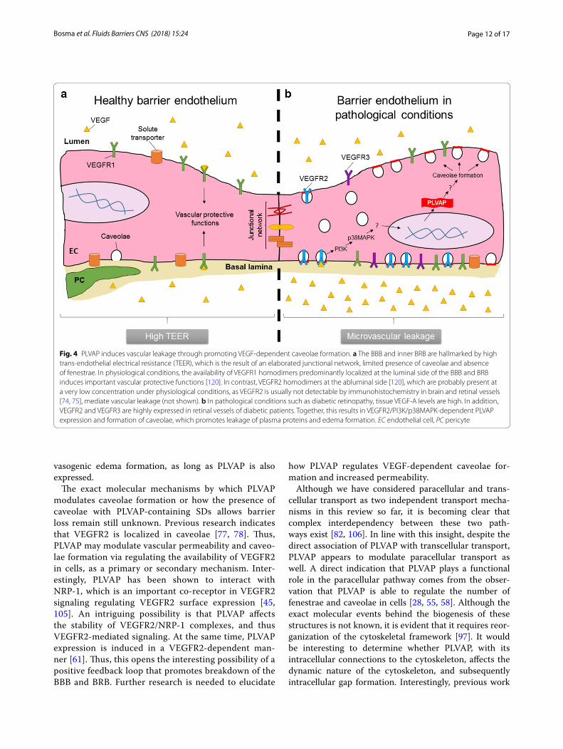
In contrast, it is relatively well-established that PLVAP plays a functional role in BBB and BRB break-down in pathological conditions (Fig. 4). With the use of short hairpin RNA (shRNA)-mediated knockdown of PLVAP expression, it was shown that PLVAP inhi-bition reduces VEGF-induced loss of BRB integrity as

Page 11 of 17Bosma et al. Fluids Barriers CNS (2018) 15:24
shown in a trans-endothelial electrical resistance assay [28]. Moreover, inhibition of PLVAP expression signifi-cantly reduced VEGF-induced permeability of 70 kDa tracers but not of 766 Da tracers in an in vitro model of retinal leakage [28, 104]. Correspondingly, siRNA-targeting of PLVAP expression reduced vascular per-meability in the oxygen-induced retinopathy (OIR) mouse model as shown by decreased extravasation of fluorescent tracers (70 kDa) and fluorescein angiog-raphy [28]. Large molecules (70 kDa) are expected to cross the endothelial barrier via transcellular transport, whereas small molecules (766 Da) cross the barrier via the paracellular pathway. Thus, these findings suggest that (VEGF-induced) leakage via the transcellular path-way is dependent on PLVAP expression. Correspond-ingly, shRNA-mediated knockdown of PLVAP expression did not prevent VEGF-induced alterations in endothe-lial junction integrity [28]. However, stress fiber forma-tion was significantly reduced. Ultrastructural analysis
revealed that knockdown of PLVAP expression in human retinal explants blocks VEGF-induced caveolae forma-tion to basal levels [28], suggesting that PLVAP induces leakage by enabling formation of caveolae, probably with PLVAP-containing SDs. The observation that inhibition of PLVAP expression does not modulate basal caveolae numbers is in agreement with the study of Herrnberger et al. [55], that did not observe a reduction in the number of caveolae in endothelial cells of Plvap-deficient mice. As previously emphasized, vesicular trafficking is a com-plex process that requires the need of multiple proteins and molecules [94–96], indicating that increased num-bers of caveolae do not necessarily implicate increased transcytosis. However, VEGF has been shown to induce the expression of several important genes involved in the transcellular transport pathway [62] and promotes cav-eolae-mediated transcytosis in vivo [85], which strongly indicates that this argument is invalid during pathological
Fig. 3 PLVAP regulates vascular development and function. a Absence of Wnt and Norrin ligands for the Lpr5/Frzd receptor complex results in inactive canonical β-catenin signaling in non-barrier endothelium and in early embryonic stages in barrier endothelium. As a consequence, cytosolic β-catenin is targeted for proteolytic degradation through phosphorylation by the “β-catenin destruction complex”, which consists of the APC/axin/GSK3b-complex. Low levels of β-catenin allow upregulation of PLVAP expression in endothelial cells. However, it is unknown how PLVAP expression is upregulated during vascular development. PLVAP expression in the developing vasculature is essential during angiogenesis. PLVAP may directly promote angiogenesis or give mechanical support to capillaries, which prevents the formation of leaky vessels after the drastic remodeling steps of angiogenesis. b The canonical β-catenin signaling pathway is active in late embryonic stages in barrier endothelium. In the presence of Wnt or Norrin ligands, the “β-catenin destruction complex” is inhibited which results in the accumulation of β-catenin in cells. Nuclear β-catenin induces the transcription of β-catenin-target genes, which results in the downregulation of PLVAP expression. Low levels of PLVAP expression induce maturation of the BRB and BBB. EC endothelial cell (Adapted from [68])
Page 12 of 17Bosma et al. Fluids Barriers CNS (2018) 15:24
vasogenic edema formation, as long as PLVAP is also expressed.
The exact molecular mechanisms by which PLVAP modulates caveolae formation or how the presence of caveolae with PLVAP-containing SDs allows barrier loss remain still unknown. Previous research indicates that VEGFR2 is localized in caveolae [77, 78]. Thus, PLVAP may modulate vascular permeability and caveo-lae formation via regulating the availability of VEGFR2 in cells, as a primary or secondary mechanism. Inter-estingly, PLVAP has been shown to interact with NRP-1, which is an important co-receptor in VEGFR2 signaling regulating VEGFR2 surface expression [45, 105]. An intriguing possibility is that PLVAP affects the stability of VEGFR2/NRP-1 complexes, and thus VEGFR2-mediated signaling. At the same time, PLVAP expression is induced in a VEGFR2-dependent man-ner [61]. Thus, this opens the interesting possibility of a positive feedback loop that promotes breakdown of the BBB and BRB. Further research is needed to elucidate
how PLVAP regulates VEGF-dependent caveolae for-mation and increased permeability.
Although we have considered paracellular and trans-cellular transport as two independent transport mecha-nisms in this review so far, it is becoming clear that complex interdependency between these two path-ways exist [82, 106]. In line with this insight, despite the direct association of PLVAP with transcellular transport, PLVAP appears to modulate paracellular transport as well. A direct indication that PLVAP plays a functional role in the paracellular pathway comes from the obser-vation that PLVAP is able to regulate the number of fenestrae and caveolae in cells [28, 55, 58]. Although the exact molecular events behind the biogenesis of these structures is not known, it is evident that it requires reor-ganization of the cytoskeletal framework [97]. It would be interesting to determine whether PLVAP, with its intracellular connections to the cytoskeleton, affects the dynamic nature of the cytoskeleton, and subsequently intracellular gap formation. Interestingly, previous work
Fig. 4 PLVAP induces vascular leakage through promoting VEGF-dependent caveolae formation. a The BBB and inner BRB are hallmarked by high trans-endothelial electrical resistance (TEER), which is the result of an elaborated junctional network, limited presence of caveolae and absence of fenestrae. In physiological conditions, the availability of VEGFR1 homodimers predominantly localized at the luminal side of the BBB and BRB induces important vascular protective functions [120]. In contrast, VEGFR2 homodimers at the abluminal side [120], which are probably present at a very low concentration under physiological conditions, as VEGFR2 is usually not detectable by immunohistochemistry in brain and retinal vessels [74, 75], mediate vascular leakage (not shown). b In pathological conditions such as diabetic retinopathy, tissue VEGF-A levels are high. In addition, VEGFR2 and VEGFR3 are highly expressed in retinal vessels of diabetic patients. Together, this results in VEGFR2/PI3K/p38MAPK-dependent PLVAP expression and formation of caveolae, which promotes leakage of plasma proteins and edema formation. EC endothelial cell, PC pericyte

Page 13 of 17Bosma et al. Fluids Barriers CNS (2018) 15:24
has shown that inhibition of PLVAP expression in bovine retinal endothelial cells significantly reduced stress fiber formation after VEGF stimulation, indicating that PLVAP may regulate intracellular gap formation [28]. Although this function of PLVAP may operate via its suspected link to actin-binding proteins, it should be noted that knockdown of PLVAP expression had only limited effects on stress fiber formation in unstimulated cells [28]. This indicates that VEGF plays a central role in this process and that the role of PLVAP is indirect. A striking phe-notype observed in these cells was that knockdown of PLVAP expression significantly blocked VEGF-induced caveolae formation, which led to the hypothesis that PLVAP regulates VEGFR2 availability [28]. Interestingly, VEGF-A has been shown to induce activation of RhoA, which is well known for its function in stress fiber forma-tion and cellular contractility [107, 108]. Hence, PLVAP may modulates stress fiber formation via induction of VEGFR2-dependent Rho GTPase signaling in cells, which is an interesting avenue to be explored in future research. Moreover, in a recent study we have found that expression of tight junctions and adherens junctions is altered in the retina of heterozygous Plvap mice [Van der Wijk et al., submitted]. Although it is not known how this altered expression is translated into functional junctional characteristics, it indicates that PLVAP affects paracellu-lar transport as well.
In short, PLVAP plays an important functional role in VEGF-induced BBB and BRB breakdown by regulating the number of caveolae with PLVAP-containing SDs. Although the sieving function of PLVAP during mac-romolecular transport in fenestrated endothelium is well established, PLVAP regulates vascular permeabil-ity in multiple and complex additional ways that remain incompletely understood.
Therapeutic perspective for cerebral edema and diabetic macular edemaTreatment approaches for vasogenic cerebral edema and DME remain limited. Available options for cerebral edema include osmotherapy, corticosteroids and surgery [109]. However, osmotherapy only has transient effects, and its overall effectiveness remains elusive [110]. In con-trast, corticosteroids are well-known to reduce cerebral edema, but therapy is associated with numerous severe side effects that may persist after treatment [6]. For this reason, the identification of novel therapeutic targets for cerebral edema is of high clinical importance. Several studies demonstrate that VEGF inhibition reduces cer-ebral edema that is caused by ischemic stroke and brain tumors [4, 8, 9]. However, VEGF is an important sign-aling molecule known to regulate pleiotropic cellular responses including neuronal survival and function [12,
14, 18], indicating that reduction of VEGF levels may have significant clinical consequences. In addition, sys-temic anti-VEGF therapy in cancer patients is associated with a high risk of stroke and other arterial thrombo-embolic events [111, 112].
Apart from the management of risk factors such as hyperglycemia and hypertension [113], current treatment options for DME consist of anti-VEGF agents, corticos-teroids and laser photocoagulation. Laser photocoagu-lation has remained the standard care of treatment for over 30 years, but currently anti-VEGF therapy is the first line of treatment for DME [11]. However, agents that block VEGF do not target the initial upstream fac-tors that lead to a compromised barrier which makes repeated injections (every 4–6 weeks) necessary [17]. In addition, a tight balance between VEGF and the pro-fibrotic growth factor termed connective tissue growth factor (CTGF) is present in the eye [15, 114]. Anti-VEGF therapy in patients with DME accompanied by severe proliferative DR has been shown to cause a shift in bal-ance of these growth factors, favoring CTGF levels, and causing retinal fibrosis [15]. Recently, it was shown that repeated intravitreal anti-VEGF injections induces reti-nal neurodegeneration and increased vascular leakage in Akita mice (Ins2Akita) [17]. Thus, this suggests that long-term anti-VEGF therapy induces retinal damage, which may eventually have significant impact on visual function. Although the effects of long-term anti-VEGF therapy in the brain remain unknown, the same charac-teristics of brain and retinal barrier endothelium and the observed neurodegenerative effects in the study of Hom-brebueno et al. [17] indicate that long-term therapy may also induce serious side effects in the brain.
In contrast to VEGF which has effects on multiple cell types, PLVAP is only expressed in endothelial cells, where it modulates caveolae formation and possibly VEGFR2 availability [28]. Consequently, targeting PLVAP expres-sion in brain and retinal endothelial cells may inhibit downstream responses of VEGF and of other mediators in endothelial cells without altering cell survival of neu-rons [12, 14, 18]. Together with the striking observation that PLVAP inhibition significantly reduced vascular leakage of large macromolecules in vitro and in vivo [28], it strongly indicates that anti-PLVAP therapy may hold great potential as a safer novel treatment option for vaso-genic cerebral edema and DME.
The widespread expression of PLVAP in non-barrier continuous endothelia and the crucial functions of PLVAP as a regulator of vascular homeostasis in fenes-trated endothelium indicate that systemic targeting of PLVAP is not recommended. However, the ease of local drug delivery in the eye, and the limited fluid and molec-ular exchange between the eye and the rest of the body

Page 14 of 17Bosma et al. Fluids Barriers CNS (2018) 15:24
indicate that local PLVAP inhibition is achievable. For instance, PLVAP expression can be inhibited via intra-vitreal injections and slow-release drug delivery systems such as implantable micropumps [115] or biodegrad-able polyesteramide microspheres [116]. In contrast to the eye, local drug delivery to the brain may be more problematic. However, in the case of tumor-related cer-ebral edema, which often requires surgery to remove as much as possible tumor tissue, anti-PLVAP therapy may be implemented during surgery to restore the func-tional properties of the BBB to prevent relapse of edema formation.
Further investigations of the therapeutic rationale and possibilities of anti-PLVAP therapy is warranted. Inter-estingly, PLVAP has been shown to modulate angiogen-esis as well [36], which indicates that PLVAP is also a target for conditions associated with angiogenesis such as age-related macular degeneration, proliferative diabetic retinopathy and cancer.
Therapeutic perspective: targeted drug delivery via caveolaeBesides the potential to restore the function of barrier endothelia via inhibition of PLVAP expression, PLVAP may also be exploited to achieve targeted drug deliv-ery across the BBB and BRB in pathological conditions. In a recent study, the therapeutic enzyme superoxide dismutase (SOD) was conjugated to antibodies against PLVAP with the ultimate goal to target SOD to caveolae of endothelial cells of pulmonary vessels, as the target substrate of the enzyme is present in endothelial vesicles [117]. Interestingly, SOD conjugated to PLVAP antibod-ies more efficiently blocked lipopolysaccharide-induced pulmonary inflammation than SOD conjugated to endothelial cells via CD31 [117]. It is interesting to assess whether such type of drug delivery has potential for the BBB and BRB.
Concluding remarksPLVAP is an important regulator of vascular permeabil-ity during embryogenesis, and after birth in fenestrated endothelium and in pathological conditions, in particu-lar of macromolecules. Although the sieving regulatory function of PLVAP as a molecular component of dia-phragms remains comprehensible, it remains incom-pletely understood how PLVAP modulates breakdown of the BBB and BRB. Previous research indicates that PLVAP induces vascular permeability via promoting VEGF-dependent caveolae formation, but the molecu-lar mechanisms remain unclear [28]. Furthermore, very little is known how VEGF induces PLVAP expression. There are indications that this occurs via a VEGFR2/PI3K/p38MAPK-dependent signaling pathway [61], but
it remains unknown which transcriptional regulators are involved in this process. More research is needed to better understand the underlying molecular and cellu-lar mechanisms that link PLVAP to vascular leakage and breakdown of the BBB and BRB. Nevertheless, after dec-ades of functioning as histological marker for endothe-lium in normal tissues outside the brain and eye, and in pathological leakage in the brain and eye without a known molecular substrate, the first functional evidence has now been provided that PLVAP plays a pivotal role in VEGF-induced BRB permeability. Together with the selective expression of PLVAP in the BBB and BRB dur-ing pathological conditions, PLVAP emerges as a novel promising therapeutic target to prevent the clinical bur-den of vasogenic cerebral edema and DME.
AbbreviationsBBB: blood–brain barrier; BRB: blood–retinal barrier; CTGF: connective tissue growth factor; DME: diabetic macular edema; FD: fenestral diaphragm; NRP-1: neuropilin-1; OIR: oxygen-induced retinopathy; p38MAPK: p38 mitogen-activated protein kinase; PAL-E: Pathologische Anatomie Leiden-Endothelium; PI3K: phosphatidylinositol 3-kinase; PLVAP: plasmalemma vesicle-associated protein; PMA: phorbol myristate acetate; PV-1: plasmalemma vesicle-associ-ated protein; SD: stomatal diaphragm; shRNA: short hairpin RNA; SOD: super-oxide dismutase; TEC: trans-endothelial channel; TGF-β: transforming growth factor beta; TNF-α: tumor necrosis factor alpha; VEGF: vascular endothelial growth factor; VEGFR: vascular endothelial growth factor receptor.
Authors’ contributionsEKB designed the content of the review, wrote the manuscript and prepared figures. CJFVN, ROS and IK advised in the design of the content of the review and contributed to editing of the manuscript. All authors read and approved the final manuscript.
Author details1 Ocular Angiogenesis Group, Departments of Ophthalmology and Medi-cal Biology, Amsterdam Cardiovascular Sciences, Amsterdam Neuroscience, Amsterdam UMC, University of Amsterdam, Meibergdreef 9, Amsterdam, The Netherlands. 2 Department of Genetic Toxicology and Cancer Biology, National Institute of Biology, Ljubljana, Slovenia. 3 Department of Ophthalmology, Uni-versity of Lausanne, Jules-Gonin Eye Hospital, Fondation Asile des Aveugles, Lausanne, Switzerland. 4 Ocular Angiogenesis Group, Department of Medical Biology, Amsterdam UMC, University of Amsterdam, Meibergdreef 15, Room L3-154, 1105 AZ Amsterdam, The Netherlands.
AcknowledgementsNot applicable.
Competing interestsThe authors declare that they have no competing interests.
Availability of data and materialsNot applicable.
Consent for publicationNot applicable.
Ethics approval and consent to participateNot applicable.
FundingThis work was made possible by the financial support of the Diabetes Fonds (Dutch Diabetes Fund, Grant 2014.00.1784) and by the following founda-tions: Landelijke Stichting voor Blinden en Slechtzienden, Novartis Fonds and

Page 15 of 17Bosma et al. Fluids Barriers CNS (2018) 15:24
MaculaFonds, that contributed through UitZicht (Grant UitZicht 2014-33), the Nederlandse Vereniging ter Verbetering van het Lot der Blinden, Rotterdamse Stichting Blindenbelangen (Grant B20140050) and Stichting Blindenhulp. This work was published with financial support from the Edmond en Marianne Blaauw Fonds voor Oogheelkunde. The funding organizations had no role in the design or conduct of this research. They provided unrestricted grants.
Publisher’s NoteSpringer Nature remains neutral with regard to jurisdictional claims in pub-lished maps and institutional affiliations.
Received: 13 July 2018 Accepted: 10 August 2018
References 1. Merali Z, Huang K, Mikulis D, Silver F, Kassner A. Evolution of blood–
brain-barrier permeability after acute ischemic stroke. PLoS ONE. 2017;12:e0171558.
2. Wolburg H, Noell S, Fallier-Becker P, MacK AF, Wolburg-Buchholz K. The disturbed blood–brain barrier in human glioblastoma. Mol Aspects Med. 2012;33:579–89.
3. Klaassen I, Van Noorden CJF, Schlingemann RO. Molecular basis of the inner blood–retinal barrier and its breakdown in diabetic macular edema and other pathological conditions. Prog Retinal Eye Res. 2013;34:19–48.
4. Gerstner ER, Duda DG, Di Tomaso E, Ryg PA, Loeffler JS, Sorensen AG, et al. VEGF inhibitors in the treatment of cerebral edema in patients with brain cancer. Nat Rev Clin Oncol. 2009;6:229–36.
5. Klein R, Klein BEK, Moss SE. Visual impairment in diabetes. Ophthalmol-ogy. 1984;91:1–9.
6. Dietrich J, Rao K, Pastorino S, Kesari S. Corticosteroids in brain cancer patients: benefits and pitfalls. Expert Rev Clin Pharmacol. 2011;4:233–42.
7. Network TDRCR. Aflibercept, Bevacizumab, or Ranibizumab for diabetic macular edema. N Engl J Med. 2015;372:1193–203.
8. Van Bruggen N, Thibodeaux H, Palmer JT, Lee WP, Fu L, Cairns B, et al. VEGF antagonism reduces edema formation and tissue damage after ischemia/reperfusion injury in the mouse brain. J Clin Invest. 1999;104:1613–20.
9. Batchelor TT, Sorensen AG, di Tomaso E, Zhang WT, Duda DG, Cohen KS, et al. AZD2171, a pan-VEGF receptor tyrosine kinase inhibitor, normal-izes tumor vasculature and alleviates edema in glioblastoma patients. Cancer Cell. 2007;11:83–95.
10. Mitchell P, Bandello F, Schmidt-Erfurth U, Lang GE, Massin P, Schlinge-mann RO, et al. The RESTORE study: ranibizumab monotherapy or combined with laser versus laser monotherapy for diabetic macular edema. Ophthalmology. 2011;118:615–25.
11. Agarwal A, Sarwar S, Sepah YJ, Nguyen QD. What have we learnt about the management of diabetic macular edema in the antivascular endothelial growth factor and corticosteroid era? Curr Opin Ophthal-mol. 2015;26:177–83.
12. Sun Y, Jin K, Xie L, Childs J, Mao XO, Logvinova A, et al. VEGF-induced neuroprotection, neurogenesis, and angiogenesis after focal cerebral ischemia. J Clin Invest. 2003;111:1843–51.
13. Zachary I. Signaling mechanisms mediating vascular protective actions of vascular endothelial growth factor. Am J Physiol Cell Physiol. 2001;280:C1375–86.
14. Saint-Geniez M, Maharaj ASR, Walshe TE, Tucker BA, Sekiyama E, Kuri-hara T, et al. Endogenous VEGF is required for visual function: evidence for a survival role on Müller cells and photoreceptors. PLoS ONE. 2008;3:e3554.
15. Van Geest RJ, Lesnik-Oberstein SY, Tan HS, Mura M, Goldschmeding R, Van Noorden CJF, et al. A shift in the balance of vascular endothelial growth factor and connective tissue growth factor by bevacizumab causes the angiofibrotic switch in proliferative diabetic retinopathy. Br J Ophthalmol. 2012;96:587–90.
16. Gerhardinger C, Brown LF, Roy S, Mizutani M, Zucker CL, Lorenzi M. Expression of vascular endothelial growth factor in the human
retina and in nonproliferative diabetic retinopathy. Am J Pathol. 1998;152:1453–62.
17. Hombrebueno JR, Ali IHA, Xu H, Chen M. Sustained intraocular VEGF neutralization results in retinal neurodegeneration in the Ins2(Akita) diabetic mouse. Sci Rep. 2015;5:18316.
18. Amato R, Biagioni M, Cammalleri M, Dal Monte M, Casini G. VEGF as a survival factor in ex vivo models of early diabetic retinopathy. Invest Opthalmol Vis Sci. 2016;57:3066–76.
19. Bradbury MW. The blood–brain barrier. Transport across the cerebral endothelium. Circ Res. 1985;57:213–22.
20. Raviola G. The structural basis of the blood–ocular barriers. Exp Eye Res. 1977;25:27–63.
21. De Bock M, Van Haver V, Vandenbroucke RE, Decrock E, Wang N, Ley-baert L. Into rather unexplored terrain-transcellular transport across the blood–brain barrier. GLIA. 2016;64:1097–123.
22. Schlingemann RO, Dingjan GM, Emeis JJ, Blok J, Warnaar SO, Ruiter DJ. Monoclonal antibody PAL-E specific for endothelium. Lab Invest. 1985;52:71–6.
23. Schlingemann RO, Bots GTAM, Duinen SG, Ruiter DJ. Differential expression of endothelium-specific antigen PAL-E in vasculature of brain tumors and preexistent brain capillaries. Ann N Y Acad Sci. 1988;529:111–4.
24. Schlingemann RO, Hofman P, Anderson L, Troost D, van der Gaag R. Vascular expression of endothelial antigen PAL-E indicates absence of blood–ocular barriers in the normal eye. Ophthalmic Res. 1997;29:130–8.
25. Schlingemann RO, Hofman P, Vrensen GFJM, Blaauwgeers HGT. Increased expression of endothelial antigen PAL-E in human diabetic retinopathy correlates with microvascular leakage. Diabetologia. 1999;42:596–602.
26. Stan RV, Tkachenko E, Niesman IR. PV1 is a key structural component for the formation of the stomatal and fenestral diaphragms. Mol Biol Cell. 2004;15:3615–30.
27. Niemelä H, Elima K, Henttinen T, Irjala H, Salmi M, Jalkanen S. Molecular identification of PAL-E, a widely used endothelial-cell marker. Blood. 2005;106:3405–9.
28. Wisniewska-Kruk J, Van Der Wijk AE, Van Veen HA, Gorgels TGMF, Vogels IMC, Versteeg D, et al. Plasmalemma vesicle-associated protein has a key role in blood–retinal barrier loss. Am J Pathol. 2016;186:1044–54.
29. Tse D, Stan RV. Morphological heterogeneity of endothelium. Semin Thromb Hemost. 2010;36:236–45.
30. Hallmann R, Mayer DN, Berg EL, Broermann R, Butcher EC. Novel mouse endothelial cell surface marker is suppressed during differentiation of the blood brain barrier. Dev Dyn. 1995;202:325–32.
31. Leppink DM, Bishop DK, Sedmak DD, Henry ML, Ferguson RM, Streeter PR, et al. Inducible expression of an endothelial cell antigen on murine myocardial vasculature in association with interstitial cellular infiltration. Transplantation. 1989;48:874–7.
32. Schlingemann RO, Rietveld FJ, Kwaspen F, van de Kerkhof PC, de Waal RM, Ruiter DJ. Differential expression of markers for endothelial cells, pericytes, and basal lamina in the microvasculature of tumors and granulation tissue. Am J Pathol. 1991;138:1335–47.
33. Schlingemann RO, Hofman P, Klooster J, Blaauwgeers HG, Van der Gaag R, Vrensen GF. Ciliary muscle capillaries have blood–tissue barrier characteristics. Exp Eye Res. 1998;66:747–54.
34. Hofman P, Hoyng P, Vanderwerf F, Vrensen GFJM, Schlingemann RO. Lack of blood–brain barrier properties in microvessels of the prelaminar optic nerve head. Investig Ophthalmol Vis Sci. 2001;42:895–901.
35. Shue EH, Carson-Walter EB, Liu Y, Winans BN, Ali ZS, Chen J, et al. Plasmalemmal vesicle associated protein-1 (PV-1) is a marker of blood–brain barrier disruption in rodent models. BMC Neurosci. 2008;9:29.
36. Carson-Walter EB, Hampton J, Shue E, Geynisman DM, Pillai PK, Sathanoori R, et al. Plasmalemmal vesicle associated protein-1 is a novel marker implicated in brain tumor angiogenesis. Clin Cancer Res. 2005;11:7643–50.
37. Hofman P, Blaauwgeers HGT, Vrensen GFJM, Schlingemann RO. Role of VEGF-A in endothelial phenotypic shift in human diabetic retinopa-thy and VEGF-A-induced retinopathy in monkeys. Ophthalmic Res. 2001;33:156–62.

Page 16 of 17Bosma et al. Fluids Barriers CNS (2018) 15:24
38. Leenstra S, Troost D, Das PK, Claessen N, Becker AE, Bosch DA. Endothe-lial cell marker PAL-E reactivity in brain tumor, developing brain, and brain disease. Cancer. 1993;72:3061–7.
39. Ghitescu L, Fixman A, Simionescu M, Simionescu N. Specific binding sites for albumin restricted to plasmalemmal vesicles of continuous capillary endothelium: receptor-mediated transcytosis. J Cell Biol. 1986;102:1304–11.
40. Xu B, deWaal RM, Mor-Vaknin N, Hibbard C, Markovitz DM, Kahn ML. The endothelial cell-specific antibody PAL-E identifies a secreted form of vimentin in the blood vasculature. Mol Cell Biol. 2004;24:9198–206.
41. Jaalouk DE, Ozawa MG, Sun J, Lahdenranta J, Schlingemann RO, Pas-qualini R, et al. The original pathologische anatomie Leiden–Endothe-lium monoclonal antibody recognizes a vascular endothelial growth factor-binding site within neuropilin-1. Cancer Res. 2007;67:9623–9.
42. Sarris M, Andersen KG, Randow F, Mayr L, Betz AG. Neuropilin-1 expres-sion on regulatory T cells enhances their interactions with dendritic cells during antigen recognition. Immunity. 2008;28:402–13.
43. Lepelletier Y, Smaniotto S, Hadj-Slimane R, Villa-Verde DMS, Nogueira AC, Dardenne M, et al. Control of human thymocyte migration by Neuropilin-1/Semaphorin-3A-mediated interactions. Proc Natl Acad Sci USA. 2007;104:5545–50.
44. Gu C, Rodriguez ER, Reimert DV, Shu T, Fritzsch B, Richards LJ, et al. Neuropilin-1 conveys semaphorin and VEGF signaling during neural and cardiovascular development. Dev Cell. 2003;5:45–57.
45. Keuschnigg J, Tvorogov D, Elima K, Salmi M, Alitalo K, Salminen T, et al. PV-1 is recognized by the PAL-E antibody and forms complexes with NRP-1. Blood. 2012;120:232–5.
46. Keuschnigg J, Henttinen T, Auvinen K, Karikoski M, Salmi M, Jalkanen S. The prototype endothelial marker PAL-E is a leukocyte trafficking molecule. Blood. 2009;114:478–84.
47. Stan RV, Roberts WG, Predescu D, Ihida K, Saucan L, Ghitescu L, et al. Immunoisolation and partial characterization of endothelial plas-malemmal vesicles (caveolae). Mol Biol Cell. 1997;8:595–605.
48. Stan RV, Ghitescu L, Jacobson BS, Palade GE. Isolation, cloning, and localization of rat PV-1, a novel endothelial caveolar protein. J Cell Biol. 1999;145:1189–98.
49. Stan RV. Structure of caveolae. Biochimica et Biophysica Acta Mol Cell Res. 2005;1746:334–48.
50. Rothberg KG, Heuser JE, Donzell WC, Ying YS, Glenney JR, Anderson RG. Caveolin, a protein component of caveolae membrane coats. Cell. 1992;68:673–82.
51. Stan RV. Multiple PV1 dimers reside in the same stomatal or fenestral diaphragm. Am J Physiol Hear Circ Physiol. 2004;286:1347–53.
52. Stan RV, Arden KC, Palade GE. cDNA and protein sequence, genomic organization, and analysis of cis regulatory elements of mouse and human PLVAP genes. Genomics. 2001;72:304–13.
53. Couet J, Li S, Okamoto T, Ikezu T, Lisanti MP. Identification of peptide and protein ligands for the caveolin-scaffolding domain. Biochemistry. 1997;272:6525–33.
54. Bearer EL, Orci L, Sors P. Endothelial fenestral diaphragms: a quick-freeze, deep-etch study. J Cell Biol. 1985;100:418–28.
55. Herrnberger L, Seitz R, Kuespert S, Bosl MR, Fuchshofer R, Tamm ER. Lack of endothelial diaphragms in fenestrae and caveolae of mutant Plvap-deficient mice. Histochem Cell Biol. 2012;138:709–24.
56. Herrnberger L, Ebner K, Junglas B, Tamm ER. The role of plasmalemma vesicle-associated protein (PLVAP) in endothelial cells of Schlemm’s canal and ocular capillaries. Exp Eye Res. 2012;105:27–33.
57. Stan RV, Tse D, Deharvengt SJ, Smits NC, Xu Y, Luciano MR, et al. The diaphragms of fenestrated endothelia: gatekeepers of vascular perme-ability and blood composition. Dev Cell. 2012;23:1203–18.
58. Herrnberger L, Hennig R, Kremer W, Hellerbrand C, Goepferich A, Kalbitzer HR, et al. Formation of fenestrae in murine liver sinusoids depends on plasmalemma vesicle-associated protein and is required for lipoprotein passage. PLoS ONE. 2014;9:e115005.
59. Tkachenko E, Tse D, Sideleva O, Deharvengt SJ, Luciano MR, Xu Y, et al. Caveolae, fenestrae and transendothelial channels retain PV1 on the surface of endothelial cells. PLoS ONE. 2012;7:e32655.
60. Rantakari P, Auvinen K, Jäppinen N, Kapraali M, Valtonen J, Karikoski M, et al. The endothelial protein PLVAP in lymphatics controls the entry of lymphocytes and antigens into lymph nodes. Nat Immunol. 2015;16:386–96.
61. Strickland LA, Jubb AM, Hongo JA, Zhong F, Burwick J, Fu L, et al. Plas-malemmal vesicle-associated protein (PLVAP) is expressed by tumour endothelium and is upregulated by vascular endothelial growth factor-A (VEGF). J Pathol. 2005;206:466–75.
62. Klaassen I, Hughes JM, Vogels IMC, Schalkwijk CG, Van Noorden CJF, Schlingemann RO. Altered expression of genes related to blood–retina barrier disruption in streptozotocin-induced diabetes. Exp Eye Res. 2009;89:4–15.
63. Wisniewska-Kruk J, Klaassen I, Vogels IMC, Magno AL, Lai CM, Van Noorden CJF, et al. Molecular analysis of blood–retinal barrier loss in the Akimba mouse, a model of advanced diabetic retinopathy. Exp Eye Res. 2014;122:123–31.
64. Bodor C, Nagy JP, Végh B, Németh A, Jenei A, MirzaHosseini S, et al. Angiotensin II increases the permeability and PV-1 expression of endothelial cells. Am J Physiol Cell Physiol. 2012;302:C267–76.
65. Chen J, Stahl A, Krah NM, Seaward MR, Joyal JS, Juan AM, et al. Retinal expression of Wnt-pathway mediated genes in low-density lipo-protein receptor-related protein 5 (Lrp5) knockout mice. PLoS ONE. 2012;7:e30203.
66. Schäfer NF, Luhmann UFO, Feil S, Berger W. Differential gene expression in Ndph-knockout mice in retinal development. Investig Ophthalmol Vis Sci. 2009;50:906–16.
67. Liebner S, Corada M, Bangsow T, Babbage J, Taddei A, Czupalla CJ, et al. Wnt/beta-catenin signaling controls development of the blood–brain barrier. J Cell Biol. 2008;183:409–17.
68. Liebner S, Plate KH. Differentiation of the brain vasculature: the answer came blowing by the Wnt. J Angiogenes Res. 2010;2:1.
69. Farber G, Hurtado R, Loh S, Monette S, Mtui J, Kopan R, et al. Glomerular endothelial cell maturation depends on ADAM10, a key regulator of Notch signaling. Angiogenesis. 2018;21:335–47.
70. Mintet E, Lavigne J, Paget V, Tarlet G, Buard V, Guipaud O, et al. Endothe-lial Hey2 deletion reduces endothelial-to-mesenchymal transition and mitigates radiation proctitis in mice. Sci Rep. 2017;7:4933.
71. Wasserman SM, Mehraban F, Komuves LG, Yang R-B, Tomlinson JE, Zhang Y, et al. Gene expression profile of human endothelial cells exposed to sustained fluid shear stress. Physiol Genomics. 2002;12:13–23.
72. Senger DR, Galli SJ, Dvorak AM, Perruzzi CA, Harvey VS, Dvorak HF. Tumor cells secrete a vascular permeability factor that promotes accu-mulation of ascites fluid. Science. 1983;219:983–5.
73. Ferrara N, Henzel WJ. Pituitary follicular cells secrete a novel heparin-binding growth factor specific for vascular endothelial cells. Biochem Biophys Res Commun. 1989;161:851–8.
74. Witmer AN, Dai J, Weich HA, Vrensen GF, Schlingemann RO. Expression of vascular endothelial growth factor receptors 1, 2, and 3 in quiescent endothelia. J Histochem Cytochem. 2002;50:767–77.
75. Witmer AN, Blaauwgeers HG, Weich HA, Alitalo K, Vrensen GFJM, Schlingemann RO. Altered expression patterns of VEGF receptors in human diabetic retina and in experimental VEGF-induced retinopathy in monkey. Investig Ophthalmol Vis Sci. 2002;43:849–57.
76. Hnasko R, Frank PG, Ben-Jonathan N, Lisanti MP. PV-1 is negatively regu-lated by VEGF in the lung of caveolin-1, but not caveolin-2, null mice. Cell Cycle. 2006;5:2012–20.
77. Tahir SA, Park S, Thompson TC. Caveolin-1 regulates VEGF-stimulated angiogenic activities in prostate cancer and endothelial cells. Cancer Biol Ther. 2009;8:2286–96.
78. Labrecque L, Royal I, Surprenant DS, Patterson C, Gingras D, Béliveau R. Regulation of vascular endothelial growth factor receptor-2 activ-ity by caveolin-1 and plasma membrane cholesterol. Mol Biol Cell. 2003;14:334–47.
79. Keaney J, Campbell M. The dynamic blood–brain barrier. FEBS J. 2015;282:4067–79.
80. van der Wijk AE, Vogels IMC, van Veen HA, van Noorden CJF, Schlinge-mann RO, Klaassen I. Spatial and temporal recruitment of the neuro-vascular unit during development of the mouse blood–retinal barrier. Tissue Cell. 2018;52:42–50.
81. Abbott NJ, Patabendige AAK, Dolman DEM, Yusof SR, Begley DJ. Structure and function of the blood–brain barrier. Neurobiol Dis. 2010;37:13–25.
Page 17 of 17Bosma et al. Fluids Barriers CNS (2018) 15:24
82. Komarova Y, Malik AB. Regulation of endothelial permeability via paracellular and transcellular transport pathways. Annu Rev Physiol. 2010;72:463–93.
83. Knowland D, Arac A, Sekiguchi KJ, Hsu M, Lutz SE, Perrino J, et al. Step-wise recruitment of transcellular and paracellular pathways underlies blood–brain barrier breakdown in stroke. Neuron. 2014;82:603–17.
84. Feng Y, Venema VJ, Venema RC, Tsai N, Behzadian MA, Caldwell RB. VEGF-induced permeability increase is mediated by caveolae. Investig Ophthalmol Vis Sci. 1999;40:157–67.
85. Hofman P, Blaauwgeers HG, Tolentino MJ, Adamis AP, Nunes Cardozo BJ, Vrensen GF, et al. VEGF-A induced hyperpermeability of blood–retinal barrier endothelium in vivo is predominantly associated with pinocy-totic vesicular transport and not with formation of fenestrations. Curr Eye Res. 2000;21:637–45.
86. Nahirney PC, Reeson P, Brown CE. Ultrastructural analysis of blood–brain barrier breakdown in the peri-infarct zone in young adult and aged mice. J Cereb Blood Flow Metab. 2016;36:413–25.
87. Stokum JA, Gerzanich V, Simard JM. Molecular pathophysiology of cerebral edema. J Cereb Blood Flow Metab. 2016;36:513–38.
88. Stan RV, Kubitza M, Palade GE. PV-1 is a component of the fenestral and stomatal diaphragms in fenestrated endothelia. Proc Natl Acad Sci USA. 1999;96:13203–7.
89. Elkadri A, Thoeni C, Deharvengt SJ, Murchie R, Guo C, Stavropoulos JD, et al. Mutations in plasmalemma vesicle associated protein result in sieving protein-losing enteropathy characterized by hypoproteinemia, hypoalbuminemia, and hypertriglyceridemia. Cell Mol Gastroenterol Hepatol. 2015;1(381–394):e7.
90. Broekaert IJ, Becker K, Gottschalk I, Körber F, Dötsch J, Thiele H, et al. Mutations in plasmalemma vesicle-associated protein cause severe syndromic protein-losing enteropathy. J Med Genet. 2018. https ://doi.org/10.1136/jmedg enet-2018-10526 2.
91. Sarin H. Physiologic upper limits of pore size of different blood capillary types and another perspective on the dual pore theory of microvascu-lar permeability. J Angiogenes Res. 2010;2:1–19.
92. Simionescu N, Simionescu M, Palade GE. Differentiated microdomains on the luminal surface of the capillary endothelium. I. Preferential distribution of anionic sites. J Cell Biol. 1981;90:605–13.
93. Simionescu M, Simionescu N, Silbert JE, Palade GE. Differentiated micro-domains on the luminal surface of the capillary endothelium. II. Partial characterization of their anionic sites. J Cell Biol. 1981;90:614–21.
94. Praefcke GJK, McMahon HT. The dynamin superfamily: universal membrane tubulation and fission molecules? Nat Rev Mol Cell Biol. 2004;5:133–47.
95. Chen YA, Scheller RH. SNARE-mediated membrane fusion. Nat Rev Mol Cell Biol. 2001;2:98–106.
96. Fra AM, Williamson E, Simons K, Parton RG. De novo formation of cave-olae in lymphocytes by expression of VIP21-caveolin. Proc Natl Acad Sci USA. 1995;92:8655–9.
97. Ioannidou S, Deinhardt K, Miotla J, Bradley J, Cheung E, Samuels-son S, et al. An in vitro assay reveals a role for the diaphragm protein PV-1 in endothelial fenestra morphogenesis. Proc Natl Acad Sci USA. 2006;103:16770–5.
98. Rantakari P, Jäppinen N, Lokka E, Mokkala E, Gerke H, Peuhu E, et al. Fetal liver endothelium regulates the seeding of tissue-resident mac-rophages. Nature. 2016;538:392–6.
99. Daneman R, Zhou L, Kebede AA, Barres BA. Pericytes are required for blood–brain barrier integrity during embryogenesis. Nature. 2010;468:562–6.
100. Umans RA, Henson HE, Mu F, Parupalli C, Ju B, Peters JL, et al. CNS angiogenesis and barrier genesis occur simultaneously. Dev Biol. 2017;425:101–8.
101. Chow BW, Gu C. Gradual suppression of transcytosis governs functional blood–retinal barrier formation. Neuron. 2017;93(1325–1333):e3.
102. Ben-Zvi A, Lacoste B, Kur E, Andreone BJ, Mayshar Y, Yan H, et al. MSFD2A is critical for the formation and function of the blood brain barrier. Nature. 2014;509:507–11.
103. Andreone BJ, Chow BW, Tata A, Lacoste B, Ben-Zvi A, Bullock K, et al. Blood–brain barrier permeability is regulated by lipid transport-dependent suppression of caveolae-mediated transcytosis. Neuron. 2017;94(581–594):e5.
104. Wisniewska-Kruk J, Hoeben KA, Vogels IMC, Gaillard PJ, Van Noorden CJF, Schlingemann RO, et al. A novel co-culture model of the blood–retinal barrier based on primary retinal endothelial cells, pericytes and astrocytes. Exp Eye Res. 2012;96:181–90.
105. Gelfand MV, Hagan N, Tata A, Oh WJ, Lacoste B, Kang KT, et al. Neu-ropilin-1 functions as a VEGFR2 co-receptor to guide developmental angiogenesis independent of ligand binding. Elife. 2014;3:e03720.
106. Armstrong SM, Khajoee V, Wang C, Wang T, Tigdi J, Yin J, et al. Co-regulation of transcellular and paracellular leak across microvascular endothelium by dynamin and Rac. Am J Pathol. 2012;180:1308–23.
107. Bryan BA, Dennstedt E, Mitchell DC, Walshe TE, Noma K, Loureiro R, et al. RhoA/ROCK signaling is essential for multiple aspects of VEGF-medi-ated angiogenesis. FASEB J. 2010;24:3186–95.
108. Chrzanowska-Wodnicka M, Burridge K. Rho-stimulated contractility drives the formation of stress fibers and focal adhesions. J Cell Biol. 1996;133:1403–15.
109. Rabinstein AA. Treatment of cerebral edema. Neurologist. 2006;12:59–73.
110. Grände P-O, Romner B. Osmotherapy in brain edema: a questionable therapy. J Neurosurg Anesthesiol. 2012;24:407–12.
111. Scappaticci FA, Skillings JR, Holden SN, Gerber H-P, Miller K, Kabbinavar F, et al. Arterial thromboembolic events in patients with metastatic carcinoma treated with chemotherapy and bevacizumab. J Natl Cancer Inst. 2007;99:1232–9.
112. Tolentino M. Systemic and ocular safety of intravitreal anti-VEGF thera-pies for ocular neovascular disease. Surv Ophthalmol. 2011;56:95–113.
113. Hammes HP, Welp R, Kempe HP, Wagner C, Siegel E, Holl RW. Risk factors for retinopathy and DME in type 2 diabetes-results from the German/Austrian DPV database. PLoS ONE. 2015;10:e0132492.
114. Kuiper EJ, Van Nieuwenhoven FA, de Smet MD, van Meurs JC, Tanck MW, Oliver N, et al. The angio-fibrotic switch of VEGF and CTGF in proliferative diabetic retinopathy. PLoS ONE. 2008;3:e2675.
115. Humayun M, Santos A, Altamirano JC, Ribeiro R, Gonzalez R, de la Rosa A, et al. Implantable micropump for drug delivery in patients with diabetic macular edema. Transl Vis Sci Technol. 2014;3:5.
116. Andrés-Guerrero V, Zong M, Ramsay E, Rojas B, Sarkhel S, Gallego B, et al. Novel biodegradable polyesteramide microspheres for controlled drug delivery in ophthalmology. J Control Release. 2015;211:105–17.
117. Shuvaev VV, Kiseleva RY, Arguiri E, Villa CH, Muro S, Christofidou-Solomi-dou M, et al. Targeting superoxide dismutase to endothelial caveolae profoundly alleviates inflammation caused by endotoxin. J Control Release. 2018;272:1–8.
118. Gousopoulos E, Proulx ST, Scholl J, Uecker M, Detmar M. Promi-nent lymphatic vessel hyperplasia with progressive dysfunction and distinct immune cell infiltration in lymphedema. Am J Pathol. 2016;186:2193–203.
119. Sarawar SR, Schlingemann RO, Kelsey A, Fleming S, Kumar S. A mono-clonal antibody stains blastemal but not tubular components of Wilms’ tumour. J Pathol. 1988;156:319–24.
120. Hudson N, Powner MB, Sarker MH, Burgoyne T, Campbell M, Ockrim ZK, et al. Differential apicobasal VEGF signaling at vascular blood–neural barriers. Dev Cell. 2014;30:541–52.
Related Documents