The Drosophila CAKI/CMG protein, a homolog of human CASK, is essential for regulation of neurotransmitter vesicle release. Authors Mauro A. Zordan 2 , Michele Massironi 1 , Maria Giovanna Ducato 1 , Geertruy te Kronnie 1 , Rodolfo Costa 2 , Carlo Reggiani 1 , Carine Chagneau 3 , Jean-René Martin 3 and Aram Megighian 1 . 1 Department of Human Anatomy and Physiology, University of Padova, 35131 Padova, Italy 2 Department of Biology, University of Padova, 35131 Padova, Italy 3 Equipe ATIPE : Bases Neurales du Mouvement chez la Drosophile, NAMC, CNRS, UMR-8620, Université Paris Sud, Orsay, France Running head Drosophila CAKI/CMG regulates neurotransmitter release Contact information: Aram Megighian, Department of Human Anatomy and Physiology, Section of Physiology, University of Padua, Italy Via Marzolo 3, 35131, Padova, ITALY E-mail: [email protected] Phone: +39 049 827 5724 Fax: +39 049 827 5301 Articles in PresS. J Neurophysiol (May 4, 2005). doi:10.1152/jn.00954.2004 Copyright © 2005 by the American Physiological Society.

Welcome message from author
This document is posted to help you gain knowledge. Please leave a comment to let me know what you think about it! Share it to your friends and learn new things together.
Transcript
The Drosophila CAKI/CMG protein, a homolog of human
CASK, is essential for regulation of neurotransmitter vesicle
release.
Authors
Mauro A. Zordan2, Michele Massironi1, Maria Giovanna Ducato1, Geertruy te
Kronnie1, Rodolfo Costa2, Carlo Reggiani1, Carine Chagneau3, Jean-René Martin3 and
Aram Megighian1.
1Department of Human Anatomy and Physiology, University of Padova, 35131
Padova, Italy
2Department of Biology, University of Padova, 35131 Padova, Italy
3Equipe ATIPE : Bases Neurales du Mouvement chez la Drosophile, NAMC, CNRS,
UMR-8620, Université Paris Sud, Orsay, France
Running head
Drosophila CAKI/CMG regulates neurotransmitter release
Contact information:
Aram Megighian,
Department of Human Anatomy and Physiology, Section of Physiology, University of
Padua, Italy
Via Marzolo 3, 35131, Padova, ITALY
E-mail: [email protected]
Phone: +39 049 827 5724
Fax: +39 049 827 5301
Articles in PresS. J Neurophysiol (May 4, 2005). doi:10.1152/jn.00954.2004
Copyright © 2005 by the American Physiological Society.

Zordan et al. : Drosophila CAKI/CMG regulates neurotransmitter release
2
Abstract
Vertebrate CASK is a member of the MAGUK family of proteins. CASK is present in
the nervous system where it binds to NEUREXIN, a transmembrane protein localized
in the pre-synaptic membrane. The Drosophila homolog of CASK is CAKI or
CAMGUK. CAKI is expressed in the nervous system of larvae and adult flies. In
adult flies the expression of caki is particularly evident in the visual brain regions
(Martin and Ollo, 1996). In order to elucidate the functional role of CASK we
employed a caki null mutant in the model organism Drosophila melanogaster (Martin
and Ollo, 1996). By means of electrophysiological methods we analyzed, in adult
flies, the spontaneous and evoked neurotransmitter release at the neuromuscular
junction (NMJ) as well as the functional status of the giant fiber pathway and of the
visual system. We found that in caki mutants synaptic activity is modified: i) the
spontaneous neurotransmitter release of the indirect flight muscle NMJ was increased,
ii) the response of the giant fiber pathway to continuous stimulation was impaired, iii)
electroretinographic responses to single and continuous repetitive stimuli were altered
and optomotor behavior was abnormal. These results support the involvement of
CAKI in neurotransmitter release and nervous system function.
Key words: Drosophila, synapse, CASK, neurotransmitter release, caki, camguk
Zordan et al. : Drosophila CAKI/CMG regulates neurotransmitter release
3
Introduction
Recently there has been a growing interest concerning pre- and post-synaptic
membrane scaffolding proteins. A major function of these proteins is the recruitment,
to specific regions of the cell membrane, of ionic channels, receptors and other
structures involved in the pre and postsynaptic machinery, allowing the precise
alignment and organization of the pre- and postsynaptic elements which are involved
in neurotransmitter release and signal transduction (Sudhof, 2001).
The membrane-associated guanylate kinases (MAGUK) constitute one class of such
scaffolding proteins. MAGUKs are characterized by various evolutionarily conserved
domains (PDZ, SH3, HOOK and GUK) involved in protein-protein interactions
(Anderson, 1996; Hata et al., 1996).
Vertebrate CASK is a MAGUK protein. Its central PDZ domain interacts with other
membrane proteins such as NEUREXINS, SYNDECAN NG2, GLYCOPHORINS
(Hata et al., 1998; Hsueh et al., 1998; Martinez-Estrada et al., 2001). CASK also has a
catalytically inactive N-terminal CaM-kinase domain (Hata et al., 1996) and forms an
evolutionarily conserved molecular complex with two other PDZ proteins, VELIS and
MINT-1. The C. elegans homologs of CASK, VELIS and MINT-1 are LIN2, LIN7
and LIN10, respectively. The Drosophila homolog of CASK is CAKI or CAMGUK
(Drummond et al., 1991; Martin and Ollo, 1996) and the putative homologs of VELIS
and MINT-1 have also been identified (MacMullin et al., 2001; Bachmann et al.,
2004). CASK is present in the nervous system where it binds pre-synaptically to
NEUREXIN (Butz et al., 1998). NEUREXIN interacts post-synaptically with
NEUROLIGIN, leading to the correct alignment of the pre- and post-synaptic
machinery (Butz et al., 1998; Tabuchi et al., 2002). CASK is also present outside the
nervous system, where it is involved in tight junction formation and maintenance (Irie
Zordan et al. : Drosophila CAKI/CMG regulates neurotransmitter release
4
et al., 1999). In C. elegans, mutations of Lin2, Lin7 or Lin10 cause alterations in
vulval epithelial cells, leading to vulva malformations (Hoskins et al., 1996; Kaech et
al., 1998). In mice, mutations of Cask cause palatal cleft malformations (Caruana and
Bernstein, 2001). CASK has also been proposed as a co-activator of TRB1, a
transcription factor involved in brain development through the activation of genes
with T-element-containing promotors (Hsueh et al., 2000). In addition, interaction of
CASK with the molecular motor KIF17 has also been shown (Mok et al., 2002).
In the nervous system the interaction of CASK with the other components of the
tripartite complex suggests a role in neurotransmitter release (Butz et al., 1998). In
this context the role of CASK remains to be elucidated. In 1996 Martin and Ollo
(Martin and Ollo, 1996) cloned the Drosophila homolog of mammalian Cask,
caki/camguk and obtained a viable mutant by P-element excision. Adult caki mutants
were characterized by a reduced locomotor behavior (Martin and Ollo, 1996). These
mutants also show altered courtship conditioning, a test which evaluates associative
and non associative memory formation (Lu et al., 2003). In Drosophila adults, the
most evident expression of caki was in the visual brain regions (Martin and Ollo,
1996). In the larval CNS, caki is expressed in synaptic regions of the ventral ganglion
and brain lobes (Martin and Ollo, 1996; Lu et al., 2003) and in the pre- and
postsynaptic region of the neuromuscular junction (NMJ) (Lu et al., 2003). Moreover,
similarly to the association of CASK with VELI, it has recently been shown that
CAKI interacts with the Drosophila homolog of VELI in the yeast two-hybrid assay
(Bachmann et al., 2004). In this study we employed electrophysiological methods to
analyze the spontaneous and evoked neurotransmitter release at the NMJ and to
explore the functional status of the giant fiber pathway and of the visual system. Our
results suggest the involvement of CAKI in neurotransmitter release and nervous

Zordan et al. : Drosophila CAKI/CMG regulates neurotransmitter release
5
system function.
Zordan et al. : Drosophila CAKI/CMG regulates neurotransmitter release
6
Materials and Methods
Fly stocks
Flies were maintained on standard medium at room temperature (22°C). Canton S
strain was used as the wild type (WT) reference strain. caki mutants were
transheterozygous for two overlapping deletions and were obtained by crossing two
lines (X-313 and X-307), each carrying a recessively lethal partial deletion of the caki
gene (Martin and Ollo, 1996).
In genetic rescue expriments we also employed a UAS-caki transgenic line bearing a
single X-chromosome insertion of the transgene coding for the complete caki cDNA.
The transgene consisted in a 3200 bp caki cDNA fragment included between the (5')
EcoRI-XhoI (3') restriction sites. The EcoRI site is located 474 bp upstream of the
ATG and the XhoI site is 1016 bp downstream of the TAG (stop codon). The
fragment was thus cloned into the pUAST transgenesis vector. In order to activate the
expression of the UAS-caki transgene in a caki mutant background, crosses were set
up in order to transfer the transgene into a X-313 background. Parallel crosses were
arranged in order to transfer an elav-GAL4 transgene (originally present in strain
C155; Bloomington, U.S.A.) into the X-307 background. Finally by crossing UAS-
caki;;X-313; individuals to elav-GAL4;;X-307 flies, a progeny was obtained which
consisted of caki mutant females, in which the the expression of the UAS-caki
transgene was also activated and caki mutant males in which the production of the
wild type caki transgene was not active.
Zordan et al. : Drosophila CAKI/CMG regulates neurotransmitter release
7
Behavioral tests
Flight test
A flight test was carried out as described by Drummond (Drummond et al., 1991).
Flies were allowed to fly inside a transparent perspex box (20x20x40 cm) towards a
light source placed above the box. Flies flying upward (toward the light) were scored
as UP, while if they flew down, but reached the opposite side of the box, they were
scored as DOWN. Flies falling inside a Petri dish placed on the bottom of the box
were scored as NULL.
Walking Optomotor response
The optomotor response was tested following the method described by (Burnet,
1968). 3–8 days-old flies were dark-adapted for 4 hr in food vials. Each fly was tested
individually for its turning behavior in a moving visual field. The latter was created
using a rotating perspex drum (diameter 8 cm; height 9 cm) the inner surface of which
was painted with alternating black and white vertical stripes. The stripes subtended an
angle of 12.4 degrees. The drum was constantly rotated at 30 rpm. Each fly was
placed in the middle arm of a T-shaped glass tube. The middle arm was painted black,
so that the fly was forced to walk out, towards the light into a choice point where it
could turn into the transparent right or left arm of the T. To test whether the turning
behavior corresponded to the moving environment, the tube was placed in the middle
of the rotating drum. A fly produced a correct response every time it chose to turn in
the same direction as the rotating stripes. For each genotype, at least 10 individuals
were tested. Each fly was given 10 trials, and each time the direction of rotation of the
striped drum was inverted. A lamp (60 W) was placed above the drum to provide a
uniform illumination of the central part of the cylinder. All tests were performed at
Zordan et al. : Drosophila CAKI/CMG regulates neurotransmitter release
8
room temperature (22° C).
Optomotor response sensitivity
A tethered fly (attached by the dorsal part of the head and the thorax to a manipulator)
was allowed to walk on top of a styrofoam ball in the center of a rotating drum with
the inner surface painted in black and white stripes (Gotz, 1970). The ball was
supported by a gentle stream of air and is easily rotated by the fly. The rotations of the
ball were recorded optoelectronically in four separate channels for forward/backward
and left/right movements (Buchner et al., 1978). The number of revolutions of the
styrofoam ball around the vertical axis, following and against the pattern of
movement was counted. After 50 counts, obtained by monitoring the revolutions of
the ball around its transverse horizontal axis (forward walking) (named a run), a new
measurement (run) was started. The stripes subtended an angle of 24° and the
rotations of the drum were calibrated to obtain a 3 Hz contrast frequency, an
experimental condition known to elicit maximal responses. The quotient of the
numbers of revolutions of the ball (rev.R/rev.F) as defined by the recording technique
represents the turning tendency of the fly (Buchner et al., 1978). The average
luminance (I) of the pattern was in the order of 300cd/m2. Each condition was
repeated with a gradual decrement of the normalized light intensity ranging from 1 to
1/10.
Electrophysiological analyses
Electroretinogram (ERG)
The ERG is an extracellular recording from the Drosophila eye that measures light-
induced depolarization of photoreceptors (the sustained response) and synaptic
Zordan et al. : Drosophila CAKI/CMG regulates neurotransmitter release
9
activation of second order neurons in the visual pathway (Hotta and Benzer, 1969;
Pak et al., 1969; Heisenberg, 1971). The latter synaptic events occur at the onset and
termination of a light pulse and are represented by the On- and Off-transients.
Cold anesthetized flies were immobilized in dental wax (Bellevue, USA); one glass
microelectrode (the reference electrode) was inserted in the median head region
between the eyes, and the other (recording electrode) was inserted into one eye just
below the cornea (Heisenberg, 1971; Sandrelli et al., 2001). After a 10-min recovery
period, flies were dark adapted for 5 min and then submitted to several 2- to 5-sec
light pulse stimuli using a DC-powered LED mounted close to the head. Recorded
signals were amplified with an intracellular amplifier (705, WPI Instruments, USA),
fed to a signal conditioner (CyberAmp, Axon Instruments, USA), lowpass filtered (3
kHz), and then sent to a PC through an A/D converter (Digidata 1200, Axon
Instruments, USA). The output signals from the signal conditioner were also
displayed on a digital oscilloscope (Tektronix, USA) for online evaluation. The
amplitude of ON and OFF transient responses and of the sustained response were
measured using appropriate software (PClamp 6.04, Axon Instruments, USA).
Latency measurement in the giant fiber pathway
Flies anaesthetized on ice, were immobilized in a slab placed on the bottom of a
perspex recording chamber and then covered, with the exception of the dorsal thorax,
with dental wax (Bellevue, USA). The flies were then submerged with adult fly
physiological saline (Kawasaki et al., 1998). A piece of polyethylene tubing (1.0 mm
ID, 1.5 OD, Clay Adams, USA) connected to a hole in the bottom of the slab,
continuously delivered fresh air to fly thorax spiracles, maintaining a constant
physiological oxygen afflux to indirect flight muscle (IFM) fibers (Ikeda and Koenig,
Zordan et al. : Drosophila CAKI/CMG regulates neurotransmitter release
10
1988). Experiments were carried out at room temperature (22° C). After allowing the
flies to recover from anesthesia for 15 min, an intracellular glass recording
microelectrode (OD 1.2 mm, ID 0.9 mm, tip diameter 0.5 µm, 5-10 MΩ resistance,
Hingelborg, Germany) was inserted into IFM fibers through a hole made in the
scutellum. Two tungsten stimulating electrodes (150 µm diameter, WPI, USA)
inserted into the fly eyes, just underneath the cornea (one electrode for each eye),
were used to stimulate the fly brain with square pulse stimuli (0.2 ms duration, 4-15 V
intensity) generated by a stimulator (Grass S88, USA) connected to a stimulus
isolation unit (Grass, SIU5, USA). An Ag/AgCl ground electrode was placed in the
bathing solution.
Synaptic transmission along the giant fiber (GF) pathway was explored by measuring
the evoked action potentials in indirect flight muscles (IFM) following brain
stimulation.
The GF pathway is a well defined neuronal circuit responsible of a stereotyped "jump
and flight" escape response in Drosophila (Levine and Wyman, 1973; Tanouye and
Wyman, 1980; Thomas and Wyman, 1984; Trimarchi and Schneiderman, 1993, 1995;
Engel and Wu, 1996, 1998; Engel et al., 2000).
Usually, stimuli above 7-9 V elicited short-latency IFM evoked action potentials,
while stimuli below 7-9 V evoked long-latency IFM action potentials. According to
Engel and Wu (Engel and Wu, 1996, 1998; Engel et al., 2000) short latency responses
are a consequence of direct giant neuron stimulation, while long-latency responses are
due to the stimulation of giant neuron afferents. Signals from the recording electrode
were amplified using an intracellular current-voltage clamp amplifier (NPI Turbo Tec,
Germany), fed to an A/D interface (Digidata 1200, Axon Instruments, USA) digitized
and recorded on a computer. Digitized data were analyzed using PClamp v.6 (Axon
Zordan et al. : Drosophila CAKI/CMG regulates neurotransmitter release
11
Instr., USA) and Mini v.5 (Synaptosoft, USA). Measurements were made on 100-150
evoked responses from 3-5 fibers per fly. Latencies were calculated from the onset of
the stimulus artifact to the beginning of the evoked response at the muscle membrane
(Engel and Wu, 1992).
Giant fiber response to continuous stimulation
Analysis of giant fiber response habituation was carried out as previously described
(Engel and Wu, 1996, 1998; Engel et al., 2000; Megighian et al., 2001) using
intracellular recording techniques as described above. Briefly, following continuous
stimulation, the giant fiber IFM Long-Latency (LL) response habituates according to
the criteria described by (Thompson and Spencer, 1966). According to (Engel and
Wu, 1996), the appearance of habituation was recognized when five consecutive
failures in the response to brain stimulation were recorded. The time of onset of
habituation was established to correspond to the last stimulus which was followed by
a response before the five consecutive failures. Intracellularly amplified IFM action
potentials were fed to a window discriminator (WPI, USA) which detected both
stimulus artifact and action potential peak, generating a corresponding square wave as
the output signal. The discriminator output was fed to an A/D converter (Digidata
1200, Axon, USA) and recorded on a PC. Recorded signals were then analyzed using
Pclamp v.6 software (Axon, USA). Each stimulation session consisted in 200
continuous stimuli delivered at a constant frequency. Five different stimulation
frequencies (0.5, 1, 2, 3 Hz) were tested for each habituation analysis. Dishabituation
was tested by directing an air puff to the fly head after having attained the five-failure
criterion for the onset of habituation. The same protocol, based on 200 consecutive
stimuli at the same frequencies used for habituation analysis, was used to study the
Zordan et al. : Drosophila CAKI/CMG regulates neurotransmitter release
12
short-latency giant fiber pathway response.
Intracellular recording of miniature end plate potentials (mepps)
Mepps were intracellularly recorded from the IFM fibers. Flies were prepared in the
recording chamber as described above. In this experimental protocol, however, the
temperature of the extracellular saline was maintained at 19°C by means of a Peltier
device. This temperature was chosen in view of the high frequency of mepps
registered in IFM fibers at room temperature: the lower temperature was previously
shown to reduce the mepps frequency, permitting a clearer discrimination of the
single events (Ikeda and Koenig, 1988). After allowing the flies to recover from
anaesthesia for 15 min, an intracellular glass recording microelectrode (OD 1.2 mm,
ID 0.9 mm, tip diameter 0.5 µm, Hingelborg, Germany) was inserted into IFM fibers
through a hole made in the scutellum. Signals from the recording electrode were
amplified with a low noise Voltage- Clamp Amplifier (NPI, Turbo Tec 10, Germany),
fed to an A/D interface (Digidata 1200, Axon Instr., USA) and stored in a PC.
Recorded signals were subsequently analyzed using the Mini Analysis software
(Synaptosoft, USA) or custom software written using the Python scripting language
(www.python.org). Mepps were recorded from 2 to 3 IFM fibers per fly. Each
recording session consisted of 60 sec of continuous recording. Particular care was
taken to record mepps only from fibers with a clear, stable and normal resting
membrane potential. Fibers with unstable resting membrane potential at the beginning
or during the recording session were discarded.
Electron microscopic analysis of IFM fibres
For transmission electron microscopy, thoraces of caki mutants and WT flies were
Zordan et al. : Drosophila CAKI/CMG regulates neurotransmitter release
13
divided in half along the midline and immediately transferred to ice-cold fixation
solution. The fixation solution contained 3% paraformaldehyde, 2% glutaraldehyde,
100mM sucrose and 2mM EGTA in 0.1 M sodium phosphate buffer at pH 7.2. Pieces
were fixed by immersion for 6 hours and washed overnight at 4 °C in 0.1 M
phosphate buffer, pH 7.2. The next day, post-fixation of specimens was carried out in
1% OsO4, followed by dehydration and imbedding in Epon. Ultra-thin cross sections
of IFM muscles were cut with a diamond knife and stained for 20 minutes in 2%
aqueous uranyl acetate followed by 30 seconds of lead citrate. Sections were
examined and photographed with a Philips 200 Electronmicroscope.
Statistical analysis
All comparisons between mutant and WT flies were performed using a Welch-
corrected t-test with Graphpad 3.0a for Macintosh (Graphpad Software, San Diego,
CA), unless otherwise noted. The significance of the difference between genotypes in
walking optomotor tests was determined by ANOVA and posthoc Tukey-Kramer
tests . Flight test data were analyzed by means of the χ2 test.
Zordan et al. : Drosophila CAKI/CMG regulates neurotransmitter release
14
Results
Behaviour
Locomotor performance of adult caki flies had been previously tested using the
Buridan paradigm (Martin and Ollo, 1996) and a significant motor impairment was
reported. In order to confirm and integrate this observation we performed flight tests
on the caki mutants (Drummond et al., 1991), and the results showed that the
proportion of flies classified as “down”, i.e. flies which show impairments in their
ability to fly (see Materials and Methods), was significantly increased in caki mutants
compared to WT flies (34.7 % in caki vs. 17.7 % in WT, respectively; N was 121 in
caki and 119 in WT; P<0.05).
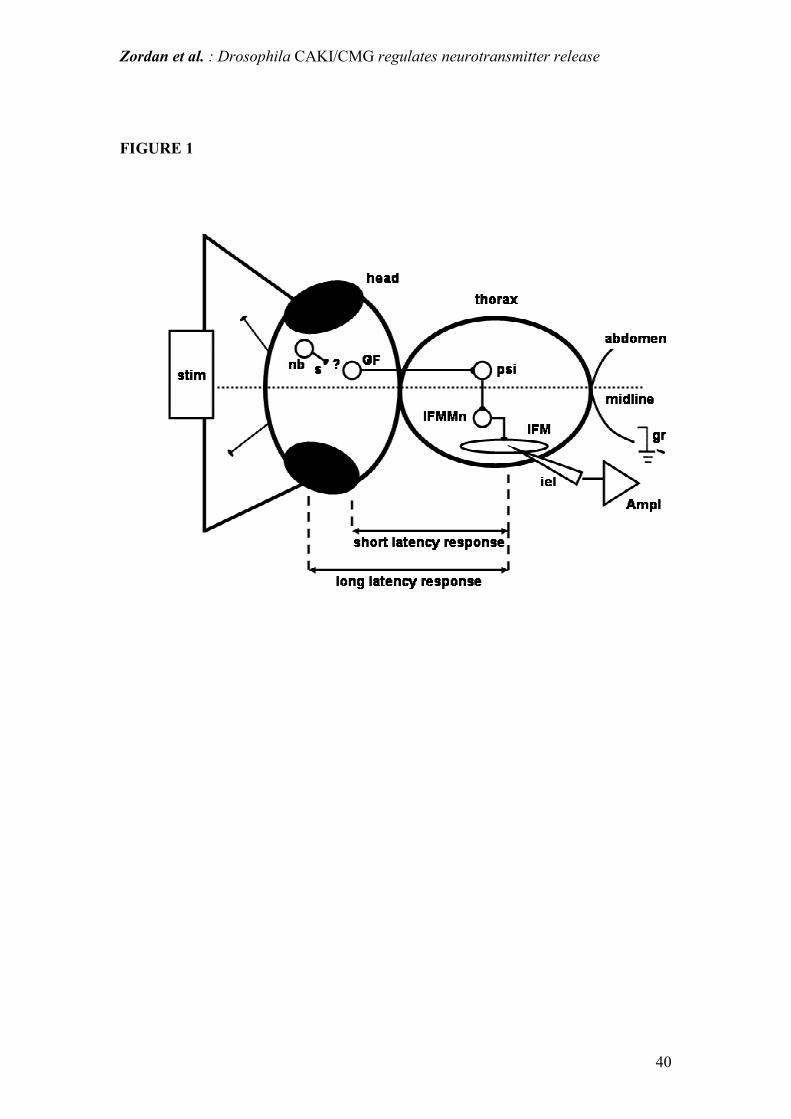
Giant fiber pathway response
Given the alterations in locomotor and flight performance, we conducted a detailed
neurophysiological analysis of the giant fiber (GF) pathway, which constitutes the
jump and escape response neuronal circuit in the adult fly (Fig. 1). Furthermore, the
GF is an example of a complete and relatively simple neuronal circuit, which is
amenable to an in situ electrophysiological analysis in whole-animal preparations.
GF neurons send long axons through the cervical connective to the thoracic ganglia,
where they indirectly (via an interneuron) stimulate the IFM motoneurons. GF
activity evokes an action potential in the IFM and this can be recorded extra or
intracellularly (Tanouye and Wyman, 1980; Engel and Wu, 1996). The GF pathway
can also be activated by direct brain stimulation using a pair of stimulating electrodes
Zordan et al. : Drosophila CAKI/CMG regulates neurotransmitter release
15
placed in the eyes (Tanouye and Wyman, 1980; Gorczyca and Hall, 1984). The IFM
action potentials thus evoked, show a dual behavior: a short-latency (SL) response
with stimuli above 6-8 V and long-latency (LL) responses with lower intensity
stimuli. The SL response is attributed to the direct stimulation of the GF neurons or
the GF itself, while the LL response results from the stimulation of GF afferents with
the interposition of at least 1 chemical synapse (Engel and Wu, 1996). With repeated
stimuli, LL response probability diminishes, showing all the characteristics of
habituation (Thompson and Spencer, 1966; Engel and Wu, 1996, 1998; Engel et al.,
2000; Megighian et al., 2001). Habituation is a form of non associative short-term
learning due to mechanisms such as homosynaptic- or extrinsic- inhibition
(Castellucci et al., 1970; Krasne and Teshiba, 1995), with the functional significance
of fine tuning the gain and sensitivity of a behavioral response (Fisher et al., 1997;
Engel and Hoy, 1999). SL responses do not show habituation (Engel and Wu, 1996).
In caki mutants both short- and long- latencies were significantly longer than in WT.
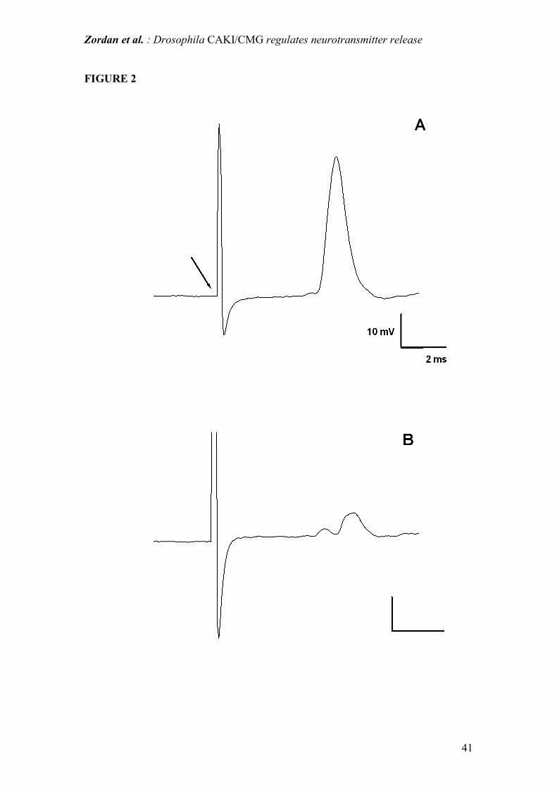
(Table 1). Furthermore, in caki flies, in some IFMs (about 5%) displaying a normal
resting membrane potential, the stimulation of the GF pathway failed to evoke a clear
response, which instead appeared as a small depolarization: an example is shown in
Fig.2. This lack of response remained unchanged with time as well as with increasing
stimulus voltage or duration, suggesting that synaptic transmission was impaired in
these fibers. We further established that in caki individuals, the ability of the GF
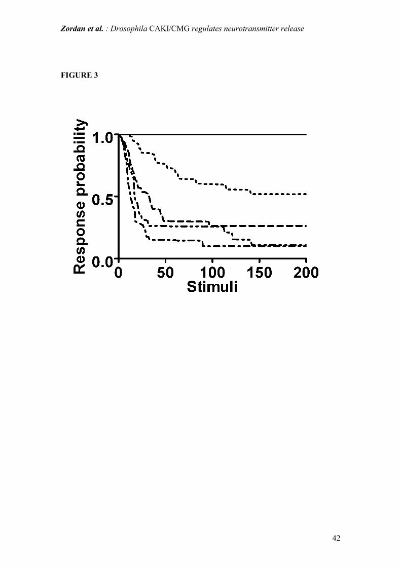
pathway to follow continuous stimulation was also impaired. In WT flies SL
responses did not show any failure up to stimulation frequencies as high as 150 Hz.
By contrast in caki mutants, SL responses showed a significant response decrement
even at low stimulation frequencies (0.5, 1, 2, 3 Hz). In the range of these test
frequencies no response failures, during continuous stimulation, were observed in WT

Zordan et al. : Drosophila CAKI/CMG regulates neurotransmitter release
16
flies, as already reported by other authors (Engel and Wu, 1996) (Fig. 3). On the other
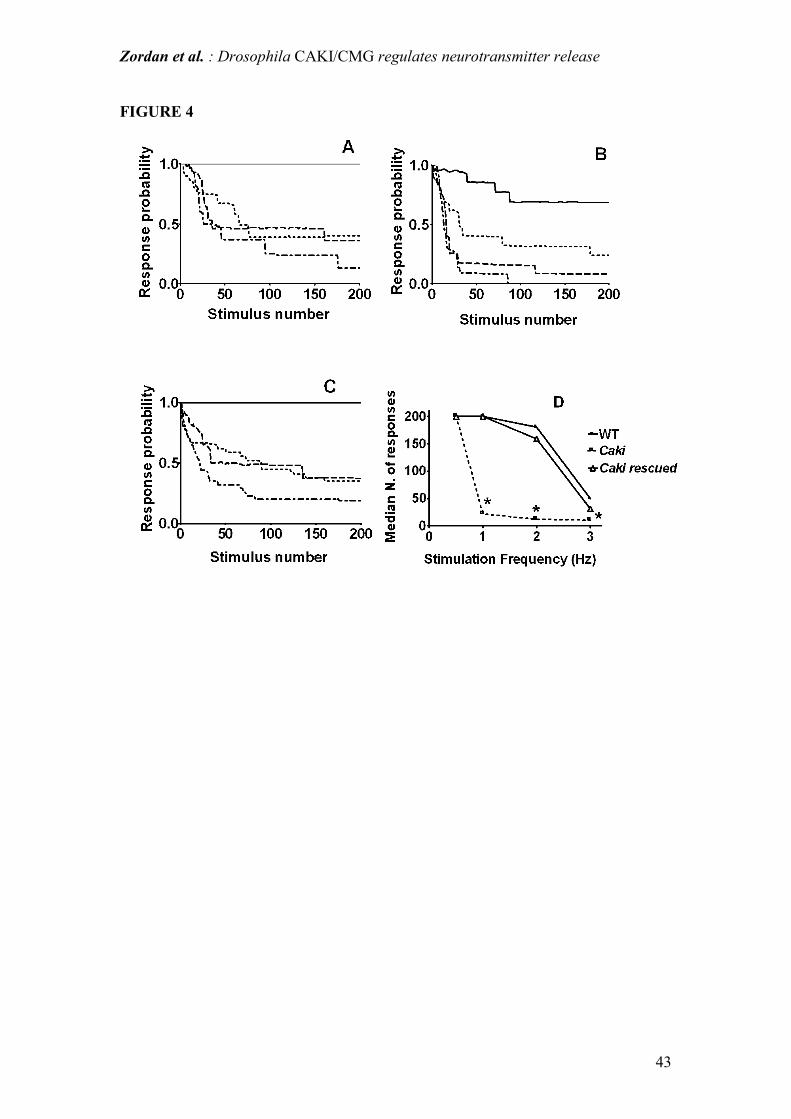
hand, LL responses showed the typical phenomenon of habituation (Engel and Wu,
1996): during continuous stimulation, the probability of LL responses diminishes with
a time course which depends on the stimulus frequencies (Fig. 4). At stimulation
frequencies between (1 and 3 Hz) the onset of habituation occurred significantly
earlier in caki than in WT flies (see Fig. 4 A, B, D). Considering the individuals
which displayed a LL response following stimulation of the GF pathway, the
percentage of caki flies which then showed habituation was lower than in the case of
the WT controls (71.4% vs 90%, respectively, determied from at least 120 responses
obtained from 40 flies). After the onset of habituation, dishabituation was obtained by
delivering a different stimulus (air puff). If dishabituation was not observed, the
responses which attained the five consecutive failure criteria were discarded from
analysis. However, as previously observed (Engel and Wu, 1996), the efficacy of air
puffs to achieve dishabituation varied, in the sense that in some cases one puff was
not sufficient to obtain the desired effect. As a rough index of this efficacy, we
analyzed the number of flies displaying dishabituation after the first air puff and we
observed that caki flies were less responsive than controls (40% vs 61%,
respectively).
Miniature end-plate potentials
As a corollary to the electrophysiological analysis at the GF neuronal circuit level,
and on the basis of the evidence of the association of CASK and it's invertebrate
homolog, with presynaptic membrane proteins (see Introduction), suggesting the
possible involvement of caki/camguk in neurotransmitter release, we examined
Zordan et al. : Drosophila CAKI/CMG regulates neurotransmitter release
17
spontaneous neurotransmitter release at the NeuroMuscular Junction (NMJ) of the
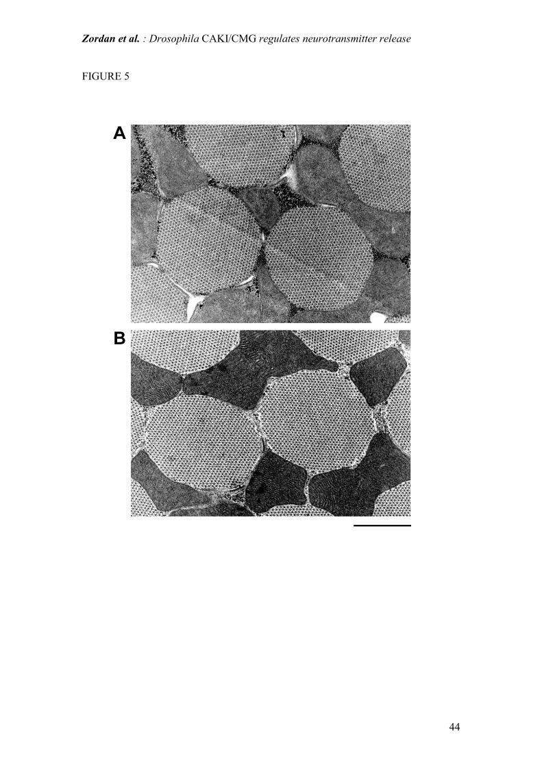
IFM. First of all, the structural and functional preservation of IFM in caki mutant flies
was assessed respectively by electron-microscopy (see Fig.5) and by measurement of
the resting membrane potential, which is a sensitive indicator of fiber membrane
integrity. The resting membrane potential of IFM fibers of caki mutant flies was not
significantly different from WT (79.1±1.6 vs 79.4±2.6, respectively, data determined
from 4 IFMs for each of 10 flies) suggesting, together with the electron-microscopy
observation, that the caki mutation does not cause overt damage to IFM fibers.
Spontaneous neurotransmitter release was monitored by intracellularly recording
miniature end-plate potentials (mepps) in IFM fibers. Only fibers displaying a resting
membrane potential of –70 mV or less were considered. Fibers with an oscillating or
unstable resting membrane potential were discarded. Average mepps frequency in
caki flies was significantly higher (23.8 ± 1 Hz, n=22205 in 10 flies) than in WT (5.8
± 1 Hz, n=4268 in 10 flies ;P<0.05). Furthermore average mepps amplitude was
greater in caki mutants (124.21±0.42 µV, n=22205, in 10 flies) than in WT
(76.47±0.74 µV, n=4268 in 10 flies). Event amplitudes were compared by
constructing normalized cumulative amplitude histograms and testing whether the
observed differences were statistically significant using a Kolmogorov-Smirnov test.
In addition, the broader distribution of the caki mepps amplitude suggests the frequent
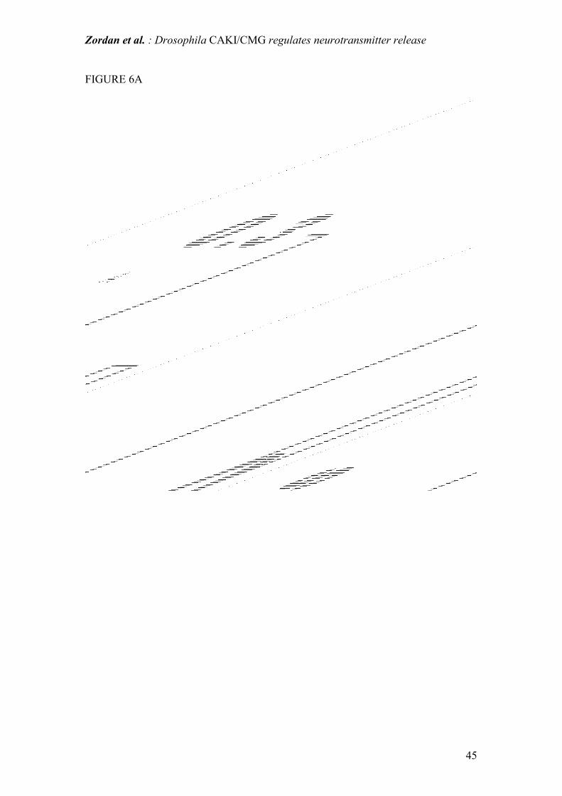
occurrence of multi-quantal neurotransmitter release (Fig. 6 A, B). Peaks
corresponding to single and multi-quantal release were detected: they corresponded to
multiples of 60 µV, suggesting that the amplitude of a single “quantum” remains
unchanged in caki mutants with respect to WT flies (Fig 6 A, B). Spontaneous
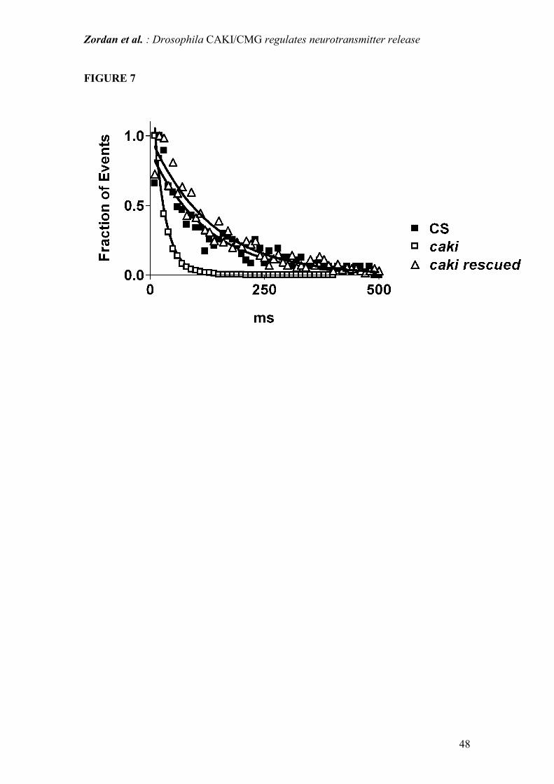
neurotransmitter release is a random process and as such can be described by a
Poisson model. Mepps latencies (i.e. intervals between successive mepps) obtained in
Zordan et al. : Drosophila CAKI/CMG regulates neurotransmitter release
18
caki mutants fit the exponential equation predicted by the Poisson model well, thus
confirming that in all experimental conditions the random nature of spontaneous
release is conserved. However, as expected on the basis of the higher frequency of
mepps (see above), the latency values were much shorter in caki mutants than in WT
(Fig. 7).
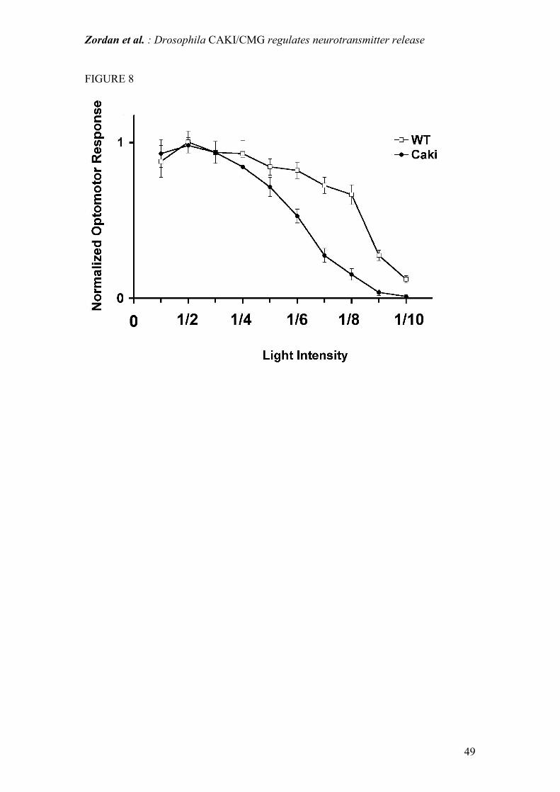
Visual function: optomotor test and ERG
Finally, in view of the high expression of CAKI in the optic lobes of adult flies
(Martin and Ollo, 1996), and also to ascertain whether nervous function was impaired
in other districts, we performed an analysis of the behavioral responses of the visual
system by means of the walking optomotor test. The frequency of correct turns (see
Materials and Methods) was significantly lower in caki (66±13 %, n=10) than in WT
flies (83±13 %, n=10, p<0.05). Furthermore we also assayed the sensitivity of the
optomotor response to light intensity. This was measured by recording the ability of a
fly, which is kept suspended with its legs resting on a freely rotating styrofoam ball
connected to a tracking system, to follow a rotating pattern. In this paradigm, the
responses of both caki mutants and WT flies decreased with decreasing light intensity
(Fig. 8). However, whereas at high light intensity (points 10/10, 8/10 and 7/10 on Fig.
7) the response of caki flies is similar to WT, the response of caki was significantly
worse than WT at light intensities below intermediate levels, suggesting an
impairment of visual function at low light intensity in caki flies. In order to ascertain
visual function directly, we performed electroretinogram (ERG) analyses in caki flies.
ERG transient (On and Off) and sustained responses were recorded in caki and WT
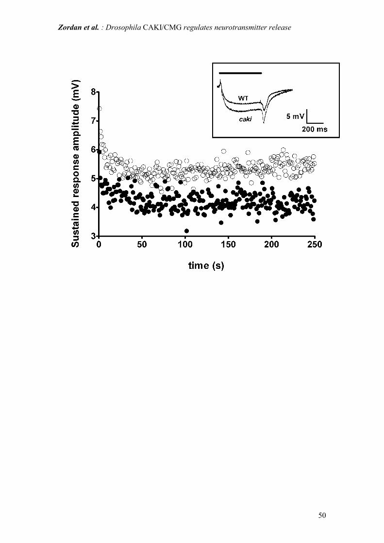
flies (see inset Fig. 9). As shown in Table 2, the sustained ERG response was higher
in caki than in WT individuals. Moreover, while the ON transient amplitude was
Zordan et al. : Drosophila CAKI/CMG regulates neurotransmitter release
19
similar in both WT and caki flies, OFF transients were significantly higher in caki
than in WT individuals. These differences remained even when a high intensity light
stimulus was used instead of a low intensity stimulus (data not shown). Adaptation of
the sustained response, induced by continuous 1 Hz stimulation, was qualitatively
similar in both caki and WT flies (Fig. 9), while the amplitudes of the ERG sustained
responses were significantly higher in caki flies during the whole stimulation period.
Caki mutant rescue
In genetic rescue expriments we employed a UAS-caki transgenic line bearing a
single X-chromosome insertion of the transgene coding for the complete caki cDNA.
Rescue was obtained by crossing UAS-caki;;X-313; individuals to elav-GAL4;;X-307
flies, the progeny of which consisted of caki mutant females, in which the expression
of the UAS-caki transgene was activated and caki mutant males in which the
production of the wild type caki transgene was not active. We thus evaluated the
capacity of the panneuronal expression (presynaptically at the level of the NMJ) of
the caki transgene to rescue the key phenotypical aspects displayed by the caki
mutant.
In particular, female progeny of the above cross showed a practically fully rescued
phenotype (not significantly different from WT) as regards the frequency of NMJ
mepps as well as the giant fiber habituation response. In these individuls IFM resting
membrane potential was 83.4±1.6; 7 IFM from 7 flies. Average NMJ mepps
frequency was 4.61 ± 1 Hz, n=1099 in 7 flies; while the average NMJ mepps
amplitude was 87.57±1.50 µV, n=1099, in 7 flies. The distribution of NMJ mepps
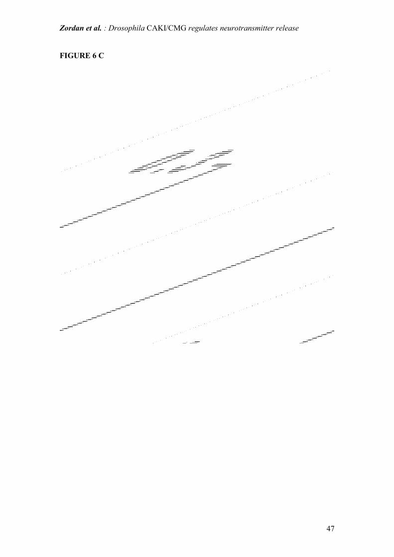
amplitudes in caki "rescued" flies was restored to the WT values (Fig. 6 C). In
addition as shown in Fig. 3 C and 4 C, the kinetics of SL and LL giant fiber responses

Zordan et al. : Drosophila CAKI/CMG regulates neurotransmitter release
20
to continuous stimulation was also clearly restored to near WT values.
On the contrary, male progeny from the same cross (in which the rescuing caki
transgene is not expressed) displayed a clearly mutant phenotype for these key aspects
(data not shown).
Zordan et al. : Drosophila CAKI/CMG regulates neurotransmitter release
21
Discussion
The main result of this study is that neurotransmitter release is altered in caki mutants:
a 4 times increase in spontaneous neurotransmitter release at the neuromuscular
junction (NMJ) was revealed by the intracellular analysis of mepps in indirect flight
muscles (IFM). Mepps were more frequent and also showed an increased mean
amplitude. Furthermore distinct peaks on the mepps amplitude distribution curve
suggest multi-quantal release in the presence of conserved quantal amplitude. Thus,
the absence of CAKI protein determines an impaired vesicle release control, leading
to frequent multiple vesicular release. However, conserved quantal amplitude
indicates that vesicle size remains unaltered, suggesting that CAKI is involved in the
regulation of neurotrasmitter release at the synaptic level. Importantly, the synaptic
localization of CAKI has been previously shown (Bachmann et al., 2004; Lu et al,
2003).
As mentioned in the introduction, CASK is part of a presynaptic complex with VELIS
and MINT-1 (Butz et al., 1998). CASK binds to the cytoplasmic tail of NEUREXIN
and, via protein 4.1, is linked to the actin cytoskeleton (Hata et al., 1996). CASK also
binds N-type Ca2+ channels through its SH3 domains (Maximov and Bezprozvanny,
2002). Such Ca2+ channels also bind MINT-1, which in turn is tightly connected to
CASK (Sudhof, 2001; Ho et al., 2003). Invertebrates express a single representative
of the Cav2 Ca2+ channel family (Spafford et al., 2003; Spafford and Zamponi, 2003;
Zamponi, 2003). Moreover, functional studies of the in Lymnea stagnalis Cav2
homolog (which corresponds to vertebrate N and P/Q-type Ca2+ channels) show that it
Zordan et al. : Drosophila CAKI/CMG regulates neurotransmitter release
22
behaves like an N-type Ca2+ channel (Spafford et al., 2003). It has also been proposed
that CASK and/or MINT colocalize with the synaptic release machinery and may
contribute to the appropriate targeting of presynaptic Ca2+ channels (Spafford and
Zamponi, 2003). Ca2+ channels and Ca2+
influx are, in fact, important regulators of
evoked and spontaneous neurotransmitter release (Rizo and Sudhof, 2002) in
vertebrates and invertebrates (Fatt and Katz, 1952; Muchnik et al., 1975; Grinnell et
al., 1989; Kidokoro and Nishikawa, 1994; Sweeney et al., 1995). Finally, MINT-1
interacts with MUNC-18, which plays an important role in neurotransmitter release,
since Munc-18 KO mice showed a complete block of spontaneous and evoked
neurotransmitter release at normally developed NMJs (Verhage et al., 2000).
Considering the structural framework described above, the results of the present work
suggest that CAKI: i) is involved in the control of exocytosis: loss of CAKI causes an
increased frequency of mepps; ii) is not involved in determining vesicle size: loss of
CAKI does not modify the amplitude of single mepps, but causes multiquantal
release. Crucially, presynaptic rescue of CAKI expression, restores wild type mepps
frequency. Our results further provide evidence that the impaired control of
neurotransmitter release leads to the depletion of presynaptic vesicle stores and
consequently to synaptic failure. The electrophysiological characterizations reported
in this paper rest on the observation of motor and learning defects displayed by caki
mutants (Martin and Ollo, 1996; Lu et al., 2003) and by the impairment in flight
ability, even in the presence of structurally normal IFM fibres (see Fig 1) observed in
the present research. Therefore we conducted a detailed neurophysiological analysis
of the giant fiber (GF) pathway, which is a well defined neuronal circuit responsible
of a stereotyped "jump and flight" escape response in Drosophila. In caki mutants, GF
pathway stimulation evoked an IFM response in 95% of IFM fibres. The remaining
Zordan et al. : Drosophila CAKI/CMG regulates neurotransmitter release
23
5% of IFM fibres did not respond at all, suggesting the complete impairment of
synaptic transmission in these IFM.
Habituation
Continuous stimulation of the GF pathway, even at low frequencies (0.5-3 Hz),
caused transmission failure in both SL and LL responses. In WT flies SL responses
show no habituation but only fatigue at very high stimulation frequencies (100-150
Hz) (Engel and Wu, 1992), thus the loss of response at low frequency (0.5-3 Hz)
observed in caki mutants is suggestive of synaptic failure. In view of the above model
for CAKI function, the transmission failure in caki mutants might be attributed to
vesicular depletion, which in turn would be determined by uncontrolled spontaneous
vesicle release. GF LL responses in WT flies show habituation even at low frequency
stimulation. The impaired LL response observed in caki mutants was reversible (as
expected in the case of true habituation) by an alternative stimulation in
approximately 70% of the cases, while in the remaining 30% it was not. The latter
may be due to the depletion of neurotransmitter stores, as proposed for SL responses,
whereas in the former it could be due to precocious habituation of these flies.
Precocious habituation in caki mutants might be explained by assuming that the
mechanisms for short-term plasticity are activated earlier in 70% of the mutants than
in WT flies.
ERGs and Optomotor response
The "visual" part of the WT brain (optic lobes) shows high levels of CAKI (Martin
and Ollo, 1996): interestingly, caki mutants, in which CAKI is lacking, show
quantitative electroretinogram (ERG) alterations and an impaired optomotor response.
Zordan et al. : Drosophila CAKI/CMG regulates neurotransmitter release
24
ERG alterations in caki flies are characterized by a significant increase, with respect
to wild type controls) in the sustained response (10.5±1.1 vs 7.0±1.4 mV) and off
transients (6.0±1.3 vs 4.2±1.0 mV). The preceding measurements were conducted on
28 wild type flies and 26 caki mutants. On the other hand, the presence of a
qualitatively normal ERG response in caki flies confirms that these flies are receptive
to visual stimuli. In fact Martin and Ollo (1996), had previously ascribed the altered
Buridan test in caki flies to locomotor impairments and not to visual defects. It seems
that photoreceptor sensitivity of caki flies to light stimuli is normal at standard light-
intensities but, as revealed by the optomotor test, photoreceptor sensitivity is
decreased at low light intensity. Since the optomotor response is the result of neural
processing of visual field movement (Gotz and Buchner, 1978; Bausenwein et al.,
1986), the impaired optomotor response in caki individuals suggests the presence of
visual system alterations which interfere with the physiological flow and integration
of the visual stimuli.
Conclusions
The results presented here provide strong support for a role of CAKI within the
tripartite complex as a regulator of vesicle release. However, CAKI does not seem to
be involved in determining vesicle size. Instead, the lack of CAKI likely produces a
depletion of presynaptic neurotransmitter stores, resulting in an increased frequency
of synaptic transmission failures. This hypothesis finds support in the observation that
targeted presynaptic expression of CAKI, in a null CAKI background, abolishes this
defect. As an alternative hypothesis, based on the expression of CAKI also at the
postsynaptic level, Lu et al. (2003) suggested that CAKI could regulate postsynaptic
CaMKII T306 autophosphorylation and thus modulate synaptic plasticity. Our data
Zordan et al. : Drosophila CAKI/CMG regulates neurotransmitter release
25
indicate a major role of CAKI in regulating the function of presynaptic elements,
although we cannot exclude a postsynaptic role. Furthermore, since CAKI is highly
evolutionarily conserved it is likely that vertebrate CASK plays a similar role in the
mammalian synapsis.
Zordan et al. : Drosophila CAKI/CMG regulates neurotransmitter release
26
Acknowledgments
The authors thank Dr. S. Ferlin and Dr. M. Amodio for technical assistance in the
execution of giant fiber pathway analysis and Mr M. deFabris for technical assistance
in Electron microscopy studies.
Grants
This study has been supported by the collaborative programme between CNR and
MIUR "Legge 449/97" (Grant N° CU04.00067 to R.C.), by grant Telethon N.
GP0048Y01 to R.C and by University of Padova Research grant N. CPDA048818 to
A.M. .
Zordan et al. : Drosophila CAKI/CMG regulates neurotransmitter release
27
References
Anderson JM (1996) Cell signalling: MAGUK magic. Curr Biol 6:382-384.
Bachmann A, Timmer M, Sierralta J, Pietrini G, Gundelfinger ED, Knust E, Thomas
U (2004) Cell type-specific recruitment of Drosophila Lin-7 to distinct
MAGUK-based protein complexes defines novel roles for Sdt and Dlg-S97. J
Cell Sci 117:1899-1909.
Bausenwein B, Wolf R, Heisenberg M (1986) Genetic dissection of optomotor
behavior in Drosophila melanogaster. Studies on wild-type and the mutant
optomotor-blindH31. J Neurogenet 3:87-109.
Buchner E, Gotz KG, Straub C (1978) Elementary detectors for vertical movement in
the visual system of Drosophila. Biol Cybern 31:235-242.
Burnet BaJB (1968) Phenogenetic studies on visual acuity in Drosophila
melanogaster. J Insect Physiol 57:855-860.
Butz S, Okamoto M, Sudhof TC (1998) A tripartite protein complex with the potential
to couple synaptic vesicle exocytosis to cell adhesion in brain. Cell 94:773-
782.
Caruana G, Bernstein A (2001) Craniofacial dysmorphogenesis including cleft palate
in mice with an insertional mutation in the discs large gene. Mol Cell Biol
21:1475-1483.
Castellucci V, Pinsker H, Kupfermann I, Kandel ER (1970) Neuronal mechanisms of
habituation and dishabituation of the gill-withdrawal reflex in Aplysia.
Science 167:1745-1748.
Zordan et al. : Drosophila CAKI/CMG regulates neurotransmitter release
28
Drummond DR, Hennessey ES, Sparrow JC (1991) Characterisation of missense
mutations in the Act88F gene of Drosophila melanogaster. Mol Gen Genet
226:70-80.
Engel JE, Wu CF (1992) Interactions of membrane excitability mutations affecting
potassium and sodium currents in the flight and giant fiber escape systems of
Drosophila. J Comp Physiol [A] 171:93-104.
Engel JE, Wu CF (1996) Altered habituation of an identified escape circuit in
Drosophila memory mutants. J Neurosci 16:3486-3499.
Engel JE, Wu CF (1998) Genetic dissection of functional contributions of specific
potassium channel subunits in habituation of an escape circuit in Drosophila. J
Neurosci 18:2254-2267.
Engel JE, Hoy RR (1999) Experience-dependent modification of ultrasound auditory
processing in a cricket escape response. J Exp Biol 202 ( Pt 20):2797-2806.
Engel JE, Xie XJ, Sokolowski MB, Wu CF (2000) A cGMP-dependent protein kinase
gene, foraging, modifies habituation-like response decrement of the giant fiber
escape circuit in Drosophila. Learn Mem 7:341-352.
Fatt P, Katz B (1952) Spontaneous subthreshold activity at motor nerve endings. J
Physiol 117:109-128.
Fisher SA, Fischer TM, Carew TJ (1997) Multiple overlapping processes underlying
short-term synaptic enhancement. Trends Neurosci 20:170-177.
Gorczyca M, Hall JC (1984) Identification of a cholinergic synapse in the giant fiber
pathway of Drosophila using conditional mutations of acetylcholine synthesis.
J Neurogenet 1:289-313.
Gotz KG (1970) Fractionation of Drosophila populations according to optomotor
traits. J Exp Biol 52:419-436.
Zordan et al. : Drosophila CAKI/CMG regulates neurotransmitter release
29
Gotz KG, Buchner E (1978) Evidence for one-way movement detection in the visual
system of Drosophila. Biol Cybern 31:243-248.
Grinnell AD, Gundersen CB, Meriney SD, Young SH (1989) Direct measurement of
ACh release from exposed frog nerve terminals: constraints on interpretation
of non-quantal release. J Physiol 419:225-251.
Hata Y, Butz S, Sudhof TC (1996) CASK: a novel dlg/PSD95 homolog with an N-
terminal calmodulin-dependent protein kinase domain identified by interaction
with neurexins. J Neurosci 16:2488-2494.
Hata Y, Nakanishi H, Takai Y (1998) Synaptic PDZ domain-containing proteins.
Neurosci Res 32:1-7.
Heisenberg M (1971) Separation of receptor and lamina potentials in the
electroretinogram of normal and mutant Drosophila. J Exp Biol 55:85-100.
Ho A, Morishita W, Hammer RE, Malenka RC, Sudhof TC (2003) A role for Mints in
transmitter release: Mint 1 knockout mice exhibit impaired GABAergic
synaptic transmission. Proc Natl Acad Sci U S A 100:1409-1414.
Hoskins R, Hajnal AF, Harp SA, Kim SK (1996) The C. elegans vulval induction
gene lin-2 encodes a member of the MAGUK family of cell junction proteins.
Development 122:97-111.
Hotta Y, Benzer S (1969) Abnormal electroretinograms in visual mutants of
Drosophila. Nature 222:354-356.
Hsueh YP, Wang TF, Yang FC, Sheng M (2000) Nuclear translocation and
transcription regulation by the membrane-associated guanylate kinase
CASK/LIN-2. Nature 404:298-302.
Hsueh YP, Yang FC, Kharazia V, Naisbitt S, Cohen AR, Weinberg RJ, Sheng M
(1998) Direct interaction of CASK/LIN-2 and syndecan heparan sulfate
Zordan et al. : Drosophila CAKI/CMG regulates neurotransmitter release
30
proteoglycan and their overlapping distribution in neuronal synapses. J Cell
Biol 142:139-151.
Ikeda K, Koenig JH (1988) Spontaneous release of multiquantal miniature excitatory
junction potentials induced by a Drosophila mutant. J Physiol 406:215-223.
Irie M, Hata Y, Deguchi M, Ide N, Hirao K, Yao I, Nishioka H, Takai Y (1999)
Isolation and characterization of mammalian homologues of Caenorhabditis
elegans lin-7: localization at cell-cell junctions. Oncogene 18:2811-2817.
Kaech SM, Whitfield CW, Kim SK (1998) The LIN-2/LIN-7/LIN-10 complex
mediates basolateral membrane localization of the C. elegans EGF receptor
LET-23 in vulval epithelial cells. Cell 94:761-771.
Kawasaki F, Mattiuz AM, Ordway RW (1998) Synaptic physiology and ultrastructure
in comatose mutants define an in vivo role for NSF in neurotransmitter
release. J Neurosci 18:10241-10249.
Kidokoro Y, Nishikawa K (1994) Miniature endplate currents at the newly formed
neuromuscular junction in Drosophila embryos and larvae. Neurosci Res
19:143-154.
Krasne FB, Teshiba TM (1995) Habituation of an invertebrate escape reflex due to
modulation by higher centers rather than local events. Proc Natl Acad Sci U S
A 92:3362-3366.
Levine JD, Wyman RJ (1973) Neurophysiology of flight in wild-type and a mutant
Drosophila. Proc Natl Acad Sci U S A 70:1050-1054.
Lu CS, Hodge JJ, Mehren J, Sun XX, Griffith LC (2003) Regulation of the
Ca2+/CaM-responsive pool of CaMKII by scaffold-dependent
autophosphorylation. Neuron 40:1185-1197.
Zordan et al. : Drosophila CAKI/CMG regulates neurotransmitter release
31
MacMullin A, Stewart B, Jacobs JR (2001) Drosophila velis/lin-7 expression in
neurons suggests function in organization of the post-synaptic membrane. In:
A. Dros. Res. Conf. 42, p 809B.
Martin JR, Ollo R (1996) A new Drosophila Ca2+/calmodulin-dependent protein
kinase (Caki) is localized in the central nervous system and implicated in
walking speed. Embo J 15:1865-1876.
Martinez-Estrada OM, Villa A, Breviario F, Orsenigo F, Dejana E, Bazzoni G (2001)
Association of junctional adhesion molecule with calcium/calmodulin-
dependent serine protein kinase (CASK/LIN-2) in human epithelial caco-2
cells. J Biol Chem 276:9291-9296.
Maximov A, Bezprozvanny I (2002) Synaptic targeting of N-type calcium channels in
hippocampal neurons. J Neurosci 22:6939-6952.
Megighian A, Zordan M, Costa R (2001) Giant neuron pathway neurophysiological
activity in per(0) mutants of Drosophila melanogaster. J Neurogenet 15:221-
231.
Mok H, Shin H, Kim S, Lee JR, Yoon J, Kim E (2002) Association of the kinesin
superfamily motor protein KIF1Balpha with postsynaptic density-95 (PSD-
95), synapse-associated protein-97, and synaptic scaffolding molecule PSD-
95/discs large/zona occludens-1 proteins. J Neurosci 22:5253-5258.
Muchnik S, Kotsias BA, Arrizurieta de Muchnik EE (1975) In vivo and in vitro
miniature end-plate potentials at various external K concentrations. Am J
Physiol 228:1733-1737.
Pak WL, Grossfield J, White NV (1969) Nonphototactic mutants in a study of vision
of Drosophila. Nature 222:351-354.
Zordan et al. : Drosophila CAKI/CMG regulates neurotransmitter release
32
Rizo J, Sudhof TC (2002) Snares and Munc18 in synaptic vesicle fusion. Nat Rev
Neurosci 3:641-653.
Sandrelli F, Campesan S, Rossetto M, Benna C, Zieger E, Megighian A, Couchman
M, Kyriacou C, Costa R (2001) Molecular Dissection of the 5' Region of no-
on-transientA of Drosophila melanogaster Reveals cis-Regulation by Adjacent
dGpi1 Sequences. Genetics 157:765-775.
Spafford JD, Zamponi GW (2003) Functional interactions between presynaptic
calcium channels and the neurotransmitter release machinery. Curr Opin
Neurobiol 13:308-314.
Spafford JD, Chen L, Feng ZP, Smit AB, Zamponi GW (2003) Expression and
modulation of an invertebrate presynaptic calcium channel alpha1 subunit
homolog. J Biol Chem 278:21178-21187.
Sudhof TC (2001) The Synaptic Cleft and Synaptic Cell Adhesion. In: Synapses
(Cowan M, Sudhof, TC, Stevens, CF, ed), pp 275-313. Baltimore: John
Hopkins University Press.
Sweeney JD, Crawford NR, Brandon TA (1995) Neuromuscular stimulation
selectivity of multiple-contact nerve cuff electrode arrays. Med Biol Eng
Comput 33:418-425.
Tabuchi K, Biederer T, Butz S, Sudhof TC (2002) CASK participates in alternative
tripartite complexes in which Mint 1 competes for binding with caskin 1, a
novel CASK-binding protein. J Neurosci 22:4264-4273.
Tanouye MA, Wyman RJ (1980) Motor outputs of giant nerve fiber in Drosophila. J
Neurophysiol 44:405-421.
Thomas JB, Wyman RJ (1984) Mutations altering synaptic connectivity between
identified neurons in Drosophila. J Neurosci 4:530-538.
Zordan et al. : Drosophila CAKI/CMG regulates neurotransmitter release
33
Thompson RF, Spencer WA (1966) Habituation: a model phenomenon for the study
of neuronal substrates of behavior. Psychol Rev 73:16-43.
Trimarchi JR, Schneiderman AM (1993) Giant fiber activation of an intrinsic muscle
in the mesothoracic leg of Drosophila melanogaster. J Exp Biol 177:149-167.
Trimarchi JR, Schneiderman AM (1995) Flight initiations in Drosophila melanogaster
are mediated by several distinct motor patterns. J Comp Physiol [A] 176:355-
364.
Verhage M, Maia AS, Plomp JJ, Brussaard AB, Heeroma JH, Vermeer H, Toonen
RF, Hammer RE, van den Berg TK, Missler M, Geuze HJ, Sudhof TC (2000)
Synaptic assembly of the brain in the absence of neurotransmitter secretion.
Science 287:864-869.
Zamponi GW (2003) Regulation of presynaptic calcium channels by synaptic
proteins. J Pharmacol Sci 92:79-83.
Zordan et al. : Drosophila CAKI/CMG regulates neurotransmitter release
34
Legends to Tables
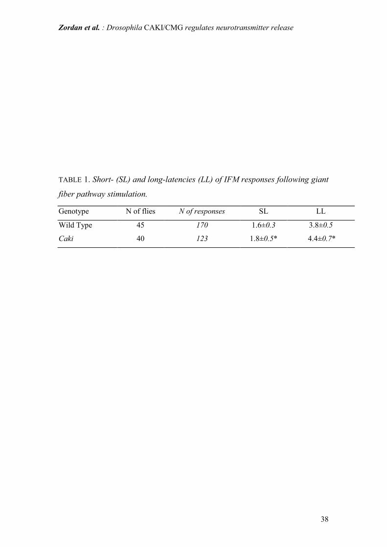
Table 1: Measured short- and long-latencies of the action potentials intracellularly
recorded in IFM fibers following brain stimulation of the giant fiber pathway.
Latencies were calculated from the onset of the stimulus artifact to the beginning of
the evoked response at the muscle membrane. Values are mean ± SD (units are ms).
Sampling frequency of 25 kHz allows a true sensitivity in the reported range.
* P<0.001 (Student's t-test for unpaired data).
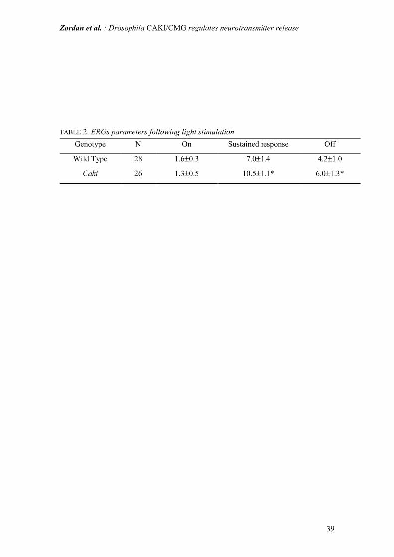
Table 2: ERG response parameters extracellularly recorded following light
stimulation of Drosophila eye. ON and OFF are the transient responses at the onset
and offset of light stimulation respectively and they are due to the synaptic activation
of second order neurons in the visual pathway. The sustained response measures light-
induced depolarization of photoreceptors (the sustained response of ERG) Values are
mean ± SD (units are mV). N is number of animals. *P<0.05 (Student's for unpaired t-
test).
Zordan et al. : Drosophila CAKI/CMG regulates neurotransmitter release
35
Figure Legends
Figure 1: diagram of giant fiber pathway circuit in an adult fly. stim = stimulator; iel
= intracellular microelectrode; ampl = amplifier; gr = ground electrode; nb = neuron
cell body; s = synapse; GF = giant fiber neuron; ? = unknown number of synapses in
the afferent pathway to the GF; psi = peripherally synapsing interneuron; IFM =
indirect flight muscle; IFMMn = IFM motoneuron;
Figure 2: intracellular recording of IFM responses evoked by brain stimulation of the
giant fiber pathway. (A) shows an example of a typical spike observed in WT
individuals; (B) shows one case in which a lack of response was observed in a caki
mutant (see text). Calibration bar units, as reported in (A) are the same in both
figures. Arrow indicates the onset of the stimulus artifact.
Figure 3: Kinetics of the short-latency giant fiber pathway response. Frequency
dependent decrement of response probability is plotted using the mean of pooled
responses from 16 WT, 23 caki mutants and 7 caki "rescued" individuals. In WT and
"rescued" flies short-latency responses did not show any failure at any of the
stimulation frequencies tested. In contrast caki mutants showed a clear response
decrement even at low stimulation frequencies (0.5, 1, 2, 3 Hz).
WT and caki rescued = line;
caki mutant: 0.5 Hz = dots ; 1 Hz = dash; 2 Hz = two dots and dash; 3 Hz = dot-dash.
Figure 4: Kinetics of habituation of the long-latency giant fiber pathway response.
Zordan et al. : Drosophila CAKI/CMG regulates neurotransmitter release
36
Frequency dependent decrement of response probability. Mean of pooled responses
from 18 WT (A), 30 caki mutants (B) and 7 caki "rescued" individuals (C).
In A, B, C : 0.5 Hz = line; 1 Hz = dots; 2 Hz = dash; 3 Hz = dot-dash.
Comparison of A, B and C shows that caki flies habituate consistently earlier than
WT and rescued individuals at all stimulation frequencies, as is clearly summarized in
D, which shows the median number of responses at each stimulation frequency. In D,
* indicates significant (p<0.05, median test) differences between caki mutant and WT.
Figure 5: Electron Microscopy Ultrastructure of myofibrils in WT (A) and caki
mutant (B) IFM. Electron microscopic examination of muscle fibers shows no
alteration in ultrastructural organization of the myofibrils in caki IFM muscles
compared to WT. Cal bar: 0.5 µm.
Figure 6: Normalized distribution of mepps amplitudes recorded from IFM fibers of
WT (A), caki mutants (B) and caki "rescued" individuals (C). Amplitude histograms
show a wider distribution for caki IFM mepps; lines show the multipeak nature of
mepps distributions.
Figure 7: Normalized distribution of mepps latencies. Curve fitting assuming a
mono-exponential decay. Spontaneous neurotransmitter release is a random process
and as such can be described by a Poisson model. Latency values obtained in caki
mutants in normal saline fit the exponential equation predicted by the Poisson model
well, thus confirming that under all experimental conditions the random nature of
spontaneous release is conserved. However, as expected on the basis of the higher
frequency of mepps (see Results), the latency values were much shorter in caki
Zordan et al. : Drosophila CAKI/CMG regulates neurotransmitter release
37
mutants in normal saline than in WT and "rescued" flies.
Figure 8: A: Optomotor response on the styrofoam ball, determined at different light-
intensity. The graph represents the normalized turning tendency of the fly
(rev.R/rev.F) (expressed as a value between 0 and 1) as function of the light-intensity.
In wild-type flies, the turning tendency slowly decreases as light intensity decreases,
and in particular presents an abrupt decrement at a light intensity of about 8/10 . In
caki mutants the turning tendency is not different from wild-type at the three highest
light intensities (10/10-6/10) however, this response rapidly decreases in parallel to
the light intensity, but more steeply than in WT flies. For wild-type flies, the data are
from 4 individuals for a total of 57 runs. For caki mutants, the data are from 4 flies for
a total of 73 runs.
Figure 9: kinetics of ERG sustained responses during 1Hz continuous light
stimulation. WT = black dots; caki mutants = white dots. Comparison of the two plots
shows that the amplitude of the sustained response remains significantly higher
(P<0.001; Kolmogorov-Smirnov test) in caki flies during the entire stimulation
period. Inset shows representative ERG extracellular recordings following light
stimulation of WT and caki mutant eyes. Comparison of the two traces shows that the
amplitudes of the sustained response and OFF transient are larger in caki flies. Top
horizontal bar represents the duration of light stimulation.
Zordan et al. : Drosophila CAKI/CMG regulates neurotransmitter release
38
TABLE 1. Short- (SL) and long-latencies (LL) of IFM responses following giant
fiber pathway stimulation.
Genotype N of flies N of responses SL LL
Wild Type 45 170 1.6±0.3 3.8±0.5
Caki 40 123 1.8±0.5* 4.4±0.7*
Zordan et al. : Drosophila CAKI/CMG regulates neurotransmitter release
39
TABLE 2. ERGs parameters following light stimulation Genotype N On Sustained response Off
Wild Type 28 1.6±0.3 7.0±1.4 4.2±1.0
Caki 26 1.3±0.5 10.5±1.1* 6.0±1.3*
Zordan et al. : Drosophila CAKI/CMG regulates neurotransmitter release
40
FIGURE 1
Zordan et al. : Drosophila CAKI/CMG regulates neurotransmitter release
41
FIGURE 2
Zordan et al. : Drosophila CAKI/CMG regulates neurotransmitter release
42
FIGURE 3
Zordan et al. : Drosophila CAKI/CMG regulates neurotransmitter release
43
FIGURE 4
Zordan et al. : Drosophila CAKI/CMG regulates neurotransmitter release
44
FIGURE 5
Zordan et al. : Drosophila CAKI/CMG regulates neurotransmitter release
45
FIGURE 6A

Zordan et al. : Drosophila CAKI/CMG regulates neurotransmitter release
46
FIGURE 6 B
Zordan et al. : Drosophila CAKI/CMG regulates neurotransmitter release
47
FIGURE 6 C
Zordan et al. : Drosophila CAKI/CMG regulates neurotransmitter release
48
FIGURE 7
Zordan et al. : Drosophila CAKI/CMG regulates neurotransmitter release
49
FIGURE 8
Zordan et al. : Drosophila CAKI/CMG regulates neurotransmitter release
50
Related Documents