Diploma Thesis The Role of Placental Hormones in the Regulation of Maternal Metabolism During Pregnancy submitted by Lisa-Catharina Lindheim date of birth: 1/12/1990 for the academic degree of Doktor der gesamten Heilkunde (Dr.med.univ.) at the Medical University of Graz Department of Obstetrics and Gynecology under supervision of Ao.Univ.-Prof. Dr.phil. Gernot Desoye and Dr.rer.nat. Ursula Hiden Graz, 8/29/2012

Welcome message from author
This document is posted to help you gain knowledge. Please leave a comment to let me know what you think about it! Share it to your friends and learn new things together.
Transcript

Diploma Thesis
The Role of Placental Hormones in the Regulation of
Maternal Metabolism During Pregnancy
submitted by
Lisa-Catharina Lindheim
date of birth: 1/12/1990
for the academic degree of
Doktor der gesamten Heilkunde
(Dr.med.univ.)
at the
Medical University of Graz
Department of Obstetrics and Gynecology
under supervision of
Ao.Univ.-Prof. Dr.phil. Gernot Desoye
and
Dr.rer.nat. Ursula Hiden
Graz, 8/29/2012

i
Eidesstattliche Erklärung
Ich erkläre hiermit ehrenwörtlich, dass ich die vorliegende Arbeit selbstständig und ohne
fremde Hilfe verfasst habe, andere als die angegebenen Quellen nicht verwendet habe
und die den benutzten Quellen wörtlich oder inhaltlich entnommenen Stellen als solche
kenntlich gemacht habe.
Graz, 29.8.2012 Lisa Lindheim

ii
Acknowledgements
I would like to thank my supervisors, Prof. Gernot Desoye and Dr. Ursula Hiden for giving
me the opportunity to write this thesis and for their unwavering enthusiasm and
dedication throughout. You were always available for all my questions and thoughts,
while making sure that every part of the thesis, from the outline to the research to the
actual writing, was my own work. I very much appreciate the time and effort that you put
into this project.
A big thank you goes out to my parents, who have always supported me in all my
academic and non-academic endeavors and who can always be relied on for plenty of
advice, encouragement, and humor. Your constant belief in my abilities made failing not
an option.
I also want to thank my brother and sister who, while not directly involved in this project,
did their part by providing a distraction during the tough parts.
Finally, I have to thank Alex, who probably suffered the most during this time and who
gave me the incentive to start writing this thesis. The first ten pages are dedicated to you.

iii
Für meine Mama

iv
Abstract
Of the multitude of functions performed by the human placenta during pregnancy,
the alteration of maternal metabolic processes by the secretion of various hormones and
cytokines is of great relevance and importance. In response to the secreted products of
the placenta, the maternal metabolism shifts from a balanced to an anabolic and later to
a catabolic state so as to provide the best possible conditions for the growth and
development of the fetus. Hyperphagia, hyperlipidemia, hyperinsulinemia, and
subsequent insulin resistance are among the changes that can be observed. This review
provides a comprehensive overview of the known and unknown aspects of the placental
regulation of maternal metabolism and also addresses the hormonal changes that can be
observed in common pathologies of pregnancy.
Research was conducted using the international online database PubMed.
Preliminary research allowed for the selection of 16 hormones and cytokines, which were
then individually researched. This process ultimately yielded 116 sources published
between the years 1982 and 2012.
A large amount of evidence exists supporting the role of estradiol, progesterone,
PGH, hPL, leptin, TNF-α, and adiponectin in the initiation and amplification of
hyperphagia, hyperlipidemia, hyperinsulinemia, and insulin resistance. The peptide
hormones hCG, CRH, hCT, PTH-rP, and ghrelin have a minor role in these changes. The
relatively recently identified adipokines visfatin, resistin, apelin, and chemerin also have
metabolic effects, but have not yet been sufficiently researched to make any statements
about their exact role and significance during gestation. Many contradictions exist
regarding their physiological concentrations, regulation, and relation to pregnancy-
related pathologies. Many adipokines are secreted in abnormal concentrations in
gestational diabetes mellitus, preeclampsia, and intrauterine growth restriction, but so far
only studies with leptin, TNF-α, and adiponectin have shown consistent results.
In conclusion, the adipokines represent an interesting point for future research, as
they are often a sign of an impending or current pathological condition of the mother or
the fetus. However, the great individual variability of adipokine concentrations will be an
obstacle to overcome before they can be widely used as a screening or diagnostic tool.

v
Zusammenfassung
In einer Schwangerschaft ist die Anpassung der mütterlichen Stoffwechselprozesse
durch die von der Plazenta sezernierten Hormone und Zytokine von groβer Wichtigkeit. In
Gegenwart dieser Faktoren wechselt die Schwangere von einer ausgeglichenen auf eine
anabole und später eine katabole Stoffwechsellage um die optimalen Bedingungen für
das Wachstum und die Entwicklung des Föten zu schaffen. Hyperphagie, Hyperlipidämie,
Hyperinsulinämie und die daraus folgende Insulinresistenz sind typische Veränderungen.
Diese Arbeit bietet einen Überblick über die bekannten und unbekannten Aspekte der
Regulation des mütterlichen Metabolismus durch die Plazenta und erörtert die
Hormonveränderungen, die in Schwangerschaftspathologien beobachtet werden können.
Die Literaturrecherche in der internationalen online Database PubMed ergab nach
anfänglicher Suche 16 Hormone und Zytokine, welche nachfolgend genauer recherchiert
wurden. Es wurden 116 Quellen, zwischen 1982 und 2012 publiziert, ausgewählt.
Die vorliegende Evidenz lässt auf eine Rolle für Estradiol, Progesteron, PGH, hPL,
Leptin, TNF-α und Adiponektin in der Entwicklung und Verstärkung von Hyperphagie,
Hyperlipidämie, Hyperinsulinämie und Insulinresistenz schlieβen. Die Peptidhormone
hCG, CRH, hCT, PTH-rP und Ghrelin spielen bei diesen Veränderungen eine
untergeordnete Rolle. Die relativ neu entdeckten Adipokine Visfatin, Resistin, Apelin und
Chemerin haben ebenfalls metabolische Effekte, sind jedoch derzeit noch nicht
ausreichend bezüglich Ihrer Funktion und Signifikanz erforscht. Es existieren viele
Widersprüche hinsichtlich ihrer physiologischen Konzentrationen, Regulation und
Zusammenhang mit Schwangerschaftspathologien. Viele der Adipokine werden in
pathologischen Zuständen wie Gestationsdiabetes, Präeklampsie und intrauteriner
Wachstumsrestriktion in abnormen Konzentrationen produziert, jedoch haben bis jetzt
nur Studien mit Leptin, TNF-α und Adiponektin übereinstimmende Resultate gezeigt.
Adipokine stellen ein interessantes zukünftiges Forschungsthema dar, da sie oft
ein Zeichen einer inzipienten oder schon bestehenden Pathologie der Mutter oder des
Föten sind. Allerdings ist die groβe individuelle Variabilität der Konzentrationen der
Adipokine ein Problem, welches es zu überwinden gilt bevor diese als Screening- oder
diagnostische Parameter genutzt werden können.

vi
Table of Contents
Abbreviations ....................................................................................................................... viii Figure Index ............................................................................................................................ x Table Index ............................................................................................................................ xi I. Introduction ........................................................................................................................ 1 A. Maternal metabolism in pregnancy ...................................................................... 1 1) The first trimester ...................................................................................... 1 2) The second trimester ................................................................................. 2 3) The third trimester ..................................................................................... 5 II. Materials and Methods ..................................................................................................... 8 III. Results ............................................................................................................................. 10 A. Steroid Hormones ................................................................................................ 10 1) Estrogens ................................................................................................. 10 i. Levels during pregnancy ................................................................ 11 ii. Functions ...................................................................................... 11 iii. Regulation and interactions with other hormones ..................... 13 iv. Pathologies .................................................................................. 13 2) Progesterone ........................................................................................... 14 i. Levels during pregnancy ................................................................ 14 ii. Functions ...................................................................................... 15 iii. Regulation and interactions with other hormones ..................... 15 iv. Pathologies .................................................................................. 16 B. Peptide Hormones ............................................................................................... 17 1) hCG ........................................................................................................... 17 i. Levels during pregnancy ................................................................ 17 ii. Functions ...................................................................................... 18 iii. Regulation and interactions with other hormones ..................... 18 iv. Pathologies .................................................................................. 19 2) hPL............................................................................................................ 19 i. Levels during pregnancy ................................................................ 19 ii. Functions ...................................................................................... 20 iii. Regulation and interactions with other hormones ..................... 21 iv. Pathologies .................................................................................. 22 3) Placental Growth Hormone ..................................................................... 22 i. Levels during pregnancy ................................................................ 22 ii. Functions ...................................................................................... 23 iii. Regulation and interactions with other hormones ..................... 24 iv. Pathologies .................................................................................. 24 4) CRH ........................................................................................................... 25 i. Levels during pregnancy ................................................................ 25 ii. Functions ...................................................................................... 26 iii. Regulation and interactions with other hormones ..................... 27 iv. Pathologies .................................................................................. 27 5) Ghrelin ..................................................................................................... 27 i. Levels during pregnancy ................................................................ 28

vii
ii. Functions ...................................................................................... 28 iii. Regulation and interactions with other hormones ..................... 28 iv. Pathologies .................................................................................. 29 6) hCT, PTH-rP .............................................................................................. 29 C. Adipokines ........................................................................................................... 30 1) Leptin ....................................................................................................... 30 i. Levels during pregnancy ................................................................ 30 ii. Functions ...................................................................................... 31 iii. Regulation and interactions with other hormones ..................... 34 iv. Pathologies .................................................................................. 35 2) TNF-α........................................................................................................ 36 i. Levels during pregnancy ................................................................ 36 ii. Functions ...................................................................................... 37 iii. Regulation and interactions with other hormones ..................... 38 iv. Pathologies .................................................................................. 38 3) Adiponectin .............................................................................................. 39 i. Levels during pregnancy ................................................................ 39 ii. Functions ...................................................................................... 39 iii. Regulation and interactions with other hormones ..................... 40 iv. Pathologies .................................................................................. 40 4) Visfatin ..................................................................................................... 41 i. Levels during pregnancy ................................................................ 42 ii. Functions ...................................................................................... 42 iii. Regulation and interactions with other hormones ..................... 43 iv. Pathologies .................................................................................. 43 5) Resistin ..................................................................................................... 44 i. Levels during pregnancy ................................................................ 45 ii. Functions ...................................................................................... 45 iii. Regulation and interactions with other hormones ..................... 45 iv. Pathologies .................................................................................. 46 6) Apelin ....................................................................................................... 46 i. Levels during pregnancy ................................................................ 46 ii. Functions ...................................................................................... 47 iii. Regulation and interactions with other hormones ..................... 47 iv. Pathologies .................................................................................. 47 7) Chemerin .................................................................................................. 48 i. Levels during pregnancy ................................................................ 48 ii. Functions ...................................................................................... 48 iii. Regulation and interactions with other hormones ..................... 48 iv. Pathologies .................................................................................. 49 D. Placental Hormones in the Fetus ........................................................................ 50 IV. Discussion ....................................................................................................................... 53
Bibliography ......................................................................................................................... 58 Appendix .............................................................................................................................. 66

viii
Abbreviations
11β-HSD 11β-hydroxysteroid dehydrogenase
ACTH adrenocorticotropic hormone
APJ receptor apelin receptor
BMI body mass index
CG chorionic gonadotropin
CNS central nervous system
CRH corticotropin-releasing hormone
E2 17β-estradiol
ESR1 and 2 estrogen receptors 1 and 2
FFA free fatty acid(s)
FSH follicle stimulating hormone
GDM gestational diabetes mellitus
GH growth hormone
GH-N pituitary growth hormone
GHSR growth hormone secretagogue receptor
GH-V placental growth hormone
GLUT glucose transporter
GnRH gonadotropin-releasing hormone
hCG human chorionic gonadotropin
hCS human chorionic somatomammotropin
hCT: human chorionic thyrotropin
HDL high-density lipoprotein
hPL human placental lactogen
IGF-I insulin-like growth factor I
IL interleukin
IUGR intrauterine growth restriction
LDL low-density lipoprotein
LH luteinizing hormone
LPL lipoprotein lipase
M-CSF macrophage colony-stimulating factor

ix
mRNA messenger RNA
NPY neuropeptide Y
PBEF Pre-B cell colony-enhancing factor
PGH placental growth hormone
PL placental lactogen
PTH-rP parathyroid hormone-related protein
TBG thyroxin-binding globulin
TG triglyceride(s)
TNF-α tumor necrosis factor-α
TNFR1 and 2 tumor necrosis factor-α receptor 1 and 2
TSH thyroid-stimulating hormone
VLDL very low-density lipoprotein

x
Figure Index
Figure 1: Physiological response of muscle, liver, and adipose tissue to insulin after feeding ................................................................................................................................... 3 Figure 2: Effects of insulin resistance on maternal metabolism during the second half of pregnancy .............................................................................................................................. 4 Figure 3: Changes in plasma concentrations of glucose and free fatty acids in non-gravid (n=14, triangles) and healthy pregnant (n=14, squares) women between 12 h fasting and 18 h fasting during the third trimester .................................................................................. 5 Figure 4: Synthesis of estradiol and estrone by the fetoplacental unit, placental progesterone synthesis ....................................................................................................... 10 Figure 5: Time course of estrogen and progesterone concentrations during pregnancy .. 14 Figure 6: Time course of hCG concentrations during pregnancy ........................................ 17 Figure 7: Time course of hPL concentrations during pregnancy ........................................ 20 Figure 8: Time course of placental growth hormone and pituitary growth hormone concentrations during pregnancy........................................................................................ 23 Figure 9: Time course of CRH concentrations during pregnancy ....................................... 26 Figure 10: Time course of ghrelin concentrations during pregnancy ................................ 28 Figure 11: Time course of placental leptin concentrations during pregnancy................... 31 Figure 12: Factors leading to the development of leptin resistance in mid- to late pregnancy ............................................................................................................................ 32 Figure 13: Dysregulation of the adipo-insular axis and pathogenesis of type 2 diabetes .. 33 Figure 14: Time course of TNF-α and adiponectin concentrations during pregnancy ....... 37
xi
Table Index
Table 1: Maternal metabolic changes during early, mid-, and late pregnancy .................... 7 Table 2: Changes in steroid hormone levels in pregnancy-related pathologies ................. 16 Table 3: Effects of estrogen, progesterone, hPL, and PGH on maternal metabolism during pregnancy ............................................................................................................................ 25 Table 4: Changes in peptide hormone levels in pregnancy-related pathologies ................ 29 Table 5: Effects of placenta-derived adipokines on the maternal metabolism during pregnancy ............................................................................................................................ 44 Table 6: Changes in adipokine levels in pregnancy-related pathologies ............................ 49 Table 7: Placental hormones and their functions in the fetus ............................................ 52

1
I. Introduction
This paper will discuss the effects of placental hormones on the metabolism of the
mother during pregnancy. Firstly, the metabolic changes of each trimester of pregnancy
will be addressed, followed by a description of the research method that was used. Then,
each of the selected hormones will be discussed as to its history, physiological
concentrations, functions, regulation, interactions with other hormones, and pathological
implications. Finally, there will be a discussion stating the merits and limitations of the
paper, as well as suggestions for future research.
A. Maternal metabolism in pregnancy
The metabolic changes occurring during pregnancy can be divided into an anabolic
and a catabolic phase. The anabolic phase corresponds to the first and second trimester
of pregnancy and is directed at nutrient storage and the buildup of reserves, which are
then mobilized in the catabolic phase of the third trimester when they are needed for
fetal growth and to prepare the mother for the demands of lactation (1,2).
1) The first trimester
In the past, it was thought that the fetus acts as a "parasite" upon the mother,
feeding off her and depleting her reserves (3). However, it has since been observed that
the metabolic changes in early pregnancy happen long before the fetus reaches a size
that would allow it to significantly impact maternal nutrient stores (3). Therefore,
maternal changes occur in preparation for the later demands of the fetus, not as a
consequence of them. Rather, these changes are brought about by hormones secreted by
the corpus luteum, placenta, and maternal organs.
One of the earliest changes that can be observed in the mother during pregnancy
is the development of hyperphagia. In the rat, hyperphagia can begin on the fourth day of
pregnancy, even before implantation, and a similar situation can be assumed in humans
(4,5). Food intake in pregnant women increases by 10-15% in the first trimester (1). The
mechanism causing this change is not fully elucidated, but the hormones progesterone,
prolactin, and human placental lactogen are probably involved as they are secreted in

2
larger than normal quantities during this time (5,6). As a consequence of hyperphagia,
body weight and fat mass increase (3,6-9). An estimated 3.3 kg of fat is stored in the first
15 weeks of pregnancy (3). These fat stores become essential to maternal tissues later in
pregnancy, since most of the circulating glucose is used by the placenta and fetus in the
third trimester (3).
Meanwhile, peripheral insulin sensitivity remains stable or slightly increased in the
first trimester, providing optimal conditions for glucose and lipid uptake (3,7,8). There is a
60-120% increase in first phase insulin response and simultaneous increased β-cell
activity and hyperinsulinemia (1,3,6,7,10). The consequence of this anabolic state is a
decrease in fasting glucose levels accompanied by a temporary low plasma lipid
concentration in the first eight weeks of pregnancy (3,7). After eight weeks, lipid levels
begin to rise and do so continuously until term(1,3). Amino acid levels decline in the first
trimester and remain low throughout gestation (1,3). This is due to increased amino acid
uptake by the placenta, increased use of amino acids for gluconeogenesis in the liver, and
increased trans-placental transfer of amino acids (3). Unlike glucose, which moves
passively across the placenta along a concentration gradient, amino acids enter the fetal
circulation via active transport (2). Thus, fetal plasma amino acid levels are high despite
low maternal levels (2).
2) The second trimester
Although the second trimester still represents the anabolic phase of pregnancy, it
differs from the first due to the development of insulin resistance around mid-gestation.
While insulin sensitivity is normal or high during the first trimester, it begins to decline
soon thereafter (7). In the second trimester, peripheral insulin response decreases by
45-70% and postprandial hyperglycemia becomes apparent (3,6). Furthermore, fasting
glucose production in the liver increases by 30%, a sign of impaired hepatic insulin
sensitivity (3). Meanwhile, hyperphagia persists, further promoted by the adipokine
leptin, and fat depots continue to increase to an estimated 4.8 kg by the end of the
second trimester (11). Intestinal calcium absorption increases (1).
There are several factors which contribute to the development of insulin
resistance. The first are the placental hormones, most of which are secreted in ever
increasing quantities as the pregnancy progresses. Initially, human placental lactogen,

3
progesterone, estrogen, and placental growth hormone were believed to be the main
causes of insulin resistance (6,12,13). However, the current opinion is that adipokines
such as TNF-α, leptin, and adiponectin play a more significant role (6,12,14). Insulin
resistance has a way of potentiating itself by creating a feed-forward mechanism by
which decreased insulin sensitivity leads to decreased lipid uptake and this
hyperlipidemia further causes insulin sensitivity to decline [see Figures 1 and 2] (1,3). To
counter the metabolic stress caused by the placental hormones, β-cell mass and insulin
secretion are augmented (6). However, β-cells are damaged by free fatty acids and
gradually lose their functionality the longer the insulin resistance persists (15). Notably,
maternal insulin levels return to normal 24 hours after the expulsion of the placenta,
further supporting the view that placental hormones are responsible for insulin resistance
(16).
Figure 1: Physiological response of muscle, liver, and adipose tissue to insulin after feeding (15). LPL = lipoprotein lipase, TG = triglycerides, FFA = free fatty acids
β-cells
insulin
Liver - ↑ glucose uptake - ↓gluconeogenesis
Muscle - ↑ glucose uptake
Adipose tissue - ↑ glucose uptake - ↑ LPL activity - ↓ lipolysis
Plasma - ↓ glucose - ↓ TG - ↓ FFA

4
Norbert Freinkel has described two states which are characteristic for maternal
metabolism during the second half of pregnancy. The first is "accelerated starvation". This
term was first described when Freinkel studied a group of pregnant women and
discovered that after a 14-hour fast, these women had significantly lower plasma glucose
and higher free fatty acid levels than non-pregnant control women [see Figure 3] (17).
These changes result from the constant metabolic demands of the fetus in addition to
those of the mother. Freinkel showed that pregnant women have a profoundly different
metabolism than non-pregnant women and that even a skipped breakfast can have a
pronounced and detrimental effect on the mother and the fetus. Further changes
observed during accelerated starvation are enhanced ketogenesis and decreased plasma
amino acids (3).
Figure 2: Effects of insulin resistance on maternal metabolism during the second half of pregnancy (15). LPL = lipoprotein lipase, TG = triglycerides, FFA = free fatty acids
β-cells
insulin
Liver - ↓ glucose uptake - ↑gluconeogenesis - ↑ TG synthesis
Muscle - ↓ glucose uptake - ↑ FA oxidation - ↓ insulin sensitivity
Adipose tissue - ↓ glucose uptake - ↓ LPL activity - ↑ lipolysis
Plasma - ↑ glucose - ↑ TG - ↑ FFA
FFA toxicity
+
+

5
The second concept, "facilitated anabolism", describes an adaptive mechanism by
which the mother seeks to constantly ensure an adequate supply of nutrients to the fetus
(3). This occurs mainly through augmented hepatic gluconeogenesis as a result of insulin
resistance, despite elevated levels of insulin and fatty acids after feeding (3). Facilitated
anabolism enables the mother to utilize fatty acids as her main source of energy, while
glucose is spared for the fetus (3). Furthermore, a high concentration gradient guarantees
an effective transfer of glucose across the placenta and must be maintained throughout
feeding and fasting periods (3).
3) The third trimester
The catabolic state which is characteristic of late gestation is achieved through
changes in insulin production and sensitivity combined with a continuing increase in
maternal food uptake (1). Accelerated starvation and facilitated anabolism become very
Figure 3: Changes in plasma concentrations of glucose and free fatty acids in non-gravid (n=14, triangles) and healthy pregnant (n=14, squares) women between 12 h fasting and 18 h fasting during the third trimester. Adapted from Hadden and McLaughlin (3)

6
apparent in late pregnancy. Total body insulin sensitivity is reduced by 45-70%, insulin
secretion is twice as high as in the non-pregnant state with a 10-15% increase in
pancreatic β-cell mass, and basal glucose levels are reduced despite increased hepatic
glucose production (1,3,7,10). Maternal skeletal muscle, cardiac muscle, and adipose
tissue reduce their glucose uptake, relying on free fatty acids and ketones as their energy
source (1,2). In late pregnancy, the placenta uses up to 40-60% of the maternal glucose
and oxygen for its own metabolism (2,8).
As the fat depots of the mother dwindle to supply the demands of herself, the
placenta, and the growing fetus, feeding and fasting periods must be optimally utilized.
The main goal is to effectively store nutrients during meals, while ensuring adequate
supply to the fetus during fasting periods through a quick mobilization of reserves (1).
Immediately after feeding, maternal glucose and free fatty acid concentrations are
elevated, allowing effective nutrient transfer to the fetus (1). At the same time, lipolysis
and ketogenesis are suppressed and amino acid uptake is increased, facilitating fat
storage and protein synthesis (1,8,10). In fasting periods, when plasma glucose is low, the
mother can quickly release the stored fatty acids and ketones and use them as an
alternate energy source, sparing glucose for the fetus (1,3,8,10). Hepatic glucose
production is also increased during fasting periods due to hepatic insulin resistance (3).
Finally, the maternal lipid profile needs to be addressed. Phospholipid, total
cholesterol, free cholesterol, and triglyceride concentrations increase throughout
gestation (1,8,10,18). An increase in plasma free fatty acids and glycerol can also be
observed (10). At term, triglyceride levels have tripled compared to week eight of
gestation, while total cholesterol, LDL-cholesterol, and HDL-cholesterol increase to a
lesser extent (1,18). In late gestation, VLDL concentrations have risen by 100-150%, while
total cholesterol levels show an increase of 20-30% (1). This is due to increased lipolysis
and decreased lipoprotein lipase activity (1,2,10)

7
First trimester Second trimester Third trimester
Food intake ↑ ↑↑ ↑↑
Fat mass ↑ ↑↑ ↑↑
Insulin production ↑ ↑↑ ↑↑↑
Glucose tolerance ↔ or ↑ ↓ ↓↓
Insulin sensitivity ↔ or ↑ ↓ ↓↓
Free fatty acids ↓ then ↑ ↑↑ ↑↑↑
Triglycerides ↓ then ↑ ↑↑ ↑↑↑
Cholesterol ↔ ↑ ↑↑
Amino acids ↓ ↓ ↓
Table 1: Maternal metabolic changes during early, mid-, and late pregnancy (1,3,7,8,18)

8
II. Materials and Methods
The main goal of this paper is to summarize and discuss the metabolic effects of
placental hormones in the mother during pregnancy. The best way to tackle this is in the
form of a review. From December 2011 to (but not including) April 2012, research was
conducted using the international online database PubMed, ultimately yielding 116
sources published between 1982 and 2012. Of these, 73 are studies and 43 reviews.
In the initial stage of research, basic knowledge of placental formation, structure,
and function as well as an overview of the metabolic changes that occur during pregnancy
were obtained through PubMed using the search terms "placenta", "pregnancy",
"metabolism", "changes", "maternal", "effect", "physiological", and "insulin resistance",
either on their own or in combination.
After basic knowledge had been established on the subject, the next task was to
compile a list of placental hormones with metabolic functions. This second stage of
research was also executed via PubMed using the search terms "placental", "hormone",
"endocrine", "trophoblast", "syncytiotrophoblast", "regulation", "metabolic", "function",
"maternal", "pregnancy", "physiological", and "secretion", alone or in combination. The
limits used were "female", "human", "adult", and "english". Publications which were not
accessible for free were acquired using the literature delivery service of the Medical
University Graz. Once several sources had been found, their respective bibliographies
were used to identify further useful publications. Only placental hormones with an effect
on the maternal metabolism were considered. This eliminated placental hormones with
functions exclusively on the fetal metabolism along with placental hormones and
cytokines that are present in the maternal circulation during pregnancy but do not have a
direct metabolic effect. An inclusion of these hormones and cytokines would by far
exceed the scope of this investigation.
Once the relevant hormones and cytokines had been identified, further research
was done on each individually, using the search terms "estrogen", "estradiol",
"progesterone", "hCG", "hPL", "placental growth hormone", "CRH", "hCT", "PTH-rP",
"ghrelin", "leptin", "leptin resistance", "TNF-alpha", "visfatin", "PBEF", "adiponectin",
"resistin", "apelin", and "chemerin", in combination with the search terms mentioned in

9
the second stage of research. Once again, the bibliographies of the relevant articles were
considered. The works of the authors Freemark, Hauguel-de Mouzon, Evain-Brion,
Guibourdenche, Murphy, and Lowry were closely examined upon recommendation by the
supervising professor, Dr. Desoye.

10
III. Results
A. Steroid Hormones
1) Estrogens
This group is comprised of the steroid hormones 17β-estradiol (E2), estrone, and
estriol (19). While in humans estrone and estriol are only present in low concentrations,
E2 is recognized as the dominant estrogen and is present at high levels during gestation
(19,20). The production of estrogens during pregnancy is of interest, as it occurs as a
collaboration between the maternal and fetal metabolism [see Figure 4]. Placental
cholesterol-derived pregnenolone is converted in the fetal adrenal glands to
dehydroepiandrosterone and then to dehydroepiandrosterone sulfate in the fetal liver,
which is then metabolized to androstenedione and testosterone in the placenta (19,21).
These products are subsequently converted into estrone and estradiol and secreted back
into the maternal circulation (19). This gives rise to the concept of the fetoplacental unit
as a site of hormone production during pregnancy.
Figure 4: Synthesis of estradiol and estrone by the fetoplacental unit, placental progesterone synthesis (8,19,21)
Mother Placenta Fetus
Cholesterol Cholesterol
Pregnenolone Pregnenolone
Dehydroepiandrosterone
Dehydroepiandrosterone sulfate
Progesterone Progesterone
Dehydroepiandrosterone sulfate
Estradiol,
estrone
Estradiol,
estrone

11
Once released into the maternal circulation, E2 exerts its effect in two different
ways. The first is the classical interaction with intracellular estrogen receptors (ESR 1 and
ESR 2), which act as ligand-activated transcription factors (20). Once activated by E2, the
ESRs dimerize and go on to modulate gene expression and protein synthesis (19,20).
These genomic actions occur slowly and induce long-term changes in maternal tissues.
However, some effects of E2 occur so rapidly that they cannot be explained by means of
this classical pathway. Recently, it has been proposed that E2 can also exert an
immediate, or non-genomic, cellular effect by binding to membrane receptors on the
outside of cells and activating protein kinase pathways (19,20). This can either cause a
rapid change in membrane properties (charge, ion channels) or influence gene expression
by means of a non-genomic-to-genomic signaling inside the cytoplasm (20). The changes
caused by estrogens during pregnancy are thought to be a combination of genomic and
non-genomic actions (19).
i. Levels during pregnancy
Estrogen production begins to increase rapidly once the placenta is large enough
to take over the function of the corpus luteum. This has been described to occur between
the sixth and ninth week of pregnancy (1,19,22). From this point onward, concentrations
continuously rise until term, reaching levels three to eight times higher than in the non-
pregnant state, according to one author [see Figure 5] (19). Another study found that
estradiol levels were 16 times higher at term than at week eight of pregnancy (18). In late
pregnancy, physiological concentrations have been reported at 30-50 nmol/l and
20 ng/ml (13,23).
ii. Functions [see Table 3]
Among the many functions of estrogens during pregnancy, including the
regulation of fetal growth, the onset of parturition, placental steroidogenesis,
glycoprotein synthesis, and neuropeptide production, the modulation of maternal lipid
metabolism must be addressed (23). It has been shown numerously that E2 causes a rise
in plasma lipid levels during mid- to late gestation (1,7,8,24,10,25). One review reported a
rise in maternal plasma triglycerides by 50-300% and a rise in total cholesterol by 50-60%,
while another states that plasma triglycerides rise by 200-310%, total cholesterol by

12
30-65%, and HDL-cholesterol by 15-40% (8,25). Yet another review puts the rise in
HDL-cholesterol at 20-30% (1). Perhaps the authors used measurements taken from
different times during the pregnancy. Since estrogen concentrations continuously rise
until term, it can be expected that the changes in lipid levels are more pronounced in late
gestation. Another reason for the discrepancy could be that some of the women studied
already had altered lipid profiles before becoming pregnant. LDL-cholesterol
concentrations also increase during pregnancy, almost doubling between weeks eight and
36 and decreasing somewhat thereafter (18).
Another pronounced effect of E2 on plasma lipid levels is the increase in very low-
density lipoprotein (VLDL) in late pregnancy (8,10). Freemark reports that VLDL levels are
2-2.5 times higher in women at term than in non-pregnant women (1). The increase can
be attributed to a higher hepatic production of VLDL as a response to stimulation by
estrogen (8). Another contributing factor to hyperlipidemia in pregnancy is an estrogen-
mediated reduction of hepatic lipase and lipoprotein lipase (1). This prevents lipids from
being broken down and absorbed in peripheral tissues and their reduced clearance leads
to increased transport back to the liver and repackaging into VLDL (1). By inhibiting
lipolysis, the estrogens also promote lipid storage and weight gain (7).
In early pregnancy, insulin sensitivity can be slightly elevated (3,7,8,13). Ryan and
Enns have suggested that this brief improvement of insulin sensitivity may be due to
enhanced insulin binding mediated by estradiol (13). The situation is quite different in
mid- and late pregnancy. García-Arencibia et al. have shown that estradiol reduces insulin
receptor gene expression and glucose transport, implicating the estrogens in the
induction of insulin resistance in the latter part of pregnancy (26). Hyperlipidemia is also
considered a contributing factor to insulin resistance. However, the contribution of
estradiol to the development of insulin resistance is relatively minor compared to that of
several other pregnancy hormones.
Finally, E2 exerts a function on the thyroid gland during pregnancy. In early
pregnancy, a rise in the hepatic production and secretion of thyroxin-binding globulin
(TBG), the major thyroid hormone transport protein, has been observed as a result of
elevated estrogen levels (27). Around mid-gestation, TBG levels peak at 2.5 times the
normal level and remain stable until term (27). This rise is one of the changes that must
occur in the normal human thyroid during pregnancy to adjust to the altered metabolic

13
demands of the mother during this time (27). Pregnancy is a state requiring higher levels
of thyroid hormones and a proportional increase in TBG is necessary for the mother to
remain euthyroid (27).
iii. Regulation and interactions with other hormones
Estrogen is produced continuously throughout pregnancy, initially by the corpus
luteum and later by the fetoplacental unit (19). Production rate is generally thought to be
influenced by luteinizing hormone from the pituitary gland, as well as substrate
availability on the maternal (cholesterol) and fetal (androgens) side (22). Estrogen
production has been found to be down-regulated by leptin and possibly by human
chorionic gonadotropin (7,22,28). There may also be a role for human placental lactogen
in the modulation of estrogen production through induction of dehydroepiandrosterone
secretion, although this hypothesis remains to be confirmed (9).
E2 acts on several other hormones of pregnancy. It up-regulates leptin at the
transcriptional level and also through non-genomic actions in maternal adipocytes and
placental explants (19,24,29-31). However, Henson et al. state that while adipose tissue
leptin production is up-regulated by estradiol, placental leptin is down-regulated (30). The
discrepancy may be due to differences in the E2 concentrations that were administered.
There is some evidence that estradiol increases the expression of both the long form of
the leptin receptor in the hypothalamus and the soluble leptin receptor (30).
Estradiol suppresses placental corticotropin-releasing hormone concentrations
(32). Simultaneously, cortisol binding protein levels double during pregnancy in the
presence of estrogen, extending the half-life of cortisol in the blood stream (32). Overall,
cortisol levels are increased by 200-300% during pregnancy, suggesting that the
suppressive action of estrogen is rather weak (32). Finally, E2 has a suppressive effect on
resistin, a novel adipokine which is thought to contribute to insulin resistance (33). The
implication of estrogen-mediated down-regulation of resistin is unclear.
iv. Pathologies
Since estradiol is thought to have a positive effect on trophoblast differentiation,
abnormalities in estrogen production are associated with impaired placental growth and

14
function (19). Decreased estradiol levels have been observed in women with
preeclampsia [see Table 2] (34).
2) Progesterone
Like the estrogens, progesterone is produced continuously throughout pregnancy,
first by the corpus luteum and later by the placenta. While pregnancy can be maintained
at low estrogen concentrations, this is not true for progesterone, making it arguably the
most important steroid hormone of pregnancy (22). Following implantation, the corpus
luteum is stimulated to sustain progesterone secretion by rising concentrations of hCG
(21). After six to ten weeks of pregnancy, hCG concentrations decline and progesterone
synthesis is relocated to placental trophoblast cells (1,6,21,22,35). There, cholesterol is
converted to pregnenolone and then to progesterone in the placental mitochondria [see
Figure 4] (8).
i. Levels during pregnancy
While progesterone concentrations are initially low during the phase of luteal
production, they rise exponentially once the placenta takes over as the main site of
steroid synthesis and continue to increase until term [see Figure 5] (1,6,21). At term,
progesterone concentrations have been reported at 150 ng/ml in one study, while
another has estimated a production rate of 300 mg/day at term (13,35). A further study
declares progesterone secretion to be eight times higher at term than at week 14 (21).
Finally, yet another study found progesterone levels to be seven times higher at term
than at week eight of pregnancy (18).
Figure 5: Time course of estrogen and progesterone concentrations during pregnancy (1)
Weeks of gestation
0 13 26 39
Co
nce
ntr
atio
n

15
ii. Functions [see Table 3]
Progesterone is considered the most important hormone for the maintenance of
pregnancy, as it promotes uterine quiescence and suppresses maternal immune response
to prevent rejection of the fetus (6,21,24,35,36). It is generally accepted that
progesterone is the main stimulant of hyperphagia in pregnancy, increasing food intake
and body weight throughout gestation (5,6). Hyperphagia is one of the maternal adaptive
mechanisms to ensure adequate nutrient reserves for the metabolic demands of mother
and fetus during pregnancy and lactation. Progesterone further contributes to weight
gain by inhibiting lipolysis and promoting fat storage (1,3,7). In concert with other
gestational hormones, progesterone thus contributes to the hyperlipidemia and free fatty
acidemia of pregnancy. This metabolic change is one of the factors leading to insulin
resistance around mid-pregnancy (1).
The rise in progesterone is proportional to the decrease in insulin sensitivity
observed during the second half of pregnancy, pointing to a role for progesterone in this
process (37). In late gestation, when levels are highest, progesterone contributes to
insulin resistance by reducing insulin binding, glucose transport, and GLUT-4 expression in
skeletal muscle and adipose tissue (1,6,12,13). This leads to postprandial hyperglycemia
and increased transfer of glucose to the fetus. Progesterone also reduces hepatic insulin
sensitivity and induces hepatic triglyceride lipase activity, augmenting gluconeogenesis
and hyperlipidemia, thereby further adding to hyperglycemia (1,18).
It has been suggested that progesterone plays a part in inducing leptin resistance
by inhibiting central nervous system response to leptin (5). However, the exact
mechanism appears to be unclear.
iii. Regulation and interactions with other hormones
The mechanisms regulating progesterone secretion are not fully elucidated.
Interestingly, progesterone concentrations are only weakly correlated with placental
mass, indicating the presence of alternate regulatory mechanisms (21). Estrogen, insulin,
insulin-like growth factor, and epidermal growth factor have been reported to increase
progesterone synthesis, while transforming growth factor-β1 has been reported to have
an inhibitory effect (21).

16
It has been observed that progesterone decreases placental leptin production
(4,24). This effect can be explained through the anti-inflammatory actions of
progesterone during pregnancy. Since leptin is an adipokine, it probably falls into the
category of pro-inflammatory cytokines suppressed by progesterone. The same is true for
resistin (33). Increasing concentrations of progesterone are associated with decreasing
levels of hCG and CRH (23,38). Since the drop in hCG levels coincides with the placental
take-over of steroid production from the corpus luteum it is difficult to say whether the
rise in progesterone inhibits hCG, lower levels of hCG promote progesterone secretion, or
both events occur as a consequence of a third hormone or other influence. The decrease
in CRH in the presence of progesterone is likely due to competitive antagonism at the
glucocorticoid receptor (38).
iv. Pathologies
High progesterone levels are associated with states of insulin resistance.
Therefore, progesterone concentrations are elevated above the normal range in
pregnancies with diabetes mellitus or gestational diabetes [see Table 2] (37). There is
also a connection between low progesterone levels and the inability to sustain a
pregnancy (21). Progesterone is the most important hormone for maintaining a safe
environment during pregnancy and concentrations lower than normal in the first ten
weeks of gestation are predictors of an impending abortion in 83% of pregnancies (21).
GDM PE IUGR
Estrogen ? ↓ ↓
Progesterone ↑
Table 2: Changes in steroid hormone levels in pregnancy-related pathologies (19,34,37). A question mark represents unclear or conflicting data while a blank space indicates a lack of data on the topic.

17
B. Peptide Hormones
1) hCG
Human chorionic gonadotropin (hCG) is a glycoprotein hormone and considered
by some to be the "key hormone of human pregnancy" because of its importance in the
process of implantation and trophoblast differentiation (35,39). Human CG is secreted
initially by the blastocyst and later by villous trophoblast cells in a pulsatile manner (40).
Two types of pulsatility can be observed, short-term pulses lasting less than one hour and
long-term pulses occurring every few hours (41). To date, the earliest stage of proven hCG
production is the 8-cell embryo (40). In the maternal circulation, hCG binds to the LH/hCG
receptor, a G-protein-coupled receptor (39,40).
i. Levels during pregnancy
Human CG is among the first hormones produced by the human embryo and large
quantities are secreted during implantation and the early stages of pregnancy, detectable
as early as eight days after fertilization (40,42). Unlike other gestational hormones, hCG
levels do not increase until term, but rather peak early on at eight to twelve weeks and
subsequently decline in the second trimester [see Figure 6] (13,39,42,43). This peak
generally lasts less than one week, after which levels remain stable until term, increasing
slightly near term (27,43). Desoye et al. reported hCG levels of 57-60 IU/ml in the first and
8-13 IU/ml in the second trimester, and an increase again in the third trimester (18). In
late pregnancy, hCG levels have been reported at 180 mg/l by one author (13).
Concentrations of hCG are directly proportional to syncytiotrophoblast formation (23,44).
Figure 6: Time course of hCG concentrations during pregnancy (13,27,39,42,43)
Weeks of gestation
0 13 26 39
Co
nce
ntr
atio
n

18
ii. Functions
As mentioned earlier, hCG plays a key role in the implantation of the blastocyst
and in stimulating the differentiation of cytotrophoblast cells to syncytiotrophoblast cells
(35,43). Furthermore, because they share a receptor, hCG acts as a "super-agonist of LH",
maintaining the corpus luteum and thus the secretion of estrogen and progesterone in
the first six weeks of pregnancy (21,35,40,43).
Human CG has a close structural similarity to thyroid-stimulating hormone (TSH),
and the receptors of the two molecules are also very similar (39). This allows hCG to
displace TSH from the TSH receptor and exert a thyroid stimulating activity in the first
trimester (27,39). Fortunately, the potency of hCG at the TSH receptor is much lower than
that of TSH itself, so it does not normally cause hyperthyroidism or thyrotoxicosis (39). In
addition to increased iodide uptake, an increase in T3, and T4 is observed, with maximum
concentrations occurring at the time of the hCG peak (39). A weak suppression of TSH has
also been measured (39). Thus, hCG acts as a weak thyroid stimulator during the first
trimester of pregnancy.
iii. Regulation and interactions with other hormones
Many factors have been implicated in the regulation of hCG production and
release. Because of the pulsatile nature of hCG secretion by trophoblast cells, three
different qualities may be influenced: pulse frequency, pulse amplitude, and total hCG
secretion (41). GnRH causes a decrease in pulse frequency, but an increase in total hCG
secretion (41). Other promoters of hCG secretion include epidermal growth factor,
leukemia inhibitory factor, IL-1, IL-6, TNF, M-CSF, and activin (23,36). Inhibitors of hCG
secretion are progesterone, inhibin, and transforming growth factor (23,36).
A point of contention is the regulation of hCG secretion by leptin. While many
authors have claimed that leptin causes a rise in hCG production, others have disputed
this (23,28,30,36,40,41,45). Coya et al. state that experiments which showed an increase
in hCG release after administration of leptin were carried out using unphysiologically high
leptin concentrations and further point out the discrepancy between the early hCG and
late leptin peaks (28). A recent study provides an explanation, stating that leptin
promotes hCG secretion only in the first trimester and not at term (40). Conversely and

19
less controversially, hCG has been shown to up-regulate the production of leptin in early
pregnancy, acting at the transcriptional level (24,30,40).
iv. Pathologies
Several pathologies are associated with overly high hCG concentrations.
Choriocarcinomas and molar pregnancies can secrete significant amounts of hCG, leading
to excessive thyroid stimulation and thyrotoxicosis in 25-64% of cases (27,39). In
pregnancies with hCG concentrations rising above normal levels, the increased thyroid
stimulation can cause hyperemesis gravidarum and, in extreme cases, also thyrotoxicosis
(27). Pregnancies with trisomy 21 fetuses also show abnormally high hCG concentrations,
reflecting a pathological trophoblast differentiation (46).
2) hPL
Human placental lactogen, initially known as human chorionic
somatomammotropin (hCS), is a polypeptide hormone derived from a gene cluster
encoding five closely related proteins (47). These are pituitary growth hormone (GH-N),
placental growth hormone (GH-V), and three lactogens, hPL-A, hPL-B, and hPL-L (9). Of
these, hPL-A is the most abundant during pregnancy, with levels three to six times higher
than hPL-B, while hPL-L has not been identified in maternal blood (9,48). Apart from
GH-N, which is synthesized in the pituitary gland, all hormones of this family are produced
by the placental syncytiotrophoblast (9,35,48). Human PL has a structural similarity of
85% to GH-N and 17% to prolactin, but functionally it is a stronger lactogen than
somatogen (5,6,9). Human PL binds to the growth hormone receptor with low affinity,
but to the prolactin receptor with a higher affinity than prolactin itself (9,13). GH and
prolactin receptors are present in many maternal and fetal tissues, including liver, white
adipose tissue, skin, cartilage, ovary, adrenal glands, kidney, breast, and pancreas (9).
There also exists a distinct PL receptor in the fetal skeletal muscle to which hPL can bind
(9).
i. Levels during pregnancy
Human PL production begins very early in pregnancy. In the placenta, it can be
detected as early as five to ten days after implantation, and in the maternal circulation

20
after six weeks (9). Human PL concentrations correlate closely with placental mass and
are higher in twin pregnancies and in pregnancies with female fetuses (6,9,35,46,48,49).
Accordingly, hPL concentrations rise linearly after six weeks and peak at 30-35 weeks to
remain stable until term [see Figure 7] (6,9). One study reports hPL levels 30 times higher
in late than in early pregnancy (50). In another study, hPL concentrations were measured
at week eight at 33 ng/ml, while in week 38 the measurement was 7.1 µg/ml (18).
Maximal hPL secretion has been estimated from 5-10 µg/ml to 1-3 g per day (6,46,47).
Handwerger states that hPL has the highest term secretion rate of any polypeptide
hormone (9). Although hPL does not cross the placenta, a small amount is secreted
directly into the fetal circulation (9). At term, this amounts to 20-30 ng/ml (9,35).
ii. Functions [see Table 3]
Human PL has a profound impact on maternal metabolism in all phases of
gestation. For many years, hPL was thought to be the dominant factor in the
development of insulin resistance in mid-gestation. In recent years, however, many
hormones have emerged as potential regulators of insulin sensitivity during pregnancy,
and it seems likely that insulin resistance is the result of the combined effects of these.
In early pregnancy, hPL contributes to weight gain and the accumulation of fat
stores by promoting hyperphagia, glucose uptake, and incorporation of glucose into
glycogen, glycerol, and fatty acids (1,9). In the catabolic phase of the third trimester, hPL
causes increased lipolysis and fat mobilization, especially during fasting periods (1,7,9).
Human PL has also been suggested as a promoter of leptin resistance in mid-pregnancy,
although the exact mechanisms of action are not fully elucidated (4,5).
Figure 7: Time course of hPL concentrations during pregnancy (1,6,9)
Weeks of gestation
0 13 26 39
Co
nce
ntr
atio
n

21
Human PL acts as an insulin antagonist, decreasing insulin sensitivity in a dose-
dependent manner. As pregnancy progresses and hPL concentrations rise, insulin
sensitivity worsens (9,28,51,52). In late pregnancy, hPL reduces glucose transport, while
increasing ketone, glycerol, and free fatty acid levels in the maternal circulation (8,9,13).
It is therefore an important contributor to insulin resistance. However, hPL is also one of
the most important hormones counteracting insulin resistance during pregnancy.
Starting in early to mid-pregnancy, hPL promotes the production and secretion of
insulin (1,9,13,28,50,53). Under the influence of hPL, pancreatic β-cell replication
increases, resulting in enhanced β-cell mass and pancreatic growth (1,6,9,28,53). Human
PL also increases the lifespan of β-cells (6,53). As a consequence, insulin levels are twice
as high in the third trimester than at the beginning of pregnancy (6). In the first half of
pregnancy, this increased insulin production successfully counteracts the diabetogenic
effects of hPL and other gestational hormones, delaying insulin resistance. However, in
late pregnancy this compensation is no longer sufficient and insulin resistance emerges.
Lastly, in preparation for parturition and lactation, hPL promotes breast
development and nesting behavior in the mother (9).
iii. Regulation and interactions with other hormones
The exact mechanisms regulating hPL secretion are not known (9). It seems that
hPL production is not related to plasma glucose, amino acid, or fatty acid levels (9).
However, levels are increased during fasting (9). A likely explanation for the regulation of
hPL is the presence of factors acting in an autocrine or paracrine manner (9). Some of the
suspected promoters of hPL secretion are 1,25-dihydroxyvitamin D3, IL-1, IL-6, retinoic
acid, thyroid hormone, and pre-β HDL (9). Earlier studies have proposed a stimulatory
effect of phospholipase A2 and arachidonic acid on hPL release (9).
Human PL itself has a regulatory role on some other gestational hormones. With
prolactin, hPL stimulates the release of parathyroid hormone-related protein (PTH-rP) and
cortisol (9). It may have an effect on estrogen production by inducing fetal
dehydroepiandrosterone secretion (9). Coya et al. demonstrated that hPL causes a time-
and dose-dependent decrease in leptin concentrations in vitro (24).

22
iv. Pathologies [see Table 4]
Human PL levels are elevated in conditions associated with impaired insulin
sensitivity, such as diabetes mellitus and gestational diabetes (1,9). On the other hand,
very low hPL levels can be observed in pregnancies complicated by preeclampsia,
maternal hypertension, and IUGR (1,6,9). In these cases, the decreased hPL production
can be seen as a sign of placental dysfunction and insufficiency (6).
There have been reports of pregnancies in which the gene locus encoding for hPL
was fully deleted in the fetus (48,49,54). Surprisingly, these pregnancies were able to be
carried to term and showed a normal outcome, although some authors have found an
association between hPL-gene deletion and fetal growth retardation (54). Due to the
close similarity of the lactogenic and somatogenic hormones, it can by hypothesized that
in the case of a complete absence of one hormone, others can partially or completely
take over its functions (48,49).
3) Placental Growth Hormone
Like hPL, placental growth hormone (PGH, GH-V) is a polypeptide hormone which
is secreted by the placental syncytiotrophoblast during pregnancy (35). Due to its close
genetic similarity to hPL and pituitary growth hormone (GH-N), PGH also binds to
somatogenic and lactogenic receptors, albeit with different affinities. The molecular
structure of PGH is more similar to GH-N than to the lactogens, differing by only 13
amino acids (1,9,47). The affinity of PGH for the somatogenic receptor is equal to that of
GH-N, while its lactogenic potential is seven times lower (1,9,48,49,55).
i. Levels during pregnancy
Like hPL, placental GH is a marker for syncytiotrophoblast formation; levels
therefore correlate with placental size and development (47,48,55). Levels are also higher
in twin pregnancies and when the fetus is female (48,56,57). PGH can be detected as
early as five weeks of pregnancy, but levels can vary significantly in the mothers (44,58).
The first detection of PGH can therefore be anytime between five and 21 weeks
(9,44,56,58). From then on, PGH concentrations continually rise until the third trimester,
peaking at 34-37 weeks and then remaining stable or declining slightly until term [see
Figure 8] (1,6,47,48,58). Maximum levels have been reported from 2.6-40 ng/ml,
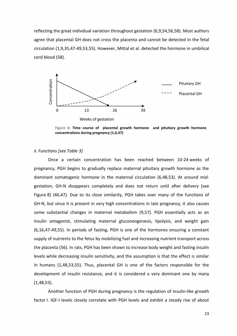
23
reflecting the great individual variation throughout gestation (6,9,54,56,58). Most authors
agree that placental GH does not cross the placenta and cannot be detected in the fetal
circulation (1,9,35,47-49,53,55). However, Mittal et al. detected the hormone in umbilical
cord blood (58).
ii. Functions [see Table 3]
Once a certain concentration has been reached between 10-24 weeks of
pregnancy, PGH begins to gradually replace maternal pituitary growth hormone as the
dominant somatogenic hormone in the maternal circulation (6,48,53). At around mid-
gestation, GH-N disappears completely and does not return until after delivery [see
Figure 8] (46,47). Due to its close similarity, PGH takes over many of the functions of
GH-N, but since it is present in very high concentrations in late pregnancy, it also causes
some substantial changes in maternal metabolism (9,57). PGH essentially acts as an
insulin antagonist, stimulating maternal gluconeogenesis, lipolysis, and weight gain
(6,16,47-49,55). In periods of fasting, PGH is one of the hormones ensuring a constant
supply of nutrients to the fetus by mobilizing fuel and increasing nutrient transport across
the placenta (56). In rats, PGH has been shown to increase body weight and fasting insulin
levels while decreasing insulin sensitivity, and the assumption is that the effect is similar
in humans (1,48,53,55). Thus, placental GH is one of the factors responsible for the
development of insulin resistance, and it is considered a very dominant one by many
(1,48,53).
Another function of PGH during pregnancy is the regulation of insulin-like growth
factor I. IGF-I levels closely correlate with PGH levels and exhibit a steady rise of about
Figure 8: Time course of placental growth hormone and pituitary growth hormone concentrations during pregnancy (1,6,47)
Weeks of gestation
0 13 26 39
Pituitary GH
Placental GH
Co
nce
ntr
atio
n

24
56% during pregnancy (1). In addition to its role in regulating fetal growth, IGF-I
stimulates the growth of maternal tissues such as uterus, breast, and thyroid gland
(1,9,35,58). It also increases maternal cardiac output and blood volume (1,9).
Finally, PGH probably also has autocrine or paracrine regulatory effects on the
placenta, as suggested by the presence of GH receptors in the villous trophoblast
(46,48,49).
iii. Regulation and interactions with other hormones
Unlike GH-N, placental GH is not secreted in a pulsatile manner and its secretion is
not controlled by growth-hormone-releasing hormone (GHRH) (6,9,47-49,53, 55).
However, many studies have shown a stimulatory effect on PGH secretion by
hypoglycemia, as well as an inhibition by glucose (1,6,9,16,47-49). This reflects the
importance of PGH as a nutrient provider for the fetus in times of low supply. PGH
secretion is inhibited by insulin, cortisol, ghrelin, and possibly leptin and up-regulated by
visfatin (44,57).
Short-term administration of PGH leads to an increase in leptin, but leptin is
decreased during chronic exposure to PGH, most likely due to the decrease in fat mass
mediated by PGH (57). PGH decreases adiponectin levels (1).
iv. Pathologies [see Table 4]
Many studies have found a correlation between PGH levels and fetal size and
development, while other authors found no relationship. Therefore, the role of placental
GH in diabetic pregnancies is uncertain. However, it is clear that PGH levels are decreased
in pregnancies with IUGR (1,6,9,16,17,47,49,55). This observation could be explained as a
consequence of inadequate fetal growth due to low levels of PGH and IGF-I, but the low
PGH levels could also be the result of placental insufficiency due to some other reason.
Evain-Brion states that low levels of PGH can be associated with fetal malnutrition. One
author claims that PGH levels are increased in women suffering from preeclampsia (58).
However, there is not yet much information on this topic.
Like hPL, PGH can be absent during pregnancy due to a gene deletion (56).
Nevertheless, the pregnancy can proceed and be carried to term, but maternal plasma

25
typically shows circulating levels of GH-N throughout as a substitute for the missing
placental hormone (55,56).
Hyperphagia Fat storage Insulin sensitivity
Insulin production
Plasma lipids
Estrogen ? ↑ in early, ↓ in late gestation
↑
Progesterone ↑ ↑ ↓ ↑
hPL ↑ ↑ ↓ ↑ ↑
PGH ↑ in early, ↓ in late gestation
↓ ↑
4) CRH
Corticotropin-releasing hormone (CRH), also known as corticotropin-releasing
factor (CRF), is a polypeptide hormone which is usually derived from the hypothalamus,
but is also secreted in significant concentrations by the placenta during human pregnancy
(38). Placental CRH is identical in size, structure, and biological activity to hypothalamic
CRH (38,59). However, unlike hypothalamic CRH, its release does not follow a circadian
rhythm, as the two hormones are controlled differently (32). During mid and late
pregnancy, CRH is produced in large quantities by the cytotrophoblast,
syncytiotrophoblast, and fetal membranes (38,59,60). It is secreted into the maternal
and, to a lesser extent, the fetal circulation (38,59,61). CRH exerts its effects by binding to
one of two G-protein-coupled receptors, corticotropin-releasing hormone receptor 1
and 2 (32).
i. Levels during pregnancy
CRH becomes detectable in maternal plasma at 8-20 weeks of gestation (32,59).
As with many other placental hormones, CRH levels can vary greatly between individual
women and are higher in twin pregnancies (32,59). After their first appearance, CRH
Table 3: Effects of estrogen, progesterone, hPL, and PGH on maternal metabolism during pregnancy (1,6,9,13,26,56). A question mark represents unclear or conflicting data while a blank space indicates a lack of data on the topic.

26
concentrations rise steadily until shortly before term and then rapidly until parturition
[see Figure 9] (59). It is generally agreed that maximum levels of CRH are seen
immediately before or during gestation, possibly at the time of maximal cervical dilation
(60). However, the reported levels vary greatly. Goland et al. found an exponential
increase of CRH levels during the last six weeks of pregnancy to concentrations of 1 ng/ml
and more, while mean CRH concentrations after 18-20 weeks were reported at 350 pg/ml
(59). Several authors found a two- to threefold increase of CRH levels throughout
pregnancy, while Frim et al. have found a 100-fold increase just in the last six to eight
weeks of pregnancy (1,32,38,60). Robinson et al. measured a 20-fold increase in CRH
concentrations five weeks before term, as compared to non-pregnant levels (61).
CRH is also secreted directly into the fetal circulation, but fetal cord CRH
concentrations are about 20 times lower than those in the mother (60).
ii. Functions
Since it is structurally identical to hypothalamic CRH, placental CRH performs
many of the same functions, namely stimulation of ACTH release (32,53). Pregnancy is
considered a state of hypercortisolism (59). This state is characterized by a stimulation of
hepatic gluconeogenesis and inhibition of insulin-dependent glucose uptake in skeletal
muscle (1). CRH also exerts important local effects, contributing to "the aseptic anti-
inflammatory process of implantation and the anti-rejection process that protects the
fetus from the maternal immune system" (32). Furthermore, CRH regulates placental
blood flow, myometrial contractility, and prostaglandin release (60).
Figure 9: Time course of CRH concentrations during pregnancy (32,59)
Weeks of gestation
0 13 26 39
Co
nce
ntr
atio
n

27
In late pregnancy, CRH levels continue to rise, but ACTH response decreases,
indicating a down-regulation of the CRH receptor in response to chronically high
concentrations (38,59). Shortly before birth, CRH levels are extremely high and it has
been proposed that CRH acts as a "pregnancy clock", determining the timing and
initiation of labor (32,59).
iii. Regulation and interactions with other hormones
Unlike hypothalamic CRH, placental CRH release is not down-regulated, but rather
stimulated by cortisol (32,38,61). Both maternal and fetal cortisol production cause a rise
in placental CRH concentrations (61). CRH concentrations also rise in the presence of IL-1,
NPY, acetylcholine, noradrenaline, vasopressin, angiotensin II, and oxytocin (60). As
mentioned earlier, estrogen down-regulates CRH levels while increasing cortisol binding
globulin (32,38,59). Several authors have found that progesterone decreases CRH levels
(38,60).
Not much is known about the effects of CRH on other gestational hormones. One
author has suggested that CRH might stimulate the release of hCG from the placenta by
an autocrine or paracrine mechanism (61).
iv. Pathologies [see Table 4]
High CRH levels are associated with all forms of maternal and fetal stress. Several
studies have confirmed increased CRH levels in preterm labor, pregnancy-induced
hypertension, and IUGR (38,60,61). Additionally, psychological stress can cause CRH levels
to increase (38,61). Other pregnancy-associated pathologies have not yet been
thoroughly investigated in regard to CRH levels.
5) Ghrelin
Ghrelin is a peptide hormone which has garnered some interest in recent years. It
is produced by many different tissues, including stomach, ovary, pancreas, neutrophils,
hypothalamus, and the placenta (16,62,63). Ghrelin is a ligand for the growth hormone
secretagogue receptor (GHSR), which is present in the central nervous system, adipose
tissue, endocrine organs, muscle tissue, and gastrointestinal tract (62,63).

28
i. Levels during pregnancy
Ghrelin levels follow an interesting pattern during pregnancy. Concentrations are
low in the first trimester, peak at mid-gestation, and subsequently decline to lower than
non-pregnant levels in the third trimester, becoming nearly undetectable in some cases
[see Figure 10] (1,16,62). After parturition, ghrelin levels once again rise to normal values
(62). Fuglsang et al. measured ghrelin levels in pregnant women after a period of fasting
(62). Maximum levels were observed at week 18 at 1.2 µg/l, and a concentration of
0.87 µg/l was observed at term (62). Another publication states that ghrelin levels are
30% lower in women in the third trimester of pregnancy than in non-pregnant women
(63).
ii. Functions
Ghrelin acts as an orexigenic hormone, increasing food uptake and promoting
weight gain and fat accretion by stimulating the differentiation of preadipocytes
(16,62,63). Ghrelin is also believed to be a contributing factor to insulin resistance by
stimulating hepatic gluconeogenesis while inhibiting pancreatic insulin secretion (63).
iii. Regulation and interactions with other hormones
Not much is known about the regulation of ghrelin, but its release might be
stimulated by fasting, while insulin causes a decrease in ghrelin concentrations (16,63).
On the other hand, ghrelin down-regulates insulin secretion, promoting
hyperglycemia (16). Placental GH, leptin, and resistin are decreased in the presence of
Figure 10: Time course of ghrelin concentrations during pregnancy (1,16,62)
Weeks of gestation
0 13 26 39
Co
nce
ntr
atio
n

29
ghrelin, while prolactin, ACTH, and cortisol are elevated (44,62,63). It has also been
shown that ghrelin has potent GH-releasing effects (62).
iv. Pathologies [see Table 4]
Ghrelin levels are low in states of decreased insulin sensitivity, such as obesity and
gestational diabetes mellitus (16,62,63). In pregnancy-induced hypertension and IUGR,
ghrelin levels are elevated (16,62).
6) hCT, PTH-rP
In the 1970s, some research was conducted into human chorionic thyrotropin
(hCT), a placental form of TSH. This hormone was believed to be secreted in small
quantities and to stimulate the thyroid gland and exert certain effects on maternal
metabolism (43). However, this research was not pursued in the following decades and
hCT has since disappeared from current publications on placental endocrine function.
Another placental hormone not receiving much attention currently is PTH-rP,
parathyroid hormone-related peptide. This polypeptide hormone influences maternal
calcium metabolism during pregnancy, increasing gastrointestinal calcium absorption,
stimulating placental calcium transport, thereby regulating fetal calcium levels (1).
Synergistically with hPL, PTH-rP increases the replication and inhibits apoptosis of
pancreatic β-cells (1). Furthermore, PTH-rP promotes breast development and liberates
calcium for breast milk synthesis (1).
GDM PE IUGR
hPL ↑ ↓ ↓
Placental GH ? ↑? ↓
CRH ↑
Ghrelin ↓ ↑
Table 4: Changes in peptide hormone levels in pregnancy-related pathologies (1,6,9,38,58,60,62). A question mark represents unclear or conflicting data while a blank space indicates a lack of data on the topic.

30
C. Adipokines
1) Leptin
There is a plethora of information and research concerning this adipokine.
Originally, leptin was identified as the product of the ob gene in 1994 by Zhang et al. and
considered to be an adipocyte-derived regulator of appetite and weight (19,23,36,45,64).
However, as more research was conducted into this hormone, it was discovered to fulfill
many other functions, including regulatory effects on angiogenesis, reproduction,
hematopoiesis, and bone mass (65). It was then discovered that the adipocyte is not the
only source of leptin, but that the gastric epithelium, brain, and placenta can also
synthesize this hormone (36,40,66,67). Placental leptin is identical to adipose cell-derived
leptin in size, structure, and immunoreactivity and is secreted in large quantities during
gestation by the syncytiotrophoblast, chorionic villi, chorion laeve, and amnion
(19,24,30,33,66,68). 95-98% of placental leptin is secreted into the maternal, 2-5% into
the fetal circulation (57,66). Leptin does not cross the placenta (69).
There are two forms of the leptin receptor, a long and a short one, the long one
being of greater importance in regulating body weight (5,65). Receptors for leptin are
abundant in the human body and can be found in the hypothalamus, arcuate nucleus,
liver, pancreatic β-cells, adipose tissue, and skeletal muscle (5,65,70). Leptin receptors are
also present in the placenta, amnion, and chorion (71).
i. Levels during pregnancy
In non-pregnant individuals, leptin concentrations are proportional to fat mass
(7,19,33,40,45,72). During pregnancy, both fat mass and leptin concentrations increase;
however, the major site of leptin production during pregnancy is not the adipose tissue,
but rather the placenta (66,73). This is evidenced by the fact that adipose tissue leptin
mRNA expression does not significantly change during pregnancy, while placental tissue
shows high amounts of leptin mRNA (66). Furthermore, leptin concentrations rise before
a significant change in fat mass is observed in early pregnancy, and they decrease
immediately after delivery of the placenta (40,73). Maternal leptin concentrations do not
show a correlation with placental mass, unlike those of other placental hormones (30).

31
However, it has been suggested that female fetuses present with higher maternal leptin
concentrations than male fetuses (74).
There is a consensus that leptin concentrations rise rapidly in early gestation to
peak in the second trimester and then decline somewhat in the third trimester, remaining
high until term [see Figure 11] (23,24,36,68). Placental leptin gene expression is at its
highest in early pregnancy (30,52). Different authors have reported leptin concentrations
to rise up to 2-50 times the normal level during gestation (66,68,70,75). The leptin peak
occurs at 22-27 weeks of pregnancy and shows levels between 19-30 µg/l (7,40,76,77).
Third trimester leptin levels have been measured at 20.4-35 µg/l (7,70,74), while at term
another study found a leptin concentration of 17.0 µg/l (57).
ii. Functions [see Table 5]
In healthy non-pregnant individuals, leptin acts as a regulator of food intake and
as an appetite suppressant by binding to receptors in the hypothalamus
(5,29,33,40,45,66). However, pregnancy is associated with weight gain although leptin
levels are high. This suggests that the mechanism of leptin action is different in pregnant
than in non-pregnant individuals.
There is evidence that pregnant women develop a leptin resistance in the second
trimester of pregnancy, blunting the anorexigenic effects of leptin in the central nervous
system (5). There are many theories as to the cause of this leptin resistance [see
Figure 12]. Leptin levels are not sufficiently high in early pregnancy to justify the
development of a down-regulation of leptin receptors during this time (5). Ladyman et al.
state that some but not all CNS leptin receptors are down-regulated in late pregnancy (5).
Figure 11: Time course of placental leptin concentrations during pregnancy (7,24,36,40)
Weeks of gestation
0 13 26 39
Co
nce
ntr
atio
n

32
However, leptin-responsive neurons may become resistant without being down-regulated
(5). Another explanation may be the decreased transport of leptin across the blood brain
barrier, as well as increased binding of leptin to soluble plasma receptors and thus a
decreased bioavailability to the hypothalamus (4,5,30,68). It is likely that leptin resistance
develops as a combination of a down-regulation of the leptin receptor, impaired leptin
signaling, and decreased availability of bioactive leptin. Leptin resistance is not only
present in the CNS, it also develops in peripheral organs such as pancreatic β-cells (65).
The causes of these changes are not fully elucidated, though several gestational
hormones are thought to be involved [see Figure 12]. The most likely candidates seem to
be prolactin and hPL, but progesterone and estradiol have also been suggested. Leptin
resistance can be induced in non-pregnant rats through infusions of hPL (5). Ladyman et
al. state that chronic activation of the prolactin receptor, as is the case in mid- to late
pregnancy, can cause leptin resistance (5). Other authors believe that the loss of the pre-
conception cyclic pattern of estradiol secretion, in addition to elevated progesterone and
subsequent changes in feeding behavior in the first trimester account for changes in
leptin responsiveness (4). Estradiol and progesterone are able to exert substantial effects
on leptin-responsive tissues as they are not regulated by maternal feed-back mechanisms
during pregnancy and reach very high concentrations (5).
Figure 12: Factors leading to the development of leptin resistance in mid- to late pregnancy (4,5,65)
Changes in
estradiol secretion
Prolactin
Progesterone
hPL
Changes in
feeding behavior
- down-regulation of
leptin receptors
- impaired leptin
signaling at the leptin
receptor
- decreased transport of
leptin across the blood-
brain barrier
- increased binding of
leptin to soluble
receptors
Leptin resistance
- loss of satiety
signals
- hyperphagia and
weight gain
- β- cell
dysfunction
- hyperinsulinemia

33
Due to leptin resistance, leptin actions during pregnancy differ from its
physiological actions in non-pregnant humans. In pregnancy, leptin contributes to the
increase in body weight and fat stores in early and mid-pregnancy by helping to induce
hyperphagia, while enhancing the mobilization of fat stores in the catabolic phase of late
pregnancy (4,45,66). Unlike conventional weight loss, weight loss due to leptin only
involves adipose tissue while sparing lean mass (66).
There are some contradictions as to the effect of leptin on insulin sensitivity.
While many authors believe that leptin is an insulin-sensitizing hormone, others claim it
decreases insulin sensitivity and inhibits insulin signaling (11,51,52,72,75,78). Possibly,
leptin has different effects on insulin sensitivity at different concentrations and at
different times during pregnancy depending on the severity of leptin resistance. It has
been observed that leptin increases skeletal muscle glucose uptake while reducing
hepatic glucose production, indicating insulin-mimetic properties (79). According to some
authors, the secretion of insulin by pancreatic β-cells is reduced in the presence of leptin,
while others have found an increase (33,65,73). Seufert describes an adipo-insular
feedback loop by which leptin from adipose tissue inhibits pancreatic insulin secretion,
maintaining glucose homeostasis [see Figure 13] (65). In leptin resistance, this feedback
loop is broken, leading to uncontrolled insulin secretion and eventually to β-cell failure
and diabetes (65).
Figure 13: Dysregulation of the adipo-insular axis and pathogenesis of type 2 diabetes. Adapted from Seufert (65)

34
In addition to its endocrine properties, leptin also exerts autocrine and paracrine
effects (30,35,36,40,66). Leptin is one of the hormones which promote trophoblast
differentiation and placental growth (40,80). It may also be a local immunomodulator,
counteracting the effects of pro-inflammatory cytokines at the maternal-fetal interface
(68).
iii. Regulation and interactions with other hormones
Although placental leptin is structurally identical to leptin from adipose tissue, the
mechanisms regulating its synthesis and release seem to be unique, although they are not
exactly known (19,33,66). However, several factors have been consistently shown to up-
regulate placental leptin production by different research teams. These are estrogen,
insulin, TNF-α, and hypoxia (19,23,24,29-31,33,40,64-66,71,72,68,81,82). A stimulation on
leptin release was also observed after administration of hCG, cortisol, IL-1, IL-6, and
forskolin (24,30,33,40,68,71). Factors thought to down-regulate leptin are hPL,
progesterone, androgens, and ghrelin (24,30,63). Although it has been suggested that
leptin is regulated by placental GH, several studies have yielded contradictory results
(57,81). GnRH does not regulate placental leptin production (81).
On the other hand, leptin positively influences the secretion of GnRH, LH, and FSH
from the hypothalamus and pituitary gland (45). Leptin also stimulates a rise in the
number of hCG pulses and pulse amplitude and up-regulates placental GH, CRH, and
various inflammatory cytokines such as IL-1, IL-6, and TNF-α (30,36,40,45,57,66,67).
Interestingly, leptin is up-regulated by TNF-α and IL-1 and IL-6 while also up-regulating
these cytokines. This mechanism can be observed in preeclampsia or diabetes mellitus,
where an excess of inflammatory products is produced in response to a systemic
pathological change in the mother. Regardless of which hormone or cytokine is elevated
first, these pathologies lead to chronically high levels of leptin, IL-1, IL-6, and other
cytokines, which continue to potentiate each other's effects and further promote
inflammation (66).
Similarly, hCG up-regulates leptin and leptin up-regulates hCG. Due to the vastly
different peaks of these two hormones in pregnancy, it is unlikely that hCG can have an
effect on leptin in late pregnancy. On the other hand, leptin levels are comparatively low
at the time of the hCG peak in early pregnancy, so a stimulation at this point also seems

35
unlikely. However, it is possible that at certain times during pregnancy, these two
hormones stimulate each other, but this probably does not occur simultaneously.
Finally, leptin acts directly at the maternal-fetal interface with TNF-α to increase
the expression of placental endothelial lipase and placental phospholipase, thereby aiding
the transport of fatty acids and cholesterol across the placenta (69).
iv. Pathologies [see Table 6]
As has already been mentioned, leptin is elevated in pathologies associated with
chronic inflammation, such as preeclampsia and diabetes mellitus (25,30,31,33,40,
51,63,66,68,70,71,80,83,84). In preeclampsia, elevated leptin levels have been observed
prior to the onset of all other symptoms (71,82,85,86). This could make leptin a useful
screening tool if levels were measured at different times throughout the pregnancy.
There have been some reports of unchanged or even decreased leptin levels associated
with preeclampsia, but the majority of studies have found that levels significantly
increase (71). The same is true for leptin concentrations in gestational diabetes mellitus.
In GDM, different studies show increased, unchanged, or decreased leptin levels
(12,25,29,70,71). However, most authors have found an increase and it has even been
suggested that high leptin levels in early gestation predict the risk for developing GDM
later on (76,80). One explanation for the various observations on leptin with GDM has
been postulated by Lappas et al., who found increased levels of adipose tissue leptin and
decreased placental leptin in GDM, with total leptin being increased (33,80). Other
conditions associated with increased leptin concentrations are pregnancy-induced
hypertension, hydatidiform mole, choriocarcinoma, and obesity (25,36,66,70,72, 83,84).
According to Hauguel-de Mouzon, there is no condition associated with a down-
regulation of placental leptin gene expression (66). However, several authors have
described decreased leptin concentrations in pregnancies with IUGR fetuses, which they
saw as a consequence of impaired placental function due to insufficient perfusion
(30,40,71,80,68). Other studies have found increased leptin concentrations with this
condition (70,74,80). Briana et al. provide an explanation by suggesting that the
pregnancies that showed increased leptin levels may have additionally been complicated
by other gestational pathologies such as preeclampsia, and that maternal characteristics
like BMI and smoking had not been taken into account, leading to falsely high

36
measurements (71). Alternatively, leptin levels may relate directly to the severity of the
disorder, appearing lower in mild IUGR and higher in severe IUGR (71). One study found
decreased leptin levels in pregnancies with macrosomic fetuses (29). Lastly, leptin levels
may be abnormally low in a state of extreme fasting or starvation (67).
2) TNF-α
Tumor necrosis factor-α is in inflammatory cytokine which is mainly produced in
monocytes, macrophages, T-cells, and neutrophils, as well as in fibroblasts and
adipocytes, which is why it is also termed an adipokine (29,50). Generally speaking, TNF-α
is correlated with fat mass and is increased in obesity and insulin resistant states (37).
During pregnancy, TNF-α can be found in the placental syncytiotrophoblast, decidua, and
amniotic fluid (37,87). In non-pregnant individuals, TNF-α production is greater in
omental than subcutaneous adipose tissue, while in pregnancy placental TNF-α
production exceeds that of omental and subcutaneous adipose tissue (37). Thus, the
placenta is most likely responsible for the increased TNF-α concentrations that can be
observed during normal human pregnancy (25).
There are two types of receptors for TNF-α, named TNFR1 and TNFR2. TNFR1 is
constitutively expressed throughout many tissues, including adipocytes, liver, endothelial
cells, granulocytes, and the placenta, while TNFR2 is localized to immune cells (64,87).
There is also a soluble form of the TNF-α receptor (64). TNF-α does not cross the placenta
(69).
i. Levels during pregnancy
TNF-α levels are closely related to the level of insulin resistance and have a
negative correlation with whole body insulin sensitivity (14,37,50,72,88). By many
authors, TNF-α is heralded as the best predictor of peripheral insulin resistance during
pregnancy (11,14,89). Thus, TNF-α can decrease briefly in early pregnancy when insulin
sensitivity is augmented (14). After 30 weeks, parallel to the development of insulin
resistance, TNF-α and TNF-α receptor concentrations begin to rise and continue to do so
until term [see Figure 14] (1,14,37,83). The onset of labor is associated with a further
increase in TNF-α (90). After delivery, maternal TNF-α levels fall rapidly, indicating both a

37
significant contribution of the placenta to total TNF-α and a return of insulin sensitivity
(14).
Many authors have measured TNF-α levels in healthy pregnant women during the
various stages of pregnancy. In the first trimester, levels of 1.56 pg/ml have been
reported and the third trimester measurements ranged from 0.9-65 pg/ml (14,25,69,
77,87,91). 94% of placental TNF-α is secreted into the maternal circulation, while the
remaining 6% are secreted into the fetal circulation (14).
ii. Functions [see Table 5]
Because TNF-α is not a pregnancy-specific cytokine, many of its effects during
pregnancy are similar to those in non-pregnant individuals. These include immune
surveillance, cell differentiation and renewal, and inflammation (87). However, TNF-α also
has some additional functions during pregnancy. In the early phase of implantation and
trophoblast invasion, TNF-α acts as an inhibitor of syncytialization and induces
trophoblast apoptosis as a means of maintaining trophoblast turnover and renewal (87).
TNF-α is also believed to cause apoptosis of vascular smooth muscle cells in spiral
arteries, contributing to the remodeling of these arteries during trophoblast invasion (87).
Another very important function of TNF-α during pregnancy is the modulation of
maternal metabolism. Like leptin, TNF-α traditionally causes an decrease in food intake
and body weight while increasing metabolism (72). Also like leptin, the overall effect of
TNF-α in combination with other hormones of pregnancy paradoxically leads to impaired
glucose tolerance and the development of insulin resistance (72). However, TNF-α is
much more closely correlated with insulin resistance than leptin, leading to the
hypothesis that it is in fact its major cause (50,88). TNF-α contributes to skeletal muscle
Figure 14: Time course of TNF-α and adiponectin concentrations during pregnancy (1,6)
Weeks of gestation
0 13 26 39
TNF-α
Adiponectin
Co
nce
ntr
atio
n

38
insulin resistance by causing a decrease in insulin receptor tyrosine phosphorylation and
GLUT-4 gene expression, leading to impaired insulin signaling and glucose disposal
(1,15,29,37,50,71,72,88). In the presence of TNF-α, insulin signaling is also decreased in
adipose and hepatic tissues and hepatic lipogenesis, cholesterol synthesis, and VLDL
production are increased (14,15,72). Furthermore, TNF-α inhibits lipoprotein lipase in
adipocytes, stimulates lipolysis, and impairs pancreatic β-cell function, resulting in
hyperglycemia and hyperlipidemia (1,15,25,37,53,71,72).
iii. Regulation and interactions with other hormones
Not much is known about the regulation of placental TNF-α production. Coughlan
et al. conducted a study with placental explants in which high glucose concentrations
stimulated TNF-α release (37). Other stimuli for placental TNF-α production are hypoxia
and infection (71). Finally, adiponectin down-regulates TNF-α (83,92,93).
On the other hand, TNF-α exerts many regulating effects on other hormones. It
increases concentrations of leptin, placental endothelial lipase and phospholipase, IL-6,
IL-8, and CRH (33,64,68,69,72,87,94). Resistin, adiponectin, and visfatin are down-
regulated in the presence of TNF-α (1,15,29,33,50,79,85,88,95,96). Contrary results have
been published regarding the effect of TNF-α on β-hCG production, with increases as well
as decreases being reported (23,87).
iv. Pathologies [see Table 6]
Since TNF-α is strongly associated with insulin resistance, elevated levels can be
observed in pregnancies with type 2 diabetes mellitus, gestational diabetes, and obesity
(12,14,25,29,37,63,64,71,88). Due to its properties as an inflammatory cytokine, elevated
TNF-α concentrations can also be observed in pregnancies complicated by preeclampsia
(14,71,82,83,87). According to Haider et al., TNF-α can be used as a marker for the
severity of preeclampsia, as concentrations are higher in more severe cases (87). There
have been several studies which have observed higher levels of TNF-α in pregnancy-
induced hypertension and placental insufficiency in combination with IUGR (71,83,87). An
increase can also be seen in chorioamnionitis and preterm labor as a result of ascending
bacteria (87). Finally, 40-70% of recurrent spontaneous abortions are associated with high
TNF-α levels (87). In this case, elevated TNF-α can either be a consequence of amniotic

39
infection leading to abortion, or the abortion can be caused by an excess of TNF-α itself
due to its cytotoxic effect on trophoblast cells (87).
3) Adiponectin
Adiponectin is a polypeptide hormone which is released exclusively from white
adipose tissue and, during pregnancy, the placenta (29,97). It is found more abundantly in
subcutaneous than in omental adipose tissue (95). Plasma adiponectin concentrations are
higher than those of any other adipokine (11,24). Although concentrations can vary
greatly amongst individuals, women exhibit higher adiponectin levels than men (98).
Adiponectin levels are negatively correlated with intraabdominal fat mass and BMI and
positively correlated with whole body insulin sensitivity, an observation which is
consistent with the anti-atherogenic and insulin-sensitizing properties of this hormone
(11,29,33,50,95,97,99,100). Adiponectin receptors are located in skeletal muscle, liver,
pancreatic β-cells, adipose tissue, and the placenta (71,101,102).
i. Levels during pregnancy [see Figure 14]
As with many adipokines, there is some controversy as to the changes in
adiponectin concentrations during pregnancy. Many authors have reported a decrease in
adiponectin levels as pregnancy progressed, while others observed no change or even an
increase (25,50, 84,103). Naruse et al. saw a 30% decrease in adiponectin concentrations
during pregnancy, which they attributed to hemodilution (83). Thus, adiponectin
production during pregnancy may be increased while plasma adiponectin concentrations
remain stable or decrease. To summarize the results of many studies, non-pregnant
women showed adiponectin levels between 0.4 ng/ml to 17 µg/ml (15,103,104). First
trimester levels have been reported at 5.2-12.3 µg/ml, second trimester levels at
5.1-11.8 µg/ml, and third trimester levels at 4.7-14.7 µg/ml (11,25,33,74,76,77,83-86,
92,96,97,99,100, 103,105-107).
ii. Functions [see Table 5]
As has been mentioned above, adiponectin has anti-atherogenic, anti-
inflammatory, and insulin-sensitizing properties (29,71,74,83,85,86,95,97,99,103).
Adiponectin improves insulin signaling by increasing insulin-induced tyrosine

40
phosphorylation of the insulin receptor in skeletal muscle and other insulin-sensitive
tissues while decreasing hepatic gluconeogenesis (25,29,33,50,71,85,88,95,101,108). In
skeletal muscle and liver, adiponectin increases the oxidation of free fatty acids, leading
to decreased triglyceride levels, further benefiting insulin sensitivity (15,92,97,98,105).
Additionally, adiponectin improves hepatic lipoprotein metabolism in response to insulin
and induces lipoprotein lipase gene expression (95,98,101). There is some evidence that
adiponectin may improve β-cell function during pregnancy (76). Overall, adiponectin
combats the effects of TNF-α and other diabetogenic hormones to reverse insulin
resistance by decreasing plasma free fatty acids, triglycerides, and glucose, even reducing
body weight (25,29,93,101,105).
iii. Regulation and interactions with other hormones
Adiponectin is regulated by feeding and fasting, decreasing as a response to
insulin secretion (95,99). Adiponectin concentrations are down-regulated in the face of
increased fat mass and obesity (11). Furthermore, this hormone is down-regulated by
placental and pituitary growth hormone, prolactin, IL-6, glucocorticoids, and
catecholamines (1,11,95,101).
A complicated relationship exists between adiponectin and TNF-α. These two
hormones are considered antagonists and an inverse correlation exists between them
(96,102). TNF-α is able to down-regulate adiponectin; however, adiponectin can also
inhibit TNF-α signaling and down-regulate its release from macrophages (1,15,25,29,33,
50,83,92,93,95,96,100). It seems that these two hormones do not coexist well, but it is
unclear which is the more dominant player. Adiponectin is able to reverse insulin
resistance to a certain degree in the first and second trimester, but as pregnancy
progresses and the hormonal cocktail leading to insulin resistance grows stronger TNF-α
emerges as the leading hormone.
iv. Pathologies [see Table 6]
Due to its insulin-sensitizing effects it could be speculated that adiponectin would
be up-regulated in insulin-resistant states such as diabetes mellitus or gestational
diabetes. However, observations by many different authors have shown that these
pathologies show decreased adiponectin levels (6,12,15,29,50,63,71,76,84,88,92,95,99,

41
102,105). Because many women who develop gestational diabetes during pregnancy have
a predisposition for this condition, it is possible that the protective mechanism of action
of adiponectin is inherently weak. Thus, impaired insulin sensitivity during pregnancy may
be a result of inadequate adiponectin production prior to conception. In fact, adiponectin
concentrations may be a useful diagnostic tool for predicting the risk for gestational
diabetes early on. Several authors have found that adiponectin concentrations are low
months before gestational diabetes manifests itself (71,76,100). Williams et al. go so far
as to claim that low adiponectin levels are a dose-dependent risk factor for gestational
diabetes, where lower adiponectin levels indicate a higher risk (100). In accordance with
these results, pregnant women with macrosomic fetuses had lower adiponectin levels
than controls in one study (29).
In preeclamptic pregnancies, adiponectin concentrations can be increased,
decreased, or unchanged (51,71,75,83-85,86,103,106). The majority of authors have
reported elevated adiponectin concentrations with this condition, possibly as a
compensatory mechanism to combat the inflammation (97). In pregnancies with IUGR
fetuses, maternal adiponectin concentrations were decreased in two studies, but more
research needs to be done in the field (71,74). In contradiction to this, one author
measured higher adiponectin levels in pregnancies with pathological uterine perfusion
(85).
4) Visfatin
Previously known as pre-B cell colony enhancing factor (PBEF), this adipokine was
first identified as the product of lymphocytes (90,94). Later, it was also found in skeletal
muscle, bone marrow, hepatic tissue, and visceral fat, hence the name change
(79,90,109,110). Visfatin levels show a negative correlation with visceral, but not
subcutaneous fat, and omental secretion has been observed to be elevated during
pregnancy (79,88,94). The placental syncytiotrophoblast, chorionic cytotrophoblast,
amniotic epithelium, mesenchymal cells, parietal decidua, and fetal capillary endothelium
have been identified as additional sites of visfatin production (71,79,94,109,110). Visfatin
binds to the insulin receptor in a non-competitive way, exerting insulin-mimetic effects
(75,94,110).

42
i. Levels during pregnancy
Because visfatin expression is up-regulated to up to seven times the normal level
in omental adipose tissue during pregnancy, it is not clear how large the contribution of
the placenta is to maternal visfatin levels (94). Some authors have reported no increase of
visfatin concentrations during pregnancy, while others have observed a rise throughout
gestation and other groups measured decreasing visfatin concentrations as pregnancy
progressed (71,75,90,94,110). As with many other placental hormones, there seems to be
great individual variability. In early to mid-gestation, concentrations have been reported
from 26-67.5 ng/ml while at term the measurements range from 6.2-695.9 ng/ml
(75,79,84,85,91,94,109). Morgan et al. state that in late pregnancy visfatin levels are
elevated by 20-50 times compared to the luteal phase of the menstrual cycle (94). A
further elevation of visfatin concentrations can be observed with the onset of labor,
possibly in response to subclinical infection (90). Briana et al. found comparable visfatin
concentrations in maternal and fetal blood, suggesting a passive transplacental transfer of
this adipokine (109).
ii. Functions [see Table 5]
The majority of authors agree that visfatin acts as an insulin-mimetic of equal
potency as insulin (84,85,88,94,109,110). Because it does not utilize the same binding site
on the insulin receptor, visfatin acts in concert with insulin to lower blood glucose,
stimulate muscle and adipocyte glucose transport, inhibit hepatic gluconeogenesis, and
promote adipogenesis (75,79,94,110). There is some evidence that visfatin can improve
insulin sensitivity as chronic exposure lowers insulin levels (79). Visfatin gene and protein
expression increase with decreasing β-cell function, suggesting a compensatory
mechanism to mitigate insulin resistance (84). A regulatory role of visfatin on
HDL-cholesterol has also been suggested (84). In recent years, several authors have
suggested that visfatin may not act as a hormone in the classical sense, but rather
operate in a paracrine or autocrine manner without any systemic effects (94). This
hypothesis resulted from the observation that visfatin concentrations are greatly
increased locally in omental adipose tissue during pregnancy, while only slightly
increasing in serum (94).
43
Visfatin also has pro-inflammatory and immunomodulating properties and acts as
a local growth regulator to accommodate membrane distension due to amniotic infection
and thereby protect against membrane tissue apoptosis (84,90,91).
iii. Regulation and interactions with other hormones
Visfatin secretion is up-regulated by glucocorticoids (88,110). There are conflicting
results as to the effect of pro-inflammatory cytokines such as TNF-α, IL-1, and IL-6 on
visfatin secretion, with positive and negative effects being reported (71,79,85,88,90,110).
One might expect visfatin, a pro-inflammatory cytokine, to be increased in the presence
of other pro-inflammatory cytokines. On the other hand, visfatin may be secreted to
counteract the impaired insulin signaling caused by an inflammatory milieu. More
research needs to be done on this topic to elucidate how visfatin concentrations change
in the face of inflammation. Briana et al. state that only placental and not adipose tissue
visfatin production is elevated by pro-inflammatory cytokines, while Fasshauer et al.
found decreased visfatin secretion in response to inflammation only in adipose tissue
(71,85). Perhaps the contradictory results were achieved due to differences in study
design and in the type of tissue that was investigated.
Visfatin levels are decreased in the presence of pituitary growth hormone,
possibly due to the negative effect of GH on insulin sensitivity (88,110). Unlike with
insulin, visfatin secretion is not regulated by fasting or feeding (110). Rather, it is
constitutively expressed in pregnant and non-pregnant individuals alike (90,110).
Mechanical stimuli such as membrane distension may trigger visfatin secretion (90).
There may also be a role for glucose and insulin in the regulation of visfatin, but this is not
yet proven (84).
iv. Pathologies [see Table 6]
Most of the pathologies associated with changes in visfatin concentrations are
related to its insulin-mimetic effects. There have been many investigations into visfatin
levels in diabetes mellitus, gestational diabetes, and obesity with contradictory results.
Visfatin levels have been reported increased, decreased, and unchanged in each of these
pathologies (71,75,79,84,85,91,94,109). Visfatin has also been suggested as a predictive

44
factor for gestational diabetes (84,94). Mastorakos et al. claim that first trimester visfatin
levels predict insulin sensitivity in the second trimester (84).
Preeclampsia presents another contradiction. Elevated as well as decreased
visfatin levels have been measured in preeclamptic women and several studies found
increased visfatin concentrations in pathological placental perfusion and IUGR
(71,75,77,85). As has been mentioned earlier, visfatin secretion is increased in
chorioamnionitis and imminent birth (90,110).
Hyperphagia Fat storage Insulin sensitivity
Insulin production
Plasma lipids
Leptin ↑
↑ in early, ↓ in late gestation
?
?
TNF-α ↓ ↓ ↑
Adiponectin ↑ ↑? ↓
Visfatin ↑ ↓
Resistin ↓
5) Resistin
This relatively novel cytokine is produced mainly by monocytes and macrophages
and to a much lesser extent by adipose tissue, skeletal muscle, and pancreatic islet cells
(63,71). In non-pregnant individuals, resistin synthesis is higher in abdominal than thigh
fat (89). During pregnancy, resistin concentrations are elevated and there is evidence that
the placenta is a source of resistin (12). The main production site of placental resistin is
the syncytiotrophoblast, but resistin can also be found in the extravillous cytotrophoblast,
decidua, and amnion (12,52,63). During pregnancy, resistin gene and protein expression is
higher in the placenta than in adipose tissue (52). Although associated with the
development of insulin resistance, resistin concentrations are independent of BMI during
gestation (12).
Table 5: Effects of placenta-derived adipokines on the maternal metabolism during pregnancy (4,45,50,51,66,71-73,78,79,88). A question mark represents unclear or conflicting data while a blank space indicates a lack of data on the topic.

45
i. Levels during pregnancy
Resistin levels are elevated during pregnancy (75,103). While most authors claim
that resistin concentrations rise continually until term, others state that while
concentrations are elevated in early gestation, they then decline progressively until term
(12,51,52,71,75,89,103). Yet another author observed elevated resistin concentrations in
the first and third, but not the second trimester (63). The change in resistin
concentrations is most likely due to placental production, as no change is observed in
adipose tissue resistin synthesis during gestation (70,89).
Resistin measurements in non-pregnant women show concentrations of
6.3-18.1 ng/ml (52,103). In early pregnancy, concentrations were measured between
5.0 and 17.9 ng/ml and late pregnancy values were between 2 and 68.2 ng/ml
(33,51,103,106). The wide spectrum indicates that resistin concentrations probably vary
greatly within the population like those of most other adipokines.
ii. Functions [see Table 5]
Resistin has been associated with the development of insulin resistance, but less
research is available on this adipokine than on many other hormones of pregnancy (12). It
has been observed that insulin sensitivity declines as a response to elevated resistin
concentrations (51,52,71,103). Experiments with mice have shown that hepatic insulin
resistance develops in the presence of high concentrations of resistin (63,71). In this
respect, resistin works like many other diabetogenic hormones, impairing glucose uptake
and thereby increasing plasma glucose and decreasing insulin sensitivity (51,70,88). In
vitro experiments have shown a decrease in GLUT-4 activity, indicating a possible
involvement of resistin in skeletal muscle insulin resistance (63,71). However, this
observation calls for further research. At this time, resistin is believed to induce only
hepatic, but not peripheral insulin resistance (33,63).
iii. Regulation and interactions with other hormones
Because resistin has not been extensively studied, not much is known about its
regulation. Estrogens, progesterone, TNF-α, corticosteroids, and ghrelin lead to decreased
resistin secretion (33,63). There seems to be a regulatory effect of insulin on resistin
46
secretion, but whether it is a positive or negative one is unclear (33,71). It is not known if
and how resistin is involved in the regulation of other hormones.
iv. Pathologies [see Table 6]
Resistin levels can be expected to be elevated in insulin resistant states, such as
obesity, diabetes mellitus, or gestational diabetes and this has been observed by some
authors (63). However, other studies have found a decrease or no change in resistin levels
in women with GDM (71). The same is true for preeclampsia, where increased, decreased,
and unchanged resistin levels have been reported (71,75,106). The majority of authors
believe resistin to be elevated in pregnancies complicated by preeclampsia as a
consequence of impaired placental hormone production (51,103). Again, more research is
required to make definitive statements on this subject.
6) Apelin
Apelin is a peptide hormone which is described as the endogenous ligand for the
G-protein coupled APJ receptor (71,104,111). Both apelin and its receptor are widely
distributed in the human body, occurring in lung, kidney, white and brown adipose tissue,
hypothalamus, GI-tract, the pregnant and lactating breast, vascular endothelial cells, and
the placenta (71,104,107,111). During pregnancy, the placenta is said to produce ten
times more apelin than adipose tissue (104). Like resistin, it is a relatively novel adipokine
and has not been studied extensively.
i. Levels during pregnancy
Preliminary observations on the changes in apelin gene and protein expression
during pregnancy are ambiguous. Several authors have reported a decrease of plasma
apelin concentrations from the first to the third trimester, while others observed an
increase in adipose tissue and placental apelin in the pregnant state (71,104,107).
Malamitsi-Puchner et al. reported that apelin concentrations decline rapidly after
parturition in both maternal and fetal plasma, pointing to a significant placental
production of this adipokine during pregnancy (112). The same authors also observed
higher fetal than maternal apelin levels during pregnancy, suggesting a mode of passive
transplacental transfer from mother to fetus (112). Kourtis et al. measured an apelin

47
concentration of 4.45 µg/ml in women during mid-pregnancy and a concentration of
5.0 µg/ml in non-pregnant control women (104).
ii. Functions
The functions of apelin cover a wide spectrum. It has a regulatory role on the
immune system, cardiovascular system, angiogenesis, brain signaling of hunger and thirst,
fat storage, and glucose homeostasis (71,107,111). During pregnancy, apelin is assumed
to have a role in the regulation of placenta formation and fetal development, largely
through the promotion of angiogenesis (112). It is also a local vasoconstrictor (107). In a
study with mice, apelin increased glucose utilization and showed a negative correlation
with oxidized LDL-cholesterol, perhaps indicating a positive effect on insulin sensitivity
and atherosclerosis (104). However, the same study showed no correlation between
apelin and markers of insulin sensitivity (104).
iii. Regulation and interactions with other hormones
Based on the research available at this time, the strongest factors regulating
apelin release are fasting and feeding (71,104,111,112). Apelin is strongly up-regulated by
insulin and therefore by feeding, while fasting strongly decreases apelin secretion
(71,104,111,112). There is a negative correlation between apelin and adiponectin levels,
but the significance of this remains to be explained (104). Finally, there may be a
regulatory role for TNF-α on apelin secretion, but once again more research is required to
make any concise statements (71).
iv. Pathologies [see Table 6]
Since there are no long-term studies with apelin, it is difficult to say how it reflects
on pregnancy-related pathologies. In a few studies, apelin levels were elevated in
pregnant women who were obese or had diabetes (104,111). However, this elevation
could only be observed if the women were hyperinsulinemic (111). Pregnancy-induced
hypertension and preeclampsia may show elevated or decreased apelin levels (71,107).
Due to its effect as a vasoconstrictor, changes in apelin concentrations may play a part in
the development of preeclampsia (107). This is definitely an interesting point which
should be the focus of more research in the future.

48
7) Chemerin
This relatively new adipokine was first described in 2003 as the ligand for the
G-protein-coupled chemokine-like-receptor 1 and joined the group of adipokines in 2007
(76,77,113). Originally, chemerin was of interest due to its pro-inflammatory properties,
but in recent years it has been investigated regarding its role as a regulator of glucose and
lipid metabolism (113). So far, adipose tissue, the liver, and the placenta have been
identified as sources of chemerin production (77). During pregnancy, the placenta
produces more chemerin than omental or subcutaneous adipose tissue (113).
i. Levels during pregnancy
Because most of the research concerning chemerin is being done in the fields of
obesity and diabetes mellitus, there is a relative paucity of papers regarding chemerin in
pregnancy. However, there have been a few publications which mention chemerin levels
in the third trimester ranging from 124.2-217.6 µg/l (76,77,113). These authors agree that
chemerin levels are increased during pregnancy compared to non-pregnant control
women (76,76,113). Pfau et al. state that chemerin concentrations are higher in the third
than in the first trimester (76).
ii. Functions
There have been contradictory publications concerning the properties of chemerin
as an adipokine. While Pfau et al. claim that chemerin has insulin-sensitizing properties
and increases glucose uptake in adipocytes, other authors state the exact opposite,
namely that chemerin impairs glucose tolerance, lowers serum insulin, stimulates
lipolysis, and reduces insulin resistance (76,77,113). Since most of these results come
from studies with mice, it is unclear how the situation is in humans. All the above authors
agree that chemerin has an important role in the differentiation of adipocytes and the
expression of adipocyte genes involved in glucose and lipid homeostasis (76,77,113).
iii. Regulation and interactions with other hormones
Chemerin secretion is up-regulated by IL-1β (113). There exists a significant
positive correlation between chemerin and leptin, plasma triglycerides, and fasting insulin
(77,113). However, it is not known whether these factors influence chemerin secretion.

49
iv. Pathologies [see Table 6]
Some authors have suggested that chemerin production is augmented as body
mass index (BMI) increases, while others found no correlation (113). One publication
shows that chemerin concentrations are unchanged in the plasma of obese pregnant
women, but increased in the cord blood of the fetuses of the same women (113). Pfau et
al. investigated chemerin concentrations in women with gestational diabetes mellitus and
found no significant change (76). However, the authors attributed this to the fact that all
women were matched to controls for fasting insulin and hypothesize that chemerin
concentrations are higher in women with hyperinsulinemia. The authors conjecture that
this elevation may either be a compensatory mechanism utilizing the insulin-sensitizing
properties of chemerin to counteract insulin resistance, or a manifestation of chemerin
resistance requiring more chemerin to maintain its physiological effects (76).
Finally, one study showed elevated chemerin concentrations in preeclamptic
women in the third trimester and six months after delivery, compared to healthy control
women (113).
GDM PE IUGR
Leptin ↑ ↑ ?
TNF-α ↑ ↑ ↑
Adiponectin ↓ ↑? ↓?
Visfatin ? ?
Resistin ? ?
Apelin ↑?
Chemerin ↑? ↑?
Table 6: Changes in adipokine levels in pregnancy-related pathologies (6,71,85,113). A question mark represents unclear or conflicting data while a blank space indicates a lack of data on the topic.

50
D. Placental Hormones in the Fetus
Although most placental hormones have an effect on fetal growth, not all are
detectable in the fetal circulation. Aside from one, all publications on placental growth
hormone have been unable to detect this hormone in the fetal circulation, meaning it
does not cross the placenta (1,6,9,44,47,53,54,56,58,114,). Other placental hormones
that do not cross the placenta are progesterone, hPL, CRH, leptin, and TNF-α; however,
these hormones are directly secreted into the fetal circulation by the placenta and can
therefore be detected in umbilical cord blood (6,9,14,33,38,43,66,69,115). For leptin and
TNF-α, it has been determined that the fraction of hormone secreted into the fetal
circulation is quite small in comparison to the total amount produced by the placenta,
while it is not known in what amounts progesterone, hPL, and CRH enter the fetus
(14,66,69). Visfatin and apelin are able to pass the placenta passively, leading to fetal
concentrations equal to or higher than those in the mother (109,112).
As the fetus grows and its organs mature, it begins to produce some of these
hormones itself, which complicates the matter as it is difficult to differentiate between
hormones of placental and fetal origin in some cases. There is evidence for fetal
production of leptin, resistin, adiponectin, and possibly ghrelin, all of which can be
detected in cord blood (16,66,103).
Some placental hormones have been investigated as to their effects in the fetus.
Human CG has a role in fetal development through its regulation of the
11β-hydroxysteroid-dehydrogenase type 2, a hormone which inactivates cortisol by
converting it to cortisone (42). By up-regulating 11β-HSD 2, hCG creates a "glucocorticoid
barrier", protecting the fetus from high levels of cortisol, which is of utmost importance in
early pregnancy (42). Human CG also stimulates dehydroepiandrosterone synthesis in the
fetal adrenal glands (42).
Human PL also has a function in fetal development, promoting the synthesis of
insulin-like growth factors, insulin, adrenocortical hormones, and surfactant, regulating
fetal metabolism, and possibly promoting fetal angiogenesis (9,43). Furthermore, hPL
levels correlate positively with fetal weight in the second and third trimester (38,60). Like
hPL, CRH stimulates the fetal adrenal glands, but also the pituitary gland and the

51
production of ACTH (38,60). Placental growth hormone cannot be detected in the fetal
circulation, yet a correlation exists with birth weight, suggesting an indirect effect on fetal
growth (38,60).
Although only 2-5% of placental leptin is secreted into the fetal circulation, it is
one of the most abundantly found placental hormones in the fetus (66). The abundance
of leptin receptors in fetal tissues such as cartilage, lung, bone, kidney, testes, and
hypothalamus, suggests an important role of this adipokine in fetal development and
growth (66,70). Leptin is involved in fetal vasculogenesis, erythropoiesis, lymphopoiesis,
and the regulation of fetal fat stores (66,70,116). Because fetal adipose tissue also
produces leptin, its concentration is thought to reflect the metabolic state of the fetus
and can be elevated in gestational diabetes or diabetes mellitus in response to
hyperinsulinemia (116). Likewise, IUGR fetuses have lower leptin levels because there is
less adipose tissue to produce it (66). Apelin receptors are present in the fetus, suggesting
a role for this adipokine in the promotion of fetal growth and possibly on angiogenesis
(112).

52
Hormone Transfer into the fetal circulation
Functions in the fetus
Fetal production
Progesterone Direct secretion by the placenta
Yes
hCG Regulation of 11β-HSD, steroidogenesis
hPL Direct secretion by the placenta
Synthesis of IGF, insulin, adrenocortical hormones, surfactant Regulation of metabolism, angiogenesis
PGH No Regulation of fetal growth?
No
CRH Direct secretion by the placenta
Stimulation of adrenal and pituitary glands
Ghrelin Yes?
Leptin Direct secretion by the placenta
Vasculogenesis, erythropoiesis, lymphopoiesis, regulation of fat stores
Yes
TNF-α Direct secretion by the placenta
Adiponectin Yes
Visfatin Diffusion
Resistin Yes
Apelin Diffusion Promotion of fetal growth, possibly angiogenesis
Table 7: Placental hormones and their functions in the fetus (6,9,14,16,33, 38,42,43,60,66,70,103,109,112). A question mark represents unclear or conflicting data while a blank space indicates a lack of data on the topic.

53
IV. Discussion
The purpose of this paper is to review the hormones that are produced by the
placenta during pregnancy which have an effect on the maternal metabolism. While
many reviews already exist regarding this topic, most of these focus on one or several
placental hormones and do not offer a comprehensive overview of all relevant hormones
and cytokines. Also, many of these reviews focus on one portion of maternal metabolism,
such as glucose or lipid homeostasis, while others address more areas rather superficially.
The goal of this paper is to provide a global overview of the metabolic changes that occur
in pregnancy and the actions and interactions of the hormones responsible for these
changes. Although the scope of this paper is quite wide, each hormone was thoroughly
researched and analyzed as to its history, functions, physiological concentrations,
regulation, influence on other hormones, and role in common pathologies of pregnancy.
The most important metabolic changes of pregnancy, such as hyperphagia, insulin
resistance, leptin resistance, facilitated anabolism, and accelerated starvation were also
discussed.
It has long been known that the placenta secretes certain factors which cause
metabolic changes in the mother during pregnancy. Initially, the steroid hormones
estrogen and progesterone were the focus of research, followed by placental GH and hPL.
The discovery of leptin in 1994 brought attention to adipose tissue as an endocrine organ
with potent effects on glucose and lipid metabolism. When it was discovered that many
other tissues including the placenta produce leptin, this led to an explosion of studies on
leptin during pregnancy. Since then, many other adipokines have been discovered,
including TNF-α, adiponectin, visfatin, resistin, apelin, and chemerin, all of which are
secreted by the placenta. Thus, research on the effects of adipokines on maternal
metabolism in pregnancy has been booming in recent years. While it was previously
thought that the steroid hormones, placental GH, and the lactogens were the main
predictors of weight gain and insulin resistance during pregnancy, these hormones are
now assigned a minor role by many authors, with the adipokines taking the lead. While
leptin, TNF-α, and adiponectin are well researched, the role of other adipokines is still
unclear.

54
In this paper, the "old" placental hormones estrogen, progesterone, hCG, hPL,
placental growth hormone, CRH, and PTH-rP have been revisited and the current opinions
regarding their functions and importance have been stated. Furthermore, the "new"
placental hormones, ghrelin, leptin, TNF-α, adiponectin, visfatin, resistin, apelin, and
chemerin have been discussed. The time course graphs of the concentrations of these
hormones allow for an interpretation of their relative significance in the different phases
of gestation. The first trimester is dominated by hCG, estrogen, progesterone, and leptin,
while concentrations of hPL and PGH are relatively low. The result of this hormonal mix is
hyperphagia and weight gain, but not yet insulin resistance. Because of the early peak and
subsequent low concentrations of hCG, it is likely not a major factor in this process, albeit
having a key role in the implantation of the blastocyst and the continuation of estrogen
and progesterone secretion in the first trimester. In the second trimester, concentrations
of estrogen, progesterone, leptin, hPL, and PGH continue to increase and are joined by
ghrelin and CRH. While the anabolic quality of the first trimester is maintained, insulin
sensitivity begins to decline. The early third trimester is characterized by very high levels
of estrogen, progesterone, leptin, hPL, PGH, and TNF-α, while adiponectin concentrations
decrease. Here, insulin resistance is very pronounced and the maternal metabolism shifts
to a catabolic state. Shortly before term, leptin and hPL decrease slightly, while CRH
increases rapidly, ushering in parturition. It can be inferred that the steroid hormones and
leptin play a major role in the changes occurring in the early first trimester and that these
changes become more pronounced as the concentrations of these hormones rise and are
joined by increasing concentrations of hPL and PGH. It is likely that these five hormones
are responsible for the development of insulin resistance in mid-pregnancy. CRH is
present in low concentrations throughout gestation, increasing only in the last weeks
before parturition. Though this hormone has a role in the induction of labor, its metabolic
effects are likely not very significant. Although it has been suggested by many authors
that TNF-α is a major predictor of insulin resistance in pregnancy, this statement is not
congruent with the fact that TNF-α concentrations begin to rise quite late in gestation
when insulin resistance is already apparent. It can, however, be assumed that TNF-α adds
to and perhaps exacerbates insulin resistance in the third trimester.
Since it is not clear how the concentrations of visfatin, apelin, and chemerin
change throughout gestation, it is difficult to hypothesize when and how strongly they

55
affect the maternal metabolism. While it is likely that these hormones affect insulin
sensitivity, glucose metabolism, and lipid metabolism, the lack of a definite pattern of
secretion suggests that this effect is probably not very pronounced.
The subject of this paper is of importance because an understanding of the
physiological changes of pregnancy and their causes allows for improved medical care of
pregnant women. Not only is it possible to give advice as to eating behavior, weight gain,
and fat mass to ensure an uncomplicated pregnancy, it may also be possible to detect and
manage pregnancy-related pathologies. Certain hormones or adipokines could be used as
screening parameters, with measurements in early, mid-, and late gestation. A baseline
value measured at the beginning of a pregnancy could serve as a reference point for
future measurements as well as a risk assessment for developing GDM or preeclampsia.
Even more ideal would be measurements taken before conception, but this would be very
difficult to achieve. Possibly, adipokines with insulin-sensitizing properties such as
adiponectin and visfatin could be used to treat gestational diabetes.
This paper has some limitations. Although the topic was thoroughly researched, it
is impossible to include all available literature. Research into the placenta as an endocrine
organ has been conducted for decades, resulting in many publications that are no longer
pertinent. Sources that were published before the year 2000 were viewed critically as to
their merit and relevance and it was attempted to include more recent literature to
maintain the relevance of this paper. Another issue that arose during the research
process was the fact that many authors quote each other, resulting in a multitude of
publications ultimately stemming from one original study. As the information is passed
along, it may be misinterpreted. Also, if many authors make the same claim it is tempting
to assume that this claim is true, when it may have only been one author's original claim
that was taken up in subsequent publications. Therefore, the original papers were
identified and considered wherever possible.
There are some discrepancies in the results of studies conducted by different
research teams. This is particularly evident in the regulation of the hormones studied and
their concentrations in gestational diabetes, preeclampsia, and IUGR. There are many
reasons for these contradictory results. Firstly, not all studies utilized the same material.
Some studies investigated hormone concentrations in the serum of pregnant women at
different times throughout the pregnancy, while other studies used placental explants

56
from the first, second, or third trimester, or cultured trophoblast cells. It is very difficult to
compare these studies since the design is so different, although a general trend can be
observed. It is not ethically feasible to subject pregnant women to invasive procedures or
experiments, therefore it is necessary to make do with biopsies or cell lines to gain
information, but these experiments occur in a tightly controlled setting with one or two
variables, very unlike the complex hormonal interactions taking place in the maternal
circulation during gestation. It is therefore questionable whether these studies give a
realistic indication of what actually happens in the pregnant woman.
A second factor which contributes to the contradictory results is the nature of
pregnancy and the development of pregnancy-related pathologies. Because pregnancy is
a continuous, ever-changing, highly individual process, different results may be obtained
at any given point. Obviously, studies using placentas or plasma from the first trimester
cannot be compared to studies of the third trimester. Similarly, studies in women with a
certain pregnancy-related pathology can yield highly dissimilar results because each
woman is in a different stage of the disease. Thus, adiponectin may be increased in the
early stage of preeclampsia as an attempt to alleviate the inflammation, and decreased in
a later stage as a result of placental insufficiency. The same is true for GDM and IUGR.
Thirdly, not all studies on pregnant women had identical criteria for including or
excluding subjects. Studies with very strict criteria can't be compared to studies which
were more lax. Some of the results may have been influenced by factors such as smoking,
BMI, or an additional pathology that was not accounted for.
Finally, many placental hormones exhibit a great individual variability in their
concentrations. This makes it difficult to determine whether a woman has an elevated,
decreased, or normal level of a certain hormone. Adipokines present some difficulty since
they are not secreted only by the placenta, but also by adipose tissue and other organs.
As there is usually not a difference in structure or function, it is not always possible to
distinguish between a placental or other origin of an adipokine. The question therefore
arises how much the placenta really contributes to the circulating hormone levels.
In the future, more research needs to be be done in the area of adipokines to
determine physiological concentrations, the contribution of the placenta, and the
relationship between increased or decreased concentrations and pathologies such as
GMD, preeclampsia, and IUGR. Furthermore, an attempt should be made to characterize

57
adipokines that show a sufficient change in concentration during pregnancy and a
significant association with pathologies of pregnancy to be of use as a diagnostic or
screening parameter. Here, it would be beneficial to focus on areas that will be of
practical use and may one day lead to the use of adipokines as a diagnostic tool or even
as a treatment. Furthermore, researchers should strive to better standardize their
research protocol, using similar criteria for inclusion and exclusion and eliminating
confounding factors such as BMI and smoking. In studies on pregnant women, certain
dates within a pregnancy could be identified and measurements could be taken only on
these dates to achieve more comparable results.

58
Bibliography
1. Freemark M. Regulation of maternal metabolism by pituitary and placental hormones: roles in fetal development and metabolic programming. Horm Res 2006; 65 (suppl 3):41-49. 2. Herrera E. Metabolic adaptations in pregnancy and their implications for the availability of substrates to the fetus. Eur J Clin Nutr 2000; 54 (suppl 1):47-51. 3. Hadden DR, McLaughlin C. Normal and abnormal maternal metabolism during pregnancy. Semin Fetal Neonatal Med 2009; 14:66-71. 4. Trujillo ML, Spuch C, Carro E, Señaris R. Hyperphagia and central mechanisms for leptin resistance during pregnancy. Endocrinology 2011; 152(4):1355-1365. 5. Ladyman SR, Augustine RA, Grattan DR. Hormone interactions regulating energy balance during pregnancy. J Neuroendocrinol 2010; 22:805-817. 6. Newbern D, Freemark M. Placental hormones and the control of maternal metabolism and fetal growth. Curr Opin Endocrinol Diabetes Obes 2011; 18:409-416. 7. Butte NF. Carbohydrate and lipid metabolism in pregnancy: normal compared with gestational diabetes mellitus. Am J Clin Nutr 2000; 71 (suppl):1256-1261. 8. Von Versen-Hoeynck FM, Powers RW. Maternal-fetal metabolism in normal pregnancy and preeclampsia. Front Biosci 2007; 12:2457-2470. 9. Handwerger S, Freemark M. The roles of placental growth hormone and placental lactogen in the regulation of human fetal growth and development. J Pediatr Endocr Met 2000; 13:343-356. 10. Herrera E. Lipid metabolism in pregnancy and its consequences in the fetus and newborn. Endocrine 2002; 19:43-55. 11. Catalano PM, Hoegh M, Minium J, Huston-Presley L, Bernard S, Kalhan S et al. Adiponectin in human pregnancy: implications for regulation of glucose and lipid metabolism. Diabetologia 2006; 49:1677-1685. 12. Nien JK, Mazaki-Tovi S, Romero R, Kusanovic JP, Erez O, Gotsch F et al. Resistin: a hormone which induces insulin resistance is increased in normal pregnancy. J Perinat Med 2007; 35(6):513-521. 13. Ryan EA, Enns L. Role of gestational hormones in the induction of insulin resistance. J Clin Endocr Metab 1988; 67(2):341-347. 14. Kirwan JP, Haugel-De Mouzon S, Lepercq J, Challier J, Huston-Presley L, Friedman JE et al. TNF-α is a predictor of insulin resistance in human pregnancy. Diabetes 2002; 51:2207-2213. 15. Ruan H, Lodish HF. Insulin resistance in adipose tissue: direct and indirect effects of tumor necrosis factor-α. Cytokine Growth F R 2003; 14:447-455. 16. Fuglsang J. Ghrelin in pregnancy and lactation. Vitam Horm 2008; 77:259-284.

59
17. Metzger BE, Ravnikar V, Vileisis RA, Freinkel N. "Accelerated starvation" and the skipped breakfast in late normal pregnancy. Lancet 1982; 1(8272):588-592. 18. Desoye G, Schweditsch MO, Pfeiffer KP, Zechner R, Kostner GM. Correlation of hormones with lipid and lipoprotein levels during normal pregnancy and postpartum. J Clin Endocr Metab 1987; 64(4):704-712. 19. Gambino YP, Maymó JL, Pérez-Pérez A, Dueñas JL, Sánchez-Margalet V, Calvo JC et al. 17Beta-estradiol enhances leptin expression in human placental cells through genomic and nongenomic actions. Biol Reprod 2010; 83:42-51. 20. Björnström L, Sjöberg M. Mechanisms of estrogen receptor signaling: convergence of genomic and nongenomic actions on target genes. Mol Endocrinol 2005; 19(4):833-842. 21. Tuckey RC. Progesterone synthesis by the human placenta. Placenta 2005; 26:273-281. 22. Strauss JF, Martinez F, Kiriakidou M. Placental steroid hormone synthesis: unique features and unanswered questions. Biol Reprod 1996; 54:303-311. 23. Chardonnens D, Cameo P, Aubert ML, Pralong FP, Islami D, Campana A et al. Modulation of human cytotrophoblastic leptin secretion by interleukin-1α and 17β-oestradiol and its effect on HCG secretion. Mol Hum Reprod 1999; 5(11):1077-1082. 24. Coya R, Martul P, Algorta J, Aniel-Quiroga MA, Busturia MA, Señaris R. Progesterone and human placental lactogen inhibit leptin secretion on cultured trophoblast cells from human placentas at term. Gynecol Endocrinol 2005; 21(1):27-32. 25. McLachlan KA, O'Neal D, Jenkins A, Alford FP. Do adiponectin, TNFα, leptin and CRP relate to insulin resistance in pregnancy? Studies in women with and without gestational diabetes, during and after pregnancy. Diabetes Metab Res Rev 2006; 22:131-138. 26. García-Arencibia M, Molero S, Dávila N, Carranza MC, Calle C. 17β-Estradiol transcriptionally represses human insulin receptor gene expression causing cellular insulin resistance. Leukemia Res 2005; 29:79-87. 27. Glinoer D. The regulation of thyroid function in pregnancy: pathways of endocrine adaptation from physiology to pathology. Endocrine Rev 1997; 18(3):404-433. 28. Coya R, Matrul P, Algorta J, Aniel-Quiroga MA, Busturia MA, Señaris R. Effect of leptin on the regulation of placental hormone secretion in cultured human placental cells. Gynecol Endocrinol 2006; 22(11):620-626. 29. Atègbo JM, Grissa O, Yessoufou A, Hichami A, Dramane KL, Moutairou K et al. Modulation of adipokines and cytokines in gestational diabetes and macrosomia. J Clin Endocr Metab 2006; 91(10):4137-4143. 30. Henson MC, Castracane VD. Leptin in pregnancy: an update. Biol Reprod 2006; 74:218-229. 31. Hauguel-de Mouzon S, Shafrir E. Carbohydrate and fat metabolism and related hormonal regulation in normal and diabetic placenta. Placenta 2001; 22:619-627.

60
32. Mastorakos G, Ilias I. Maternal and fetal hypothalamic-pituitary-adrenal axes during pregnancy and postpartum. Ann NY Acad Sci 2003; 997:136-149. 33. Lappas M, Yee K, Permezel M, Rice GE. Release and regulation of leptin, resistin, and adiponectin from human placenta, fetal membranes, and maternal adipose tissue and skeletal muscle from normal and gestational diabets mellitus-complicated pregnancies. J Endocrinol 2005; 186:457-465. 34. Page NM. Neurokinin B and preeclampsia: a decade of discovery. Reprod Biol Endocrin 2010; 8:4. 35. Guibourdenche J, Fournier T, Masassiné A, Evain-Brion D. Development and hormonal functions of the human placenta. Folia Histochem Cyto 2009; 47(5):35-42. 36. Cameo P, Bischof P, Calvo JC. Effect of leptin on progesterone, human chorionic gonadotropin, and interleukin-6 secretion by human term trophoblast cells in culture. Biol Reprod 2003; 68:472-477. 37. Coughlan MT, Oliva K, Georgiou HM, Permezel MH, Rice GE. Glucose-induced release of tumour necrosis factor-alpha from human placental and adipose tissue in gestational diabetes mellitus. Diabetic Med 2001; 18:921-927. 38. Majzoub JA, Karalis KP. Placental corticotropin-releasing hormone: function and regulation. Am J Obstet Gynecol 1999; 180:242-246. 39. Yoshimura M, Hershman JM. Thyrotropic action of human chorionic gonadotropin. Thyroid 1995; 5(5):425-434. 40. Maymó JL, Pérez-Pérez A, Sánchez-Margalet V, Dueñas JL, Calvo JC, Varone CL. Up-regulation of placental leptin by human chorionic gonadotropin. Endocrinology 2009; 150:304-313. 41. Islami D, Chardonnens D, Campana A, Bischof P. Comparison of the effects of GnRH-I and GnRH-II on HCG synthesis and secretion by first trimester trophoblast. Mol Hum Reprod 2001; 7(1):3-9. 42. Ni XT, Duan T, Yang Z, Guo CM, Li JN, Sun K. Role of human chorionic gonadotropin in maintaining 11β-hydroxysteroid dehydrogenase type 2 expression in human placental syncytiotrophoblasts. Placenta 2009; 30:1023-1028. 43. Gude NM, Roberts CT, Kalionis B, King RG. Growth and function of the normal human placenta. Thromb Res 2004; 114:397-407. 44. Zeck W, Widberg C, Maylin E, Desoye G, Lang U, McIntyre D et al. Regulation of placental growth hormone secretion in a human trophoblast model - The effects of hormones and adipokines. Pediatr Res 2008; 63(4):353-357. 45. Islami D, Bischof P, Chardonnens D. Possible interactions between leptin, gonadotropin-releasing hormone (GnRH-I and II) and human chorionic gonadotropin (hCG). Eur J Obstet Gyn R B 2003; 110:169-175. 46. Evain-Brion D, Malassine A. Human placenta as an endocrine organ. Growth Horm IGF Res 2003; 13:34-37.

61
47. Alsat E, Guibourdenche J, Couturier A, Evain-Brion D. Physiological role of human placental growth hormone. Mol Cell Endocrinol 1998; 140:121-127. 48. Lacroix MC, Guibourdenche J, Frendo JL, Pidoux G, Evain-Brion D. Placental growth hormones. Endocrine 2002; 19(1):73-79. 49. Lacroix MC, Guibourdenche J, Frendo JL, Muller F, Evain-Brion D. Human placental growth hormone - A review. Placenta 2002; 23(suppl A):87-94. 50. Barbour LA, Kirwan JP, McCurdy CE, Catalano PM, Hernandez TL, Friedman JE. Cellular mechanisms for insulin resistance in normal pregnancy and gestational diabetes. Diabetes Care 2007; 30 (suppl 2):112-119. 51. Chen D, Dong M, Fang Q, He J, Wang Z, Yang X. Alterations of serum resistin in normal pregnancy and preeclampsia. Clin Sci 2005; 108:81-84. 52. Yura S, Sagawa N, Itoh H, Kakui K, Nuamah M, Korita D et al. Resistin is expressed in the human placenta. J Clin Endocr Metab 2003; 88(3):1394-1397. 53. Barbour LM, Shao J, Qiao L, Leitner W, Anderson M, Friedman JE et al. Human placental growth hormone increases expression of the p85 regulatory unit of phosphatidylinositol 3-kinase and triggers severe insulin resistance in skeletal muscle. Endocrinology 2004; 145(3):1144-1150. 54. Rygaard K, Revol A, Esquivel-Escobedo D, Beck B, Barrera-Saldaña HA. Absence of human placental lactogen and placental growth hormone (HGH-V) during pregnancy: PCR analysis of the deletion. Hum Genet 1998; 102:87-92. 55. Evain-Brion D. Maternal endocrine adaptations to placental hormones in humans. Acta Paediatr 1999; 428 (suppl):12-16). 56. Lønberg U, Damm P, Andersson A, Main KM, Chellakooty M, Lauenborg J et al. Increase in maternal placental growth hormone during pregnancy and disappearance during parturition in normal and growth hormone-deficient pregnancies. Am J Obstet Gynecol 2003; 188(1):247-251. 57. Coutant R, Boux de Casson F, Douay O, Mathieu E, Rouleau S, Beringue F et al. Relationships between placental GH concentration and maternal smoking, newborn gender, and maternal leptin: possible implications for birth weight. J Clin Endocr Metab 2001; 86(10):4854-4859. 58. Mittal P, Espinoza J, Hassan S, Kusanovic JP, Edwin SS, Nien JK et al. Placental growth hormone is increased in the maternal and fetal serum of patients with preeclampsia. J Matern Fetal Neonatal Med 2007; 20(9):651-659. 59. Goland RS, Conwell IM, Warren WB, Wardlaw SL. Placental corticotropin-releasing hormone and pituitary-adrenal function during pregnancy. Neuroendocrinology 1992; 56(5):742-749. 60. Fadalti M, Pezzani I, Cobellis L, Springolo F, Petrovec MM, Ambrosini G et al. Placental corticotropin-releasing factor: an update. Ann NY Acad Sci 2000; 900:89-94. 61. Robinson BG, Emanuel RL, Frim DM, Majzoub JA. Glucocorticoid stimulates expression of corticotropin-releasing hormone gene in human placenta. P Natl Acad Sci USA 1988; 85:5244-5248.

62
62. Fuglsang J, Skjærbæk C, Espelund U, Frystyk J, Fiskert S, Flyvberg A et al. Ghrelin and its relationship to growth hormones during normal pregnancy. Endocrinology 2005; 62:554-559. 63. Palik E, Baranyi E, Melczer Z, Audikovsky M, Szöcs A, Winkler G et al. Elevated serum acylated (biologically active) ghrelin and resistin levels associate with pregnancy-induced weight gain and insulin resistance. Diabetes Res Clin Pr 2007; 76(3):351-357. 64. Mantzoros CS, Moschos S, Avramopoulos I, Kaklamani V, Liolios A, Doulgerakis DE et al. Leptin concentrations in relation to body mass index and the tumor necrosis factor-α system in humans. J Clin Endocr Metab 1997; 82(10):3408-3413. 65. Seufert J. Leptin effects on pancreatic β-cell gene expression and function. Diabetes 2004; 53 (suppl 1):152-158. 66. Hauguel-de Mouzon S, Lepercq J, Catalano P. The known and unknown of leptin in pregnancy. Am J Obstet Gynecol 2006; 194:1537-1545. 67. Friedman JM, Halaas JL. Leptin and the regulation of body weight in mammals. Nature 1998; 395:763-770. 68. Ashworth CJ, Hoggard N, Thomas L, Mercer JG, Wallace JM, Lea RG. Placental leptin. Rev Reprod 2000; 5:18-24. 69. Gauster M, Hiden U, Van Poppel M, Frank S, Wadsack C, Hauguel-de Mouzon S et al. Dysregulation of placental endothelial lipase in obese women with gestational diabetes mellitus. Diabetes 2011; 60:2457-2464. 70. Sagawa N, Yura S, Itoh H, Mise H, Kakui K, Korita D et al. Role of leptin in pregnancy - A review. Placenta 2002; 23 (suppl A):80-86. 71. Briana DD, Malamitsi-Puchner A. Adipocytokines in normal and complicated pregnancies. Reprod Sci 2009; 16(10):921-937. 72. Kirchgessner TG, Uysal KT, Wiesbrock SM, Marino MW, Hotamisligil GS. Tumor necrosis factor-α contributes to obesity-related hyperleptinemia by regulating leptin release from adipocytes. J Clin Invest 1997; 100:2777-2782. 73. McIntyre HD, Huston-Presley L, Chang AM, Amini SB, Callaway LK, Kirwan JP et al. Hormonal and metabolic factors associated with variations in insulin sensitivity in human pregnancy. Diabetes Care 2010; 33(2):356-360. 74. Kyriakakou M, Malamitsi-Puchner A, Militsi H, Boutsikou T, Margeli A, Hassiakos D et al. Leptin and adiponectin concentrations in intrauterine growth restricted and appropriate for gestational age fetuses, neonates, and their mothers. Eur J Endocrinol 2008; 158:343-348. 75. Hu W, Wang Z, Wang H, Huang H, Dong M. Serum visfatin levels in late pregnancy and preeclampsia. Acta Obstet Gyn Scan 2008; 87(4):413-418. 76. Pfau D, Stepan H, Kratzsch J, Verlohren M, Verlohren H et al. Circulating levels of the adipokine chemerin in gestational diabetes mellitus. Horm Res Paediatr 2010; 74:56-61.

63
77. Stepan H, Philipp A, Roth I, Kralisch S, Jank A, Schaarschmidt W et al. Serum levels of the adipokine chemerin are increased in preeclampsia during and 6 months after pregnancy. Regul Peptides 2011; 168:69-72. 78. Bouchard L, Monpetit A, Thibault S, St-Pierre J, Guay S, Perron P et al. Leptin gene epigenetic adaptation to impaired glucose metabolism during pregnancy. Diabetes Care 2010; 33(11):2436-1441. 79. Krzyzanowska K, Krugluger W, Mittermayer F, Rahman R, Haider D, Shnawa N et al. Increased visfatin concentrations in women with gestational diabetes mellitus. Clin Sci 2006; 110:605-609. 80. Grisaru-Granovsky S, Samueloff A, Elstein D. The role of leptin in fetal growth: a short review from conception to delivery. Eur J Obstet Gyn R B 2008; 136:146-150. 81. Sir-Peterman T, Maliqueo M, Palomino A, Vantman D, Recabarren SE, Wildt L. Episodic leptin release is independent of luteinizing hormone secretion. Hum Reprod 1999; 14(11):2695-2699. 82. Anim-Nyame N, Sooranna SR, Steer PJ, Johnson MR. Longitudinal analysis of maternal plasma leptin concentrations during normal pregnancy and pre-eclampsia. Hum Reprod 2000; 15(9):2033-2036). 83. Naruse K, Yamasaki M, Umekage H, Sado T, Sakamoto Y, Morikawa H. Peripheral blood concentrations of adiponectin, an adipocyte-specific plasma protein, in normal pregnancy and preeclampsia. J Reprod Immunol 2005; 65:65-75. 84. Mastorakos G, Valsamakis G, Papatheodorou DC, Barlas I, Margeli A, Boutsiadis A et al. The role of adipocytokines in insulin resistance in normal pregnancy: visfatin concentrations in early pregnancy predict insulin sensitivity. Clin Chem 2007; 53(8):1477-1483. 85. Fasshauer M, Blüher M, Stumvoll M, Tönessen P, Faber R, Stepan H. Differential regulation of visfatin and adiponectin in pregnancies with normal and abnormal placental function. Clin Endocrinol 2007; 66:434-439. 86. Ouyang Y, Chen H, Chen H. Reduced plasma adiponectin and elevated leptin in pre-eclampsia. Int J Gynecol Obstet 2007; 98:110-114. 87. Haider S, Knöfler M. Human tumour necrosis factor: physiological and pathological roles in placenta and endometrium. Placenta 2009; 30:111-123. 88. Kralisch S, Klein J, Lossner U, Bluher M, Pachke R, Stumvoll M et al. Hormonal regulation of the novel adipocytokine visfatin in 3T3-L1 adipocytes. J Endocrinol 2005; 185: 1-8. 89. Ryan EA. Hormones and insulin resistance during pregnancy. Lancet 2003; 362:1777-1778. 90. Ognjanovic S, Bryant-Greenwood GD. Pre-B-cell colony-enhancing factor, a novel cytokine of human fetal membranes. Am J Obstet Gynecol 2002; 187:1051-1058. 91. Ma Y, Chang Y, Wang J, Cheng H, Zhou S, Li X. The changes of visfatin in serum and its expression in fat and placental tissue in pregnant women with gestational diabetes. Diabetes Res Clin Pr 2010; 90:60-65.

64
92. Worda C, Leipold H, Gruber C, Kautzky-Willer A, Knöfler M, Bancher-Todesca D. Decreased plasma adiponectin concentrations in women with gestational diabetes mellitus. Am J Obstet Gynecol 2004; 191:2120-2124. 93. Matsubara M, Maruoka S, Katayose S. Inverse relationship between plasma adiponectin and leptin concentrations in normal-weight and obese women. Eur J Endocrinol 2002; 147:173-180. 94. Morgan SA, Bringolf JB, Seidel ER. Visfatin expression is elevated in normal human pregnancy. Peptides 2008; 29:1382-1389. 95. Chandran M, Ciaraldi T, Philipps SA, Henry RR. Adiponectin: more than just another fat cell hormone? Diabetes Care 2003; 26(8):2442-2450. 96. Cseh K, Baranyi E, Melczer Z, Kaszás E, Palik E, Winkler G. Plasma adiponectin and pregnancy-induced insulin resistance. Diabetes Care 2004; 27(1):274-275. 97. Ramsay JE, Jamieson N, Greer IA, Sattar N. Paradoxical elevation in adiponectin concentrations in women with preeclampsia. Hypertension 2003; 42:891-894. 98. Tschritter O, Fritsche A, Thamer C, Haap M, Shirkavnda F, Rahe S et al. Plasma adiponectin concentrations predict insulin sensitivity of both glucose and lipid metabolism. Diabetes 2003; 52:239-243. 99. Tsai P, Yu C, Hsu S, Lee Y, Huang I, Ho S et al. Maternal plasma adiponectin concentrations at 24 to 31 weeks of gestation: negative association with gestational diabetes mellitus. Nutrition 2005; 21:1095-1099. 100. Williams MA, Qiu C, Muy-Rivera M, Vadachkoria S, Song T, Luthy DA. Plasma adiponectin concentrations in early pregnancy and subsequent risk of gestational diabetes mellitus. J Clin Endocr Metab 2004; 89(5):2306-2311. 101. Nilsson L, Binart N, Bohlooly-Y M, Bramnert M, Egecioglu E, Kindblom J et al. Prolactin and growth hormone regulate adiponectin secretion and receptor expression in adipose tissue. Biochem Bioph Res Co 2005; 331:1120-1126. 102. Chen J, Tan B, Kerteris E, Zervou S, Digby J, Hillhouse EW. Secretion of adiponectin by human placenta: differential modulation of adiponectin and its receptors by cytokines. Diabetologia 2006; 49:1292-1302. 103. Cortelazzi D, Corbetta S, Rouzoni S, Pelle F, Marconi A, Cozzi V. Maternal and foetal resistin and adiponectin concentrations in normal and complicated pregnancies. Clin Endocrinol 2007; 66:447-453. 104. Kourtis A, Gkiomisi A, Mouzaki M, Makedou K, Anastasilakis AD, Toulis KA. Apelin levels in normal pregnancy. Clin Endocrinol 2011; 75:367-371. 105. Ranheim T, Haugen F, Staff AC, Braekke K, Harsem NK, Drevon CA. Adiponectin is reduced in gestational diabetes in normal weight women. Acta Obstet Gynecol Scand 2004; 83:341-347. 106. Hendler I, Blackwell SC, Mehta SH, Whitty JE, Russell E, Sorokin Y et al. The levels of leptin, adiponectin, and resistin in normal weight, overweight, and obese pregnant women with and without preeclampsia. Am J Obstet Gynecol 2005; 193:979-983.

65
107. Cobellis L, De Falco M, Mastrogiacomo A, Giraldi D, Dattilo D, Colacurci N et al. Modulation of apelin and APJ receptor in normal and preeclampsia-complicated placentas. Histol Histopathol 2007; 22:1-8. 108. Yamauchi T, Kamon J, Minokoshi Y, Ito Y, Waki H, Uchida S et al. Adiponectin stimulates glucose utilization and fatty-acid oxidation by activating AMP-activated protein kinase. Nat Med 2002; 8(11):1288-1295. 109. Malamitsi-Puchner A, Briana DD, Gourgiotis D, Boutsikou M, Baka S, Hassiakos D. Blood visfatin concentrations in normal full-term pregnancies. Acta Paediatr 2007; 96:526-529. 110. Stephens JM, Vidal-Puig AJ. An update on visfatin/pre-B cell colony-enhancing factor, an ubiquitously expressed, illusive cytokine that is regulated in obesity. Curr Opin Lipidol 2006; 17:128-131. 111. Boucher J, Masri B, Daviaud D, Gesta S, Guigné C, Mazzucotelli A et al. Apelin, a newly identified adipokine up-regulated by insulin and obesity. Endocrinology 2005; 146(4):1764-1771. 112. Malamitsi-Puchner A, Gourgiotis D, Boutsikou M, Hassiakos D, Briana DD. Circulating apelin concentrations in mother-infant pairs at term. Acta Paediatr 2007; 96:1751-1754. 113. Barker G, Lim R, Rice GE, Lappas M. Increased chemerin concentrations in fetuses of obese mothers and correlation with maternal insulin sensitivity. J Matern Fetal Neonatal Med 2012; Epub ahead of print. 114. McIntyre HD, Serek R, Crane DL, Veveris-Lowe T, Parry A, Johnson S et al. Placental Growth hormone (GH), GH-binding protein, and insulin-like growth factor axis in normal, growth-retarded, and diabetic pregnancies: correlations with fetal growth. J Clin Endocr Metab 2000; 85(3):1143-1150. 115. Lowry PJ. Has the mechanism by which the endocrine placenta scavenges the mother whilst sparing the foetus been unmasked? J Mol Endocrinol 2003; 31:341-347. 116. Lepercq J, Cauzac M, Lahlou N, Timsit J, Girard J, Auwerx J et al. Overexpression of placental leptin in diabetic pregnancy - A critical role for insulin. Diabetes 1998; 47:847-850.

66
Appendix
Figure 2: Physiological response of muscle, liver, and adipose tissue to insulin after feeding (15). LPL = lipoprotein lipase, TG = triglycerides, FFA = free fatty acids
Figure 2: Effects of insulin resistance on maternal metabolism during the second half of pregnancy (15). LPL = lipoprotein lipase, TG = triglycerides, FFA = free fatty acids
β-cells
insulin
Liver - ↓ glucose uptake - ↑gluconeogenesis - ↑ TG synthesis
Muscle - ↓ glucose uptake - ↑ FA oxidation - ↓ insulin sensitivity
Adipose tissue - ↓ glucose uptake - ↓ LPL activity - ↑ lipolysis
Plasma - ↑ glucose - ↑ TG - ↑ FFA
FFA toxicity
+
+
β-cells
insulin
Liver - ↑ glucose uptake - ↓gluconeogenesis
Muscle - ↑ glucose uptake
Adipose tissue - ↑ glucose uptake - ↑ LPL activity - ↓ lipolysis
Plasma - ↓ glucose - ↓ TG - ↓ FFA

67
Figure 3: Changes in plasma concentrations of glucose and free fatty acids in non-gravid (n=14, triangles) and healthy pregnant (n=14, squares) women between 12 h fasting and 18 h fasting during the third trimester. Adapted from Hadden and McLaughlin (3)

68
First trimester Second trimester Third trimester
Food intake ↑ ↑↑ ↑↑
Fat mass ↑ ↑↑ ↑↑
Insulin production ↑ ↑↑ ↑↑↑
Glucose tolerance ↔ or ↑ ↓ ↓↓
Insulin sensitivity ↔ or ↑ ↓ ↓↓
Free fatty acids ↓ then ↑ ↑↑ ↑↑↑
Triglycerides ↓ then ↑ ↑↑ ↑↑↑
Cholesterol ↔ ↑ ↑↑
Amino acids ↓ ↓ ↓
Table 1: Maternal metabolic changes during early, mid-, and late pregnancy (1,3,7,8,18)

69
Figure 4: Synthesis of estradiol and estrone by the fetoplacental unit, placental progesterone synthesis (8,19,21)
Mother Placenta Fetus
Cholesterol Cholesterol
Pregnenolone Pregnenolone
Dehydroepiandrosterone
Dehydroepiandrosterone sulfate
Progesterone Progesterone
Dehydroepiandrosterone sulfate
Estradiol,
estrone
Estradiol,
estrone

70
GDM PE IUGR
Estrogen ? ↓ ↓
Progesterone ↑
hPL ↑ ↓ ↓
Placental GH ? ↑? ↓
CRH ↑
Ghrelin ↓ ↑
Leptin ↑ ↑ ?
TNF-α ↑ ↑ ↑
Adiponectin ↓ ↑? ↓?
Visfatin ? ?
Resistin ? ?
Apelin ↑?
Chemerin ↑? ↑?
Compilation of tables 2,4,6: Changes in placental hormone levels in pregnancy-related pathologies (1,6,9,19,34,37,38,58,60,62,71,85,113). A question mark represents unclear or conflicting data while a blank space indicates a lack of data on the topic.

71
Compilation of tables 3 and 5: Effects of placental hormones on maternal metabolism during pregnancy (1,4,6,9,13,26,45,50,51,56,66,71-73,79,88). A question mark represents unclear or conflicting data while a blank space indicates a lack of data on the topic.
Hyperphagia Fat storage Insulin sensitivity
Insulin production
Plasma lipids
Estrogen ? ↑ in early, ↓ in late gestation
↑
Progesterone ↑ ↑ ↓ ↑
hPL ↑ ↑ ↓ ↑ ↑
PGH ↑ in early, ↓ in late gestation
↓ ↑
Leptin ↑
↑ in early, ↓ in late gestation
?
?
TNF-α ↓ ↓ ↑
Adiponectin ↑ ↑? ↓
Visfatin ↑ ↓
Resistin ↓

72
Figure 5: Time course of estrogen and progesterone concentrations during pregnancy (1)
Figure 6: Time course of hCG concentrations during pregnancy (13,27,39,42,43)
Figure 7: Time course of hPL concentrations during pregnancy (1,6,9)
Weeks of gestation
0 13 26 39
Weeks of gestation
0 13 26 39
Weeks of gestation
0 13 26 39
Co
nce
ntr
atio
n
Co
nce
ntr
atio
n
Co
nce
ntr
atio
n
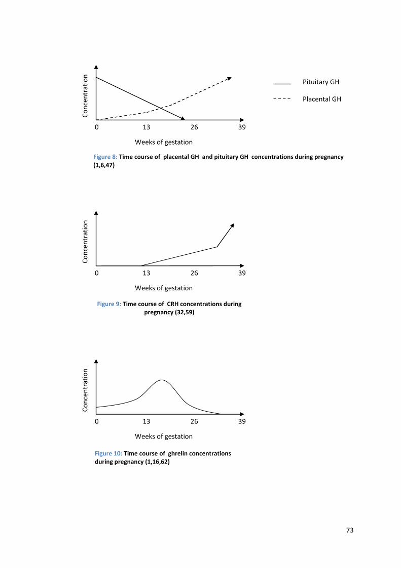
73
Figure 8: Time course of placental GH and pituitary GH concentrations during pregnancy (1,6,47)
Figure 9: Time course of CRH concentrations during pregnancy (32,59)
Figure 10: Time course of ghrelin concentrations during pregnancy (1,16,62)
Weeks of gestation
0 13 26 39
Pituitary GH
Placental GH
Weeks of gestation
0 13 26 39
Weeks of gestation
0 13 26 39
Co
nce
ntr
atio
n
Co
nce
ntr
atio
n
Co
nce
ntr
atio
n

74
Figure 11: Time course of placental leptin concentrations during pregnancy (7,24,36,40)
Figure 14: Time course of TNF-α and adiponectin concentrations during pregnancy (1,6)
Weeks of gestation
0 13 26 39
Weeks of gestation
0 13 26 39
TNF-α
Adiponectin
Co
nce
ntr
atio
n
Co
nce
ntr
atio
n

75
Figure 12: Factors leading to the development of leptin resistance in mid- to late pregnancy (4,5,65)
Figure 13: Dysregulation of the adipo-insular axis and pathogenesis of type 2 diabetes. Adapted from Seufert (65)
Changes in
estradiol secretion
Prolactin
Progesterone
hPL
Changes in
feeding behavior
- down-regulation of
leptin receptors
- impaired leptin
signaling at the leptin
receptor
- decreased transport of
leptin across the blood-
brain barrier
- increased binding of
leptin to soluble
receptors
Leptin resistance
- loss of satiety
signals
- hyperphagia and
weight gain
- β- cell
dysfunction
- hyperinsulinemia

76
Hormone Transfer into the fetal circulation
Functions in the fetus
Fetal production
Progesterone Direct secretion by the placenta
Yes
hCG Regulation of 11β-HSD, steroidogenesis
hPL Direct secretion by the placenta
Synthesis of IGF, insulin, adrenocortical hormones, surfactant Regulation of metabolism, angiogenesis
PGH No Regulation of fetal growth?
No
CRH Direct secretion by the placenta
Stimulation of adrenal and pituitary glands
Ghrelin Yes?
Leptin Direct secretion by the placenta
Vasculogenesis, erythropoiesis, lymphopoiesis, regulation of fat stores
Yes
TNF-α Direct secretion by the placenta
Adiponectin Yes
Visfatin Diffusion
Resistin Yes
Apelin Diffusion Promotion of fetal growth, possibly angiogenesis
Table 7: Placental hormones and their functions in the fetus (6,9,14,16,33, 38,42,43,60,66,70,103,109,112). A question mark represents unclear or conflicting data while a blank space indicates a lack of data on the topic.
Related Documents


