REVIEW published: 17 August 2018 doi: 10.3389/fphys.2018.01091 Frontiers in Physiology | www.frontiersin.org 1 August 2018 | Volume 9 | Article 1091 Edited by: Emilio A. Herrera, Universidad de Chile, Chile Reviewed by: Carina Mallard, University of Gothenburg, Sweden Charles Andrew Ducsay, Loma Linda University School of Medicine, United States Paola Casanello, Pontificia Universidad Católica de Chile, Chile *Correspondence: Amanda N. Sferruzzi-Perri [email protected] † These authors have contributed equally to this work Specialty section: This article was submitted to Embryonic and Developmental Physiology, a section of the journal Frontiers in Physiology Received: 20 April 2018 Accepted: 23 July 2018 Published: 17 August 2018 Citation: Napso T, Yong HEJ, Lopez-Tello J and Sferruzzi-Perri AN (2018) The Role of Placental Hormones in Mediating Maternal Adaptations to Support Pregnancy and Lactation. Front. Physiol. 9:1091. doi: 10.3389/fphys.2018.01091 The Role of Placental Hormones in Mediating Maternal Adaptations to Support Pregnancy and Lactation Tina Napso † , Hannah E. J. Yong † , Jorge Lopez-Tello and Amanda N. Sferruzzi-Perri* Department of Physiology, Development and Neuroscience, Centre for Trophoblast Research, University of Cambridge, Cambridge, United Kingdom During pregnancy, the mother must adapt her body systems to support nutrient and oxygen supply for growth of the baby in utero and during the subsequent lactation. These include changes in the cardiovascular, pulmonary, immune and metabolic systems of the mother. Failure to appropriately adjust maternal physiology to the pregnant state may result in pregnancy complications, including gestational diabetes and abnormal birth weight, which can further lead to a range of medically significant complications for the mother and baby. The placenta, which forms the functional interface separating the maternal and fetal circulations, is important for mediating adaptations in maternal physiology. It secretes a plethora of hormones into the maternal circulation which modulate her physiology and transfers the oxygen and nutrients available to the fetus for growth. Among these placental hormones, the prolactin-growth hormone family, steroids and neuropeptides play critical roles in driving maternal physiological adaptations during pregnancy. This review examines the changes that occur in maternal physiology in response to pregnancy and the significance of placental hormone production in mediating such changes. Keywords: pregnancy, placenta, hormones, maternal adaptations, metabolism, fetal growth, endocrine, cardiovascular INTRODUCTION Pregnancy is a dynamic and precisely coordinated process involving systemic and local changes in the mother that support the supply of nutrients and oxygen to the baby for growth in utero and in the subsequent lactation. Inappropriate adaptation of maternal physiology may lead to complications of pregnancy, such as gestational diabetes, preeclampsia, fetal growth restriction, fetal overgrowth and pre-term birth; which can have immediate consequences for fetal and maternal health. Furthermore, these pregnancy complications can also lead to long-term health consequences for the mother and infant. Altered fetal growth is associated with an increased risk of the offspring developing obesity, type-2 diabetes and cardiovascular disease in adulthood (Hales and Barker, 2001; Barker, 2004; Fowden et al., 2006). Moreover, women who develop gestational diabetes or preeclampsia are more likely to develop type-2 diabetes or cardiovascular disease in later life (Kim et al., 2002; Petry et al., 2007). Maternal adaptations to pregnancy are largely mediated by the placenta; the functional interface between the mother and fetus that secretes hormones and growth factors into the mother with physiological effects. This review aims to provide an overview of the physiological changes that occur in the mother in response to pregnancy and to discuss the role of key placental hormones in mediating such adaptations. In particular, this review focuses

Welcome message from author
This document is posted to help you gain knowledge. Please leave a comment to let me know what you think about it! Share it to your friends and learn new things together.
Transcript
REVIEWpublished: 17 August 2018
doi: 10.3389/fphys.2018.01091
Frontiers in Physiology | www.frontiersin.org 1 August 2018 | Volume 9 | Article 1091
Edited by:
Emilio A. Herrera,
Universidad de Chile, Chile
Reviewed by:
Carina Mallard,
University of Gothenburg, Sweden
Charles Andrew Ducsay,
Loma Linda University School of
Medicine, United States
Paola Casanello,
Pontificia Universidad Católica de
Chile, Chile
*Correspondence:
Amanda N. Sferruzzi-Perri
†These authors have contributed
equally to this work
Specialty section:
This article was submitted to
Embryonic and Developmental
Physiology,
a section of the journal
Frontiers in Physiology
Received: 20 April 2018
Accepted: 23 July 2018
Published: 17 August 2018
Citation:
Napso T, Yong HEJ, Lopez-Tello J and
Sferruzzi-Perri AN (2018) The Role of
Placental Hormones in Mediating
Maternal Adaptations to Support
Pregnancy and Lactation.
Front. Physiol. 9:1091.
doi: 10.3389/fphys.2018.01091
The Role of Placental Hormones inMediating Maternal Adaptations toSupport Pregnancy and Lactation
Tina Napso †, Hannah E. J. Yong †, Jorge Lopez-Tello and Amanda N. Sferruzzi-Perri*
Department of Physiology, Development and Neuroscience, Centre for Trophoblast Research, University of Cambridge,
Cambridge, United Kingdom
During pregnancy, the mother must adapt her body systems to support nutrient and
oxygen supply for growth of the baby in utero and during the subsequent lactation.
These include changes in the cardiovascular, pulmonary, immune and metabolic systems
of the mother. Failure to appropriately adjust maternal physiology to the pregnant state
may result in pregnancy complications, including gestational diabetes and abnormal
birth weight, which can further lead to a range of medically significant complications
for the mother and baby. The placenta, which forms the functional interface separating
the maternal and fetal circulations, is important for mediating adaptations in maternal
physiology. It secretes a plethora of hormones into the maternal circulation which
modulate her physiology and transfers the oxygen and nutrients available to the fetus
for growth. Among these placental hormones, the prolactin-growth hormone family,
steroids and neuropeptides play critical roles in drivingmaternal physiological adaptations
during pregnancy. This review examines the changes that occur in maternal physiology
in response to pregnancy and the significance of placental hormone production
in mediating such changes.
Keywords: pregnancy, placenta, hormones, maternal adaptations, metabolism, fetal growth, endocrine,
cardiovascular
INTRODUCTION
Pregnancy is a dynamic and precisely coordinated process involving systemic and local changesin the mother that support the supply of nutrients and oxygen to the baby for growth in uteroand in the subsequent lactation. Inappropriate adaptation of maternal physiology may lead tocomplications of pregnancy, such as gestational diabetes, preeclampsia, fetal growth restriction,fetal overgrowth and pre-term birth; which can have immediate consequences for fetal andmaternal health. Furthermore, these pregnancy complications can also lead to long-term healthconsequences for the mother and infant. Altered fetal growth is associated with an increased riskof the offspring developing obesity, type-2 diabetes and cardiovascular disease in adulthood (Halesand Barker, 2001; Barker, 2004; Fowden et al., 2006). Moreover, women who develop gestationaldiabetes or preeclampsia aremore likely to develop type-2 diabetes or cardiovascular disease in laterlife (Kim et al., 2002; Petry et al., 2007). Maternal adaptations to pregnancy are largely mediated bythe placenta; the functional interface between the mother and fetus that secretes hormones andgrowth factors into the mother with physiological effects. This review aims to provide an overviewof the physiological changes that occur in the mother in response to pregnancy and to discuss therole of key placental hormones in mediating such adaptations. In particular, this review focuses
Napso et al. Placental Hormones and Maternal Adaptations
on the importance of the prolactin-growth hormone family(e.g., prolactin, placental lactogen and growth hormone), steroids(estrogens and progesterone) and neuropeptides (serotonin,melatonin and oxytocin) in adaptations of maternal physiologyduring pregnancy. Where possible, this review draws uponfindings in women and animal models, including rodentsand sheep. However, differences exist between species in thespecific hormones produced by the placenta, the access ofthese hormones to the maternal circulation, and the relativeproportion of conceptus mass to maternal size (hence constrainton the mother to provide resources for fetal growth; Haig, 2008;Carter, 2012; Fowden and Moore, 2012). Where such differencesbetween species exist, these have been highlighted and discussedas necessary in the relevant sections. Nevertheless, althoughsome effects described may not be applicable to all species, thedifferent animal models of pregnancy still provide novel insightinto the fundamental mechanisms of maternal adaptation duringgestation.
ADAPTATIONS IN MATERNALPHYSIOLOGY DURING PREGNANCY ANDLACTATION
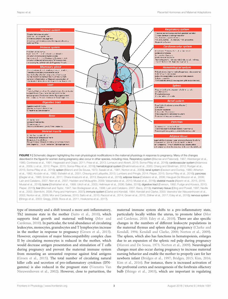
Most tissues and organs in the mother respond to the pregnantstate. Changes include alterations in size, morphology, functionand responsiveness of tissues and organs to hormonal andmetabolic cues. These changes arise in the cardiovascular,pulmonary, immune, and metabolic systems of the mother(Figure 1). Some of these changes are seen from very earlyin pregnancy, prior to the establishment of a fully functionalplacenta, highlighting that non-placental factors may also beimportant (Paller et al., 1989; Drynda et al., 2015). The specificnature of changes in maternal physiology depends on the stageof the pregnancy and appears to track with alterations in themetabolic requirements of the mother versus the developingfetus.
Alterations in the maternal cardiovascular system begin veryearly in gestation (Chapman et al., 1998) and ultimately lead tosystemic vasodilation and increased blood perfusion of maternalorgans, including the gravid uterus. Systemic vascular resistanceis reduced by 25–30% and accompanied by a 40% increase incardiac output during human pregnancy; while in mice, bloodpressure decreases by 15% and cardiac output is increased by48% (Bader et al., 1955; Kulandavelu et al., 2006; Soma-Pillayet al., 2016). Renal blood flow and glomerular filtration rates arealso increased (Davison and Dunlop, 1980; Soma-Pillay et al.,2016). The renin-angiotensin-aldosterone system (RAAS) whichis a major determinant for sodium balance during gestation, isprogressively upregulated toward term with associated plasmavolume expansion (Elsheikh et al., 2001; Tkachenko et al.,2014). This rise in blood volume, which is required to copewith the oxygen requirements of the maternal organs and theconceptus growth, plateaus by the late gestation, resulting inan increase in total blood volume by approximately 30% at theend of pregnancy (Chang and Streitman, 2012). There is also anincrease in the numbers of red blood cells in the mother during
pregnancy, due to proliferation of erythroid progenitors in thespleen (Bustamante et al., 2008). Pulmonary function is alsoaltered and encompasses changes in ventilation rates and bloodgases. For instance, lung tidal volume and minute ventilationincreases by 30–50% (Hegewald and Crapo, 2011). As a resultof increased oxygen consumption during hyperventilation, thereis greater carbon dioxide production, which leads to chronicrespiratory alkalosis that is compensated by an increased renalexcretion of bicarbonate (Weinberger et al., 1980). Overall,these adaptations ensure the well-being of the mother, whilealso providing an adequate blood flow to the placenta for fetalnutrition, oxygenation and maturation.
There are also alterations inmaternalmetabolic and endocrinestate during gestation. In early pregnancy, the maternalpancreatic β-cell mass expands due to both hyperplasia andhypertrophy of islets, which for example in rats, results ina >50% increase (Ackermann and Gannon, 2007; Rieck andKaestner, 2010). The threshold for glucose-stimulated insulinproduction is also lowered and maternal circulating insulinconcentration is greater compared to the non-pregnant state. Inearly pregnancy, when fetal demands are relatively low, wholebody maternal insulin sensitivity is unchanged or increasedand there is accumulation of energy reserves in the mother.In particular, early pregnancy is associated with adipocytehypertrophy, increased lipogenesis and lipid storage and relatesto improved insulin sensitivity of white adipose tissue in themother (Hadden and Mclaughlin, 2009; Mcilvride et al., 2017).Interestingly, in pregnant mice, brown adipose stores of thedam also switch to a white adipose tissue-like phenotype inearly gestation (Mcilvride et al., 2017). Additionally, glycogenaccumulates in the liver, which also increases in size from earlygestation (Bustamante et al., 2010). In contrast, late pregnancy isassociated with diminishedmaternal tissue insulin sensitivity anda concomitant increase in lipolysis and hepatic gluconeogenesis(Freemark et al., 2002; Lain and Catalano, 2007; Musial et al.,2016). Despite the pregnancy-related rise in leptin and insulinconcentrations, maternal appetite increases in pregnancy (Villaret al., 1992; Douglas et al., 2007; Hadden and Mclaughlin, 2009;Díaz et al., 2014). Together, these metabolic and endocrinealterations increase lipid and glucose availability for the rapidlygrowing fetus in late gestation. Intriguingly in rodents, wholebody responsiveness to insulin starts to improve near term,which may be important for conserving nutrients for maternaluse, as parturition and lactation approach (Musial et al., 2016).There are also notable changes in maternal bone metabolismduring pregnancy. In particular, intestinal calcium absorptionis enhanced in the mother during pregnancy via upregulationof 1,25-dihydroxyvitamin D levels, improved renal conservationand increased calcium mobilization from the maternal skeleton(Hellmeyer et al., 2006). These processes support the supply ofcalcium for the formation, growth and mineralization of the fetalskeleton (King, 2000; Kalkwarf and Specker, 2002).
The immune system of the mother during pregnancy is tightlyregulated to prevent an unwanted immune response against thepaternal antigens present in the developing conceptus (Racicotet al., 2014; Groen et al., 2015; Zöllner et al., 2017). As gestationprogresses, there is suppression of the pro-inflammatory Th1
Frontiers in Physiology | www.frontiersin.org 2 August 2018 | Volume 9 | Article 1091
Napso et al. Placental Hormones and Maternal Adaptations
FIGURE 1 | Schematic diagram highlighting the main physiological modifications in the maternal physiology in response to pregnancy. Many of the changes
described in the figure for women during pregnancy also occur in other species, including mice. Respiratory system (Macrae and Palavradji, 1967; Weinberger et al.,
1980; Contreras et al., 1991; Hegewald and Crapo, 2011; Frise et al., 2013; Lomauro and Aliverti, 2015; Soma-Pillay et al., 2016); cardiovascular system (Adamova
et al., 2009; Li et al., 2012; Pieper, 2015; Soma-Pillay et al., 2016); hematological system (Shakhmatova et al., 2000; Chang and Streitman, 2012; Rodger et al.,
2015; Soma-Pillay et al., 2016); spleen (Maroni and De Sousa, 1973; Sasaki et al., 1981; Norton et al., 2009); renal system (Davison and Dunlop, 1980; Atherton
et al., 1982; Krutzén et al., 1992; Elsheikh et al., 2001; Cheung and Lafayette, 2013; Lumbers and Pringle, 2014; Pieper, 2015; Soma-Pillay et al., 2016); pancreas
(Ziegler et al., 1985; Ernst et al., 2011; Ohara-Imaizumi et al., 2013; Baeyens et al., 2016); adipose tissue (Catalano et al., 2006; Hauguel-De Mouzon et al., 2006;
Lain and Catalano, 2007; Nien et al., 2007; Hadden and Mclaughlin, 2009; Valsamakis et al., 2010; Musial et al., 2016); skeletal muscle (Alperin et al., 2015, 2016;
Musial et al., 2016); bone (Shahtaheri et al., 1999; Ulrich et al., 2003; Hellmeyer et al., 2006; Salles, 2016); digestive tract (Everson, 1992; Fudge and Kovacs, 2010;
Pieper, 2015); liver (Munnell and Taylor, 1947; Van Bodegraven et al., 1998; Lain and Catalano, 2007; Bacq, 2013); mammary tissue (Elling and Powell, 1997; Neville
et al., 2002; Sternlicht, 2006; Pang and Hartmann, 2007); immune system (Clarke and Kendall, 1994; Kendall and Clarke, 2000; Veenstra Van Nieuwenhoven et al.,
2002; Norton et al., 2009; Mor and Cardenas, 2010; Saito et al., 2010; Racicot et al., 2014; Groen et al., 2015; Zöllner et al., 2017; Edey et al., 2018); nervous system
(Shingo et al., 2003; Gregg, 2009; Roos et al., 2011; Hoekzema et al., 2017).
type of immunity and a shift toward a more anti-inflammatory,Th2 immune state in the mother (Saito et al., 2010), whichsupports fetal growth and maternal well-being (Mor andCardenas, 2010). In particular, the total abundance of circulatingleukocytes, monocytes, granulocytes and T lymphocytes increasein the mother in response to pregnancy (Groen et al., 2015).However, expression of major histocompatibility complex classII by circulating monocytes is reduced in the mother, whichwould decrease antigen presentation and stimulation of T cellsduring pregnancy and prevent the maternal immune systemfrom mounting an unwanted response against fetal antigens(Groen et al., 2015). The total number of circulating naturalkiller cells and secretion of pro-inflammatory cytokines (IFN-gamma) is also reduced in the pregnant state (Veenstra VanNieuwenhoven et al., 2002). However, close to parturition, the
maternal immune system shifts to a pro-inflammatory state,particularly locally within the uterus, to promote labor (Morand Cardenas, 2010; Edey et al., 2018). There are also specificchanges in the numbers of different leukocyte populations inthe maternal thymus and spleen during pregnancy (Clarke andKendall, 1994; Kendall and Clarke, 2000; Norton et al., 2009).The spleen, which also has functions in hematopoiesis, enlargesdue to an expansion of the splenic red pulp during pregnancy(Maroni and De Sousa, 1973; Norton et al., 2009). Neurologicalchanges must also occur during pregnancy to increase maternalnursing behavior and enable the mother to properly care for hernewborn infant (Bridges et al., 1997; Bridges, 2015; Kim, 2016;Kim et al., 2016). For instance, there is increased activation ofthe prefrontal cortex and neurogenesis of the forebrain olfactorybulb (Shingo et al., 2003), which are important in regulating
Frontiers in Physiology | www.frontiersin.org 3 August 2018 | Volume 9 | Article 1091
Napso et al. Placental Hormones and Maternal Adaptations
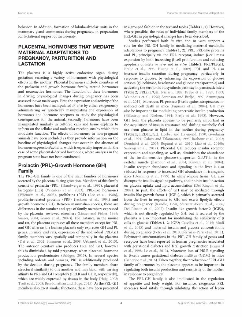
behavior. In addition, formation of lobulo-alveolar units in themammary gland commences during pregnancy, in preparationfor lactational support of the neonate.
PLACENTAL HORMONES THAT MEDIATEMATERNAL ADAPTATIONS TOPREGNANCY, PARTURITION ANDLACTATION
The placenta is a highly active endocrine organ duringgestation; secreting a variety of hormones with physiologicaleffects in the mother. Placental hormones include members ofthe prolactin and growth hormone family, steroid hormonesand neuroactive hormones. The function of these hormonesin driving physiological changes during pregnancy has beenassessed in twomain ways. First, the expression and activity of thehormones have been manipulated in vivo by either exogenouslyadministering or genetically manipulating the expression ofhormones and hormone receptors to study the physiologicalconsequences for the animal. Secondly, hormones have beenmanipulated similarly in cultured cells and tissue explants toinform on the cellular and molecular mechanisms by which theymodulate function. The effects of hormones in non-pregnantanimals have been included as they provide information on thebaseline of physiological changes that occur in the absence ofhormone expression/activity, which is especially important in thecase of some placental-derived hormones, where analyses in thepregnant state have not been conducted.
Prolactin (PRL)-Growth Hormone (GH)FamilyThe PRL-GH family is one of the main families of hormonessecreted by the placenta during gestation. Members of this familyconsist of prolactin (PRL) (Handwerger et al., 1992), placentallactogens (PLs) (Wiemers et al., 2003), PRL-like hormones(Wiemers et al., 2003), proliferins (PLF) (Lee et al., 1988),proliferin-related proteins (PRP) (Jackson et al., 1994) andgrowth hormone (GH). Between mammalian species, there aredifferences in the number and type of family members expressedby the placenta [reviewed elsewhere (Linzer and Fisher, 1999;Soares, 2004; Soares et al., 2007)]. For instance, in the mouseand rat, the placenta expresses all these members except for PRLand GH whereas the human placenta only expresses GH and PLgenes. In mice and rats, expression of the individual PRL-GHfamily members vary spatially and temporally in the placenta(Dai et al., 2002; Simmons et al., 2008; Urbanek et al., 2015).The anterior pituitary also produces PRL and GH; howeverthis is diminished by mid-pregnancy, when placental hormoneproduction predominates (Bridges, 2015). In several speciesincluding rodents and humans, PRL is additionally producedby the decidua during pregnancy. The family members sharestructural similarity to one another and may bind, with varyingaffinity to PRL and GH receptors (PRLR and GHR, respectively),which are widely expressed by tissues in the body (Haig, 2008;Trott et al., 2008; Ben-Jonathan andHugo, 2015). As the PRL-GHmembers also exert similar functions, these have been presented
in a grouped fashion in the text and tables (Tables 1, 2). However,where possible, the roles of individual family members of thePRL-GH in physiological changes have been described.
Studies performed both in vivo and in vitro support arole for the PRL-GH family in mediating maternal metabolicadaptations to pregnancy (Tables 1, 2). PRL, PRL-like proteinsand PL, principally via the PRL receptor, induce β-cell massexpansion by both increasing β-cell proliferation and reducingapoptosis of islets in vivo and in vitro (Table 2; PRL/PL/GH;Brelje et al., 1993; Huang et al., 2009). PRL and PL alsoincrease insulin secretion during pregnancy, particularly inresponse to glucose, by enhancing the expression of glucosesensors (glucokinase, hexokinase and glucose transporter-2) andactivating the serotonin biosynthesis pathway in pancreatic islets(Table 2; PRL/PL/GH; Nielsen, 1982; Brelje et al., 1989, 1993;Weinhaus et al., 1996; Sorenson and Brelje, 1997; Arumugamet al., 2014). Moreover, PL protects β-cells against streptozotocin-induced cell death in mice (Fujinaka et al., 2004). GH mayalso be important for modulating pancreatic insulin production(Billestrup and Nielsen, 1991; Brelje et al., 1993). However,GH from the placenta appears to be primarily important inthe acquisition of insulin resistance and shifting metabolic fueluse from glucose to lipid in the mother during pregnancy(Table 1; PRL/PL/GH; Horber and Haymond, 1990; Goodmanet al., 1991; Galosy and Talamantes, 1995; Barbour et al., 2002;Dominici et al., 2005; Boparai et al., 2010; Liao et al., 2016b;Sairenji et al., 2017). Placental GH reduces insulin receptorexpression and signaling, as well as, diminishes the abundanceof the insulin-sensitive glucose-transporter, GLUT-4, in theskeletal muscle (Barbour et al., 2004; Kirwan et al., 2004).Insulin receptor abundance and signaling in the liver is alsoreduced in response to increased GH abundance in transgenicmice (Dominici et al., 1999). In white adipose tissue, GH alsodisrupts the insulin signaling pathway, and inhibits insulin actionon glucose uptake and lipid accumulation (Del Rincon et al.,2007). In part, the effects of GH may be mediated throughinsulin-like growth factor-1 (IGF1), which is primarily secretedfrom the liver in response to GH and exerts lipolytic effectsduring pregnancy (Randle, 1998; Sferruzzi-Perri et al., 2006;Del Rincon et al., 2007). Insulin-like growth factor-2 (IGF2),which is not directly regulated by GH, but is secreted by theplacenta is also important for modulating the sensitivity of β
cells to glucose (Tables 1, 2; IGF2; Casellas et al., 2015; Modiet al., 2015) and maternal insulin and glucose concentrationsduring pregnancy (Petry et al., 2010; Sferruzzi-Perri et al., 2011).Polymorphisms/mutations in the PRL-GH family of genes andreceptors have been reported in human pregnancies associatedwith gestational diabetes and fetal growth restriction (Rygaardet al., 1998; Le et al., 2013). Moreover, loss of PRLR signalingin β-cells causes gestational diabetes mellitus (GDM) in mice(Banerjee et al., 2016). Taken together, the production of PRL-GHfamily of hormones by the placenta appears to be important inregulating both insulin production and sensitivity of the motherin response to pregnancy.
The PRL-GH family is also implicated in the regulationof appetite and body weight. For instance, exogenous PRLincreases food intake through inhibiting the action of leptin
Frontiers in Physiology | www.frontiersin.org 4 August 2018 | Volume 9 | Article 1091
Napso et al. Placental Hormones and Maternal Adaptations
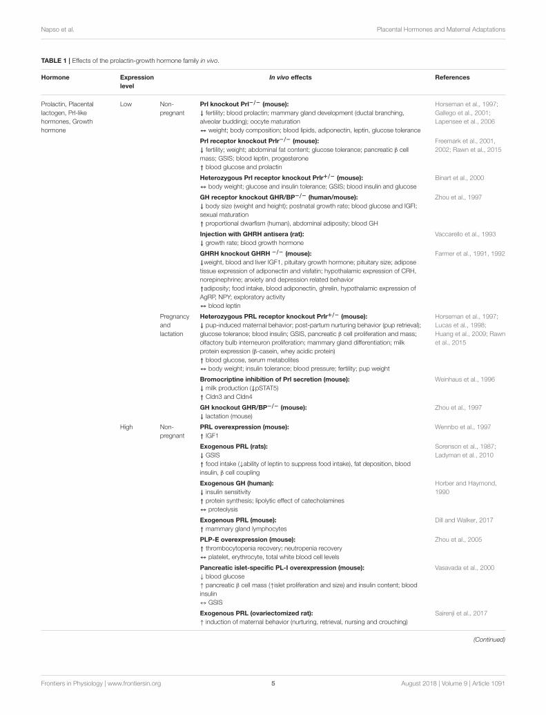
TABLE 1 | Effects of the prolactin-growth hormone family in vivo.
Hormone Expression
level
In vivo effects References
Prolactin, Placental
lactogen, Prl-like
hormones, Growth
hormone
Low Non-
pregnant
Prl knockout Prl−/− (mouse):
↓ fertility; blood prolactin; mammary gland development (ductal branching,
alveolar budding); oocyte maturation
↔ weight; body composition; blood lipids, adiponectin, leptin, glucose tolerance
Horseman et al., 1997;
Gallego et al., 2001;
Lapensee et al., 2006
Prl receptor knockout Prlr−/− (mouse):
↓ fertility; weight; abdominal fat content; glucose tolerance; pancreatic β cell
mass; GSIS; blood leptin, progesterone
↑ blood glucose and prolactin
Freemark et al., 2001,
2002; Rawn et al., 2015
Heterozygous Prl receptor knockout Prlr+/− (mouse):
↔ body weight; glucose and insulin tolerance; GSIS; blood insulin and glucose
Binart et al., 2000
GH receptor knockout GHR/BP−/− (human/mouse):
↓ body size (weight and height); postnatal growth rate; blood glucose and IGFI;
sexual maturation
↑ proportional dwarfism (human), abdominal adiposity; blood GH
Zhou et al., 1997
Injection with GHRH antisera (rat):
↓ growth rate; blood growth hormone
Vaccarello et al., 1993
GHRH knockout GHRH −/− (mouse):
↓weight, blood and liver IGF1, pituitary growth hormone; pituitary size; adipose
tissue expression of adiponectin and visfatin; hypothalamic expression of CRH,
norepinephrine; anxiety and depression related behavior
↑adiposity; food intake, blood adiponectin, ghrelin, hypothalamic expression of
AgRP, NPY; exploratory activity
↔ blood leptin
Farmer et al., 1991, 1992
Pregnancy
and
lactation
Heterozygous PRL receptor knockout Prlr+/− (mouse):
↓ pup-induced maternal behavior; post-partum nurturing behavior (pup retrieval);
glucose tolerance; blood insulin; GSIS, pancreatic β cell proliferation and mass;
olfactory bulb interneuron proliferation; mammary gland differentiation; milk
protein expression (β-casein, whey acidic protein)
↑ blood glucose, serum metabolites
↔ body weight; insulin tolerance; blood pressure; fertility; pup weight
Horseman et al., 1997;
Lucas et al., 1998;
Huang et al., 2009; Rawn
et al., 2015
Bromocriptine inhibition of Prl secretion (mouse):
↓ milk production (↓pSTAT5)
↑ Cldn3 and Cldn4
Weinhaus et al., 1996
GH knockout GHR/BP−/− (mouse):
↓ lactation (mouse)
Zhou et al., 1997
High Non-
pregnant
PRL overexpression (mouse):
↑ IGF1
Wennbo et al., 1997
Exogenous PRL (rats):
↓ GSIS
↑ food intake (↓ability of leptin to suppress food intake), fat deposition, blood
insulin, β cell coupling
Sorenson et al., 1987;
Ladyman et al., 2010
Exogenous GH (human):
↓ insulin sensitivity
↑ protein synthesis; lipolytic effect of catecholamines
↔ proteolysis
Horber and Haymond,
1990
Exogenous PRL (mouse):
↑ mammary gland lymphocytes
Dill and Walker, 2017
PLP-E overexpression (mouse):
↑ thrombocytopenia recovery; neutropenia recovery
↔ platelet, erythrocyte, total white blood cell levels
Zhou et al., 2005
Pancreatic islet-specific PL-I overexpression (mouse):
↓ blood glucose
↑ pancreatic β cell mass (↑islet proliferation and size) and insulin content; blood
insulin
↔ GSIS
Vasavada et al., 2000
Exogenous PRL (ovariectomized rat):
↑ induction of maternal behavior (nurturing, retrieval, nursing and crouching)
Sairenji et al., 2017
(Continued)
Frontiers in Physiology | www.frontiersin.org 5 August 2018 | Volume 9 | Article 1091
Napso et al. Placental Hormones and Maternal Adaptations
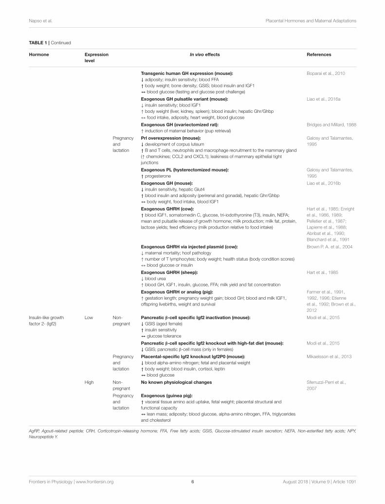
TABLE 1 | Continued
Hormone Expression
level
In vivo effects References
Transgenic human GH expression (mouse):
↓ adiposity; insulin sensitivity; blood FFA
↑ body weight; bone density; GSIS; blood insulin and IGF1
↔ blood glucose (fasting and glucose post challenge)
Boparai et al., 2010
Exogenous GH pulsatile variant (mouse):
↓ insulin sensitivity; blood IGF1
↑ body weight (liver, kidney, spleen); blood insulin; hepatic Ghr/Ghbp
↔ food intake, adiposity, heart weight, blood glucose
Liao et al., 2016a
Exogenous GH (ovariectomized rat):
↑ induction of maternal behavior (pup retrieval)
Bridges and Millard, 1988
Pregnancy
and
lactation
Prl overexpression (mouse):
↓ development of corpus luteum
↑ B and T cells, neutrophils and macrophage recruitment to the mammary gland
(↑ chemokines; CCL2 and CXCL1); leakiness of mammary epithelial tight
junctions
Galosy and Talamantes,
1995
Exogenous PL (hysterectomized mouse):
↑ progesterone
Galosy and Talamantes,
1995
Exogenous GH (mouse):
↓ insulin sensitivity, hepatic Glut4
↑ blood insulin and adiposity (perirenal and gonadal), hepatic Ghr/Ghbp
↔ body weight, food intake, blood IGF1
Liao et al., 2016b
Exogenous GHRH (cow):
↑ blood IGF1, somatomedin C, glucose, tri-iodothyronine (T3), insulin, NEFA;
mean and pulsatile release of growth hormone; milk production; milk fat, protein,
lactose yields; feed efficiency (milk production relative to food intake)
Hart et al., 1985; Enright
et al., 1986, 1989;
Pelletier et al., 1987;
Lapierre et al., 1988;
Abribat et al., 1990;
Blanchard et al., 1991
Exogenous GHRH via injected plasmid (cow):
↓ maternal mortality; hoof pathology
↑ number of T lymphocytes; body weight; health status (body condition scores)
↔ blood glucose or insulin
Brown P. A. et al., 2004
Exogenous GHRH (sheep):
↓ blood urea
↑ blood GH, IGF1, insulin, glucose, FFA; milk yield and fat concentration
Hart et al., 1985
Exogenous GHRH or analog (pig):
↑ gestation length; pregnancy weight gain; blood GH; blood and milk IGF1,
offspring livebirths, weight and survival
Farmer et al., 1991,
1992, 1996; Etienne
et al., 1992; Brown et al.,
2012
Insulin-like growth
factor 2- (Igf2)
Low Non-
pregnant
Pancreatic β-cell specific Igf2 inactivation (mouse):
↓ GSIS (aged female)
↑ insulin sensitivity
↔ glucose tolerance
Modi et al., 2015
Pancreatic β-cell specific Igf2 knockout with high-fat diet (mouse):
↓ GSIS; pancreatic β-cell mass (only in females)
Modi et al., 2015
Pregnancy
and
lactation
Placental-specific Igf2 knockout Igf2P0 (mouse):
↓ blood alpha-amino nitrogen; fetal and placental weight
↑ body weight; blood insulin, cortisol, leptin
↔ blood glucose
Mikaelsson et al., 2013
High Non-
pregnant
No known physiological changes Sferruzzi-Perri et al.,
2007
Pregnancy
and
lactation
Exogenous (guinea pig):
↑ visceral tissue amino acid uptake, fetal weight; placental structural and
functional capacity
↔ lean mass; adiposity; blood glucose, alpha-amino nitrogen, FFA, triglycerides
and cholesterol
AgRP, Agouti-related peptide; CRH, Corticotropin-releasing hormone; FFA, Free fatty acids; GSIS, Glucose-stimulated insulin secretion; NEFA, Non-esterified fatty acids; NPY,
Neuropeptide Y.
Frontiers in Physiology | www.frontiersin.org 6 August 2018 | Volume 9 | Article 1091
Napso et al. Placental Hormones and Maternal Adaptations
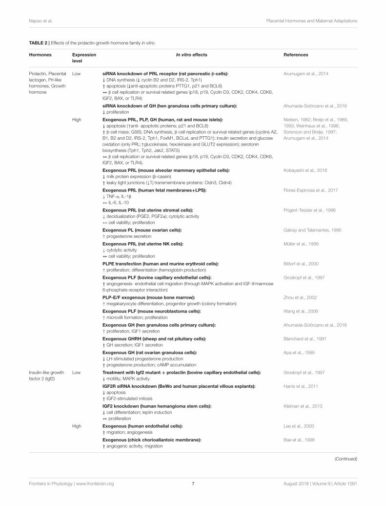
TABLE 2 | Effects of the prolactin-growth hormone family in vitro.
Hormones Expression
level
In vitro effects References
Prolactin, Placental
lactogen, Prl-like
hormones, Growth
hormone
Low siRNA knockdown of PRL receptor (rat pancreatic β-cells):
↓ DNA synthesis (↓ cyclin B2 and D2, IRS-2, Tph1)
↑ apoptosis (↓anti-apoptotic proteins PTTG1, p21 and BCL6)
↔ β cell replication or survival related genes (p18, p19, Cyclin D3, CDK2, CDK4, CDK6,
IGF2, BAX, or TLR4)
Arumugam et al., 2014
siRNA knockdown of GH (hen granulosa cells primary culture):
↓ proliferation
Ahumada-Solórzano et al., 2016
High Exogenous PRL, PLP, GH (human, rat and mouse islets):
↓ apoptosis (↑anti- apoptotic proteins; p21 and BCL6)
↑ β-cell mass; GSIS; DNA synthesis, β-cell replication or survival related genes (cyclins A2,
B1, B2 and D2, IRS-2, Tph1, FoxM1, BCLxL and PTTG1); insulin secretion and glucose
oxidation (only PRL;↑glucokinase, hexokinase and GLUT2 expression); serotonin
biosynthesis (Tph1, Tph2, Jak2, STAT5)
↔ β cell replication or survival related genes (p18, p19, Cyclin D3, CDK2, CDK4, CDK6,
IGF2, BAX, or TLR4).
Nielsen, 1982; Brelje et al., 1989,
1993; Weinhaus et al., 1996;
Sorenson and Brelje, 1997;
Arumugam et al., 2014
Exogenous PRL (mouse alveolar mammary epithelial cells):
↓ milk protein expression (β-casein)
↑ leaky tight junctions (↓Tj transmembrane proteins: Cldn3, Cldn4)
Kobayashi et al., 2016
Exogenous PRL (human fetal membranes+LPS):
↓ TNF-α, IL-1β
↔ IL-6, IL-10
Flores-Espinosa et al., 2017
Exogenous PRL (rat uterine stromal cells):
↓ decidualization (PGE2, PGF2α); cytolytic activity
↔ cell viability; proliferation
Prigent-Tessier et al., 1996
Exogenous PL (mouse ovarian cells):
↑ progesterone secretion
Galosy and Talamantes, 1995
Exogenous PRL (rat uterine NK cells):
↓ cytolytic activity
↔ cell viability; proliferation
Müller et al., 1999
PLPE transfection (human and murine erythroid cells):
↑ proliferation; differentiation (hemoglobin production)
Bittorf et al., 2000
Exogenous PLF (bovine capillary endothelial cells):
↑ angiogenesis- endothelial cell migration (through MAPK activation and IGF-II/mannose
6-phosphate receptor interaction)
Groskopf et al., 1997
PLP-E/F exogenous (mouse bone marrow):
↑ megakaryocyte differentiation, progenitor growth (colony formation)
Zhou et al., 2002
Exogenous PLF (mouse neuroblastoma cells):
↑ microvilli formation; proliferation
Wang et al., 2006
Exogenous GH (hen granulosa cells primary culture):
↑ proliferation; IGF1 secretion
Ahumada-Solórzano et al., 2016
Exogenous GHRH (sheep and rat pituitary cells):
↑ GH secretion; IGF1 secretion
Blanchard et al., 1991
Exogenous GH (rat ovarian granulosa cells):
↓ LH-stimulated progesterone production
↑ progesterone production; cAMP accumulation
Apa et al., 1995
Insulin-like growth
factor 2 (Igf2)
Low Treatment with Igf2 mutant + prolactin (bovine capillary endothelial cells):
↓ motility; MAPK activity
Groskopf et al., 1997
IGF2R siRNA knockdown (BeWo and human placental villous explants):
↓ apoptosis
↑ IGF2-stimulated mitosis
Harris et al., 2011
IGF2 knockdown (human hemangioma stem cells):
↓ cell differentiation; leptin induction
↔ proliferation
Kleiman et al., 2013
High Exogenous (human endothelial cells):
↑ migration; angiogenesis
Lee et al., 2000
Exogenous (chick chorioallantoic membrane):
↑ angiogenic activity; migration
Bae et al., 1998
(Continued)
Frontiers in Physiology | www.frontiersin.org 7 August 2018 | Volume 9 | Article 1091
Napso et al. Placental Hormones and Maternal Adaptations
TABLE 2 | Continued
Hormones Expression
level
In vitro effects References
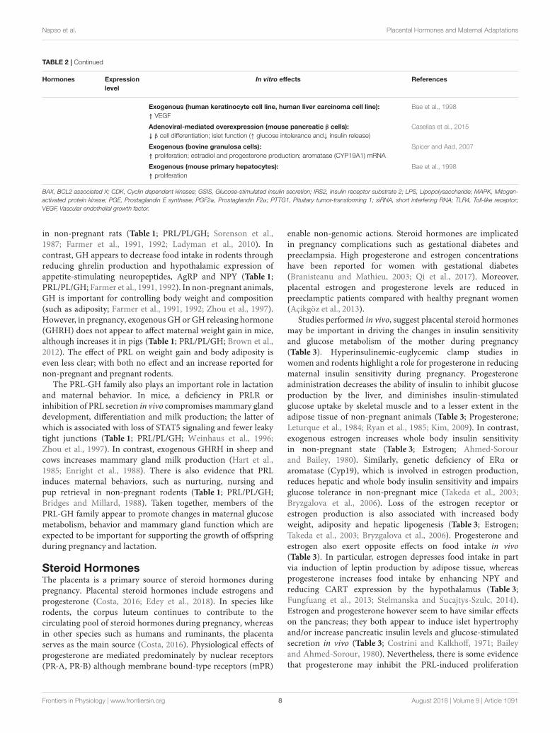
Exogenous (human keratinocyte cell line, human liver carcinoma cell line):
↑ VEGF
Bae et al., 1998
Adenoviral-mediated overexpression (mouse pancreatic β cells):
↓ β cell differentiation; islet function (↑ glucose intolerance and↓ insulin release)
Casellas et al., 2015
Exogenous (bovine granulosa cells):
↑ proliferation; estradiol and progesterone production; aromatase (CYP19A1) mRNA
Spicer and Aad, 2007
Exogenous (mouse primary hepatocytes):
↑ proliferation
Bae et al., 1998
BAX, BCL2 associated X; CDK, Cyclin dependent kinases; GSIS, Glucose-stimulated insulin secretion; IRS2, Insulin receptor substrate 2; LPS, Lipopolysaccharide; MAPK, Mitogen-
activated protein kinase; PGE, Prostaglandin E synthase; PGF2α, Prostaglandin F2α; PTTG1, Pituitary tumor-transforming 1; siRNA, short interfering RNA; TLR4, Toll-like receptor;
VEGF, Vascular endothelial growth factor.
in non-pregnant rats (Table 1; PRL/PL/GH; Sorenson et al.,1987; Farmer et al., 1991, 1992; Ladyman et al., 2010). Incontrast, GH appears to decrease food intake in rodents throughreducing ghrelin production and hypothalamic expression ofappetite-stimulating neuropeptides, AgRP and NPY (Table 1;PRL/PL/GH; Farmer et al., 1991, 1992). In non-pregnant animals,GH is important for controlling body weight and composition(such as adiposity; Farmer et al., 1991, 1992; Zhou et al., 1997).However, in pregnancy, exogenous GH or GH releasing hormone(GHRH) does not appear to affect maternal weight gain in mice,although increases it in pigs (Table 1; PRL/PL/GH; Brown et al.,2012). The effect of PRL on weight gain and body adiposity iseven less clear; with both no effect and an increase reported fornon-pregnant and pregnant rodents.
The PRL-GH family also plays an important role in lactationand maternal behavior. In mice, a deficiency in PRLR orinhibition of PRL secretion in vivo compromises mammary glanddevelopment, differentiation and milk production; the latter ofwhich is associated with loss of STAT5 signaling and fewer leakytight junctions (Table 1; PRL/PL/GH; Weinhaus et al., 1996;Zhou et al., 1997). In contrast, exogenous GHRH in sheep andcows increases mammary gland milk production (Hart et al.,1985; Enright et al., 1988). There is also evidence that PRLinduces maternal behaviors, such as nurturing, nursing andpup retrieval in non-pregnant rodents (Table 1; PRL/PL/GH;Bridges and Millard, 1988). Taken together, members of thePRL-GH family appear to promote changes in maternal glucosemetabolism, behavior and mammary gland function which areexpected to be important for supporting the growth of offspringduring pregnancy and lactation.
Steroid HormonesThe placenta is a primary source of steroid hormones duringpregnancy. Placental steroid hormones include estrogens andprogesterone (Costa, 2016; Edey et al., 2018). In species likerodents, the corpus luteum continues to contribute to thecirculating pool of steroid hormones during pregnancy, whereasin other species such as humans and ruminants, the placentaserves as the main source (Costa, 2016). Physiological effects ofprogesterone are mediated predominately by nuclear receptors(PR-A, PR-B) although membrane bound-type receptors (mPR)
enable non-genomic actions. Steroid hormones are implicatedin pregnancy complications such as gestational diabetes andpreeclampsia. High progesterone and estrogen concentrationshave been reported for women with gestational diabetes(Branisteanu and Mathieu, 2003; Qi et al., 2017). Moreover,placental estrogen and progesterone levels are reduced inpreeclamptic patients compared with healthy pregnant women(Açikgöz et al., 2013).
Studies performed in vivo, suggest placental steroid hormonesmay be important in driving the changes in insulin sensitivityand glucose metabolism of the mother during pregnancy(Table 3). Hyperinsulinemic-euglycemic clamp studies inwomen and rodents highlight a role for progesterone in reducingmaternal insulin sensitivity during pregnancy. Progesteroneadministration decreases the ability of insulin to inhibit glucoseproduction by the liver, and diminishes insulin-stimulatedglucose uptake by skeletal muscle and to a lesser extent in theadipose tissue of non-pregnant animals (Table 3; Progesterone;Leturque et al., 1984; Ryan et al., 1985; Kim, 2009). In contrast,exogenous estrogen increases whole body insulin sensitivityin non-pregnant state (Table 3; Estrogen; Ahmed-Sorourand Bailey, 1980). Similarly, genetic deficiency of ERα oraromatase (Cyp19), which is involved in estrogen production,reduces hepatic and whole body insulin sensitivity and impairsglucose tolerance in non-pregnant mice (Takeda et al., 2003;Bryzgalova et al., 2006). Loss of the estrogen receptor orestrogen production is also associated with increased bodyweight, adiposity and hepatic lipogenesis (Table 3; Estrogen;Takeda et al., 2003; Bryzgalova et al., 2006). Progesterone andestrogen also exert opposite effects on food intake in vivo(Table 3). In particular, estrogen depresses food intake in partvia induction of leptin production by adipose tissue, whereasprogesterone increases food intake by enhancing NPY andreducing CART expression by the hypothalamus (Table 3;Fungfuang et al., 2013; Stelmanska and Sucajtys-Szulc, 2014).Estrogen and progesterone however seem to have similar effectson the pancreas; they both appear to induce islet hypertrophyand/or increase pancreatic insulin levels and glucose-stimulatedsecretion in vivo (Table 3; Costrini and Kalkhoff, 1971; Baileyand Ahmed-Sorour, 1980). Nevertheless, there is some evidencethat progesterone may inhibit the PRL-induced proliferation
Frontiers in Physiology | www.frontiersin.org 8 August 2018 | Volume 9 | Article 1091
Napso et al. Placental Hormones and Maternal Adaptations
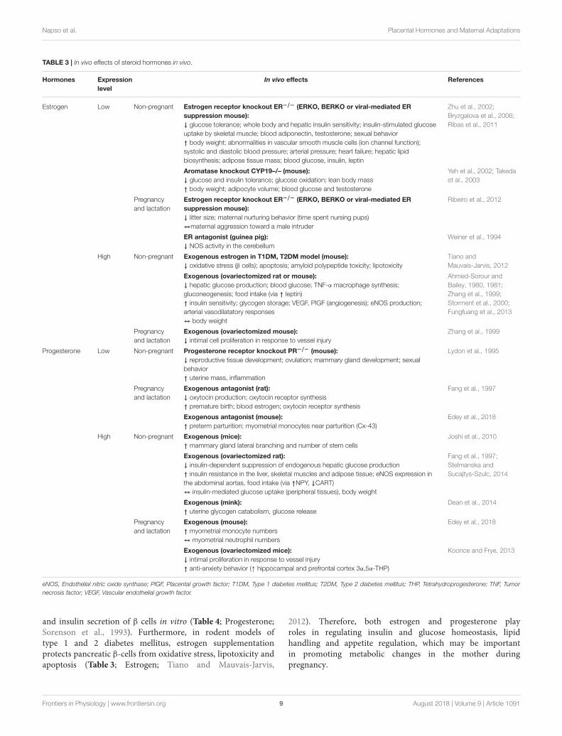
TABLE 3 | In vivo effects of steroid hormones in vivo.
Hormones Expression
level
In vivo effects References
Estrogen Low Non-pregnant Estrogen receptor knockout ER−/− (ERKO, BERKO or viral-mediated ER
suppression mouse):
↓ glucose tolerance; whole body and hepatic insulin sensitivity; insulin-stimulated glucose
uptake by skeletal muscle; blood adiponectin, testosterone; sexual behavior
↑ body weight; abnormalities in vascular smooth muscle cells (ion channel function);
systolic and diastolic blood pressure; arterial pressure; heart failure; hepatic lipid
biosynthesis; adipose tissue mass; blood glucose, insulin, leptin
Zhu et al., 2002;
Bryzgalova et al., 2006;
Ribas et al., 2011
Aromatase knockout CYP19–/– (mouse):
↓ glucose and insulin tolerance; glucose oxidation; lean body mass
↑ body weight; adipocyte volume; blood glucose and testosterone
Yeh et al., 2002; Takeda
et al., 2003
Pregnancy
and lactation
Estrogen receptor knockout ER−/− (ERKO, BERKO or viral-mediated ER
suppression mouse):
↓ litter size; maternal nurturing behavior (time spent nursing pups)
↔maternal aggression toward a male intruder
Ribeiro et al., 2012
ER antagonist (guinea pig):
↓ NOS activity in the cerebellum
Weiner et al., 1994
High Non-pregnant Exogenous estrogen in T1DM, T2DM model (mouse):
↓ oxidative stress (β cells); apoptosis; amyloid polypeptide toxicity; lipotoxicity
Tiano and
Mauvais-Jarvis, 2012
Exogenous (ovariectomized rat or mouse):
↓ hepatic glucose production; blood glucose; TNF-α macrophage synthesis;
gluconeogenesis; food intake (via ↑ leptin)
↑ insulin sensitivity; glycogen storage; VEGF, PlGF (angiogenesis); eNOS production;
arterial vasodilatatory responses
↔ body weight
Ahmed-Sorour and
Bailey, 1980, 1981;
Zhang et al., 1999;
Storment et al., 2000;
Fungfuang et al., 2013
Pregnancy
and lactation
Exogenous (ovariectomized mouse):
↓ intimal cell proliferation in response to vessel injury
Zhang et al., 1999
Progesterone Low Non-pregnant Progesterone receptor knockout PR−/− (mouse):
↓ reproductive tissue development; ovulation; mammary gland development; sexual
behavior
↑ uterine mass, inflammation
Lydon et al., 1995
Pregnancy
and lactation
Exogenous antagonist (rat):
↓ oxytocin production; oxytocin receptor synthesis
↑ premature birth; blood estrogen; oxytocin receptor synthesis
Fang et al., 1997
Exogenous antagonist (mouse):
↑ preterm parturition; myometrial monocytes near parturition (Cx-43)
Edey et al., 2018
High Non-pregnant Exogenous (mice):
↑ mammary gland lateral branching and number of stem cells
Joshi et al., 2010
Exogenous (ovariectomized rat):
↓ insulin-dependent suppression of endogenous hepatic glucose production
↑ insulin resistance in the liver, skeletal muscles and adipose tissue; eNOS expression in
the abdominal aortas, food intake (via ↑NPY, ↓CART)
↔ insulin-mediated glucose uptake (peripheral tissues), body weight
Fang et al., 1997;
Stelmanska and
Sucajtys-Szulc, 2014
Exogenous (mink):
↑ uterine glycogen catabolism, glucose release
Dean et al., 2014
Pregnancy
and lactation
Exogenous (mouse):
↑ myometrial monocyte numbers
↔ myometrial neutrophil numbers
Edey et al., 2018
Exogenous (ovariectomized mice):
↓ intimal proliferation in response to vessel injury
↑ anti-anxiety behavior (↑ hippocampal and prefrontal cortex 3α,5α-THP)
Koonce and Frye, 2013
eNOS, Endothelial nitric oxide synthase; PlGF, Placental growth factor; T1DM, Type 1 diabetes mellitus; T2DM, Type 2 diabetes mellitus; THP, Tetrahydroprogesterone; TNF, Tumor
necrosis factor; VEGF, Vascular endothelial growth factor.
and insulin secretion of β cells in vitro (Table 4; Progesterone;Sorenson et al., 1993). Furthermore, in rodent models oftype 1 and 2 diabetes mellitus, estrogen supplementationprotects pancreatic β-cells from oxidative stress, lipotoxicity andapoptosis (Table 3; Estrogen; Tiano and Mauvais-Jarvis,
2012). Therefore, both estrogen and progesterone playroles in regulating insulin and glucose homeostasis, lipidhandling and appetite regulation, which may be importantin promoting metabolic changes in the mother duringpregnancy.
Frontiers in Physiology | www.frontiersin.org 9 August 2018 | Volume 9 | Article 1091
Napso et al. Placental Hormones and Maternal Adaptations
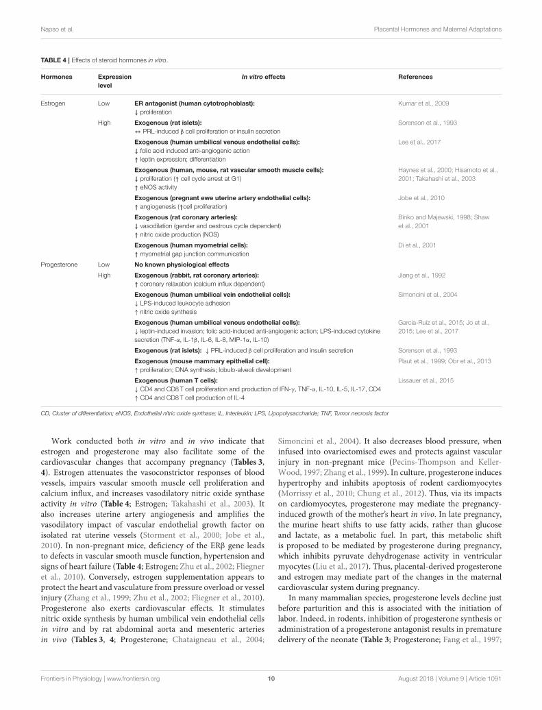
TABLE 4 | Effects of steroid hormones in vitro.
Hormones Expression
level
In vitro effects References
Estrogen Low ER antagonist (human cytotrophoblast):
↓ proliferation
Kumar et al., 2009
High Exogenous (rat islets):
↔ PRL-induced β cell proliferation or insulin secretion
Sorenson et al., 1993
Exogenous (human umbilical venous endothelial cells):
↓ folic acid induced anti-angiogenic action
↑ leptin expression; differentiation
Lee et al., 2017
Exogenous (human, mouse, rat vascular smooth muscle cells):
↓ proliferation (↑ cell cycle arrest at G1)
↑ eNOS activity
Haynes et al., 2000; Hisamoto et al.,
2001; Takahashi et al., 2003
Exogenous (pregnant ewe uterine artery endothelial cells):
↑ angiogenesis (↑cell proliferation)
Jobe et al., 2010
Exogenous (rat coronary arteries):
↓ vasodilation (gender and oestrous cycle dependent)
↑ nitric oxide production (NOS)
Binko and Majewski, 1998; Shaw
et al., 2001
Exogenous (human myometrial cells):
↑ myometrial gap junction communication
Di et al., 2001
Progesterone Low No known physiological effects
High Exogenous (rabbit, rat coronary arteries):
↑ coronary relaxation (calcium influx dependent)
Jiang et al., 1992
Exogenous (human umbilical vein endothelial cells):
↓ LPS-induced leukocyte adhesion
↑ nitric oxide synthesis
Simoncini et al., 2004
Exogenous (human umbilical venous endothelial cells):
↓ leptin-induced invasion; folic acid-induced anti-angiogenic action; LPS-induced cytokine
secretion (TNF-α, IL-1β, IL-6, IL-8, MIP-1α, IL-10)
Garcia-Ruíz et al., 2015; Jo et al.,
2015; Lee et al., 2017
Exogenous (rat islets): ↓ PRL-induced β cell proliferation and insulin secretion Sorenson et al., 1993
Exogenous (mouse mammary epithelial cell):
↑ proliferation; DNA synthesis; lobulo-alveoli development
Plaut et al., 1999; Obr et al., 2013
Exogenous (human T cells):
↓ CD4 and CD8T cell proliferation and production of IFN-γ, TNF-α, IL-10, IL-5, IL-17, CD4
↑ CD4 and CD8T cell production of IL-4
Lissauer et al., 2015
CD, Cluster of differentiation; eNOS, Endothelial nitric oxide synthase; IL, Interleukin; LPS, Lipopolysaccharide; TNF, Tumor necrosis factor
Work conducted both in vitro and in vivo indicate thatestrogen and progesterone may also facilitate some of thecardiovascular changes that accompany pregnancy (Tables 3,4). Estrogen attenuates the vasoconstrictor responses of bloodvessels, impairs vascular smooth muscle cell proliferation andcalcium influx, and increases vasodilatory nitric oxide synthaseactivity in vitro (Table 4; Estrogen; Takahashi et al., 2003). Italso increases uterine artery angiogenesis and amplifies thevasodilatory impact of vascular endothelial growth factor onisolated rat uterine vessels (Storment et al., 2000; Jobe et al.,2010). In non-pregnant mice, deficiency of the ERβ gene leadsto defects in vascular smooth muscle function, hypertension andsigns of heart failure (Table 4; Estrogen; Zhu et al., 2002; Fliegneret al., 2010). Conversely, estrogen supplementation appears toprotect the heart and vasculature from pressure overload or vesselinjury (Zhang et al., 1999; Zhu et al., 2002; Fliegner et al., 2010).Progesterone also exerts cardiovascular effects. It stimulatesnitric oxide synthesis by human umbilical vein endothelial cellsin vitro and by rat abdominal aorta and mesenteric arteriesin vivo (Tables 3, 4; Progesterone; Chataigneau et al., 2004;
Simoncini et al., 2004). It also decreases blood pressure, wheninfused into ovariectomised ewes and protects against vascularinjury in non-pregnant mice (Pecins-Thompson and Keller-Wood, 1997; Zhang et al., 1999). In culture, progesterone induceshypertrophy and inhibits apoptosis of rodent cardiomyocytes(Morrissy et al., 2010; Chung et al., 2012). Thus, via its impactson cardiomyocytes, progesterone may mediate the pregnancy-induced growth of the mother’s heart in vivo. In late pregnancy,the murine heart shifts to use fatty acids, rather than glucoseand lactate, as a metabolic fuel. In part, this metabolic shiftis proposed to be mediated by progesterone during pregnancy,which inhibits pyruvate dehydrogenase activity in ventricularmyocytes (Liu et al., 2017). Thus, placental-derived progesteroneand estrogen may mediate part of the changes in the maternalcardiovascular system during pregnancy.
In many mammalian species, progesterone levels decline justbefore parturition and this is associated with the initiation oflabor. Indeed, in rodents, inhibition of progesterone synthesis oradministration of a progesterone antagonist results in prematuredelivery of the neonate (Table 3; Progesterone; Fang et al., 1997;
Frontiers in Physiology | www.frontiersin.org 10 August 2018 | Volume 9 | Article 1091
Napso et al. Placental Hormones and Maternal Adaptations
Kota et al., 2013). In humans, circulating progesterone levelscontinue to be high until birth. Commencement of labor istherefore proposed to be related to a functional withdrawal ofprogesterone activity in the myometrium of women (Brown A.G. et al., 2004; Norwitz and Caughey, 2011). In experimentalanimals, progesterone reduces the production of prostaglandinsand decreases the expression of contraction-associated genesincluding oxytocin and prostaglandin receptors, gap junctionproteins and ion channels in the myometrium (Table 3;Progesterone; Fang et al., 1997; Soloff et al., 2011; Edey et al.,2018). Together, these progesterone-mediated actions decreasecontractility of uterine smooth muscle cells and maintain uterinequiescence until term. In contrast to progesterone, estrogen levelsrise prior to term and estrogen promotes the expression ofcontraction-associated genes and contraction of themyometrium(Table 4; Estrogen; Nathanielsz et al., 1998; Di et al., 2001;Chandran et al., 2014). Therefore, in many species, the highratio of estrogen to progesterone in the maternal circulationis thought to contribute the onset of labor. Parturition isassociated with an influx of inflammatory cells and release ofpro-inflammatory cytokines, including interleukin (IL)-1β andtumor necrosis factor (TNF)-α, in the myometrium, cervix andfetal membranes (Golightly et al., 2011). In mice, progesteronereduces the expression of pro-inflammatory cytokines, includingIL-1β and IL-6 by the uterus and trophoblast and may modulatethe abundance of myometrial monocytes (Table 3; Estrogen;Edey et al., 2018). Progesterone also decreases the ability ofLPS to induce pro-inflammatory cytokine secretion by humanmyometrium and placental explants (Youssef et al., 2009; Garcia-Ruíz et al., 2015). It also diminishes the ability of estrogento induce the infiltration of macrophages and neutrophils intothe uterus, and decreases LPS-induced leukocyte adhesion tohuman umbilical vein cells (Simoncini et al., 2004). Thus,it is perhaps not surprising that progesterone receptor nullmice demonstrate chronic uterine inflammation, particularlyin response to estrogen treatment (Table 3; Estrogen; Lydonet al., 1995). There is also evidence that placental steroidsparticipate in cervical softening, by regulating the expressionof matrix remodeling enzymes as well as leukocyte infiltrationand function (Chinnathambi et al., 2014; Gopalakrishnan et al.,2016; Berkane et al., 2017). In addition to regulating the eventsleading to parturition, recent data suggest that during the courseof pregnancy, both estrogen and progesterone contribute to thematernal tolerance of the fetus by modulating proliferation andcytokine expression of CD4 and CD8T cells and enhancingthe suppressive function of T-regulatory cells (Mao et al., 2010;Robinson and Klein, 2012; Lissauer et al., 2015).
Additionally, both estrogen and progesterone are keystimulators of mammary gland development. For instance,progesterone stimulates proliferation of mammary stem cellsand mammary epithelium (Tables 3, 4; Progesterone; Joshiet al., 2010; Lee et al., 2013). In mice, deficiency of theprogesterone receptor restricts mammary gland development,whereas exogenous progesterone induces ductal side branchingand lobuloalveolar differentiation and development (Table 3;Progesterone; Plaut et al., 1999; Joshi et al., 2010). In addition,both estrogen and progesterone may have indirect effects on
mammary gland development by regulating prolactin secretionfrom the pituitary gland (Rezaei et al., 2016).
Maternal behavior during and after birth are regulated bythe steroid hormones. Estrogen stimulates maternal nurturingbehavior in numerous species, including rats, mice, sheep andprimates (Bridges, 2015). In particular, maternal care is inducedby estrogen treatment, whereas the converse happens when ERα
expression is suppressed; deficiency of ERα increases the latencyto pup retrieval and reduces the length of time dams spendnursing and licking their pups (Table 3; Estrogen; Ribeiro et al.,2012). Findings from animal models suggest that progesteroneplays a role in regulating anxiety and depression-related behavior.For instance, exogenous progesterone stimulates anti-anxiety andanti-depressive actions in mouse dams (Table 3; Progesterone;Koonce and Frye, 2013). In contrast, progesterone withdrawalincreases these types of behaviors (Gulinello et al., 2002).Thus, placental-derived steroids may modulate several aspects ofmaternal physiology which are beneficial to both pregnancy andpost-partum support of the offspring.
Neuroactive HormonesOne major target of placental hormones is the maternal brainand related neuroendocrine organs such as the hypothalamus andpituitary glands. These neuroendocrine effects enable the motherto respond and adapt accordingly to her environment, so as tomitigate the adverse effects of stress and maintain homeostasis(Voltolini and Petraglia, 2014). Neuroactive hormones alsoprepare and enable the future mother to adequately carefor her young (Lévy, 2016). In addition to their impact onthe maternal neuroendocrine system, these hormones haveadditional functions in vivo and in vitro functions as well, whichare detailed in Tables 5, 6, respectively.
Melatonin and SerotoninMelatonin and its precursor, serotonin, are tryptophan-derivedhormones with well-known neuroendocrine impacts. In humans,circulating concentrations of melatonin and serotonin increaseas pregnancy advances (Lin et al., 1996; Nakamura et al.,2001). In the non-pregnant state, melatonin and serotoninare primarily produced by the pineal gland and the brain,respectively. However, the enzymes involved in melatoninand serotonin biosynthesis are also expressed by the humanplacenta throughout gestation (Iwasaki et al., 2005; Solimanet al., 2015; Laurent et al., 2017). The mouse placenta similarlyexpresses the enzymes needed for serotonin synthesis (Wuet al., 2016), although work is required to assess if melatoninsynthesizing enzymes are also expressed. The rat placenta doesnot produce melatonin de novo due to the lack of synthesizingenzymes (Tamura et al., 2008). However, the same studydemonstrated that conditioned medium from cultured termrat placentas stimulated melatonin release by the maternalpineal gland (Tamura et al., 2008). These findings suggestthat placental-derived factors may indirectly regulate melatoninlevels by the mother during pregnancy. Placental expressionof melatonin, serotonin and their respective enzymes, alsoremains to be investigated in other species such as rabbitsand sheep, which are commonly used in pregnancy-related
Frontiers in Physiology | www.frontiersin.org 11 August 2018 | Volume 9 | Article 1091
Napso et al. Placental Hormones and Maternal Adaptations
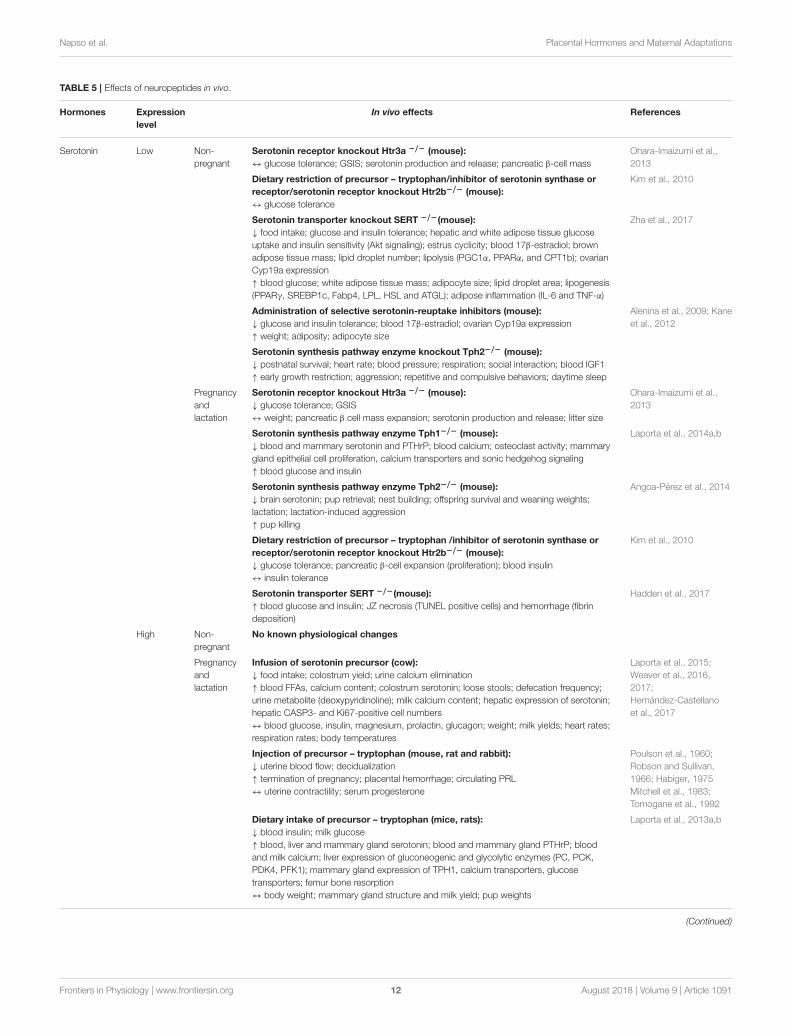
TABLE 5 | Effects of neuropeptides in vivo.
Hormones Expression
level
In vivo effects References
Serotonin Low Non-
pregnant
Serotonin receptor knockout Htr3a −/− (mouse):
↔ glucose tolerance; GSIS; serotonin production and release; pancreatic β-cell mass
Ohara-Imaizumi et al.,
2013
Dietary restriction of precursor – tryptophan/inhibitor of serotonin synthase or
receptor/serotonin receptor knockout Htr2b−/− (mouse):
↔ glucose tolerance
Kim et al., 2010
Serotonin transporter knockout SERT −/−(mouse):
↓ food intake; glucose and insulin tolerance; hepatic and white adipose tissue glucose
uptake and insulin sensitivity (Akt signaling); estrus cyclicity; blood 17β-estradiol; brown
adipose tissue mass; lipid droplet number; lipolysis (PGC1α, PPARα, and CPT1b); ovarian
Cyp19a expression
↑ blood glucose; white adipose tissue mass; adipocyte size; lipid droplet area; lipogenesis
(PPARγ, SREBP1c, Fabp4, LPL, HSL and ATGL); adipose inflammation (IL-6 and TNF-α)
Zha et al., 2017
Administration of selective serotonin-reuptake inhibitors (mouse):
↓ glucose and insulin tolerance; blood 17β-estradiol; ovarian Cyp19a expression
↑ weight; adiposity; adipocyte size
Alenina et al., 2009; Kane
et al., 2012
Serotonin synthesis pathway enzyme knockout Tph2−/− (mouse):
↓ postnatal survival; heart rate; blood pressure; respiration; social interaction; blood IGF1
↑ early growth restriction; aggression; repetitive and compulsive behaviors; daytime sleep
Pregnancy
and
lactation
Serotonin receptor knockout Htr3a −/− (mouse):
↓ glucose tolerance; GSIS
↔ weight; pancreatic β cell mass expansion; serotonin production and release; litter size
Ohara-Imaizumi et al.,
2013
Serotonin synthesis pathway enzyme Tph1−/− (mouse):
↓ blood and mammary serotonin and PTHrP; blood calcium; osteoclast activity; mammary
gland epithelial cell proliferation, calcium transporters and sonic hedgehog signaling
↑ blood glucose and insulin
Laporta et al., 2014a,b
Serotonin synthesis pathway enzyme Tph2−/− (mouse):
↓ brain serotonin; pup retrieval; nest building; offspring survival and weaning weights;
lactation; lactation-induced aggression
↑ pup killing
Angoa-Pérez et al., 2014
Dietary restriction of precursor – tryptophan /inhibitor of serotonin synthase or
receptor/serotonin receptor knockout Htr2b−/− (mouse):
↓ glucose tolerance; pancreatic β-cell expansion (proliferation); blood insulin
↔ insulin tolerance
Kim et al., 2010
Serotonin transporter SERT −/−(mouse):
↑ blood glucose and insulin; JZ necrosis (TUNEL positive cells) and hemorrhage (fibrin
deposition)
Hadden et al., 2017
High Non-
pregnant
No known physiological changes
Pregnancy
and
lactation
Infusion of serotonin precursor (cow):
↓ food intake; colostrum yield; urine calcium elimination
↑ blood FFAs, calcium content; colostrum serotonin; loose stools; defecation frequency;
urine metabolite (deoxypyridinoline); milk calcium content; hepatic expression of serotonin;
hepatic CASP3- and Ki67-positive cell numbers
↔ blood glucose, insulin, magnesium, prolactin, glucagon; weight; milk yields; heart rates;
respiration rates; body temperatures
Laporta et al., 2015;
Weaver et al., 2016,
2017;
Hernández-Castellano
et al., 2017
Injection of precursor – tryptophan (mouse, rat and rabbit):
↓ uterine blood flow; decidualization
↑ termination of pregnancy; placental hemorrhage; circulating PRL
↔ uterine contractility; serum progesterone
Poulson et al., 1960;
Robson and Sullivan,
1966; Habiger, 1975
Mitchell et al., 1983;
Tomogane et al., 1992
Dietary intake of precursor – tryptophan (mice, rats):
↓ blood insulin; milk glucose
↑ blood, liver and mammary gland serotonin; blood and mammary gland PTHrP; blood
and milk calcium; liver expression of gluconeogenic and glycolytic enzymes (PC, PCK,
PDK4, PFK1); mammary gland expression of TPH1, calcium transporters, glucose
transporters; femur bone resorption
↔ body weight; mammary gland structure and milk yield; pup weights
Laporta et al., 2013a,b
(Continued)
Frontiers in Physiology | www.frontiersin.org 12 August 2018 | Volume 9 | Article 1091
Napso et al. Placental Hormones and Maternal Adaptations
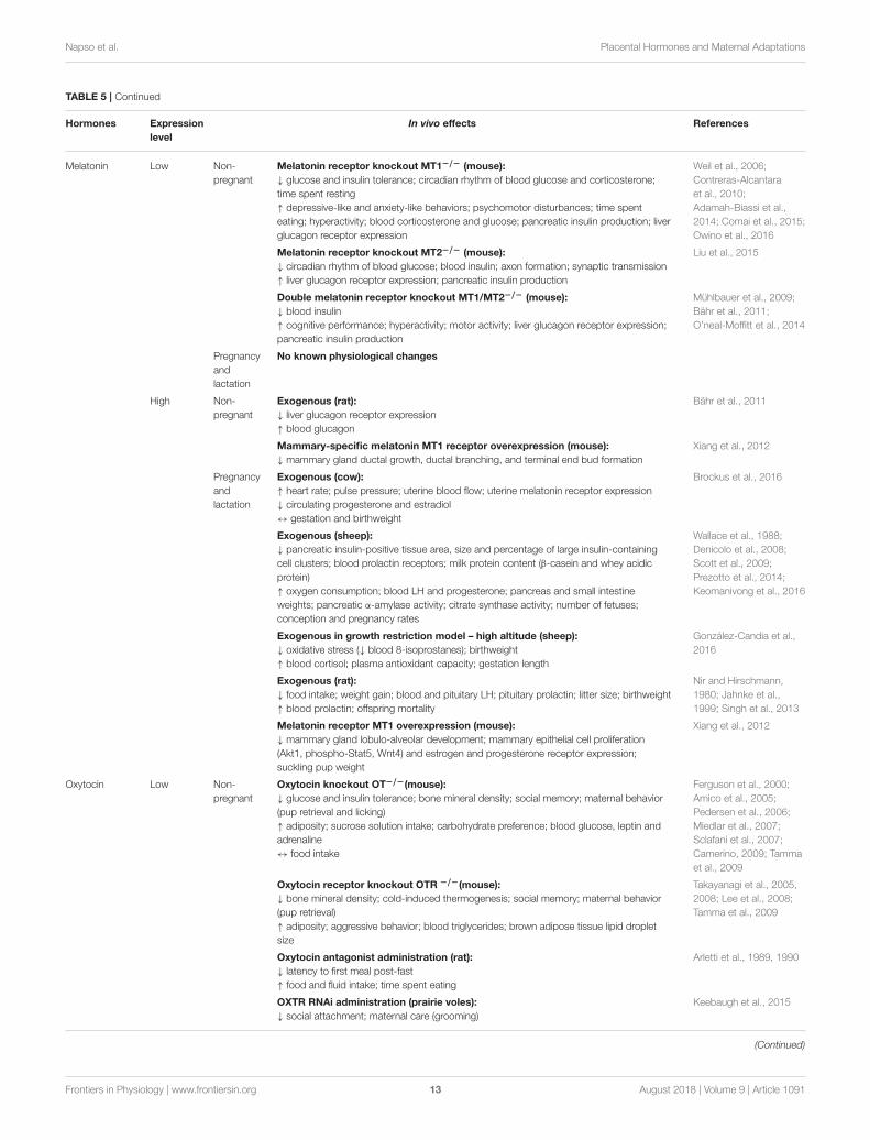
TABLE 5 | Continued
Hormones Expression
level
In vivo effects References
Melatonin Low Non-
pregnant
Melatonin receptor knockout MT1−/− (mouse):
↓ glucose and insulin tolerance; circadian rhythm of blood glucose and corticosterone;
time spent resting
↑ depressive-like and anxiety-like behaviors; psychomotor disturbances; time spent
eating; hyperactivity; blood corticosterone and glucose; pancreatic insulin production; liver
glucagon receptor expression
Weil et al., 2006;
Contreras-Alcantara
et al., 2010;
Adamah-Biassi et al.,
2014; Comai et al., 2015;
Owino et al., 2016
Melatonin receptor knockout MT2−/− (mouse):
↓ circadian rhythm of blood glucose; blood insulin; axon formation; synaptic transmission
↑ liver glucagon receptor expression; pancreatic insulin production
Liu et al., 2015
Double melatonin receptor knockout MT1/MT2−/− (mouse):
↓ blood insulin
↑ cognitive performance; hyperactivity; motor activity; liver glucagon receptor expression;
pancreatic insulin production
Mühlbauer et al., 2009;
Bähr et al., 2011;
O’neal-Moffitt et al., 2014
Pregnancy
and
lactation
No known physiological changes
High Non-
pregnant
Exogenous (rat):
↓ liver glucagon receptor expression
↑ blood glucagon
Bähr et al., 2011
Mammary-specific melatonin MT1 receptor overexpression (mouse):
↓ mammary gland ductal growth, ductal branching, and terminal end bud formation
Xiang et al., 2012
Pregnancy
and
lactation
Exogenous (cow):
↑ heart rate; pulse pressure; uterine blood flow; uterine melatonin receptor expression
↓ circulating progesterone and estradiol
↔ gestation and birthweight
Brockus et al., 2016
Exogenous (sheep):
↓ pancreatic insulin-positive tissue area, size and percentage of large insulin-containing
cell clusters; blood prolactin receptors; milk protein content (β-casein and whey acidic
protein)
↑ oxygen consumption; blood LH and progesterone; pancreas and small intestine
weights; pancreatic α-amylase activity; citrate synthase activity; number of fetuses;
conception and pregnancy rates
Wallace et al., 1988;
Denicolo et al., 2008;
Scott et al., 2009;
Prezotto et al., 2014;
Keomanivong et al., 2016
Exogenous in growth restriction model – high altitude (sheep):
↓ oxidative stress (↓ blood 8-isoprostanes); birthweight
↑ blood cortisol; plasma antioxidant capacity; gestation length
González-Candia et al.,
2016
Exogenous (rat):
↓ food intake; weight gain; blood and pituitary LH; pituitary prolactin; litter size; birthweight
↑ blood prolactin; offspring mortality
Nir and Hirschmann,
1980; Jahnke et al.,
1999; Singh et al., 2013
Melatonin receptor MT1 overexpression (mouse):
↓ mammary gland lobulo-alveolar development; mammary epithelial cell proliferation
(Akt1, phospho-Stat5, Wnt4) and estrogen and progesterone receptor expression;
suckling pup weight
Xiang et al., 2012
Oxytocin Low Non-
pregnant
Oxytocin knockout OT−/−(mouse):
↓ glucose and insulin tolerance; bone mineral density; social memory; maternal behavior
(pup retrieval and licking)
↑ adiposity; sucrose solution intake; carbohydrate preference; blood glucose, leptin and
adrenaline
↔ food intake
Ferguson et al., 2000;
Amico et al., 2005;
Pedersen et al., 2006;
Miedlar et al., 2007;
Sclafani et al., 2007;
Camerino, 2009; Tamma
et al., 2009
Oxytocin receptor knockout OTR −/−(mouse):
↓ bone mineral density; cold-induced thermogenesis; social memory; maternal behavior
(pup retrieval)
↑ adiposity; aggressive behavior; blood triglycerides; brown adipose tissue lipid droplet
size
Takayanagi et al., 2005,
2008; Lee et al., 2008;
Tamma et al., 2009
Oxytocin antagonist administration (rat):
↓ latency to first meal post-fast
↑ food and fluid intake; time spent eating
Arletti et al., 1989, 1990
OXTR RNAi administration (prairie voles):
↓ social attachment; maternal care (grooming)
Keebaugh et al., 2015
(Continued)
Frontiers in Physiology | www.frontiersin.org 13 August 2018 | Volume 9 | Article 1091
Napso et al. Placental Hormones and Maternal Adaptations
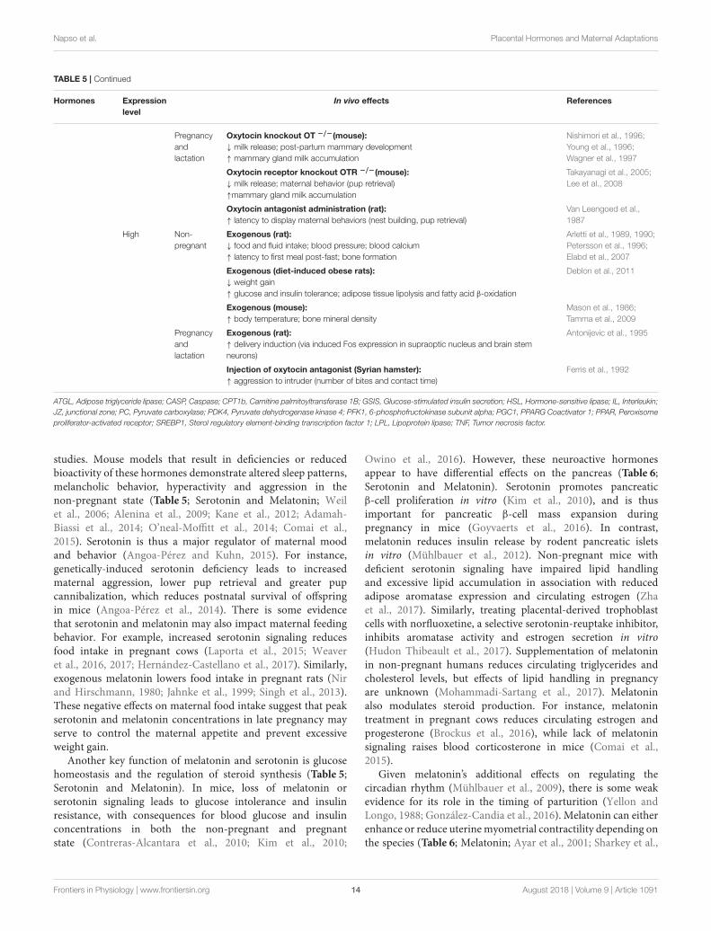
TABLE 5 | Continued
Hormones Expression
level
In vivo effects References
Pregnancy
and
lactation
Oxytocin knockout OT −/−(mouse):
↓ milk release; post-partum mammary development
↑ mammary gland milk accumulation
Nishimori et al., 1996;
Young et al., 1996;
Wagner et al., 1997
Oxytocin receptor knockout OTR −/−(mouse):
↓ milk release; maternal behavior (pup retrieval)
↑mammary gland milk accumulation
Takayanagi et al., 2005;
Lee et al., 2008
Oxytocin antagonist administration (rat):
↑ latency to display maternal behaviors (nest building, pup retrieval)
Van Leengoed et al.,
1987
High Non-
pregnant
Exogenous (rat):
↓ food and fluid intake; blood pressure; blood calcium
↑ latency to first meal post-fast; bone formation
Arletti et al., 1989, 1990;
Petersson et al., 1996;
Elabd et al., 2007
Exogenous (diet-induced obese rats):
↓ weight gain
↑ glucose and insulin tolerance; adipose tissue lipolysis and fatty acid β-oxidation
Deblon et al., 2011
Exogenous (mouse):
↑ body temperature; bone mineral density
Mason et al., 1986;
Tamma et al., 2009
Pregnancy
and
lactation
Exogenous (rat):
↑ delivery induction (via induced Fos expression in supraoptic nucleus and brain stem
neurons)
Antonijevic et al., 1995
Injection of oxytocin antagonist (Syrian hamster):
↑ aggression to intruder (number of bites and contact time)
Ferris et al., 1992
ATGL, Adipose triglyceride lipase; CASP, Caspase; CPT1b, Carnitine palmitoyltransferase 1B; GSIS, Glucose-stimulated insulin secretion; HSL, Hormone-sensitive lipase; IL, Interleukin;
JZ, junctional zone; PC, Pyruvate carboxylase; PDK4, Pyruvate dehydrogenase kinase 4; PFK1, 6-phosphofructokinase subunit alpha; PGC1, PPARG Coactivator 1; PPAR, Peroxisome
proliferator-activated receptor; SREBP1, Sterol regulatory element-binding transcription factor 1; LPL, Lipoprotein lipase; TNF, Tumor necrosis factor.
studies. Mouse models that result in deficiencies or reducedbioactivity of these hormones demonstrate altered sleep patterns,melancholic behavior, hyperactivity and aggression in thenon-pregnant state (Table 5; Serotonin and Melatonin; Weilet al., 2006; Alenina et al., 2009; Kane et al., 2012; Adamah-Biassi et al., 2014; O’neal-Moffitt et al., 2014; Comai et al.,2015). Serotonin is thus a major regulator of maternal moodand behavior (Angoa-Pérez and Kuhn, 2015). For instance,genetically-induced serotonin deficiency leads to increasedmaternal aggression, lower pup retrieval and greater pupcannibalization, which reduces postnatal survival of offspringin mice (Angoa-Pérez et al., 2014). There is some evidencethat serotonin and melatonin may also impact maternal feedingbehavior. For example, increased serotonin signaling reducesfood intake in pregnant cows (Laporta et al., 2015; Weaveret al., 2016, 2017; Hernández-Castellano et al., 2017). Similarly,exogenous melatonin lowers food intake in pregnant rats (Nirand Hirschmann, 1980; Jahnke et al., 1999; Singh et al., 2013).These negative effects on maternal food intake suggest that peakserotonin and melatonin concentrations in late pregnancy mayserve to control the maternal appetite and prevent excessiveweight gain.
Another key function of melatonin and serotonin is glucosehomeostasis and the regulation of steroid synthesis (Table 5;Serotonin and Melatonin). In mice, loss of melatonin orserotonin signaling leads to glucose intolerance and insulinresistance, with consequences for blood glucose and insulinconcentrations in both the non-pregnant and pregnantstate (Contreras-Alcantara et al., 2010; Kim et al., 2010;
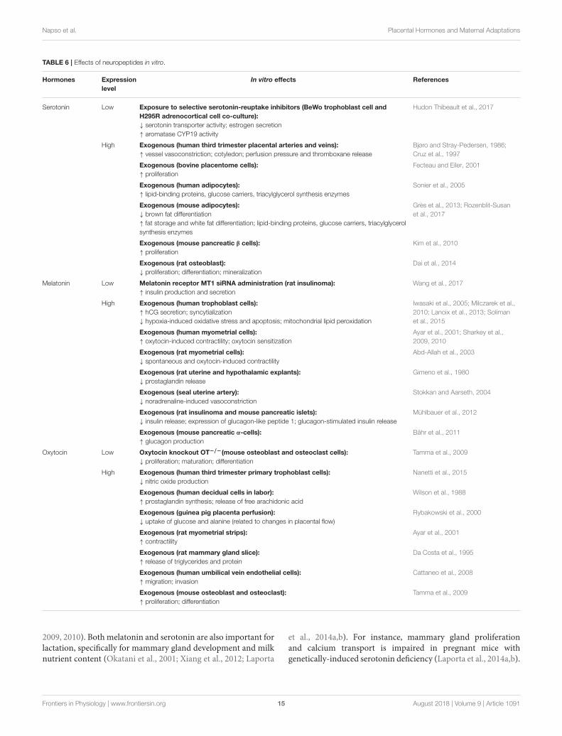
Owino et al., 2016). However, these neuroactive hormonesappear to have differential effects on the pancreas (Table 6;Serotonin and Melatonin). Serotonin promotes pancreaticβ-cell proliferation in vitro (Kim et al., 2010), and is thusimportant for pancreatic β-cell mass expansion duringpregnancy in mice (Goyvaerts et al., 2016). In contrast,melatonin reduces insulin release by rodent pancreatic isletsin vitro (Mühlbauer et al., 2012). Non-pregnant mice withdeficient serotonin signaling have impaired lipid handlingand excessive lipid accumulation in association with reducedadipose aromatase expression and circulating estrogen (Zhaet al., 2017). Similarly, treating placental-derived trophoblastcells with norfluoxetine, a selective serotonin-reuptake inhibitor,inhibits aromatase activity and estrogen secretion in vitro(Hudon Thibeault et al., 2017). Supplementation of melatoninin non-pregnant humans reduces circulating triglycerides andcholesterol levels, but effects of lipid handling in pregnancyare unknown (Mohammadi-Sartang et al., 2017). Melatoninalso modulates steroid production. For instance, melatonintreatment in pregnant cows reduces circulating estrogen andprogesterone (Brockus et al., 2016), while lack of melatoninsignaling raises blood corticosterone in mice (Comai et al.,2015).
Given melatonin’s additional effects on regulating thecircadian rhythm (Mühlbauer et al., 2009), there is some weakevidence for its role in the timing of parturition (Yellon andLongo, 1988; González-Candia et al., 2016). Melatonin can eitherenhance or reduce uterinemyometrial contractility depending onthe species (Table 6; Melatonin; Ayar et al., 2001; Sharkey et al.,
Frontiers in Physiology | www.frontiersin.org 14 August 2018 | Volume 9 | Article 1091
Napso et al. Placental Hormones and Maternal Adaptations
TABLE 6 | Effects of neuropeptides in vitro.
Hormones Expression
level
In vitro effects References
Serotonin Low Exposure to selective serotonin-reuptake inhibitors (BeWo trophoblast cell and
H295R adrenocortical cell co-culture):
↓ serotonin transporter activity; estrogen secretion
↑ aromatase CYP19 activity
Hudon Thibeault et al., 2017
High Exogenous (human third trimester placental arteries and veins):
↑ vessel vasoconstriction; cotyledon; perfusion pressure and thromboxane release
Bjøro and Stray-Pedersen, 1986;
Cruz et al., 1997
Exogenous (bovine placentome cells):
↑ proliferation
Fecteau and Eiler, 2001
Exogenous (human adipocytes):
↑ lipid-binding proteins, glucose carriers, triacylglycerol synthesis enzymes
Sonier et al., 2005
Exogenous (mouse adipocytes):
↓ brown fat differentiation
↑ fat storage and white fat differentiation; lipid-binding proteins, glucose carriers, triacylglycerol
synthesis enzymes
Grès et al., 2013; Rozenblit-Susan
et al., 2017
Exogenous (mouse pancreatic β cells):
↑ proliferation
Kim et al., 2010
Exogenous (rat osteoblast):
↓ proliferation; differentiation; mineralization
Dai et al., 2014
Melatonin Low Melatonin receptor MT1 siRNA administration (rat insulinoma):
↑ insulin production and secretion
Wang et al., 2017
High Exogenous (human trophoblast cells):
↑ hCG secretion; syncytialization
↓ hypoxia-induced oxidative stress and apoptosis; mitochondrial lipid peroxidation
Iwasaki et al., 2005; Milczarek et al.,
2010; Lanoix et al., 2013; Soliman
et al., 2015
Exogenous (human myometrial cells):
↑ oxytocin-induced contractility; oxytocin sensitization
Ayar et al., 2001; Sharkey et al.,
2009, 2010
Exogenous (rat myometrial cells):
↓ spontaneous and oxytocin-induced contractility
Abd-Allah et al., 2003
Exogenous (rat uterine and hypothalamic explants):
↓ prostaglandin release
Gimeno et al., 1980
Exogenous (seal uterine artery):
↓ noradrenaline-induced vasoconstriction
Stokkan and Aarseth, 2004
Exogenous (rat insulinoma and mouse pancreatic islets):
↓ insulin release; expression of glucagon-like peptide 1; glucagon-stimulated insulin release
Mühlbauer et al., 2012
Exogenous (mouse pancreatic α-cells):
↑ glucagon production
Bähr et al., 2011
Oxytocin Low Oxytocin knockout OT−/−(mouse osteoblast and osteoclast cells):
↓ proliferation; maturation; differentiation
Tamma et al., 2009
High Exogenous (human third trimester primary trophoblast cells):
↓ nitric oxide production
Nanetti et al., 2015
Exogenous (human decidual cells in labor):
↑ prostaglandin synthesis; release of free arachidonic acid
Wilson et al., 1988
Exogenous (guinea pig placenta perfusion):
↓ uptake of glucose and alanine (related to changes in placental flow)
Rybakowski et al., 2000
Exogenous (rat myometrial strips):
↑ contractility
Ayar et al., 2001
Exogenous (rat mammary gland slice):
↑ release of triglycerides and protein
Da Costa et al., 1995
Exogenous (human umbilical vein endothelial cells):
↑ migration; invasion
Cattaneo et al., 2008
Exogenous (mouse osteoblast and osteoclast):
↑ proliferation; differentiation
Tamma et al., 2009
2009, 2010). Both melatonin and serotonin are also important forlactation, specifically for mammary gland development and milknutrient content (Okatani et al., 2001; Xiang et al., 2012; Laporta
et al., 2014a,b). For instance, mammary gland proliferationand calcium transport is impaired in pregnant mice withgenetically-induced serotonin deficiency (Laporta et al., 2014a,b).
Frontiers in Physiology | www.frontiersin.org 15 August 2018 | Volume 9 | Article 1091
Napso et al. Placental Hormones and Maternal Adaptations
Conversely, supplementation of a serotonin precursor increasesmammary calcium transporter expression and milk calciumcontent in lactating mice and cows (Laporta et al., 2013a,b, 2015;Weaver et al., 2016, 2017; Hernández-Castellano et al., 2017). Incontrast to serotonin, increased melatonin signaling is associatedwith reduced ductal growth and branching, as well as impairedterminal end bud formation in the non-pregnant state (Xianget al., 2012). Thus, during lactation, these mice with increasedmelatonin signaling have impaired mammary gland lobulo-alveolar development and reduced milk protein content, whichreduces the weight of suckling pups (Xiang et al., 2012). Indeed,a recent study showed antenatal melatonin supplementationfurther exacerbated the growth restriction of offspring andraised circulating maternal cortisol in a sheep model of fetalgrowth restriction (González-Candia et al., 2016). Nevertheless,melatonin supplementation during pregnancy confers significantbeneficial neuroprotective effects on the fetus and enhancesmaternal antioxidant capacity (Miller et al., 2014; González-Candia et al., 2016; Castillo-Melendez et al., 2017). Therefore,while melatonin supplementation shows promise for use inthe clinic, particularly for enhancing the neurodevelopmentaloutcomes of offspring in growth compromised pregnancies,the potential adverse outcomes for both mother and childmust also be considered and should be assessed in furtherstudies.
OxytocinAnother key neuroendocrine factor is oxytocin. Oxytocin iswidely known for its role in triggering maternal nursing behavior(Bosch and Neumann, 2012). This is mediated by oxytocin’sactions on the maternal brain, as well as, the mammary glands.Indeed, a greater rise in circulating oxytocin concentrationsfrom early to late pregnancy in pregnant women, is associatedwith a stronger bond between a mother and her infant (Levineet al., 2007). Concurrently, placental expression of oxytocin alsopeaks at term in humans (Kim S. C. et al., 2017). The ratplacenta also produces oxytocin (Lefebvre et al., 1992), whileplacental expression in other species remains unclear. Reducedoxytocin signaling decreases maternal nurturing behavior suchas pup retrieval in rats (Van Leengoed et al., 1987). It alsodecreases the willingness of female voles to care for, groomand lick unrelated pups (Keebaugh et al., 2015). Low oxytocinsignaling can additionally impair social bonding in voles andmice (Ferguson et al., 2000; Takayanagi et al., 2005; Lee et al.,2008; Keebaugh et al., 2015), while high levels builds trust andcooperation in a group setting to facilitate group survival inhumans (Declerck et al., 2010; De Dreu et al., 2010). Moreover,a lack of oxytocin disrupts mammary gland proliferation andlobuloalveolar development, which impairs milk release fromthe mammary tissues in mice (Nishimori et al., 1996; Wagneret al., 1997). Therefore, high oxytocin levels enable the motherto bond better and protect her newborn, when it is mostvulnerable.
Oxytocin is also important in the process of parturition(Table 6; Oxytocin); it stimulates the contraction of smoothmuscle cells in the myometrium (Ayar et al., 2001; Arrowsmithand Wray, 2014), by inducing calcium influx and stimulating
prostaglandin release (Wilson et al., 1988; Voltolini and Petraglia,2014; Kim S. H. et al., 2017). Cardiovascular effects of oxytocininclude its ability to significantly lower blood pressure in non-pregnant rats (Petersson et al., 1996). There is also some evidencethat oxytocin induces anti-inflammatory and antioxidant effectsin the heart under hypoxic conditions in non-pregnant rats(Gutkowska and Jankowski, 2012). Nevertheless, the specificcardiovascular effects of oxytocin in pregnancy remain to beexplored.
Studies performed in non-pregnant rodents show thatoxytocin also affects metabolic function in vivo (Table 5;Oxytocin). In particular, loss of oxytocin reduces glucose andinsulin tolerance and increases adiposity (Camerino, 2009),whereas exogenous oxytocin has the reverse effect (Deblon et al.,2011). Studies are however, required to determine whether therise in oxytocin in late pregnancy (Levine et al., 2007) mayserve to improve insulin sensitivity in the mother in preparationfor the metabolic requirements of delivery and lactation. Thereis some evidence that oxytocin may additionally play a rolein controlling energy expenditure and thermoregulation duringpregnancy. Even with a similar diet and activity level to controlmice, oxytocin-deficient mice become obese due to reducedenergy expenditure from poor thermoregulation in the non-pregnant state (Chaves et al., 2013). Furthermore, exogenousoxytocin in non-pregnant mice causes a rise in body temperature(Mason et al., 1986; Tamma et al., 2009). Nevertheless, whetheroxytocin may play a role in controlling heat dissipation dueto the increased maternal energy expenditure during pregnancyrequires exploration. Exogenous oxytocin also reduces foodintake in non-pregnant rats (Arletti et al., 1989, 1990). However,the role of oxytocin in appetite regulation during pregnancyremains to be explored. There is also evidence for oxytocin’spossible involvement in maternal bone metabolism and calciumhomeostasis during pregnancy and lactation. For instance,oxytocin stimulates both bone resorption and bone formationby osteoclasts and osteoblasts respectively in vitro (Tammaet al., 2009). Moreover, oxytocin administration in rats reducescirculating calcium with an overall skew toward bone formation(Elabd et al., 2007). These findings may suggest that the peakin circulating oxytocin toward term promote the restoration ofdepleted maternal skeletal calcium stores.
Other Neuroactive HormonesIn addition to the aforementioned melatonin, serotonin andoxytocin, the human placenta also produces neuroactivehormones such as kisspeptin and thyrotropin-releasing hormone(TRH), which may function in adapting maternal physiologyto support pregnancy (Bajoria and Babawale, 1998; De Pedroet al., 2015). In humans, circulating kisspeptin rises throughoutpregnancy to concentrations 10,000-fold that of the non-pregnant state, with the placenta speculated as a major source(Horikoshi et al., 2003). In the non-pregnant state, kisspeptin canboth stimulate and impede glucose stimulated insulin secretionin mice (Bowe et al., 2009; Song et al., 2014). The nature of theeffect may partly relate to differences in the actions of kisspeptinisoforms on pancreatic islets (Bowe et al., 2012). Kisspeptinmay also have effects on the maternal cardiovascular system,
Frontiers in Physiology | www.frontiersin.org 16 August 2018 | Volume 9 | Article 1091
Napso et al. Placental Hormones and Maternal Adaptations
given its reported vasoconstrictive effects on vascular smoothmuscle cells and fibrotic effects on the heart in non-pregnantrats (Mead et al., 2007; Zhang et al., 2017). Studies in humanshighlight the importance of regulating kisspeptin productionduring gestation; increased placental kisspeptin is associatedwith pre-eclampsia (Whitehead et al., 2013; Matjila et al., 2016)and reduced circulating kisspeptin is observed in women withhypertension and diabetes during pregnancy (Cetkovic et al.,2012; Matjila et al., 2016). Like the human, the murine placentaproduces kisspeptin. Although a kisspeptin-deficient mouse hasbeen established, previous work has been focused on feto-placental outcomes, with no examination of maternal physiology(Herreboudt et al., 2015). Studies are required to determine theconsequences of abnormal placental kisspeptin on the maternalphysiology during pregnancy.
In the non-pregnant state, hypothalamic TRH stimulatesrelease of thyroid-stimulating hormone and PRL from thepituitary (Hershman et al., 1973; Vale et al., 1973; Askew andRamsden, 1984). However, during pregnancy, the placenta servesas an additional source of TRH (Bajoria and Babawale, 1998).Excess TRH in pregnancy raises blood concentrations of thyroid-stimulating hormone and PRL in humans, rhesusmonkeys, sheepand rats (Thomas et al., 1975; Azukizawa et al., 1976; Rotiet al., 1981; Moya et al., 1986; Lu et al., 1998). Conversely, alack of TRH reduces blood PRL in mice (Rabeler et al., 2004;Yamada et al., 2006). Thyroid hormones are necessary for optimalbrain development as well as thyroid function (Miranda andSousa, 2018). Impaired TRH signaling is associated with anxiety-like and depressive-like behavior in non-pregnant mice (Zenget al., 2007; Sun et al., 2009) and there is some evidence whichsuggests a link between thyroid dysfunction and poor maternalmood during pregnancy in humans (Basraon and Costantine,2011). However, whether any direct causal relationship betweenplacental hormones, like TRH and perinatal depression remainsunclear. Additionally, TRH is implicated in glucose homeostasisand appetite regulation. For example, mice with TRH deficiencyare hyperglycaemic, due to an impaired insulin response toglucose (Yamada et al., 1997). Reduced TRH signaling alsoimpedes leptin production and ghrelin acylation, which resultsin less energy conservation during fasting and a lower bodymass in the non-pregnant state (Groba et al., 2013; Mayerl et al.,2015). Investigations are warranted to identify whether TRHmaycontribute to the regulation of glucose handling and appetite inthe mother during pregnancy.
Additional HormonesThe placenta also produces numerous other hormones withpleiotropic effects. Several key ones, which have been implicatedin pregnancy failure or disorders of pregnancy such ashypertension, hyperglycemia and hypercalcemia, are discussedhere. The hormones presented here are by no means exhaustiveand were selected primarily on their major associationswith abnormal maternal physiology during pregnancy. Thegonadotropin, chorionic gonadotropin (CG); transforminggrowth factor β (TGF β) family member, activin; angiogenicfactor, relaxin; bone metabolism-associated parathyroid
hormone-related protein (PTHrP) and energy homeostasisregulator, leptin are reviewed (Tables 7, 8).
Chorionic Gonadotropin (CG)CG, is secreted by the human (hCG) and equine (eCG) placenta,although hCG has been more extensively studied. hCG is alarge glycoprotein composed of α and β subunits, of whichthe α subunit identical to luteinizing hormone (LH), folliclestimulating hormone (FSH) and thyroid stimulating hormone(TSH). As a result, hCG can interact with LH, FSH and TSHreceptors. In women, hCG is secreted from the trophoblastfrom very early in gestation and is thought to be the firstplacental hormone to act on the mother (Ogueh et al., 2011).Indeed, maternal circulating hCG concentrations peak in the firsttrimester and then decline toward term (Ogueh et al., 2011).In early pregnancy, hCG maintains corpus luteum allowingthe continued secretion of ovarian progesterone and estrogensuntil the steroidogenic activity of the fetal-placental unit cancompensate for maternal ovarian function (Fournier et al.,2015). In particular, hCG increases the abundance of low-density lipoprotein receptor and thus uptake of cholesterolfor steroidogenesis. It also enhances the expression and/oractivity of steroidogenic enzymes including 3β-hydroxysteroidand aromatase. There is also some evidence which suggestshCG may inhibit factors that promote luteal demise, such asthe prostaglandins. The high levels of hCG in early pregnancyare also sufficient to bind to the TSH receptor and may actto increase maternal thyroid hormone production, which asmentioned previously, may exert effects in the mother and fetus.
CG may also play important autocrine and paracrine rolesat the maternal-fetal interface. Administration of hCG antiseraprevents implantation in marmoset in vivo (Hearn et al., 1988).Recent proteomic analysis of estrogen and hCG treated humanendometrial epithelial cells demonstrates that hCG targetspathways involved in metabolism, basement membrane and cellconnectivity, proliferation and differentiation, cellular adhesion,extracellular-matrix organization, developmental growth, growthfactor regulation and cell signaling (Greening et al., 2016). Suchpathways are likely to be important for placental development,as attenuating hCG signaling disrupts trophoblast differentiationin vitro (Shi et al., 1993). In contrast, supplementing humantrophoblast cells with hCG increases their differentiation,migration, invasion and adhesion to uterine epithelial cells, anddecreases their leptin secretion in vitro (Table 8; hCG; Shi et al.,1993; Prast et al., 2008; Lee C. L. et al., 2013; Chen et al., 2015).hCG also promotes angiogenic vascular endothelial growthfactor secretion by both trophoblast and endometrial epithelialcells (Islami et al., 2003a; Berndt et al., 2006) and enhancesendothelial tube formation and migration (Zygmunt et al., 2002).Furthermore, hCG is key in suppressing the maternal immunesystem from mounting a response against paternal antigenscarried by the allogenic conceptus. Administration of hCG in amouse model of spontaneous abortion significantly reduces thenumber of fetal resorptions due to improved immune toleranceof the fetus (Schumacher et al., 2013). In vitro, hCG enhancesproliferation of immunosuppressive uterine natural killer cells(Kane et al., 2009), and the production of immunosuppressing
Frontiers in Physiology | www.frontiersin.org 17 August 2018 | Volume 9 | Article 1091
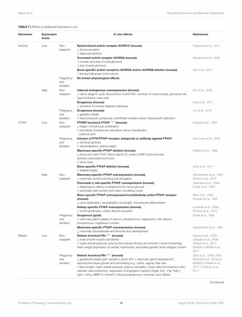
Napso et al. Placental Hormones and Maternal Adaptations
TABLE 7 | Effects of additional hormones in vivo.
Hormones Expression
levels
In vivo effects References
Activins Low Non-
pregnant
Dysfunctional activin receptor ACVR1C (mouse):
↓ fat accumulation
↑ adipocyte lipolysis
Yogosawa et al., 2013
Truncated activin receptor ACVR2A (mouse):
↑ number and area of renal glomeruli
↓ size of renal glomeruli
Maeshima et al., 2000
Bone-specific activin receptors ACVR2A and/or ACVR2B deletion (mouse):
↑ femoral trabecular bone volume
Goh et al., 2017
Pregnancy
and
lactation
No known physiological effects
High Non-
pregnant
Induced endogenous overexpression (mouse):
↑ estrus stage in cycle; blood activin A and FSH; numbers of corpora lutea; granulosa cell
layer thickness; ovary size
Kim et al., 2008
Exogenous (mouse):
↑ activation of muscle catabolic pathways
Ding et al., 2017
Pregnancy
and
lactation
Exogenous (mouse):
↓ gestation length
↑ blood pressure; proteinuria; endothelial oxidative stress; fetal growth restriction
Lim et al., 2015
PTHrP Low Non-
pregnant
PTHRP knockout PTHrP −/−(mouse):
↓ height; chondrocyte proliferation
↑ premature chondrocyte maturation; bone mineralization
- Lethal at birth
Karaplis et al., 1994
Pregnancy
and
lactation
Infusion of PTH/PTHrP receptor antagonist or antibody against PTHrP:
↓ decidual apotosis
↑ decidualization; uterine weight
Vanhouten et al., 2003
Mammary-specific PTHrP deletion (mouse):
↓ blood and milk PTHrP; blood vitamin D; urinary cAMP; bone turnover;
lactation-associated bone loss
↑ bone mass
Williams et al., 1998
Bone-specific PTHrP deletion (mouse):
↑ skeletal fragility
Kirby et al., 2011
High Non-
pregnant
Mammary-specific PTHrP overexpression (mouse):
↓ mammary ductal branching and elongation
Wysolmerski et al., 1995;
Dunbar et al., 2001
Pancreatic β cell-specific PTHrP overexpression (mouse):
↓ diabetogenic effects of streptozotocin; blood glucose
↑ pancreatic islet number and mass; circulating insulin
Vasavada et al., 1996;
Porter et al., 1998
Bone-specific PTHrP overexpression/constitutively active PTHrP receptor
(mouse):
↓ bone ossification, mineralization and length; chondrocyte differentiation
Weir et al., 1996;
Schipani et al., 1997
Kidney-specific PTHrP overexpression (mouse):
↑ renal hypertrophy; urinary albumin excretion
Izquierdo et al., 2006;
Romero et al., 2010
Pregnancy
and
lactation
Exogenous (goat):
↑ mammary gland uptake of calcium, phosphorous, magnesium; milk calcium,
phosphorous, magnesium content
Barlet et al., 1992
Mammary-specific PTHrP overexpression (mouse):
↓ mammary lobuloalveolar and terminal duct development
Wysolmerski et al., 1995
Relaxin Low Non-
pregnant
Relaxin knockout Rln−/− (mouse):
↓ renal smooth muscle cell density
↑ mean arterial pressure; lung function (airway fibrosis and smooth muscle thickening);
heart weight (expression of cardiac hypertrophy associated genes); renal collagen content
Samuel et al., 2003;
Lekgabe et al., 2006;
Debrah et al., 2011;
Mirabito Colafella et al.,
2017
Pregnancy
and
lactation
Relaxin knockout Rln−/− (mouse):
↓ gestational weight gain; lactation; blood sFlt-1; mammary gland development;
reproductive tissue growth and remodeling (e.g., cervix, vagina); litter size
↑ labor length; mean arterial pressure; plasma osmolality; urinary albumin/creatinine ratio;
vascular vasoconstriction; expression of angiogenic markers (Vegfa, Esr1, Pgr, Rxfp1,
Egln1, Hif1a, MMP14, Ankrd37); blood progesterone; mammary duct dilation
Zhao et al., 1999, 2000;
Marshall et al., 2016a,b;
Mirabito Colafella et al.,
2017; O’sullivan et al.,
2017
(Continued)
Frontiers in Physiology | www.frontiersin.org 18 August 2018 | Volume 9 | Article 1091
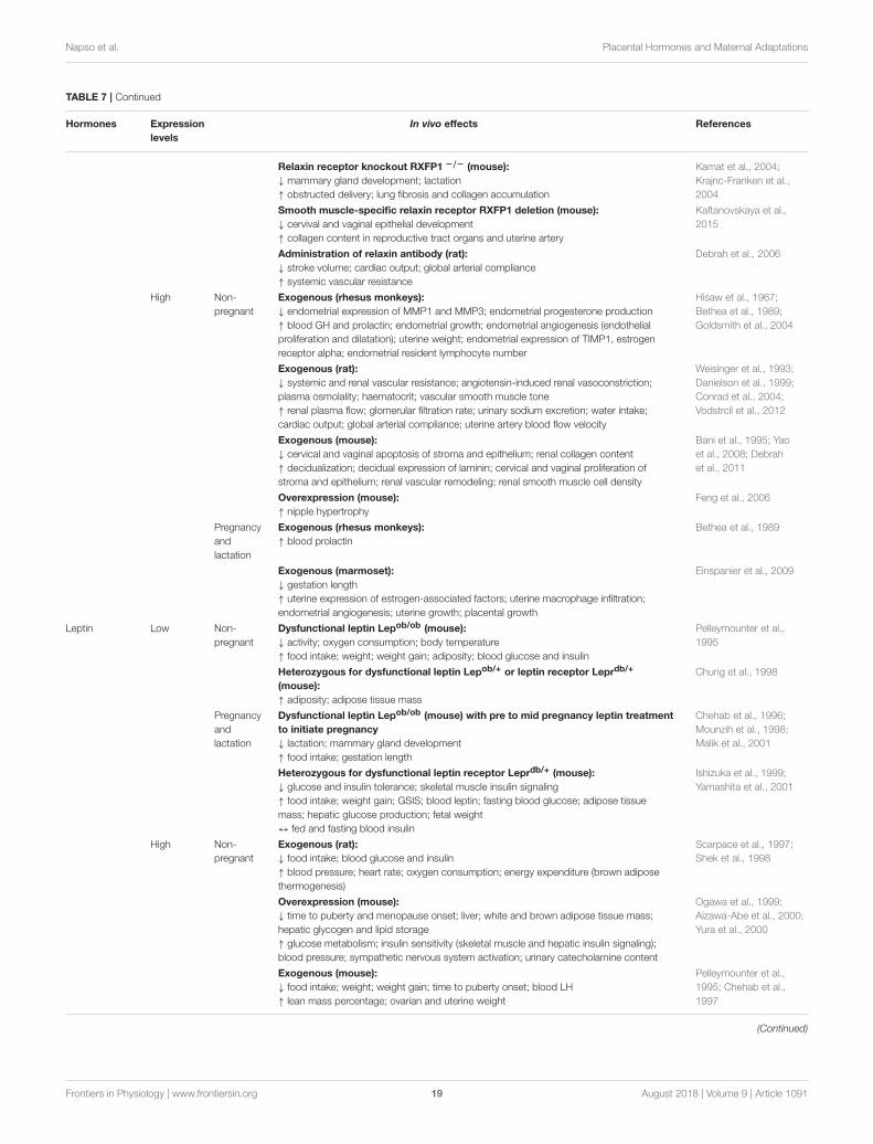
Napso et al. Placental Hormones and Maternal Adaptations
TABLE 7 | Continued
Hormones Expression
levels
In vivo effects References
Relaxin receptor knockout RXFP1 −/− (mouse):
↓ mammary gland development; lactation
↑ obstructed delivery; lung fibrosis and collagen accumulation
Kamat et al., 2004;
Krajnc-Franken et al.,
2004
Smooth muscle-specific relaxin receptor RXFP1 deletion (mouse):
↓ cervival and vaginal epithelial development
↑ collagen content in reproductive tract organs and uterine artery
Kaftanovskaya et al.,
2015
Administration of relaxin antibody (rat):
↓ stroke volume; cardiac output; global arterial compliance
↑ systemic vascular resistance
Debrah et al., 2006
High Non-
pregnant
Exogenous (rhesus monkeys):
↓ endometrial expression of MMP1 and MMP3; endometrial progesterone production
↑ blood GH and prolactin; endometrial growth; endometrial angiogenesis (endothelial
proliferation and dilatation); uterine weight; endometrial expression of TIMP1, estrogen
receptor alpha; endometrial resident lymphocyte number
Hisaw et al., 1967;
Bethea et al., 1989;
Goldsmith et al., 2004
Exogenous (rat):
↓ systemic and renal vascular resistance; angiotensin-induced renal vasoconstriction;
plasma osmolality; haematocrit; vascular smooth muscle tone
↑ renal plasma flow; glomerular filtration rate; urinary sodium excretion; water intake;
cardiac output; global arterial compliance; uterine artery blood flow velocity
Weisinger et al., 1993;
Danielson et al., 1999;
Conrad et al., 2004;
Vodstrcil et al., 2012
Exogenous (mouse):
↓ cervical and vaginal apoptosis of stroma and epithelium; renal collagen content
↑ decidualization; decidual expression of laminin; cervical and vaginal proliferation of
stroma and epithelium; renal vascular remodeling; renal smooth muscle cell density
Bani et al., 1995; Yao
et al., 2008; Debrah
et al., 2011
Overexpression (mouse):
↑ nipple hypertrophy
Feng et al., 2006
Pregnancy
and
lactation
Exogenous (rhesus monkeys):
↑ blood prolactin
Bethea et al., 1989
Exogenous (marmoset):
↓ gestation length
↑ uterine expression of estrogen-associated factors; uterine macrophage infiltration;
endometrial angiogenesis; uterine growth; placental growth
Einspanier et al., 2009
Leptin Low Non-
pregnant
Dysfunctional leptin Lepob/ob (mouse):
↓ activity; oxygen consumption; body temperature
↑ food intake; weight; weight gain; adiposity; blood glucose and insulin
Pelleymounter et al.,
1995
Heterozygous for dysfunctional leptin Lepob/+ or leptin receptor Leprdb/+
(mouse):
↑ adiposity; adipose tissue mass
Chung et al., 1998
Pregnancy
and
lactation
Dysfunctional leptin Lepob/ob (mouse) with pre to mid pregnancy leptin treatment
to initiate pregnancy
↓ lactation; mammary gland development
↑ food intake; gestation length
Chehab et al., 1996;
Mounzih et al., 1998;
Malik et al., 2001
Heterozygous for dysfunctional leptin receptor Leprdb/+ (mouse):
↓ glucose and insulin tolerance; skeletal muscle insulin signaling
↑ food intake; weight gain; GSIS; blood leptin; fasting blood glucose; adipose tissue
mass; hepatic glucose production; fetal weight
↔ fed and fasting blood insulin
Ishizuka et al., 1999;
Yamashita et al., 2001
High Non-
pregnant
Exogenous (rat):
↓ food intake; blood glucose and insulin
↑ blood pressure; heart rate; oxygen consumption; energy expenditure (brown adipose
thermogenesis)
Scarpace et al., 1997;
Shek et al., 1998
Overexpression (mouse):
↓ time to puberty and menopause onset; liver; white and brown adipose tissue mass;
hepatic glycogen and lipid storage
↑ glucose metabolism; insulin sensitivity (skeletal muscle and hepatic insulin signaling);
blood pressure; sympathetic nervous system activation; urinary catecholamine content
Ogawa et al., 1999;
Aizawa-Abe et al., 2000;
Yura et al., 2000
Exogenous (mouse):
↓ food intake; weight; weight gain; time to puberty onset; blood LH
↑ lean mass percentage; ovarian and uterine weight
Pelleymounter et al.,
1995; Chehab et al.,
1997
(Continued)
Frontiers in Physiology | www.frontiersin.org 19 August 2018 | Volume 9 | Article 1091
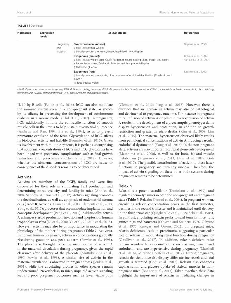
Napso et al. Placental Hormones and Maternal Adaptations
TABLE 7 | Continued
Hormones Expression
levels
In vivo effects References
Pregnancy
and
lactation
Overexpression (mouse):
↓ food intake; fetal weight
↑ blood pressure; pregnancy-associated rise in blood leptin
Sagawa et al., 2002
Exogenous (mouse):
↓ food intake; weight gain; GSIS; fed blood insulin; fasting blood insulin and leptin;
adipose tissue mass; fetal and placental weights; placental leptin
↑ fed blood glucose
Kulkarni et al., 1997;
Yamashita et al., 2001
Exogenous (rat):
↑ blood pressure; proteinuria; blood markers of endothelial activation (E-selectin and
ICAM-1)
↔ food intake; weight
Ibrahim et al., 2013
cAMP, Cyclic adenosine monophosphate; FSH, Follicle stimulating hormone; GSIS, Glucose-stimulated insulin secretion; ICAM-1, Intercellular adhesion molecule 1; LH, Luteinizing
hormone; MMP, Matrix metalloproteinase; TIMP, Tissue inhibitor of metalloproteinase.
IL-10 by B cells (Fettke et al., 2016). hCG can also modulatethe immune system even in a non-pregnant state, as shownby its efficacy in preventing the development of autoimmunediabetes in a mouse model (Khil et al., 2007). In pregnancy,hCG additionally inhibits the contractile function of smoothmuscle cells in the uterus to help sustain myometrial quiescence(Ambrus and Rao, 1994; Eta et al., 1994), so as to preventpremature expulsion of the fetus. Glycosylation of hCG affectsits biological activity and half-life (Fournier et al., 2015). Givenits involvement with multiple systems, it is perhaps unsurprisingthat abnormal concentrations of hCG and hCG glycoforms havebeen linked with pregnancy complications such as fetal growthrestriction and preeclampsia (Chen et al., 2012). However,whether the abnormal concentrations of hCG are cause orconsequence of the disorders remains to be determined.
ActivinsActivins are members of the TGFβ family and were firstdiscovered for their role in stimulating FSH production anddetermining estrus cyclicity and fertility in mice (Ahn et al.,2004; Sandoval-Guzmán et al., 2012). Activin signaling promotesthe decidualization, as well as, apoptosis of endometrial stromacells (Table 8; Activins; Tessier et al., 2003; Clementi et al., 2013;Yong et al., 2017); processes that accommodate implantation andconceptus development (Peng et al., 2015). Additionally, activinA enhances steroid production, invasion and apoptosis of humantrophoblast in vitro (Ni et al., 2000; Yu et al., 2012; Li et al., 2015).However, activins may also be of importance in modulating thephysiology of the mother during pregnancy (Table 7; Activins).In normal human pregnancy, activin A concentrations graduallyrise during gestation and peak at term (Fowler et al., 1998).The placenta is thought to be the main source of activin Ain the maternal circulation during pregnancy, given the rapidclearance after delivery of the placenta (Muttukrishna et al.,1997; Fowler et al., 1998). A similar rise of activin in thematernal circulation is observed in pregnant ewes (Jenkin et al.,2001), while the circulating profiles in other species remainundetermined. Nevertheless, in mice, impaired activin signalingleads to poor pregnancy outcomes such as fewer viable pups
(Clementi et al., 2013; Peng et al., 2015). However, there isevidence that an increase in activin may also be pathologicaland detrimental to pregnancy outcome. For instance in pregnantmice, infusion of activin A or plasmid overexpression of activinA results in the development of a preeclamptic phenotype; damsdisplay hypertension and proteinuria, in addition to growthrestriction and greater in utero deaths (Kim et al., 2008; Limet al., 2015). The maternal hypertension observed likely resultsfrom pathological concentrations of activin A inducing vascularendothelial dysfunction (Yong et al., 2015). In the non-pregnantstate, activins are also important for renal glomeruli development(Maeshima et al., 2000), as well as, for bone, fat and musclemetabolism (Yogosawa et al., 2013; Ding et al., 2017; Gohet al., 2017). The possible contributions of activin to these latterfunctions in pregnancy are currently unclear. Therefore, theimpact of activin signaling on these other body systems duringpregnancy remains to be determined.
RelaxinRelaxin is a potent vasodilator (Danielson et al., 1999), andregulates hemodynamics in both the non-pregnant and pregnantstate (Table 7; Relaxin; Conrad et al., 2004). In pregnant women,circulating relaxin concentration peaks in the first trimester,declines in the second trimester and is maintained until deliveryin the third trimester (Quagliarello et al., 1979; Seki et al., 1985).In contrast, circulating relaxin peaks toward term in mice, rats,guinea pigs and hamsters (O’byrne and Steinetz, 1976; O’byrneet al., 1976; Renegar and Owens, 2002). In pregnant mice,relaxin deficiency leads to proteinuria, suggesting a particularrole of relaxin in modulating renal function during pregnancy(O’sullivan et al., 2017). In addition, relaxin-deficient miceremain sensitive to vasoconstrictors such as angiotensin andendothelin, and are hypertensive during pregnancy (Marshallet al., 2016a; Mirabito Colafella et al., 2017). During pregnancy,relaxin-deficient mice also display stiffer uterine vessels and fetalgrowth is retarded (Gooi et al., 2013). Relaxin also enhancescapillarisation and glucose uptake of skeletal muscles in non-pregnant mice (Bonner et al., 2013). Taken together, these datahighlight the importance of relaxin in mediating changes in
Frontiers in Physiology | www.frontiersin.org 20 August 2018 | Volume 9 | Article 1091
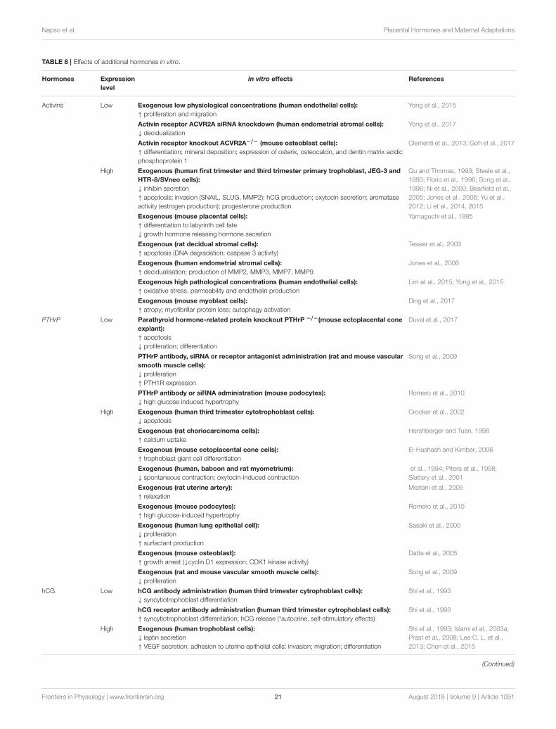
Napso et al. Placental Hormones and Maternal Adaptations
TABLE 8 | Effects of additional hormones in vitro.
Hormones Expression
level
In vitro effects References
Activins Low Exogenous low physiological concentrations (human endothelial cells):
↑ proliferation and migration
Yong et al., 2015
Activin receptor ACVR2A siRNA knockdown (human endometrial stromal cells):
↓ decidualization
Yong et al., 2017
Activin receptor knockout ACVR2A−/− (mouse osteoblast cells):
↑ differentiation; mineral deposition; expression of osterix, osteocalcin, and dentin matrix acidic
phosphoprotein 1
Clementi et al., 2013; Goh et al., 2017
High Exogenous (human first trimester and third trimester primary trophoblast, JEG-3 and
HTR-8/SVneo cells):
↓ inhibin secretion
↑ apoptosis; invasion (SNAIL, SLUG, MMP2); hCG production; oxytocin secretion; aromatase
activity (estrogen production); progesterone production
Qu and Thomas, 1993; Steele et al.,
1993; Florio et al., 1996; Song et al.,
1996; Ni et al., 2000; Bearfield et al.,
2005; Jones et al., 2006; Yu et al.,
2012; Li et al., 2014, 2015
Exogenous (mouse placental cells):
↑ differentiation to labyrinth cell fate
↓ growth hormone releasing hormone secretion
Yamaguchi et al., 1995
Exogenous (rat decidual stromal cells):
↑ apoptosis (DNA degradation; caspase 3 activity)
Tessier et al., 2003
Exogenous (human endometrial stromal cells):
↑ decidualisation; production of MMP2, MMP3, MMP7, MMP9
Jones et al., 2006
Exogenous high pathological concentrations (human endothelial cells):
↑ oxidative stress, permeability and endothelin production
Lim et al., 2015; Yong et al., 2015
Exogenous (mouse myoblast cells):
↑ atropy; myofibrillar protein loss; autophagy activation
Ding et al., 2017
PTHrP Low Parathyroid hormone-related protein knockout PTHrP −/−(mouse ectoplacental cone
explant):
↑ apoptosis
↓ proliferation; differentiation
Duval et al., 2017
PTHrP antibody, siRNA or receptor antagonist administration (rat and mouse vascular
smooth muscle cells):
↓ proliferation
↑ PTH1R expression
Song et al., 2009
PTHrP antibody or siRNA administration (mouse podocytes):
↓ high glucose induced hypertrophy
Romero et al., 2010
High Exogenous (human third trimester cytotrophoblast cells):
↓ apoptosis
Crocker et al., 2002
Exogenous (rat choriocarcinoma cells):
↑ calcium uptake
Hershberger and Tuan, 1998
Exogenous (mouse ectoplacental cone cells):
↑ trophoblast giant cell differentiation
El-Hashash and Kimber, 2006
Exogenous (human, baboon and rat myometrium):
↓ spontaneous contraction; oxytocin-induced contraction
et al., 1994; Pitera et al., 1998;
Slattery et al., 2001
Exogenous (rat uterine artery):
↑ relaxation
Meziani et al., 2005
Exogenous (mouse podocytes):
↑ high glucose-induced hypertrophy
Romero et al., 2010
Exogenous (human lung epithelial cell):
↓ proliferation
↑ surfactant production
Sasaki et al., 2000
Exogenous (mouse osteoblast):
↑ growth arrest (↓cyclin D1 expression; CDK1 kinase activity)
Datta et al., 2005
Exogenous (rat and mouse vascular smooth muscle cells):
↓ proliferation
Song et al., 2009
hCG Low hCG antibody administration (human third trimester cytrophoblast cells):
↓ syncytiotrophoblast differentiation
Shi et al., 1993
hCG receptor antibody administration (human third trimester cytrophoblast cells):
↑ syncytiotrophoblast differentiation; hCG release (*autocrine, self-stimulatory effects)
Shi et al., 1993
High Exogenous (human trophoblast cells):
↓ leptin secretion
↑ VEGF secretion; adhesion to uterine epithelial cells; invasion; migration; differentiation
Shi et al., 1993; Islami et al., 2003a;
Prast et al., 2008; Lee C. L. et al.,
2013; Chen et al., 2015
(Continued)
Frontiers in Physiology | www.frontiersin.org 21 August 2018 | Volume 9 | Article 1091
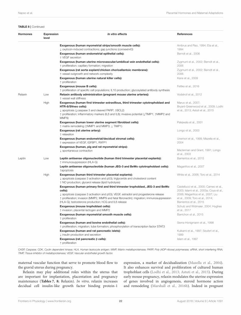
Napso et al. Placental Hormones and Maternal Adaptations
TABLE 8 | Continued
Hormones Expression
level
In vitro effects References
Exogenous (human myometrial strips/smooth muscle cells):
↓ oxytocin-induced contractions; gap junctions (connexin43)
Ambrus and Rao, 1994; Eta et al.,
1994
Exogenous (human endometrial epithelial cells):
↑ VEGF secretion
Berndt et al., 2006
Exogenous (human uterine microvascular/umbilical vein endothelial cells):
↑ proliferation; capillary formation; migration
Zygmunt et al., 2002; Berndt et al.,
2006
Exogenous (rat aorta explant/chicken chorioallantoic membrane):
↑ vessel outgrowth and network complexity
Zygmunt et al., 2002; Berndt et al.,
2006
Exogenous (human uterine natural killer cells):
↑ proliferation
Kane et al., 2009
Exogenous (mouse B cells):
↑ proliferation of specific cell populations; IL10 production; glycosylated antibody synthesis
Fettke et al., 2016
Relaxin Low Relaxin antibody administration (pregnant mouse uterine arteries):
↑ vessel wall stiffness
Vodstrcil et al., 2012
High Exogenous (human first trimester extravillous, third trimester cytotrophoblast and
HTR-8/SVneo cells):
↓ apoptosis (↓caspase 3 and cleaved PARP; ↑BCL2)
↑ proliferation; inflammatory markers (IL6 and IL8); invasive potential (↓TIMP1; ↑MMP2 and
MMP9)
Maruo et al., 2007;
Bryant-Greenwood et al., 2009; Lodhi
et al., 2013; Astuti et al., 2015
Exogenous (human lower uterine segment fibroblast cells):
↑ matrix remodeling (↑MMP1 and MMP3; ↓ TIMP1)
Palejwala et al., 2001
Exogenous (rat uterine artery):
↑ relaxation
Longo et al., 2003
Exogenous (human endometrial/decidual stromal cells):
↑ expression of VEGF, IGFBP1, RXFP1
Unemori et al., 1999; Mazella et al.,
2004
Exogenous (human, pig and rat myometrial strips):
↓ spontaneous contraction Maclennan and Grant, 1991; Longo
et al., 2003
Leptin Low Leptin antisense oligonucleotide (human third trimester placental explants):
↑ immunosuppression (HLA-G)
Barrientos et al., 2015
Leptin antisense oligonucleotide (human JEG-3 and BeWo cytotrophoblast cells):
↑apoptosis
Magariños et al., 2007
High Exogenous (human third trimester placental explants):
↓ apoptosis (caspase 3 activation and p53); triglyceride and cholesterol content
↑ NO production; glycerol release (lipid hydrolysis)
White et al., 2006; Toro et al., 2014
Exogenous (human primary first and third trimester trophoblast, JEG-3 and BeWo
cells):
↓ apoptosis (caspase 3 activation and p53); VEGF, estradiol and progesterone release
↑ proliferation; invasion (MMP2, MMP9 and fetal fibronectin); migration; immunosuppression
(HLA-G); testosterone production; hCG and IL6 release
Castellucci et al., 2000; Cameo et al.,
2003; Islami et al., 2003a; Coya et al.,
2006; Magariños et al., 2007; Liu
et al., 2009; Toro et al., 2014;
Barrientos et al., 2015
Exogenous (mouse trophoblast cells):
↑ invasion; placental lactogen and MMP2
Schulz and Widmaier, 2004; Hughes
et al., 2017
Exogenous (human myometrial smooth muscle cells):
↑ proliferation
Barrichon et al., 2015
Exogenous (human and bovine endothelial cells):
↑ proliferation; migration; tube formation; phosphorylation of transcription factor STAT3
Sierra-Honigmann et al., 1998
Exogenous (human and rat pancreatic islets):
↓ insulin production and secretion
Kulkarni et al., 1997; Seufert et al.,
1999
Exogenous (rat pancreatic β cells):
↑ proliferation
Islam et al., 1997
CASP, Caspase; CDK, Cyclin dependent kinase; HLA, Human leukocyte antigen; MMP, Matrix metalloproteinase; PARP, Poly (ADP-ribose) polymerase; siRNA, short interfering RNA;
TIMP, Tissue inhibitor of metalloproteinase; VEGF, Vascular endothelial growth factor.
maternal vascular function that serve to promote blood flow tothe gravid uterus during pregnancy.
Relaxin may play additional roles within the uterus thatare important for implantation, placentation and pregnancymaintenance (Tables 7, 8; Relaxin). In vitro, relaxin increasesdecidual cell insulin-like growth factor binding protein-1
expression, a marker of decidualization (Mazella et al., 2004).It also enhances survival and proliferation of cultured humantrophoblast cells (Lodhi et al., 2013; Astuti et al., 2015). Duringearly mouse pregnancy, relaxin modulates the uterine expressionof genes involved in angiogenesis, steroid hormone actionand remodeling (Marshall et al., 2016b). Indeed in pregnant
Frontiers in Physiology | www.frontiersin.org 22 August 2018 | Volume 9 | Article 1091
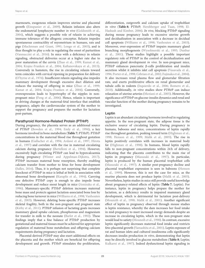
Napso et al. Placental Hormones and Maternal Adaptations
marmosets, exogenous relaxin improves uterine and placentalgrowth (Einspanier et al., 2009). Relaxin infusion also altersthe endometrial lymphocyte number in vivo (Goldsmith et al.,2004), which suggests a possible role of relaxin in achievingimmune tolerance of the allogenic conceptus. Relaxin impedesspontaneous contractility of myometrium in humans, rats andpigs (Maclennan and Grant, 1991; Longo et al., 2003), and isthus thought to play a role in regulating the onset of parturition(Vannuccini et al., 2016). In mice with a deficiency in relaxinsignaling, obstructed deliveries occur at a higher rate due topoor maturation of the cervix (Zhao et al., 1999; Kamat et al.,2004; Krajnc-Franken et al., 2004; Kaftanovskaya et al., 2015).Conversely in hamsters, the rise in circulating relaxin towardterm coincides with cervical ripening in preparation for delivery(O’byrne et al., 1976). Insufficient relaxin signaling also impedesmammary development through excessive duct dilation andreduces the nursing of offspring in mice (Zhao et al., 1999;Kamat et al., 2004; Krajnc-Franken et al., 2004). Conversely,overexpression leads to hypertrophy of the nipples in non-pregnant mice (Feng et al., 2006). Hence, relaxin is importantin driving changes at the maternal-fetal interface that establishpregnancy, adapts the cardiovascular system of the mother tosupport the pregnancy and prepares the mother for lactationpost-partum.
Parathyroid Hormone-Related Protein (PTHrP)During pregnancy, the placenta serves as an additional sourceof PTHrP (Bowden et al., 1994; Emly et al., 1994), a keyhormone involved in bonemetabolism (Table 7; PTHrP). PTHrPconcentrations in the maternal blood rise throughout gestationin humans (Gallacher et al., 1994; Ardawi et al., 1997; Hirotaet al., 1997) and correlate with the rise in maternal circulatingcalcium during pregnancy (Bertelloni et al., 1994). However,excessively high circulating PTHrP can lead to hypercalcaemiaduring pregnancy (Winter and Appelman-Dijkstra, 2017).PTHrP increases maternal bone resorption, thereby enablingcalcium transfer from mother to fetus for bone development(Salles, 2016). Thus, it is perhaps not surprising that completeknockout of PTHrP in mice is lethal at birth in association withabnormal bone development (Karaplis et al., 1994). Carryingone defective PTHrP copy is enough to also impede bonedevelopment and reduce snout length in mice (Amizuka et al.,1996). Mammary-specific PTHrP deletion increases maternalbone mass and protects against lactation-associated bone loss byreducing bone turnover inmice (Williams et al., 1998; Vanhoutenet al., 2003). However, deleting bone-specific PTHrP increasesskeletal fragility, both in the non-pregnant and pregnant state(Kirby et al., 2011). PTHrP infusion of lactating goats increasesmammary gland uptake calcium, phosphorous and magnesiumfor transfer in milk to the neonate (Barlet et al., 1992). Thesefindings imply that a fine balance of PTHrP production bygestational andmaternal tissues must be achieved for appropriateregulation of maternal bone metabolism and offspring calciumrequirements during pregnancy and lactation.
Placental-derived PTHrP may also exert additional effects onthe placenta and the mother which are beneficial for offspringdevelopment and growth. PTHrP stimulates the proliferation,
differentiation, outgrowth and calcium uptake of trophoblastin vitro (Table 8; PTHrP; Hershberger and Tuan, 1998; El-Hashash and Kimber, 2006). In vivo, blocking PTHrP signalingduring mouse pregnancy leads to excessive uterine growthand decidualization in association with a decrease in decidualcell apoptosis (Williams et al., 1998; Vanhouten et al., 2003).Moreover, over-expression of PTHrP impairs mammary glandbranching morphogenesis (Wysolmerski et al., 1995; Dunbaret al., 2001). These studies highlight a possible importantregulatory role of PTHrP in the control of decidualization andmammary gland development in vivo. In non-pregnant mice,PTHrP enhances pancreatic β-cells proliferation and insulinsecretion whilst it inhibits islet cell apoptosis (Vasavada et al.,1996; Porter et al., 1998; Cebrian et al., 2002; Fujinaka et al., 2004).It also increases renal plasma flow and glomerular filtrationrate, and exerts proliferative effects on renal glomerular andtubule cells in rodents (Izquierdo et al., 2006; Romero et al.,2010). Additionally, in vitro studies show PTHrP can inducerelaxation of uterine arteries (Meziani et al., 2005). However, thesignificance of PTHrP on glucose-insulin dynamics and renal andvascular function of the mother during pregnancy remains to beinvestigated.
LeptinLeptin is an abundant circulating hormone involved in regulatingappetite. In the non-pregnant state, the adipose tissue is theexclusive source of circulating leptin. During pregnancy inhumans, baboons and mice, concentrations of leptin rapidlyrise throughout gestation, peaking toward term (Highman et al.,1998; Henson et al., 1999; Malik et al., 2005). The rise inleptin positively correlates with increases in maternal bodyfat (Highman et al., 1998). In humans, blood leptin rapidlyfalls to non-pregnant concentrations within 24 h of delivery,indicating that the placenta contributes to the main rise ofleptin in pregnancy (Masuzaki et al., 1997). In particular,leptin is produced by the human placental trophoblast cells(Masuzaki et al., 1997). A similar post-pregnancy decline andplacental trophoblast expression is seen in baboons (Hensonet al., 1999). However, this is not the case for mice, as themurine placenta does not produce leptin (Malik et al., 2005).Nevertheless, leptin studies inmice still provide useful knowledgeabout pregnancy-related effects of leptin (Table 7; Leptin). Forinstance, leptin in pregnancy helps prepare the mother forlactation, as a deficiency results in impaired mammary glanddevelopment, which is detrimental for lactation post-delivery(Mounzih et al., 1998; Malik et al., 2001). Another significanteffect of leptin in pregnancy observed through mouse studiesis leptin resistance, whereby the dam increases her food intakein mid-pregnancy to meet increased energy demands despite anincrease in circulating leptin, which in the non-pregnant statewould lead to satiety (Mounzih et al., 1998). In contrast, excessiveleptin significantly decreases maternal food intake and restrictsfeto-placental growth (Yamashita et al., 2001). Leptin exposure ofrat and human islets and cultured insulinoma cells significantlydecreases insulin production in vitro, demonstrating that leptinmay be directly involved in glucose metabolism (Table 8; Leptin;Kulkarni et al., 1997). Indeed dysfunctional leptin signaling in
Frontiers in Physiology | www.frontiersin.org 23 August 2018 | Volume 9 | Article 1091
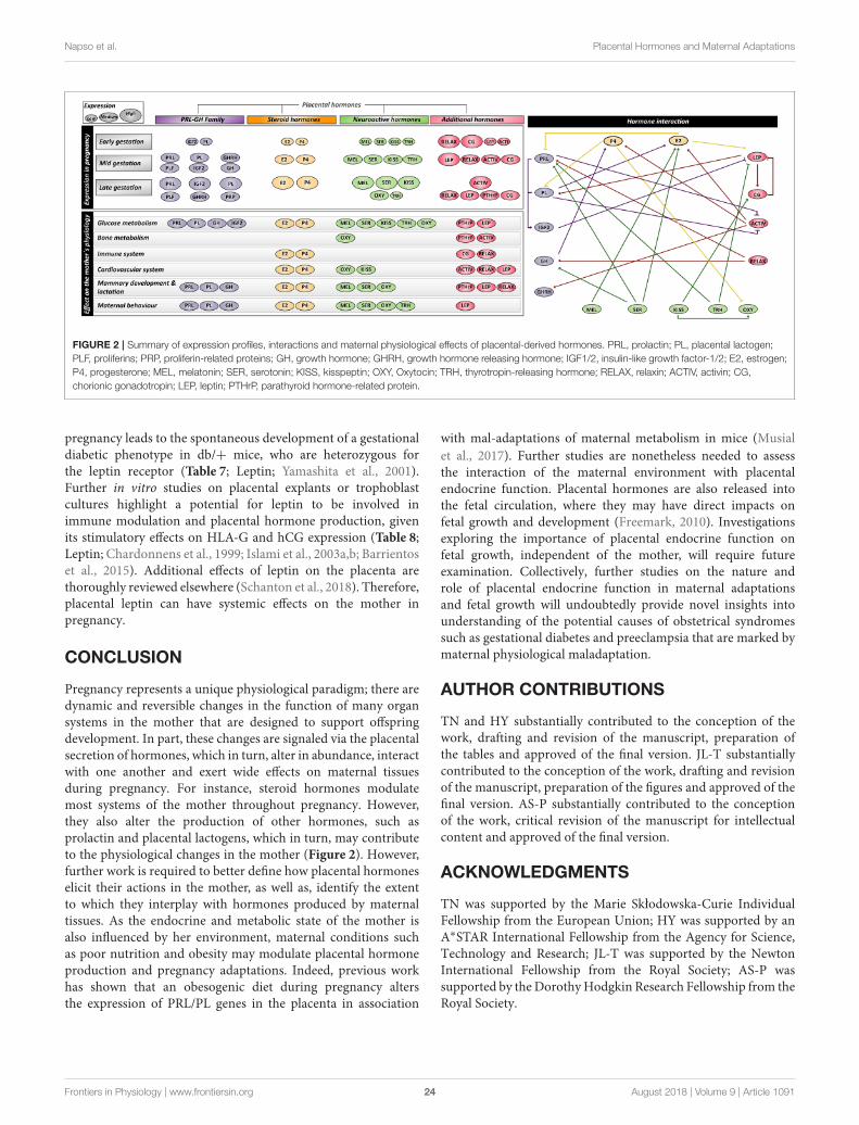
Napso et al. Placental Hormones and Maternal Adaptations
FIGURE 2 | Summary of expression profiles, interactions and maternal physiological effects of placental-derived hormones. PRL, prolactin; PL, placental lactogen;
PLF, proliferins; PRP, proliferin-related proteins; GH, growth hormone; GHRH, growth hormone releasing hormone; IGF1/2, insulin-like growth factor-1/2; E2, estrogen;
P4, progesterone; MEL, melatonin; SER, serotonin; KISS, kisspeptin; OXY, Oxytocin; TRH, thyrotropin-releasing hormone; RELAX, relaxin; ACTIV, activin; CG,
chorionic gonadotropin; LEP, leptin; PTHrP, parathyroid hormone-related protein.
pregnancy leads to the spontaneous development of a gestationaldiabetic phenotype in db/+ mice, who are heterozygous forthe leptin receptor (Table 7; Leptin; Yamashita et al., 2001).Further in vitro studies on placental explants or trophoblastcultures highlight a potential for leptin to be involved inimmune modulation and placental hormone production, givenits stimulatory effects on HLA-G and hCG expression (Table 8;Leptin; Chardonnens et al., 1999; Islami et al., 2003a,b; Barrientoset al., 2015). Additional effects of leptin on the placenta arethoroughly reviewed elsewhere (Schanton et al., 2018). Therefore,placental leptin can have systemic effects on the mother inpregnancy.
CONCLUSION
Pregnancy represents a unique physiological paradigm; there aredynamic and reversible changes in the function of many organsystems in the mother that are designed to support offspringdevelopment. In part, these changes are signaled via the placentalsecretion of hormones, which in turn, alter in abundance, interactwith one another and exert wide effects on maternal tissuesduring pregnancy. For instance, steroid hormones modulatemost systems of the mother throughout pregnancy. However,they also alter the production of other hormones, such asprolactin and placental lactogens, which in turn, may contributeto the physiological changes in the mother (Figure 2). However,further work is required to better define how placental hormoneselicit their actions in the mother, as well as, identify the extentto which they interplay with hormones produced by maternaltissues. As the endocrine and metabolic state of the mother isalso influenced by her environment, maternal conditions suchas poor nutrition and obesity may modulate placental hormoneproduction and pregnancy adaptations. Indeed, previous workhas shown that an obesogenic diet during pregnancy altersthe expression of PRL/PL genes in the placenta in association
with mal-adaptations of maternal metabolism in mice (Musialet al., 2017). Further studies are nonetheless needed to assessthe interaction of the maternal environment with placentalendocrine function. Placental hormones are also released intothe fetal circulation, where they may have direct impacts onfetal growth and development (Freemark, 2010). Investigationsexploring the importance of placental endocrine function onfetal growth, independent of the mother, will require futureexamination. Collectively, further studies on the nature androle of placental endocrine function in maternal adaptationsand fetal growth will undoubtedly provide novel insights intounderstanding of the potential causes of obstetrical syndromessuch as gestational diabetes and preeclampsia that are marked bymaternal physiological maladaptation.
AUTHOR CONTRIBUTIONS
TN and HY substantially contributed to the conception of thework, drafting and revision of the manuscript, preparation ofthe tables and approved of the final version. JL-T substantiallycontributed to the conception of the work, drafting and revisionof the manuscript, preparation of the figures and approved of thefinal version. AS-P substantially contributed to the conceptionof the work, critical revision of the manuscript for intellectualcontent and approved of the final version.
ACKNOWLEDGMENTS
TN was supported by the Marie Skłodowska-Curie IndividualFellowship from the European Union; HY was supported by anA∗STAR International Fellowship from the Agency for Science,Technology and Research; JL-T was supported by the NewtonInternational Fellowship from the Royal Society; AS-P wassupported by theDorothyHodgkin Research Fellowship from theRoyal Society.
Frontiers in Physiology | www.frontiersin.org 24 August 2018 | Volume 9 | Article 1091
Napso et al. Placental Hormones and Maternal Adaptations
REFERENCES
Abd-Allah, A. R., El-Sayed El, S. M., Abdel-Wahab, M. H., and Hamada,F. M. (2003). Effect of melatonin on estrogen and progesterone receptorsin relation to uterine contraction in rats. Pharmacol. Res. 47, 349–354.doi: 10.1016/S1043-6618(03)00014-8
Abribat, T., Lapierre, H., Dubreuil, P., Pelletier, G., Gaudreau, P., Brazeau,P., et al. (1990). Insulin-like growth factor-I concentration in Holsteinfemale cattle: variations with age, stage of lactation and growth hormone-releasing factor administration. Domest. Anim. Endocrinol. 7, 93–102.doi: 10.1016/0739-7240(90)90058-8
Açikgöz, S., Bayar, U. O., Can, M., Güven, B., Mungan, G., Dogan, S., et al. (2013).Levels of oxidized LDL, estrogens, and progesterone in placenta tissues andserum paraoxonase activity in preeclampsia. Mediators Inflamm. 2013:862982.doi: 10.1155/2013/862982
Ackermann, A. M., and Gannon, M. (2007). Molecular regulation of pancreaticbeta-cell mass development, maintenance, and expansion. J. Mol. Endocrinol.
38, 193–206. doi: 10.1677/JME-06-0053Adamah-Biassi, E. B., Hudson, R. L., and Dubocovich, M. L. (2014).
Genetic deletion of MT1 melatonin receptors alters spontaneous behavioralrhythms in male and female C57BL/6 mice. Horm. Behav. 66, 619–627.doi: 10.1016/j.yhbeh.2014.08.012
Adamova, Z., Ozkan, S., and Khalil, R. A. (2009). Vascular and cellular calciumin normal and hypertensive pregnancy. Curr. Clin. Pharmacol. 4, 172–190.doi: 10.2174/157488409789375320
Ahmed-Sorour, H., and Bailey, C. J. (1980). Role of ovarian hormones in the long-term control of glucose homeostasis. Interaction with insulin, glucagon andepinephrine. Horm. Res. 13, 396–403. doi: 10.1159/000179307
Ahmed-Sorour, H., and Bailey, C. J. (1981). Role of ovarian hormones in the long-term control of glucose homeostasis, glycogen formation and gluconeogenesis.Ann. Nutr. Metab. 25, 208–212. doi: 10.1159/000176496
Ahn, J. M., Jung, H. K., Cho, C., Choi, D., Mayo, K. E., and Cho, B. N. (2004).Changes in the reproductive functions of mice due to injection of a plasmidexpressing an inhibin alpha-subunit into muscle: a transient transgenic model.Mol. Cells 18, 79–86.
Ahumada-Solórzano, S. M., Martínez-Moreno, C. G., Carranza, M., Ávila-Mendoza, J., Luna-Acosta, J. L., Harvey, S., et al. (2016). Autocrine/paracrineproliferative effect of ovarian GH and IGF-I in chicken granulosa cell cultures.Gen. Comp. Endocrinol. 234, 47–56. doi: 10.1016/j.ygcen.2016.05.008
Aizawa-Abe, M., Ogawa, Y., Masuzaki, H., Ebihara, K., Satoh, N., Iwai, H., et al.(2000). Pathophysiological role of leptin in obesity-related hypertension. J. Clin.Invest. 105, 1243–1252. doi: 10.1172/JCI8341
Alenina, N., Kikic, D., Todiras, M., Mosienko, V., Qadri, F., Plehm, R.,et al. (2009). Growth retardation and altered autonomic control in micelacking brain serotonin. Proc. Natl. Acad. Sci. U.S.A. 106, 10332–10337.doi: 10.1073/pnas.0810793106
Alperin, M., Kaddis, T., Pichika, R., Esparza, M. C., and Lieber, R. L. (2016).Pregnancy-induced adaptations in intramuscular extracellular matrix of ratpelvic floor muscles. Am. J. Obstet. Gynecol. 215, 210 e211–210 e217.doi: 10.1016/j.ajog.2016.02.018
Alperin, M., Lawley, D. M., Esparza, M. C., and Lieber, R. L. (2015). Pregnancy-induced adaptations in the intrinsic structure of rat pelvic floor muscles. Am. J.
Obstet. Gynecol. 213, 191 e191–191 e197. doi: 10.1016/j.ajog.2015.05.012Ambrus, G., and Rao, C. V. (1994). Novel regulation of pregnant
human myometrial smooth muscle cell gap junctions byhuman chorionic gonadotropin. Endocrinology 135, 2772–2779.doi: 10.1210/endo.135.6.7988470
Amico, J. A., Vollmer, R. R., Cai, H. M., Miedlar, J. A., and Rinaman, L. (2005).Enhanced initial and sustained intake of sucrose solution in mice with anoxytocin gene deletion. Am. J. Physiol. Regul. Integr. Comp. Physiol. 289,R1798–R1806. doi: 10.1152/ajpregu.00558.2005
Amizuka, N., Karaplis, A. C., Henderson, J. E., Warshawsky, H., Lipman, M. L.,Matsuki, Y., et al. (1996). Haploinsufficiency of parathyroid hormone-relatedpeptide (PTHrP) results in abnormal postnatal bone development. Dev. Biol.175, 166–176. doi: 10.1006/dbio.1996.0104
Angoa-Pérez, M., Kane, M. J., Sykes, C. E., Perrine, S. A., Church, M. W.,and Kuhn, D. M. (2014). Brain serotonin determines maternal behavior andoffspring survival. Genes Brain Behav. 13, 579–591. doi: 10.1111/gbb.12159
Angoa-Pérez, M., and Kuhn, D. M. (2015). Neuronal serotonin in theregulation of maternal behavior in rodents. Neurotransmitter (Houst) 2:e615.doi: 10.14800/nt.615
Antonijevic, I. A., Leng, G., Luckman, S. M., Douglas, A. J., Bicknell, R. J.,and Russell, J. A. (1995). Induction of uterine activity with oxytocin inlate pregnant rats replicates the expression of c-fos in neuroendocrine andbrain stem neurons as seen during parturition. Endocrinology 136, 154–163.doi: 10.1210/endo.136.1.7828526
Apa, R., Lanzone, A., Miceli, F., Mastrandrea, M., Macchione, E., Caruso, A., et al.(1995). Growth hormone-releasing factor stimulates meiotic maturation infollicle- and cumulus-enclosed rat oocyte.Mol. Cell. Endocrinol. 112, 195–201.doi: 10.1016/0303-7207(95)03599-3
Ardawi, M. S., Nasrat, H. A., and BA’Aqueel, H. S. (1997). Calcium-regulatinghormones and parathyroid hormone-related peptide in normal humanpregnancy and postpartum: a longitudinal study. Eur. J. Endocrinol. 137,402–409. doi: 10.1530/eje.0.1370402
Arletti, R., Benelli, A., and Bertolini, A. (1989). Influence of oxytocin onfeeding behavior in the rat. Peptides 10, 89–93. doi: 10.1016/0196-9781(89)90082-X
Arletti, R., Benelli, A., and Bertolini, A. (1990). Oxytocin inhibits food and fluidintake in rats. Physiol. Behav. 48, 825–830. doi: 10.1016/0031-9384(90)90234-U
Arrowsmith, S., and Wray, S. (2014). Oxytocin: its mechanism of action andreceptor signalling in the myometrium. J. Neuroendocrinol. 26, 356–369.doi: 10.1111/jne.12154
Arumugam, R., Fleenor, D., and Freemark, M. (2014). Knockdown of prolactinreceptors in a pancreatic beta cell line: effects on DNA synthesis, apoptosis, andgene expression. Endocrine 46, 568–576. doi: 10.1007/s12020-013-0073-1
Askew, R. D., and Ramsden, D. B. (1984). Effect of repeated stimulation bythyrotropin-releasing hormone (TRH) on thyrotropin and prolactin secretionin perfused euthyroid and hypothyroid rat pituitary fragments. Horm. Res. 20,269–276. doi: 10.1159/000180007
Astuti, Y., Nakabayashi, K., Deguchi, M., Ebina, Y., and Yamada, H.(2015). Human recombinant H2 relaxin induces AKT and GSK3betaphosphorylation and HTR-8/SVneo cell proliferation. Kobe J. Med. Sci. 61,E1–8. doi: 10.24546/81008925
Atherton, J. C., Dark, J. M., Garland, H. O., Morgan, M. R., Pidgeon, J., andSoni, S. (1982). Changes in water and electrolyte balance, plasma volumeand composition during pregnancy in the rat. J. Physiol. (Lond). 330, 81–93.doi: 10.1113/jphysiol.1982.sp014330
Ayar, A., Kutlu, S., Yilmaz, B., and Kelestimur, H. (2001). Melatonin inhibitsspontaneous and oxytocin-induced contractions of rat myometrium in vitro.Neuro Endocrinol. Lett. 22, 199–207.
Azukizawa, M., Murata, Y., Ikenoue, T., Martin, C. B. Jr., and Hershman,J. M. (1976). Effect of thyrotropin-releasing hormone on secretion ofthyrotropin, prolactin, thyroxine, and triiodothyronine in pregnantand fetal rhesus monkeys. J. Clin. Endocrinol. Metab. 43, 1020–1028.doi: 10.1210/jcem-43-5-1020
Bacq, Y. (2013). “The liver in normal pregnancy,” in Madame Curie Bioscience
Database. (Austin, TX: Landes Bioscience).Bader, R. A., Bader, M. E., Rose, D. F., and Braunwald, E. (1955). Hemodynamics
at rest and during exercise in normal pregnancy as studies by cardiaccatheterization. J. Clin. Invest. 34, 1524–1536. doi: 10.1172/JCI103205
Bae, M. H., Lee, M. J., Bae, S. K., Lee, O. H., Lee, Y. M., Park, B. C., et al.(1998). Insulin-like growth factor II (IGF-II) secreted from HepG2 humanhepatocellular carcinoma cells shows angiogenic activity. Cancer Lett. 128,41–46. doi: 10.1016/S0304-3835(98)00044-5
Baeyens, L., Hindi, S., Sorenson, R. L., and German, M. S. (2016). beta-Cell adaptation in pregnancy. Diabetes Obes. Metab. 18(Suppl. 1), 63–70.doi: 10.1111/dom.12716
Bähr, I., Mühlbauer, E., Schucht, H., and Peschke, E. (2011). Melatoninstimulates glucagon secretion in vitro and in vivo. J. Pineal Res. 50, 336–344.doi: 10.1111/j.1600-079X.2010.00848.x
Bailey, C. J., and Ahmed-Sorour, H. (1980). Role of ovarian hormones in the long-term control of glucose homeostasis. Effects of insulin secretion. Diabetologia19, 475–481. doi: 10.1007/BF00281829
Bajoria, R., and Babawale, M. (1998). Ontogeny of endogenous secretion ofimmunoreactive-thyrotropin releasing hormone by the human placenta. J.Clin. Endocrinol. Metab. 83, 4148–4155. doi: 10.1210/jcem.83.11.5216
Frontiers in Physiology | www.frontiersin.org 25 August 2018 | Volume 9 | Article 1091
Napso et al. Placental Hormones and Maternal Adaptations
Banerjee, R. R., Cyphert, H. A., Walker, E. M., Chakravarthy, H., Peiris, H.,Gu, X., et al. (2016). Gestational diabetes mellitus from inactivation ofprolactin receptor and mafb in islet beta-cells. Diabetes 65, 2331–2341.doi: 10.2337/db15-1527
Bani, G., Maurizi, M., Bigazzi, M., and Bani Sacchi, T. (1995). Effects of relaxinon the endometrial stroma. Studies in mice. Biol. Reprod 53, 253–262.doi: 10.1095/biolreprod53.2.253
Barbour, L. A., Shao, J., Qiao, L., Leitner, W., Anderson, M., Friedman, J.E., et al. (2004). Human placental growth hormone increases expressionof the p85 regulatory unit of phosphatidylinositol 3-kinase and triggerssevere insulin resistance in skeletal muscle. Endocrinology 145, 1144–1150.doi: 10.1210/en.2003-1297
Barbour, L. A., Shao, J., Qiao, L., Pulawa, L. K., Jensen, D. R., Bartke,A., et al. (2002). Human placental growth hormone causes severe insulinresistance in transgenic mice. Am. J. Obstet. Gynecol. 186, 512–517.doi: 10.1067/mob.2002.121256
Barker, D. J. (2004). The developmental origins of well-being. Philos. Trans. R. Soc.Lond. B Biol. Sci. 359, 1359–1366. doi: 10.1098/rstb.2004.1518
Barlet, J. P., Champredon, C., Coxam, V., Davicco, M. J., and Tressol, J.C. (1992). Parathyroid hormone-related peptide might stimulate calciumsecretion into the milk of goats. J. Endocrinol. 132, 353–359. doi: 10.1677/joe.0.1320353
Barrichon, M., Hadi, T., Wendremaire, M., Ptasinski, C., Seigneuric, R., Marcion,G., et al. (2015). Dose-dependent biphasic leptin-induced proliferationis caused by non-specific IL-6/NF-kappaB pathway activation in humanmyometrial cells. Br. J. Pharmacol. 172, 2974–2990. doi: 10.1111/bph.13100
Barrientos, G., Toro, A., Moschansky, P., Cohen, M., Garcia, M. G., Rose, M.,et al. (2015). Leptin promotes HLA-G expression on placental trophoblastsvia the MEK/Erk and PI3K signaling pathways. Placenta 36, 419–426.doi: 10.1016/j.placenta.2015.01.006
Basraon, S., and Costantine, M. M. (2011). Mood disorders in pregnantwomen with thyroid dysfunction. Clin. Obstet. Gynecol. 54, 506–514.doi: 10.1097/GRF.0b013e3182273089
Bearfield, C., Jauniaux, E., Groome, N., Sargent, I. L., and Muttukrishna, S. (2005).The secretion and effect of inhibin A, activin A and follistatin on first-trimestertrophoblasts in vitro. Eur. J. Endocrinol. 152, 909–916. doi: 10.1530/eje.1.01928
Ben-Jonathan, N., and Hugo, E. (2015). Prolactin (PRL) in adiposetissue: regulation and functions. Adv. Exp. Med. Biol. 846, 1–35.doi: 10.1007/978-3-319-12114-7_1
Berkane, N., Liere, P., Oudinet, J. P., Hertig, A., Lefèvre, G., Pluchino, N., et al.(2017). From pregnancy to preeclampsia: a key role for estrogens. Endocr. Rev.38, 123–144. doi: 10.1210/er.2016-1065
Berndt, S., Perrier D’hauterive, S., Blacher, S., Péqueux, C., Lorquet, S., Munaut,C., et al. (2006). Angiogenic activity of human chorionic gonadotropin throughLH receptor activation on endothelial and epithelial cells of the endometrium.FASEB J. 20, 2630–2632. doi: 10.1096/fj.06-5885fje
Bertelloni, S., Baroncelli, G. I., Pelletti, A., Battini, R., and Saggese, G. (1994).Parathyroid hormone-related protein in healthy pregnant women.Calcif. TissueInt. 54, 195–197. doi: 10.1007/BF00301677
Bethea, C. L., Cronin, M. J., Haluska, G. J., and Novy, M. J. (1989). The effect ofrelaxin infusion on prolactin and growth hormone secretion in monkeys. J.Clin. Endocrinol. Metab. 69, 956–962. doi: 10.1210/jcem-69-5-956
Billestrup, N., and Nielsen, J. H. (1991). The stimulatory effect of growthhormone, prolactin, and placental lactogen on beta-cell proliferation isnot mediated by insulin-like growth factor-I. Endocrinology 129, 883–888.doi: 10.1210/endo-129-2-883
Binart, N., Helloco, C., Ormandy, C. J., Barra, J., Clément-Lacroix, P., Baran, N.,et al. (2000). Rescue of preimplantatory egg development and embryoimplantation in prolactin receptor-deficient mice after progesteroneadministration. Endocrinology 141, 2691–2697. doi: 10.1210/endo.141.7.7568
Binko, J., and Majewski, H. (1998). 17β-Estradiol reduces vasoconstriction inendothelium-denuded rat aortas through inducible NOS. Am. J. Physiol. Heart
Circ. Physiol. 274, H853–H859. doi: 10.1152/ajpheart.1998.274.3.H853Bittorf, T., Jaster, R., Soares, M. J., Seiler, J., Brock, J., Friese, K., et al. (2000).
Induction of erythroid proliferation and differentiation by a trophoblast-specific cytokine involves activation of the JAK/STAT pathway. J. Mol.
Endocrinol. 25, 253–262. doi: 10.1677/jme.0.0250253
Bjøro, K., and Stray-Pedersen, S. (1986). Effects of vasoactive autacoids on differentsegments of human umbilicoplacental vessels. Gynecol. Obstet. Invest. 22, 1–6.doi: 10.1159/000298881
Blanchard, M. M., Goodyer, C. G., Charrier, J., Kann, G., Garcia-Villar, R.,Bousquet-Melou, A., et al. (1991). GRF treatment of late pregnant ewes altersmaternal and fetal somatotropic axis activity. Am. J. Physiol. 260, E575–580.doi: 10.1152/ajpendo.1991.260.4.E575
Bonner, J. S., Lantier, L., Hocking, K. M., Kang, L., Owolabi, M., James, F. D., et al.(2013). Relaxin treatment reverses insulin resistance in mice fed a high-fat diet.Diabetes 62, 3251–3260. doi: 10.2337/db13-0033
Boparai, R. K., Arum, O., Khardori, R., and Bartke, A. (2010). Glucose homeostasisand insulin sensitivity in growth hormone-transgenic mice: a cross-sectionalanalysis. Biol. Chem. 391, 1149–1155. doi: 10.1515/bc.2010.124
Bosch, O. J., and Neumann, I. D. (2012). Both oxytocin and vasopressin aremediators of maternal care and aggression in rodents: from central release tosites of action. Horm. Behav. 61, 293–303. doi: 10.1016/j.yhbeh.2011.11.002
Bowden, S. J., Emly, J. F., Hughes, S. V., Powell, G., Ahmed, A., Whittle, M. J.,et al. (1994). Parathyroid hormone-related protein in human term placenta andmembranes. J. Endocrinol. 142, 217–224. doi: 10.1677/joe.0.1420217
Bowe, J. E., Foot, V. L., Amiel, S. A., Huang, G. C., Lamb, M. W., Lakey, J.,et al. (2012). GPR54 peptide agonists stimulate insulin secretion from murine,porcine and human islets. Islets 4, 20–23. doi: 10.4161/isl.18261
Bowe, J. E., King, A. J., Kinsey-Jones, J. S., Foot, V. L., Li, X. F.,O’byrne, K. T., et al. (2009). Kisspeptin stimulation of insulin secretion:mechanisms of action in mouse islets and rats. Diabetologia 52, 855–862.doi: 10.1007/s00125-009-1283-1
Branisteanu, D. D., and Mathieu, C. (2003). Progesterone in gestationaldiabetes mellitus: guilty or not guilty? Trends Endocrinol. Metab. 14, 54–56.doi: 10.1016/S1043-2760(03)00003-1
Brelje, T. C., Allaire, P., Hegre, O., and Sorenson, R. L. (1989). Effect ofprolactin versus growth hormone on islet function and the importance of usinghomologous mammosomatotropic hormones. Endocrinology 125, 2392–2399.doi: 10.1210/endo-125-5-2392
Brelje, T. C., Scharp, D. W., Lacy, P. E., Ogren, L., Talamantes, F.,Robertson, M., et al. (1993). Effect of homologous placental lactogens,prolactins, and growth hormones on islet B-cell division and insulinsecretion in rat, mouse, and human islets: implication for placental lactogenregulation of islet function during pregnancy. Endocrinology 132, 879–887.doi: 10.1210/endo.132.2.8425500
Bridges, R. S. (2015). Neuroendocrine regulation of maternal behavior. Front.Neuroendocrinol. 36, 178–196. doi: 10.1016/j.yfrne.2014.11.007
Bridges, R. S., and Millard, W. J. (1988). Growth hormone is secreted by ectopicpituitary grafts and stimulates maternal behavior in rats. Horm. Behav. 22,194–206. doi: 10.1016/0018-506X(88)90066-9
Bridges, R. S., Robertson, M. C., Shiu, R. P., Sturgis, J. D., Henriquez, B. M.,and Mann, P. E. (1997). Central lactogenic regulation of maternal behaviorin rats: steroid dependence, hormone specificity, and behavioral potenciesof rat prolactin and rat placental lactogen I. Endocrinology 138, 756–763.doi: 10.1210/endo.138.2.4921
Brockus, K. E., Hart, C. G., Gilfeather, C. L., Fleming, B. O., and Lemley,C. O. (2016). Dietary melatonin alters uterine artery hemodynamicsin pregnant Holstein heifers. Domest. Anim. Endocrinol. 55, 1–10.doi: 10.1016/j.domaniend.2015.10.006
Brown, A. G., Leite, R. S., and Strauss, J. F. III. (2004). Mechanisms underlying“functional” progesterone withdrawal at parturition.Ann. N. Y. Acad. Sci. 1034,36–49. doi: 10.1196/annals.1335.004
Brown, P. A., Davis, W. C., and Draghia-Akli, R. (2004). Immune-enhancingeffects of growth hormone-releasing hormone delivered by plasmid injectionand electroporation.Mol. Ther. 10, 644–651. doi: 10.1016/j.ymthe.2004.06.1015
Brown, P. A., Khan, A. S., Draghia-Akli, R., Pope, M. A., Bodles-Brakhop,A. M., and Kern, D. R. (2012). Effects of administration of two growthhormone-releasing hormone plasmids to gilts on sow and litter performancefor the subsequent three gestations. Am. J. Vet. Res. 73, 1428–1434.doi: 10.2460/ajvr.73.9.1428
Bryant-Greenwood, G. D., Yamamoto, S. Y., Sadowsky, D. W., Gravett, M. G.,and Novy, M. J. (2009). Relaxin stimulates interleukin-6 and interleukin-8 secretion from the extraplacental chorionic cytotrophoblast. Placenta 30,599–606. doi: 10.1016/j.placenta.2009.04.009
Frontiers in Physiology | www.frontiersin.org 26 August 2018 | Volume 9 | Article 1091
Napso et al. Placental Hormones and Maternal Adaptations
Bryzgalova, G., Gao, H., Ahren, B., Zierath, J. R., Galuska, D., Steiler, T. L., et al.(2006). Evidence that oestrogen receptor-alpha plays an important role inthe regulation of glucose homeostasis in mice: insulin sensitivity in the liver.Diabetologia 49, 588–597. doi: 10.1007/s00125-005-0105-3
Bustamante, J. J., Copple, B. L., Soares, M. J., and Dai, G. (2010). Geneprofiling of maternal hepatic adaptations to pregnancy. Liver Int. 30, 406–415.doi: 10.1111/j.1478-3231.2009.02183.x
Bustamante, J. J., Dai, G., and Soares, M. J. (2008). Pregnancy and lactationmodulate maternal splenic growth and development of the erythroid lineagein the rat and mouse. Reprod. Fertil. Dev. 20, 303–310. doi: 10.1071/RD07106
Cameo, P., Bischof, P., and Calvo, J. C. (2003). Effect of leptin onprogesterone, human chorionic gonadotropin, and interleukin-6 secretionby human term trophoblast cells in culture. Biol. Reprod. 68, 472–477.doi: 10.1095/biolreprod.102.006122
Camerino, C. (2009). Low sympathetic tone and obese phenotype in oxytocin-deficient mice. Obesity (Silver. Spring). 17, 980–984. doi: 10.1038/oby.2009.12
Carter, A. M. (2012). Evolution of placental function in mammals: the molecularbasis of gas and nutrient transfer, hormone secretion, and immune responses.Physiol. Rev. 92, 1543–1576. doi: 10.1152/physrev.00040.2011
Casellas, A., Mallol, C., Salavert, A., Jimenez, V., Garcia, M., Agudo, J., et al. (2015).Insulin-like growth factor 2 overexpression induces beta-cell dysfunction andincreases beta-cell susceptibility to damage. J. Biol. Chem. 290, 16772–16785.doi: 10.1074/jbc.M115.642041
Castellucci, M., De Matteis, R., Meisser, A., Cancello, R., Monsurrò, V., Islami,D., et al. (2000). Leptin modulates extracellular matrix molecules andmetalloproteinases: possible implications for trophoblast invasion. Mol. Hum.
Reprod. 6, 951–958. doi: 10.1093/molehr/6.10.951Castillo-Melendez, M., Yawno, T., Sutherland, A., Jenkin, G., Wallace, E. M., and
Miller, S. L. (2017). effects of antenatal melatonin treatment on the cerebralvasculature in an ovine model of fetal growth restriction. Dev. Neurosci. 39,323–337. doi: 10.1159/000471797
Catalano, P. M., Hoegh, M., Minium, J., Huston-Presley, L., Bernard, S.,Kalhan, S., et al. (2006). Adiponectin in human pregnancy: implicationsfor regulation of glucose and lipid metabolism. Diabetologia 49, 1677–1685.doi: 10.1007/s00125-006-0264-x
Cattaneo, M. G., Chini, B., and Vicentini, L. M. (2008). Oxytocin stimulatesmigration and invasion in human endothelial cells. Br. J. Pharmacol. 153,728–736. doi: 10.1038/sj.bjp.0707609
Cebrian, A., García-Ocaña, A., Takane, K. K., Sipula, D., Stewart, A. F., andVasavada, R. C. (2002). Overexpression of parathyroid hormone-related proteininhibits pancreatic beta-cell death in vivo and in vitro. Diabetes 51, 3003–3013.doi: 10.2337/diabetes.51.10.3003
Cetkovic, A., Miljic, D., Ljubic, A., Patterson, M., Ghatei, M., Stamenkovic,J., et al. (2012). Plasma kisspeptin levels in pregnancies with diabetes andhypertensive disease as a potential marker of placental dysfunction andadverse perinatal outcome. Endocr. Res. 37, 78–88. doi: 10.3109/07435800.2011.639319
Chandran, S., Cairns, M. T., O’brien, M., and Smith, T. J. (2014). Transcriptomiceffects of estradiol treatment on cultured human uterine smooth muscle cells.Mol. Cell. Endocrinol. 393, 16–23. doi: 10.1016/j.mce.2014.05.020
Chang, J., and Streitman, D. (2012). Physiologic adaptations to pregnancy. Neurol.Clin. 30, 781–789. doi: 10.1016/j.ncl.2012.05.001
Chapman, A. B., Abraham, W. T., Zamudio, S., Coffin, C., Merouani, A.,Young, D., et al. (1998). Temporal relationships between hormonal andhemodynamic changes in early human pregnancy. Kidney Int. 54, 2056–2063.doi: 10.1046/j.1523-1755.1998.00217.x
Chardonnens, D., Cameo, P., Aubert, M. L., Pralong, F. P., Islami, D., Campana,A., et al. (1999). Modulation of human cytotrophoblastic leptin secretion byinterleukin-1α and 17β-oestradiol and its effect on HCG secretion. Mol. Hum.
Reprod. 5, 1077–1082. doi: 10.1093/molehr/5.11.1077Chataigneau, T., Zerr, M., Chataigneau, M., Hudlett, F., Hirn, C.,
Pernot, F., et al. (2004). Chronic treatment with progesterone but notmedroxyprogesterone acetate restores the endothelial control of vasculartone in the mesenteric artery of ovariectomized rats. Menopause 11, 255–263.doi: 10.1097/01.GME.0000097847.95550.E3
Chaves, V. E., Tilelli, C. Q., Brito, N. A., and Brito, M. N. (2013). Role of oxytocinin energy metabolism. Peptides 45, 9–14. doi: 10.1016/j.peptides.2013.04.010
Chehab, F. F., Lim, M. E., and Lu, R. (1996). Correction of the sterility defect inhomozygous obese female mice by treatment with the human recombinantleptin. Nat. Genet. 12, 318–320. doi: 10.1038/ng0396-318
Chehab, F. F., Mounzih, K., Lu, R., and Lim, M. E. (1997). Early onset ofreproductive function in normal female mice treated with leptin. Science 275,88–90. doi: 10.1126/science.275.5296.88
Chen, J. Z., Sheehan, P. M., Brennecke, S. P., and Keogh, R. J. (2012). Vesselremodelling, pregnancy hormones and extravillous trophoblast function. Mol.
Cell. Endocrinol. 349, 138–144. doi: 10.1016/j.mce.2011.10.014Chen, L., Xie, Y., Fan, J., Sui, L., Xu, Y., Zhang, N., et al. (2015). HCG induces
beta1,4-GalT I expression and promotes embryo implantation. Int. J. Clin. Exp.Pathol. 8, 4673–4683.
Cheung, K. L., and Lafayette, R. A. (2013). Renal physiology of pregnancy. Adv.Chronic Kidney Dis. 20, 209–214. doi: 10.1053/j.ackd.2013.01.012
Chinnathambi, V., Blesson, C. S., Vincent, K. L., Saade, G. R., Hankins,G. D., Yallampalli, C., et al. (2014). Elevated testosterone levels duringrat pregnancy cause hypersensitivity to angiotensin II and attenuation ofendothelium-dependent vasodilation in uterine arteries. Hypertension 64,405–414. doi: 10.1161/HYPERTENSIONAHA.114.03283
Chung, E., Yeung, F., and Leinwand, L. A. (2012). Akt and MAPK signalingmediate pregnancy-induced cardiac adaptation. J Appl. Physiol. (1985) 112,1564–1575. doi: 10.1152/japplphysiol.00027.2012
Chung, W. K., Belfi, K., Chua, M., Wiley, J., Mackintosh, R., Nicolson, M., et al.(1998). Heterozygosity for Lep(ob) or Lep(rdb) affects body composition andleptin homeostasis in adult mice. Am. J. Physiol. 274, R985–R990.
Clarke, A. G., and Kendall, M. D. (1994). The thymus in pregnancy: the interplayof neural, endocrine and immune influences. Immunol. Today 15, 545–551.doi: 10.1016/0167-5699(94)90212-7
Clementi, C., Tripurani, S. K., Large, M. J., Edson, M. A., Creighton, C.J., Hawkins, S. M., et al. (2013). Activin-like kinase 2 functions in peri-implantation uterine signaling in mice and humans. PLoS Genet. 9:e1003863.doi: 10.1371/journal.pgen.1003863
Comai, S., Ochoa-Sanchez, R., Dominguez-Lopez, S., Bambico, F. R., andGobbi, G. (2015). Melancholic-Like behaviors and circadian neurobiologicalabnormalities in melatonin MT1 receptor knockout mice. Int. J.
Neuropsychopharmacol. 18. doi: 10.1093/ijnp/pyu075Conrad, K. P., Debrah, D. O., Novak, J., Danielson, L. A., and Shroff, S. G. (2004).
Relaxin modifies systemic arterial resistance and compliance in conscious,nonpregnant rats. Endocrinology 145, 3289–3296. doi: 10.1210/en.2003-1612
Contreras-Alcantara, S., Baba, K., and Tosini, G. (2010). Removal of melatoninreceptor type 1 induces insulin resistance in the mouse. Obesity (Silver. Spring).18, 1861–1863. doi: 10.1038/oby.2010.24
Contreras, G., Gutiérrez, M., Beroíza, T., Fantín, A., Oddó, H., Villarroel, L., et al.(1991). Ventilatory drive and respiratory muscle function in pregnancy. Am.
Rev. Respir. Dis. 144, 837–841. doi: 10.1164/ajrccm/144.4.837Costa, M. A. (2016). The endocrine function of human placenta: an overview.
Reprod. Biomed. Online 32, 14–43. doi: 10.1016/j.rbmo.2015.10.005Costrini, N. V., and Kalkhoff, R. K. (1971). Relative effects of pregnancy, estradiol,
and progesterone on plasma insulin and pancreatic islet insulin secretion. J.Clin. Invest. 50, 992–999. doi: 10.1172/JCI106593
Coya, R., Martul, P., Algorta, J., Aniel-Quiroga, M. A., Busturia, M. A., andSeñarís, R. (2006). Effect of leptin on the regulation of placental hormonesecretion in cultured human placental cells. Gynecol. Endocrinol. 22, 620–626.doi: 10.1080/09513590601012587
Crocker, I., Kaur, M., Hosking, D. J., and Baker, P. N. (2002). Rescue oftrophoblast apoptosis by parathyroid hormone-related protein. BJOG 109,218–220. doi: 10.1111/j.1471-0528.2002.01033.x
Cruz, M. A., Gallardo, V., Miguel, P., Carrasco, G., and Gonzalez, C. (1997).Serotonin-induced vasoconstriction is mediated by thromboxane releaseand action in the human fetal-placental circulation. Placenta 18, 197–204.doi: 10.1016/S0143-4004(97)90093-X
Díaz, P., Powell, T. L., and Jansson, T. (2014). The role of placentalnutrient sensing in maternal-fetal resource allocation. Biol. Reprod. 91:82.doi: 10.1095/biolreprod.114.121798
Da Costa, T. H., Taylor, K., Ilic, V., and Williamson, D. H. (1995). Regulationof milk lipid secretion: effects of oxytocin, prolactin and ionomycin ontriacylglycerol release from rat mammary gland slices. Biochem. J. 308( Pt 3),975–981. doi: 10.1042/bj3080975
Frontiers in Physiology | www.frontiersin.org 27 August 2018 | Volume 9 | Article 1091

Napso et al. Placental Hormones and Maternal Adaptations
Dai, G., Lu, L., Tang, S., Peal, M. J., and Soares, M. J. (2002). Prolactin familyminiarray: a tool for evaluating uteroplacental-trophoblast endocrine cellphenotypes. Reproduction 124, 755–765. doi: 10.1530/rep.0.1240755
Dai, S. Q., Yu, L. P., Shi, X., Wu, H., Shao, P., Yin, G. Y., et al. (2014). Serotoninregulates osteoblast proliferation and function in vitro. Braz. J. Med. Biol. Res.
47, 759–765. doi: 10.1590/1414-431X20143565Danielson, L. A., Sherwood, O. D., and Conrad, K. P. (1999). Relaxin is a
potent renal vasodilator in conscious rats. J. Clin. Invest. 103, 525–533.doi: 10.1172/JCI5630
Datta, N. S., Chen, C., Berry, J. E., and Mccauley, L. K. (2005). PTHrP signalingtargets cyclin D1 and induces osteoblastic cell growth arrest. J. Bone Miner. Res.
20, 1051–1064. doi: 10.1359/JBMR.050106Davison, J. M., and Dunlop, W. (1980). Renal hemodynamics and tubular function
normal human pregnancy. Kidney Int. 18, 152–161. doi: 10.1038/ki.1980.124Dean, M., Hunt, J., Mcdougall, L., and Rose, J. (2014). Uterine glycogen
metabolism in mink during estrus, embryonic diapause and pregnancy. J.Reprod. Dev. 60, 438–446. doi: 10.1262/jrd.2014-013
Deblon, N., Veyrat-Durebex, C., Bourgoin, L., Caillon, A., Bussier, A. L.,Petrosino, S., et al. (2011). Mechanisms of the anti-obesity effects of oxytocinin diet-induced obese rats. PLoS ONE 6:e25565. doi: 10.1371/journal.pone.0025565
Debrah, D. O., Debrah, J. E., Haney, J. L., Mcguane, J. T., Sacks, M. S.,Conrad, K. P., et al. (2011). Relaxin regulates vascular wall remodeling andpassive mechanical properties in mice. J. Appl. Physiol. (1985) 111, 260–271.doi: 10.1152/japplphysiol.00845.2010
Debrah, D. O., Novak, J., Matthews, J. E., Ramirez, R. J., Shroff, S. G., and Conrad,K. P. (2006). Relaxin is essential for systemic vasodilation and increased globalarterial compliance during early pregnancy in conscious rats. Endocrinology147, 5126–5131. doi: 10.1210/en.2006-0567
Declerck, C. H., Boone, C., and Kiyonari, T. (2010). Oxytocin and cooperationunder conditions of uncertainty: the modulating role of incentives and socialinformation. Horm. Behav. 57, 368–374. doi: 10.1016/j.yhbeh.2010.01.006
De Dreu, C. K., Greer, L. L., Handgraaf, M. J., Shalvi, S., Van Kleef, G.A., Baas, M., et al. (2010). The neuropeptide oxytocin regulates parochialaltruism in intergroup conflict among humans. Science 328, 1408–1411.doi: 10.1126/science.1189047
Del Rincon, J. P., Iida, K., Gaylinn, B. D., Mccurdy, C. E., Leitner, J. W.,Barbour, L. A., et al. (2007). Growth hormone regulation of p85alphaexpression and phosphoinositide 3-kinase activity in adipose tissue: mechanismfor growth hormone-mediated insulin resistance. Diabetes 56, 1638–1646.doi: 10.2337/db06-0299
Denicolo, G., Morris, S. T., Kenyon, P. R., Morel, P. C., and Parkinson, T. J. (2008).Melatonin-improved reproductive performance in sheep bred out of season.Anim. Reprod. Sci. 109, 124–133. doi: 10.1016/j.anireprosci.2007.10.012
De Pedro, M. A., Morán, J., Díaz, I., Murias, L., Fernández-Plaza, C., Gonzále, C.,et al. (2015). Circadian Kisspeptin expression in human term placenta. Placenta36, 1337–1339. doi: 10.1016/j.placenta.2015.09.009
Dill, R., and Walker, A. M. (2017). Role of prolactin in promotion of immunecell migration into the mammary gland. J. Mammary Gland Biol. Neoplasia 22,13–26. doi: 10.1007/s10911-016-9369-0
Ding, H., Zhang, G., Sin, K. W., Liu, Z., Lin, R. K., Li, M., et al. (2017). ActivinA induces skeletal muscle catabolism via p38beta mitogen-activated proteinkinase. J. Cachexia Sarcopenia Muscle 8, 202–212. doi: 10.1002/jcsm.12145
Di, W. L., Lachelin, G. C., Mcgarrigle, H. H., Thomas, N. S., and Becker, D.L. (2001). Oestriol and oestradiol increase cell to cell communication andconnexin43 protein expression in human myometrium. Mol. Hum. Reprod. 7,671–679. doi: 10.1093/molehr/7.7.671
Dominici, F. P., Argentino, D. P., Muñoz, M. C., Miquet, J. G., Sotelo, A. I., andTuryn, D. (2005). Influence of the crosstalk between growth hormone andinsulin signalling on the modulation of insulin sensitivity. Growth Horm. IGF
Res. 15, 324–336. doi: 10.1016/j.ghir.2005.07.001Dominici, F. P., Cifone, D., Bartke, A., and Turyn, D. (1999). Loss of
sensitivity to insulin at early events of the insulin signaling pathway inthe liver of growth hormone-transgenic mice. J. Endocrinol. 161, 383–392.doi: 10.1677/joe.0.1610383
Douglas, A. J., Johnstone, L. E., and Leng, G. (2007). Neuroendocrine mechanismsof change in food intake during pregnancy: a potential role for brain oxytocin.Physiol. Behav. 91, 352–365. doi: 10.1016/j.physbeh.2007.04.012
Drynda, R., Peters, C. J., Jones, P. M., and Bowe, J. E. (2015). The role of non-placental signals in the adaptation of islets to pregnancy.Horm. Metab. Res. 47,64–71. doi: 10.1055/s-0034-1395691
Dunbar, M. E., Dann, P., Brown, C. W., Van Houton, J., Dreyer, B., Philbrick, W.P., et al. (2001). Temporally regulated overexpression of parathyroid hormone-related protein in the mammary gland reveals distinct fetal and pubertalphenotypes. J. Endocrinol. 171, 403–416. doi: 10.1677/joe.0.1710403
Duval, C., Dilworth, M. R., Tunster, S. J., Kimber, S. J., and Glazier, J. D. (2017).PTHrP is essential for normal morphogenetic and functional development ofthe murine placenta. Dev. Biol. 430, 325–336. doi: 10.1016/j.ydbio.2017.08.033
Edey, L. F., Georgiou, H., O’dea, K. P., Mesiano, S., Herbert, B. R., Lei, K., et al.(2018). Progesterone, the maternal immune system and the onset of parturitionin the mouse. Biol. Reprod. 98, 376–395. doi: 10.1093/biolre/iox146
Einspanier, A., Lieder, K., Husen, B., Ebert, K., Lier, S., Einspanier, R., et al. (2009).Relaxin supports implantation and early pregnancy in the marmoset monkey.Ann. N. Y. Acad. Sci. 1160, 140–146. doi: 10.1111/j.1749-6632.2009.03947.x
Elabd, S. K., Sabry, I., Hassan, W. B., Nour, H., and Zaky, K. (2007). Possibleneuroendocrine role for oxytocin in bone remodeling. Endocr. Regul. 41,131–141.
El-Hashash, A. H., and Kimber, S. J. (2006). PTHrP induces changes incell cytoskeleton and E-cadherin and regulates Eph/Ephrin kinases andRhoGTPases in murine secondary trophoblast cells. Dev. Biol. 290, 13–31.doi: 10.1016/j.ydbio.2005.10.010
Elling, S. V., and Powell, F. C. (1997). Physiological changes in the skin duringpregnancy. Clin. Dermatol. 15, 35–43. doi: 10.1016/S0738-081X(96)00108-3
Elsheikh, A., Creatsas, G., Mastorakos, G., Milingos, S., Loutradis, D.,and Michalas, S. (2001). The renin-aldosterone system during normaland hypertensive pregnancy. Arch. Gynecol. Obstet. 264, 182–185.doi: 10.1007/s004040000104
Emly, J. F., Gregory, J., Bowden, S. J., Ahmed, A., Whittle, M. J., Rushton, D.I., et al. (1994). Immunohistochemical localization of parathyroid hormone-related protein (PTHrP) in human term placenta and membranes. Placenta 15,653–660. doi: 10.1016/S0143-4004(05)80411-4
Enright, W. J., Chapin, L. T., Moseley, W. M., and Tucker, H. A. (1988). Effectsof infusions of various doses of bovine growth hormone-releasing factor ongrowth hormone and lactation in Holstein cows. J. Dairy Sci. 71, 99–108.doi: 10.3168/jds.S0022-0302(88)79530-2
Enright,W. J., Chapin, L. T., Moseley,W.M., Zinn, S. A., Kamdar, M. B., Krabill, L.F., et al. (1989). Effects of infusions of various doses of bovine growth hormone-releasing factor on blood hormones and metabolites in lactating Holstein cows.J. Endocrinol. 122, 671–679. doi: 10.1677/joe.0.1220671
Enright, W. J., Chapin, L. T., Moseley, W. M., Zinn, S. A., and Tucker, H.A. (1986). Growth hormone-releasing factor stimulates milk production andsustains growth hormone release in Holstein cows. J. Dairy Sci. 69, 344–351.doi: 10.3168/jds.S0022-0302(86)80412-X
Ernst, S., Demirci, C., Valle, S., Velazquez-Garcia, S., and Garcia-Ocaña, A.(2011). Mechanisms in the adaptation of maternal beta-cells during pregnancy.Diabetes Manag. (Lond). 1, 239–248. doi: 10.2217/dmt.10.24
Eta, E., Ambrus, G., and Rao, C. V. (1994). Direct regulation of humanmyometrialcontractions by human chorionic gonadotropin. J. Clin. Endocrinol. Metab. 79,1582–1586.
Etienne, M., Bonneau, M., Kann, G., and Deletang, F. (1992). Effects ofadministration of growth hormone-releasing factor to sows during lategestation on growth hormone secretion, reproductive traits, and performanceof progeny from birth to 100 kilograms live weight. J. Anim. Sci. 70, 2212–2220.doi: 10.2527/1992.7072212x
Everson, G. T. (1992). Gastrointestinal motility in pregnancy. Gastroenterol. Clin.North Am. 21, 751–776.
Fang, X., Wong, S., and Mitchell, B. F. (1997). Effects of RU486 on estrogen,progesterone, oxytocin, and their receptors in the rat uterus during lategestation. Endocrinology 138, 2763–2768. doi: 10.1210/endo.138.7.5247
Farmer, C., Dubreuil, P., Pelletier, G., Petitclerc, D., Gaudreau, P., and Brazeau,P. (1991). Effects of active immunization against somatostatin (SRIF) and/orinjections of growth hormone-releasing factor (GRF) during gestation onhormonal and metabolic profiles in sows. Domest. Anim. Endocrinol. 8,415–422. doi: 10.1016/0739-7240(91)90009-9
Farmer, C., Petitclerc, D., Pelletier, G., and Brazeau, P. (1992). Lactationperformance of sows injected with growth hormone-releasing factor
Frontiers in Physiology | www.frontiersin.org 28 August 2018 | Volume 9 | Article 1091
Napso et al. Placental Hormones and Maternal Adaptations
during gestation and(or) lactation. J. Anim. Sci. 70, 2636–2642.doi: 10.2527/1992.7092636x
Farmer, C., Robert, S., and Matte, J. J. (1996). Lactation performance of sows feda bulky diet during gestation and receiving growth hormone-releasing factorduring lactation. J. Anim. Sci. 74, 1298–1306. doi: 10.2527/1996.7461298x
Fecteau, K. A., and Eiler, H. (2001). Placenta detachment: unexpected highconcentrations of 5-hydroxytryptamine (serotonin) in fetal blood andits mitogenic effect on placental cells in bovine. Placenta 22, 103–110.doi: 10.1053/plac.2000.0596
Feng, S., Bogatcheva, N. V., Kamat, A. A., Truong, A., and Agoulnik, A. I.(2006). Endocrine effects of relaxin overexpression in mice. Endocrinology 147,407–414. doi: 10.1210/en.2005-0626
Ferguson, J. N., Young, L. J., Hearn, E. F., Matzuk,M.M., Insel, T. R., andWinslow,J. T. (2000). Social amnesia in mice lacking the oxytocin gene. Nat. Genet. 25,284–288. doi: 10.1038/77040
Ferris, C. F., Foote, K. B., Meltser, H. M., Plenby, M. G., Smith, K. L., and Insel, T.R. (1992). Oxytocin in the amygdala facilitates maternal aggression. Ann. N. Y.Acad. Sci. 652, 456–457. doi: 10.1111/j.1749-6632.1992.tb34382.x
Fettke, F., Schumacher, A., Canellada, A., Toledo, N., Bekeredjian-Ding, I., Bondt,A., et al. (2016). Maternal and fetal mechanisms of B cell regulation duringpregnancy: human chorionic gonadotropin stimulates B cells to Produce IL-10 while alpha-fetoprotein drives them into apoptosis. Front. Immunol. 7:495.doi: 10.3389/fimmu.2016.00495
Fliegner, D., Schubert, C., Penkalla, A., Witt, H., Kararigas, G., Dworatzek, E., et al.(2010). Female sex and estrogen receptor-beta attenuate cardiac remodelingand apoptosis in pressure overload. Am. J. Physiol. Regul. Integr. Comp. Physiol.
298, R1597–R1606. doi: 10.1152/ajpregu.00825.2009Flores-Espinosa, P., Preciado-Martínez, E., Mejía-Salvador, A., Sedano-
González, G., Bermejo-Martínez, L., Parra-Covarruvias, A., et al. (2017).Selective immuno-modulatory effect of prolactin upon pro-inflammatoryresponse in human fetal membranes. J. Reprod. Immunol. 123, 58–64.doi: 10.1016/j.jri.2017.09.004
Florio, P., Lombardo, M., Gallo, R., Di Carlo, C., Sutton, S., Genazzani, A. R., et al.(1996). Activin A, corticotropin-releasing factor and prostaglandin F2 alphaincrease immunoreactive oxytocin release from cultured human placental cells.Placenta 17, 307–311. doi: 10.1016/S0143-4004(96)90054-5
Fournier, T., Guibourdenche, J., and Evain-Brion, D. (2015). Review: hCGs:different sources of production, different glycoforms and functions. Placenta36(Suppl. 1), S60–S65. doi: 10.1016/j.placenta.2015.02.002
Fowden, A. L., Giussani, D. A., and Forhead, A. J. (2006). Intrauterineprogramming of physiological systems: causes and consequences. Physiology(Bethesda). 21, 29–37. doi: 10.1152/physiol.00050.2005
Fowden, A. L., and Moore, T. (2012). Maternal-fetal resourceallocation: co-operation and conflict. Placenta 33(Suppl. 2), e11–e15.doi: 10.1016/j.placenta.2012.05.002
Fowler, P. A., Evans, L. W., Groome, N. P., Templeton, A., and Knight, P. G.(1998). A longitudinal study of maternal serum inhibin-A, inhibin-B, activin-A, activin-AB, pro-alphaC and follistatin during pregnancy. Hum. Reprod. 13,3530–3536. doi: 10.1093/humrep/13.12.3530
Freemark, M. (2010). Placental hormones and the control of fetal growth. J. Clin.Endocrinol. Metab. 95, 2054–2057. doi: 10.1210/jc.2010-0517
Freemark, M., Avril, I., Fleenor, D., Driscoll, P., Petro, A., Opara, E., et al.(2002). Targeted deletion of the PRL receptor: effects on islet development,insulin production, and glucose tolerance. Endocrinology 143, 1378–1385.doi: 10.1210/endo.143.4.8722
Freemark, M., Fleenor, D., Driscoll, P., Binart, N., and Kelly, P. (2001). Bodyweight and fat deposition in prolactin receptor-deficient mice. Endocrinology142, 532–537. doi: 10.1210/endo.142.2.7979
Frise, C., Noori, M., and Williamson, C. (2013). Severe metabolic alkalosis inpregnancy. Obstet. Med. 6, 138–140. doi: 10.1258/om.2012.120030
Fudge, N. J., and Kovacs, C. S. (2010). Pregnancy up-regulates intestinalcalcium absorption and skeletal mineralization independently of the vitaminD receptor. Endocrinology 151, 886–895. doi: 10.1210/en.2009-1010
Fujinaka, Y., Sipula, D., Garcia-Ocaña, A., and Vasavada, R. C. (2004).Characterization of mice doubly transgenic for parathyroid hormone-related protein and murine placental lactogen: a novel role for placentallactogen in pancreatic beta-cell survival. Diabetes 53, 3120–3130.doi: 10.2337/diabetes.53.12.3120
Fungfuang, W., Terada, M., Komatsu, N., Moon, C., and Saito, T. R. (2013).Effects of estrogen on food intake, serum leptin levels and leptin mRNAexpression in adipose tissue of female rats. Lab. Anim. Res. 29, 168–173.doi: 10.5625/lar.2013.29.3.168
Gallacher, S. J., Fraser, W. D., Owens, O. J., Dryburgh, F. J., Logue, F. C., Jenkins,A., et al. (1994). Changes in calciotrophic hormones and biochemical markersof bone turnover in normal human pregnancy. Eur. J. Endocrinol. 131, 369–374.doi: 10.1530/eje.0.1310369
Gallego, M. I., Binart, N., Robinson, G. W., Okagaki, R., Coschigano, K. T.,Perry, J., et al. (2001). Prolactin, growth hormone, and epidermal growthfactor activate Stat5 in different compartments of mammary tissue and exertdifferent and overlapping developmental effects. Dev. Biol. 229, 163–175.doi: 10.1006/dbio.2000.9961
Galosy, S. S., and Talamantes, F. (1995). Luteotropic actions of placentallactogens at midpregnancy in the mouse. Endocrinology 136, 3993–4003.doi: 10.1210/endo.136.9.7649108
Garcia-Ruíz, G., Flores-Espinosa, P., Preciado-Martínez, E., Bermejo-Martínez, L.,Espejel-Nuñez, A., Estrada-Gutierrez, G. et al. (2015). In vitro progesteronemodulation on bacterial endotoxin-induced production of IL-1beta, TNFalpha,IL-6, IL-8, IL-10, MIP-1alpha, and MMP-9 in pre-labor human term placenta.Reprod. Biol. Endocrinol. 13:115. doi: 10.1186/s12958-015-0111-3
Gimeno, M. F., Landa, A., Sterin-Speziale, N., Cardinali, D. P., and Gimeno,A. L. (1980). Melatonin blocks in vitro generation of prostaglandinby the uterus and hypothalamus. Eur. J. Pharmacol. 62, 309–317.doi: 10.1016/0014-2999(80)90098-9
Goh, B. C., Singhal, V., Herrera, A. J., Tomlinson, R. E., Kim, S., Faugere,M. C., et al. (2017). Activin receptor type 2A (ACVR2A) functions directlyin osteoblasts as a negative regulator of bone mass. J. Biol. Chem. 292,13809–13822. doi: 10.1074/jbc.M117.782128
Goldsmith, L. T., Weiss, G., Palejwala, S., Plant, T. M., Wojtczuk, A., Lambert,W. C., et al. (2004). Relaxin regulation of endometrial structure andfunction in the rhesus monkey. Proc. Natl. Acad. Sci. U.S.A. 101, 4685–4689.doi: 10.1073/pnas.0400776101
Golightly, E., Jabbour, H. N., and Norman, J. E. (2011). Endocrine immuneinteractions in human parturition. Mol. Cell. Endocrinol. 335, 52–59.doi: 10.1016/j.mce.2010.08.005
González-Candia, A., Veliz, M., Araya, C., Quezada, S., Ebensperger, G., Seron-Ferre, M., et al. (2016). Potential adverse effects of antenatal melatonin as atreatment for intrauterine growth restriction: findings in pregnant sheep. Am J
Obstet Gynecol 215, 245 e241–245 e247. doi: 10.1016/j.ajog.2016.02.040Goodman, H. M., Tai, L. R., Ray, J., Cooke, N. E., and Liebhaber, S.
A. (1991). Human growth hormone variant produces insulin-like andlipolytic responses in rat adipose tissue. Endocrinology 129, 1779–1783.doi: 10.1210/endo-129-4-1779
Gooi, J. H., Richardson, M. L., Jelinic, M., Girling, J. E., Wlodek, M. E., Tare,M., et al. (2013). Enhanced uterine artery stiffness in aged pregnant relaxinmutant mice is reversed with exogenous relaxin treatment. Biol. Reprod. 89:18.doi: 10.1095/biolreprod.113.108118
Gopalakrishnan, K., Mishra, J. S., Chinnathambi, V., Vincent, K. L., Patrikeev,I., Motamedi, M., et al. (2016). Elevated testosterone reduces uterineblood flow, spiral artery elongation, and placental oxygenation in pregnantrats. Hypertension 67, 630–639. doi: 10.1161/HYPERTENSIONAHA.115.06946
Goyvaerts, L., Schraenen, A., and Schuit, F. (2016). Serotonin competenceof mouse beta cells during pregnancy. Diabetologia 59, 1356–1363.doi: 10.1007/s00125-016-3951-2
Greening, D. W., Nguyen, H. P., Evans, J., Simpson, R. J., and Salamonsen, L.A. (2016). Modulating the endometrial epithelial proteome and secretomein preparation for pregnancy: the role of ovarian steroid and pregnancyhormones. J. Proteomics 144, 99–112. doi: 10.1016/j.jprot.2016.05.026
Gregg, C. (2009). Pregnancy, prolactin and white matter regeneration. J. Neurol.Sci. 285, 22–27. doi: 10.1016/j.jns.2009.06.040
Grès, S., Canteiro, S., Mercader, J., and Carpene, C. (2013). Oxidation of high dosesof serotonin favors lipid accumulation inmouse and human fat cells.Mol. Nutr.
Food Res. 57, 1089–1099. doi: 10.1002/mnfr.201200681Groba, C., Mayerl, S., VanMullem, A. A., Visser, T. J., Darras, V. M., Habenicht, A.
J., et al. (2013). Hypothyroidism compromises hypothalamic leptin signaling inmice.Mol. Endocrinol. 27, 586–597. doi: 10.1210/me.2012-1311
Frontiers in Physiology | www.frontiersin.org 29 August 2018 | Volume 9 | Article 1091
Napso et al. Placental Hormones and Maternal Adaptations
Groen, B., Van Der Wijk, A. E., Van Den Berg, P. P., Lefrandt, J. D., Van DenBerg, G., Sollie, K. M., et al. (2015). Immunological Adaptations to Pregnancyin Women with Type 1 Diabetes. Sci. Rep. 5:13618. doi: 10.1038/srep13618
Groskopf, J. C., Syu, L. J., Saltiel, A. R., and Linzer, D. I. (1997). Proliferininduces endothelial cell chemotaxis through a G protein-coupled, mitogen-activated protein kinase-dependent pathway. Endocrinology 138, 2835–2840.doi: 10.1210/endo.138.7.5276
Gulinello, M., Gong, Q. H., and Smith, S. S. (2002). Progesterone withdrawalincreases the alpha4 subunit of the GABA(A) receptor in male rats inassociation with anxiety and altered pharmacology-a comparison with femalerats. Neuropharmacology 43, 701–714. doi: 10.1016/S0028-3908(02)00171-5
Gutkowska, J., and Jankowski, M. (2012). Oxytocin revisited: itsrole in cardiovascular regulation. J. Neuroendocrinol. 24, 599–608.doi: 10.1111/j.1365-2826.2011.02235.x
Habiger, V. W. (1975). Serotonin effect on the fetus and the feto-maternalrelationship in the rat. Arzneimittelforschung. 25, 626–632.
Hadden, C., Fahmi, T., Cooper, A., Savenka, A. V., Lupashin, V. V., Roberts,D. J., et al. (2017). Serotonin transporter protects the placental cells againstapoptosis in caspase 3-independent pathway. J. Cell. Physiol. 232, 3520–3529.doi: 10.1002/jcp.25812
Hadden, D. R., and Mclaughlin, C. (2009). Normal and abnormal maternalmetabolism during pregnancy. Semin. Fetal Neonatal Med. 14, 66–71.doi: 10.1016/j.siny.2008.09.004
Haig, D. (2008). Placental growth hormone-related proteins and prolactin-relatedproteins. Placenta 29(Suppl. A), S36–S41. doi: 10.1016/j.placenta.2007.09.010
Hales, C. N., and Barker, D. J. (2001). The thrifty phenotype hypothesis. Br. Med.
Bull. 60, 5–20. doi: 10.1093/bmb/60.1.5Handwerger, S., Richards, R. G., andMarkoff, E. (1992). The physiology of decidual
prolactin and other decidual protein hormones. Trends Endocrinol. Metab. 3,91–95. doi: 10.1016/1043-2760(92)90019-W
Harris, L. K., Crocker, I. P., Baker, P. N., Aplin, J. D., and Westwood, M. (2011).IGF2 actions on trophoblast in human placenta are regulated by the insulin-likegrowth factor 2 receptor, which can function as both a signaling and clearancereceptor. Biol. Reprod. 84, 440–446. doi: 10.1095/biolreprod.110.088195
Hart, I. C., Chadwick, P. M., James, S., and Simmonds, A. D. (1985). Effect ofintravenous bovine growth hormone or human pancreatic growth hormone-releasing factor on milk production and plasma hormones and metabolites insheep. J. Endocrinol. 105, 189–196. doi: 10.1677/joe.0.1050189
Hauguel-De Mouzon, S., Lepercq, J., and Catalano, P. (2006). The known andunknown of leptin in pregnancy. Am. J. Obstet. Gynecol. 194, 1537–1545.doi: 10.1016/j.ajog.2005.06.064
Haynes, M. P., Sinha, D., Russell, K. S., Collinge, M., Fulton, D., Morales-Ruiz,M., et al. (2000). Membrane estrogen receptor engagement activates endothelialnitric oxide synthase via the PI3-kinase-Akt pathway in human endothelialcells. Circ. Res. 87, 677–682. doi: 10.1161/01.RES.87.8.677
Hearn, J. P., Gidley-Baird, A. A., Hodges, J. K., Summers, P. M., and Webley, G. E.(1988). Embryonic signals during the peri-implantation period in primates. J.Reprod. Fertil. Suppl. 36, 49–58.
Hegewald, M. J., and Crapo, R. O. (2011). Respiratory physiology in pregnancy.Clin. Chest Med. 32, 1-13, vii. doi: 10.1016/j.ccm.2010.11.001
Hellmeyer, L., Ziller, V., Anderer, G., Ossendorf, A., Schmidt, S., and Hadji,P. (2006). Biochemical markers of bone turnover during pregnancy:a longitudinal study. Exp. Clin. Endocrinol. Diabetes 114, 506–510.doi: 10.1055/s-2006-951627
Henson, M. C., Castracane, V. D., O’neil, J. S., Gimpel, T., Swan, K. F., Green, A. E.,et al. (1999). Serum leptin concentrations and expression of leptin transcriptsin placental trophoblast with advancing baboon pregnancy. J. Clin. Endocrinol.Metab. 84, 2543–2549. doi: 10.1210/jc.84.7.2543
Hernández-Castellano, L. E., Hernandez, L. L., Weaver, S., and Bruckmaier, R. M.(2017). Increased serum serotonin improves parturient calcium homeostasis indairy cows. J. Dairy Sci. 100, 1580–1587. doi: 10.3168/jds.2016-11638
Herreboudt, A. M., Kyle, V. R., Lawrence, J., Doran, J., and Colledge, W. H. (2015).Kiss1 mutant placentas show normal structure and function in the mouse.Placenta 36, 52–58. doi: 10.1016/j.placenta.2014.10.016
Hershberger, M. E., and Tuan, R. S. (1998). Placental 57-kDa Ca(2+)-bindingprotein: regulation of expression and function in trophoblast calcium transport.Dev. Biol. 199, 80–92. doi: 10.1006/dbio.1998.8926
Hershman, J. M., Kojima, A., and Friesen, H. G. (1973). Effect of thyrotropin-releasing hormone on human pituitary thyrotropin, prolactin, placentallactogen, and chorionic thyrotropin. J. Clin. Endocrinol. Metab. 36, 497–501.doi: 10.1210/jcem-36-3-497
Highman, T. J., Friedman, J. E., Huston, L. P., Wong, W. W., and Catalano, P. M.(1998). Longitudinal changes in maternal serum leptin concentrations, bodycomposition, and resting metabolic rate in pregnancy. Am. J. Obstet. Gynecol.
178, 1010–1015. doi: 10.1016/S0002-9378(98)70540-XHirota, Y., Anai, T., andMiyakawa, I. (1997). Parathyroid hormone-related protein
levels in maternal and cord blood. Am. J. Obstet. Gynecol. 177, 702–706.doi: 10.1016/S0002-9378(97)70167-4
Hisamoto, K., Ohmichi, M., Kurachi, H., Hayakawa, J., Kanda, Y., Nishio, Y., et al.(2001). Estrogen induces the Akt-dependent activation of endothelial nitric-oxide synthase in vascular endothelial cells. J. Biol. Chem. 276, 3459–3467.doi: 10.1074/jbc.M005036200
Hisaw, F. L., Hisaw, F. L. Jr., and Dawson, A. B. (1967). Effects of relaxin onthe endothelium of endometrial blood vessels in monkeys (Macaca mulatta).Endocrinology 81, 375–385. doi: 10.1210/endo-81-2-375
Hoekzema, E., Barba-Müller, E., Pozzobon, C., Picado, M., Lucco, F., García-García, D., et al. (2017). Pregnancy leads to long-lasting changes in human brainstructure. Nat. Neurosci. 20, 287–296. doi: 10.1038/nn.4458
Horber, F. F., and Haymond, M. W. (1990). Human growth hormone preventsthe protein catabolic side effects of prednisone in humans. J. Clin. Invest. 86,265–272. doi: 10.1172/JCI114694
Horikoshi, Y., Matsumoto, H., Takatsu, Y., Ohtaki, T., Kitada, C., Usuki, S.,et al. (2003). Dramatic elevation of plasma metastin concentrations in humanpregnancy: metastin as a novel placenta-derived hormone in humans. J. Clin.Endocrinol. Metab. 88, 914–919. doi: 10.1210/jc.2002-021235
Horseman, N. D., Zhao,W., Montecino-Rodriguez, E., Tanaka, M., Nakashima, K.,Engle, S. J., et al. (1997). Defective mammopoiesis, but normal hematopoiesis,in mice with a targeted disruption of the prolactin gene. EMBO J. 16,6926–6935. doi: 10.1093/emboj/16.23.6926
Huang, C., Snider, F., and Cross, J. C. (2009). Prolactin receptor is requiredfor normal glucose homeostasis and modulation of beta-cell mass duringpregnancy. Endocrinology 150, 1618–1626. doi: 10.1210/en.2008-1003
Hudon Thibeault, A. A., Laurent, L., Vo Duy, S., Sauve, S., Caron, P., Guillemette,C., et al. (2017). Fluoxetine and its active metabolite norfluoxetine disruptestrogen synthesis in a co-culture model of the feto-placental unit. Mol. Cell.
Endocrinol. 442, 32–39. doi: 10.1016/j.mce.2016.11.021Hughes, C. K., Xie, M. M., Mccoski, S. R., and Ealy, A. D. (2017). Activities
for leptin in bovine trophoblast cells. Domest. Anim. Endocrinol. 58, 84–89.doi: 10.1016/j.domaniend.2016.09.001
Ibrahim, H. S., Omar, E., Froemming, G. R., and Singh, H. J. (2013). Leptinincreases blood pressure and markers of endothelial activation duringpregnancy in rats. Biomed Res. Int. 2013, 298401. doi: 10.1155/2013/298401
Ishizuka, T., Klepcyk, P., Liu, S., Panko, L., Liu, S., Gibbs, E. M., et al. (1999). Effectsof overexpression of humanGLUT4 gene onmaternal diabetes and fetal growthin spontaneous gestational diabetic C57BLKS/J Lepr(db/+) mice. Diabetes 48,1061–1069. doi: 10.2337/diabetes.48.5.1061
Islami, D., Bischof, P., and Chardonnens, D. (2003a). Modulation of placentalvascular endothelial growth factor by leptin and hCG. Mol. Hum. Reprod. 9,395–398. doi: 10.1093/molehr/gag053
Islami, D., Bischof, P., and Chardonnens, D. (2003b). Possible interactionsbetween leptin, gonadotrophin-releasing hormone (GnRH-I and II) andhuman chorionic gonadotrophin (hCG). Eur. J. Obstet. Gynecol. Reprod. Biol.110, 169–175. doi: 10.1016/S0301-2115(03)00185-4
Islam,M. S.,Morton, N.M., Hansson, A., and Emilsson, V. (1997). Rat insulinoma-derived pancreatic beta-cells express a functional leptin receptor that mediatesa proliferative response. Biochem. Biophys. Res. Commun. 238, 851–855.doi: 10.1006/bbrc.1997.7399
Iwasaki, S., Nakazawa, K., Sakai, J., Kometani, K., Iwashita, M., Yoshimura, Y., et al.(2005).Melatonin as a local regulator of human placental function. J. Pineal Res.39, 261–265. doi: 10.1111/j.1600-079X.2005.00244.x
Izquierdo, A., López-Luna, P., Ortega, A., Romero, M., Guitiérrez-Tarrés, M. A.,Arribas, I., et al. (2006). The parathyroid hormone-related protein system anddiabetic nephropathy outcome in streptozotocin-induced diabetes. Kidney Int.69, 2171–2177. doi: 10.1038/sj.ki.5000195
Frontiers in Physiology | www.frontiersin.org 30 August 2018 | Volume 9 | Article 1091
Napso et al. Placental Hormones and Maternal Adaptations
Jackson, D., Volpert, O. V., Bouck, N., and Linzer, D. I. (1994). Stimulation andinhibition of angiogenesis by placental proliferin and proliferin-related protein.Science 266, 1581–1584. doi: 10.1126/science.7527157
Jahnke, G., Marr, M., Myers, C., Wilson, R., Travlos, G., and Price, C.(1999). Maternal and developmental toxicity evaluation of melatoninadministered orally to pregnant Sprague-Dawley rats. Toxicol. Sci. 50, 271–279.doi: 10.1093/toxsci/50.2.271
Jenkin, G., Ward, J., Loose, J., Schneider-Kolsky, M., Young, R., Canny, B., et al.(2001). Physiological and regulatory roles of activin A in late pregnancy. Mol.
Cell. Endocrinol. 180, 131–138. doi: 10.1016/S0303-7207(01)00504-4Jiang, C. W., Sarrel, P. M., Lindsay, D. C., Poole-Wilson, P. A., and
Collins, P. (1992). Progesterone induces endothelium-independent relaxationof rabbit coronary artery in vitro. Eur. J. Pharmacol. 211, 163–167.doi: 10.1016/0014-2999(92)90524-8
Jobe, S. O., Ramadoss, J., Koch, J. M., Jiang, Y., Zheng, J., andMagness, R. R. (2010).Estradiol-17beta and its cytochrome P450- and catechol-O-methyltransferase-derived metabolites stimulate proliferation in uterine artery endothelial cells:role of estrogen receptor-alpha versus estrogen receptor-beta. Hypertension 55,1005–1011. doi: 10.1161/HYPERTENSIONAHA.109.146399
Jones, R. L., Findlay, J. K., Farnworth, P. G., Robertson, D. M., Wallace,E., and Salamonsen, L. A. (2006). Activin A and inhibin A differentiallyregulate human uterine matrix metalloproteinases: potential interactionsduring decidualization and trophoblast invasion. Endocrinology 147, 724–732.doi: 10.1210/en.2005-1183
Joshi, P. A., Jackson, H. W., Beristain, A. G., Di Grappa, M. A., Mote, P. A., Clarke,C. L., et al. (2010). Progesterone induces adult mammary stem cell expansion.Nature 465, 803–807. doi: 10.1038/nature09091
Jo, Y. S., Lee, G. S., Nam, S. Y., and Kim, S. J. (2015). Progesterone inhibitsleptin-induced invasiveness of BeWo cells. Int. J. Med. Sci. 12, 773–779.doi: 10.7150/ijms.11610
Kaftanovskaya, E. M., Huang, Z., Lopez, C., Conrad, K., and Agoulnik, A. I. (2015).Conditional deletion of the relaxin receptor gene in cells of smooth musclelineage affects lower reproductive tract in pregnant mice. Biol. Reprod. 92:91.doi: 10.1095/biolreprod.114.127209
Kalkwarf, H. J., and Specker, B. L. (2002). Bone mineral changes during pregnancyand lactation. Endocrine 17, 49–53. doi: 10.1385/ENDO:17:1:49
Kamat, A. A., Feng, S., Bogatcheva, N. V., Truong, A., Bishop, C. E., and Agoulnik,A. I. (2004). Genetic targeting of relaxin and insulin-like factor 3 receptors inmice. Endocrinology 145, 4712–4720. doi: 10.1210/en.2004-0515
Kane, M. J., Angoa-Peréz, M., Briggs, D. I., Sykes, C. E., Francescutti,D. M., Rosenberg, D. R., et al. (2012). Mice genetically depleted ofbrain serotonin display social impairments, communication deficits andrepetitive behaviors: possible relevance to autism. PLoS ONE 7:e48975.doi: 10.1371/journal.pone.0048975
Kane, N., Kelly, R., Saunders, P. T., and Critchley, H. O. (2009). Proliferationof uterine natural killer cells is induced by human chorionic gonadotropinand mediated via the mannose receptor. Endocrinology 150, 2882–2888.doi: 10.1210/en.2008-1309
Karaplis, A. C., Luz, A., Glowacki, J., Bronson, R. T., Tybulewicz, V. L.,Kronenberg, H. M., et al. (1994). Lethal skeletal dysplasia from targeteddisruption of the parathyroid hormone-related peptide gene. Genes Dev. 8,277–289. doi: 10.1101/gad.8.3.277
Keebaugh, A. C., Barrett, C. E., Laprairie, J. L., Jenkins, J. J., and Young, L. J. (2015).RNAi knockdown of oxytocin receptor in the nucleus accumbens inhibitssocial attachment and parental care in monogamous female prairie voles. Soc.Neurosci. 10, 561–570. doi: 10.1080/17470919.2015.1040893
Kendall, M. D., and Clarke, A. G. (2000). The thymus in the mouse changes itsactivity during pregnancy: a study of the microenvironment. J. Anat. 197(Pt 3),393–411. doi: 10.1046/j.1469-7580.2000.19730393.x
Keomanivong, F. E., Lemley, C. O., Camacho, L. E., Yunusova, R., Borowicz,P. P., Caton, J. S., et al. (2016). Influence of nutrient restriction andmelatonin supplementation of pregnant ewes on maternal and fetal pancreaticdigestive enzymes and insulin-containing clusters. Animal 10, 440–448.doi: 10.1017/S1751731115002219
Khil, L. Y., Jun, H. S., Kwon, H., Yoo, J. K., Kim, S., Notkins, A. L., et al.(2007). Human chorionic gonadotropin is an immune modulator and canprevent autoimmune diabetes in NOD mice. Diabetologia 50, 2147–2155.doi: 10.1007/s00125-007-0769-y
Kim, C., Newton, K. M., and Knopp, R. H. (2002). Gestational diabetes and theincidence of type 2 diabetes: a systematic review. Diabetes Care 25, 1862–1868.doi: 10.2337/diacare.25.10.1862
Kim, H., Toyofuku, Y., Lynn, F. C., Chak, E., Uchida, T., Mizukami, H., et al.(2010). Serotonin regulates pancreatic beta cell mass during pregnancy. Nat.Med. 16, 804–808. doi: 10.1038/nm.2173
Kim, J. K. (2009). Hyperinsulinemic-euglycemic clamp to assess insulin sensitivityin vivo.Methods Mol. Biol. 560, 221–238. doi: 10.1007/978-1-59745-448-3_15
Kim, M. N., Park, M. N., Jung, H. K., Cho, C., Mayo, K. E., and Cho, B. N.(2008). Changes in the reproductive function and developmental phenotypes inmice following intramuscular injection of an activin betaA-expressing plasmid.Reprod. Biol. Endocrinol. 6:63. doi: 10.1186/1477-7827-6-63
Kim, P. (2016). Human maternal brain plasticity: adaptation to parenting. NewDir. Child Adolesc. Dev. 2016, 47–58. doi: 10.1002/cad.20168
Kim, P., Strathearn, L., and Swain, J. E. (2016). Thematernal brain and its plasticityin humans. Horm. Behav. 77, 113–123. doi: 10.1016/j.yhbeh.2015.08.001
Kim, S. C., Lee, J. E., Kang, S. S., Yang, H. S., Kim, S. S., and An, B. S. (2017).The regulation of oxytocin and oxytocin receptor in human placenta accordingto gestational age. J. Mol. Endocrinol. 59, 235–243. doi: 10.1530/JME-16-0223
Kim, S. H., Bennett, P. R., and Terzidou, V. (2017). Advances in the role ofoxytocin receptors in human parturition. Mol. Cell. Endocrinol. 449, 56–63.doi: 10.1016/j.mce.2017.01.034
King, J. C. (2000). Physiology of pregnancy and nutrient metabolism. Am. J. Clin.
Nutr 71, 1218S−1225S. doi: 10.1093/ajcn/71.5.1218sKirby, B. J., Ardeshirpour, L., Woodrow, J. P., Wysolmerski, J. J., Sims, N. A.,
Karaplis, A. C., et al. (2011). Skeletal recovery after weaning does not requirePTHrP. J. Bone Miner. Res. 26, 1242–1251. doi: 10.1002/jbmr.339
Kirwan, J. P., Varastehpour, A., Jing, M., Presley, L., Shao, J., Friedman, J. E.,et al. (2004). Reversal of insulin resistance postpartum is linked to enhancedskeletal muscle insulin signaling. J. Clin. Endocrinol. Metab. 89, 4678–4684.doi: 10.1210/jc.2004-0749
Kleiman, A., Keats, E. C., Chan, N. G., and Khan, Z. A. (2013).Elevated IGF2 prevents leptin induction and terminal adipocytedifferentiation in hemangioma stem cells. Exp. Mol. Pathol. 94, 126–136.doi: 10.1016/j.yexmp.2012.09.023
Kobayashi, K., Tsugami, Y., Matsunaga, K., Oyama, S., Kuki, C., and Kumura, H.(2016). Prolactin and glucocorticoid signaling induces lactation-specific tightjunctions concurrent with beta-casein expression in mammary epithelial cells.Biochim. Biophys. Acta 1863, 2006–2016. doi: 10.1016/j.bbamcr.2016.04.023
Koonce, C. J., and Frye, C. A. (2013). Progesterone facilitates exploration, affectiveand social behaviors among wildtype, but not 5alpha-reductase Type 1 mutant,mice. Behav. Brain Res. 253, 232–239. doi: 10.1016/j.bbr.2013.07.025
Kota, S. K., Gayatri, K., Jammula, S., Kota, S. K., Krishna, S. V., Meher, L. K., et al.(2013). Endocrinology of parturition. Indian J. Endocrinol. Metab. 17, 50–59.doi: 10.4103/2230-8210.107841
Krajnc-Franken, M. A., Van Disseldorp, A. J., Koenders, J. E., Mosselman,S., Van Duin, M., and Gossen, J. A. (2004). Impaired nipple developmentand parturition in LGR7 knockout mice. Mol. Cell. Biol. 24, 687–696.doi: 10.1128/MCB.24.2.687-696.2004
Krutzén, E., Olofsson, P., Bäck, S. E., and Nilsson-Ehle, P. (1992). Glomerularfiltration rate in pregnancy: a study in normal subjects and in patientswith hypertension, preeclampsia and diabetes. Scand. J. Clin. Lab. Invest. 52,387–392. doi: 10.3109/00365519209088374
Kulandavelu, S., Qu, D., and Adamson, S. L. (2006). Cardiovascular functionin mice during normal pregnancy and in the absence of endothelial NOsynthase. Hypertension 47, 1175–1182. doi: 10.1161/01.HYP.0000218440.71846.db
Kulkarni, R. N., Wang, Z. L., Wang, R. M., Hurley, J. D., Smith, D. M., Ghatei,M. A., et al. (1997). Leptin rapidly suppresses insulin release from insulinomacells, rat and human islets and, in vivo, in mice. J. Clin. Invest. 100, 2729–2736.doi: 10.1172/JCI119818
Kumar, P., Kamat, A., and Mendelson, C. R. (2009). Estrogen receptoralpha (ERalpha) mediates stimulatory effects of estrogen on aromatase(CYP19) gene expression in human placenta. Mol. Endocrinol. 23, 784–793.doi: 10.1210/me.2008-0371
Ladyman, S. R., Augustine, R. A., and Grattan, D. R. (2010). Hormone interactionsregulating energy balance during pregnancy. J. Neuroendocrinol. 22, 805–817.doi: 10.1111/j.1365-2826.2010.02017.x
Frontiers in Physiology | www.frontiersin.org 31 August 2018 | Volume 9 | Article 1091
Napso et al. Placental Hormones and Maternal Adaptations
Lain, K. Y., and Catalano, P. M. (2007). Metabolic changes in pregnancy. Clin.Obstet. Gynecol. 50, 938–948. doi: 10.1097/GRF.0b013e31815a5494
Lanoix, D., Lacasse, A. A., Reiter, R. J., and Vaillancourt, C. (2013). Melatonin: thewatchdog of villous trophoblast homeostasis against hypoxia/reoxygenation-induced oxidative stress and apoptosis. Mol. Cell. Endocrinol. 381, 35–45.doi: 10.1016/j.mce.2013.07.010
Lapensee, C. R., Horseman, N. D., Tso, P., Brandebourg, T. D., Hugo,E. R., and Ben-Jonathan, N. (2006). The prolactin-deficient mousehas an unaltered metabolic phenotype. Endocrinology 147, 4638–4645.doi: 10.1210/en.2006-0487
Lapierre, H., Pelletier, G., Petitclerc, D., Dubreuil, P., Morisset, J., Gaudreau, P.,et al. (1988). Effect of human growth hormone-releasing factor (1-29)NH2 ongrowth hormone release and milk production in dairy cows. J. Dairy Sci. 71,92–98. doi: 10.3168/jds.S0022-0302(88)79529-6
Laporta, J., Keil, K. P., Vezina, C. M., and Hernandez, L. L. (2014a).Peripheral serotonin regulates maternal calcium trafficking in mammaryepithelial cells during lactation in mice. PLoS ONE 9:e110190.doi: 10.1371/journal.pone.0110190
Laporta, J., Keil, K. P., Weaver, S. R., Cronick, C. M., Prichard, A. P., Crenshaw,T. D., et al. (2014b). Serotonin regulates calcium homeostasis in lactation byepigenetic activation of hedgehog signaling. Mol. Endocrinol. 28, 1866–1874.doi: 10.1210/me.2014-1204
Laporta, J., Moore, S. A., Weaver, S. R., Cronick, C. M., Olsen, M., Prichard, A.P., et al. (2015). Increasing serotonin concentrations alter calcium and energymetabolism in dairy cows. J. Endocrinol. 226, 43–55. doi: 10.1530/JOE-14-0693
Laporta, J., Peters, T. L., Merriman, K. E., Vezina, C. M., and Hernandez, L. L.(2013a). Serotonin (5-HT) affects expression of liver metabolic enzymes andmammary gland glucose transporters during the transition from pregnancy tolactation. PLoS ONE 8:e57847. doi: 10.1371/journal.pone.0057847
Laporta, J., Peters, T. L., Weaver, S. R., Merriman, K. E., and Hernandez,L. L. (2013b). Feeding 5-hydroxy-l-tryptophan during the transition frompregnancy to lactation increases calcium mobilization from bone in rats.Domest. Anim. Endocrinol. 44, 176–184. doi: 10.1016/j.domaniend.2013.01.005
Laurent, L., Deroy, K., St-Pierre, J., Côté, F., Sanderson, J. T., andVaillancourt, C. (2017). Human placenta expresses both peripheral andneuronal isoform of tryptophan hydroxylase. Biochimie 140, 159–165.doi: 10.1016/j.biochi.2017.07.008
Lee, C. L., Chiu, P. C., Hautala, L., Salo, T., Yeung, W. S., Stenman, U. H., et al.(2013). Human chorionic gonadotropin and its free beta-subunit stimulatetrophoblast invasion independent of LH/hCG receptor. Mol. Cell. Endocrinol.
375, 43–52. doi: 10.1016/j.mce.2013.05.009Lee, H. J., Caldwell, H. K., Macbeth, A. H., Tolu, S. G., and Young,W. S. III. (2008).
A conditional knockoutmouse line of the oxytocin receptor. Endocrinology 149,3256–3263. doi: 10.1210/en.2007-1710
Lee, H. J., Gallego-Ortega, D., Ledger, A., Schramek, D., Joshi, P., Szwarc, M.M., et al. (2013). Progesterone drives mammary secretory differentiation viaRankL-mediated induction of Elf5 in luminal progenitor cells. Development
140, 1397–1401. doi: 10.1242/dev.088948Lee, O. H., Bae, S. K., Bae, M. H., Lee, Y. M., Moon, E. J., Cha, H. J., et al. (2000).
Identification of angiogenic properties of insulin-like growth factor II in in vitroangiogenesis models. Br. J. Cancer 82, 385–391. doi: 10.1054/bjoc.1999.0931
Lee, S. J., Talamantes, F., Wilder, E., Linzer, D. I., and Nathans, D. (1988).Trophoblastic giant cells of themouse placenta as the site of proliferin synthesis.Endocrinology 122, 1761–1768. doi: 10.1210/endo-122-5-1761
Lee, W. S., Lu, Y. C., Kuo, C. T., Chen, C. T., and Tang, P. H. (2017). Effectsof female sex hormones on folic acid-induced anti-angiogenesis. Acta Physiol.
(Oxf). 222:e13001. doi: 10.1111/apha.13001Lefebvre, D. L., Giaid, A., and Zingg, H. H. (1992). Expression of the oxytocin gene
in rat placenta. Endocrinology 130, 1185–1192.Lekgabe, E. D., Royce, S. G., Hewitson, T. D., Tang, M. L., Zhao, C., Moore, X.
L., et al. (2006). The effects of relaxin and estrogen deficiency on collagendeposition and hypertrophy of nonreproductive organs. Endocrinology 147,5575–5583. doi: 10.1210/en.2006-0533
Le, T. N., Elsea, S. H., Romero, R., Chaiworapongsa, T., and Francis,G. L. (2013). Prolactin receptor gene polymorphisms are associatedwith gestational diabetes. Genet. Test. Mol. Biomarkers 17, 567–571.doi: 10.1089/gtmb.2013.0009
Leturque, A., Burnol, A. F., Ferré, P., and Girard, J. (1984). Pregnancy-inducedinsulin resistance in the rat: assessment by glucose clamp technique. Am. J.
Physiol. 246, E25–31. doi: 10.1152/ajpendo.1984.246.1.E25Levine, A., Zagoory-Sharon, O., Feldman, R., and Weller, A. (2007).
Oxytocin during pregnancy and early postpartum: individual patternsand maternal-fetal attachment. Peptides 28, 1162–1169. doi: 10.1016/j.peptides.2007.04.016
Lévy, F. (2016). Neuroendocrine control of maternal behavior in non-human and human mammals. Ann. Endocrinol. (Paris). 77, 114–125.doi: 10.1016/j.ando.2016.04.002
Liao, S., Vickers, M. H., Evans, A., Stanley, J. L., Baker, P. N., and Perry, J.K. (2016a). Comparison of pulsatile vs. continuous administration of humanplacental growth hormone in female C57BL/6J mice. Endocrine 54, 169–181.doi: 10.1007/s12020-016-1060-0
Liao, S., Vickers, M. H., Stanley, J. L., Ponnampalam, A. P., Baker, P. N., andPerry, J. K. (2016b). The placental variant of human growth hormone reducesmaternal insulin sensitivity in a dose-dependent manner in C57BL/6J Mice.Endocrinology 157, 1175–1186. doi: 10.1210/en.2015-1718
Li, J., Umar, S., Amjedi, M., Iorga, A., Sharma, S., Nadadur, R. D., et al. (2012).New frontiers in heart hypertrophy during pregnancy. Am. J. Cardiovasc. Dis.
2, 192–207.Lim, R., Acharya, R., Delpachitra, P., Hobson, S., Sobey, C. G., Drummond,
G. R., et al. (2015). Activin and NADPH-oxidase in preeclampsia: insightsfrom in vitro and murine studies. Am. J. Obstet. Gynecol. 212, 86 e81–86 e12.doi: 10.1016/j.ajog.2014.07.021
Lin, B., Zhu, S., and Shao, B. (1996). Changes of plasma levels of monoaminesin normal pregnancy and pregnancy-induced hypertension women and theirsignificance. Zhonghua Fu Chan Ke Za Zhi 31, 670–672.
Linzer, D. I., and Fisher, S. J. (1999). The placenta and the prolactin family ofhormones: regulation of the physiology of pregnancy. Mol. Endocrinol. 13,837–840. doi: 10.1210/mend.13.6.0286
Lissauer, D., Eldershaw, S. A., Inman, C. F., Coomarasamy, A., Moss, P. A.,and Kilby, M. D. (2015). Progesterone promotes maternal-fetal toleranceby reducing human maternal T-cell polyfunctionality and inducing aspecific cytokine profile. Eur. J. Immunol. 45, 2858–2872. doi: 10.1002/eji.201445404
Liu, D., Wei, N., Man, H. Y., Lu, Y., Zhu, L. Q., and Wang, J. Z.(2015). The MT2 receptor stimulates axonogenesis and enhances synaptictransmission by activating Akt signaling. Cell Death Differ. 22, 583–596.doi: 10.1038/cdd.2014.195
Liu, H., Wu, Y., Qiao, F., and Gong, X. (2009). Effect of leptin on cytotrophoblastproliferation and invasion. J. Huazhong Univ. Sci. Technol. Med. Sci. 29,631–636. doi: 10.1007/s11596-009-0519-0
Liu, L. X., Rowe, G. C., Yang, S., Li, J., Damilano, F., Chan, M. C., et al. (2017).PDK4 inhibits cardiac pyruvate oxidation in late pregnancy. Circ. Res. 121,1370–1378. doi: 10.1161/CIRCRESAHA.117.311456
Li, Y., Klausen, C., Cheng, J. C., Zhu, H., and Leung, P. C. (2014). Activin A, B, andAB increase human trophoblast cell invasion by up-regulating N-cadherin. J.Clin. Endocrinol. Metab. 99, E2216–2225. doi: 10.1210/jc.2014-2118
Li, Y., Klausen, C., Zhu, H., and Leung, P. C. (2015). Activin A Increaseshuman trophoblast invasion by inducing SNAIL-mediated MMP2 up-regulation through ALK4. J. Clin. Endocrinol. Metab. 100, E1415–1427.doi: 10.1210/jc.2015-2134
Lodhi, R. S., Nakabayashi, K., Suzuki, K., Yamada, A. Y., Hazama, R.,Ebina, Y., et al. (2013). Relaxin has anti-apoptotic effects on humantrophoblast-derived HTR-8/SV neo cells. Gynecol. Endocrinol. 29, 1051–1054.doi: 10.3109/09513590.2013.829444
Lomauro, A., and Aliverti, A. (2015). Respiratory physiology ofpregnancy: physiology masterclass. Breathe (Sheff) 11, 297–301.doi: 10.1183/20734735.008615
Longo, M., Jain, V., Vedernikov, Y. P., Garfield, R. E., and Saade, G. R. (2003).Effects of recombinant human relaxin on pregnant rat uterine artery andmyometrium in vitro. Am. J. Obstet. Gynecol. 188, 1468–1474; discussion1474-1466. doi: 10.1067/mob.2003.454
Lucas, B. K., Ormandy, C. J., Binart, N., Bridges, R. S., and Kelly, P. A. (1998). Nullmutation of the prolactin receptor gene produces a defect in maternal behavior.Endocrinology 139, 4102–4107. doi: 10.1210/endo.139.10.6243
Frontiers in Physiology | www.frontiersin.org 32 August 2018 | Volume 9 | Article 1091
Napso et al. Placental Hormones and Maternal Adaptations
Lu, C. C., Chen, J. J., Tsai, S. C., Chien, E. J., Chien, C. H., and Wang, P. S. (1998).Increase of thyrotropin response to thyrotropin-releasing hormone (TRH) andTRH release in rats during pregnancy. Chin. J. Physiol. 41, 211–216.
Lumbers, E. R., and Pringle, K. G. (2014). Roles of the circulating renin-angiotensin-aldosterone system in human pregnancy. Am. J. Physiol. Regul.
Integr. Comp. Physiol. 306, R91–101. doi: 10.1152/ajpregu.00034.2013Lydon, J. P., Demayo, F. J., Funk, C. R., Mani, S. K., Hughes, A. R.,
Montgomery, C. A. Jr., et al. (1995). Mice lacking progesterone receptorexhibit pleiotropic reproductive abnormalities. Genes Dev. 9, 2266–2278.doi: 10.1101/gad.9.18.2266
Maclennan, A. H., and Grant, P. (1991). Human relaxin. In vitro response ofhuman and pig myometrium. J. Reprod. Med. 36, 630–634.
Macrae, D. J., and Palavradji, D. (1967). Maternal acid-base changesin pregnancy. J. Obstet. Gynaecol. Br. Commonw. 74, 11–16.doi: 10.1111/j.1471-0528.1967.tb03925.x
Maeshima, A., Shiozaki, S., Tajima, T., Nakazato, Y., Naruse, T., and Kojima,I. (2000). Number of glomeruli is increased in the kidney of transgenicmice expressing the truncated type II activin receptor. Biochem. Biophys. Res.
Commun. 268, 445–449. doi: 10.1006/bbrc.2000.2171Magariños, M. P., Sánchez-Margalet, V., Kotler, M., Calvo, J. C., and Varone, C.
L. (2007). Leptin promotes cell proliferation and survival of trophoblastic cells.Biol. Reprod. 76, 203–210. doi: 10.1095/biolreprod.106.051391
Malik, N. M., Carter, N. D., Murray, J. F., Scaramuzzi, R. J., Wilson,C. A., and Stock, M. J. (2001). Leptin requirement for conception,implantation, and gestation in the mouse. Endocrinology 142, 5198–5202.doi: 10.1210/endo.142.12.8535
Malik, N. M., Carter, N. D., Wilson, C. A., Scaramuzzi, R. J., Stock, M.J., and Murray, J. F. (2005). Leptin expression in the fetus and placentaduring mouse pregnancy. Placenta 26, 47–52. doi: 10.1016/j.placenta.2004.03.009
Mao, G., Wang, J., Kang, Y., Tai, P., Wen, J., Zou, Q., et al. (2010). Progesteroneincreases systemic and local uterine proportions of CD4+CD25+ Tregcells during midterm pregnancy in mice. Endocrinology 151, 5477–5488.doi: 10.1210/en.2010-0426
Maroni, E. S., and De Sousa, M. A. (1973). The lymphoid organs during pregnancyin the mouse. A comparison between a syngeneic and an allogeneic mating.Clin. Exp. Immunol. 13, 107–124.
Marshall, S. A., Leo, C. H., Senadheera, S. N., Girling, J. E., Tare, M., and Parry, L.J. (2016a). Relaxin deficiency attenuates pregnancy-induced adaptation of themesenteric artery to angiotensin II in mice. Am. J. Physiol. Regul. Integr. Comp.
Physiol. 310, R847–R857. doi: 10.1152/ajpregu.00506.2015Marshall, S. A., Ng, L., Unemori, E. N., Girling, J. E., and Parry, L. J.
(2016b). Relaxin deficiency results in increased expression of angiogenesis- andremodelling-related genes in the uterus of early pregnant mice but does notaffect endometrial angiogenesis prior to implantation. Reprod. Biol. Endocrinol.14:11. doi: 10.1186/s12958-016-0148-y
Maruo, N., Nakabayashi, K., Wakahashi, S., Yata, A., and Maruo, T. (2007). Effectsof recombinant H2 relaxin on the expression of matrix metalloproteinasesand tissue inhibitor metalloproteinase in cultured early placental extravilloustrophoblasts. Endocrine 32, 303–310. doi: 10.1007/s12020-008-9034-5
Mason, G. A., Caldwell, J. D., Stanley, D. A., Hatley, O. L., Prange, A. J. Jr., andPedersen, C. A. (1986). Interactive effects of intracisternal oxytocin and othercentrally active substances on colonic temperatures of mice. Regul. Pept. 14,253–260. doi: 10.1016/0167-0115(86)90008-X
Masuzaki, H., Ogawa, Y., Sagawa, N., Hosoda, K., Matsumoto, T., Mise,H., et al. (1997). Nonadipose tissue production of leptin: leptin as anovel placenta-derived hormone in humans. Nat. Med. 3, 1029–1033.doi: 10.1038/nm0997-1029
Matjila, M., Millar, R., Van Der Spuy, Z., and Katz, A. (2016). Elevatedplacental expression at the maternal-fetal interface but diminished maternalcirculatory kisspeptin in preeclamptic pregnancies. Pregnancy Hypertens. 6,79–87. doi: 10.1016/j.preghy.2015.11.001
Mayerl, S., Liebsch, C., Visser, T. J., and Heuer, H. (2015). Absence of TRH receptor1 in male mice affects gastric ghrelin production. Endocrinology 156, 755–767.doi: 10.1210/en.2014-1395
Mazella, J., Tang, M., and Tseng, L. (2004). Disparate effects of relaxin andTGFbeta1: relaxin increases, but TGFbeta1 inhibits, the relaxin receptor and
the production of IGFBP-1 in human endometrial stromal/decidual cells.Hum.
Reprod. 19, 1513–1518. doi: 10.1093/humrep/deh274Mcilvride, S., Mushtaq, A., Papacleovoulou, G., Hurling, C., Steel, J., Jansen, E.,
et al. (2017). A progesterone-brown fat axis is involved in regulating fetalgrowth. Sci. Rep. 7:10671. doi: 10.1038/s41598-017-10979-7
Mead, E. J., Maguire, J. J., Kuc, R. E., and Davenport, A. P. (2007).Kisspeptins: a multifunctional peptide system with a role in reproduction,cancer and the cardiovascular system. Br. J. Pharmacol. 151, 1143–1153.doi: 10.1038/sj.bjp.0707295
Meziani, F., Van Overloop, B., Schneider, F., and Gairard, A. (2005). Parathyroidhormone-related protein-induced relaxation of rat uterine arteries: influenceof the endothelium during gestation. J. Soc. Gynecol. Investig. 12, 14–19.doi: 10.1016/j.jsgi.2004.07.005
Miedlar, J. A., Rinaman, L., Vollmer, R. R., and Amico, J. A. (2007). Oxytocingene deletion mice overconsume palatable sucrose solution but not palatablelipid emulsions.Am. J. Physiol. Regul. Integr. Comp. Physiol. 293, R1063–R1068.doi: 10.1152/ajpregu.00228.2007
Mikaelsson, M. A., Constância, M., Dent, C. L., Wilkinson, L. S., and Humby, T.(2013). Placental programming of anxiety in adulthood revealed by Igf2-nullmodels. Nat. Commun. 4:2311. doi: 10.1038/ncomms3311
Milczarek, R., Hallmann, A., Sokołowska, E., Kaletha, K., and Klimek, J.(2010). Melatonin enhances antioxidant action of alpha-tocopheroland ascorbate against NADPH- and iron-dependent lipid peroxidationin human placental mitochondria. J. Pineal Res. 49, 149–155.doi: 10.1111/j.1600-079X.2010.00779.x
Miller, S. L., Yawno, T., Alers, N. O., Castillo-Melendez, M., Supramaniam, V.G., Vanzyl, N., et al. (2014). Antenatal antioxidant treatment with melatoninto decrease newborn neurodevelopmental deficits and brain injury causedby fetal growth restriction. J. Pineal Res. 56, 283–294. doi: 10.1111/jpi.12121
Mirabito Colafella, K. M., Samuel, C. S., and Denton, K. M. (2017). Relaxincontributes to the regulation of arterial pressure in adult female mice. Clin. Sci.131, 2795–2805. doi: 10.1042/CS20171225
Miranda, A., and Sousa, N. (2018). Maternal hormonal milieu influence on fetalbrain development. Brain Behav. 8:e00920. doi: 10.1002/brb3.920
Mitchell, J. A., Hammer, R. E., and Goldman, H. (1983). Serotonin-induceddisruption of implantation in the rat: II. Suppression of decidualization. Biol.Reprod 29, 151–156. doi: 10.1095/biolreprod29.1.151
Modi, H., Jacovetti, C., Tarussio, D., Metref, S., Madsen, O. D., Zhang, F. P., et al.(2015). Autocrine action of IGF2 regulates adult beta-cell mass and function.Diabetes 64, 4148–4157. doi: 10.2337/db14-1735
Mohammadi-Sartang, M., Ghorbani, M., and Mazloom, Z. (2017). Effects ofmelatonin supplementation on blood lipid concentrations: a systematicreview and meta-analysis of randomized controlled trials. Clin. Nutr.
doi: 10.1016/j.clnu.2017.11.003. [Epub ahead of print].Mor, G., and Cardenas, I. (2010). The immune system in pregnancy:
a unique complexity. Am J Reprod Immunol 63, 425–433.doi: 10.1111/j.1600-0897.2010.00836.x
Morrissy, S., Xu, B., Aguilar, D., Zhang, J., and Chen, Q. M. (2010). Inhibitionof apoptosis by progesterone in cardiomyocytes. Aging Cell 9, 799–809.doi: 10.1111/j.1474-9726.2010.00619.x
Mounzih, K., Qiu, J., Ewart-Toland, A., and Chehab, F. F. (1998). Leptinis not necessary for gestation and parturition but regulates maternalnutrition via a leptin resistance state. Endocrinology 139, 5259–5262.doi: 10.1210/endo.139.12.6523
Moya, F., Mena, P., Heusser, F., Foradori, A., Paiva, E., Yazigi, R., et al.(1986). Response of the maternal, fetal, and neonatal pituitary-thyroidaxis to thyrotropin-releasing hormone. Pediatr. Res. 20, 982–986.doi: 10.1203/00006450-198610000-00018
Mühlbauer, E., Albrecht, E., Bazwinsky-Wutschke, I., and Peschke, E. (2012).Melatonin influences insulin secretion primarily via MT(1) receptors in ratinsulinoma cells (INS-1) andmouse pancreatic islets. J. Pineal Res. 52, 446–459.doi: 10.1111/j.1600-079X.2012.00959.x
Mühlbauer, E., Gross, E., Labucay, K., Wolgast, S., and Peschke, E. (2009).Loss of melatonin signalling and its impact on circadian rhythms inmouse organs regulating blood glucose. Eur. J. Pharmacol. 606, 61–71.doi: 10.1016/j.ejphar.2009.01.029
Frontiers in Physiology | www.frontiersin.org 33 August 2018 | Volume 9 | Article 1091
Napso et al. Placental Hormones and Maternal Adaptations
Müller, H., Liu, B., Croy, B. A., Head, J. R., Hunt, J. S., Dai, G., et al. (1999). Uterinenatural killer cells are targets for a trophoblast cell-specific cytokine, prolactin-like protein A. Endocrinology 140, 2711–2720. doi: 10.1210/endo.140.6.6828
Munnell, E. W., and Taylor, H. C. (1947). Liver Blood Flow in Pregnancy-HepaticVein Catheterization. J. Clin. Invest. 26, 952–956. doi: 10.1172/JCI101890
Musial, B., Fernandez-Twinn, D. S., Vaughan, O. R., Ozanne, S. E., Voshol,P., Sferruzzi-Perri, A. N., et al. (2016). Proximity to Delivery Alters InsulinSensitivity and Glucose Metabolism in Pregnant Mice. Diabetes 65, 851–860.doi: 10.2337/db15-1531
Musial, B., Vaughan, O. R., Fernandez-Twinn, D. S., Voshol, P., Ozanne, S. E.,Fowden, A. L., et al. (2017). A Western-style obesogenic diet alters maternalmetabolic physiology with consequences for fetal nutrient acquisition in mice.J. Physiol. (Lond). 595, 4875–4892. doi: 10.1113/JP273684
Muttukrishna, S., Child, T. J., Groome, N. P., and Ledger, W. L. (1997). Source ofcirculating levels of inhibin A, pro alpha C-containing inhibins and activin A inearly pregnancy. Hum. Reprod. 12, 1089–1093. doi: 10.1093/humrep/12.5.1089
Nakamura, Y., Tamura, H., Kashida, S., Takayama, H., Yamagata, Y., Karube,A., et al. (2001). Changes of serum melatonin level and its relationshipto feto-placental unit during pregnancy. J. Pineal Res. 30, 29–33.doi: 10.1034/j.1600-079X.2001.300104.x
Nanetti, L., Raffaelli, F., Giulietti, A., Sforza, G., Raffaele Giannubilo, S., Ciavattini,A., et al. (2015). Oxytocin, its antagonist Atosiban, and preterm labor: arole for placental nitric oxide. J. Matern. Fetal Neonatal Med. 28, 611–616.doi: 10.3109/14767058.2014.927859
Nathanielsz, P. W., Jenkins, S. L., Tame, J. D., Winter, J. A., Guller, S., and Giussani,D. A. (1998). Local paracrine effects of estradiol are central to parturition in therhesus monkey. Nat. Med. 4, 456–459. doi: 10.1038/nm0498-456
Neville, M. C., Mcfadden, T. B., and Forsyth, I. (2002). Hormonal regulationof mammary differentiation and milk secretion. J. Mammary Gland Biol.
Neoplasia 7, 49–66. doi: 10.1023/A:1015770423167Nielsen, J. H. (1982). Effects of growth hormone, prolactin, and placental lactogen
on insulin content and release, and deoxyribonucleic acid synthesis in culturedpancreatic islets. Endocrinology 110, 600–606. doi: 10.1210/endo-110-2-600
Nien, J. K., Mazaki-Tovi, S., Romero, R., Erez, O., Kusanovic, J. P., Gotsch,F., et al. (2007). Plasma adiponectin concentrations in non-pregnant,normal and overweight pregnant women. J. Perinat. Med. 35, 522–531.doi: 10.1515/JPM.2007.123
Nir, I., and Hirschmann, N. (1980). Melatonin-induced changes in blood andpituitary luteinizing hormone and prolactin levels during the perinatal periodin rat dams. J Neural Transm 49, 219–228. doi: 10.1007/BF01252127
Nishimori, K., Young, L. J., Guo, Q., Wang, Z., Insel, T. R., and Matzuk, M. M.(1996). Oxytocin is required for nursing but is not essential for parturitionor reproductive behavior. Proc. Natl. Acad. Sci. U.S.A. 93, 11699–11704.doi: 10.1073/pnas.93.21.11699
Ni, X., Luo, S., Minegishi, T., and Peng, C. (2000). Activin A in JEG-3 cells:potential role as an autocrine regulator of steroidogenesis in humans. Biol.Reprod. 62, 1224–1230. doi: 10.1095/biolreprod62.5.1224
Norton, M. T., Fortner, K. A., Bizargity, P., and Bonney, E. A. (2009). Pregnancyalters the proliferation and apoptosis of mouse splenic erythroid lineagecells and leukocytes. Biol. Reprod. 81, 457–464. doi: 10.1095/biolreprod.109.076976
Norwitz, E. R., and Caughey, A. B. (2011). Progesterone supplementation and theprevention of preterm birth. Rev. Obstet. Gynecol. 4, 60–72.
Obr, A. E., Grimm, S. L., Bishop, K. A., Pike, J. W., Lydon, J. P., andEdwards, D. P. (2013). Progesterone receptor and Stat5 signaling cross talkthrough RANKL in mammary epithelial cells.Mol. Endocrinol. 27, 1808–1824.doi: 10.1210/me.2013-1077
O’byrne, E., M., Sawyer, W. K., Butler, M. C., and Steinetz, B. G. (1976).Serum immunoreactive relaxin and softening of the uterine cervix in pregnanthamsters. Endocrinology 99, 1333–1335. doi: 10.1210/endo-99-5-1333
O’byrne, E. M., and Steinetz, B. G. (1976). Radioimmunoassay (RIA) of relaxin insera of various species using an antiserum to porcine relaxin. Proc. Soc. Exp.Biol. Med. 152, 272–276. doi: 10.3181/00379727-152-39377
Ogawa, Y., Masuzaki, H., Hosoda, K., Aizawa-Abe, M., Suga, J., Suda,M., et al. (1999). Increased glucose metabolism and insulin sensitivityin transgenic skinny mice overexpressing leptin. Diabetes 48, 1822–1829.doi: 10.2337/diabetes.48.9.1822
Ogueh, O., Clough, A., Hancock, M., and Johnson, M. R. (2011). A longitudinalstudy of the control of renal and uterine hemodynamic changes of pregnancy.Hypertens. Pregnancy 30, 243–259. doi: 10.3109/10641955.2010.484079
Ohara-Imaizumi, M., Kim, H., Yoshida, M., Fujiwara, T., Aoyagi, K., Toyofuku,Y., et al. (2013). Serotonin regulates glucose-stimulated insulin secretion frompancreatic beta cells during pregnancy. Proc. Natl. Acad. Sci. U.S.A. 110,19420–19425. doi: 10.1073/pnas.1310953110
Okatani, Y., Wakatsuki, A., Shinohara, K., Taniguchi, K., and Fukaya, T.(2001). Melatonin protects against oxidative mitochondrial damage inducedin rat placenta by ischemia and reperfusion. J. Pineal Res. 31, 173–178.doi: 10.1034/j.1600-079x.2001.310212.x
O’neal-Moffitt, G., Pilli, J., Kumar, S. S., and Olcese, J. (2014). Genetic deletionof MT(1)/MT(2) melatonin receptors enhances murine cognitive and motorperformance. Neuroscience 277, 506–521. doi: 10.1016/j.neuroscience.2014.07.018
O’sullivan, K. P., Marshall, S. A., Cullen, S., Saunders, T., Hannan, N. J.,Senadheera, S. N., et al. (2017). Evidence of proteinuria, but no othercharacteristics of pre-eclampsia, in relaxin-deficient mice. Reprod. Fertil. Dev.29, 1477–1485. doi: 10.1071/RD16056
Owino, S., Contreras-Alcantara, S., Baba, K., and Tosini, G. (2016). Melatoninsignaling controls the daily rhythm in blood glucose levels independentof peripheral clocks. PLoS ONE 11:e0148214. doi: 10.1371/journal.pone.0148214
Palejwala, S., Stein, D. E., Weiss, G., Monia, B. P., Tortoriello, D., and Goldsmith,L. T. (2001). Relaxin positively regulates matrix metalloproteinase expressionin human lower uterine segment fibroblasts using a tyrosine kinase signalingpathway. Endocrinology 142, 3405–3413. doi: 10.1210/endo.142.8.8295
Paller, M. S., Gregorini, G., and Ferris, T. F. (1989). Pressor responsiveness inpseudopregnant and pregnant rats: role of maternal factors. Am. J. Physiol. 257,R866–R871. doi: 10.1152/ajpregu.1989.257.4.R866
Pang, W. W., and Hartmann, P. E. (2007). Initiation of human lactation: secretorydifferentiation and secretory activation. J. Mammary Gland Biol. Neoplasia 12,211–221. doi: 10.1007/s10911-007-9054-4
Pecins-Thompson, M., and Keller-Wood, M. (1997). Effects of progesterone onblood pressure, plasma volume, and responses to hypotension. Am. J. Physiol.
272, R377–385. doi: 10.1152/ajpregu.1997.272.1.R377Pedersen, C. A., Vadlamudi, S. V., Boccia, M. L., and Amico, J. A. (2006). Maternal
behavior deficits in nulliparous oxytocin knockout mice. Genes Brain Behav. 5,274–281. doi: 10.1111/j.1601-183X.2005.00162.x
Pelletier, G., Petitclerc, D., Lapierre, H., Bernier-Cardou, M., Morisset,J., Gaudreau, P., et al. (1987). Injection of synthetic human growthhormone-releasing factors in dairy cows. 1. Effect on feed intakeand milk yield and composition. J. Dairy Sci. 70, 2511–2517.doi: 10.3168/jds.S0022-0302(87)80319-3
Pelleymounter, M. A., Cullen, M. J., Baker, M. B., Hecht, R., Winters, D., Boone,T., et al. (1995). Effects of the obese gene product on body weight regulation inob/ob mice. Science 269, 540–543. doi: 10.1126/science.7624776
Peng, J., Fullerton, P. T. Jr., Monsivais, D., Clementi, C., Su, G. H., andMatzuk, M. M. (2015). Uterine activin-like kinase 4 regulates trophoblastdevelopment during mouse placentation. Mol. Endocrinol. 29, 1684–1693.doi: 10.1210/me.2015-1048
Petersson, M., Alster, P., Lundeberg, T., and Uvnäs-Moberg, K. (1996). Oxytocincauses a long-term decrease of blood pressure in female and male rats. Physiol.Behav. 60, 1311–1315. doi: 10.1016/S0031-9384(96)00261-2
Petry, C. J., Evans, M. L., Wingate, D. L., Ong, K. K., Reik, W., Constância,M., et al. (2010). Raised late pregnancy glucose concentrations in micecarrying pups with targeted disruption of H19delta13. Diabetes 59, 282–286.doi: 10.2337/db09-0757
Petry, C. J., Ong, K. K., and Dunger, D. B. (2007). Does the fetal genotypeaffect maternal physiology during pregnancy? Trends Mol. Med. 13, 414–421.doi: 10.1016/j.molmed.2007.07.007
Pieper, P. G. (2015). Use of medication for cardiovascular disease duringpregnancy. Nat. Rev. Cardiol. 12, 718–729. doi: 10.1038/nrcardio.2015.172
Pitera, A. E., Smith, G. C., Wentworth, R. A., and Nathanielsz, P. W. (1998).Parathyroid hormone-related peptide (1 to 34) inhibits in vitro oxytocin-stimulated activity of pregnant baboon myometrium. Am. J. Obstet. Gynecol.
179, 492–496. doi: 10.1016/S0002-9378(98)70385-0
Frontiers in Physiology | www.frontiersin.org 34 August 2018 | Volume 9 | Article 1091
Napso et al. Placental Hormones and Maternal Adaptations
Plaut, K., Maple, R., Ginsburg, E., and Vonderhaar, B. (1999). Progesteronestimulates DNA synthesis and lobulo-alveolar development inmammary glands in ovariectomized mice. J. Cell Physiol. 180, 298–304.doi: 10.1002/(SICI)1097-4652(199908)180:2<298::AID-JCP17>3.0.CO;2-V
Porter, S. E., Sorenson, R. L., Dann, P., Garcia-Ocana, A., Stewart, A. F.,and Vasavada, R. C. (1998). Progressive pancreatic islet hyperplasia in theislet-targeted, parathyroid hormone-related protein-overexpressing mouse.Endocrinology 139, 3743–3751. doi: 10.1210/endo.139.9.6212
Poulson, E., Botros, M., and Robson, J. M. (1960). Effect of 5-hydroxytryptamine and iproniazid on pregnancy. Science 131, 1101–1102.doi: 10.1126/science.131.3407.1101
Prast, J., Saleh, L., Husslein, H., Sonderegger, S., Helmer, H., and Knöfler,M. (2008). Human chorionic gonadotropin stimulates trophoblast invasionthrough extracellularly regulated kinase and AKT signaling. Endocrinology 149,979–987. doi: 10.1210/en.2007-1282
Prezotto, L. D., Lemley, C. O., Camacho, L. E., Doscher, F. E., Meyer, A. M., Caton,J. S., et al. (2014). Effects of nutrient restriction andmelatonin supplementationon maternal and foetal hepatic and small intestinal energy utilization. J. Anim.
Physiol. Anim. Nutr. (Berl). 98, 797–807. doi: 10.1111/jpn.12142Prigent-Tessier, A., Pageaux, J. F., Fayard, J. M., Lagarde, M., Laugier, C.,
and Cohen, H. (1996). Prolactin up-regulates prostaglandin E2 productionthrough increased expression of pancreatic-type phospholipase A2 (type I)and prostaglandin G/H synthase 2 in uterine cells. Mol. Cell. Endocrinol. 122,101–108. doi: 10.1016/0303-7207(96)03888-9
Qi, X., Gong, B., Yu, J., Shen, L., Jin, W., Wu, Z., et al. (2017). Decreasedcord blood estradiol levels in related to mothers with gestationaldiabetes. Medicine (Baltimore). 96:e6962. doi: 10.1097/MD.0000000000006962
Quagliarello, J., Szlachter, N., Steinetz, B. G., Goldsmith, L. T., and Weiss, G.(1979). Serial relaxin concentrations in human pregnancy. Am. J. Obstet.
Gynecol. 135, 43–44.Qu, J., and Thomas, K. (1993). Regulation of inhibin secretion in human placental
cell culture by epidermal growth factor, transforming growth factors, andactivin. J. Clin. Endocrinol. Metab. 77, 925–931.
Rabeler, R., Mittag, J., Geffers, L., Rüther, U., Leitges, M., Parlow, A. F., et al.(2004). Generation of thyrotropin-releasing hormone receptor 1-deficient miceas an animal model of central hypothyroidism.Mol. Endocrinol. 18, 1450–1460.doi: 10.1210/me.2004-0017
Racicot, K., Kwon, J. Y., Aldo, P., Silasi, M., andMor, G. (2014). Understanding thecomplexity of the immune system during pregnancy.Am J Reprod Immunol 72,107–116. doi: 10.1111/aji.12289
Randle, P. J. (1998). Regulatory interactions between lipids and carbohydrates:the glucose fatty acid cycle after 35 years. Diabetes Metab. Rev. 14, 263–283.doi: 10.1002/(sici)1099-0895(199812)14:4<263::aid-dmr233>3.0.co;2-c
Rawn, S. M., Huang, C., Hughes, M., Shaykhutdinov, R., Vogel, H. J., and Cross,J. C. (2015). Pregnancy hyperglycemia in prolactin receptor mutant, but notprolactin mutant, mice and feeding-responsive regulation of placental lactogengenes implies placental control of maternal glucose homeostasis. Biol. Reprod.93:75. doi: 10.1095/biolreprod.115.132431
Renegar, R. H., and Owens, C. R. III. (2002). Measurement of plasma and tissuerelaxin concentrations in the pregnant hamster and fetus using a homologousradioimmunoassay. Biol. Reprod. 67, 500–505. doi: 10.1095/biolreprod67.2.500
Rezaei, R., Wu, Z., Hou, Y., Bazer, F. W., and Wu, G. (2016). Aminoacids and mammary gland development: nutritional implications formilk production and neonatal growth. J. Anim. Sci. Biotechnol. 7:20.doi: 10.1186/s40104-016-0078-8
Ribas, V., Drew, B. G., Le, J. A., Soleymani, T., Daraei, P., Sitz, D., et al.(2011). Myeloid-specific estrogen receptor alpha deficiency impairs metabolichomeostasis and accelerates atherosclerotic lesion development. Proc. Natl.Acad. Sci. U.S.A. 108, 16457–16462. doi: 10.1073/pnas.1104533108
Ribeiro, A. C., Musatov, S., Shteyler, A., Simanduyev, S., Arrieta-Cruz, I., Ogawa,S., et al. (2012). siRNA silencing of estrogen receptor-alpha expressionspecifically in medial preoptic area neurons abolishes maternal care in femalemice. Proc. Natl. Acad. Sci. U.S.A. 109, 16324–16329. doi: 10.1073/pnas.1214094109
Rieck, S., and Kaestner, K. H. (2010). Expansion of beta-cell massin response to pregnancy. Trends Endocrinol. Metab. 21, 151–158.doi: 10.1016/j.tem.2009.11.001
Robinson, D. P., and Klein, S. L. (2012). Pregnancy and pregnancy-associatedhormones alter immune responses and disease pathogenesis. Horm. Behav. 62,263–271. doi: 10.1016/j.yhbeh.2012.02.023
Robson, J. M., and Sullivan, F. M. (1966). Analysis of actions of 5-hydroxytryptamine in pregnancy. J. Physiol. (Lond). 184, 717–732.doi: 10.1113/jphysiol.1966.sp007943
Rodger, M., Sheppard, D., Gándara, E., and Tinmouth, A. (2015). Haematologicalproblems in obstetrics. Best Pract. Res. Clin. Obstet. Gynaecol. 29, 671–684.doi: 10.1016/j.bpobgyn.2015.02.004
Romero, M., Ortega, A., Izquierdo, A., López-Luna, P., and Bosch, R. J. (2010).Parathyroid hormone-related protein induces hypertrophy in podocytes viaTGF-beta(1) and p27(Kip1): implications for diabetic nephropathy. Nephrol.Dial. Transplant 25, 2447–2457. doi: 10.1093/ndt/gfq104
Roos, A., Robertson, F., Lochner, C., Vythilingum, B., and Stein, D. J. (2011).Altered prefrontal cortical function during processing of fear-relevant stimuliin pregnancy. Behav. Brain Res. 222, 200–205. doi: 10.1016/j.bbr.2011.03.055
Roti, E., Gnudi, A., Braverman, L. E., Robuschi, G., Emanuele, R., Bandini, P.,et al. (1981). Human cord blood concentrations of thyrotropin, thyroglobulin,and iodothyronines after maternal administration of thyrotropin-releasinghormone. J. Clin. Endocrinol. Metab. 53, 813–817. doi: 10.1210/jcem-53-4-813
Rozenblit-Susan, S., Chapnik, N., and Froy, O. (2017). Serotonin preventsdifferentiation into brown adipocytes and induces transdifferentiation intowhite adipocytes. Int. J. Obes (Lond). 42, 704–710. doi: 10.1038/ijo.2017.261
Ryan, E. A., O’sullivan, M. J., and Skyler, J. S. (1985). Insulin action duringpregnancy. Studies with the euglycemic clamp technique. Diabetes 34, 380–389.doi: 10.2337/diab.34.4.380
Rybakowski, C., Niemax, K., Goepel, E., and Schröder, H. J. (2000). The effect ofoxytocin, prostaglandin E2 and acetylsalicylic acid on flow distribution andon the transfer of alanine, glucose and water in isolated perfused guinea pigplacentae. Placenta 21, 126–131. doi: 10.1053/plac.1999.0459
Rygaard, K., Revol, A., Esquivel-Escobedo, D., Beck, B. L., and Barrera-Saldana, H.A. (1998). Absence of human placental lactogen and placental growth hormone(HGH-V) during pregnancy: PCR analysis of the deletion. Hum. Genet. 102,87–92. doi: 10.1007/s004390050658
Sagawa, N., Yura, S., Itoh, H., Mise, H., Kakui, K., Korita, D., et al. (2002).Role of leptin in pregnancy–a review. Placenta 23(Suppl. A), S80–S86.doi: 10.1053/plac.2002.0814
Sairenji, T. J., Ikezawa, J., Kaneko, R., Masuda, S., Uchida, K., Takanashi,Y., et al. (2017). Maternal prolactin during late pregnancy is important ingenerating nurturing behavior in the offspring. Proc. Natl. Acad. Sci. U.S.A. 114,13042–13047. doi: 10.1073/pnas.1621196114
Saito, S., Nakashima, A., Shima, T., and Ito, M. (2010). Th1/Th2/Th17 andregulatory T-cell paradigm in pregnancy. Am. J. Reprod Immunol. 63, 601–610.doi: 10.1111/j.1600-0897.2010.00852.x
Salles, J. P. (2016). Bone metabolism during pregnancy. Ann. Endocrinol. (Paris).77, 163–168. doi: 10.1016/j.ando.2016.04.004
Samuel, C. S., Zhao, C., Bathgate, R. A., Bond, C. P., Burton, M. D., Parry, L. J., et al.(2003). Relaxin deficiency in mice is associated with an age-related progressionof pulmonary fibrosis. FASEB J. 17, 121–123. doi: 10.1096/fj.02-0449fje
Sandoval-Guzmán, T., Gongrich, C., Moliner, A., Guo, T., Wu, H., Broberger,C., et al. (2012). Neuroendocrine control of female reproductive functionby the activin receptor ALK7. FASEB J. 26, 4966–4976. doi: 10.1096/fj.11-199059
Sasaki, K., Matsumura, G., and Ito, T. (1981). Effects of pregnancy onerythropoiesis in the splenic red pulp of the mouse: a quantitative electronmicroscopic study.Arch. Histol. Jpn. 44, 429–438. doi: 10.1679/aohc1950.44.429
Sasaki, Y., Morimoto, T., Saito, H., Suzuki, M., Ichizuka, K., and Yanaihara, T.(2000). The role of parathyroid hormone-related protein in intra-tracheal fluid.Endocr. J. 47, 169–175. doi: 10.1507/endocrj.47.169
Scarpace, P. J., Matheny, M., Pollock, B. H., and Tümer, N. (1997). Leptin increasesuncoupling protein expression and energy expenditure. Am. J. Physiol. 273,E226–230. doi: 10.1152/ajpendo.1997.273.1.E226
Schanton, M., Maymó, J. L., Pérez-Pérez, A., Sánchez-Margalet, V., and Varone, C.L. (2018). Involvement of leptin in the molecular physiology of the placenta.Reproduction 155, R1–R12. doi: 10.1530/REP-17-0512
Schipani, E., Lanske, B., Hunzelman, J., Luz, A., Kovacs, C. S., Lee, K., et al. (1997).Targeted expression of constitutively active receptors for parathyroid hormoneand parathyroid hormone-related peptide delays endochondral bone formation
Frontiers in Physiology | www.frontiersin.org 35 August 2018 | Volume 9 | Article 1091
Napso et al. Placental Hormones and Maternal Adaptations
and rescues mice that lack parathyroid hormone-related peptide. Proc. Natl.Acad. Sci. U.S.A. 94, 13689–13694. doi: 10.1073/pnas.94.25.13689
Schulz, L. C., andWidmaier, E. P. (2004). The effect of leptin onmouse trophoblastcell invasion. Biol. Reprod. 71, 1963–1967. doi: 10.1095/biolreprod.104.032722
Schumacher, A., Heinze, K., Witte, J., Poloski, E., Linzke, N., Woidacki,K., et al. (2013). Human chorionic gonadotropin as a central regulatorof pregnancy immune tolerance. J. Immunol. 190, 2650–2658.doi: 10.4049/jimmunol.1202698
Sclafani, A., Rinaman, L., Vollmer, R. R., and Amico, J. A. (2007). Oxytocinknockout mice demonstrate enhanced intake of sweet and nonsweetcarbohydrate solutions. Am. J. Physiol. Regul. Integr. Comp. Physiol. 292,R1828–1833. doi: 10.1152/ajpregu.00826.2006
Scott, P. R., Sargison, N. D., Macrae, A. I., and Gough, M. R. (2009).Melatonin treatment prior to the normal breeding season increasesfetal number in United Kingdom sheep flocks. Vet. J. 182, 198–202.doi: 10.1016/j.tvjl.2008.07.010
Seki, K., Uesato, T., Tabei, T., and Kato, K. (1985). The secretory patterns of relaxinand human chorionic gonadotropin in human pregnancy. Endocrinol. Jpn. 32,741–744. doi: 10.1507/endocrj1954.32.741
Seufert, J., Kieffer, T. J., Leech, C. A., Holz, G. G., Moritz, W., Ricordi, C., et al.(1999). Leptin suppression of insulin secretion and gene expression in humanpancreatic islets: implications for the development of adipogenic diabetesmellitus. J. Clin. Endocrinol. Metab. 84, 670–676. doi: 10.1210/jc.84.2.670
Sferruzzi-Perri, A. N., Owens, J. A., Pringle, K. G., Robinson, J. S., andRoberts, C. T. (2006). Maternal insulin-like growth factors-I and -II act viadifferent pathways to promote fetal growth. Endocrinology 147, 3344–3355.doi: 10.1210/en.2005-1328
Sferruzzi-Perri, A. N., Owens, J. A., Standen, P., Taylor, R. L., Heinemann, G. K.,Robinson, J. S., et al. (2007). Early treatment of the pregnant guinea pig withIGFs promotes placental transport and nutrient partitioning near term. Am. J.
Physiol. Endocrinol. Metab. 292, E668–676. doi: 10.1152/ajpendo.00320.2006Sferruzzi-Perri, A. N., Vaughan, O. R., Coan, P. M., Suciu, M. C., Darbyshire,
R., Constancia, M., et al. (2011). Placental-specific Igf2 deficiency altersdevelopmental adaptations to undernutrition in mice. Endocrinology 152,3202–3212. doi: 10.1210/en.2011-0240
Shahtaheri, S. M., Aaron, J. E., Johnson, D. R., and Purdie, D. W. (1999). Changesin trabecular bone architecture in women during pregnancy. Br. J. Obstet.Gynaecol. 106, 432–438. doi: 10.1111/j.1471-0528.1999.tb08296.x
Shakhmatova, E. I., Osipova, N. A., and Natochin, Y. V. (2000). Changes inosmolality and blood serum ion concentrations in pregnancy. Hum. Physiol.
26, 92–95. doi: 10.1007/BF02760724Sharkey, J. T., Cable, C., and Olcese, J. (2010). Melatonin sensitizes human
myometrial cells to oxytocin in a protein kinase C alpha/extracellular-signalregulated kinase-dependent manner. J. Clin. Endocrinol. Metab. 95, 2902–2908.doi: 10.1210/jc.2009-2137
Sharkey, J. T., Puttaramu, R., Word, R. A., and Olcese, J. (2009). Melatoninsynergizes with oxytocin to enhance contractility of humanmyometrial smoothmuscle cells. J. Clin. Endocrinol. Metab. 94, 421–427. doi: 10.1210/jc.2008-1723
Shaw, L., Taggart, M., and Austin, C. (2001). Effects of the oestrous cycleand gender on acute vasodilatory responses of isolated pressurized ratmesenteric arteries to 17 beta-oestradiol. Br. J. Pharmacol. 132, 1055–1062.doi: 10.1038/sj.bjp.0703908
Shek, E.W., Brands, M.W., andHall, J. E. (1998). Chronic leptin infusion increasesarterial pressure. Hypertension 31, 409–414. doi: 10.1161/01.HYP.31.1.409
Shingo, T., Gregg, C., Enwere, E., Fujikawa, H., Hassam, R., Geary, C., et al. (2003).Pregnancy-stimulated neurogenesis in the adult female forebrain mediated byprolactin. Science 299, 117–120. doi: 10.1126/science.1076647
Shi, Q. J., Lei, Z. M., Rao, C. V., and Lin, J. (1993). Novel role of human chorionicgonadotropin in differentiation of human cytotrophoblasts. Endocrinology 132,1387–1395. doi: 10.1210/endo.132.3.7679981
Sierra-Honigmann, M. R., Nath, A. K., Murakami, C., García-Cardeña G, G.,Papapetropoulos, A., Sessa, W. C., et al. (1998). Biological action of leptin as anangiogenic factor. Science 281, 1683–1686. doi: 10.1126/science.281.5383.1683
Simmons, D. G., Rawn, S., Davies, A., Hughes, M., and Cross, J. C. (2008). Spatialand temporal expression of the 23 murine Prolactin/Placental Lactogen-relatedgenes is not associated with their position in the locus. BMC Genomics 9:352.doi: 10.1186/1471-2164-9-352
Simoncini, T., Mannella, P., Fornari, L., Caruso, A., Willis, M. Y., Garibaldi,S., et al. (2004). Differential signal transduction of progesterone andmedroxyprogesterone acetate in human endothelial cells. Endocrinology 145,5745–5756. doi: 10.1210/en.2004-0510
Singh, H. J., Saleh, H. I., Gupalo, S., and Omar, E. (2013). Effect of melatoninsupplementation on pregnancy outcome in Wistar-Kyoto and Sprague-Dawleyrats. Sheng Li Xue Bao 65, 149–157.
Slattery, M. M., O’leary, M., J., and Morrison, J. J. (2001). Effect of parathyroidhormone-related peptide on human and rat myometrial contractility in vitro.Am. J. Obstet. Gynecol. 184, 625–629. doi: 10.1067/mob.2001.110695
Soares, M. J. (2004). The prolactin and growth hormone families: pregnancy-specific hormones/cytokines at the maternal-fetal interface. Reprod. Biol.
Endocrinol. 2:51. doi: 10.1186/1477-7827-2-51Soares, M. J., Konno, T., and Alam, S. M. (2007). The prolactin family: effectors
of pregnancy-dependent adaptations. Trends Endocrinol. Metab. 18, 114–121.doi: 10.1016/j.tem.2007.02.005
Soliman, A., Lacasse, A. A., Lanoix, D., Sagrillo-Fagundes, L., Boulard, V., andVaillancourt, C. (2015). Placental melatonin system is present throughoutpregnancy and regulates villous trophoblast differentiation. J. Pineal Res. 59,38–46. doi: 10.1111/jpi.12236
Soloff, M. S., Jeng, Y. J., Izban, M. G., Sinha, M., Luxon, B. A., Stamnes, S. J., et al.(2011). Effects of progesterone treatment on expression of genes involved inuterine quiescence. Reprod. Sci. 18, 781–797. doi: 10.1177/1933719111398150
Soma-Pillay, P., Nelson-Piercy, C., Tolppanen, H., and Mebazaa, A. (2016).Physiological changes in pregnancy. Cardiovasc. J. Afr. 27, 89–94.doi: 10.5830/CVJA-2016-021
Song, G. J., Fiaschi-Taesch, N., and Bisello, A. (2009). Endogenous parathyroidhormone-related protein regulates the expression of PTH type 1 receptor andproliferation of vascular smooth muscle cells. Mol. Endocrinol. 23, 1681–1690.doi: 10.1210/me.2009-0098
Song, W. J., Mondal, P., Wolfe, A., Alonso, L. C., Stamateris, R., Ong, B. W., et al.(2014). Glucagon regulates hepatic kisspeptin to impair insulin secretion. CellMetab. 19, 667–681. doi: 10.1016/j.cmet.2014.03.005
Song, Y., Keelan, J., and France, J. T. (1996). Activin-A stimulates, whiletransforming growth factor beta 1 inhibits, chorionic gonadotrophinproduction and aromatase activity in cultured human placental trophoblasts.Placenta 17, 603–610. doi: 10.1016/S0143-4004(96)80078-6
Sonier, B., Lavigne, C., Arseneault, M., Ouellette, R., and Vaillancourt, C. (2005).Expression of the 5-HT2A serotoninergic receptor in human placenta andchoriocarcinoma cells: mitogenic implications of serotonin. Placenta 26,484–490. doi: 10.1016/j.placenta.2004.08.003
Sorenson, R. L., and Brelje, T. C. (1997). Adaptation of islets of Langerhansto pregnancy: beta-cell growth, enhanced insulin secretion andthe role of lactogenic hormones. Horm. Metab. Res. 29, 301–307.doi: 10.1055/s-2007-979040
Sorenson, R. L., Brelje, T. C., and Roth, C. (1993). Effects of steroid and lactogenichormones on islets of Langerhans: a new hypothesis for the role of pregnancysteroids in the adaptation of islets to pregnancy. Endocrinology 133, 2227–2234.doi: 10.1210/endo.133.5.8404674
Sorenson, R. L., Johnson, M. G., Parsons, J. A., and Sheridan, J. D. (1987).Decreased glucose stimulation threshold, enhanced insulin secretion, andincreased beta cell coupling in islets of prolactin-treated rats. Pancreas 2,283–288. doi: 10.1097/00006676-198705000-00006
Spicer, L. J., and Aad, P. Y. (2007). Insulin-like growth factor (IGF) 2 stimulatessteroidogenesis and mitosis of bovine granulosa cells through the IGF1receptor: role of follicle-stimulating hormone and IGF2 receptor. Biol. Reprod.77, 18–27. doi: 10.1095/biolreprod.106.058230
Steele, G. L., Currie, W. D., Yuen, B. H., Jia, X. C., Perlas, E., and Leung,P. C. (1993). Acute stimulation of human chorionic gonadotropin secretionby recombinant human activin-A in first trimester human trophoblast.Endocrinology 133, 297–303. doi: 10.1210/endo.133.1.8319577
Stelmanska, E., and Sucajtys-Szulc, E. (2014). Enhanced food intake byprogesterone-treated female rats is related to changes in neuropeptidegenes expression in hypothalamus. Endokrynol. Pol. 65, 46–56.doi: 10.5603/EP.2014.0007
Sternlicht, M. D. (2006). Key stages in mammary gland development: the cuesthat regulate ductal branching morphogenesis. Breast Cancer Res. 8:201.doi: 10.1186/bcr1368
Frontiers in Physiology | www.frontiersin.org 36 August 2018 | Volume 9 | Article 1091
Napso et al. Placental Hormones and Maternal Adaptations
Stokkan, K. A., and Aarseth, J. J. (2004). Melatonin reduces noradrenaline-inducedvasoconstriction in the uterine artery of pregnant hooded seals (Cystophoracristata). Pflugers Arch. 447, 405–407. doi: 10.1007/s00424-003-1198-5
Storment, J. M., Meyer, M., and Osol, G. (2000). Estrogen augmentsthe vasodilatory effects of vascular endothelial growth factor in theuterine circulation of the rat. Am. J. Obstet. Gynecol. 183, 449–453.doi: 10.1067/mob.2000.105910
Sun, Y., Zupan, B., Raaka, B. M., Toth, M., and Gershengorn, M. C. (2009). TRH-receptor-type-2-deficient mice are euthyroid and exhibit increased depressionand reduced anxiety phenotypes. Neuropsychopharmacology 34, 1601–1608.doi: 10.1038/npp.2008.217
Takahashi, K., Ohmichi, M., Yoshida, M., Hisamoto, K., Mabuchi, S., Arimoto-Ishida, E., et al. (2003). Both estrogen and raloxifene cause G1 arrest of vascularsmooth muscle cells. J. Endocrinol. 178, 319–329. doi: 10.1677/joe.0.1780319
Takayanagi, Y., Kasahara, Y., Onaka, T., Takahashi, N., Kawada, T., and Nishimori,K. (2008). Oxytocin receptor-deficient mice developed late-onset obesity.Neuroreport 19, 951–955. doi: 10.1097/WNR.0b013e3283021ca9
Takayanagi, Y., Yoshida, M., Bielsky, I. F., Ross, H. E., Kawamata, M., Onaka,T., et al. (2005). Pervasive social deficits, but normal parturition, in oxytocinreceptor-deficient mice. Proc. Natl. Acad. Sci. U.S.A. 102, 16096–16101.doi: 10.1073/pnas.0505312102
Takeda, K., Toda, K., Saibara, T., Nakagawa, M., Saika, K., Onishi, T., et al.(2003). Progressive development of insulin resistance phenotype in male micewith complete aromatase (CYP19) deficiency. J. Endocrinol. 176, 237–246.doi: 10.1677/joe.0.1760237
Tamma, R., Colaianni, G., Zhu, L. L., Dibenedetto, A., Greco, G., Montemurro,G., et al. (2009). Oxytocin is an anabolic bone hormone. Proc. Natl. Acad. Sci.U.S.A. 106, 7149–7154. doi: 10.1073/pnas.0901890106
Tamura, H., Takayama, H., Nakamura, Y., Reiter, R. J., and Sugino, N. (2008).Fetal/placental regulation of maternal melatonin in rats. J. Pineal Res. 44,335–340. doi: 10.1111/j.1600-079X.2007.00537.x
Tessier, C., Prigent-Tessier, A., Bao, L., Telleria, C. M., Ferguson-Gottschall,S., Gibori, G. B., et al. (2003). Decidual activin: its role in the apoptoticprocess and its regulation by prolactin. Biol. Reprod. 68, 1687–1694.doi: 10.1095/biolreprod.102.011684
Thomas, A. L., Jack, P. M., Manns, J. G., and Nathanielsz, P. W. (1975). Effectof synthetic thyrotrophin releasing hormone on thyrotrophin and prolactinconcentractions in the peripheral plasma of the pregnant ewe, lamb fetus andneonatal lamb. Biol. Neonate 26, 109–116. doi: 10.1159/000240722
Tiano, J. P., and Mauvais-Jarvis, F. (2012). Importance of oestrogen receptors topreserve functional beta-cell mass in diabetes.Nat. Rev. Endocrinol. 8, 342–351.doi: 10.1038/nrendo.2011.242
Tkachenko, O., Shchekochikhin, D., and Schrier, R. W. (2014). Hormonesand hemodynamics in pregnancy. Int J Endocrinol Metab 12:e14098.doi: 10.5812/ijem.14098
Tomogane, H., Mistry, A. M., and Voogt, J. L. (1992). Late pregnancyand rat choriocarcinoma cells inhibit nocturnal prolactin surgesand serotonin-induced prolactin release. Endocrinology 130, 23–28.doi: 10.1210/endo.130.1.1727699
Toro, A. R., Maymó, J. L., Ibarbalz, F. M., Pérez-Pérez, A., Maskin, B., Faletti,A. G., et al. (2014). Leptin is an anti-apoptotic effector in placental cellsinvolving p53 downregulation. PLoS ONE 9:e99187. doi: 10.1371/journal.pone.0099187
Trott, J. F., Vonderhaar, B. K., and Hovey, R. C. (2008). Historical perspectives ofprolactin and growth hormone as mammogens, lactogens and galactagogues—agog for the future!. J. Mammary Gland Biol. Neoplasia 13, 3–11.doi: 10.1007/s10911-008-9064-x
Ulrich, U., Miller, P. B., Eyre, D. R., Chesnut, C. H. III., Schlebusch, H., and Soules,M. R. (2003). Bone remodeling and bone mineral density during pregnancy.Arch. Gynecol. Obstet. 268, 309–316. doi: 10.1007/s00404-002-0410-8
Unemori, E. N., Erikson, M. E., Rocco, S. E., Sutherland, K. M., Parsell,D. A., Mak, J., et al. (1999). Relaxin stimulates expression of vascularendothelial growth factor in normal human endometrial cells in vitro andis associated with menometrorrhagia in women. Hum. Reprod. 14, 800–806.doi: 10.1093/humrep/14.3.800
Urbanek, M. O., Nawrocka, A. U., and Krzyzosiak, W. J. (2015). Small RNADetection by in Situ Hybridization Methods. Int. J. Mol. Sci. 16, 13259–13286.doi: 10.3390/ijms160613259
Vaccarello, M. A., Diamond, F. B. Jr., Guevara-Aguirre, J., Rosenbloom, A. L.,Fielder, P. J., Gargosky, S., et al. (1993). Hormonal and metabolic effectsand pharmacokinetics of recombinant insulin-like growth factor-I in growthhormone receptor deficiency/Laron syndrome. J. Clin. Endocrinol. Metab. 77,273–280.
Vale, W., Blackwell, R., Grant, G., and Guillemin, R. (1973). TRF and thyroidhormones on prolactin secretion by rat anterior pituitary cells in vitro.Endocrinology 93, 26–33. doi: 10.1210/endo-93-1-26
Valsamakis, G., Kumar, S., Creatsas, G., and Mastorakos, G. (2010). The effects ofadipose tissue and adipocytokines in human pregnancy. Ann. N. Y. Acad. Sci.1205, 76–81. doi: 10.1111/j.1749-6632.2010.05667.x
Van Bodegraven, A. A., Böhmer, C. J., Manoliu, R. A., Paalman, E., Van Der Klis,A. H., Roex, A. J., et al. (1998). Gallbladder contents and fasting gallbladdervolumes during and after pregnancy. Scand. J. Gastroenterol. 33, 993–997.doi: 10.1080/003655298750027047
Vanhouten, J. N., Dann, P., Stewart, A. F., Watson, C. J., Pollak, M., Karaplis, A.C., et al. (2003). Mammary-specific deletion of parathyroid hormone-relatedprotein preserves bone mass during lactation. J. Clin. Invest. 112, 1429–1436.doi: 10.1172/JCI200319504
Van Leengoed, E., Kerker, E., and Swanson, H. H. (1987). Inhibition of post-partum maternal behaviour in the rat by injecting an oxytocin antagonist intothe cerebral ventricles. J. Endocrinol. 112, 275–282. doi: 10.1677/joe.0.1120275
Vannuccini, S., Bocchi, C., Severi, F. M., Challis, J. R., and Petraglia, F. (2016).Endocrinology of human parturition. Ann. Endocrinol. (Paris). 77, 105–113.doi: 10.1016/j.ando.2016.04.025
Vasavada, R. C., Cavaliere, C., D’ercole, A. J., Dann, P., Burtis, W. J., Madlener, A.L., et al. (1996). Overexpression of parathyroid hormone-related protein in thepancreatic islets of transgenic mice causes islet hyperplasia, hyperinsulinemia,and hypoglycemia. J. Biol. Chem. 271, 1200–1208. doi: 10.1074/jbc.271.2.1200
Vasavada, R. C., Garcia-Ocaña, A., Zawalich,W. S., Sorenson, R. L., Dann, P., Syed,M., et al. (2000). Targeted expression of placental lactogen in the beta cells oftransgenic mice results in beta cell proliferation, islet mass augmentation, andhypoglycemia. J. Biol. Chem. 275, 15399–15406. doi: 10.1074/jbc.275.20.15399
Veenstra Van Nieuwenhoven, A. L., Bouman, A., Moes, H., Heineman, M. J.,De Leij, L. F., Santema, J., et al. (2002). Cytokine production in naturalkiller cells and lymphocytes in pregnant women compared with womenin the follicular phase of the ovarian cycle. Fertil. Steril. 77, 1032–1037.doi: 10.1016/S0015-0282(02)02976-X
Villar, J., Cogswell, M., Kestler, E., Castillo, P., Menendez, R., andRepke, J. T. (1992). Effect of fat and fat-free mass deposition duringpregnancy on birth weight. Am. J. Obstet. Gynecol. 167, 1344–1352.doi: 10.1016/S0002-9378(11)91714-1
Vodstrcil, L. A., Tare, M., Novak, J., Dragomir, N., Ramirez, R. J., Wlodek, M. E.,et al. (2012). Relaxin mediates uterine artery compliance during pregnancy andincreases uterine blood flow. FASEB J. 26, 4035–4044. doi: 10.1096/fj.12-210567
Voltolini, C., and Petraglia, F. (2014). Neuroendocrinology ofpregnancy and parturition. Handb. Clin. Neurol. 124, 17–36.doi: 10.1016/B978-0-444-59602-4.00002-2
Wagner, K. U., Young, W. S. III., Liu, X., Ginns, E. I., Li, M., Furth,P. A., et al. (1997). Oxytocin and milk removal are required forpost-partum mammary-gland development. Genes Funct. 1, 233–244.doi: 10.1046/j.1365-4624.1997.00024.x
Wallace, J. M., Robinson, J. J., Wigzell, S., and Aitken, R. P. (1988). Effectof melatonin on the peripheral concentrations of LH and progesteroneafter oestrus, and on conception rate in ewes. J. Endocrinol. 119, 523–530.doi: 10.1677/joe.0.1190523
Wang, J. W., Jiang, Y. N., Huang, C. Y., Huang, P. Y., Huang, M. C., Cheng, W.T., et al. (2006). Proliferin enhances microvilli formation and cell growth ofneuroblastoma cells.Neurosci. Res. 56, 80–90. doi: 10.1016/j.neures.2006.05.011
Wang, S. J., Liu, W. J., Wang, L. K., Pang, X. S., and Yang, L. G. (2017). The role ofMelatonin receptor MTNR1A in the action of Melatonin on bovine granulosacells.Mol. Reprod. Dev. 84, 1140–1154. doi: 10.1002/mrd.22877
Weaver, S. R., Prichard, A. P., Endres, E. L., Newhouse, S. A., Peters, T. L.,Crump, P. M., et al. (2016). Elevation of circulating serotonin improvescalcium dynamics in the peripartum dairy cow. J. Endocrinol. 230, 105–123.doi: 10.1530/JOE-16-0038
Weaver, S. R., Prichard, A. S., Maerz, N. L., Prichard, A. P., Endres, E. L.,Hernández-Castellano, L. E., et al. (2017). Elevating serotonin pre-partum
Frontiers in Physiology | www.frontiersin.org 37 August 2018 | Volume 9 | Article 1091
Napso et al. Placental Hormones and Maternal Adaptations
alters the Holstein dairy cow hepatic adaptation to lactation. PLoS ONE
12:e0184939. doi: 10.1371/journal.pone.0184939Weil, Z. M., Hotchkiss, A. K., Gatien, M. L., Pieke-Dahl, S., and Nelson, R.
J. (2006). Melatonin receptor (MT1) knockout mice display depression-likebehaviors and deficits in sensorimotor gating. Brain Res. Bull. 68, 425–429.doi: 10.1016/j.brainresbull.2005.09.016
Weinberger, S. E., Weiss, S. T., Cohen, W. R., Weiss, J. W., and Johnson,T. S. (1980). Pregnancy and the lung. Am. Rev. Respir. Dis. 121, 559–581.doi: 10.1164/arrd.1980.121.3.559
Weiner, C. P., Lizasoain, I., Baylis, S. A., Knowles, R. G., Charles, I. G.,and Moncada, S. (1994). Induction of calcium-dependent nitric oxidesynthases by sex hormones. Proc. Natl. Acad. Sci. USA. 91, 5212–5216.doi: 10.1073/pnas.91.11.5212
Weinhaus, A. J., Stout, L. E., and Sorenson, R. L. (1996). Glucokinase, hexokinase,glucose transporter 2, and glucose metabolism in islets during pregnancy andprolactin-treated islets in vitro: mechanisms for long term up-regulation ofislets. Endocrinology 137, 1640–1649. doi: 10.1210/endo.137.5.8612496
Weir, E. C., Philbrick, W. M., Amling, M., Neff, L. A., Baron, R., andBroadus, A. E. (1996). Targeted overexpression of parathyroid hormone-related peptide in chondrocytes causes chondrodysplasia and delayedendochondral bone formation. Proc. Natl. Acad. Sci. U.S.A. 93, 10240–10245.doi: 10.1073/pnas.93.19.10240
Weisinger, R. S., Burns, P., Eddie, L. W., and Wintour, E. M. (1993). Relaxinalters the plasma osmolality-arginine vasopressin relationship in the rat. J.Endocrinol. 137, 505–510. doi: 10.1677/joe.0.1370505
Wennbo, H., Kindblom, J., Isaksson, O. G., and Törnell, J. (1997). Transgenicmice overexpressing the prolactin gene develop dramatic enlargement ofthe prostate gland. Endocrinology 138, 4410–4415. doi: 10.1210/endo.138.10.5461
Whitehead, C. L., Walker, S. P., Ye, L., Mendis, S., Kaitu’u-Lino, T. J., Lappas, M.,et al. (2013). Placental specific mRNA in the maternal circulation are globallydysregulated in pregnancies complicated by fetal growth restriction. J. Clin.Endocrinol. Metab. 98, E429–436. doi: 10.1210/jc.2012-2468
White, V., González, E., Capobianco, E., Pustovrh, C., Martínez, N., Higa,R., et al. (2006). Leptin modulates nitric oxide production and lipidmetabolism in human placenta. Reprod. Fertil. Dev. 18, 425–432. doi: 10.1071/RD05105
Wiemers, D. O., Shao, L.-J., Ain, R., Dai, G., and Soares, M. J. (2003).The mouse prolactin gene family locus. Endocrinology 144, 313–325.doi: 10.1210/en.2002-220724
Williams, E. D., Leaver, D. D., Danks, J. A., Moseley, J. M., andMartin, T. J. (1994).Effect of parathyroid hormone-related protein (PTHrP) on the contractility ofthe myometrium and localization of PTHrP in the uterus of pregnant rats. J.Reprod. Fertil. 102, 209–214. doi: 10.1530/jrf.0.1020209
Williams, E. D., Major, B. J., Martin, T. J., Moseley, J. M., and Leaver, D. D.(1998). Effect of antagonism of the parathyroid hormone (PTH)/PTH-relatedprotein receptor on decidualization in rat uterus. J. Reprod. Fertil. 112, 59–67.doi: 10.1530/jrf.0.1120059
Wilson, T., Liggins, G. C., and Whittaker, D. J. (1988). Oxytocin stimulates therelease of arachidonic acid and prostaglandin F2 alpha from human decidualcells. Prostaglandins 35, 771–780. doi: 10.1016/0090-6980(88)90149-9
Winter, E. M., and Appelman-Dijkstra, N. M. (2017). Parathyroid hormone-related protein-induced hypercalcemia of pregnancy successfully reversedby a dopamine agonist. J. Clin. Endocrinol. Metab. 102, 4417–4420.doi: 10.1210/jc.2017-01617
Wu, H. H., Choi, S., and Levitt, P. (2016). Differential patterning of genes involvedin serotoninmetabolism and transport in extra-embryonic tissues of themouse.Placenta 42, 74–83. doi: 10.1016/j.placenta.2016.03.013
Wysolmerski, J. J., Mccaughern-Carucci, J. F., Daifotis, A. G., Broadus, A.E., and Philbrick, W. M. (1995). Overexpression of parathyroid hormone-related protein or parathyroid hormone in transgenic mice impairs branchingmorphogenesis during mammary gland development. Development 121,3539–3547.
Xiang, S., Mao, L., Yuan, L., Duplessis, T., Jones, F., Hoyle, G. W.,et al. (2012). Impaired mouse mammary gland growth and developmentis mediated by melatonin and its MT1G protein-coupled receptor viarepression of ERalpha, Akt1, and Stat5. J. Pineal Res. 53, 307–318.doi: 10.1111/j.1600-079X.2012.01000.x
Yamada, M., Saga, Y., Shibusawa, N., Hirato, J., Murakami, M., Iwasaki,T., et al. (1997). Tertiary hypothyroidism and hyperglycemia in micewith targeted disruption of the thyrotropin-releasing hormone gene.Proc. Natl. Acad. Sci. U.S.A. 94, 10862–10867. doi: 10.1073/pnas.94.20.10862
Yamada, M., Shibusawa, N., Ishii, S., Horiguchi, K., Umezawa, R., Hashimoto, K.,et al. (2006). Prolactin secretion in mice with thyrotropin-releasing hormonedeficiency. Endocrinology 147, 2591–2596. doi: 10.1210/en.2005-1326
Yamaguchi, M., Endo, H., Tasaka, K., and Miyake, A. (1995). Mouse growthhormone-releasing factor secretion is activated by inhibin and inhibited byactivin in placenta. Biol. Reprod. 53, 368–372. doi: 10.1095/biolreprod53.2.368
Yamashita, H., Shao, J., Ishizuka, T., Klepcyk, P. J., Muhlenkamp, P., Qiao, L.,et al. (2001). Leptin administration prevents spontaneous gestational diabetesin heterozygous Lepr(db/+) mice: effects on placental leptin and fetal growth.Endocrinology 142, 2888–2897. doi: 10.1210/endo.142.7.8227
Yao, L., Agoulnik, A. I., Cooke, P. S., Meling, D. D., and Sherwood, O. D. (2008).Relaxin acts on stromal cells to promote epithelial and stromal proliferationand inhibit apoptosis in the mouse cervix and vagina. Endocrinology 149,2072–2079. doi: 10.1210/en.2007-1176
Yeh, S., Tsai, M. Y., Xu, Q., Mu, X. M., Lardy, H., Huang, K. E., et al. (2002).Generation and characterization of androgen receptor knockout (ARKO)mice:an in vivo model for the study of androgen functions in selective tissues. Proc.Natl. Acad. Sci. U.S.A. 99, 13498–13503. doi: 10.1073/pnas.212474399
Yellon, S. M., and Longo, L. D. (1988). Effect of maternal pinealectomy andreverse photoperiod on the circadian melatonin rhythm in the sheep andfetus during the last trimester of pregnancy. Biol. Reprod. 39, 1093–1099.doi: 10.1095/biolreprod39.5.1093
Yogosawa, S., Mizutani, S., Ogawa, Y., and Izumi, T. (2013). Activin receptor-like kinase 7 suppresses lipolysis to accumulate fat in obesity throughdownregulation of peroxisome proliferator-activated receptor gamma andC/EBPalpha. Diabetes 62, 115–123. doi: 10.2337/db12-0295
Yong, H. E. J., Murthi, P., Kalionis, B., Keogh, R. J., and Brennecke, S. P.(2017). Decidual ACVR2A regulates extravillous trophoblast functions ofadhesion, proliferation, migration and invasion in vitro. Pregnancy Hypertens.12, 189–193. doi: 10.1016/j.preghy.2017.11.002
Yong, H. E., Murthi, P., Wong, M. H., Kalionis, B., Cartwright, J. E., Brennecke, S.P., et al. (2015). Effects of normal and high circulating concentrations of activinA on vascular endothelial cell functions and vasoactive factor production.Pregnancy Hypertens. 5, 346–353. doi: 10.1016/j.preghy.2015.09.006
Young, W. S. III., Shepard, E., Amico, J., Hennighausen, L., Wagner, K. U.,Lamarca, M. E., et al. (1996). Deficiency in mouse oxytocin prevents milkejection, but not fertility or parturition. J. Neuroendocrinol. 8, 847–853.doi: 10.1046/j.1365-2826.1996.05266.x
Youssef, R. E., Ledingham, M. A., Bollapragada, S. S., O’gorman, N., Jordan,F., Young, A., et al. (2009). The role of toll-like receptors (TLR-2 and−4)and triggering receptor expressed on myeloid cells 1 (TREM-1) in humanterm and preterm labor. Reprod. Sci. 16, 843–856. doi: 10.1177/1933719109336621
Yu, L., Li, D., Liao, Q. P., Yang, H. X., Cao, B., Fu, G., et al. (2012). High levels ofactivin A detected in preeclamptic placenta induce trophoblast cell apoptosisby promoting nodal signaling. J. Clin. Endocrinol. Metab. 97, E1370–E1379.doi: 10.1210/jc.2011-2729
Yura, S., Ogawa, Y., Sagawa, N., Masuzaki, H., Itoh, H., Ebihara, K., et al. (2000).Accelerated puberty and late-onset hypothalamic hypogonadism in femaletransgenic skinny mice overexpressing leptin. J. Clin. Invest. 105, 749–755.doi: 10.1172/JCI8353
Zeng, H., Schimpf, B. A., Rohde, A. D., Pavlova, M. N., Gragerov, A., andBergmann, J. E. (2007). Thyrotropin-releasing hormone receptor 1-deficientmice display increased depression and anxiety-like behavior. Mol. Endocrinol.
21, 2795–2804. doi: 10.1210/me.2007-0048Zhang, L., Fishman, M. C., and Huang, P. L. (1999). Estrogen mediates the
protective effects of pregnancy and chorionic gonadotropin in a mousemodel of vascular injury. Arterioscler. Thromb. Vasc. Biol. 19, 2059–2065.doi: 10.1161/01.ATV.19.9.2059
Zhang, Y., Hou, Y., Wang, X., Ping, J., Ma, Z., Suo, C., et al. (2017). The effectsof kisspeptin-10 on serum metabolism and myocardium in rats. PLoS ONE
12:e0179164. doi: 10.1371/journal.pone.0179164
Frontiers in Physiology | www.frontiersin.org 38 August 2018 | Volume 9 | Article 1091
Napso et al. Placental Hormones and Maternal Adaptations
Zhao, L., Roche, P. J., Gunnersen, J. M., Hammond, V. E., Tregear, G.W., Wintour,E. M., et al. (1999). Mice without a functional relaxin gene are unable to delivermilk to their pups. Endocrinology 140, 445–453. doi: 10.1210/endo.140.1.6404
Zhao, L., Samuel, C. S., Tregear, G. W., Beck, F., and Wintour, E. M. (2000).Collagen studies in late pregnant relaxin null mice. Biol. Reprod. 63, 697–703.doi: 10.1095/biolreprod63.3.697
Zha, W., Ho, H. T. B., Hu, T., Hebert, M. F., and Wang, J. (2017).Serotonin transporter deficiency drives estrogen-dependent obesity andglucose intolerance. Sci. Rep. 7:1137. doi: 10.1038/s41598-017-01291-5
Zhou, B., Kong, X., and Linzer, D. I. (2005). Enhanced recovery fromthrombocytopenia and neutropenia in mice constitutively expressinga placental hematopoietic cytokine. Endocrinology 146, 64–70.doi: 10.1210/en.2004-1011
Zhou, B., Lum, H. E., Lin, J., and Linzer, D. I. (2002). Two placental hormonesare agonists in stimulating megakaryocyte growth and differentiation.Endocrinology 143, 4281–4286. doi: 10.1210/en.2002-220447
Zhou, Y., Xu, B. C., Maheshwari, H. G., He, L., Reed, M., Lozykowski,M., et al. (1997). A mammalian model for Laron syndrome produced bytargeted disruption of the mouse growth hormone receptor/binding proteingene (the Laron mouse). Proc. Natl. Acad. Sci. U.S.A. 94, 13215–13220.doi: 10.1073/pnas.94.24.13215
Zhu, Y., Bian, Z., Lu, P., Karas, R. H., Bao, L., Cox, D., et al. (2002). Abnormalvascular function and hypertension in mice deficient in estrogen receptor beta.Science 295, 505–508. doi: 10.1126/science.1065250
Ziegler, B., Lucke, S., Besch, W., and Hahn, H. J. (1985). Pregnancy-associatedchanges in the endocrine pancreas of normoglycaemic streptozotocin-treatedWistar rats. Diabetologia 28, 172–175.
Zöllner, J., Howe, L. G., Edey, L. F., O’dea, K. P., Takata, M., Gordon, F.,et al. (2017). The response of the innate immune and cardiovascular systemsto LPS in pregnant and nonpregnant mice. Biol. Reprod. 97, 258–272.doi: 10.1093/biolre/iox076
Zygmunt, M., Herr, F., Keller-Schoenwetter, S., Kunzi-Rapp, K.,Münstedt, K., Rao, C. V., et al. (2002). Characterization ofhuman chorionic gonadotropin as a novel angiogenic factor. J.
Clin. Endocrinol. Metab. 87, 5290–5296. doi: 10.1210/jc.2002-020642
Conflict of Interest Statement: The authors declare that the research wasconducted in the absence of any commercial or financial relationships that couldbe construed as a potential conflict of interest.
Copyright © 2018 Napso, Yong, Lopez-Tello and Sferruzzi-Perri. This is an open-
access article distributed under the terms of the Creative Commons Attribution
License (CC BY). The use, distribution or reproduction in other forums is permitted,
provided the original author(s) and the copyright owner(s) are credited and that the
original publication in this journal is cited, in accordance with accepted academic
practice. No use, distribution or reproduction is permitted which does not comply
with these terms.
Frontiers in Physiology | www.frontiersin.org 39 August 2018 | Volume 9 | Article 1091
Related Documents