International Scholarly Research Network ISRN Otolaryngology Volume 2011, Article ID 931813, 8 pages doi:10.5402/2011/931813 Review Article The Role of p53 and MDM2 in Head and Neck Cancer N. Denaro, 1 C. Lo Nigro, 1 G. Natoli, 1 E. G. Russi, 2 V. Adamo, 3 and M. C. Merlano 1 1 Oncology Department, ASO Santa Croce e Carle Cuneo, Via Michele Coppino 21, 12100 Cuneo, Italy 2 Radiotherapy Department, ASO Santa Croce e Carle Cuneo, Via Michele Coppino 21, 12100 Cuneo, Italy 3 Oncology Department, Policlinico G Martino, Via Consolare Valeria 1, 98100 Messina, Italy Correspondence should be addressed to N. Denaro, [email protected] Received 6 September 2011; Accepted 29 September 2011 Academic Editors: K. Ishikawa and M. Khyatti Copyright © 2011 N. Denaro et al. This is an open access article distributed under the Creative Commons Attribution License, which permits unrestricted use, distribution, and reproduction in any medium, provided the original work is properly cited. Head and neck cancer is a complex disorder that includes mostly squamous cell carcinomas that can develop in the throat, larynx, nose, sinuses, and mouth. Etiopathogenesis is due to tobacco and alcohol consumption and to infection by human papillomavirus (HPV) type 16/18. Tumors often develop within preneoplastic fields of genetically altered cells. Most head and neck cancers result from multistep accumulation of genetic alterationsm resulting in clonal outgrowth of transformed cells. These DNA changes are caused by a variety of mechanisms like endogenous mutations and exogenous mutations. Dysregulated molecular pathway includes alterations of critical inhibitor of cyclin CDK complexes, inactivating mutations of p53 gene, and activation of oncogenes and growth factors. This paper attempts to review the role of p53 and MDM2 genetic aberrations and pathways in head and neck cancer. 1. Introduction Head and neck squamous cell carcinoma (HNSCC) remains a major clinical challenge in oncology and represents the sixth most common neoplasm in the world today [1]. The prognosis of patients with HNSCC is not signifi- cantly improved in recent years despite the strengthening of diagnostic and therapeutic approaches. This failure is essen- tially due to marked clinical heterogeneity of the biological behavior of these tumors, resulting in the accumulation of multiple gene mutations, often different from each other tumor. Several unique genetic mutations combine to cause head and neck cancer. However, it is still unclear which are driver mutations, which events must occur firstly, and if a specific order is involved in molecular tumorigenesis. Additionally, the role of environmental exposure (alcohol and smoking) and viral carcinogenesis has to be clearly assessed. Recently, studies on the mechanisms underlying the deregulation of proliferation have allowed to identify several oncogenes and tumor suppressor genes involved. The tumor suppressor TP53 and its negative regulator mouse double minute 2 (MDM2) play crucial roles in carcinogenesis. P53 tumor suppressor, as a gatekeeper, plays a major role in sensing and responding to a variety of stress to maintain cellular homeostasis. Alterations in the p53 gene are described in almost all malignancies [2]. In HNSCC, p53 mutations are generally considered to be an early event in tumorigenesis which most commonly occurs in guanosine nucleotide probably due to exposure to carcinogens in tobacco smoke and also potentially as a consequence of alcohol consumption [3, 4]. 2. Pathology The p53-MDM2 paradigm represents the best studied rela- tionship between a tumor suppressor gene which functions as a transcription factor and an oncogene which works as an E3 protein ligase. Lack of p53 function precludes p53-trigge- red apoptosis or cell-cycle arrest. Some mutations can also exert dominant negative effects on p63 and p73, two related proteins with a key role in apoptosis and differentiation [5]. The MDM2 gene is a cellular proto-oncogene amplified in 25%–40% of all human cancers. In HNSCC, the reported frequency of MDM2 expression or upregulation is high, ranging from 40% to 80% [6]. MDM2 gene maps to chromosome 12q13-14 and was originally identified as a highly amplified gene present on double minutes in a spontaneously transformed tumorigenic derivative of a Balb/c cell line called 3T3DM [7]. Human MDM2 is 491 amino acids long and interacts through its N-terminal domain with an α-helix present in the transactivation domain of p53 [8, 9].

Welcome message from author
This document is posted to help you gain knowledge. Please leave a comment to let me know what you think about it! Share it to your friends and learn new things together.
Transcript
International Scholarly Research NetworkISRN OtolaryngologyVolume 2011, Article ID 931813, 8 pagesdoi:10.5402/2011/931813
Review Article
The Role of p53 and MDM2 in Head and Neck Cancer
N. Denaro,1 C. Lo Nigro,1 G. Natoli,1 E. G. Russi,2 V. Adamo,3 and M. C. Merlano1
1 Oncology Department, ASO Santa Croce e Carle Cuneo, Via Michele Coppino 21, 12100 Cuneo, Italy2 Radiotherapy Department, ASO Santa Croce e Carle Cuneo, Via Michele Coppino 21, 12100 Cuneo, Italy3 Oncology Department, Policlinico G Martino, Via Consolare Valeria 1, 98100 Messina, Italy
Correspondence should be addressed to N. Denaro, [email protected]
Received 6 September 2011; Accepted 29 September 2011
Academic Editors: K. Ishikawa and M. Khyatti
Copyright © 2011 N. Denaro et al. This is an open access article distributed under the Creative Commons Attribution License,which permits unrestricted use, distribution, and reproduction in any medium, provided the original work is properly cited.
Head and neck cancer is a complex disorder that includes mostly squamous cell carcinomas that can develop in the throat, larynx,nose, sinuses, and mouth. Etiopathogenesis is due to tobacco and alcohol consumption and to infection by human papillomavirus(HPV) type 16/18. Tumors often develop within preneoplastic fields of genetically altered cells. Most head and neck cancers resultfrom multistep accumulation of genetic alterationsm resulting in clonal outgrowth of transformed cells. These DNA changes arecaused by a variety of mechanisms like endogenous mutations and exogenous mutations. Dysregulated molecular pathway includesalterations of critical inhibitor of cyclin CDK complexes, inactivating mutations of p53 gene, and activation of oncogenes andgrowth factors. This paper attempts to review the role of p53 and MDM2 genetic aberrations and pathways in head and neck cancer.
1. Introduction
Head and neck squamous cell carcinoma (HNSCC) remainsa major clinical challenge in oncology and represents thesixth most common neoplasm in the world today [1].
The prognosis of patients with HNSCC is not signifi-cantly improved in recent years despite the strengthening ofdiagnostic and therapeutic approaches. This failure is essen-tially due to marked clinical heterogeneity of the biologicalbehavior of these tumors, resulting in the accumulation ofmultiple gene mutations, often different from each othertumor. Several unique genetic mutations combine to causehead and neck cancer. However, it is still unclear whichare driver mutations, which events must occur firstly, andif a specific order is involved in molecular tumorigenesis.Additionally, the role of environmental exposure (alcoholand smoking) and viral carcinogenesis has to be clearlyassessed. Recently, studies on the mechanisms underlyingthe deregulation of proliferation have allowed to identifyseveral oncogenes and tumor suppressor genes involved.The tumor suppressor TP53 and its negative regulatormouse double minute 2 (MDM2) play crucial roles incarcinogenesis. P53 tumor suppressor, as a gatekeeper, playsa major role in sensing and responding to a variety of stressto maintain cellular homeostasis. Alterations in the p53 geneare described in almost all malignancies [2].
In HNSCC, p53 mutations are generally considered tobe an early event in tumorigenesis which most commonlyoccurs in guanosine nucleotide probably due to exposureto carcinogens in tobacco smoke and also potentially as aconsequence of alcohol consumption [3, 4].
2. Pathology
The p53-MDM2 paradigm represents the best studied rela-tionship between a tumor suppressor gene which functionsas a transcription factor and an oncogene which works as anE3 protein ligase. Lack of p53 function precludes p53-trigge-red apoptosis or cell-cycle arrest. Some mutations can alsoexert dominant negative effects on p63 and p73, two relatedproteins with a key role in apoptosis and differentiation [5].
The MDM2 gene is a cellular proto-oncogene amplifiedin 25%–40% of all human cancers. In HNSCC, the reportedfrequency of MDM2 expression or upregulation is high,ranging from 40% to 80% [6].
MDM2 gene maps to chromosome 12q13-14 and wasoriginally identified as a highly amplified gene present ondouble minutes in a spontaneously transformed tumorigenicderivative of a Balb/c cell line called 3T3DM [7].
Human MDM2 is 491 amino acids long and interactsthrough its N-terminal domain with an α-helix present inthe transactivation domain of p53 [8, 9].
2 ISRN Otolaryngology
Overexpression of MDM2 can occur by increased tran-scription or by enhanced translation [6]. The MDM familyincludes MDM2 and MDMx proteins, both critical negativeregulators. MDM2 is an E3 ubiquitin ligase that targets p53for ubiquitination and degradation. In response to DNAdamage, insulin, growth factors, amino acids, and energystatus, MDM2 could be phosphorylated on various sitesthrough different pathways, including p38/AKT7Mtor/S6K1pathway and Atm/c-Abl pathway. MDM2 regulates cellproliferation, senescence, and apoptosis through targetingp53. Therefore, MDM2 is a p53 target which in turnserves to limit the amount of p53 via its efficient targetingdegradation. Close spatial coexpression of p53, MDM2, andMIB1 (an E3 ubiquitin-protein ligase) immunoreactivity wasobserved at the invasive front of the HNSCC and in the basaland suprabasal layers of the nontumors epithelium in all p53positive cases (Figure 2) [8].
Although p53 and MDM2 relationship is vital to regulateproliferation and apoptosis, several other proteins are impli-cated in the regulation of p53 stability in HNSCC, so p53-MDM2 is a central but integrated part of the complex cellularnetwork. For example MDM2 is regulated by p14ARF (onChromosome 9p21: CDKN2A gene) which directly bindsto and Inhibits the function of MDM2, thus leading tostabilization of p53. There are at least two proteins encodedfrom CDKN2A locus: p14ARF and p16INK4; usually CDKN2Amutations affect p16INK4 or both proteins, suggesting thatthis is the principal susceptibility gene. In the absence ofgenetic damage p53 transcriptional activity is inert. About50% of human tumors types carry a p53 mutation [2–4]. Most of mutationsare localized within the DNA-bindingdo-main, thereby affecting p53 transcriptional activity. In-activation of p53 function in head and neck carcinogenesis isfrequently due to MDM2 binding. At the same time, MDM2low expression is associated to mutations in p53 that preventupregulation of MDM2 [8].
MDM2 upregulation in HNSCC has not been foundto be associated with underlying MDM2 amplification(Figure 1) [9].
3. P53 and MDM2 as Markers of Risk, Prognosis,and Predictors of Response
P53 and MDM2 were recently studied to evaluate their roleas predictors of clinical outcome. In many solid tumors, theirmutations correlate with outcome. In the last decade, it hasbeen demonstrated that the single-nucleotide polymorphism(SNP), arginine or proline at codon 72 of the p53 gene,is associated with the risk for development of variousneoplasm; MDM2 SNP309, a single-nucleotide T to Gpolymorphism located in the MDM2 gene promoter, hasbeen reported to correlate with cancer risk and outcome, butresults, of published studies and subsequent meta-analyses,about this association remains contradictory [10–14].
It is generally accepted that HNSCC arises from acommon premalignant progenitor which is transformed asa result of subsequent mutations that lead to the acquisitionof a neoplastic phenotype (aggressiveness and invasion).
MDM2 and p53 play an important role in carcinogenesismultistep: it has been shown that mutations in these genesare associated with dysplastic and neoplastic changes [2–4].
Girod et al. (1995) published their experience about acorrelation between p53 and MDM2 mutations and gradeof dysplasia. The increase in the number of p53 and MDM2positive biopsies was correlated with loss of differentiation inthe premalignant and malignant lesions. In late stages of thedisease, the number of biopsies that expressed both p53 andMDM2 increased [15].
The transforming potential of MDM2 has beenattributed to the overproduction of the protein. TheMDM2 polymorphism SNP309 was found to increase levelsof MDM2 RNA and MDM2 protein with a subsequentattenuation of p53 pathway in vitro [14, 16].
Increased 17p13 loss of heterozygosis (LOH) has alsobeen documented in poorly differentiated tumors leading tothe suggestion that loss of p53 function may be implicated inthe transition from preinvasive to invasive HNSCC. Previousstudies have demonstrated that chromosome loss at 9p21and 10q22 and MDM2 oncogene amplification are the othermore common genetic alterations in these tumors [17, 18].
Millon et al. (2001) analyzed MDM2 gene amplification,mRNA, and protein expression in tumor specimens from62 head and neck patients. MDM2 gene amplification andmRNA overexpression was found to be infrequent, 7% and9% respectively. MDM2 immunohistochemistry was positivein 47% of the cell, and thus, more than half of the tumorsdisplay no or low levels of MDM2 protein. In contrast,MDM2 protein was always detectable in basal and parabasalcells of morphologically normal epithelium outside theinvasively growing tumor; similarly, the total amountof MDM2 transcripts analyzed by reverse transcriptase-polymerase chain reaction is reduced in tumor samplescompared to normal tissues [19].
Although alterations in p53 appear to correlate to anearlier onset, probably this finding in HNSCC is primary sitedependent (it seems more actual for hypopharynx and larynxrather than oral cavity oropharynx). A recent meta-analysisby Zhuo et al. reported no association between the p53 codon72 polymorphism and risk of oral carcinoma [13].
Recently, Wan et al. (2011) performed a meta-analysison cancer risk (27,813 cases with various tumor typesand 30,295 controls) to clarify the potential interactionbetween MDM2 SNP309 and p53 mutational status. Thedata reviewed indicated that variant homozygote 309GGand heterozygote 309TG were associated with a significantincreased risk of all tumor types (homozygote comparison:odds ratio (OR) = 1.25, 95% confidence interval (CI) =1.13–1.37; heterozygote comparison: OR = 1.10, 95%CI =1.03–1.17). Moreover, the combination of GG and TG withp53 codon 72 significantly increased the risk of cancer [20].However, no association was reported between MDM2SNP309 and tumor susceptibility in the stratified analysis byp53 mutational status (GG versus TT: OR = 1.17, 95% CI =0.75–1.82 and TG versus TT: OR = 1.09, 95% CI = 0.89–1.34 for positive p53 mutational status; GG versus TT: OR =0.95, 95% CI = 0.72–1.25 and TG versus TT: OR = 1.06,95% CI=0.85–1.30 for negative p53 mutational status) [21].

ISRN Otolaryngology 3
(growth factors)
“ ”
Oncogenes
Oncogenes
Oncogenes
Oncogenes
PP P P Ub
UbUb
Ub
+ +
+
+
+
++
+
++++
E2F-1
Rb
(Myc, E1A) (Ras)Survival signals
PTENAktARFGenotoxic stress
Kinases
Cyclin D
Cdk4Ink4a
Mdm2
Bax, NOXA,
Apaf-1
Apoptosis
Stable Instable
Degradationby the 26S
proteasome
HAUSPBcl-2,
survivin
(E7)
p53 p53 p53S15S20S46T55
Metastable
Puma, Fas, and
Bcl-XL, and
ATM and Chk2(E6 and E1B55k ?)
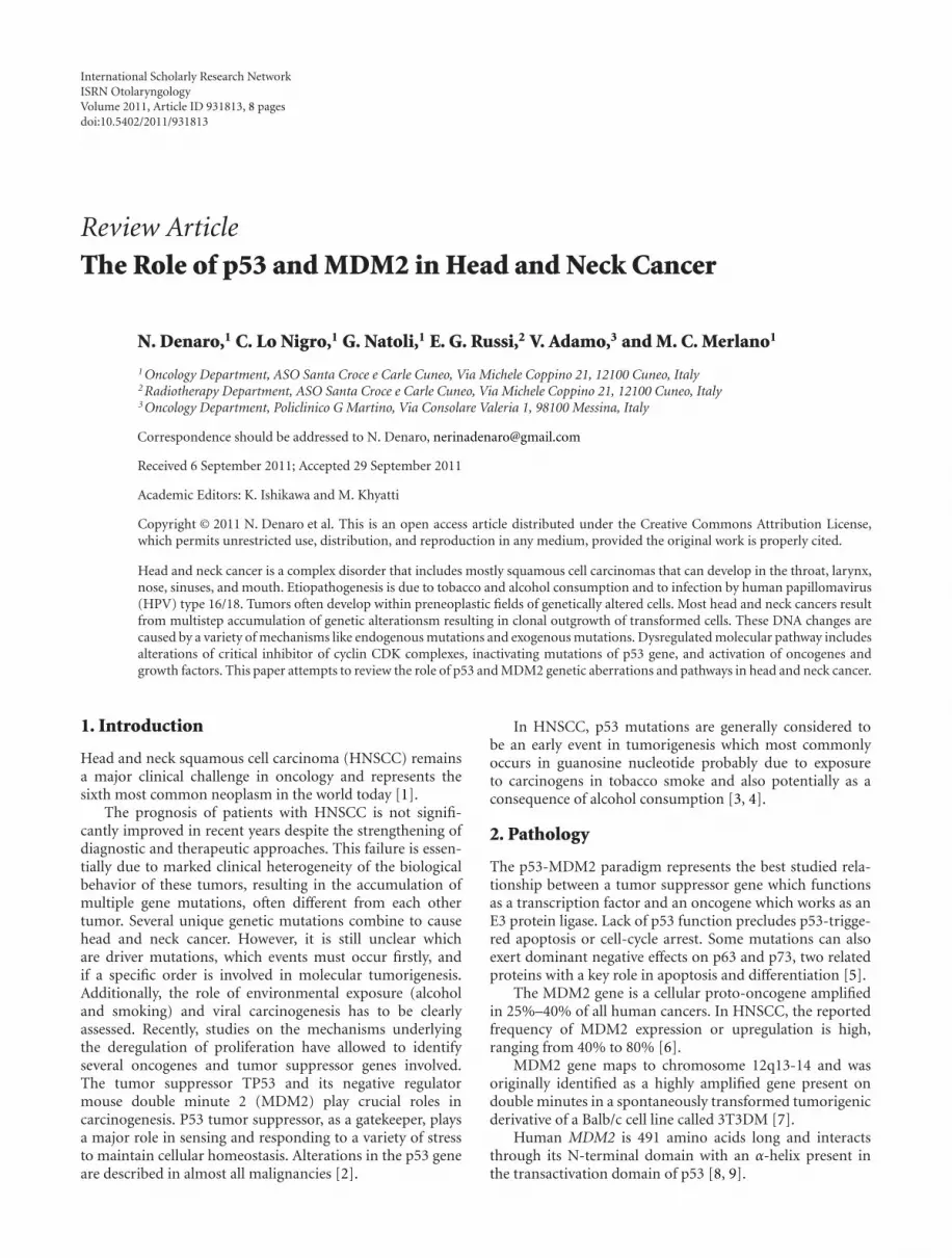
Figure 1: p53 and MDM2 Network: Apoptosis (http://www.celldeath.de/). In a normal growing viable cell, the p53 protein is inert.MDM2 directly interacts with p53 and thereby catalyzes ubiquitination of p53. Ubiquitination of p53 can be reversed by the action ofthe deubiquitinating enzyme HAUSP (also known as USP7, it is an ubiquitin-specific protease that acts as suppressor) which thereby canrescue p53 from degradation. P53 is stabilized in response to genotoxic stress such as DNA damage which leads to its phosphorylationat several specific serine and threonine residues. Phosphorylated p53 translocate into the nucleus where it activates the transcription ofproapoptotic genes and suppresses the transcription of antiapoptotic genes, thus inducing apoptosis. p53-mediated apoptosis signaling isdependent on the interplay of many regulatory factors, including protooncogenes as well as tumor-suppressors. MDM2 activity is positivelyregulated by the action of the Akt kinase: when phosphorylated by Akt, MDM2 is able to translocate from the cytosol to the nucleus, whereit unfolds its inhibitory effect on p53. Akt kinase, on the other hand, is activated in response to survival signals coming from growth factorreceptors. MDM2-mediated suppression of p53 is blocked by the action of the ARF tumor suppressor. By binding to MDM2, ARF preventsthe interaction between MDM2 and p53 and therefore stabilizes and activates p53. ARF expression is dependent on the transcription factorE2F-1 which is regulated by the retinoblastoma (Rb) tumor-suppressor and by the action of oncogenes. As an example, mitogenic signals leadto the activation of oncogenes such as c-myc and ras which among others activate E2F-1, resulting in increased ARF activity, stabilization ofp53 and induction of apoptosis. Therefore, increased mitogenic signalling or inappropriate oncogenic activity not necessarily causes excessiveproliferation but in cells with intact p53 signalling pathways can act as apoptosis inducers.
Yu et al. (2011) demonstrated an earlier onset of HNSCCwhen MDM2 promoter and p53 codon 72 are mutated. Indetail, their finding suggest that both MDM2 promoter poly-morphism and p53 codon 72 polymorphism may contributeto nonoropharyngeal cancer risk and that MDM2 SNP309 G-allele and p53 codon 72 SNP may accelerate the developmentof nonoropharyngeal cancer in women (an early age atonset of nonoropharyngeal cancer in an allele-dose responsemanner) [22].
In the same paper, Yu et al. published the results of sixstudies meta-analyses on the association between MDM2SNP309 and the risk or age at onset in HNSCC [23–27]. Theyshowed that MDM2 SNP309 was not significantly associatedwith risk of HNSCC. A possible explanation is either that theeffect of the MDM2 SNP309 on HNSCC risk may be modestand could not be detected in this study, or the effect canbe modified by other SNPs in other genes. Indeed, patientswho carried the two to three risk genotypes (i.e., MDM2SNP309 GT/TT, MDM2 SNP2164 AA, and p53 codon 72CC) appeared to have an increased risk of nonoropharyngealcancer, and this risk was more pronounced among eversmokers and ever drinkers [22].
This data has been further confirmed in the Asiaticpopulation: in a study of 103 pts, the GG genotype of MDM2SNP309 was associated (P = 0.032) with an earlier onset ofHNSCC. The average age at tumor onset was 65.6 years forTT, 62.9 years for TG, and 56.7 years for GG. The patientswith the GG genotype had a significantly earlier tumor onsetin comparison to those with the TT genotype [24].
The prognostic role of p53 in HNSCC was firstly analyzedin a critical review by Oh and Mao (1997). They concludethat tumors overexpressing p53 tend to be more aggressiveand to have the shortest survival, but most studies did notconfirm such a correlation. Additionally, in patients withearly laryngeal or hypopharyngeal cancers, p53 expressionhas no adverse effect on survival [4].
In a series of 115 patients with HNSCC immunohisto-chemistry (IHC) for p53, this gatekeeper loses its inde-pendent prognostic value when cyclin D1 is overexpressed.Cyclin D1 overexpression likes to be able to overcome theeffects caused by p53 deletion [28].
Poeta and colleagues (2007) reported on a multicenterprospective analysis of p53 status and survival data for 420cases of surgically treated HNSCC from all anatomical

4 ISRN Otolaryngology
(a) (b) (c)
(d) (e) (f)
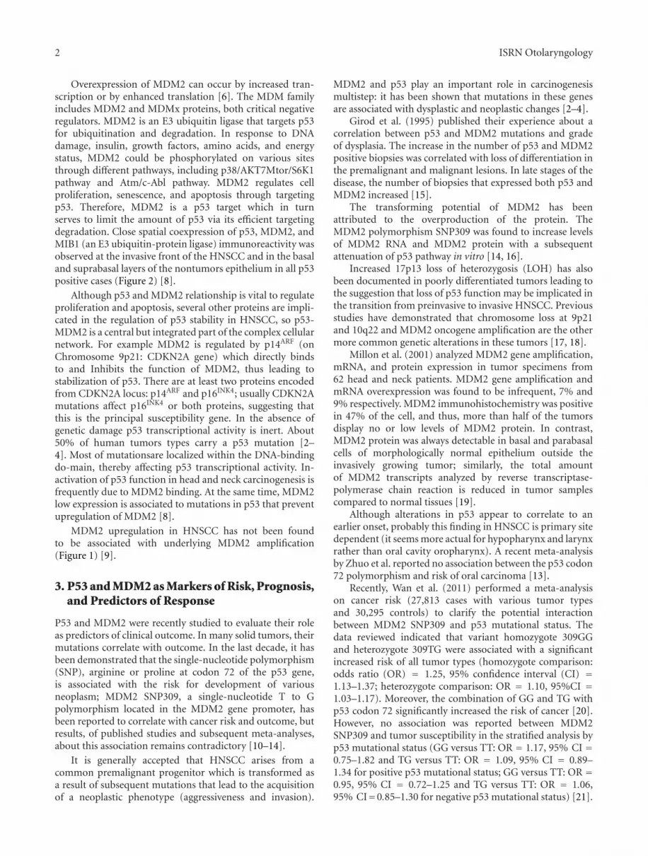
Figure 2: P53/MDM2 regulation [8]. (a) Regulation p53/MDM2. MDM2 inhibits p53 through an autoregulatory loop MDM2 directly bindsto the transactivation domain of p53 and inhibits its transcriptional activity, inducing the ubiquitination and proteasomal degradation ofp53, by exporting p53 out of the nucleus. ARF binds to MDM2 and sequesters MDM2 into the nucleolus, leading to the stabilization of p53.(b) P53 can lead to induction of apoptosis via intrinsic (mitochondrial) and extrinsic (death receptor) apoptosis pathways. (c) P53 activationcan halt cell-cycle progression in G1-S and G2-M phase through p21, Gadd45, and 14-3-3-σ proteins. (d) P53 regulates senescence throughp21-Rb-E2f signaling pathway. (e) P53 can suppress angiogenesis through the downregulation of antiangiogenenic proteins. (f) P53 plays acritical role in DNA damage repair. DNA damage and replication errors can activate ataxia telangiectasia mutated (ATM) and activate ataxiateleangiectasia and Rad kinases.
subsites. Mutational analysis of all of the coding exons ofthe p53 gene demonstrated an association between p53mutation and survival, with p53 mutations being signifi-cantly associated with a shorter overall survival in HNSCCcompared with wild-type cases. P53 phosphorylation is veryimportant to avoid MDM2 negative regulator control; in thisway, several cancers showed phosphorylation on three N-terminal (Ser15, Thr18, and Ser20) residues. In most cases,phosphorylation is associated with protein stabilization[29].
Vlatkovia et al. (2011) analyzed the influence of p53and MDM2 on survival. In their experience, neither p53nor MDM2 alone were significantly associated with outcomein Kaplan-Meier analyses although MDM2 expression wasfound to be an independent parameter associated with
increased survival (P < 0.03; OR = 0.63; 95% CI =0.41–0.96) by Cox multivariate analysis. Additionally, theyinvestigated the expression of MTBP (a-MDM2-bindingprotein). MTBP can contribute to p53/MDM2 homeostasis;it acts as an inhibitor of tumor progression in a subset ofhead and neck cancer patients. Low expression of MTBPis significantly associated with reduced overall survival inHNSCC patients [30].
Previous in vitro studies have shown that response of cellsexposed to anticancer agents is strongly influenced by SNPat codon 72 in wild-type p53. In vivo, the outcome ofchemoradiotherapy of squamous carcinomas is more favor-able in cancers retaining a wild-type 72R allele, such caseshaving higher response rates and longer survival than thosewith wild-type 72P [20].

ISRN Otolaryngology 5
About clinical implications of these findings, Perrone etal. (2010) reported that the loss of function (transactivationactivities) of p53 mutant proteins may predict a significantlow pathological complete response rate and suboptimalresponse to cisplatin-based neoadjuvant chemotherapy inpatients with oral cavity SCC [31].
The relationship between mutated p53 status and lowlevels of MDM2 found in cell lines is also observed to acertain extent in primary tumor samples. Overall, there isa high frequency of TP53 mutation and underexpression ofMDM2 in the head and neck tumors. Moreover, a significantdecreased MDM2 expression is observed in those patientswith advanced tumor stage and lower 3-year survival[17, 30].
Recent studies have shown that inhibition of ribosomalbiogenesis can activate p53 through ribosomal protein- (RP-)mediated suppression of MDM2 E3 ligase activity. Mutationsin MDM2 that disrupt RP binding have been detected inhuman cancers; however, the physiological significance of theRP-MDM2 interaction is not completely understood.
Miliani de Marval and Zhang (2011) generated micecarrying a mutation that disrupts MDM2’s binding to RPL11and RPL5 to analyze this interaction. Despite being devel-opmentally normal and maintaining an intact p53 responseto DNA damage, the MDM2C305F mice demonstrated adiminished p53 response to perturbations in ribosomalbiogenesis [32].
The results of some of the most impressive studies oncorrelation of p53-MDM2, outcome and age of onset aresummarized in Table 1.
4. HPV and p53-MDM2 Relationship
The increasing incidence of oral squamous cell carcinoma(OSCC) correlated with human papilloma virus (HPV) ledto study the influence of HPV on this tumor. There is a grow-ing body of evidence suggesting that oropharyngeal HPV-HNSCC is a distinctive clinicopathological and molecularentity [33].
The two polymorphisms of MDM2 (309SNP TT andGT/GG) were analyzed on the basis of the seropositivityo negativity. Chen et al. (2010) conducted a case-controlstudy finding an association between TT genotypes andHPV16 L1 seronegativity for OSCC risk and GT/GG andHPV16 L1 seropositivity (OR = 1.25, 95% CI = 1.06–2.19)and 2.81 (95%CI = 1.67–4.74). The authors reported thatOR was 5.57 (95% CI = 2.93–10.6) for those with boththe TT genotype and HPV16 L1 seropositivity. Similarresults were observed for the MDM2 SNP2164 (AA andAG/GG) polymorphism. Moreover, there was a borderlinesignificant interaction between the individual or combinedMDM2 genotypes of the two polymorphisms and HPV16L1 seropositivity on risk of OSCC (P(int) = 0.060 forMDM2 SNP309, P(int) = 0.009 for MDM2 SNP2164,and P(int) = 0.005 for the combined MDM2 genotypes).Notably, the effects of MDM2 mutations were particularlypronounced in never smokers and never drinkers, and fororopharyngeal as opposed to oral cavity cancer. These results
underline the central role of MDM2 in head and neck cancerdevelopment and suggest a correlation between MDM2 andHPV16 status. In the same study, the risk of OSCC has beenassociated with HPV16 L1 seropositivity, and it is modifiedby MDM2 promoter polymorphisms [34].
5. EGFR and p53/MDM2 Pathway
The epidermal growth factor receptor (EGFR) and its ligandsare fundamentally important for cell division, migration,adhesion, invasion, and angiogenesis. EGFR overexpressionhas been reported in >80% of HNSCC tumors and is foundto be associated with a more aggressive phenotype, poorprognosis, and resistance to chemotherapeutic agents [35].
To our knowledge, no direct correlations have beenreported among p53/MDM2 polymorphism and EGFR.Although they are considered in few studies as a panelof biological markers both predictive for chemotherapyresponse and HNSCC risk.
Prado et al. (2010) reported a correlation between mRNAexpression levels of COX-2, EGFR and p53 tested in oralleukoplakias. There were not correlations with sex, ageand localization of the leukoplakia. However, by meansof nonparametric statistics (Spearman’s rank correlationcoefficient), a positive linear correlation was found betweenEGFR and p53 mRNA expression levels (Spearman rho =0.6, n = 24, P = 0.003) [36, 37].
6. Future Perspectives: p53 Target Therapy
Alterations in p53 gene have been associated to an aggressivephenotype; therefore, many preclinical and clinical trialsinvestigated the way to restore wild type p53. Two approacheshave reached more convincing results: p53 targeting smallmolecules and gene therapies [39].
During the past several years, a number of small-molecule, p53-MDM2-binding inhibitors have been devel-oped in order to restore p53 activity. Nutlin-3 is a potent andselective small-molecule MDM2 antagonist that has shownconsiderable promise in preclinical studies [40]. Anotherapproach has been studied by develop an adenovirus(ONYX-015) specifically engineered to selectively replicateand lyse p53-deficient cells and spare normal cells [41, 42].
P53 reactivating (p53RA) small molecules have beentested in head and neck squamous cell lines; they inducedapoptosis with a dose-dependent increase in p53 proteinexpressions, resulting in upregulation of p21waf1 and acti-vation of the intrinsic apoptotic pathway [39]. Roh et al.(2011) tested four molecules (PRIMA1, CP31398, RITA, andnutlin 3), showing that each p53RA effectively restored p53function. Additional data on cell lines suggested a synergisticanti tumor effect [41].
Selective intratumoral replication and tumor-selectivetissue destruction have been documented in phase I andII clinical trials of intratumoral injections of ONYX-015with or without chemotherapy in patients with recurrent orrefractory HNSCC [42].

6 ISRN Otolaryngology
Table 1: Some of the most impressive studies on p53-MDM2 correlations.
Author Pts Biological predictors/prognosticators Correlations p
Michalides et al. [28] 198 MDM2, MTBP, p53, and HNSCC OS∗
Low MDM2 versushigh MDM2
0.248
P53+ AND low MDM2versus other status
0.035
Poeta et al. [29] 53 P53 and RR after P-based NACT 4/15 CR versus 20/38 NR 0.12
Nakashima et al. [24] 76 P53, MDM2, Rb, and dysplasia
HNSCC P53 66% MDM2 46%
Hyperplastic lesions P53 55% MDM2 31%
Dysplastic lesion P53 64% MDM2 44%
Hamid et al. [27] 62 MDM2, p53, HNSCC OS, and stage
P53+ AND low MDM2 0.07
P53+ AND IHC staining 0.018
MDM2 and T stage 0.10
MDM2 and T4 versus T1 0.015
Low MDM2 and 3 y OS 0.034
Chen et al. [34] 660∗∗ MDM2 polymorphism and HPV 16 seropositivity
MDM2-rs2279744 TTversus GT
OR = 0.64
MDM2-rs2279744 TTversus GG
OR = 0.60
MDM2-rs2279744 TTversus GT/GG
OR = 0.62
MDM2-rs2279744 TTversus AG
OR = 2.20
MDM2-rs2279744 TTversus AG/GG
OR = 2.05
Gasco and Crook [17] 2073∗∗∗ MDM2, p53, and HNSCC onsetMDM2 SNP309 0.638
MDM2 A2164G 0.580
P53 codon 72 0.193
Agarwal et al. [38] 128∗∗∗∗ MDM2, p53, and prognosis
MDM2 mut/p53+AND stage III/IV
0.0009
MDM2 mut/p53+AND N+
0.0325
Sullivan et al. [20] 73 p53 and OS
P53 72R versus p53 72P 0.007
wt p53 (72R or 72P)versus no wt P53
0.0001
P53 72R versus p53 72P +72R
0.02
Bergamaschi et al. [5] 70 p53, p73, OS, and response
CR: p53 wt versusno wt P53
0.0001
PFS: p53 wt versusno wt P53
0.0007
PFS: p53 72R versus 72P 0.008
OS: p53 72R versus 72P 0.0044
PFS: p73−/p53+ versusp73+/p53+
0.05
Hamid et al. [27] 420 p53 and OSp53+ versus p53− 0.074
p53+ disruptiveversusp53+ nondisruptive
0.001
RR = 1.17∗= OS after surgery; ∗∗= 335 controls; ∗∗∗= 1090 control population; ∗∗∗∗= 33 premalignant lesion and 30 control normal oral tissues.
OS = overall survival; NACT = neoadjuvant chemotherapy; OR = odd ratio; RR = response rate; CR = complete response.IHC = immuhistochemical; NR = nonresponse;3y = 3 year; p53+ = p53 mutated; p53 codon 72 = mutation in codon 72.72R = arginine at codon 72 of p53.72P = proline at codon 72 of p53.p73−/p53+ = non-p73-inactivating p53 mutations. p73+/p53+ = p73-inactivative and p53 mutations.
ISRN Otolaryngology 7
Another approach of targeting p53 mutations involvesgene replacement using a replication-defective adenoviralvector containing the wild-type p53 gene (Ad-p53 or RPR-INGN-201). In preclinical studies, Ad-p53 vector systemhas been shown to induce apoptosis in neoplastic cellsregardless of their p53 status and to reduce tumor growthin mouse xenografts. In a phase I study, good tolerabilitywas reported and a phase II study combining the viruswith cytotoxic chemotherapy achieved some tumor response.Further studies on p53 target therapy are on going [43].
7. Discussion
The prognostic and predictive role of p53 has been con-firmed, while some concerns on the reliability of MDM2mutation depend on the presence of a few data on it.
In the general population, an increased risk or an earlierage of onset of HNSCC has been reported for patients withp53 R72P, while the association between age of onset andprognosis in patients with MDM2 SNP309 need, furtherconfirmations. However, it appears that MDM2 and p53together could be used as predictive markers of response butneither p53 alone nor MDM2 correlates with patients overallsurvival.
In fact, several studies reported that only the combina-tion of p53 positive with concurrent low MDM2 resultedin a statistically significant association with reduced survival[21, 30, 31, 38].
Therefore, a new biomarker, the combination of p53positive and MDM2 low level, should be considered for itsprognostic role. Further studies on the role of p53/MDM2are warranted especially in HPV positive HNSCC. In con-clusion, the field of epidemiology has traditionally evaluatedwhether lifestyle factors are associated with HNSCC risk.However, with an increasing understanding of the molecularprocesses that underlie carcinogenesis, the field of molecularepidemiology has emerged. The ultimate goal of this progressis to personalize the approach for each patient. There is aneed to identify drug-specific predictive biomarkers in orderto better tailor chemotherapy regimens to individual patientswith head and neck cancer. In cell and animal models, p53 isthe critical factor for the outcome of genotoxic stress, such asthe one triggered by many cancer therapies. The research andstudy of biological markers with phenotypic and functionalperspectives and clinical application is designed to acquirebasic information in order to better know and understandthe natural history of preclinical and clinical tumor, withoutneglecting the practical aspects related to the identificationof prognostic and predictive indicators of different typesof local or systemic treatment. P53 and MDM2 are bothimportant players in the DNA damage repair. Detection ofMDM2 protein expression by immunohistochemical may bean important diagnostic tool in the future. Some mutationsof both p53 and MDM2 correlate with worst prognosis.The application of molecular predictors of clinical outcomewould be extremely useful to allow rational selection ofpatients who are more likely to benefit from treatmentand to spare unnecessary toxicity to those with a poorchance of response. A model which correlate several markers
(EGFR status, HPV, p53 mutations, and MDM2 status) withold prognostic known factors may allow a more targetedtherapy.
References
[1] A. Argiris, M. V. Karamouzis, D. Raben, and R. L. Ferris,“Head and neck cancer,” The Lancet, vol. 371, no. 9625, pp.1695–1709, 2008.
[2] P. Lothaire, E. A. D. Dequanter, Y. Lalami et al., “Molecularmarkers of head and neck squamous cell carcinoma: promis-ing signs in need of prospective evaluation,” Head and Neck,vol. 28, no. 3, pp. 256–269, 2006.
[3] B. Perez-Ordonez, M. Beauchemin, and R. C. K. Jordan,“Molecular biology of squamous cell carcinoma of the headand neck,” Journal of Clinical Pathology, vol. 59, no. 5, pp. 445–453, 2006.
[4] Y. Oh and L. Mao, “Biomarkers in head and neck carcinoma,”Current Opinion in Oncology, vol. 9, no. 3, pp. 247–256, 1997.
[5] D. Bergamaschi, M. Gasco, L. Hiller et al., “p53 polymorphisminfluences response in cancer chemotherapy via modulation ofp73-dependent apoptosis,” Cancer Cell, vol. 3, no. 4, pp. 387–402, 2003.
[6] L. Cahilly-Snyder, T. Yang-Feng, U. Francke, and D. L. George,“Molecular analysis and chromosomal mapping of amplifiedgenes isolated from a transformed mouse 3T3 cell line,”Somatic Cell and Molecular Genetics, vol. 13, no. 3, pp. 235–244, 1987.
[7] Z. Wang and B. Li, “Mdm2 links genotoxic stress andmetabolism to p53,” Protein and Cell, vol. 1, no. 12, pp. 1063–1072, 2010.
[8] S. Shangary and S. Wang, “Targeting the MDM2-p53 interac-tion for cancer therapy,” Clinical Cancer Research, vol. 14, no.17, pp. 5318–5324, 2008.
[9] S. F. Huang, I. H. Chen, C. T. Liao, H. M. Wang, S. H. Liou, andL. L. Hsieh, “Combined effects of MDM2 SNP 309 and p53mutation on oral squamous cell carcinomas associated withareca quid chewing,” Oral Oncology, vol. 45, no. 1, pp. 16–22,2009.
[10] R. Nenutil, J. Smardova, S. Pavlova et al., “Discriminatingfunctional and non-functional p53 in human tumours by p53and MDM2 immunohistochemistry,” Journal of Pathology, vol.207, no. 3, pp. 251–259, 2005.
[11] X. Wang, J. Wang, and X. Jiang, “MdmX protein is essential forMdm2 protein-mediated p53 polyubiquitination,” Journal ofBiological Chemistry , vol. 286, no. 27, pp. 23725–23734, 2011.
[12] Y. A. Valentin-Vega, J. A. Barboza, G. P. Chau, A. K. El-Naggar,and G. Lozano, “High levels of the p53 inhibitor MDM4 inhead and neck squamous carcinomas,” Human Pathology, vol.38, no. 10, pp. 1553–1562, 2007.
[13] X. L. Zhuo, Q. Li, Y. Zhou et al., “Study on TP53 codon 72polymorphisms with oral carcinoma susceptibility,” Archivesof Medical Research, vol. 40, no. 7, pp. 625–634, 2009.
[14] P. Alhopuro, S. K. Ylisaukko-Oja, W. J. Koskinen et al., “TheMDM2 promoter polymorphism SNP309T→G and the riskof uterine leiomyosarcoma, colorectal cancer, and squamouscell carcinoma of the head and neck,” Journal of MedicalGenetics, vol. 42, no. 9, pp. 694–698, 2005.
[15] S. C. Girod, D. Cesarz, U. Fischer, and G. R. F. Krueger,“Detection of p53 and MDM2 protein expression in head andneck carcinogenesis,” Anticancer Research, vol. 15, no. 4, pp.1453–1457, 1995.
8 ISRN Otolaryngology
[16] G. L. Bond, W. Hu, E. E. Bond et al., “A single nucleotidepolymorphism in the MDM2 promoter attenuates the p53tumor suppressor pathway and accelerates tumor formationin humans,” Cell, vol. 119, no. 5, pp. 591–602, 2004.
[17] M. Gasco and T. Crook, “p53 family members and chemore-sistance in cancer: what we know and what we need to know,”Drug Resistance Updates, vol. 6, no. 6, pp. 323–328, 2003.
[18] F. Toledo and G. M. Wahl, “Regulating the p53 pathway: invitro hypotheses, in vivo veritas,” Nature Reviews Cancer, vol.6, no. 12, pp. 909–923, 2006.
[19] R. Millon, D. Muller, I. Schultz et al., “Loss of MDM2expression in human head and neck squamous cell carcinomasand clinical significance,” Oral Oncology, vol. 37, no. 8, pp.620–631, 2001.
[20] A. Sullivan, N. Syed, M. Gasco et al., “Polymorphism in wild-type p53 modulates response to chemotherapy in vitro and invivo,” Oncogene, vol. 23, no. 19, pp. 3328–3337, 2004.
[21] Y. Wan, W. Wu, Z. Yin, P. Guan, and B. Zhou, “MDM2SNP309, gene-gene interaction, and tumor susceptibility: anupdated meta-analysis,” BMC Cancer, vol. 11, article 208,2011.
[22] H. Yu, Y.-J. Huang, Z. Liu et al., “Effects of MDM2 promoterpolymorphisms and p53 codon 72 polymorphism on risk andage at onset of squamous cell carcinoma of the head andneck,” Molecular Carcinogenesis, vol. 50, no. 9, pp. 697–706,2011.
[23] G. L. Clayman, A. K. El-Naggar, S. M. Lippman et al.,“Adenovirus-mediated p53 gene transfer in patients withadvanced recurrent head and neck squamous cell carcinoma,”Journal of Clinical Oncology, vol. 16, no. 6, pp. 2221–2232,1998.
[24] M. Nakashima, S. Kondo, Y. Shimizu et al., “Impact of MDM2single nucleotide polymorphism on tumor onset in head andneck squamous cell carcinoma,” Acta Oto-Laryngologica, vol.128, no. 7, pp. 808–813, 2008.
[25] C. Misra, M. Majumder, S. Bajaj, S. Ghosh, B. Roy, and S.Roychoudhury, “Polymorphisms at p53, p73, and MDM2 locimodulate the risk of tobacco associated leukoplakia and oralcancer,” Molecular Carcinogenesis, vol. 48, no. 9, pp. 790–800,2009.
[26] H. F. Tu, H. W. Chen, S. Y. Kao, S. C. Lin, C. J. Liu, and K. W.Chang, “MDM2 SNP 309 and p 53 codon 72 polymorphismsare associated with the outcome of oral carcinoma patientsreceiving postoperative irradiation,” Radiotherapy and Oncol-ogy, vol. 87, no. 2, pp. 243–252, 2008.
[27] S. Hamid, Y. H. Yang, K. N. L. Peng et al., “MDM2 SNP309does not confer an increased risk to oral squamous cellcarcinoma but may modulate the age of disease onset,” OralOncology, vol. 45, no. 6, pp. 496–500, 2009.
[28] R. J. A. M. Michalides, N. M. J. Van Veelen, P. M. P. Kristel etal., “Overexpression of cyclin D1 indicates a poor prognosisin squamous cell carcinoma of the head and neck,” Archives ofOtolaryngology, vol. 123, no. 5, pp. 497–502, 1997.
[29] M. L. Poeta, J. Manola, M. A. Goldwasser et al., “TP53mutations and survival in squamous-cell carcinoma of thehead and neck,” New England Journal of Medicine, vol. 357, no.25, pp. 2552–2561, 2007.
[30] N. Vlatkovia, A. El-Fert, T. Devling et al., “Loss of MTBPexpression is associated with reduced survival in a biomarker-defined subset of patients with squamous cell carcinoma ofthe head and neck,” Cancer, vol. 117, no. 13, pp. 2939–2950,2011.
[31] F. Perrone, P. Bossi, B. Cortelazzi et al., “TP53 mutations andpathologic complete response to neoadjuvant cisplatin andfluorouracil chemotherapy in resected oral cavity squamouscell carcinoma,” Journal of Clinical Oncology, vol. 28, no. 5, pp.761–766, 2010.
[32] P. L. Miliani de Marval and Y. Zhang, “The RP-Mdm2-p53pathway and tumorigenesis,” Oncotarget, vol. 2, no. 3, pp. 234–238, 2011.
[33] A. Psyrri, E. Boutati, and S. Karageorgopoulou, “Humanpapillomavirus in head and neck cancers: biology, prognosis,hope of treatment, and vaccines,” Anti-Cancer Drugs, vol. 22,no. 7, pp. 586–590, 2011.
[34] X. Chen, E. M. Sturgis, D. Lei, K. Dahlstrom, Q. Wei, and G. Li,“Human papillomavirus seropositivity synergizes with MDM2variants to increase the risk of oral squamous cell carcinoma,”Cancer Research, vol. 70, no. 18, pp. 7199–7208, 2010.
[35] M. E. Sharafinski, R. L. Ferris, S. Ferrone, and J. R. Grandis,“Epidermal growth factor receptor targeted therapy of squa-mous cell carcinoma of the head and neck,” Head and Neck,vol. 32, no. 10, pp. 1412–1421, 2010.
[36] L. Farnebo, A. Jedlinski, A. Ansell et al., “Proteins and singlenucleotide polymorphisms involved in apoptosis, growthcontrol, and DNA repair predict cisplatin sensitivity in headand neck cancer cell lines,” International Journal of MolecularMedicine, vol. 24, no. 4, pp. 549–556, 2009.
[37] S. M. D. Prado, J. L. L. Cedrun, R. L. Rey et al., “Evaluation ofCOX-2, EGFR, and p53 as biomarkers of non-dysplastic oralleukoplakias,” Experimental and Molecular Pathology, vol. 89,no. 2, pp. 197–203, 2010.
[38] S. Agarwal, M. Mathur, A. Srivastava, and R. Ralhan,“MDM2/p53 co-expression in oral premalignant and malig-nant lesions: potential prognostic implications,” Oral Oncol-ogy, vol. 35, no. 2, pp. 209–216, 1999.
[39] H. Shen and C. G. Maki, “Pharmacologic activation of p53 bysmall-molecule MDM2 antagonists,” Current PharmaceuticalDesign, vol. 17, no. 6, pp. 560–568, 2011.
[40] W. Wang and W. S. El-Deiry, “Restoration of p53 to limittumor growth,” Current Opinion in Oncology, vol. 20, no. 1,pp. 90–96, 2008.
[41] J. L. Roh, S. K. Kang, I. L. Minn, J. A. Califano, D. Sidransky,and W. M. Koch, “p53-Reactivating small molecules induceapoptosis and enhance chemotherapeutic cytotoxicity in headand neck squamous cell carcinoma,” Oral Oncology, vol. 47,pp. 8–15, 2011.
[42] J. Nemunaitis, I. Ganly, F. Khuri et al., “Selective replicationand oncolysis in p53 mutant tumors with ONYX-015, an E1B-55kD gene-deleted adenovirus, in patients with advanced headand neck cancer: a phase II trial,” Cancer Research, vol. 60, no.22, pp. 6359–6366, 2000.
[43] J. R. Bischoff, D. H. Kirn, A. Williams et al., “An adenovirusmutant that replicates selectively in p53-deficient humantumor cells,” Science, vol. 274, no. 5286, pp. 373–376, 1996.
Related Documents


![Evolution of the p53-MDM2 pathway1158898/FULLTEXT01.pdf · ation [9]. In vertebrates, MDM2 belongs to a family with two members, MDM2 and MDM4. To date, members of the p53/p63/p73](https://static.cupdf.com/doc/110x72/5e6a22570899fb6605504c19/evolution-of-the-p53-mdm2-1158898fulltext01pdf-ation-9-in-vertebrates-mdm2.jpg)
