The Role of Oxytocin in the Stress and Anxiety Response by Rose C. Mantella BS, Allegheny College, 1998 Submitted to the Graduate Faculty of the School of Pharmacy in partial fulfillment of the requirements for the degree of Doctor of Philosophy University of Pittsburgh October 12, 2004

Welcome message from author
This document is posted to help you gain knowledge. Please leave a comment to let me know what you think about it! Share it to your friends and learn new things together.
Transcript

The Role of Oxytocin in the Stress and Anxiety Response
by
Rose C. Mantella
BS, Allegheny College, 1998
Submitted to the Graduate Faculty of
the School of Pharmacy in partial fulfillment
of the requirements for the degree of
Doctor of Philosophy
University of Pittsburgh
October 12, 2004

UNIVERSITY OF PITTSBURGH
SCHOOL OF PHARMACY, DEPARTMENT OF PHARMACEUTICAL SCIENCES
The Role of Oxytocin in the Stress and Anxiety Response
This dissertation was presented
by
Rose C. Mantella
It was defended on
October 12, 2004
and approved by
Janet A. Amico, MD, Professor of Medicine ___________________________________ (Committee Chair, Advisor)
Sharon Corey, PhD, Assistant Professor of Pharmacy ___________________________________ (Committee Member)
Samuel Poloyac, PhD, Assistant Professor of Pharmacy ___________________________________ (Committee Member)
Linda Rinaman, PhD, Assistant Professor of Neuroscience ___________________________________ (Committee Member)
Regis R. Vollmer, PhD, Professor of Pharmacy ___________________________________ (Committee Member)
ii

The Role of Oxytocin in the Stress and Anxiety Response
Rose C. Mantella, PhD
University of Pittsburgh, 2004
Centrally released oxytocin (OT) is believed to attenuate stress-induced activation of the
hypothalamic pituitary adrenal (HPA) axis as well as being anxiolytic. Therefore, it is expected that
OT deficient (OT-/-) mice that do not synthesize or release OT centrally or peripherally will display
enhanced HPA axis activation, as well as increased anxiety-related behavior compared to wildtype
(OT+/+) mice. To test this hypothesis, OT-/- mice were exposed to shaker stress, (psychogenic
stressor), cholecystokinin- (CCK) administration (physical stressor), or the elevated plus maze
(EPM), a behavioral test of anxiety.
Female OT-/- mice released more corticosterone than OT+/+ mice in response to shaker
stress. Shaker stress exposure activated Fos in OT neurons of the paraventricular nucleus of the
hypothalamus (PVN) of male and female OT+/+ mice and corticotropin-releasing hormone (CRH)
within the PVN of male and female mice of both genotypes. In addition, shaker stress exposure
revealed that Fos expression in the medial nucleus of the amygdala (MeA) was lower in female OT-/-
than OT+/+ mice. Genotypic differences in corticosterone release and Fos activation of the MeA in
response to shaker stress exposure were not observed in male mice. Furthermore, similar genotypic
(and/or sex) differences were not revealed in response to CCK-administration.
OT is also anxiolytic in female mice. Female OT-/- mice tested in the EPM displayed
increased anxiety-related behavior compared to OT+/+ mice. In response to EPM exposure Fos
expression in the MeA was greater in female OT-/- mice than OT+/+ mice. Surprisingly, male OT-/-
mice tested in the EPM displayed decreased anxiety-related behavior compared to OT+/+ mice, but
did not display genotypic differences in the Fos expression within the MeA.
iii

The results of this thesis suggest that OT is anxiolytic and attenuates HPA activation in
female, but not male mice. Furthermore, it appears that OT plays a modulatory role in the processing
of psychogenic stressors, but may not be involved in the processing of physical or systemic stressors.
More specifically, it is possible that OT plays a role in behavioral and physiological responses that
depend upon neuronal processing within the MeA.
iv

TABLE OF CONTENTS I. Overview .......................................................................................................................................1
A. Introduction ...............................................................................................................................1 B. Oxytocin ....................................................................................................................................3 C. Stress .........................................................................................................................................8 D. Anxiety ....................................................................................................................................19 E. Involvement of Oxytocin in Stress and Anxiety .....................................................................24 F. Oxytocin Deficient Mouse ......................................................................................................24 G. Specific Objectives of the Research ........................................................................................26
II. Materials and Methods ............................................................................................................29
A. Breeding and Maintenance of Wildtype and Oxytocin Deficient Mice ..................................29 B. Genotype Determination of Mice Using Polymerase Chain Reaction (PCR).........................29 C. Plasma Corticosterone Analysis ..............................................................................................30 D. Immunocytochemistry.............................................................................................................30 E. Shaker Stress ...........................................................................................................................32 F. Cholecystokinin Administration..............................................................................................33 G. Lateral Ventricle Cannulation Surgery and Infusions .............................................................33 H. Elevated Plus Maze Testing ....................................................................................................34 I. Determination of the Estrous Cycle ........................................................................................35 J. Statistical Analysis ..................................................................................................................35
III. Stress and the Oxytocin Deficient Mouse ...............................................................................37
A. Experimental Design ...............................................................................................................37 B. Results .....................................................................................................................................40 C. Discussion ...............................................................................................................................49
IV. Anxiety and the Oxytocin Deficient Mouse............................................................................55
A. Experimental Design ...............................................................................................................55 B. Results .....................................................................................................................................57 C. Discussion ...............................................................................................................................64
V. Forebrain Activation of the OT Deficient Mouse in Response to Stress and Anxiety............67
A. Experimental Design ...............................................................................................................68 B. Results .....................................................................................................................................69 C. Discussion ...............................................................................................................................85
VI. Conclusion...............................................................................................................................92 VII. References ...............................................................................................................................98
v

LIST OF TABLES Table 1. Oxytocin Efferents of the Paraventricular Nucleus of the Hypothalamus..............................5 Table 2. Oxytocin Receptor Localization in the Rat and Mouse. .........................................................7
vi

LIST OF FIGURES Figure 1. Oxytocin Peptide ...................................................................................................................3 Figure 2. Structure of the Mouse Oxytocin Gene .................................................................................4 Figure 3. Structure of the Mouse Oxytocin Receptor Gene..................................................................6 Figure 4. Schematic Representation of the Hypothalamic Pituitary Adrenal Axis.............................10 Figure 5. Projections to the Paraventricular Nucleus of the Hypothalamus from Brain Areas Involved
in the Stress Response .................................................................................................................18 Figure 6. Afferents and Stimulus Processing of Anxiety....................................................................22 Figure 7. Efferents of the Amygdala to Anxiety-Related Brain Areas ...............................................23 Figure 8. Corticosterone Response to Shaker Stress in Group-Housed Female Mice........................41 Figure 9. Corticosterone Response to Shaker Stress in Individually Housed Female Mice...............42 Figure 10. Diurnal Plasma Corticosterone Concentrations in Female Mice......................................43 Figure 11. Corticosterone Response to Central Administration of Corticotrophin Releasing Hormone
in Female Mice. ...........................................................................................................................44 Figure 12. Corticosterone Response to Repeated Shaker Stress in Female Mice...............................45 Figure 13. Corticosterone Response to Shaker Stress Across the Estrous Cycle ...............................46 Figure 14. Corticosterone Response to Shaker Stress in Male Mice ..................................................47 Figure 15. Corticosterone Response to Cholecystokinin Administration in Fasted Male Mice. ........48 Figure 16. Corticosterone Response to Cholecystokinin Administration in Non-fasted Male Mice..48 Figure 17. Behavior of Female Wildtype and Oxytocin Deficient Mice in the Elevated Plus Maze. 58 Figure 18. Behavior of Female Wildtype Mice in the Elevated Plus Maze Following Administration
of an Oxytocin Antagonist...........................................................................................................59 Figure 19. Behavior of Female Oxytocin Deficient Mice in the Elevated Plus Maze Following
Administration of Synthetic Oxytocin.........................................................................................60 Figure 20. Behavior of Female Oxytocin Deficient Mice in the Elevated Plus Maze Following
Administration of Synthetic Vasopressin. ...................................................................................61
vii
Figure 21. Behavior of Female Oxytocin Deficient Mice in the Elevated Plus Maze After Administration of an Oxytocin Antagonist Followed by Oxytocin.............................................62
Figure 22. Behavior of Male Wildtype and Oxytocin Deficient Mice in the Elevated Plus Maze.....63 Figure 23. Stress-Related Brain Pathways Examined for Fos Activation. ........................................68 Figure 24. Shaker Stress-Induced Fos Activation in Oxytocin Neurons of the Hypothalamus of
Female Mice ................................................................................................................................72 Figure 25. Shaker Stress-Induced Fos Activation in Vasopressin Neurons of the Hypothalamus of
Female Mice ................................................................................................................................73 Figure 26. Shaker Stress-Induced Fos Activation in Corticotropin Releasing Hormone Neurons of
the Hypothalamus of Female Mice..............................................................................................74 Figure 27. Shaker Stress Induced Neural Activation of the Limbic Forebrain of Female Mice ........75 Figure 28. Fos Activation of Oxytocin Immunoreactive Neurons in the Paraventricular Nucleus of
the Hypothalamus of Wildtype Male Mice. ................................................................................76 Figure 29. Fos Activation of Corticotropin Releasing Hormone Immunoreactive Neurons in the
Paraventricular Nucleus of the Hypothalamus of Male Mice .....................................................76 Figure 30. Cholecystokinin Induced Fos Activation in Oxytocin Neurons of the Paraventricular
Nucleus of Wildtype Male Mice .................................................................................................77 Figure 31. Cholecystokinin Induced Fos Activation in Vasopressin Neurons of the Hypothalamus of
Male Mice....................................................................................................................................78 Figure 32. Cholecystokinin Induced Neural Activation of the Limbic Forebrain of Female Mice....80 Figure 33. Cholecystokinin Induced Neural Activation of the Limbic Forebrain in Male Mice........81 Figure 34. Elevated Plus Maze Induced Neural Activation of the Limbic Forebrain in Female Mice
.....................................................................................................................................................83 Figure 35. Elevated Plus Maze Induced Neural Activation of the Medial Amygdala of Male Mice .84
viii

I. Overview
A. Introduction
A healthy physiological and behavioral response to acute stress is crucial for our ability to
deal with everyday challenges, either systemic or psychological. The initial response to an acute
stress is protective, enhancing immune function, promoting memory of dangerous events, increasing
blood pressure and heart rate to meet the physical and emotional demands to react to the stressor.
Failure to terminate the acute response to stress may contribute to greater vulnerability to illness.
Overactivity of the stress system is believed to play an important role in the pathogenesis of certain
chronic diseases, such as affective or panic disorders, anorexia, depression, coronary heart disease,
and functional gastrointestinal disturbances [31].
An organism’s adaptive response to stressful stimuli is mediated, in part, by the hypothalamic
pituitary adrenal (HPA) axis. There are many neurotransmitters and neuropeptides that regulate HPA
activation to stress. Oxytocin (OT) is a neuropeptide, which is synthesized within the paraventricular
(PVN) and supraoptic nuclei (SON) of the hypothalamus and is released from the hypothalamo-
neurohypophyseal system in response to a number of stressors. The significance of this increased
peripheral release of OT is not known. However, OT is also widely distributed throughout the
central nervous system [19] and many studies implicate an inhibitory role of central OT on HPA
activation in response to stress and anxiety behavior.
Initial studies in humans have focused on the role of peripheral OT in female reproduction
and lactation. However, OT is present in the brains and peripheral circulation of both males and
females. The fact that OT is present in males and females may indicate a functional role of OT
beyond that of female reproduction. Studies performed in human subjects suggest that elevated OT
may attenuate the HPA axis stress response. For instance, adrenocorticotropin hormone (ACTH) and
cortisol release are attenuated in lactating women versus non-lactating women following exercise
1

stress [1]. Furthermore, in response to psychosocial stress, overall blood pressure [118], ACTH and
cortisol levels [71] were blunted in women following breast-feeding (when plasma OT levels are
elevated) compared to women that did not breast-feed. However, these studies do not correlate OT
expression with stress hormone levels and there are a variety of confounding factors during the peri-
partum period, such as the release of other hormones (i.e. prolactin), making it difficult to ascribe the
blunted stress hormone effect to OT alone.
Similar findings to those of humans have been reported in animal models of stress and
anxiety. OT is released within the brain and into the circulation of male, as well as virgin female rats
in response to stressors that contain a psychogenic component. These stressors include forced
swimming in male [216,217] and female rats [209], various forms of social stress in male [48,50] and
female rats [138], and shaker stress in male rats [142]. Infusion of synthetic OT into the lateral
ventricles of estrogen-treated ovariectomized rats decreased the corticosterone response to
psychogenic restraint stress [210] and noise stress [211]. Infusion of an OT receptor antagonist into
the lateral ventricles augmented the basal and stress-induced release of andrenocorticotropin
hormone (ACTH) and corticosterone in female rats forced to swim [140] and in male and female rats
exposed to an elevated plus-maze (EPM) [140] or to repeated airpuffs [138]. In addition, OT is
believed to be anxiolytic in female laboratory rats [11,140,211] and mice [129]. Central
administration of OT to estrogen-primed ovariectomized rats [211] or mice [129] decreased anxiety-
related behavior in the elevated plus-maze (EPM). OT infused into the amygdala of ovariectomized
estrogen treated rats significantly increased open field activity (decreased anxiety) and increased the
time spent in open arms of the EPM (anxiolytic effect) [11]. However, the anxiolytic effect of OT
has not been reported in male rats or mice. These sex differences may be due to gonadal steroids.
Estrogen has been reported to facilitate OT mRNA expression [150,185], OT receptor binding [221],
and stress- and anxiety-related effects of OT [128,129]. Collectively the data suggest that OT
2

inhibits the response of the HPA axis to stress in male and female rodents, as well as produces
anxiolysis in female rats and mice.
Although many studies conducted prior to the development of the OT deficient mouse
suggest that exogenous administration of OT attenuates the stress and anxiety response, or that
administration of an OT antagonist enhances the stress and anxiety response, it has not been possible
to determine whether the absence of OT affects the stress and anxiety responses. The central
hypothesis of this thesis is that the absence of OT will result in an enhanced corticosterone response
to stress and/or anxiety. Therefore, OT deficient mice will display an increased response of the HPA
axis to stress and greater anxiety-related behavior than wildtype mice. In addition, OT deficient mice
will display differences in the degree of activation of stress- and anxiety-related brain pathways
compared to wildtype mice.
B. Oxytocin
1. Gene and Peptide Structure
OT was first discovered in 1909 using posterior pituitary gland extracts [40]. Dale
described the powerful role of OT in producing uterine contractility. In 1953 du Vigneaud
described the structure of the OT peptide [44]. OT contains nine amino acids consisting of a six
amino acid ring structure, created by a disulfide bridge between Cys residues in the 1 and 6
position, and a three amino acid side chain (Figure 1).
CysTyr
Ile
Asp
Glu
CysPro Leu
Gly
S
S
CysTyr
Ile
Asp
Glu
CysPro Leu
Gly
S
S
Figure 1. Oxytocin Peptide
3
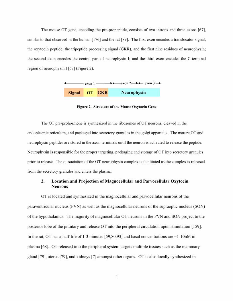
The mouse OT gene, encoding the pre-propeptide, consists of two introns and three exons [67],
similar to that observed in the human [176] and the rat [89]. The first exon encodes a translocator signal,
the oxytocin peptide, the tripeptide processing signal (GKR), and the first nine residues of neurophysin;
the second exon encodes the central part of neurophysin I; and the third exon encodes the C-terminal
region of neurophysin I [67] (Figure 2).
GKRSignal OT Neurophysin
exon 1 exon 2 exon 3
GKRSignal OT Neurophysin
exon 1 exon 2 exon 3
Figure 2. Structure of the Mouse Oxytocin Gene
The OT pre-prohormone is synthesized in the ribosomes of OT neurons, cleaved in the
endoplasmic reticulum, and packaged into secretory granules in the golgi apparatus. The mature OT and
neurophysin peptides are stored in the axon terminals until the neuron is activated to release the peptide.
Neurophysin is responsible for the proper targeting, packaging and storage of OT into secretory granules
prior to release. The dissociation of the OT-neurophysin complex is facilitated as the complex is released
from the secretory granules and enters the plasma.
2. Location and Projection of Magnocellular and Parvocellular Oxytocin Neurons
OT is located and synthesized in the magnocellular and parvocellular neurons of the
paraventricular nucleus (PVN) as well as the magnocellular neurons of the supraoptic nucleus (SON)
of the hypothalamus. The majority of magnocellular OT neurons in the PVN and SON project to the
posterior lobe of the pituitary and release OT into the peripheral circulation upon stimulation [159].
In the rat, OT has a half-life of 1-3 minutes [59,80,93] and basal concentrations are ~1-10nM in
plasma [68]. OT released into the peripheral system targets multiple tissues such as the mammary
gland [79], uterus [79], and kidneys [7] amongst other organs. OT is also locally synthesized in
4

many peripheral tissues, such as the heart [91], the ovary [88] and uterus [116] in the female
reproductive system, and the testis and prostate of the male reproductive system [87].
Parvocellular OT efferents of the PVN project to extra-hypothalamic brain areas and release OT
into the brain. OT is present in cerebrospinal fluid at concentrations between 10-50pM [68] and has a
half-life of approximately 28 min [93]. OT projections target multiple areas of the brain, including the
olfactory nucleus [19], the limbic system [19,188,189], and the brain stem [19,179,188,189] (Refer to
Table 1). At this time, the OT projections of the mouse have not been well defined.
Brain Regions Containing OT Efferents Olfactory system Anterior olfactory nucleus [19] Olfactory tubercle Piriform cortex Entorhinal/perirhinal area Basal ganglia Caudoputamen Ventral pallidum Limbic system Lateral septal nucleus Bed nucleus of the stria terminalis [19,82] Hippocampus [19,188] Central amygdala [19,188,189]Medial amygdala [188,189] Dorsal subiculum Ventral subiculum Thalamus and Hypothalamus Periventricular thalamic nucleus Ventromedial hypothalamic nucleus Medial preoptic area Supraoptic nucleus Paraventricular nucleus Supramammillary nucleus [188,189]Brain Stem Substantia nigra [188,189] Tegmental area Dorsal raphe nucleus [19, 188,189] Nucleus of the solitary tract [179, 188,189Dorsal motor nucleus of the vagus [19, 179, 188,Locus coeruleus [19, 188,189]
Table 1. Oxytocin Efferents of the Paraventricular Nucleus of the Hypothalamus
The references cited in this table are the results of tracing studies performed in male rats. Similar studies in mice and female rats have not been published.
5

3. Oxytocin Receptor
The OT receptor gene sequence has been identified in numerous mammalian species,
including the human [101], pig [62], rat [169], sheep [88], and mouse [108]. Similar to the human
[83] and the rat [169], the mouse OT receptor gene contains 3 introns and 4 exons [108] (Figure 3).
Exons 1 and 2 correspond to the 5’-prime noncoding region. Exon 3 encodes a portion of the 5’-
prime noncoding region, the start codon (ATG), and the first 6 of the 7 transmembrane regions.
Exon 4 contains the sequence encoding the seventh transmembrane domain, the stop codon (TGA),
the COOH terminus, and the entire 3’-noncoding region [108].
Figure 3. Structure of the Mouse Oxytocin Receptor Gene
OT receptor mRNA is expressed in various tissues of the rat including the uterus [111],
mammary gland [136,190], heart [91], kidney [7], and the brain [146,195,219]. In addition, the OT
receptor is also expressed in the mammary gland [63] and brain of the mouse [63,85,86]. Within the
brain of the rat and mouse, expression of the OT receptor gene has been identified in many different
areas. These areas include the olfactory system, cortex, basal ganglia, limbic system, thalamus,
hypothalamus, and brain stem (refer to Table 2).
exon 2exon 2exon 1exon 1
ATG
exon 3 exon 4
I II III IV V VI VII TGAATG
exon 3 exon 4
I II III IV V VI VII TGA
6

R
at
Mou
se
B
rain
Reg
ions
M
ale
Fem
ale
M
ale
Fem
ale
O
lfact
ory
syst
em
Olfa
ctor
y bu
lb
[63+ ]
Ant
erio
r olfa
ctor
y nu
cleu
s
[1
46*,
195# ]
[
195# ,2
19*]
[85# ,8
6# ]
[6
3+ ,85# ]
Olfa
ctor
y tu
berc
le
[146
*]
[21
9*]
Pi
rifor
m c
orte
x
[
219*
]
[8
5# ]
[63+ ,8
5# ] En
torh
inal
/per
irhin
al a
rea
[8
6# ]
[63+ ]
Bas
al g
angl
ia
Cau
dopu
tam
en
[1
95# ]
[19
5# ,219
*]
[8
5# ]
[85# ]
Ven
tral p
allid
um
[195
# ]
[
195# ,2
19*]
Li
mbi
c sy
stem
La
tera
l sep
tal n
ucle
us
[146
*,19
5# ]
[19
5# ,219
*]
[8
5# ,86# ]
[6
3+ ,85# ]
Bed
nuc
leus
of t
he st
ria te
rmin
alis
[146
*,19
5# ]
[19
5# ,219
*]
[8
5# ]
[85# ]
Hip
poca
mpu
s
[146
*,19
5# ]
[19
5# ,219
*]
[8
5# ,86# ]
[85# ]
Cen
tral a
myg
dala
[1
95# ]
[19
5#,2
19*]
[85# ]
[8
5# ] M
edia
l am
ygda
la
[195
# ]
[
195# ,2
19*]
[86# ]
Dor
sal s
ubic
ulum
[1
95# ]
[19
5# ,219
*]
Ven
tral s
ubic
ulum
[195
# ]
[195
# ,219
*]
Thal
amus
and
Hyp
otha
lam
us
Periv
entri
cula
r tha
lam
ic n
ucle
us
[1
95# ]
[19
5# ,219
*]
[8
6# ]
V
entro
med
ial h
ypot
hala
mic
nuc
leus
[1
46*,
195# ]
[
195# ,2
19*]
[86# ]
[6
3+ ] M
edia
l pre
optic
are
a
[63+ ]
Supr
aopt
ic n
ucle
us
[146
*]
[21
9*]
Para
vent
ricul
ar n
ucle
us
[146
*]
[21
9*]
Supr
amam
mill
ary
nucl
eus
[146
*]
[19
5# ,219
*]
[6
3+ ] B
rain
Ste
m
Subs
tant
ia n
igra
[146
*]
[21
9*]
Tegm
enta
l are
a
[6
3+ ] D
orsa
l rap
he n
ucle
us
[
219*
]
[6
3+ ] N
ucle
us o
f the
solit
ary
tract
[6
3+ ] D
orsa
l mot
or n
ucle
us o
f the
vag
us
[1
46*,
195# ]
[195
# ,219
*]
[6
3+ ] Lo
cus c
oeru
leus
[146
*]
Tab
le 2
. O
xyto
cin
Rec
epto
r L
ocal
izat
ion
in th
e R
at a
nd M
ouse
.
Num
bers
in th
e ta
ble
refe
r to
stud
ies c
ited
in th
e re
fere
nce
list.
Em
pty
spac
es re
fer t
o br
ain
area
s tha
t wer
e no
t eva
luat
ed a
nd/o
r did
not
dis
play
oxy
toci
n re
cept
ors.
Ref
eren
ces i
nclu
de st
udie
s tha
t mea
sure
d m
RN
A e
xpre
ssio
n*, o
xyto
cin
rece
ptor
bin
ding
# , or o
xyto
cin
rece
ptor
gen
e ex
pres
sion
usi
ng a
Lac
-Z re
porte
r m
ouse
+ .
7
C. Stress
1. Definition and Classification of Stress
Stress is a concept that is difficult to define fully because its interpretation tends to vary
according to individual disciplines. In 1936 Hans Selye, a pioneer in the physiological and
pathophysiological principles in the exploration of stress, defined stress as “the nonspecific response
of the body to any demand upon it, including bacterial infection, toxins, and various physical stimuli
(i.e. surgery, exercise)” [183]. However, Selye’s definition of stress as a “nonspecific” response of
the body has been challenged. Although he did not define stress, Walter Cannon was the first to
introduce the term “homeostasis” in reference to the stress response [22]. According to Cannon,
homeostasis is the product of multiple physiological systems that maintain steady-state in an
organism [22]. According to Cannon, a nonspecific stress response would not have provided an
antage in natural selection and would not have evolved [22]. The definition has been refined over
time to describe stress as selective pressure from the physical and social environment that threaten or
challenge an organism’s homeostasis and elicit physiologically and behaviorally adaptive responses
that are specific to the stressor [30,31,206].
Stressors can be defined as conditions that endanger, or are perceived to endanger, the well-
being of an individual. The current literature broadly categorizes stressors as psychogenic (based on
either a conditioned or an unconditioned response) or physical/systemic stimuli [73,77].
Psychogenic stressors affect emotional processes and may result in behavioral changes such as
anxiety or fear. Systemic stressors include cold, heat, hypoglycemia, hemorrhage, pain, and
chemical or noxious stimuli. The stressors described above are commonly used in animal research.
However, many stressors are both physical and psychogenic. Exposure to stressors result in a series
of coordinated responses composed of alterations in behavior, autonomic function and the secretion
adv
8
of multiple hormones including adrenocorticotropin hormone (ACTH) and cortisol/corticosterone
(discussed below).
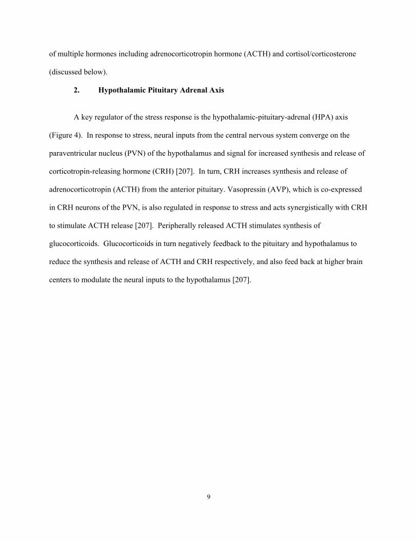
. Hypothalamic Pituitary Adrenal Axis
A key regulator of the stress response is the hypothalamic-pituitary-adrenal (HPA) axis
(Figure 4). In response to stress, neural inputs from the central nervous system converge on the
paraventricular nucleus (PVN) of the hypothalamus and signal for increased synthesis and release of
corticotropin-releasing hormone (CRH) [207]. In turn, CRH increases synthesis and release of
adrenocorticotropin (ACTH) from the anterior pituitary. Vasopressin (AVP), which is co-expressed
in CRH neurons of the PVN, is also regulated in response to stress and acts synergistically with CRH
to stimulate ACTH release [207]. Peripherally released ACTH stimulates synthesis of
glucocorticoids. Glucocorticoids in turn negatively feedback to the pituitary and hypothalamus to
reduce the synthesis and release of ACTH and CRH respectively, and also feed back at higher brain
centers to modulate the neural inputs to the hypothalamus [207].
2
9

10
3. Neurocircuitry
Multiple brain structures are involved in the organization of responses to stressful stimuli.
mong them are the hypothalamus, limbic brain areas (i.e. the lateral septum, the hippocampus, the
mygdala), hindbrain regions (i.e. the nucleus of the tractus solitarius and the locus coeruleus), the
arabrachial nucleus, and raphe nucleus. One of the most prevalent hypotheses regarding
eurocircuit regulation of the HPA axis proposes the categorization of psychogenic (processive,
xteroceptive, neurogenic) and physical (interoceptive, systemic) responses to stress. It has been
eorized that the psychogenic class of stressors require forebrain or limbic processing and
tegration prior to HPA axis activation, while the physical class of stressors is dependent upon
flexive, direct hind-brain pathways to the PVN to activate the HPA axis. Stimuli that fall within
e category of psychogenic stress include novelty and restraint, while physical stressors include
emorrhage, cold exposure, immune challenge and pain. Each stressor is believed to activate a
Figure 4. Schematic Representation of the Hypothalamic Pituitary Adrenal Axis
+
+
Anterior pituitary
ACTH
++
++
Anterior pituitary
ACTH
STRESS
Hypothalamus (PVN)
Adrenal Cortex
CRH
-
-
Adrenal Cortex
Corticosterone
STRESS
Hypothalamus (PVN)
Adrenal Cortex
CRH
--
--
Adrenal Cortex
Corticosterone
A
a
p
n
e
th
in
re
th
h
Amico Lab
Anterior Pituitary
Amico Lab
ACTH

“signature” pathway that is unique to that particular stressor. While stressors that are classified as
psychogenic tend to activate a combination of distinctly different neuronal pathways from stressors
that are classified as physical, there is some overlap in the brain areas activated by each stressor.
a) Stress-Related Hindbrain Projections to the Paraventricular Nucleus of the Hypothalamus
Generically speaking, stressors that consist of a physical or systemic stimulus directly
activate the PVN through hindbrain projections. Stimuli that demand immediate physiological
responses have a direct unimpeded pathway to the PVN region, by way of the brainstem, eliciting an
ACTH and corticosterone response. These hindbrain PVN-projecting neurons are positioned to
evoke rapid, reflexive activation of the HPA axis, which is faster than psychogenic stress-induced
activation.
ucleus of the Solitary Tract
olitary tract (NTS), which is located in the medulla, receives and
integrates sensory information from most major organs of the body (Figure 5). Fos activation
(discussed in the introduction of Chapter V) within the NTS increases following most stressors
classified as physical, including administration of the nauseogenics cholecystokinin (CCK) [145] and
lithium chloride [110], immune challenge by interleukin-1 [51,178] or lipopolysaccharide
administration [109], hypoxia [193], hypovolemia [107], hypotension [107], and footshock [178].
The PVN receives its major catecholaminergic (i.e. noradrenergic and adrenergic) input from the
NTS [38]. Fibers from this area innervate the medial parvocellular zone of the PVN [38]. The
catecholaminergic input represents a major HPA excitatory pathway, promoting CRH [158] and
ACTH [158,192] release and CRH gene transcription [157]. However, the NTS is also activated
during psychogenic stressors such as restraint [36], swim [36], and fear conditioning induced by
footshock [155]. The NTS also receives afferents from limbic forebrain circuits, including the
N
The nucleus of the s
11
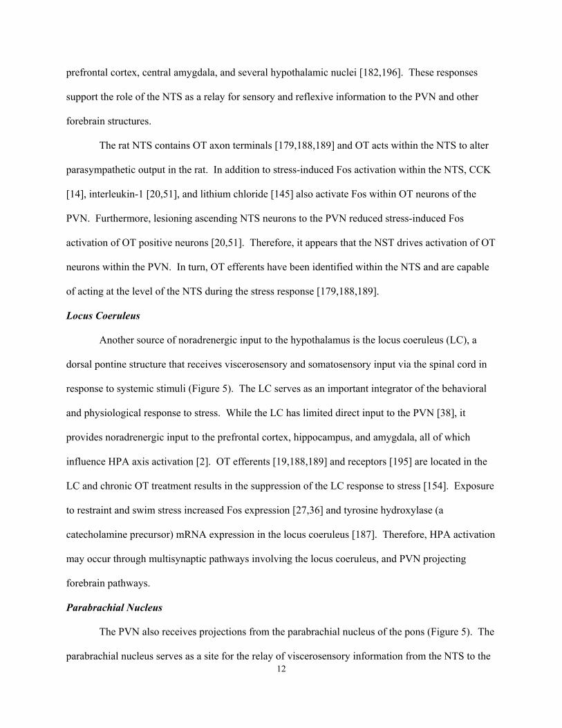
prefrontal cortex, central amygdala, and several hypothalamic nuclei [182,196]. These responses
support the role of the NTS as a relay for sensory and reflexive information to the PVN and other
forebrain structures.
The rat NTS contains OT axon terminals [179,188,189] and OT acts within the NTS to alter
parasympathetic output in the rat. In addition to stress-induced Fos activation within the NTS, CCK
[14], in e
ble
leus
drenergic input to the hypothalamus is the locus coeruleus (LC), a
dorsal p rd in
ioral
ed in the
re
ation
. The
parabrachial nucleus serves as a site for the relay of viscerosensory information from the NTS to the
terleukin-1 [20,51], and lithium chloride [145] also activate Fos within OT neurons of th
PVN. Furthermore, lesioning ascending NTS neurons to the PVN reduced stress-induced Fos
activation of OT positive neurons [20,51]. Therefore, it appears that the NST drives activation of OT
neurons within the PVN. In turn, OT efferents have been identified within the NTS and are capa
of acting at the level of the NTS during the stress response [179,188,189].
Locus Coeru
Another source of nora
ontine structure that receives viscerosensory and somatosensory input via the spinal co
response to systemic stimuli (Figure 5). The LC serves as an important integrator of the behav
and physiological response to stress. While the LC has limited direct input to the PVN [38], it
provides noradrenergic input to the prefrontal cortex, hippocampus, and amygdala, all of which
influence HPA axis activation [2]. OT efferents [19,188,189] and receptors [195] are locat
LC and chronic OT treatment results in the suppression of the LC response to stress [154]. Exposu
to restraint and swim stress increased Fos expression [27,36] and tyrosine hydroxylase (a
catecholamine precursor) mRNA expression in the locus coeruleus [187]. Therefore, HPA activ
may occur through multisynaptic pathways involving the locus coeruleus, and PVN projecting
forebrain pathways.
Parabrachial Nucleus
The PVN also receives projections from the parabrachial nucleus of the pons (Figure 5)
12

PVN [106,175], bed nucleus of the stria terminalis [106,175], and amygdala [106,175]. The
parabrachial nucleus relays information on cardiovascular tone and pain perception to the PVN,
however the precise role of this structure in HPA integration is not known.
b) Stress-Related Forebrain Projections to the Paraventricular Nucleus of the
ine
erformed in Chapter V.
Stria Terminalis
f
7].
f the
r BNST
n of
axis via excitation of inhibitory BNST projections to the parvocellular PVN.
the BNST receives GABAergic projections from the amygdala and projects
to the P
Hypothalamus
Psychogenic stressors, which require active processing by the brain to consciously determ
whether a stimulus is a threat, arrive at the PVN via a multisynaptic pathway. These forebrain
structures, including regions such as the hippocampus, bed nucleus of the stria terminalis, amygdala,
and septum, are critical for emotional responses and conditioned stress. Possible interaction of OT
with the following brain areas are discussed in reference to the experiments p
Bed Nucleus of the
The bed nucleus of the stria terminalis (BNST) plays an integrative role in the regulation o
the HPA axis response to stress by linking many forebrain regions such as the amygdala and
hippocampus with the hypothalamic and brainstem regions (Figure 5). The BNST provides
GABAergic input to the parvocellular PVN, suggesting an inhibitory action on PVN neurons [3
However, the action of the BNST (inhibitory versus excitatory) is dependent upon the division o
BNST (anterior versus posterior) that is stimulated during the stress response. The BNST is divided
into anterior and posterior components based on cyto- and chemoarchitecture [95,96]. The posterior
BNST receives glutamatergic projections from the subiculum [37]. Lesions of the posterio
enhance expression of CRH mRNA [75], whereas stimulation of the subnuclei of the posterior
division of the BNST reduces corticosterone release [46]. These data support an inhibitory actio
the BNST on the HPA
The anterior division of
VN as well as the brainstem [43]. Lesions of the anterior division of the BNST decrease
13

expression of CRH mRNA in the PVN [75] and stimulation of the anterior division of the BN
result in increased corticosterone secretion [46]. These data support an excitatory action of the
posterior BNST on the HPA axis through disinhibition of inhibitory BNST projections to the
ST
parvocellular PVN. The BNST is a prime structure to integrate limbic information from inhibitory
and/or excitatory als for HPA axis inhibition or activation.
Amygd
plays
and brainstem inputs to the amygdala arise from regions
involve
rtical
[177] or ether inhalation [49]. While the MeA has very few direct projections to the PVN, it has an
sources into sign
ala
The amygdala, which contains oxytocinergic terminals, is a limbic system structure that
a part in the mediation of the neuroendocrine and autonomic responses to stress (Figure 5). The
amygdala has been implicated in assigning emotional significance to sensory information.
Specifically, the amygdala appears to be an essential component of circuitry underlying stress and
fear-related responses. Hypothalamic
d in behavior and autonomic systems [171]. Cortical and thalamic inputs, which are
glutamatergic (excitatory), supply sensory information to the amygdala [147].
In the rat, amygdala nuclei are divided into three groups; the basolateral nuclei, the co
nuclei, and the centromedial nuclei [130]. Although many subnuclei of the amygdala are implicated
in HPA axis modulation, this review will only discuss the role of the centromedial nuclei, which
include the BNST and the central and medial nuclei of the amygdala, in the modulation of HPA
activation during stress (discussed in Chapter V in greater detail).
The medial nucleus of the amygdala (MeA) plays a role in HPA axis integration in response
to numerous psychological stressors. Selective stimulation of the MeA increases corticosterone
release in anesthetized rats [47] and may increase adrenal sensitivity to ACTH [172]. Furthermore,
MeA Fos induction can be observed following stimuli that activate psychogenic pathways, including
restraint [36,42], novelty [49] and fear conditioning through administration of footshock [156], but is
less far pronounced in conjunction with physical responses to stimuli such as cytokine stimulation
14

extensive network of GABAergic projections to PVN-projecting regions, including the anterior
division of the BNST and the medial preoptic area, as well as other hypothalamic nuclei that pro
to the PVN [24]. The BNST, medial preoptic area, and PVN surrounding hypothalamic nucle
predominantly GABAergic [37], suggesting that MeA-PVN relays are composed of sequential
GABA projections. The stimulatory effect of the MeA on corticosterone rele
ject
i are
ase can be blocked by
e BNST [52,53], supporting the importance of these relays in HPA integration.
Therefo
xis
ferential
puff
with
ng the PVN,
includin
-
te the PVN by disinhibition.
Hippoc
lesions of th
re the MeA likely activates the PVN through disinhibition.
Unlike the MeA, the central amygdala (CeA) is implicated in the integration of the HPA a
to mostly physical stressors. This hypothesis is supported by Fos mapping data showing pre
induction of the CeA by stressors such as hemorrhage [194], cytokine infusions [177] and lithium
chloride injection [110,218], while stimuli such as novelty, restraint [36], footshock [177] or air-
startle [194] show minimal CeA Fos response. However, the CeA has little direct interaction
the PVN [65,125]. The CeA has connections with brainstem structures innervati
g the nucleus of the solitary tract, parabrachial nucleus, and the dorsal motor nucleus of the
vagus [182,196]. In addition, there is evidence for forebrain relay projections to the posterior
division of the BNST [43,161]. The BNST contains large populations of GABAergic neurons; in
combination with the predominantly GABAergic phenotype of CeA projection neurons, the CeA
BNST-PVN circuit may utilize two GABA synapses, and thus activa
ampus
The hippocampus is involved in terminating the HPA axis responses to stress (Figure 5).
Hippocampal lesions prolong corticosterone and/or ACTH release following exposure to restraint
[74,76], footshock [99], and open field exposure [76]. However, hippocampal lesions are without
effect on HPA axis responses to ether [76] or hypoxia [16], indicating that the involvement of the
hippocampus in HPA axis integration is dependent upon the type of stressor. Therefore it appears
15
that the hippocampus plays a role in inhibiting HPA axis activation in response to psychogenic, b
not physical, stressors.
The hippocampus can inhibit HPA activation via glucocorticoid negative feedback. Two
corticosteroid receptors have been identified, the mineralocorticoid receptor (MR) and the
glucocorticoid receptor (GR) [132,197]. Low concentrations of corticosterone are believed to
activate MR, while higher corticosterone concentrations are believed to activate GR during st
Both the MR and the GR are expressed in the hippocampus [104,162
ut
ress.
]. The MRs of the hippocampus
mediate
ucleus [144] contain populations of GABAergic neurons that may serve to relay
inhibito
neurons of the lateral septum are robustly induced by a variety of psychogenic stressors, such as
tonic inhibitory influence of corticosterone on HPA activity [8] and show a stress-induced
increase in density [60]. GRs of the hippocampus mediate negative feedback due to stress-induced
corticosterone release [162] and show a stress-induced decrease in GR mRNA [72,78].
Glucocorticoid receptor activity within the hippocampus modulates HPA axis activation in response
to stress.
Hippocampal inhibition of the HPA axis is specifically mediated by a restricted set of
neurons in the ventral subiculum. Lesions of the ventral subiculum enhance responsiveness to
restraint and open field exposure, but not to ether vapors [74,76]. Projections from the ventral
subiculum of the hippocampus are predominantly glutamatergic [204]. Ventral subicular efferents
contact PVN-projecting regions, such as the posterior BNST, medial preoptic area, and the
dorsomedial hypothalamic nucleus [37]. The BNST [37], medial preoptic area [37], and dorsomedial
hypothalamic n
ry information to PVN neurons [15].
Lateral Septum
The lateral septum is responsible for the modulation of neuroendocrine, behavioral and
autonomic responses to anxiety and stress (Figure 5). Lesions of the lateral septum are known to
produce extreme anxiety and aggression, also known as “septal rage” [164]. Fos mapping shows
16
novelty [49], fear-conditioned behavior [21], and social stress [126], but show little induction
following activation of physical stress pathways [49,178,194]. However, lateral septal neurons do
not proj
egions
HPA
ect directly to the PVN, but innervate the medial preoptic area, anterior and lateral
hypothalamus [164]. The lateral septum sends GABAergic projections to PVN-projecting r
that are predominantly GABAergic. Therefore, like the amygdala, the lateral septum produces
activation through the disinhibition of inhibitory projections to the PVN.
17
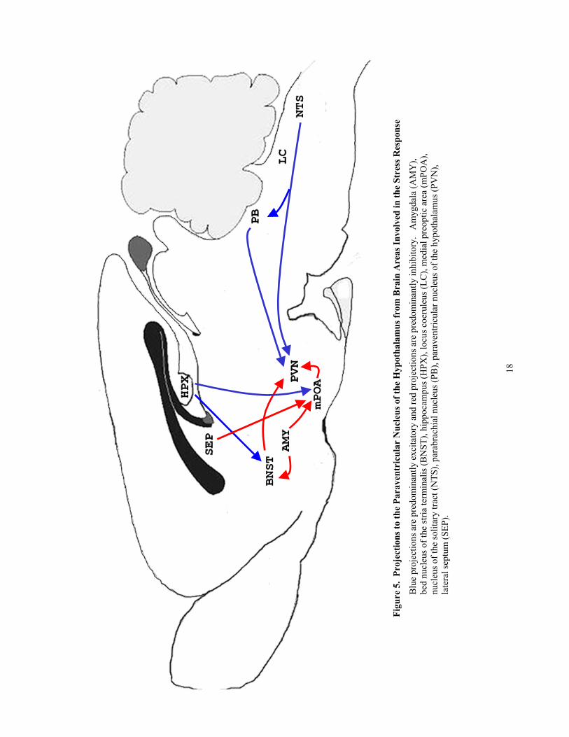
Figu
re 5
. Pr
ojec
tions
to th
e Pa
rave
ntri
cula
r N
ucle
us o
f the
Hyp
otha
lam
us fr
om B
rain
Are
as In
volv
ed in
the
Ss
o
Blu
e pr
ojec
tions
are
pre
dom
inan
tly e
xcita
tory
and
red
proj
ectio
ns a
re p
redo
min
antly
inhi
bito
ry.
Am
ygda
la (A
), be
d nu
cleu
s of t
he st
ria te
rmin
alis
(BN
ST),
hipp
ocam
pus (
HPX
), lo
cus c
oeru
leus
(LC
), m
edia
l pre
optic
are
a (m
nucl
eus o
f the
solit
ary
tract
(NTS
), pa
rabr
achi
al n
ucle
us (P
B),
para
vent
ricul
ar n
ucle
us o
f the
hyp
otha
lam
us (P
V
late
ral s
eptu
m (S
EP).
tres
Res
pns
e
MY
POA
), N
),
18

D. Anxiety
1. Definition and Classification of Anxiety
Anxiety is a common human emotional reaction that occurs in response to environmental
and/or physiological stressors. At mild levels anxiety is considered “norma n ing the senses
and mobilizing the body to respond to the stressor. However, anxiety is detrim en it
interferes with a person’s ability to function in normal daily activities, or w s
inappropriately triggered by little or no external stressful stimuli. The anxi in humans
has been defined by psychological symptoms such as worry, restlessness, or fear and physiological
symptoms such as sweating, elevated heart rate, or trembling [9]. The Ame atric
Association describes several forms of anxiety disorders, which currently include: generalized
anxiety disorder, obsessive-compulsive disorder, phobias, panic disorder, and post-traumatic stress
disorder [9]. Each type of anxiety disorder exhibits a unique combination of sy e
cases overlap. For example, generalized anxiety disorder is characterized b worry and
negative emotional affect. Post-traumatic stress disorder is also characteriz ve emotional
affect as well as increased stress reactivity. Overwhelming fear during a panic attack is a feature of
panic disorder and phobias result in extreme fear and avoidance of a specific object or situation.
Currently, there is little understanding of the underlying cause of anxiety at a neurobiological level.
Based on the behavioral symptoms of many of the anxiety disorder ely that
neural pathways involved in the fear response are also involved in at least s sorders.
However, the current literature provides conflicting opinions on the distinction between anxiety and
fear. Anxiety is defined as an emotional anticipation of an aversive situation that is difficult to
predict or control, and is likely to occur. It is usually considered a more general state of distress
prompted by generalized cues, lasting for long periods of time once activated, and physiological
arousal that lacks adaptive significance [28]. On the other hand, fear is usually elicited by an
l”, e
hen
ety r
rica
y ex
ed b
s it s
ome
hanc
ental wh
anxiety i
esponse
n Psychi
mptoms that in som
cessive
y negati
eems lik
anxiety di
19

identifiable, threatening stimulus with escape or avoidance as a goal [28]. To date much of the
literatur te
ehavio
s.
haviors of
imal
nt
usually
d
an
s), an
ethologically based test, measures a mouse’s natural tendency to explore an unfamiliar environment
e at mpting to define the neurobiology of anxiety has relied on studies of fear-related
b r.
To gain a better understanding of the neurobiology of anxiety, behavioral animal models
have been developed to reproduce some of the symptoms observed in human anxiety disorder
Behavioral animal models of human anxiety disorders rely on of a variety of ethological be
rodents that have been interpreted to be “anxiety-like” [56,184].
Animal tests of anxiety can be divided into two categories. These include tests based on
conditioned fear or unconditioned fear, in which anxiety is generated by exposure to a novel
environment or situation [120,160]. Conditioned anxiety animal models test the ability of an an
to suppress a behavior in response to the delivery of an unavoidable form of punishment. Therefore
the behavior of the animal tends to be repressed [32]. Conditioning models allow for experimental
control over behavioral baselines, but require extensive behavioral training of the animals and
multiple experimental controls for non-specific treatment effects on learning/memory, appetite,
and/or perception of the punishment. Conditioned anxiety tests can be further categorized as conflict
or non-conflict based tasks. In a non-conflict based task the rodent is re-exposed to an environme
or situation that resulted in fear or anxiety-related behavior. Conditioned tasks based on conflict
involve punishment (i.e. shock) in response to innate behaviors (i.e. eating or drinking).
The second classification of anxiety testing relies on unconditioned responses. Procedures
based on unconditioned behavior can be distinguished by the expression or inhibition of responses.
Tests falling into the former category include those that result in the expression of unconditione
defensive reactions, such as freezing, startle, or ultrasonic vocalization to an anxiogenic stimulus.
However, the majority of unconditioned anxiety tests result in behavioral inhibition in response to
anxiogenic stimulus. For instance, the elevated plus-maze (discussed in Materials and Method
20

opposed with its innate fear of novel or aversive environments [13,167]. Unlike conditioned
behavior animal models, unconditioned response paradigms do not require training and are less
susceptible
to variability in motivational processes.
as
of stimulus-specific fear models
as oppo
ble in
a) Afferents and Stimulus Processing of Anxiety
In response to anxiety or fear stimuli, sensory information (i.e. auditory, visual,
somatosensory) is relayed from peripheral receptor cells to the dorsal thalamus, which is the neuronal
interface between sensory stimuli and forebrain structures [113,115] (Figure 6). Afferent sensory
inputs from the thalamus are then relayed directly to the amygdala and cortical brain regions, such as
the primary visual (occipital), auditory (temporal), or tactile (post-central gyrus) cortex, which then
relay sensory information to the amygdala [90]. However, olfactory sensory input has input to the
amygdala either directly or through the entorhinal cortex (a portion of the hippocampal formation).
From the entorhinal cortex there are direct projections to the amygdala and the hippocampus which
projects to the amygdala [64]. The amygdala, in turn sends reciprocal projections to the
hippocampus [171]. The connections between the entorhinal cortex, hippocampus and the amygdala
provide a neuroanatomical substrate for the interaction between storage and recall of the memory of a
fear- or anxiety-inducing stimulus and the emotion related to that stimulus [64]. The amygdala also
2. Neurocircuitry of the Anxiety Response
Although there is a close correspondence between fear and anxiety, the study of anxiety h
relied heavily on the use of fear conditioned animal models. Much of the literature reporting
neuroanatomical activation of anxiety-related pathways makes use
sed to less stimulus specific models, which may be more relevant to human generalized
anxiety disorders. However, the use of fear-conditioning in animals has been extremely valua
understanding the brain systems that are involved in anxiety. Possible interaction of OT with many
of the following brain areas is discussed in reference to the experiments performed in Chapter V.
21

has strong reciprocal projections to the thalamus [171]. The thalamocortico-amygdala connec
could account for an unconscious fear and anxiety response [114].
tions,
b) Efferents and the Anxiety Response
to
ediated by
Figure 6. Afferents and Stimulus Processing of Anxiety.
Cortex
Dorsal Thalamus
stimulation Cortex
Dorsal Thalamus
stimulation
Amygdala
Hippocampus
y Cortex
Entorhinal
(Sensory Stimuli)
olfactory
Amygdala
Hippocampus
y Cortex
Entorhinal
(Sensory Stimuli)
olfactory
Primary Sensor
AnxietySensor
Anxiety
Primary
The amygdala receives highly processed sensory information from all modalities through its
lateral and basolateral nuclei. The basolateral nucleus of the amygdala sends extensive projections
the striatum and the BNST [171]. These projections may be responsible for motor responses critical
in the “fight or flight” responses to threatening stimuli. The remainder of the afferents of the lateral
and basolateral nuclei project to the central nucleus of the amygdala, which then project to a variety
of brain areas that mediate the emotional and physiological reactions to fear and anxiety [41,171]
(Figure 7).
Sympathetic activation and hormonal release associated with anxiety and fear is m
stimulation of the hypothalamus via projections from the central nucleus of the amygdala (CeA), bed
nucleus of the stria terminalis (BNST) and locus coeruleus (LC). The hypothalamus integrates
information it receives from a variety of brain structures into a coordinated pattern of sympathetic
22

and neuroendocrine responses. Stimulation of the lateral hypothalamus directly by the CeA or
through the (LC) activates the sympathetic system resulting in increased in blood pressure and heart
rate, sweating, piloerection, and pupil dilation [171]. Activation of the PVN from CeA projections
relayed through the BNST [37] results in HPA activation. Lastly, CeA projections to the dorsal
motor nucleus of the vagus result in parasympathetic inhibition associated with anxiety, including
gastrointestinal and genitourinary disturbances [28].
It should be noted that through the use of immunohistochemical labeling for Fos it has
been determined that the medial nucleus of the amygdala is also activated during exposure to the
elevated plus maze [45,186]. The medial amygdala (MeA) receives projections from the
entorhinal cortex, frontal cortex, and hypothalamus, and projects to the BNST, hypothalamic
nuclei, and thalamus [1 n forms of anxiety-
inducing stimuli activate the MeA. However, activation of afferent and efferent pathways of the
MeA have no en
71]. Therefore, it is possible that exposure to certai
t be investigated extensively.
Figure 7. Efferents of the Amygdala to Anxiety-Related Brain Areas
Abbreviations, bed nucleus of the stria terminalis (BNST), dorsal motor nucleus of the vagus (DMV), paraventricular nucleus of the hypothalamus (PVN).
BNST
PVN
BNST
PVN
Amygdala CoeruleusLateral
Hypothalamus
Amygdala CoeruleusLateral
Hypothalamus
Striatum
Locus
DMV
Striatum
Locus
DMV
23

E. Involvement of Oxytocin in Stress and Anxiety
Peripheral and central release of OT accompanies secretion of corticosterone and ACTH in
response to certain forms of stress. Central and peripheral OT is released in male and female rats in
response to stressors that contain a psychogenic component. These stressors include forced
swimming in male [216,217] and female rats [209], various forms of social stress in male [48,50] a
female rats [138], and shaker stress in male rats [142]
nd
. Chronic infusion of synthetic OT into the
lateral v
and
s
response to emotional stress, and the presence of
OT receptors throughout anxiety related brain areas, including the hypothalamus and amygdala,
suggest a potential role for OT in the modulation of anxious behavior. Following central
administration of OT, an anxiolytic-like effect has been described in rats. Central administration of
OT into the lateral ventricles of rats [211] and estrogen-treated ovariectomized mice [129] resulted in
decreased anxiety-related behavior in the elevated plus maze. Oxytocin infused into the central
nucleus of the amygdala, but not the ventromedial nucleus of the hypothalamus, resulted in decreased
anxiety-related behavior of rats in the elevated plus maze and open field, indicating brain region-
specific effects [11]. However, aside from the findings of Bale et al. there is little functional
information regarding the role OT in anxiety-related behavior.
F. Oxytocin
entricles of ovariectomized female rats decreased the corticosterone response to a
psychogenic (noise) stress and reduced anxiety-related behavior [211]. Infusion of an OT receptor
antagonist into the lateral ventricles disinhibited the basal and stress induced release of ACTH
corticosterone in male and female rats exposed to an elevated plus-maze [140], repeated airpuffs
[138], and female rats forced to swim [140]. Thus activation of central OT signaling mechanisms i
believed to exert inhibitory control over the stress response.
Parvocellular OT neurons are activated in
Deficient Mouse
1. Production of the Oxytocin Deficient Mouse
24

Similar to the human OT gene, the mouse gene for OT-neurophysin I consists of three exons:
the first l
ophysin
the OT
ion
ng et al. was used for all of the
experim
ions of the
P
VP expression is reduced in the OT-
/- mouse, it is possible that genomic response elements within the OT gene that regulate the
expression of AVP are altered in the OT-/- mouse.
exon encodes a signal peptide, the nine peptide OT hormone, the tripeptide processing signa
(GKR), and the first nine amino acids of neurophysin; the second exon encodes the central part of the
neurophysin (residues 10-76); and the third exon encodes the COOH-terminal region of neur
(residues 77-93/95) [67]. Currently there are three different versions of the oxytocin deficient mouse
(OT-/-). Gross et al. replaced all three exons of the oxytocin (OT) gene, eliminating the
preproOT/neurophysin coding sequence [66,213]. Nishimori et al. deleted the first exon of
gene [141]. This deletion resulted in the elimination of the initiation ATG codon, the processing
signal, the OT peptide, and the first few amino acids of neurophysin. Young et al. replaced the
second and third exons of the OT gene with a neomyocin resistance cassette, resulting in the delet
of the carrier polypeptide [223]. Therefore, although OT is transcribed, it is not packaged or
transported out of the cell. The OT-/- mouse created by You
ents discussed in this thesis (refer to Chapter II, section A).
2. Central Peptide Expression
Through different methods of evaluation it has been confirmed that all three mutat
OT gene sequence resulted in the elimination of OT. Using in situ hybridization histochemistry it
was determined that oxytocin transcripts were absent from the PVN and SON of the hypothalamus of
OT deficient mice in which all three exons were deleted compared to wildtype mice of the same
129/Sv-Black Swiss background [143]. CRH transcript levels were not different in the PVN of OT-/-
and OT+/+ mice [143]. However, in situ hybridization histochemistry also revealed decreased AV
mRNA in the PVN and SON of OT-/- mice compared to OT+/+ mice [143]. The AVP gene is
closely linked to the OT gene [67]. While it is not known why A
25

In a different version of the OT-/- mouse Nishimori et al. also performed in situ hybridization
histochemistry to confirm that OT mRNA was not synthesized [141]. OT/neurophysin mRNA was
not present in the PVN and SON of OT-/- mice. In addition, AVP mRNA content in the PVN and
SON of OT-/- mice is not altered in OT-/- mice. Although this transgenic mouse does not synthesize
OT, the synthesis of AVP mRNA is intact.
Young et al. also performed in situ hybridization histochemistry in addition to
immunocytochemistry to confirm that the production of OT was reduced [223]. Using a prob
for exon 1 of the OT gene, OT transcripts in the PVN of OT heterozygous and OT deficient
were 53% and 1% of the wildtype OT mRNA, respectively [223]. Immunohistochemistry using
antibodies to neurophysin confirmed the findings obtained using in situ hybridization
histochemistry. Abundant staining of OT neurophysin was evaluated in the PVN, SON,
posterior pituitary of wildtype mice [223]. However, there was no
e
mice
and
detectable OT protein in the
PVN and S situ hybridization histochemistry Young et al. also
measured the expression of other genes normally expressed in the PVN and SON. Corticotropin-
releasing hormone mRNA expression was not altered in the PVN of OT-/- mice compared to
OT+/+ mice [223]. Compared to OT+/+ mice AVP mRNA is reduced by 26% in the PVN and
30% in the SON of OT-/- mice [223]. However, AVP related behavioral differences have not
been observed in OT-/- mice.
sponse
OT deficient mice in order to elucidate the role
of OT in stress and anxiety responses. Prior to the development of the oxytocin deficient mouse,
ON of OT-/- mice [223]. Using in
G. Specific Objectives of the Research
The overall objective of this research was to evaluate the role of OT in the stress and anxiety
response. Anxiety-related behavior, the corticosterone response and neuronal activation in re
to stress- and anxiety-related stimuli were studied in
26

researchers have relied on exogenous administration of OT, OT antagonists, or OT antisense
oligonu
ess and anxiety responses. The specific objectives of
T
ker stress,
was achieved by administering systemic cholecystokinin (CCK), a physical stressor, to
male OT+/+ and OT-/- mice and evaluating the corticosterone response.
2. To evaluate anxiety-related behavior of the OT deficient mouse.
A. To determine the effect of OT on anxiety-related behavior. This was achieved by placing
male and female OT+/+ and OT-/- mice in the elevated plus maze.
B. To determine if the effect of OT is dependent upon binding at the OT receptor. This was
eral ventricles of OT-/- mice, or an OT antagonist
cleotides to study the effects of OT on the stress and anxiety response. Although
pharmacological studies have provided a tremendous amount of information regarding the role of OT
in the stress and anxiety responses, there are limitations and a lot of variability in these studies.
Therefore, OT deficient mice that do not synthesize or release central or peripheral OT are a unique
animal model to test the role of OT in the str
this study are:
1. To evaluate HPA axis activation in response to psychogenic and systemic stressors in the O
deficient mouse.
A. To determine the effect of OT on the corticosterone response to a psychogenic stressor.
This was achieved by exposing male and female OT+/+ and OT-/- mice to sha
which has been defined as a psychogenic stressor, and evaluating the corticosterone
response.
B. To determine the effects of OT on the corticosterone response to a physical stressor. This
achieved by infusing OT into the lat
into the lateral ventricles of OT+/+ mice, prior to testing in the elevated-plus maze. In
addition, an OT antagonist was infused into the lateral ventricles of OT-/- mice followed
by infusion of OT, prior to testing in the elevated plus maze.
27
3. To evaluate activation of stress- and anxiety-related pathways following exposure to a
or stress-related stimuli in OT deficient mice.
A. To determine if OT signaling pathways contribute in the modulation of the HPA axis
response to a psychogenic stressor. This was achieved by exposing OT+/+ and OT-/-
mice to shaker stress, a psychogenic stressor. Brain tissue was harvested and then
processed for immunocytochemistry for Fos and CRH, AVP, or OT, and quantifying Fo
acti
nxiety-
s
vation in the PVN and stress-related limbic brain areas.
a systemic stressor. This was achieved by administering cholecystokinin, a
C. s contribute in the modulation of anxiety-related
xiety-related
B. To determine if OT signaling pathways contribute in the modulation of the HPA axis
response to
nauseogenic, to OT+/+ and OT-/- mice. Brain tissue was harvested and then processed
for immunocytochemistry for Fos and CRH, AVP, or OT, and quantifying Fos activation
in the PVN and stress-related limbic brain areas.
To determine if OT signaling pathway
behavior. This was achieved by exposing OT+/+ and OT-/- mice to the elevated-plus
maze. Brain tissue was harvested and then processed for immunocytochemistry for Fos
and CRH, AVP, or OT, and quantifying Fos activation in the PVN and an
limbic brain areas.
28
II. Materials and Methods
The protocols were approved by the Institutional Animal Care and Use Committee of the
Univers
A. Breeding and Maintenance of Wildtype and Oxytocin Deficient Mice
Male and female wildtype (OT+/+) and OT deficient (OT-/-) mice of C57BL/6 strain were
used for these studies. The OT-/- mice were developed by Scott Young, (National Institute of Mental
Health, Bethesda, MD) [222] and breeding pairs for this study were purchased from Jackson
Laboratories (Bar Harbor, ME). OT+/+ mice were created by breeding OT+/+ male and female
mice. OT-/- mice were created by breeding female OT+/- mice with male OT-/- mice. This breeding
paradigm was used to eliminate the necessity for cross-fostering pups. In addition, all pups are
exposed to peripheral circulating OT in utero during development, limiting the variability in the
prenatal development of OT+/+ and OT-/- pups.
Animals were bred and housed in the viral free quarters of the University of Pittsburgh
Animal Facility under a 12-h light/dark cycle (lights on at 0700 h). Mice were housed in standard
suspended cages in groups of up to four animals per cage with free access to water and food (Prolab
RMH 3000 5P00, LabDiet/Purina). During testing animals were removed from group housing and
acclimated to single housing for a week prior to the test day unless otherwise stated.
. Genotype Determination of Mice Using Polymerase Chain Reaction (PCR)
To identify the genotype of the mice approximately 0.5 cm of mouse-tail was digested and
NA was extracted and prepared for polymerase chain reaction (PCR). The DNA sample was
issolved in 100µl of 10mM Tris-HCl and 1mM EDTA. 50µl PCR reactions containing 2µl (100ng)
f the DNA sample, 5.0µl 10X PCR buffer minus Mg (Gibco BRL, Gaithersburg, MD), 2.5µl 10mM
NTPs (Invitrogen, Carlsbad, CA), 2.5µl 50mM MgCl2 (Gibco BRL, Gaithersburg, MD), 0.5µl Taq
ity of Pittsburgh.
B
D
d
o
d
29

DNA polymerase (Gibco BRL, G were heated for 5 min at 95oC
and the or
Primer pairs, synthesized at the University of Pittsburgh Sequence facility, were designed for
PCR that detect either the wild-type allele (OT, 332 bp) or the mutant allele (neomycin resistance
cassette, 430 bp). Primer pairs for the wild-type allele are (forward) TCG CTC TGC CAC AGT
CCG GAT TC and (reverse) TCA GTG TTC TGA GCT GCA AAC C, and for the mutant allele are
(forward) AGA GGC TAT TCG GCT ATG ACT G and (reverse) TTC GTC CAG ATC ATC CTG
ATC.
as
levels were measured by radioimmunoassay using
a comm ).
aithersburg, MD) and 1-2µl primers
n cycled 30 (35 for OT) times at 94oC for 40 sec (45 sec for OT) and 63oC (55oC for OT) f
1 min.
C. Plasma Corticosterone Analysis
Trunk blood was collected into heparinized tubes on ice, centrifuged at 4oC, and plasma w
stored at –20oC until assay. Plasma corticosterone
ercially available kit purchased from Diagnostic Products Corporation (Los Angeles, CA
The range of this is assay is 0ng/ml - 2000ng/ml and the minimum detection limit for corticosterone
is 5.7ng/ml. The intra-assay precision (coefficient of variation) for the assay is 3.7% + 0.2 and the
ED50 is 122.4ng/ml + 2.8.
D. Immunocytochemistry
erfused
with 0.1
es
Mice were anesthetized by intraperitoneal (ip) injection of ketamine/xylazine, and p
5M saline followed by 4% paraformaldehyde fixative (0.1M sodium phosphate buffer
containing 4% paraformaldehyde, 1.4% L-lysine, and 0.2% sodium metaperiodate) 60-75 minut
after experimental testing. Fixed brains were removed from the skull, post-fixed in 4%
paraformaldehyde at 4oC for 12-18 hours, and transferred to 25% sucrose solution (4oC) for 24-72
hours before sectioning. Coronal tissue sections were cut (35µm thick) using a freezing stage
30

microtome. Tissue was stored in cryoprotectant [205] at –20oC until immunocytochemical
processing.
Sections were removed from cryoprotectant and rinsed in several changes of 0.1M sodium
48h
ImmunoResearch Laboratories, West Grove, PA) for 1h at room temperature. Sections were
rinsed and processed using the Vectastain Elite avidin-biotin immunoperoxidase method (Vector
Laboratories, Burlington, CA). A sodium acetate buffer solution of diaminobenzadine (DAB), nickel
sulfate, and H2O2 was used to generate blue-black nuclear cFos immunolabeling.
The tissue was then processed for OT (rabbit anti-OT, 1:30,000; Chemicon, Temecula, CA),
AVP (rabbit anti-AVP, 1:20,000; Chemicon, Temecula, CA), or CRH (rabbit anti-CRH, 1:10,000;
Peninsula Laboratories, Belmont CA). The tissue was incubated in antisera diluted in 0.1M sodium
phosphate buffer containing 1% normal donkey serum and 0.3% Triton X-100 for 48-72 hours at
4oC. Tissue was rinsed in several changes of 0.1M sodium phosphate buffer and then incubated in
600) for 1 h at room temperature. Sections were then rinsed
and pro
-
raded
parvocellular neurons of the PVN and OT and AVP magnocellular neurons of the supraoptic nucleus
phosphate buffer, treated for 30 minutes in 1% sodium borohydride (Sigma, St. Louis, MO), and
rinsed again in sodium phosphate buffer. Antisera were diluted in sodium phosphate buffer
containing 0.3% Triton X-100 and 1% normal donkey serum. Tissue sections were incubated for
at 4oC in rabbit anti-c-fos (1:50,000; provided by Drs. Philip Larson and Jens Mikkelsen, Planum
Institute, Denmark), rinsed, and then incubated in biotinylated donkey anti-rabbit IgG (1:600;
Jackson
biotinylated donkey anti-rabbit IgG (1:
cessed using the Vectastain Elite avidin-biotin immunoperoxidase method (Vector
Laboratories, Burlington, CA). OT, AVP, and CRH immunolabeling was generated using a non
enhanced DAB reaction to create brown immunoprecipitate. Immunolabeled tissue sections were
mounted onto Superfrost Plus glass slides (Fisher Scientific, Pittsburgh, PA), cleared in g
alcohols and xylene, and coverslipped using Histomount (VWR, Bridgeport, NJ).
A quantitative analysis of cFos expression in OT, AVP, and CRH magnocellular and/or
31

of the hypothalamus (SON) was conducted in anatomically matched tissue sections. For thi
purpose, ana
s
tomically matched brain sections containing OT-positive neurons (in OT +/+ mice only,
since O
d for
.
of the medial and
lateral B
-5
s
E. Shaker Stress
Shaker stress, originally described as an environmental stress, was chosen because it is a
predominantly psychogenic stressor [137] that releases corticosterone and OT, but not AVP, in rats
[69,142]. Mice were individually placed in an opaque plastic beaker (27cm diameter and 36cm
height) that was fixed to a shaking platform (Dubnoff Shaker Model 3575). Mice were subjected to
T immunolabeling is absent in OT -/- mice; 4-5 brain sections from bregma –0.58 to –1.22),
AVP-positive neurons (in all mice; 4-5 brain sections from bregma –0.58 to –1.22), and CRH-
positive neurons (in all mice; 2-3 brain sections from bregma –0.58 to -0.94mm) were selecte
analysis [57]. Unlike in rats, in which parvocellular and magnocellular divisions of the PVN are
easily distinguished, there is as yet no conventional method to separate these two divisions in mice
OT, AVP, and CRH cells were considered Fos-positive when their nuclei contained blue-black
immunoreactivity. The average numbers of activated, phenotypically identified neurons within the
PVN and SON per section (both sides) were calculated for each mouse.
A quantitative analysis was also conducted in the bed nucleus of the stria terminalis
(BNST; combined counts of the anterodorsal, anterolateral and anteroventral nuclei
NST), medial (MeA) and central nuclei (CeA) of the amygdala, and the medial preoptic area
(mPOA) of OT+/+ and OT-/- mice. For each mouse, brain sections containing the anatomically
matched levels of the BNST (3-4 brain sections in bregma 0.62 to 0.26mm), the MeA (4-6 sections in
bregma –0.82 to –1.70mm), the CeA (6-8 sections in bregma –0.82 to 1.70mm), and the mPOA (3
brain sections in bregma 0.5 to –0.10mm) were selected for analysis [57]. Cells were considered
cFos positive when their nuclei contained blue-black immunoreactivity. The average number of cell
per section (both sides) in each brain region was calculated for each mouse.
32

shaker stress (180cycles/min) for 10 minutes, and then returned to their home cage and sacrific
rapid decapitation for corticosterone assessment or perfused for immunocytochemical processing at
the times specified in each of the experiments. The duration of shaker stress was chosen based on
studies performed in rats showing increased peripheral and central OT and corticosterone relea
after 5+ min of shaker stress [69]. Control mice that did not receive shaker stress were sacrificed at
the same time. Studies were conducted between 0800-1200h, which is the nadir of corticosterone
secretion in rodents [29,224]. To keep environmental stress to a minimum during the experiments,
mice were housed, exposed to shaker stress, and sacrificed in different rooms. On the day of the
experiment mice were tested as paired cohorts according to genotype and treatment group.
ed via
se
F. Cholecystokinin Administration
Cholecystokinin (CCK; Bachem, King of Prussia, PA) was chosen as a systemic stressor
because it is an anxiogenic/nauseogenic agent that results in the activation of the HPA axis [97], and
OT activation [200] and release [198]. Mice of both genotypes were either normal fed or overnight
food deprived for 18h. The following morning mice were injected i.p. with saline or CCK (10µg/kg
dissolved in 0.9% saline). The dose used in this study was based on prior studies determining that
10µg/kg CCK produced anorexia in fasted OT+/+ and OT-/- mice and activated PVN OT neurons of
OT+/+ mice [122]. Mice were sham injected daily for seven days prior to the day of experimentation
to control for the possible increase in corticosterone due to the pain of injection. To keep
environmental stress to a minimum, sacrifice took place in a different room from drug
G. Lateral Ventricle Cannulation Surgery and Infusions
OT+/+ and OT-/- mice were anesthetized with ketamine (1mg/10g bodyweight, ip) and
xylazine (0.025mg/10g bodyweight, ip) for stereotaxic placement of cannulae into the lateral
ventricles. The top of the animal’s head was shaved and a 1-mm midline incision was made across
administration.
33

the top of the skull. After cleaning the periosteum, a 1mm hole was drilled 1.00-mm lateral and
0.5mm posterior to bregma and the tip of a 26-gauge stainless steel infusion cannula was placed 2.00
mm below the skull surface in the lateral ventricle. The cannula was secured to the skull with denta
cement and a stylus was inserted to maintain patency. Mice were allowed to recover for one week
prior to behavioral testing. Patency of the guided cannula was confirmed by the movement of an air
bubble placed in the PE-10 tubing connecting a 25µl Hamilton syringe to the guide cannula. Prior
euthanizing, we infused 2µl of India ink dissolved in aCSF to confirm cannula placement. Only mice
that showed correct placement of the cannula were included in the analyses.
l
to
H. Elevated Plus Maze Testing
logically validated animal models of anxiety is the
elevated
s, (2)
number of
nd on occasion (5) time spent in the center (risk assessment). This index
represents the tendency of the animal to explore the aversive open, brightly lit, elevated environment,
rather than remaining in the preferred enclosed, dark environment. An animal exhibiting a decrease
in open arm entries or time would be considered to have an increased level of anxiety. The EPM has
been validated in mice and rats through the administration of anxiolytic drugs, which increase the
One of the best-documented and pharmaco
plus maze (EPM) task. The elevated plus maze is a conflict test based on the tendency of
rats [152] and mice [119] to explore an unfamiliar environment versus the tendency of rodents to
avoid the aversive properties of a brightly-lit, open, elevated space. The apparatus has four arms
extending from a central platform that form the shape of a plus. The mouse EPM is positioned on a
platform 40 cm above the floor, consists of two opposite facing open arms (30x5cm) and two closed
arms (30x5 cm with 15cm high walls) with a central area (5x5cm). The animal is placed in the
center, at the junction of the four arms, and allowed to explore the maze during a 5 minute test
session. The critical independent variables are: (1) percentage of entries into the open arm
percentage of time spent in the open arms, (3) number of entries in the closed arms, (4)
total arm entries, a
34
percentage of open arm entries, and anxiogenic drugs, which decrease the percentage of open arm
entries [119,152].
Approximately one week prior to testing, mice were transferred from group to individual
housing. During a 4-day acclimation and on the test day, mice were brought to the holding area
outside of the behavioral testing room for 1h. Mice were tested in the elevated plus maze (EPM)
between 1300-1700h and mice were paired by genotype and/or treatment during each test session.
Mice were placed on the central platform, facing an open arm and the number and duration of entries
into open or closed arms were videotaped for each mouse for 5 min and later sc
ored by a single
observer blinded to treatment and genotype of each mouse. An arm entry was defined as all four
paws entering an arm of the EPM.
nal
ss
J. Statistical Analysis
Results are presented as group mean +
I. Determination of the Estrous Cycle
Vaginal smears were obtained by flushing 50µl of distilled, autoclaved water into the vagi
cavity and retrieving it with a plastic pipette tip. The fluid was then immediately placed onto a gla
slide (Fisher Scientific, Pittsburgh, PA) and viewed under a microscope at low magnification. The
vaginal cytology at each stage of the estrous cycle is as follows: proestrus, signaled by the
appearance of polynucleated epithelial cells; estrus, represented by masses of cornified cells;
metestrus, represented by scattered leukocytes and dispersed non-nucleated cells; diestrus,
represented by many leukocytes with some epithelial cells. This was repeated everyday for 2-3
estrous cycles (12-14 days) at 10am each morning to accurately determine the vaginal cycle of each
mouse [34].
SE. Statistical analysis was performed using
StatView software (Abacus Concepts, Inc., Berkeley, CA). Data was assessed for normal
distribution prior to statistical comparison. All data presented in this thesis were normally
35

distributed. Therefore, pairwise differences were analyzed by a two-tailed t test and differences
between genotypes and treatments were determined using a two-way analysis of variance (ANOVA).
A repea
l F
ted measures ANOVA was conducted to determine differences between genotypes and/or
treatments across time. If the ANOVA revealed a statistically significant difference in the overal
ratio, post-hoc pairwise comparisons were done using the Bonferroni/Dunn test. A statistically
significant effect was accepted when p < 0.05.
36
III. Stress and the Oxytocin Deficient Mouse
Results of this chapter have been published in part [3,124].
Centrally released OT is believed to attenuate the response to stress in laboratory rodents
[61,210,211]. OT is released within the brain and/or into the circulation of male and female rats in
response to various psychogenic and physical stressors. These stressors include restraint stress i
male rats [94], forced swimming in male [94,216
n
,217] and female rats [209], 24h dehydration [94],
emorrhage [94], various forms of social stress in male [48,50] and female rats [138], and shaker
tress in male rats [142]. Infusion of synthetic OT into the lateral ventricles of estrogen-treated
variectomized rats decreased the corticosterone response to psychogenic restraint stress [210] and
oise stress [211], and reduced anxiety-related behavior [211]. Infusion of an OT receptor antagonist
to the lateral ventricles augmented the basal and stress-induced release of andrenocorticotropin
ormone (ACTH) and corticosterone in male and female rats exposed to an elevated plus-maze
PM) [140], repeated airpuffs [138], and female rats forced to swim [140].
To assess the role of the central oxytocinergic system in the stress response, plasma
orticosterone concentrations were measured after the termination of multiple stressors, both
sychogenic and physical, in male and female OT knockout and wildtype mice. Therefore, if central
T attenuates the HPA axis response to stress, it was hypothesized that OT knockout mice will have
igher concentrations of corticosterone in plasma than wildtype mice in response to stress.
A. Experimental Design
1. Psychogenic Stress
xperiment 1A. Corticosterone Response to Shaker Stress in Group Housed Female Mice
The purpose of this experiment was to determine if shaker stress would release corticosterone
mice and to determine the time course of corticosterone release following termination of stress.
he second purpose of the experiment was to determine if OT plays a role in modulating the
h
s
o
n
in
h
(E
c
p
O
h
E
in
T
37
corticosterone respo s ice were
-/- n=6) or 30 (OT+/+ n=4, OT-/- n=6)
minutes
e
=7)
the
in an effort to
reduce
trol
Experiment 2. Diurnal Plasma Corticosterone Concentrations in Female Mice
The purpose of this experiment was to determine whether genotypic differences in the
orticosterone response to shaker stress are due to alterations in the diurnal rhythm of corticosterone
OT+/+
(N=13)
nse to haker stress. Group-housed female OT+/+ and OT-/- m
subjected to shaker stress and sacrificed 10 (OT+/+ n=4, OT
post-stress. Control mice (OT+/+ n=3, OT-/- n=6), not exposed to shaker stress but
maintained the same as mice receiving stress, were sacrificed at the same times. Trunk blood was
obtained for measures of corticosterone. Compared to control mice, corticosterone concentrations
increased following shaker stress 10 and 30 minutes after stress. Because genotype differences were
similar 10 and 30 minutes following shaker stress, subsequent studies only examined corticosteron
levels in mice 10 min after termination of shaker stress.
Experiment 1B. Corticosterone Response to Shaker Stress in Individually Housed Mice
Housing conditions (group housed vs. individually housed) have been shown to influence
corticosterone levels in rats. Therefore, Experiment IA was repeated in OT+/+ (n=7) and OT-/- (n
mice that were individually housed for the week prior to and during the experiment in contrast to
first study in which animals were group housed. Mice were individually housed
variability in corticosterone levels measured within experimental groups. Mice were
subjected to shaker stress and sacrificed 10 minutes following termination of the stressor. Con
OT+/+ (n=8) and OT-/- (n=8) mice, which were not exposed to shaker stress but were maintained
and handled as mice receiving stress, were sacrificed at the same time. Trunk blood was obtained for
measures of plasma corticosterone.
c
in OT-/- mice. To determine minimum and maximum plasma corticosterone release, female
and OT-/- (N=14) mice were sacrificed at the expected circadian nadir (AM, 2 hours after
lights on) or at circadian peak (PM, 1 hour before lights off) of a 12/12h light/dark cycle [135].
Trunk blood was collected at sacrifice to determine plasma corticosterone levels.
38

Experiment 3. Corticosterone Response to Central Administration of Corticotrophin Releasin
g Hormone in Female Mice
To determine if the genotypic difference in corticosterone response to shaker stress occurs at
the HPA axis, ovine corticotrophin releasing hormone (oCRH) was administered into the lateral
ventricles of female mice of both genotypes. Female OT+/+ (N=10) and OT-/- (N=12) mice received
artificial cerebrospinal fluid (aCSF) or 30ng oCRH (Bachem, Torrence, CA) into the lateral
ventricles in a volume of 2µl over 2 minutes. The dose and route of administration was chosen
because it was reported to activate the HPA axis and release ACTH [18]. Thirty minutes following
infusion mice were sacrificed and blood was collected for determination of corticosterone levels.
Experiment 4. Corticosterone Response to Repeated Shaker Stress in Female Mice
Repeated exposure to shaker stress has been found to dampen the corticosterone response and
attenuate release of plasma OT in rats [69]. Therefore, the purpose of this experiment was to
determine if repeated exposure to shaker stress would eliminate the genotypic differences in the
corticosterone response. Plasma corticosterone concentrations were compared after acute shaker
stress in mice that were exposed to daily shaker stress for 9 days. OT+/+ and OT-/- mice were
divided into four groups. Conditioned stressed mice (OT+/+ n=10, OT-/- n=15) received shaker
stress daily, for 9 days. On the 10th day of the experiment, animals were sacrificed following shaker
stress. Naïve stressed mice (OT+/+ n-11, OT-/- n=12) were handled for 9 days and received acute
stress on the 10th day of the experiment. Conditioned control mice (OT+/+ n=5, OT-/- n=7) received
10 minutes of shaker stress daily, for 9 days. On the 10th day of the experiment, animals were
sacrificed without being exposed to shaker stress. Naïve control mice (OT+/+ n=9, OT-/- n=8) were
handled daily for 9 days, but not exposed to shaker stress, and were sacrificed on the 10th day of the
experiment. Trunk blood was collected at sacrifice to determine plasma corticosterone levels.
Experiment 5. Corticosterone Response to Shaker Stress Across the Estrous Cycle
39

To determine if the corticosterone response to shaker stress is influenced by the stage of the
strous cycle, OT+/+ and OT-/- mice were sacrificed after shaker stress at each of the following
stages of the estrous cycle: diestrus (OT+/+ n=6, OT-/- n=6), proestrus (OT+/+ n=5, OT-/- n=8), and
estrus (OT+/+ n= 6, OT-/- n=6). Trunk blood was collected at sacrifice to determine corticosterone.
Vaginal cytology was used to predict the stage of the cycle at which mice were to be studied with
shaker stress. Only mice with regular cycles were used in these experiments (94% of the total
number of OT+/+ mice and 91% of the total number of OT-/- mice tested).
Experiment 6. Corticosterone Response to Shaker Stress in Male Mice
The purpose of this experiment was to determine if exposure to shaker stress would result in
genotypic differences of the corticosterone response in male mice. Male mice were exposed to
shaker stress and sacrificed 10 (OT+/+ n=8, OT-/- n=8) or 30 (OT+/+ n=8, OT-/- n=7) minutes
following termination of shaker stress. Control mice (OT+/+ n=8, OT-/- n=10), which did not
receive shaker stress, were sacrificed at the same time. Trunk blood was collected for measurement
of plasma corticosterone.
2. Physical/Systemic Stress
Experiment 7. Corticosterone Response to Cholecystokinin Administration in Fasted and Non-fasted
The purpose of this experiment was to determine genotypic differences in the corticosterone
response to cholecystokinin (CCK) in fasted and non-fasted mice. Male fasted or non-fasted (normal
fed) OT+/+ and OT-/- mice were injected with CCK (n=6 of each genotype fasted and n=6 of each
genotype non-fasted) or saline (n=6 of each genotype fasted and n=6 of each genotype non-fasted),
sacrificed 30 min later, and corticosterone levels were determined.
Experiment 1A.. Corticosterone Response to Shaker Stress in Group Housed Female Mice
e
Mice
B. Results
1. Psychogenic Stress
40

In group-housed mice, plasma corticosterone concentrations significantly increased in both
genotypes at 10 and 30 minutes post-termination of a 10-minute shaker stress (ANOVA,
F(2,25)=26.60, p < 0.0001) compared to non-stressed mice (Figure 8). The increase in corticosterone
was similar 10 and 30 minutes following shaker stress. Furthermore, OT-/- mice released more
corticosterone than OT+/+ mice 10 min (p < 0.0002) and 30 min (p < 0.04) post-termination of
shaker stress (Figure 8).
800
300
400
500
ntrol 10-min post-terminationof shaker stress
30-min post-terminationof shaker stress
Cor
ticos
tero
ne (n
g/m
l)
3 6 4 6 4 6
p < 0.04700
OT+/+ femalesOT-/- females
p < 0.0002
0
100
200
600
Co
8. Corticosterone Response to Shaker Stress in Group-Housed Female Mice.
lasma c measured 10 or 30 min following termination of a 10 min shaker stress or in control mice that did not receive shaker
corticosterone increase was greater in OT-/- versus OT+/+ mice at 10 min and 30 min (ANOVA, F(2,25)=26.60, m
ually Housed Mice
As was the case with group-housed mice, plasma corticosterone concentrations increased in
both genotypes 10 minutes after termination of shaker stress compared to mice not receiving this
stress (Figure 9). Moreover, corticosterone concentrations increased to a greater degree in OT-/-
mice versus OT+/+ mice (ANOVA, F (1,12)=51.15, p < 0.02).
Figure
P orticosterone concentrations of group housed female wildtype (OT+/+) and OT deficient (OT-/-) mice were
stress. Shaker stress resulted in a rise in plasma corticosterone concentrations in both genotypes, but the
p<0.0001). The number of mice per group is located in the data bars. Figure was reprinted with permission froreference [124].
Experiment 1B. Corticosterone Response to Shaker Stress in Individ
41

Figure 9. Corticosterone Response to Shaker Stress in Individually Housed Female Mice.
Plasma corticosterone concentrations were measured in individually housed female (OT+/+) and OT deficient (OT-/-) mice 10 min following termination of a 10 min shaker stress or in control mice that did not receive shaker stress. Plasma corticosterone concentrations increased in both genotypes after shaker stress, but to a greater degree in OT-/- versus OT+/+ mice at 10 min (ANOVA, F(1,12)=51.15, p<0.02). The number of mice per group is located in or above the data bars. Figure was reprinted with permission from reference [124].
asma Corticosterone Concentrations in Female Mice
Analysis of plasma corticosterone levels at the expected circadian nadir and peak revealed an
increase in basal corticosterone in both OT+/+ (ANOVA, F(1,11) = 29.61, p < 0.0002) and OT-/- mice
Experiment 2. Diurnal Pl
400
500
600
700
800
Cor
ticos
tero
ne (n
g/m
l)
OT+/+ femalesp < 0.02
0
100
200
300
Control 10-min post-termination of shakerstress
OT-/- females
n=8 n=87 7
(ANOVA, F(1,12) = 40.65, p < 0.0001) at the circadian peak (Figure 10). Minimum and maximum
plasma corticosterone levels did not differ between female OT+/+ and OT-/- mice.
42
Figure 10. Diurnal Plasma Corticosterone Concentrations in Female Mice.
Diurnal plasma corticosterone concentrations were measured in individually housed female wildtype (OT+/+) and OT deficient (OT-/-) mice. Female OT+/+ and OT-/- mice were sacrificed at circadian nadir (AM, 2 hours after lights on) or at circadian peak (PM, 1 hour before lights off) on a 12/12 light/dark cycle. Analysis of plasma
OT+/+ (ANOVA, F(1,11) = 29.61, p = 0.0002) and OT-/- mice (ANOVA, F(1,12) = 40.65, p < 0.0001) at the circadian peak. Plasma corticosterone values id not differ between female OT+/+ (ANOVA, F(1,14) = 0.51, p = 0.82) and OT-/- mice (ANOVA, F(1,9) = 0.002, p = .96). The number of mice per group is located in or above the data bars.
rmone in Female Mice
Plasma corticosterone was released in female OT+/+ (p < 0.03) and OT-/- (p < 0.001) mice
thirty minutes following administration of oCRH compared to aCSF treated mice (Figure 11).
However, plasma corticosterone concentrations increased to the same degree in female mice of both
genotypes (ANOVA, F(1,18) = 0.059, p = 0.45).
corticosterone revealed an increase in baseline corticosterone of female
OT+/+ females
0
100
200
300
400
500
600
700
800
AM PM
Cor
ticos
tero
ne (n
g/m
l)
OT-/- females
n=8 n=85 6
d0
Experiment 3. Corticosterone Response to Central Administration of Corticotrophin Releasing
Ho
43
0
100
200
300
400
500
600
700
800
aCSF oCRH
Cor
ticos
tero
ne (n
g/m
l)
OT+/+ femalesOT-/- females
4 6 6 6
Figure 11. Corticosterone Response to Central Administration of Corticotrophin Releasing Hormone in Female Mice.
0 fluid
d i n SF
ce
Experiment 4. Corticosterone Response to Repeated Shaker Stress
The corticosterone response of mice repeatedly exposed to shaker stress was compared to
mice receiving a single episode of acute shaker stress. Basal corticosterone levels of conditioned
control mice were the same as naïve control mice of both genotypes (Figure 12). The corticosterone
response of conditioned stressed OT+/+ (p < 0.04) and OT-/- (p < 0.01) mice was attenuated
compared to naïve stressed (Figure 12). However, repeated exposure to shaker stress did not abolish
the difference in corticosterone release between genotype. Conditioned stressed OT-/- mice had
higher plasma corticosterone concentrations compared to OT+/+ mice (p < 0.01, Figure 12), despite
repeated exposure to shaker stress.
Plasma corticosterone concentrations were measured in female wildtype (OT+/+) and OT deficient (OT-/-) mice 3minutes following administration of ovine corticotropin releasing hormone (oCRH) or artificial cerebrospinal (aCSF). Administration of oCRH into the lateral ventricles of OT+/+ and OT-/- mice resulte n an increase iplasma corticosterone compared to aC treated mice of both genotypes. Plasma corticosterone increased to the same degree in female OT+/+ and OT-/- mice (ANOVA, F(1,18) = 0.059, p = 0.45). The number of mi per group is located in the data bars.
44

0
100
200
300
400
500
600
700
800
Naïve Control ConditionedControl
Naïve Stressed ConditionedStressed
Cor
ticos
tero
ne (n
g/m
l)
OT+/+ femalesOT-/- females
n=9 n=8 n=5 n=711 12 10 15
p<0.02p<0.01
Figure 12. Corticosterone Response to Repeated Shaker Stress in Female Mice
ated aïve
control mice. Naïve stressed OT-/- mice released more corticosterone than OT+/+ mice following shaker stress.
n or
xperiment 5. Corticosterone Response to Shaker Stress Across the Estrous Cycle
OT-/- mice released more corticosterone than OT+/+ mice when exposed to shaker stress
(ANOVA, F(1,30)=32.08, p < 0.0001; Figure 13). Significant genotypic differences in the
corticosterone response to shaker stress were observed at each stage of the estrous cycle.
Corticosterone increase in response to shaker stress was higher in OT-/- compared to OT+/+ mice in
the diestrus (p < 0.03), proestrus (p < 0.004), and estrus (p < 0.003) stages of the estrous cycle
(Figure 13). Plasma corticosterone levels following shaker stress across the estrous cycle (ANOVA,
F(2,30) = 9.03, p < 0.008). Corticosterone levels in response to shaker stress were lower in mice in
estrus that in diestrus (p < 0.003) or proestrus (p < 0.003). However, there was not an interaction
between genotype and stage of the estrous cycle in reference to the corticosterone response to shaker
stress (ANOVA, F(2,30) = 0.881, p>0.05). Vaginal cytology verified 4-day estrous cycles in both
genotypes.
Plasma corticosterone response in female wildtype (OT+/+) and OT deficient (OT-/-) mice following repeshaker stress. Conditioned control OT+/+ and OT-/- mice released similar amounts of corticosterone as n
Conditioned stressed OT-/- mice released more corticosterone than OT+/+ mice following shaker stress. Inaddition, conditioned control OT+/+ (p < 0.04) and OT-/- (p < 0.01) mice displayed an attenuated corticosteroneresponse to shaker stress compared to mice of the same genotype. The number of mice per group is located iabove the data bars. Figure was reprinted with permission from reference [124].
E
45

100
200
300
400
500
600
700
800
900
Cor
ticos
tero
ne (n
g/m
l)
OT+/+ femalesOT-/- females
18 20 6
p < 0.0001
p < 0.03p < 0.004
p < 0.003
06 5 66 8
Figure 13. Corticosterone Response to Shaker Stress Across the Estrous Cycle
Plasma corticosterone concentrations were measured across the estrous cycle of
All Stages Diestrus Proestrus Estrus
female wildtype (OT+/+) and OT ficient (OT-/-) mice following shaker stress. OT-/- mice released more corticosterone than OT+/+ mice when
were not influenced r in OT-/- compared
to OT+/+ mice during the diestrus, proestrus, and estrus stages of the estrous cycle. The number of mice per group is locate
deexposed to shaker stress. Genotypic differences in the corticosterone response to shaker stressby the stage of the estrous cycle. Corticosterone increase in response to shaker stress was highe
d in or above the data bars. Figure was reprinted with permission from reference [124].
Experiment 6. Corticosterone Response to Shaker Stress in Male Mice
Plasma corticosterone concentrations increased following termination of a 10-minute shaker
stress compared to control mice that did not receive shaker stress (ANOVA, F(2,43)= 218.03, p <
0.0001; Figure 14). Unlike female mice, plasma corticosterone concentrations increased to a similar
degree in male OT+/+ and OT-/- mice (ANOVA, F(2,43) = 0.09, p = 0.77).
46

0
100
200
300
400
500
600
700
800
Control 10-min post-terminationof shaker stress
30-min post-terminationof shaker stress
Cor
ticos
tero
ne (n
g/m
l)
OT+/+ malesOT-/- males
n=8 n=10 8 8 8 7
Figure 14. Corticosterone Response to Shaker Stress in Male Mice lasma corticosterone concentrations were measured in male wildtype (OT+/+) and OT deficient (OT-/-) mice at 10 in or 30 min following termination of a 10 min shaker stress or in control mice that did not receive shaker stress.
gree in female mice of both genotypes (ANOVA, F = 0.09, p = 0.77). The number of mice per group is located in or above the data bars.
PmShaker stress resulted in a rise in plasma corticosterone concentrations to a similar de
(2,43)
xperiment 7. Corticosterone Response to Cholecystokinin Administration in Fasted and Non-fasted
sul se in
/+ NOVA, F(1,20)=0.05,
=0.82) and normal fed (ANOVA, F(1,19)=0.4, p=0.53) mice.
2. Physical/Systemic Stress
EMale Mice
Intraperitoneal administration of CCK in fasted (ANOVA, F(1,20)=43.46, p<0.0001; Figure
15) or normal fed (ANOVA,F(1,19)=145.31, p<0.0001; Figure 16) male mice re ted in an increa
corticosterone release compared to saline treated mice. However, plasma corticosterone
concentrations increased to the same degree in OT+ and OT-/- fasted (A
p
47

lasma corticosterone concentrations were measured in fasted male wildtype (OT+/+) and OT deficient (OT-/-) mice injected with cholecystokinin (CCK). Intraperitoneal administration of CCK resulted in increased
compared to mice administered saline.
Figure 16. Corticosterone Response to Cholecystokinin Administration in Non-fasted Male Mice.
lasma corticosterone concentrations were measured in non-fasted male wildtype (OT+/+) and OT deficient (OT-/-) mice injected with cholecystokinin (CCK). Intraperitoneal administration of CCK resulted in increased orticosterone release in non-fasted (ANOVA, F(1,19)=145.31, p<0.0001) male mice compared to mice administered
saline. Plasma corticosterone increased to the same degree in OT+/+ and OT-/- non-fasted mice (ANOVA, F(1,19)=0.40, p>0.05). The number of mice per group is located in or above the data bars.
Figure 15. Corticosterone Response to Cholecystokinin Administration in Fasted Male Mice.
0
100
200
300
400
500
600
700
Saline CCK
Cor
ticos
tero
ne (n
g/m
l)
OT+/+ malesOT-/- males
6 6 6 6
800
P
corticosterone release in fasted (ANOVA, F(1,20)=43.46, p<0.0001) male mice Plasma corticosterone increased to the same degree in OT+/+ and OT-/- fasted mice (ANOVA, F(1,20)=0.05, p>0.05). The number of mice per group is located in or above the data bars.
P
c
0
100
400
Cor
ticos
tero
ne (n
g/m
l)
n=6 n=66
200
300
500
600
800
Saline CCK
700OT+/+ malesOT-/- males
6
48

C. Discussion
The glucocorticoid (corticosterone) response was measured in male and female OT-/- and
OT+/+ mice that were exposed to shaker stress or administered cholecystokinin. Because OT is
believed to attenuate the response of the HPA axis to stress, it was hypothesized that OT-/- mice that
lack OT would release more corticosterone following stress exposure than OT +/+ mice. This study
demonstrated that the absence of OT is associated with higher plasma corticosterone concentrations
in female, but not male, mice exposed to shaker stress, a psychogenic stress. However,
dministration of cholecystokinin, a physical stressor, did not result in enhanced corticosterone
sterone levels
sor (CCK
physical-stress induced neuronal activation, it is likely that shaker stress and CCK administration
activate distinctly different brain areas. Therefore, the inhibitory role of OT upon HPA activation
may be dependent upon the neuronal pathways activated upstream of the HPA axis.
Shaker stress was selected for investigation because it is an a psychogenic stress that has
been reported to release corticosterone and OT, but not AVP, in rats [69,142]. Mice, like rats,
released corticosterone after shaker stress. If OT is modulatory, mice that do not produce or release
OT should display different levels of corticosterone compared to wildtype mice. This was the case,
in that female OT-/- mice released more corticosterone than OT+/+ mice in response to shaker stress.
Therefore, OT-/- mice, which are not capable of producing OT, displayed an inability to attenuate the
HPA axis response to shaker stress.
and
ts show lower levels of corticosterone [17] and decreased anxiety-related behavior compared to
a
release in male OT-/- mice compared to OT+/+ mice. Genotypic differences in cortico
were revealed in response to a psychogenic stressor (shaker), but not a physical stres
administration). Based on the existing literature examining differences in psychogenic- versus
Exposure to shaker stress resulted in increased corticosterone response in both group
individually housed female OT-/- mice compared to OT+/+ mice. Housing conditions (i.e. group vs.
individual) have been shown to influence the stress and anxiety response in rodents. Group housed
ra
49

individually housed rats [148]. Furthermore, OT is a neuropeptide associated with social behavior
[220].
can be
expecte
erone
pic differences in the corticosterone response to shaker stress
Chronic administration of OT into the brains of male rats increased the amount of social
interaction with other male rats [214]. To ensure that the genotypic differences in plasma
corticosterone were due to shaker stress exposure and not due to social environment, female OT+/+
and OT-/- mice were both group and individually housed prior to testing. Therefore, the enhanced
corticosterone release evaluated in female OT-/- mice compared to OT+/+ mice was not due to
housing conditions or social environment.
Although the mice in these studies were sacrificed at the same time of day (circadian nadir)
to eliminate the problem of diurnal variation, baseline corticosterone levels were measured. This
would confirm that the observed genotypic differences in response to stress were due to variations in
neuronal processing upstream of the HPA axis and not due to alterations in the physiology of the
HPA axis. The peak and nadir circadian concentrations of corticosterone occur during dark and light
hours, respectively, in both genotypes and the magnitude of the circadian peak does not differ
between genotypes. Because greater variability in basal corticosterone concentrations
d at the circadian peak of corticosterone secretion, studies were conducted in the early
morning, at the circadian nadir of corticosterone secretion when fluctuations in plasma corticost
secretion are at a minimum. Higher plasma concentrations of corticosterone in stressed OT-/- versus
OT+/+ mice are not likely to be explained by spontaneous plasma corticosterone fluctuation. In
support of this concept is the finding that plasma corticosterone concentrations in non-stressed mice
did not differ between genotypes. Therefore, OT is not necessary to establish normal diurnal
corticosterone production and genoty
are not due to abnormalities in diurnal variation.
Central administration of CRH into female OT+/+ and OT-/- mice, also confirmed that the
physiology of the HPA axis is normal in OT-/- mice. CRH, which is synthesized within the medial
parvocellular neurons of the PVN, can stimulate the release of ACTH from the anterior lobe of the
50

pituitary gland [4,207]. In turn, ACTH signals the adrenal gland to release corticosterone. Therefore,
central administration of CRH is a reliable pharmacological stimulus of the HPA axis. Infusion of
CRH into the lateral ventricles resulted in increased corticosterone secretion in female mice. Female
OT+/+ and OT-/- mice did not display genotypic differences in their corticosterone release in
response to CRH administration. This implies that function and activation of the HPA axis is similar
in mice of both genotypes. However, it should be noted that central administration of CRH into the
lateral ventricles results in increased anxiety-related behavior in mice [133] and rats [149] tested in
the elev
ion of
ice of
both
r
exposure, since habituation was also observed in
OT-/- m
ated plus maze. Therefore, it is difficult to determine if the corticosterone response of the
mice tested in this study is due to central CRH administration, CRH-induced anxiety, or a
combination of the two. Thus, it may be useful to repeat this study using peripheral administration of
CRH to activate ACTH release (and in turn corticosterone) from the anterior pituitary.
Several reports about the adaptation of the HPA axis to repeated stress in rats have been
published [70,84,98,121]. Repeated exposure of rats to the same stressor results in the habituat
the corticosterone response to that stressor. The corticosterone response to shaker stress was
attenuated if rats were repeatedly exposed to this stress [69]. Similarly, in the present study, m
both genotypes that were habituated to daily shaker stress for nine days had a lower plasma
corticosterone response to shaker stress on the 10th day compared to responses of naïve mice subject
to the same stress only once. Despite the attenuated corticosterone response observed in mice of
genotypes, OT-/- mice still had a significantly higher plasma corticosterone response to shaker stress
compared to OT+/+ mice. These findings support the view that endogenous OT attenuates the
corticosterone response to repeated or acute shaker stress. However, OT by itself cannot account fo
the attenuation of corticosterone to repeated stress
ice.
Importantly the genotypic difference in the corticosterone response following shaker stress
was independent of stage of the estrous cycle. OT-/- mice exposed to shaker stress released more
51

corticosterone than OT+/+ mice during each stage of the estrous cycle. Estrous cyclicity (as
determined by vaginal smears) was similar in both genotypes. Therefore, the stress-induced hyper-
responsiveness of OT-/- mice compared to OT+/+ mice was present regardless of stage of the cycle.
Gonadal steroids are known to affect HPA axis function under basal conditions and following
stressful stimuli. Both basal [10,25] and stress-induced release of plasma corticosterone [202] have
been reported to vary across the estrous cycle of rats and to be highest at proestrous, the time of
maximal estrogen secretion [58]. It was not the primary intent of this study to determine if the
magnitude of the stress-induced corticosterone release differed across the estrous cycle of mice.
However, this study determined that stress-induced release of corticosterone differed acros
estrous cycle of mice. Corticosterone concentrations were lower during estrus than diestrous or
proestrous in OT+/+ and OT-/- mice following shaker stress. However, the number of m
s the
ice per
experim d
o
o shaker
ment
mRNA expression [150,185] and OT receptor binding [221] are facilitated by
estradio e
ts
ental group was not sufficient to determine if stress-induced corticosterone release differe
across the estrous cycle of mice of each genotype.
Male mice administered shaker stress had increased plasma corticosterone compared t
control mice, but the magnitude of the response was similar in male OT+/+ and OT-/- mice. Male
OT-/- mice, unlike female OT-/- mice, do not release more corticosterone than OT+/+ mice t
stress. However, the reasons for the sex differences in the corticosterone response to shaker stress
are not understood. The HPA axis is sensitive to gonadal steroids. Many studies have shown that
female rats display HPA hyperactivity compared to male rats [25,35,112,165] and that HPA
hyperreactivity is associated with variations in gonadal hormone levels [102,103,165]. Pretreat
of estrogen to ovariectomized female rats enhances corticosterone secretion following stress
[25,55,202]. In contrast, testosterone administration inhibits HPA activation in response to stress
[201,203]. OT
l, and areas of the brain involved in the stress response, such as the amygdala, BNST, and th
mPOA are hormone responsive. Specifically, estrogen pre-treatment of ovariectomized mice resul
52

in increased OT receptor binding in the amygdala [221] and lateral septum [129,221], both ar
are traditionally activated by psychogenic stressors. Furthermore, regions of high specific binding i
the male rat brain, such as the BNST and the central amygdala, display low OT binding in the mouse
brain [86]. It is possible that differences in OT receptor expression, and in turn neuronal activa
during psychogenic stressors result in these sex differences. Therefore, it is possible that estradiol
and/or progesterone modulate OT actions in the stress response of mice.
Because male OT-/- mice that were exposed to a psychogenic stressor did not release m
corticosterone than OT+/+ mice, a physical stressor known to activate the OT system in the rat wa
studied. Peripheral administration of CCK is a potent stimulus of the HPA axis [97], as well as a
effective anorexigenic agent [198]. The 10µg/kg dose of CCK has been found to increase ACTH
levels [97] and activate OT, but not AVP, neurons in the PVN of rats [142]. Similarly, in male m
CCK was found to increase corticosterone levels. Moreover, this large dose of CCK is capable of
producing visceral illness in OT+/+ and OT-/- mice.
eas that
n
tion
ore
s
n
ice
Although mice do not have a complete emetic
center,
3,
y in
ults in
and do not vomit, they do exhibit behavioral changes. OT+/+ and OT-/- mice exhibited
decreased mobility and increased grooming, which is also observed in rats after administration of a
nauseogenic [199]. CCK administration resulted in increased corticosterone levels in OT+/+ and
OT-/- mice compared to saline treated controls. However, a genotypic difference in the
corticosterone response to CCK was not observed. This coincides with the behavioral findings. 1,
and 10µg/kg CCK resulted in decreased food intake of fasted OT+/+ and OT-/- mice [122].
However, OT-/- mice consumed equivalent amounts of food as OT+/+ mice following CCK
injection. Therefore, OT does not appear to influence the behavioral response or HPA activit
response to choleystokinin administration in male mice.
In summary, the absence of normally functioning OT systems in female OT-/- mice res
increased corticosterone release in response to psychogenic stressors. Therefore, the differences in
53
stress-induced corticosterone release in female OT+/+ and OT-/- mice are not due to differences in
HPA axis function. Morning and evening baseline corticosterone levels were similar in both
genotypes. Moreover, CRH administration into the lateral ventricles of female OT+/+ and OT-/-
mice resulted in an equivalent increase in plasma corticosterone in both genotypes. Therefore, the
differences in stress-induced corticosterone release in female OT+/+ and OT-/- mice are not due to
differences in HPA axis function. Moreover, it has been determined that estradiol facilitates OT
binding
t
e
tic
ine
ergic
and enhances oxytocinergic actions, but the genotypic differences in corticosterone release
following a psychogenic stressor were not dependent upon the stage of the estrous cycle. However i
is possible that the inhibitory role of OT in HPA axis activation is dependent upon the presence of
estradiol and/or progesterone since similar genotypic corticosterone differences were not found in
male mice exposed to psychogenic stressors. Lastly, the inhibitory role of OT on HPA activation is
also dependent the type of stressor. While genotypic differences in the corticosterone response wer
elucidated during exposure to psychogenic stress, similar genotypic differences in the corticosterone
response to cholecystokinin-induced stress were not observed. These findings suggest that gene
deletion of OT alters the HPA axis response to specific types of psychogenic stressors in mice. It
appears that the effects of OT on the stress response are dependent upon neuronal activation up-
stream of the HPA axis. Therefore, further study of the neuronal activation is necessary to determ
the role of oxytocinergic pathways on the HPA axis response in male and female mice exposed to
psychological and physical stressors. This thesis will attempt to examine the role of oxytocin
pathways in the stress response (discussed in Chapter V).
54

IV. Anxiety and the Oxytocin Deficient Mouse
Results of this chapter have been published in part [3,123].
OT is believed to be anxiolytic in female laboratory rats [11,139,211] and mice [129].
Central administration of OT to estrogen-primed ovariectomized rats [211] and mice [129] decrease
anxiety-related behavior in the elevated plus-maze (EPM). OT infused into the amygdala of
ovariectomized estrogen treated rats resulted in increased open field activity (decreased anxiety) a
an increase in the time spent in the open arms of the EPM (anxiolytic effect) [11]. Collectively the
data support a possible anxiolytic role for OT in female rats or mice.
To assess the role of central oxytocinergic systems in anxiety-related behavior, OT-/- and
OT+/+ mice were studied. If central OT reduces anxiety, then OT-/- mice that lack OT pathways
will display greater anxiety-related behavior than OT+/+ mice. Experiments were conducted using
the EPM, which has been validated as a test of anxiety in rats [152] and mice [119]. Synthetic OT
was administered into the lateral cerebral ventricles of OT -/- mice and their behavior was compared
in the EPM with OT -/- mice that received injections of artificial cerebrospinal fluid (aCSF). In
addition, OT+/+ mice were tested in the EPM in the presence and absence of a centrally administer
competitive OT receptor antagonist, d[Dtyr(Et)2,Thr4]ornithine vasotocin (Atosiban), to determin
whether endogenous OT was anxiolytic and if the anxiolytic function of OT was at the OT receptor.
A subobjective of the present study was to determine if the anxiety-related behavior of male
mice was similar to that of female mice. Male OT-/- mice have been reported to demonstrate less
anxiety-like behavior than OT+/+ mice during EPM testing
d
nd
ed
e
[212]. This observation in male mice
ontrasts with studies suggesting an anxiolytic effect of OT in female rats [11,211] and mice [129].
herefore, male mice of each genotype were also tested for anxiety related behavior in the EPM.
A. Experimental Design
Experiment 1. Behavior of Female OT+/+ and OT-/- Mice in the Elevated Plus Maze
c
T
55

The purpos is xiety-related
=8) were tested in the EPM and the number
and dur
Administration of an Oxytocin Antagonist
The purpose of this experiment was to determine if blocking endogenous OT would alter
anxiety-related behavior. Atosiban (20 ng, n=7 or 100ng, n= 6) or an equivalent volume of aCSF
(n=9) was infused into the lateral ventricles of female OT+/+ mice. Five min post-infusion, mice
were placed in the EPM and the number and duration of entries into open or closed arms were
recorded for 5 min.
Experiment 3. Behavior of Female OT+/+ and OT-/- Mice in the Elevated Plus Maze Following
ExperimAdministration of an Oxytocin Antagonist Prior to Oxytocin
The purpose of this experiment was to determine whether the anxiolytic function of OT was
at the OT receptor. On the day of testing, female OT-/- mice (n=8) received central infusions of
Atosiban (100ng) followed by a second infusion of OT (2 ng) five minutes later. These doses were
chosen based on the findings of Experiments 2 and 3. Control OT-/- female mice (n=7) received an
ond infusion of aCSF five minutes later. Five min post-infusion,
e of th experiment was to determine genotype differences in an
behavior in female mice. OT+/+ (n=7) and OT-/- mice (n
ation of entries into open or closed arms were recorded for 5 min.
Experiment 2. Behavior of Female OT+/+ and OT-/- Mice in the Elevated Plus Maze Following
Administration of Synthetic Oxytocin or Vasopressin
The purpose of this experiment was to determine if administering OT to OT-/- mice would
influence anxiety-related behavior and whether the effect is specific for OT. Female OT-/- mice
received infusions of OT (2ng, n=9), aCSF (n=8), or AVP (2ng, n=7) into the lateral ventricles.
AVP, a peptide closely related to OT, was used as a second control. Five min post-infusion, mice
were placed in the EPM and the number and duration of entries into open or closed arms were
recorded for 5 min.
ent 4. Behavior of Female OT+/+ and OT-/- Mice in the Elevated Plus Maze Following
infusion of aCSF followed by a sec
56

mice w ere
s Maze
ior
female mice. Naïve OT+/+ (n=8) and OT-/- male mice (n=8) were placed in the EPM and the number
and duration of entries into open or closed arms were recorded for 5 min.
mice displayed more anxiety-like behavior than OT+/+ mice in the EPM. The
s
ease in anxiety-like behavior was not due to
altered
ere placed in the EPM and the number and duration of entries into open or closed arms w
recorded for 5 min.
Experiment 5. Behavior of Male OT+/+ and OT-/- Mice in the Elevated Plu
The purpose of this experiment was to determine genotype differences in anxiety-related behav
in
B. Results
Experiment 1. Behavior of Female OT+/+ and OT-/- Mice in the Elevated Plus Maze
Female OT-/-
percentage of entries (p < 0.0002) and time spent (p < 0.003) in the open arms of the EPM was les
n OT-/- mice than OT+/+ mice (Figure 17). The incri
locomotor activity, as overall activity in the closed and total arm entries was not different
between genotype (Figure 17).
57
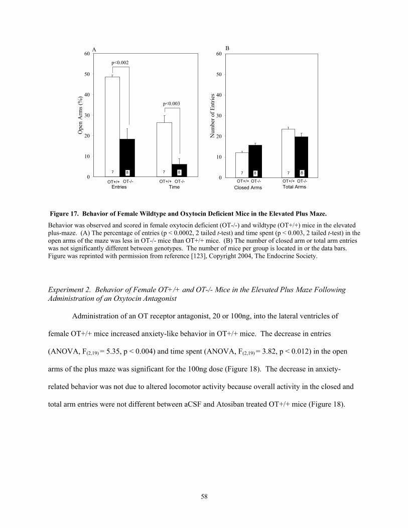
10
30
40
Ope
n A
rms (
%)
7
Time
7 8
Entries
8
p<0.002
10
30
40
50
Num
ber o
f Ent
ries
Closed Arms Total Arms
7 8 7 8
50
60 60A B
20
p<0.003
20
0Entries TimeOT+/+ OT-/- OT+/+ OT-/-
0Closed Arms Total ArmsOT+/+ OT-/- OT+/+ OT-/-
Behavior was observed and scored in female oxytocin deficient (OT-/-) and wildtype (OT+/+) mice in the elevated the
open arms of the maze was less in OT-/- mice than OT+/+ mice. (B) The number of closed arm or total arm entries
xperiment 2. Behavior of Female OT+/+ and OT-/- Mice in the Elevated Plus Maze Following dministration of an Oxytocin Antagonist
Administration of an OT receptor antagonist, 20 or 100ng, into the lateral ventricles of
male OT+/+ mice increased anxiety-like behavior in OT+/+ mice. The decrease in entries
NOVA, F(2,19) = 5.35, p < 0.004) and time spent (ANOVA, F(2,19) = 3.82, p < 0.012) in the open
rms of the plus maze was significant for the 100ng dose (Figure 18). The decrease in anxiety-
lated behavior was not due to altered locomotor activity because overall activity in the closed and
tal arm entries were not different between aCSF and Atosiban treated OT+/+ mice (Figure 18).
Figure 17. Behavior of Female Wildtype and Oxytocin Deficient Mice in the Elevated Plus Maze.
plus-maze. (A) The percentage of entries (p < 0.0002, 2 tailed t-test) and time spent (p < 0.003, 2 tailed t-test) in
was not significantly different between genotypes. The number of mice per group is located in or the data bars. Figure was reprinted with permission from reference [123], Copyright 2004, The Endocrine Society.
EA
fe
(A
a
re
to
58

0
10
20
30
40
50
60
Entries Time
Ope
n A
rms (
%)
aCSFAtosiban (20ng)Atosiban (100ng)
OT+/+
9 7 6 9 7
n=6
0
10
20
30
40
50
60
Closed Arms Total ArmsN
umbe
r of E
ntrie
s
aCSFAtosiban (20ng)Atosiban (100ng)
OT+/+
9 7 6 9 7 6
A B
p<0.004
p<0.012
Figure 18. Behavior of Female Wildtype Mice in the Elevated Plus Maze Following Administration of anOxytocin Antagonist.
) Administration of an oxytocin antagonist (Atosiban, 20 and 100ng) into the lateral ventricles of wildtype T+/+) mice decreased the percentage of entries (ANOVA, F(2,19) = 5.35, p < 0.004) and time spent (ANOVA,
(2,19) = 3.82, p < 0.012) in the open arms of the plus maze in a dose dependent manner. (B) The number of closed rs.
(A(OFarm or total arm entries did not differ between genotype. The number of mice per group is located in the data baFigure was reprinted with permission from reference [123], Copyright 2004, The Endocrine Society. Experim tion of Synthetic Oxytocin or Vasopressin
Administration of synthetic OT 2ng into the lateral ventricle of female OT-/- mice enhanced the
percentage of entries (p < 0.003) and time spent (p < 0.004) in the open arms of the plus maze
compared to OT-/- mice that received aCSF icv (Figure 19). However, administration of AVP 2ng
into the lateral ventricle of female OT-/- mice did not alter the percentage of entries or time spent in
the open arms of the plus-maze compared to aCSF treated OT-/- mice (Figure 20). Neither OT
(Figure 19) nor AVP (Figure 20) impaired locomotor function.
ent 3. Behavior of Female OT-/- Mice in the Elevated Plus Maze Following Administra
59

enhanced the percentage of entries (p < 0.003, 2 tailed t-test) and time spent (p < 0.004, 2 tailed t-test) in the open
20
30
40
50
60
Ope
n A
rms (
%)
aCSF
OT (2ng)
p<0.003
p<0.004
10
20
30
40
50
60
Num
ber o
f Ent
ries
aCSF
OT (2ng)
0Entries Time
n=89 n=8 9
0Closed Arms Total Arms
8 9 8 9
OT-/- OT-/-
A B
10
Figure 19. Behavior of Female Oxytocin Deficient Mice in the Elevated Plus Maze Following Administration of Synthetic Oxytocin.
(A) Administration of synthetic oxytocin (OT, 2ng) into the lateral ventricles of oxytocin deficient (OT-/-) mice
arms of the plus maze compared to OT-/- mice that received aCSF icv. (B) The number of closed arm or total arm entries did not differ between genotype. The number of mice per group is located in or above the data bars. Figure was reprinted with permission from reference [123], Copyright 2004, The Endocrine Society.
60

ce
0
10
20
30
40
50
60
Entries Time
Ope
n A
rms (
%)
aCSFAVP (2ng)
7 7n=7 n=7
0
10
20
30
40
50
60
Closed Arms Total Arms
Num
ber o
f Ent
ries
aCSFAVP (2ng)
7 7 7 7
OT-/- OT-/-
A B
Figure 20. Behavior of Female Oxytocin Deficient Mice in the Elevated Plus Maze Following Administration of Synthetic Vasopressin.
(A) Administration of arginine vasopressin (AVP, 2ng) into the lateral ventricles of oxytocin deficient (OT-/-) midid not alter the percentage of entries (p > 0.05, 2 tailed t-test) or time spent (p > 0.05, 2 tailed t-test) in the open arms of the plus-maze compared to artificial cerebrospinal fluid treated OT-/- mice. (B) Closed arm and total arm entries were not significantly different between female OT+/+ and OT-/- mice. The number of mice per group is located in or above the data bars. Figure was reprinted with permission from reference [123], Copyright 2004, The
ndocrine Society. E
61

Experiment 4. Behavior of Female OT-/- Mice in the Elevated Plus Maze Following Administration f an Oxytocin Antagonist Prior to Oxytocin
Central administration of 100ng Atosiban prior to 2ng OT infusion into the lateral ventricles
f female OT-/- mice prevented the anxiolytic effects of OT. OT-/- mice treated with Atosiban prior
OT made the same percentage of entries (p = 0.90) and spent the same amount of time (p = 0.72)
in the open arms of the EPM as OT-/- mice infused with aCSF (Figure 21). Overall activity in the
closed and total arm entries was not different between mice receiving aCSF and Atosiban followed
by OT.
eated with artifical cerebrospinal fluid (aCSF). (B) Closed arm and total arm entries were not gnificantly different between female OT-/- aCSF/aCSF treated mice compared to Atosiban/OT treated mice. The umber of mice per group is located in or above the data bars. Figure was reprinted with permission from reference 23], Copyright 2004, The Endocrine Society.
o
o
to
0
10
20
30
40
50
60
Entries Time
Ope
n A
rms (
%)
aCSF/aCSF
Atosiban/OT
0
10
20
30
40
50
60
Closed Arms Total Arms
Num
ber o
f Ent
ries
aCSF/aCSF
Atosiban/OT
7
OT-/-
7 8 7
n=8
7 8 8
OT-/-
A B
Figure 21. Behavior of Female Oxytocin Deficient Mice in the Elevated Plus Maze After Administration of an Oxytocin Antagonist Followed by Oxytocin.
(A) Oxytocin deficient (OT-/-) mice treated with Atosiban (an oxytocin antagonist, 100ng) prior to oxytocin (OT, 2ng) made the same percentage of entries and spent the same amount of time in the open arms of the elevated plus maze as OT-/- mice trsin[1
62

Experiment 5. Behavior of Male OT+/+ and OT-/- Mice in the Elevated Plus Maze
Male OT-/- mice displayed less anxiety-like behavior than OT+/+ mice in the plus maze
(Figure 22). The percentage of entries (p < 0.007) and time spent (p < 0.004) in the open arms of the
maze was greater in OT-/- mice than OT+/+ mice. The increase in anxiety-like behavior was not due
to altered locomotor activity, as overall activity in the closed and total arm entries were not different
between genotype (Figure 22).
Figure 22. Behavior of Male Wildtype and Oxytocin Deficient Mice in the Elevated Plus Maze.
ehavior was observed and scored in male oxytocin deficient (OT-/-) and wildtype (OT+/+) mice in the elevated
f mice per group is located in the data bars.
0
10
20
30
40
50
60
Entries Time
Ope
n A
rms (
%)
OT+/+ OT-/-Entries
OT+/+ OT-/-Time
0
10
20
30
40
50
60
Closed Arms Total Arms
Num
ber o
f Ent
ries
OT+/+ OT-/- OT+/+ OT-/-Closed Arms Total Arms
8 8 8 8 8 8 8 8
p<0.007
p<0.004
A B
Bplus-maze. (A) The percentage of entries (p < 0.007, 2 tailed t-test) and time spent (p < 0.004, 2 tailed t-test) in the open arms of the maze was more in OT-/- mice than OT+/+ mice. (B) The number of closed arm or total arm entries did not differ between genotype. The number o
63

C. Discussion
This study evaluated the role of OT in modulating anxiety behavior. Because OT is belie
to be anxiolytic, it was hypothesized that OT-/- mice would display increased anxiety-related
behavior compared to wildtype mice. These experiments demonstrate that female OT-/- mice display
greater anxiety-related behavior in the EPM, which is attributed to the absence of functional OT
pathways.
ved
Female OT-/- mice spent less time in the open arms of the EPM, an index of anxiety-related
behavior, compared to female OT+/+ mice. This observation is consistent with the concept that
activation of oxytocinergic neurons and the subsequent release of OT reduces the amount of anxiety-
related behavior observed in a novel environment. Thus, female OT-/- mice, which lack the ability to
synthesize and release OT, displayed increased anxiety-related behavior.
Intracerebroventricular administration of OT into the lateral ventricles of female OT-/- mice
reduced anxiety-related behavior. Furthermore, the findings of this study are consistent with data
showing decreased anxiety-related behavior in female rats [211] and mice [129] administered OT
centrally and tested in the EPM. In addition, the decrease in anxiety-related behavior in OT-/- mice
is OT dependent. Central administration of an equivalent dose of AVP, a peptide closely related to
OT, did not alter anxiety-related behavior. This observation suggests that the OT receptor must be
activated for OT to exert its anxiolytic affect.
lockade of endogenous OT with an OT receptor antagonist increased anxiety-related behavior. This
is the first time that blockade of endogenous OT in the mouse has been shown to increase anxiety-
related behavior in the EPM. Furthermore, infusion of the same OT receptor antagonist into the
lateral ventricles of female OT-/- mice prior to administration of synthetic OT blocked the anxiolytic
affect of OT in OT-/- female mice, supporting that the anxiolytic affect of OT is mediated via the OT
Similarly, central administration of an OT receptor antagonist into the lateral ventricles of
female OT+/+ mice decreased the number of open arm entries in the EPM, demonstrating that
b
64

receptor. The findings in female mice are similar to those of female rats, which displayed an increase
in anxie
in the experimental design of each study may account for the variations in the baseline
behavio
y-
in
ty-related behavior in the EPM following administration of an OT receptor antagonist [139].
Thus, the anxiolytic effect of OT in C57BL/6 mice is mediated by the OT receptor.
A minor concern with this study is that female mice of both genotypes displayed differences
in baseline entries and time spent in the open arms of the EPM in Experiments 1, 2, 3 and 4.
Differences
ral response. Differences in the baseline behavioral response may be due to some mice
undergoing cannulation surgeries and receiving central administration of vasopressin, Atosiban,
and/or oxytocin. Because of the expected variation in anxiety-related behavior, separate control
groups were included for each individual study. Therefore, baseline EPM entries and time spent in
the open arms should not be compared between studies.
Male OT-/- mice were tested using the same behavioral task, the EPM. Surprisingly, unlike
female OT-/- mice, male OT-/- mice displayed decreased anxiety-like behavior in the EPM and the
open field compared to male OT+/+ mice. Winslow and colleagues also reported decreased anxiet
related behavior in male OT-/- mice compared to male OT+/+ mice in the EPM [212]. Furthermore,
in male rats tested in the EPM, infusion of an OT receptor antagonist decreased anxiety-related
behavior in the EPM [139]. This study and those of others do not support an anxiolytic role for OT
in male rats or mice tested in the EPM.
Areas of the brain involved in anxiety processing, such as the BNST, amygdala and mPOA
are hormone responsive. OT mRNA expression [150,185] and OT receptor binding [221] are
facilitated by estradiol. Furthermore, regions of high specific binding in the male rat brain, such as
the BNST and the central amygdala, display low OT binding in the mouse brain [86]. Differences
OT receptor function and in turn neuronal activation during anxiety result in sex and genotype
differences. The interaction between gonadal hormones and OT function in response to anxiety can
be evaluated by testing male and female OT+/+ and OT-/- mice with the elevated plus maze in the
65

presence or absence of gonadal hormones. A similar study has been performed by McCarthy et al.
[129]. Estrogen-primed ovariectomized mice that were systemically administered OT displayed
decreased anxiety-like behavior in the EPM compared to mice receiving estrogen alone, OT alone, or
neither
e EPM
le in
ient
l
s OT receptors via central administration of
an OT r
n
,
estrogen nor OT [129]. In addition, central administration of OT into the lateral ventricles of
estrogen-primed ovariectomized mice also resulted in decreased anxiety-related behavior in th
compared to ovariectomized that received OT without estrogen [129]. Therefore, the anxiolytic role
of OT may be dependent upon the presence of estradiol and/or progesterone.
In summary, these findings indicate that oxytocinergic pathways play an anxiolytic ro
female mice tested in the EPM. The absence of normally functioning OT systems in an OT defic
mouse results in increased anxiety-related behavior. The enhanced anxiety was reversed by centra
administration of exogenous OT. Blockade of endogenou
eceptor antagonist to OT+/+ female mice resulted in increased anxiety-related behavior in the
EPM. In addition, infusion of an OT receptor antagonist prior to administration of exogenous OT i
OT-/- female mice inhibited the anxiolytic effects of OT. These findings suggest that genetic
gonadal steroid, and/or pharmacological interruption of OT may inhibit activation of OT receptors in
brain regions involved in the anxiety response.
66
V. Forebrain Activation of the OT Deficient Mouse in
Response to Stress and Anxiety
Results of this chapter have been published in part [3,124].
Neural circuits that coordinate the stress and anxiety response to a stimulus include
projections from the amygdala, lateral septum, and hippocampus to the medial parvocellular
paraventricular nucleus of the hypothalamus (PVN; the site of neurons that release corticotrophin
releasing hormone) via a neural circuit that involves synapses in the bed nucleus of the stria
terminalis (BNST) and the medial preoptic area of the hypothalamus (mPOA) [23,73,77,161]. OT
immunoreactive neurons originating in the PVN project to the BNST [33,82], and a few OT
immunoreactive fibers have been identified in the amygdala [33,82]. In addition, the projections of
OT neurons correspond to the location of OT receptors in the limbic system and include the BNST
[85], CeA [85], MeA [86], lateral septum [63,85,86], mPOA [63], and hippocampus [85,86]. OT and
its receptor are located in brain areas that modulate the hypothalamic pituitary adrenal (HPA) axis
response to stress and anxiety. Although a number of these areas participate in regulating both
anxiety behavior and HPA axis activity to stressful stimuli, it is not known whether OT modulates
the actions of these brain areas during the stress and anxiety response.
Many studies have explored the central pathways mediating stress and anxiety by mapping
euronal activation using the proto-oncogene protein product Fos, a marker of neuronal activation.
he Fos gene is expressed immediately (within minutes) in response to extracellular stimuli and
lays an important role in signal transduction and transcription regulation in cells. In 1988 it was
iscovered that neurons also express Fos when stimulated [170] and in 1989 the first application of
os to stress studies was made [26]. Studies have demonstrated forebrain patterns of Fos expression
response to various stress and anxiety models, such as restraint [6,26,36,81], swim stress [36], foot
hock [156], and anxiety [45]. Therefore, to define potential OT-sensitive stress and anxiety circuitry
the forebrain, the effect of OT on stress- and anxiety-induced Fos activation was examined in male
n
T
p
d
F
in
s
in
67

and fema ice ex plus maze
exposure. The brain ar ed to limbic stress pathways,
pecifically the CeA, MeA, BNST, and mPOA (Figure 23). These brain areas were chosen because
they contain OT neurons and receptors. In addition, the amygdala plays a critical role in modulating
the stress response. The amygdala has very few direct projections to the PVN. Therefore, the
amygdala projects to the BNST and mPOA, which are PVN-projecting brain areas. Therefore, it is
likely that OT mediates the stress and anxiety responses through this pathway.
la
le m posed to shaker stress, cholecystokinin administration, or elevated
eas examined for Fos activation were limit
s
Figure 23. Stress-Related Brain Pathways Examined for Fos Activation.
Bed nucleus of the stria terminalis (BNST), central nucleus of the amygdala (CeA), medial nucleus of the amygda(MeA), medial preoptic area (mPOA), paraventricular nucleus of the hypothalamus (PVN).
BNSTCeA
MeAmPOA
PVN
MeA
A. Experimental Design
Experiment 1. Shaker Stress Induced Fos Expression in Stress-Related Forebrain Pathways
The purpose of this experiment was to evaluate genotypic differences in Fos expression in
hypothalamic and forebrain areas associated with the stress response to psychogenic stress. Naive
female and male OT+/+ and OT-/- mice were exposed to shaker stress for 10 min (n=3 of each sex)
and returned to their home cages. Sixty to seventy-five minutes later mice were anesthetized,
perfused, and brains were harvested for immunocytochemistry. Control mice (n=3 of each sex), left
undisturbed in their home cage, were perfused at the same time.
68

Experiment 2. Cholecystokinin Induced Fos Expression in Stress-Related Forebrain Pathways
The purpose of this experiment was to evaluate genotypic differences in Fos expression
hypothalamic and forebrain areas associated with the stress response to a physical/systemic stressor.
Naive female and male OT+/+ and OT-/- mice were administered 10µg/kg CCK, intraperitoneally
(n=3 of each sex) or the same volume of saline (n=3 of each sex). Sixty to seventy-five minute
mice were anesthetized, perfused, and brains were harvested for immunocytochemistry.
for immunocytochemistry. Control mice (n=3 of each sex) that were not exposed
in
s later
Experiment 3. Elevated Plus Maze Exposure Induced Fos Expression in Anxiety-Related Forebrain Pathways
The purpose of this experiment was to evaluate genotype differences in Fos expression in
hypothalamic and forebrain areas associated with anxiety-related behavior. Naïve OT+/+ and OT-/-
female and male mice were placed in the elevated plus maze for 5 minutes and returned to their home
cages. Approximately one-hour later mice were anesthetized, perfused, and brains were harvested
to the EPM were
lso perfused at the same time.
xperiment 1. Shaker Stress Induced Fos Expression in Stress-Related Forebrain Pathways
Ten minutes of shaker stress robustly activated Fos in the PVN of female OT+/+ and OT-/-
mice compared to control mice. As expected, similar numbers of OT-immunoreactive neurons were
counted in the PVN of control OT+/+ mice (99.69 +
a
B. Results
E
3.90 OT cells per section) and in OT+/+ mice
exposed to shaker stress (92.14 + 12.53 OT cells per section; p > 0.05). OT immunoreactive neurons
were not observed in OT-/- mice (Figure 24 A and B). Shaker stress activated Fos in a small, but
significant subset of OT positive magno- and/or parvocellular PVN neurons in OT+/+ mice (p <
0.007; Figure 24 F). Fos activation did not increase in the SON of either OT+/+ or OT-/- mice after
shaker stress exposure (Figure 24 G and H).
69
Control (OT+/+, 111.6+3.2 cells per section; OT-/- 89.6+9.1 cells per section) and shaker
tress exposed mice (OT+/+, 97.27+1.7 cells per section; OT-/- 97.0+18.0 cells per section) of both
genotype displayed similar number of AVP immunoreactive neurons in the PVN of the
hypothalamus (ANOVA, F(1,8)=1.186, p > 0.05). In contrast to stressor-induced activation of OT
neurons in OT +/+ mice, AVP-positive PVN neurons were not activated after shaker stress in mice of
either genotype (Figure 25 A-F). In addition, Fos activation was specific to the PVN and did not
increase in the SON of OT+/+ and OT-/- mice exposed to shaker stress. (Figure 25 G and H)
s
Control (OT+/+, 116+1 CRH cells per section; OT-/-, 108.96+2.29 CRH cells per section)
and shaker stress exposed mice (OT+/+, 124.17+7.62 CRH cells per section; OT-/-, 126.97+12.99)
did not display genotypic differences in the amount of CRH-immunoreactive cells in the PVN of the
hypothalamus (ANOVA, F(1,6) = 3.884, p > 0.05). Significantly greater numbers of CRH-positive
PVN neurons were activated to express Fos in OT+/+ (ANOVA, F(1,3) = 62.25, p < 0.004) and OT-/-
mice (ANOVA, F(1,3) = 44.81, p < 0.001) after shaker stress compared to activation in control mice
(Figure 26). The number of CRH neurons expressing Fos after shaker stress was not different in OT-
/- mice compared to OT+/+ mice (ANOVA, F(1,8) = 6.72, p = 0.06; Figure 26).
l OT+/+ and OT-/- mice displayed similar amounts of Fos immunoreactivity in
mber
of Fos-p
VN
Female contro
the BNST, MeA, CeA, and mPOA. Ten minutes of shaker stress significantly increased the nu
ositive neurons in each of the forebrain regions evaluated compared to control mice of both
genotypes. However, a statistically significant effect of genotype on stress-induced Fos expression
was observed in the MeA but not in the other forebrain regions (Figure 27). Stress-induced
activation of MeA was lower in OT-/- mice compared to activation in OT+/+ mice exposed to shaker
stress (ANOVA, F(1,8) =9.379, p < 0.02; Figure 27).
Like female mice, similar numbers of OT-immunoreactive neurons were counted in the P
of male control OT+/+ mice (101.33 + 13.03 OT cells per section) and in OT+/+ mice exposed to
shaker stress (102.03 + 10.73 OT cells per section; p > 0.05). OT-immunoreactive neurons were not
70

observe of
e 28). Fos
d in OT-/- mice. Ten minutes of shaker stress also activated Fos in the PVN of male mice
both genotype compared to Fos activation in control mice. Shaker stress activated Fos in a small
subset of OT positive magno- and/or parvocellular neurons of the PVN (p < 0.001; Figur
activation did not increase in the SON in either male OT+/+ or OT-/- mice after shaker stress.
Control (OT+/+, 114.36+8.34 cells per section; OT-/- 96.86+9.28 cells per section) and
shaker stress exposed mice (OT+/+, 123.80+23.4 cells per section; OT-/- 130.55+25.15 cells per
section) of both genotype displayed similar number of AVP immunoreactive neurons in the PVN of
the hyp othalamus (ANOVA, F(1,8)=0.086, p > 0.05). Similar to female mice, AVP-positive neurons
of the PVN and SON were not activated in male mice.
Control (OT+/+, 76.03 + 8.15 CRH cells per section; OT-/-, 69.00 + 4.00 CRH cells per
section) and shaker stress exposed male mice (OT+/+, 75.00 + 5.00 CRH cells per section; OT-/-,
86.94 + 5.53 CRH cells per section) did not display genotypic differences in the amount of CRH-
immunoreactive cells in the PVN of the hypothalamus (ANOVA, F(1,6) = 0.131, p > 0.05). Shaker
stress activated Fos in CRH neurons of the PVN in male OT+/+ (ANOVA, F(1,3) = 2021.62, p <
0.0001) and OT-/- mice (ANOVA, F(1,3) = 172.44, p < 0.001) compared to control mice (Figure 29).
The nu
male
( NOVA, F(1,8) = 1.970, p > 0.05).
mber of CRH neurons expressing Fos after shaker stress was not different in OT-/- mice
compared to OT+/+ mice (ANOVA, F(1,6) = 0.85, p >0.05).
Based on the findings of Fos activation in the forebrains of female mice exposed to shaker
stress, Fos was quantified in the MeA of male mice. Compared to control mice of both genotypes,
shaker stress significantly activated Fos in the MeA (ANOVA, F(1,8) = 31.009, p < 0.0005) of
mice. Unlike female mice, the number of Fos immunoreactive cells was not different in the MeA of
male OT-/- and OT+/+ mice exposed to shaker stress A
71
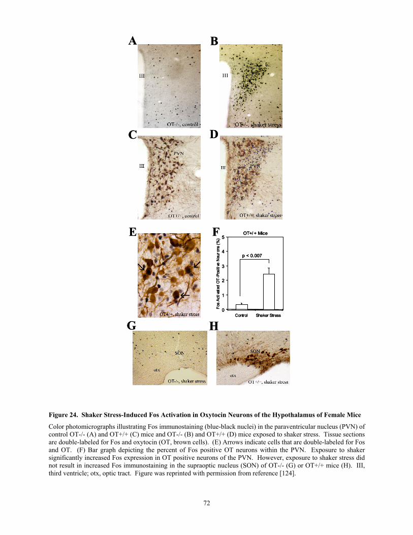
A B
C D
0
1
5
Control Shaker Stress
tiva
T-P
Neu
r%
)
p < 0.007
A B
C D
0
1
5
Control Shaker Stress
tiva
T-P
Neu
r%
)
p < 0.007
0
1
5
Control Shaker Stress
tiva
T-P
Neu
r%
)
p < 0.007
E F
G H
OT+/ + Mice
2
3
4
Fos
Acte
d O
ositi
veon
s (
E F
G H
OT+/ + Mice
2
3
4
Fos
Acte
d O
ositi
veon
s (
OT+/ + Mice
2
3
4
Fos
Acte
d O
ositi
veon
s (
Figure 24. Shaker Stress-Induced Fos Activation in Oxytocin Neurons of the Hypothalamus of Female Mice
Color photomicrographs illustrating Fos immunostaining (blue-black nuclei) in the paraventricular nucleus
(PVN) of control OT-/- (A) and OT+/+ (C) mice and OT-/- (B) and OT+/+ (D) mice exposed to shaker stress. Tissue sections are double-labeled for Fos and oxytocin (OT, brown cells). (E) Arrows indicate cells that are double-labeled for Fos nd OT. (F) Bar graph depicting the percent of Fos positive OT neurons within the PVN. Exposure to shaker gnificantly increased Fos expression in OT positive neurons of the PVN. However, exposure to shaker stress did ot result in increased Fos immunostaining in the supraoptic nucleus (SON) of OT-/- (G) or OT+/+ mice (H). III, ird ventricle; otx, optic tract. Figure was reprinted with permission from reference [124].
asinth
72

A B
C D
E F
G H
A B
C D
E F
G H
Figure 25. Shaker Stress-Induced Fos Activation in Vasopressin Neurons of the Hypothalamus of Female
-/- (G) or OT+/+ mice (H). III, third ventricle; otx, optic tract. Figure was reprinted with permission from reference [124].
Mice
Color photomicrographs illustrating Fos immunostaining (blue-black nuclei) in the paraventricular nucleus (PVN) of control OT-/- (A) and OT+/+ (C) mice and OT-/- (B) and OT+/+ (D) mice exposed to shaker stress. Tissue sections are double-labeled for Fos and arginine vasopressin (AVP, brown cells). Exposure to shaker did not increase Fos expression in AVP positive neurons of the PVN of OT-/- (E) or OT+/+ mice (F). In addition, exposure to shaker stress did not result in increased Fos immunostaining in the supraoptic nucleus (SON) of OT
73

one Neurons of the
Hypothalamus of Fem
olor photomicrographs illustrating Fos immunostaining (blue-black nuclei) in the paraventricular nucleus (PVN) of control OT-/- (A) and OT+/+ (C) mice and OT-/- (B) and OT+/+ (D) mice exposed to shaker stress. Tissue sections
-labeled for Fos and CRH in OT-/- (B) and OT+/+ mice (D). (E) Bar graph depicting the percent of Fos
A BA B
Figure 26. Shaker Stress-Induced Fos Activation in Corticotropin Releasing Hormale Mice.
C D
E
0
10
20
30
40
50
60
70
80
90
100
Control Shaker Stress
Fos
Act
ivat
ed C
RH
-Pos
itive
Neu
rons
(%) OT+/+
OT-/-
33n=3 n=3
C D
E
0
10
20
30
40
50
60
70
80
90
100
Control Shaker Stress
Fos
Act
ivat
ed C
RH
-Pos
itive
Neu
rons
(%) OT+/+
OT-/-
33n=3 n=3
C
are double-labeled for Fos and corticotropin releasing hormone (CRH, brown cells). Arrows indicate cells that are doublepositive CRH neurons within the PVN. Exposure to shaker significantly increased Fos expression in CRH positive neurons of the PVN in OT+/+ and OT-/- mice (ANOVA, F(1,3) = 62.25, p<0.004). There was not a genotypic difference in the number of Fos positive CRH neurons in response to shaker stress (ANOVA, F(1,8) = 6.72, p = 0.06). The number of mice per group is located in or above the data bars. Figure was reprinted with permission from reference [124].
74

Figure 27. Shaker Stress Induced Neural Activation of the Limbic Forebrain of Female Mice Shaker stress significantly activated Fos in the bed nucleus of the stria terminalis (BNST; ANOVA, F(1,8) = 54.75, p < 0.0001), medial (MeA; ANOVA, F(1,8) = 159.97, p < 0.0001) and central (CeA; ANONA, F(1,8) = 9.11, p < 0.02) nuclei of the amygdala, and medial preoptic area (mPOA; ANOVA, F(1,8) = 39.74, p < 0.0002). Shaker stress significantly activated Fos in all the brain areas evaluated compared to control mice of both genotypes. The number f Fos immunoreactive cells was lower in the MeA of OT-/- mice than OT+/+ mice exposed to shaker stress. The
om
0
50
100
150
200
250
300
350
400
450
500
Control Shaker Stress
0
100
200
300
400
500
600
700
800
900
1000
Control Shaker Stress
mPOA
0
50
100
150
200
250
300
350
400
450
500
Control Shaker Stress
BNST
0
50
100
150
200
250
300
350
400
450
500
Control Shaker Stress
p < 0.02
MeA CeA
OT+/+
OT-/-
Num
ber o
f Fos
Act
ivat
ed C
ells
Per
Sec
tion
3
33
33
33
3
3
33 3
33
3
3
0
50
100
150
200
250
300
350
400
450
500
Control Shaker Stress
0
100
200
300
400
500
600
700
800
900
1000
Control Shaker Stress
mPOA
0
50
100
150
200
250
300
350
400
450
500
Control Shaker Stress
BNST
0
50
100
150
200
250
300
350
400
450
500
Control Shaker Stress
p < 0.02
MeA CeA
OT+/+
OT-/-
OT+/+
OT-/-
Num
ber o
f Fos
Act
ivat
ed C
ells
Per
Sec
tion
3
33
33
33
3
3
33 3
33
3
3
onumber of mice per group is located in the data bars. Portions of this figure were reprinted with permission frreference [124].
75

Figure 28. Fos Activation of Oxytocin Immunoreactive Neurons
OT+/+ Male Mice
0
1
2
3
4
5
Control Shaker Stress
Fos
Act
ivat
ed O
T-P
ositi
ve N
euro
ns (%
)
p = 0.001
3 3
in the Paraventricular Nucleus of the Hypothalam
tocin (OT) positive neurons of the paraventricular nucleus of ce (p < 0.001). The number of mice per group is located in the data bars.
igure 29. Fos Activation of Corticotropin Releasing Hormone Immunoreactive Neurons in the araventricular Nucleus of the Hypothalamus of Male Mice
Exposure to shaker stress significantly increased Fos expression in corticotropin releasing hormone neurons (CRH) ositive neurons of the paraventricular nucleus of the hypothalamus (PVN) of wildtype (OT+/+; ANOVA, F(1,3) =
65.89, p < 0.0001) and OT deficient (OT-/-; ANOVA, F(1,3) = 108.55, p < 0.002) compared to control mice. There as not a genotypic difference in the number of Fos positive CRH neurons in response to shaker stress. The number f mice per group is located in or above the data bars.
us of Wildtype Male Mice.
Exposure to shaker stress significantly increased Fos expression in oxythe hypothalamus PVN of wildtype (OT+/+) mi
100
40
0
10
20
30
n=3 n=2 2 3
50
60
70
80
90
Control Shaker Stress
Fos
Act
ivat
ed C
RH
-Pos
itive
Neu
rons
(%)
OT+/+ malesOT-/- males
FP
p
wo
76

Experiment 2. Cholecystokinin Induced Fos Expression in Stress-Related Forebrain Pathways
Peripheral administration of CCK activated Fos in OT positive magno and/or parvocellular
neurons of the PVN of OT+/+ mice compared to saline treated controls (p < 0.009; Figure 30).
Similar numbers of OT-immunoreactive neurons were counted in the PVN of control OT+/+ mice
(85.52+8.19 cells per section) and in OT+/+ mice administered CCK (74.31+1.26 cells per section; p
< 0.05). OT immunoreactive neurons were not observed in OT-/- mice.
di ice t
A B
DC
0
2
4
6
8
10
12
14
16
18
20
Control CCK
Fos
Activ
ated
OT-
Pos
itive
Neu
rons
(%)
n=3 3
p < 0.009
OT+/+ Mice
A B
DC
0
2
4
6
8
10
12
14
16
18
20
Control CCK
Fos
Activ
ated
OT-
Pos
itive
Neu
rons
(%)
n=3 3
p < 0.009
OT+/+ Mice
Figure 30. Cholecystokinin Induced Fos Activation in Oxytocin Neurons of the Paraventricular Nucleus of Wildtype Male Mice
Color photomicrographs illustrating Fos immunostaining (blue-black nuclei) in the paraventricular nucleus of the hypothalamus (PVN) of control (A) and cholecystokinin-administered (CCK; B) OT+/+ mice. (C) Arrows in catecells that are double-labeled for Fos and OT in OT+/+ m administered CCK. (D) Bar graph depicting the percenof Fos positive OT neurons within the PVN (p < 0.009). The number of mice per group is located in or above the data bars.
77

78
Control (OT+/+, 112.67+3.34 cells per section, OT-/-, 112+4 cells per section) and CCK-
a tered mice (OT+/+, 104.28+dminis 6.75 cells per section, OT-/-, 105,27+0.82 cells per section) of
both genotype displayed similar numbers of AVP-immunoreactive neurons in the PVN of the
hypothalamus. Furthermore, CCK administration did not activate AVP-positive neurons of the PVN
(Figure 31).
Figure 31. Cholecystokinin Induced Fos Activation in Vasopressin Neurons of the Hypothalamus of Male
Color photomicrographs illustrating Fos immunostaining (blue-black nuclei) in the paraventricular nucleus (PVN) of
A B
C D
A B
C D
Mice
control OT-/- (A) and OT+/+ (B) mice and OT-/- and (C) OT-/- and OT+/+ mice administered cholecystokinin (CCK) (D). Tissue sections are double labeled for Fos and arginine vasopressin (AVP; brown cells). CCK administration did not result in increased Fos expression in AVP positive neurons of the PVN of OT+/+ or OT-/- mice.

Control (61.67+2.03 cells per section) and CCK-administered mice (74.75+6.38) did not
display treatment differences in the amount of CRH-immunoreactive cells in the PVN of the
hypothalamus (ANOVA, F(1,3)=0.49, p > 0.05). Mice that received CCK administration display
increased Fos activation in CRH-positive neurons of the PVN mice compared to control mice
(ANOVA, F(1,3)
ed
=58.72, p < 0.005). However, it was not possible to determine whether there were
genotypic differences in CRH neuron activation in response to CCK administration in this study.
Qualitative assessment of the PVN of female OT-/- and OT+/+ mice revealed similar
findings to those of male mice. CCK-administration activated Fos in OT positive magnocellular and
parvocellular neurons of the PVN of OT+/+ mice compared to saline treated controls. Furthermore,
CCK administration activated AVP- and CRH-positive neurons in the PVN of mice of both
genotypes. However, genotypic differences in the activation of CRH or AVP neurons in response to
CCK administration were not observed in female mice. Peripheral administration of CCK to female
mice significantly activated Fos in the BNST (ANOVA, F(1,6)=90.32, p<0.0001), CeA (ANOVA,
F(1,6)=135.9, p<0.0001), and mPOA (ANOVA, F(1,6)=42.73, p<0.0006), but not the MeA (p > 0.05)
compared to control mice of both genotypes (Figure 32). However, female OT+/+ and OT-/- mice
displayed similar amounts of Fos activation in the BNST, CeA, and mPOA following CCK
administration.
Peripheral administration of CCK to male mice also resulted in increased Fos expression in
the BNST (ANOVA, F(1,6)=9.91, p<0.02) and the CeA (ANOVA, F(1,7)=40.54, p<0.02), but not the
MeA or the mPOA (Figure 33). Male OT+/+ and OT-/- mice displayed similar numbers of Fos
immunoreactive cells in the BNST and the CeA following CCK administration.
79

Figure 32. Cholecystokinin Induced Neural Activation of the Limbic Forebrain of Female Mice
ANOVA, F(1,6)=90.32, p < 0.0001), central nucleus of the amygdala (CeA; ANOVA, F(1,6)=135.9, p < 0.0001), and f the amygdala (MeA;
n in the BNST, CeA, nd mPOA following CCK administration. The number of mice per group is located in the data bars.
300
400
500
250
300
400
450
500
tio 300
400
500
250
300
400
450
500
tio
0
100
0
Saline CCK
0
100
200
300
400
500
0
200
400
600
800
1000
0
50
100
150
200
Saline CCK
mPOA BNST
OT+/+
OT-/-umer
of
osct
ived
Clls
r S
e
33 33 3 333
n=3
0
100
0
Saline CCK
0
100
200
300
400
500
0
200
400
600
800
1000
0
50
100
150
200
Saline CCK
mPOA BNST
OT+/+
OT-/-
OT+/+
OT-/-umer
of
osct
ived
Clls
r S
e
33 33 3 333
n=350
150
20
250
350
450
50
150
250
350
450
Saline CCK
100
300
500
700
900
Saline CCK
350
MeA CeA
Nb
F A
ate
Pec
n
3 333 33 350
150
20
250
350
450
50
150
250
350
450
Saline CCK
100
300
500
700
900
Saline CCK
350
MeA CeA
Nb
F A
ate
Pec
n
3 333 33 3
Cholecystokinin administration significantly activated Fos in the bed nucleus of the stria terminalis (BNST;
medial preoptic area (mPOA; ANOVA, F(1,6)=42.73, p < 0.0006), but not the medial nucleus op>0.05). However, female OT+/+ and OT-/- mice displayed similar amounts of Fos activatioa
80
OT+/+
OT-/-umer
of
osct
ived
Clls
r S
e
33 33 3 333
n=3

50
100150
200250
300350
400450
500
100
200300400
500600
700800900
1000
0
50
100
150
200
250
300
350
400
450
500
Saline CCK0
50
100
150
200
250
300
350
400
450
500
Saline CCK
mPOA BNST
MeA CeA
OT+/+
OT-/-
Num
ber o
f Fos
Act
ivat
ed C
ells
Per
Sec
tion
3
33
33
33
3
3
33
33
3
3
3
50
100150
200250
300350
400450
500
100
200300400
500600
700800900
1000
0
50
100
150
200
250
300
350
400
450
500
Saline CCK0
50
100
150
200
250
300
350
400
450
500
Saline CCK
mPOA BNST
MeA CeA
OT+/+
OT-/-
OT+/+
OT-/-
Num
ber o
f Fos
Act
ivat
ed C
ells
Per
Sec
tion
3
33
33
33
3
3
33
33
3
3
3
0Saline CCK
0Saline CCK
0Saline CCK
0Saline CCK
Figure 33. Cholecystokinin Induced Neural Activation of the Limbic Forebrain in Male Mice
Cholecystokinin administration significantly activated Fos in the bed nucleus of the stria terminalis (BNST; ANOVA, F(1,6)=9.91, p < 0.02) and central nucleus of the amygdala (CeA; ANOVA, F(1,6)=40.54, p < 0.02), but not the medial preoptic area (mPOA; p > 0.05) or the medial nucleus of the amygdala (MeA; p>0.05). Male OT+/+ and OT-/- mice displayed similar amounts of Fos activation in the BNST and CeA following CCK administration. The number of mice per group is located in the data bars.
81

Experiment 3. Elevated Plus Maze Induced Fos Expression in Anxiety-Related Forebrain Pathways
Exposure to the elevated plus maze (EPM) for five minutes did not activate neurons within
e PVN of male or female OT+/+ or OT-/- mice compared to control mice. However, exposure to
the EPM activated Fos in anxiety-related brain areas of male and female OT+/+ and OT-/- mice.
These areas include, the BNST (ANOVA, F(1,8)=16.49, p < 0.004), MeA (ANOVA, F(1,8)= 76.10, p <
0.0001), and CeA (ANOVA, F(1,8)=8.71, p < 0.02), but not the mPOA (p > 0.05; Figure 34). In
addition, female mice exposed to the elevated plus maze expressed a genotypic difference in the
number of Fos immunoreactive cells in the MeA (ANOVA, F(1,8)=5.38, p<0.05; Figure 34). A post
hoc analysis revealed that OT-/- mice displayed greater numbers of Fos immunoreactive cells in the
MeA compared to OT+/+ mice (p < 0.005).
Based on the findings of Fos activation in the forebrains of female mice exposed to the EPM,
Fos was quantified in the MeA of male mice. Compared to control mice of both genotypes, EPM
significantly activated Fos in the MeA of male mice (ANOVA, F(1,6)=8.38, p < 0.03; Figure 35).
Unlike female mice, the number of Fos immunoreactive cells was not different in the MeA of male
OT-/- and OT+/+ mice exposed to the EPM (ANOVA, F(1,6)=0.183, p>0.05).
th
82

200
300
400
500
100
600
700
800
1000
50
0
400
500
Control EPM
50
150
250
400
500
Control EPM
/-
Num
ber o
f Fos
Act
ivat
ed C
ells
Per
Sec
tion
3
33
n=3
33
3
3
33
333
n=3
200
300
400
500
100
600
700
800
1000
50
0
400
500
Control EPM
50
150
250
400
500
Control EPM
/-/-
Num
ber o
f Fos
Act
ivat
ed C
ells
Per
Sec
tion
3
33
n=3
33
3
3
33
333
n=3
450900
0
100150
200
250
30
350
450
0
100
200
300
350
450
p < 0.005
mPOA BNST
MeA CeA
n=3 n=3
450900
0
100150
200
250
30
350
450
0
100
200
300
350
450
p < 0.005
mPOA BNST
MeA CeA
n=3 n=3
050
100150
250
350
Control EPM0
200
300
400
500
Control EPM
OT+/+
OT-
050
100150
250
350
Control EPM0
200
300
400
500
Control EPM
OT+/+
OT-
OT+/+
OT-
Figure 34. Elevated Plus Maze Induced Neural Activation of the Limbic Forebrain in Female Mice Exposure to the elevated plus maze activated Fos in the bed nucleus of the stria terminalis (BNST; ANOVA, F(1,8)=16.49, p < 0.004), medial (MeA; ANOVA, F(1,8)=76.10, p<0.0001) and central nucleus of the amygdala (CeA; ANOVA, F(1,8)=8.71, p<0.02), but not the medial preoptic nucleus (mPOA) compared to control female mice. The number of Fos immunoreactive cells was greater in the MeA of female OT-/- mice than OT+/+ mice exposed to the elevated plus maze. The number of mice per group is located in the data bars.
83

MeA
0
50
100
150
200
250
300
350
400
450
500
Control EPM
Num
ber o
f Fos
Act
ivat
ed C
ells
Per
Sec
tion
OT+/+ malesOT-/- males
3 33 3
=8.38,
Figure 35. Elevated Plus Maze Induced Neural Activation of the Medial Amygdala of Male Mice
Exposure to the elevated plus maze activated Fos in the medial nucleus of the amygdala (MeA; ANOVA, F(1,6)p < 0.03). However, unlike female mice, the number of Fos immunoreactive cells in the MeA of male mice did not differ between genotype. The number of mice per group is located in the data bars.
84
C. Discussion
The pattern of neuronal activation after shaker stress, CCK administration, or EPM exposure
was determined by mapping Fos immunoreactivity in select hypothalamic and forebrain regions of
OT+/+ and OT-/- mice. This study demonstrates that the absence of OT results in altered Fos
activation of the PVN and/or the MeA, which correlates with genotypic differences in the
corticosterone response to psychogenic stress and anxiety-related behavior. Specifically, diminished
activation of medial amygdala neurons and a trend towards enhanced activation of CRH
immunoreactive neurons of the PVN in female OT-/- versus OT+/+ mice suggests a neural correlate
for the genotypic difference observed in the corticosterone response to shaker stress. Moreover,
enhanced activation of MeA neurons in female OT-/- versus OT+/+ mice suggests a neural correlate
for the genotypic difference anxiety-related behavior observed in the EPM. This study supports
recent reports suggesting that the MeA may be a target for OT actions in mice [54,129,143] or rats
[210].
Shaker stress, a psychogenic stressor, has been reported to release corticosterone and OT, but
rone in mice as it does in
rats (refer to Chapter III) and activates Fos expression within the PVN of both genotypes. Shaker
stress activates Fos in OT-positive neurons of the PVN of male and female OT+/+ mice, but not OT-
/- mice. Moreover AVP-positive hypothalamic neurons were not activated by shaker stress in mice
of either genotype, consistent with the finding in rats that shaker stress selectively releases OT, but
not AVP [142]. AVP within the PVN acts synergistically with CRH to stimulate ACTH and in turn
corticosterone secretion [5,166]. Therefore enhanced release of AVP is unlikely to account for the
greater stress-induced release of corticosterone in OT-/- mice.
To determine whether the lack of OT may alter activation of CRH neurons in the PVN,
double immunohistochemistry for Fos and CRH was performed in hypothalamic brain sections of
not AVP, within the PVN and into the plasma of rats [69,142]. This study confirms the findings in
the rat using an OT deficient mouse model. Shaker stress releases corticoste
85

control and shaker stress exposed mice of both genotypes. Shaker stress activated Fos in CRH-
positive
shaker. However, lack of OT appears to result in increased CRH activation and expression
[143] in
osed OT-/- mice displayed
decreas on,
neurons in the PVN of male and female mice of both genotypes compared to control mice.
In addition, the number of activated CRH-positive neurons of female OT-/- mice tended to be higher
(approaching statistical significance, p = 0.06) than in OT+/+ mice, although the difference did not
reach statistical significance. Recently male OT-/- mice exposed to 4 hours of restraint stress were
reported to have greater abundance of CRH mRNA than OT+/+ mice [143]. Unlike in situ
hybridization histochemistry that is a measure of CRH mRNA expression, double
immunohistochemistry for Fos and CRH is an assessment of the activation of CRH-positive neurons.
Therefore, these findings are a quantitative assessment of the population of CRH neurons activated in
response to shaker stress. These findings may not be reflective of the abundance of CRH mRNA
expressed in response to shaker stress. Perhaps it will be possible to identify statistically significant
increases in CRH activation if OT-/- mice are administered longer or more intense exposure to
platform
mice exposed to psychogenic stressors.
Unlike shaker stress, exposure to the EPM, a measure of anxiety-related behavior, did not
result in increased Fos expression in the PVN of male or female mice. It is possible that the duration
of the stimulus was not long enough (or intense enough) to result in Fos activation of the PVN of
male or female mice. This theory is supported by a study performed by Duncan et al. [45]. Rats
tested in the EPM for 5 minutes did not reveal Fos activation within the PVN compared to unhandled
control rats [45]. However, 15 minutes of EPM exposure resulted in increased Fos activation within
the PVN of male rats compared to control cohorts [186].
Exposure to shaker stress and the EPM resulted in increased Fos expression in the limbic
forebrain of OT-/- and OT+/+ mice. Moreover, shaker stress exp
ed Fos activation, while OT-/- mice exposed to the EPM displayed increased Fos activati
in the medial amygdala (MeA) compared to OT+/+ mice. It is not known why, in comparison to
86

OT+/+ mice, female OT-/- mice display increased Fos activation following EPM exposure versu
decreased Fos activation following shaker stress. This discrepancy may be due to differences in the
activation of brain areas that project to the MeA, such as the subiculum, hippocampus, and
hypothalamus. Further studies examining stress- and anxiety-related brain areas projecting to the
MeA would be necessary to confirm this hypothesis. However, the MeA appears to be a target area
for the actions of OT. Altered Fos activation in the medial amygdala of OT-/- versus OT+/+
was also reported in male mice following 1h restraint stress [143] and a social mem
suggesting differences in forebrain processing in response to these stimuli. Moreover, infusion of
synthetic OT into the lateral ventricles of ovariectomized rats exposed to restraint stress reduced
stress-induced Fos activation in specific forebrain regions [210]. The OT pathways as well as OT
receptors, which have been identified in the limbic forebrain, are in an anatomical position to
modulate the HPA axis response to stress. No studies to date have reported OT release within th
MeA of mice during stress, although the present results suggests that the medial amygdala in
particular appears to be a target area for the actio
s
mice
ory task [54],
e
ns of OT. A genetic absence of OT alters activation
of this n ut
of
re, it is possible that OT modulates
forebra
ucleus when mice are exposed to psychogenic stressors. The PVN receives little direct inp
from the medial amygdala, but the MeA sends inhibitory GABAergic projections to the BNST and
mPOA in rats [24], which in turn send GABAergic projections to the medial parvocellular PVN
[161]. Consequently, activation of the MeA results in activation of the HPA axis via disinhibition
inhibitory limbic forebrain projections to the PVN. The medial amygdala may be one of the brain
areas that can account, at least in part, for heightened stress-induced corticosterone response and
enhanced anxiety-related behavior in OT-/- mice. Therefo
in projections leading into the PVN, specifically projections terminating on CRH neurons
within the PVN.
CCK administration releases corticosterone (refer to Chapter III) and increases Fos
expression within the PVN and SON of both OT+/+ and OT-/- mice. Like shaker stress, CCK
87

administration activated OT, but not AVP neurons, within the PVN of mice. In addition, CCK
administration activated CRH neurons of the PVN in mice, but not to a different degree between
genotypes. It was not possible to quantitatively determine statistically significant genotypic
differences in Fos activation of CRH-positive neurons because of the few number of tissue section
per mouse. However, a qualitative assessment did not reveal differences in Fos activated CRH
neurons. Similar CRH activation in the PVN of OT+/+ and OT-/- mice (as assessed qualitatively)
may reflect the comparable corticosterone release in OT+/+ and OT-/- mice. CCK penetrates the
blood-brain barrier poorly and the excitation of OT cells appears to be due to peripheral actions.
Systemically administered CCK binds to CCK-A receptors of gastrointestinal afferents, resulting
the stimulation of gastric sensory input to the nucleus of the solitary tract and the dorsal motor
nucleus of the vagus [127,134,163]. In turn, these hindbrain areas relay the sensory signal to t
hypothalamus and forebrain. Previous studies have determined that male OT+/+ and OT-/- mice di
not display differences in the amount of Fos activation in the ascending hindbrain areas (i.e., a
postrema, nucleus of the solitary tract, and the dorsal motor nucleus of the vagus) that result in PVN
activation [122]. Furthermore, CCK administration resulted in similar Fos activation in the CeA and
BNST, without activating the MeA of male and female OT+/+ and OT-/-mice. Therefore, OT
deficiency does not appear to play a role in the modulation of the CCK-induced stress response.
Unlike in the rat, the magnocellular and parvocellular neurons in the PVN of the mouse
cannot be segregated. Therefore, it is difficult to determine if magnocellular, parvocellular, or both
subsets of OT neurons of the PVN were activated using immunohistochemistry for Fos and OT.
Retrograde tracing using systemic administration of Fluorogold confirmed that parvocellular an
magnocellular OT and AVP neurons are intermingled in the PVN of OT+/+ and OT-/- mice
(unpublished obser
s
in
he
d
rea
d
vations, JA Amico and L Rinaman). Fluorogold labeling applied systemically
rier, identifies brain and spinal cord neurons whose axon terminals lie outside the blood-brain bar
including hypothalamic endocrine neurons [117,131]. Therefore, future studies employing
88

Fluorogold labeling in addition to dual immunohistochemistry for OT and Fos are necessary to
determine which subset of OT neurons of the PVN are activated in response to shaker stress,
cholecystokinin administration, and elevated plus maze exposure.
It should be noted that, while double immunohistochemistry is a valuable technique for
identifying neuroendocrine cells within the PVN, it may not be sensitive enough to identify the e
population of AVP and/or CRH in the PVN of mice. CRH neurons are located in the medial
parvocellular subdivision of the PVN in rats [191]. However, there are limitations to using double
immunohistochemistry for Fos and CRH to evaluate genotypic differences in the activation of CR
positive neurons. It appears that without an adrenalectomy or pre-treatment with colchicine most
CRH antibodies only stain for a subset of CRH neurons in the PVN of mice. Removal of circul
steroids in rodents by adrenalectomy is a potent stimulus to CRH neurons of the PVN, which resp
to adrenalectomy with increased CRH mRNA and peptide [100,180,215]. Colchicine is another
experimental tool that is traditionally used to enhance the number of immunoreactive cells in the
central nervous system [39]. Colchicine blocks neuronal transport of both peptides and mRNA
[39,105]. However, colchicine has been demonstrated to induce fos expression in CRH neurons of
the PVN [26] and elevate plasma ACTH and corticosterone concentrations in rats [168]. Both
adrenalectomy and colchicine pre-treatment would result in an increased number of CRH-positive
ntire
H-
ating
ond
cells, b -
s
of
ed
K-
ut would not allow us to examine the differences in the CRH function of OT+/+ and OT-/
mice in response to shaker stress. Therefore, evaluating Fos activation of CRH-positive neurons may
be problematic in determining genotypic differences in CRH function. Furthermore, AVP neuron
that co-express CRH and respond to stress are also located in the medial parvocellular subdivision
the PVN in rats [191]. AVP expression in this subset of neurons is not easily detected with
immunocytochemistry alone. Therefore it is necessary to enhance AVP expression via
adrenalectomy [180,181] or administration of colchicine [180,181]. Fos activation was not observ
in the AVP immunoreactive neurons of the PVN in either genotype after shaker stress or CC
89

administration but it is not known if all of the AVP neurons in the PVN of mice are capable of b
detected by immunohistochemistry. Although immunohistochemistry selectively identified Fo
activation in OT, but not AVP, PVN neurons following shaker stres
eing
s
s in mice, it is also possible that
all of th
thin
et
to
e is
in the
/or GC
e AVP expressing neurons of the PVN were not identified with this technique.
The hippocampus, which plays a primary role in the inhibition of the HPA axis as well as
modulation of the stress and anxiety response, was not evaluated in this study. OT neurons wi
the PVN project to and OT receptors are located in the hippocampus. However, it is not known if
there are differences in Fos activation of the hippocampus OT+/+ and OT-/- mice exposed to shaker
stress or EPM exposure. It is possible that the hippocampus, in addition to the MeA, may be a targ
area for the OT in response to stress and anxiety. Future studies examining activation of the
hippocampus in response to stress in the presence and absence of OT are necessary to answer this
question.
Corticosterone released from the adrenals in response to stress provides negative feedback
the HPA axis indirectly through the hippocampus and the amygdala. Both the hippocampus and
MeA contain corticosteroid receptors [104]. Two corticosteroid receptors have been identified, the
mineralocorticoid receptor (MR) and the glucocorticoid receptor (GR) [132,197]. Corticosteron
able to activate both receptors but corticosterone has a ten-fold higher affinity for MR than for GR
[162]. Therefore, the glucocorticoid concentration determines which receptor is activated. Low
concentrations of corticosterone are believed to activate MR and higher concentrations are believed
to activate GR during stress. The GR has a broad expression pattern, whereas the MR has restricted
expression in the neurons of the hippocampus and amygdala [104,162]. In addition, administration
of OT results in decreased expression of GR mRNA and increased expression of MR mRNA
hippocampus of the rat [153]. Therefore, it is possible that OT deficiency alters MC and
function in OT-/- mice.
90

In summary the absence of normally functioning OT systems in OT-/- mice results in the
altered activation of the MeA, and perhaps other regions. The MeA is an important area of the
limbic circuitry responsible for processing anxiety and psychogenic stress. Anxiety stimuli and
psychogenic stressors requiring higher-order processing of a stimulus, such as shaker stress in rats
e
r
st
heightened anxiety-related behavior and stress-induced corticosterone response in OT-/-
mice.
[69,142], restraint stress in male OT-/- mice [143], or female rats [210] activate limbic forebrain
pathways. Exposure to shaker stress and the EPM resulted in altered Fos expression in the MeA
forebrain of OT-/- compared to OT+/+ mice. The OT pathways as well as OT receptors, which hav
been identified in the limbic forebrain, are in an anatomical position to modulate the HPA axis
response to stress. The MeA in particular appears to be a target area for the actions of OT. A genetic
absence of OT alters activation of this nucleus when mice are exposed to an anxiety stimuli o
psychogenic stressors. The medial amygdala may be one of the brain areas that can account, at lea
in part, for
Therefore, it is possible that OT modulates forebrain projections leading into the PVN,
specifically projections terminating on CRH neurons within the PVN.
91

VI. Conclusion
The experimental findings presented in this thesis provide insight into the role of OT in t
stress and anxiety response. Pharmacological studies have consistently shown that central OT m
be an anxiolytic, as well as play an inhibitory role in the up-regulation of the HPA axis in response
stress in females. Using an OT deficient mouse model, these studies have determined that the
inhibition of anxiety and the HPA axis in response to psychogenic stressors is dependent upon the
presence of OT.
OT is responsible for the inhibition of corticosterone release in response to psychogenic
stressors. The genotypic differences in the corticosterone response to shaker stress are not due to
altered activation of the HPA axis. Firstly, OT+/+ and OT-/- mice have similar diurnal plasma
corticosterone concentrations over a 24h period. Secondly, intracerebroventricular administration of
CRH resulted in similar corticosterone release in both genotypes. Lastly, a genotypic difference in
corticosterone release is dependent upon the type of stressor administered to OT+/+ and OT-/- m
For instance, shaker stress elicited a genotype difference in the corticoste
he
ay
to
ice.
rone response but CCK
dministration resulted in similar increases in corticosterone between OT+/+ and OT-/- male mice.
herefore, it appears as if the differences in HPA axis activation in response to certain stressors are
ue to differences in neuronal activation up-stream of the HPA axis, possibly at the level of the
edial nucleus of the amygdala.
OT also plays an anxiolytic role. Female OT deficient mice displayed enhanced anxiety-
lated behavior compared to wildtype mice. Administration of an OT antagonist to OT+/+ mice
imicked the anxiogenic behavior of OT-/- mice, while administration of OT into the lateral
entricles attenuated enhanced anxiety-related behavior in OT-/- mice. Therefore, the anxiolytic role
f OT is dependent upon binding at the OT receptor.
a
T
d
m
re
m
v
o
92

Many studies have suggested th fically estradiol, facilitate OT
binding -
g
ectomizing female OT+/+ and OT-/- mice eliminates the ability of OT to inhibit the
corticos that
or.
ver,
f
the
os
sulted in increased Fos activation in the MeA of OT-/- mice
compar
nd
MeA
The findings presented in this thesis provide insight into the role of OT in the stress and
anxiety response. The experiments in this thesis have determined that OT inhibits activation of the
at ovarian hormones, speci
and enhance oxytocinergic actions. Although similar genotypic differences in shaker stress
induced corticosterone release were not observed in male mice, the genotypic difference in the
corticosterone response of female mice was not dependent upon the estrous cycle. Similarly, male
OT-/- mice displayed decreased anxiety-related behavior compared to OT+/+ mice. This findin
opposes the increased anxiety behavior evaluated in female mice. However, preliminary studies
suggest that ovari
terone response to psychogenic stress and anxiety-related behavior. Therefore, it appears
the presence of estradiol enhances the ability of OT to inhibit the HPA axis and anxiogenic behavi
To date, there is no direct evidence that OT is released into the MeA after stress. Howe
OT receptors are located in the MeA, suggesting a role for OT in the limbic system. Psychogenic
stressors result in Fos activation of the MeA and OT neurons of the PVN. In addition, stimulation o
the MeA activates the HPA axis, while excitotoxic lesions of the MeA result in the suppression of
HPA axis activation in response to psychogenic stress. The findings of these studies suggest that
MeA may be responsible for altered neuroendocrine response to shaker stress and anxiety response
detected in OT-/- mice. Exposure to shaker stress and the EPM resulted in increased Fos expression
in the limbic forebrain of OT+/+ and OT-/- mice. Shaker stress exposure resulted in decreased F
activation, while EPM exposure re
ed to OT+/+ mice. The deletion of OT alters the activation of the MeA when mice are
exposed to psychogenic stressors. The MeA may be partly responsible for the increased anxiety a
heightened corticosterone response in OT-/- mice. Therefore, it is possible that OT modulates
projections, which are relayed through the BNST and mPOA of the hypothalamus, to CRH neurons
in the medial parvocellular PVN of the hypothalamus.
93

HPA axis in response to psychogenic stress, as well as inhibiting the anxiety response in female
mice. Moreover, it appears that OT acts at the level of the MeA to inhibit anxiety behavior and the
HPA axis response to psychogenic stress.
Future Directions
Stress Response of Wildtype and Oxytocin Deficient Mice Exposed to Different Stressors
Examining the stress response of OT+/+ and OT-/- mice that have been exposed to an
expanded repertoire of stressors would be valuable in further exploring the hypothesis of oxytocin
mediating stress-induced forebrain activation versus hindbrain activation. Increasing the number of
psychogenic stressors, preferably stressors that have been shown to activate the MeA as well as OT
neurons
ly
, would be interesting to determine whether genotypic differences in the corticosterone
response to stress only occur following psychogenic stress. It would also be interesting to more ful
explore a number of physical stressors to determine if stress-induced hindbrain activation results in
genotypic differences in corticosterone release similar to that found following shaker stress.
Central Administration of Oxytocin to Oxytocin Deficient Mice Exposed to Shaker Stress
In addition to expanding the number and type of stressors, it would support the current
hypothesis to prove that central administration of oxytocin is capable of attenuating the enhanced
stress response of OT-/- mice compared to OT+/+ mice. Administration of OT into the lateral
ventricles of OT-/- mice exposed to EPM attenuated the enhanced anxiety-like behavior in
comparison to OT+/+ mice. It is expected that similar studies administering OT into the lateral
ventricles of OT-/- mice exposed to shaker stress would attenuate the enhanced corticosterone
response of OT-/- mice. This study would be useful in confirming that the presence of OT is
necessary to attenuate the corticosterone response to shaker stress.
Evaluating Gender Differences in Oxytocin Deficient Mice
in Response to Stress and Anxiety
At this time it is not known why male and female mice respond differently to shaker stress
and elevated plus maze exposure. Exploring the sex differences between male and female mice
94

exposed to shaker stress and the EPM may be useful in evaluating the interaction between OT an
gonadal hormones. The HPA axis is sensitive to gonadal steroids. Pretreatment of estrogen to
ovariectomized female rats enhances cortic
d
osterone secretion following stress [25,55,202]. In
e administration inhibits HPA activation in response to stress [201,203]. OT
d areas of
the brai one
the
trogen
this
s
by
tress-
contrast, testosteron
mRNA expression [150,185] and OT receptor binding [221] are facilitated by estradiol, an
n involved in the stress response, such as the amygdala, BNST, and the mPOA are horm
responsive. Pilot studies conducted comparing ovariectomized OT+/+ and OT-/- mice to intact
OT+/+ and OT-/- mice implicated that estrogen and/or progesterone interact with OT to attenuate the
corticosterone response to shaker stress, as well as reduce anxiety-related behavior in the EPM. It
appears that removing gonadal steroids in female mice eliminates the ability of OT to attenuate
corticosterone response to stress and reduce anxiety-like behavior. These preliminary findings
support studies conducted by McCarthy et al. reporting that administration of OT to ovariectomized
mice was not able to inhibit anxiogenic behavior in the EPM without the administration of es
[129]. The number of mice included in these studies was too few to include the findings in
thesis. However, these pilot studies provide an interesting starting-point to examine the gender
differences in the stress and anxiety response of OT-/- mice. Examining the role of OT in the stres
and anxiety responses of ovariectomized and gonadectomized OT+/+ and OT-/- mice, followed
the replacement of gonadal steroids would provide incite into the gender differences of the s
induced corticosterone response and anxiety-related behavior of OT-/- mice.
Expanding the Methods of Analysis Used to Examine Activation of the Central Nervous System
While double immunohistochemistry is a valuable technique for identifying neuroend
cells within the PVN, it may not be sensitive enough to identify the
ocrine
entire population of AVP and/or
CRH CRH in the PVN of mice. Unlike OT and AVP neurons, CRH neurons (and AVP-containing
neurons) do not contain a ready releasable pool of peptide making the neurons difficult to identify
95
with immunohistochemistry. Therefore, it may be helpful to also process and evaluate tissue using
situ hybridization for mRNA or hnRNA of OT, AVP, and/or CRH.
The Role of the Hippocampus in the Stress Response of Oxytocin Deficient Mice
in
The hippocampus, also plays a primary role in the inhibition of the HPA axis as well as
modulation of the stress and anxiety response, was not evaluated in this study. OT neurons within
the PVN project to and OT receptors are located in the hippocampus. As discussed in Chapter V, it
is not known if there are differences in Fos activation of the hippocampus OT+/+ and OT-/- mice
exposed to shaker stress or EPM exposure. It is possible that the hippocampus, in addition to the
MeA, may be a target area for the OT in response to stress and anxiety. Future studies examining
Fos activation of the hippocampus in response to stress in the presence and absence of OT are
necessary to answer this question.
The Function of Corticosteroid Receptors in Oxytocin Deficient Mice
Corticosterone released from the adrenals in response to stress provides negative feedback to
the HPA axis indirectly through the hippocampus and the amygdala. Both the hippocampus and
MeA contain corticosteroid receptors [104]. As discussed in Chapter V, corticosterone is able to
activate both receptors but corticosterone has a ten-fold higher affinity for mineralocorticoid
receptors (MR) than for glucocorticoid receptors (GR) [162]. Low concentrations of corticosterone
are believed to activate MR and higher concentrations are believed to activate GR during stress.
Furthermore, administration of OT results in decreased expression of GR mR
NA and increased
expression of MR mRNA in the hippocampus of the rat [153]. It is not known if the enhanced
corticosterone release of OT-/- mice in response to stress alters the binding affinity or number of GR
and/or MR. Therefore, evaluating the number and binding affinity of MC and GC receptors in the
amygdala and hippocampus of mice of both genotypes would provide information on how oxytocin
deficiency may alter MC and/or GC receptor function in OT-/- mice.
96
Health Consequences Due to Enhanced Glucocorticoid Response to Stress in Oxytocin Deficient
Mice
Oxytocin deficient mice also provide a unique animal model to evaluate the short and long-
rm he cal
nhanced corticosterone response to an acute stress is
orticosterone release of OT-/-
ice co
nse
te alth consequences in response to enhanced HPA activation. Many organs and physiologi
systems are sensitive to glucocorticoids. Glucocorticoids influence cardiovascular tone, immunity,
metabolism, neural function, and behavior. During an acute response to stress glucocorticoids
influence the increase blood pressure and cardiac output [173], inhibit synthesis and release of
cytokines and other mediators that promote immune and inflammatory reactions [208], mobilize
energy stores and increase circulating glucose [174], and enhance synaptic plasticity [151] and
neuronal excitability [12,92] in the hippocampus leading to enhanced learning and memory.
However, it is not known whether e
advantageous or detrimental to an organism. Therefore, the increased c
m mpared to OT+/+ in response to certain stressors may be helpful, resulting in improved
mobilization, or harmful, resulting in the “over-stimulation” of the physiological and central respo
to stress.
97

VII. References
[1] Altemus, M., Deuster, P.A., Galliven, E., Carter, C.S. and Gold, P.W., Suppression of hypothalmic-pituitary-adrenal axis responses to stress in lactating women, J Clin Endocrinol
[2] Amaral, D.G. and Sinnamon, H.M., The locus coeruleus: neurobiology of a central
[3] Amico, J.A., Mantella, R.C., Vollmer, R.R. and Li, X., Anxiety and stress responses in
[4] Antoni, F.A., Oxytocin receptors in rat adenohypophysis: evidence from radioligand binding
[5] Antoni, F.A., Vasopressinergic control of pituitary adrenocorticotropin secretion comes of
[6] Arnold, F.J., De Lucas Bueno, M., Shiers, H., Hancock, D.C., Evan, G.I. and Herbert, J., lar
corticotropin-releasing factor in unstressed or stressed male rats, Neuroscience, 51 (1992)
[7] Arpin-Bott, M.P., Waltisperger, E., Freund-Mercier, M.J. and Stoeckel, M.E., Two oxytocin-
localization by in vitro and in vivo autoradiography, J Endocrinol, 153 (1997) 49-59. orticoid
receptor as a mediator of glucocorticoid response, Neuron, 1 (1988) 877-900.
APA Press, Washington, D.C., 1995.
adrenocorticotropin in the rat: sexual dimorphism and changes across the estrous cycle,
[11] Bale, T.L., Davis, A.M., Auger, A.P., Dorsa, D.M. and McCarthy, M.M., CNS region- oxytocin receptor expression: importance in regulation of anxiety and sex behavior, J
Neurosci, 21 (2001) 2546-52. [12] eck, S.G., List, T.J. and Choi, K.C., Long and short term administration of corticosterone
alters CA1 hippocampal neuronal properties., Neuroendocrinology, 60 (1994) 261-272. [13] elzung, C. and Griebel, G., Measuring normal and pathological anxiety-like behaviour in
mice: a review, Behav Brain Res, 125 (2001) 141-9. 4] Blevins, J.E., Eakin, T.J., Murphy, J.A., Schwartz, M.W. and Baskin, D.G., Oxytocin
innervation of caudal brainstem nuclei activated by cholecystokinin, Brain Res, 993 (2003) 0-41.
[15] Bowers, G., Cullinan, W.E. and Herman, J.P., Region-specific regulation of glutamic acid ecarboxylase (GAD) mRNA expression in central stress circuits, J Neurosci, 18 (1998)
5938-47. [16] radbury, M.J., Strack, A.M. and Dallman, M.F., Lesions of the hippocampal efferent
pathway (fimbria-fornix) do not alter sensitivity of adrenocorticotropin to feedback inhibition y corticosterone in rats, Neuroendocrinology, 58 (1993) 396-407.
[17] Brown, K.J. and Grunberg, N.E., Effects of housing on male and female rats: crowding tresses male but calm females, Physiol Behav, 58 (1995) 1085-9.
[18] Bruhn, T.O., Sutton, S.W., Plotsky, P.M. and Vale, W.W., Central administration of orticotropin-releasing factor modulates oxytocin secretion in the rat, Endocrinology, 119
(1986) 1558-63.
Metab, 80 (1995) 2954-9.
noradrenergic nucleus, Prog Neurobiol, 9 (1977) 147-96.
female oxytocin deficient mice, J Neuroendocrinol, 16 (2004) 319-24.
studies, Endocrinology, 119 (1986) 2393-5.
age, Front Neuroendocrinol, 14 (1993) 76-122.
Expression of c-fos in regions of the basal limbic forebrain following intracerebroventricu
377-90.
binding site subtypes in rat kidney: pharmacological characterization, ontogeny and
[8] Arriza, J.L., Simerly, R.B., Swanson, L.W. and Evans, R.M., Neuronal mineraloc
[9] Association, A.P., Diagnostic and statistical manual of mental disorders, 4th edn (DSM IV).
[10] Atkinson, H.C. and Waddell, B.J., Circadian variation in basal plasma corticosterone and
Endocrinology, 138 (1997) 3842-8.
specific
B
B
[1
3
d
B
b
s
c
98

[19] Buijs, R.M., De Vries, G.J. and distribution and synaptic release of oxytocin in the central nervous system. In J.A. Amico and A.G. Robinson (Eds.), Oxytocin:
6. [20]
ontribute differentially to systemic interleukin-1β−induced 138.
[21] ., Davis, M. and Watson, S.J., ction
[23] .W., Projections of the ventral premammillary
[24] ., Organization of projections from the -
[25] e n steroids on hypothalamic-pituitary-adrenal regulation in the female rat, J
[26] U S A,
[27] omparison of
rosci, 13 (1993) 5126-38. s
)
rats
[30] stress and stress system disorders, JAMA, 267 (1992) 1244-
[31] e.
[32] anxiety, Neurosci Biobehav Rev, 22
[33] n neuronal sensitivity and oxytocin-binding sites in the amygdala of the rat:
[34] ry Vol. 1, 1992, pp. 1-23.
[36] S.J., Pattern and time ce,
p Neurol, 332 (1993) 1-20.
Van Leeuwen, F.W., The
clinical and laboratory studies, Elsevier Science Publishers, New York, 1985, pp. 77-8Buller, K.M., Xu, Y., Dayas, C.V. and Day, T.A., Dorsal and ventral medullary catecholamine cell groups chypothalamic pituitary adrenal axis responses, Neuroendocrinology, 73 (2001) 129-Campeau, S., Falls, W.A., Cullinan, W.E., Helmreich, D.LElicitation and reduction of fear: behavioural and neuroendocrine indices and brain induof the immediate-early gene c-fos, Neuroscience, 78 (1997) 1087-104.
[22] Cannon, W.B., Organization for physiological homeostasis, Physiol Rev, 9 (1929) 399-431. Canteras, N.S., Simerly, R.B. and Swanson, Lnucleus, J Comp Neurol, 324 (1992) 195-212. Canteras, N.S., Simerly, R.B. and Swanson, L.Wmedial nucleus of the amygdala: a PHAL study in the rat, J Comp Neurol, 360 (1995) 21345. Carey, M.P., Deterd, C.H., de Koning, J., Helmerhorst, F. and de Kloet, E.R., The influencof ovariaEndocrinol, 144 (1995) 311-21. Ceccatelli, S., Villar, M.J., Goldstein, M. and Hokfelt, T., Expression of c-Fos immunoreactivity in transmitter-characterized neurons after stress, Proc Natl Acad Sci86 (1989) 9569-9573. Chan, R.K., Brown, E.R., Ericsson, A., Kovacs, K.J. and Sawchenko, P.E., A ctwo immediate-early genes, c-fos and NGFI-B, as markers for functional activation in stress-related neuroendocrine circuitry, J Neu
[28] Charney, D.S. and Deutch, A., A functional neuroanatomy of anxiety and fear: implicationfor the pathophysiology and treatment of anxiety disorders, Crit Rev Neurobiol, 10 (1996419-46.
[29] Cheifetz, P.N., The daily rhythm of the secretion of corticotrophin and corticosterone inand mice., J Endocrinol, 49 (1971) 11-12. Chrousos, G.P., The concept of1252. Chrousos, G.P., Stressors, stress, and neuroendocrine integration of the adaptive responsThe 1997 Hans Selye Memorial Lecture, Ann N Y Acad Sci, 851 (1998) 311-35. Clement, Y. and Chapouthier, G., Biological bases of(1998) 623-33. Condes-Lara, M., Veinante, P., Rabai, M. and Freund-Mercier, M.J., Correlation between oxytocielectrophysiological and histoautoradiographic study, Brain Res, 637 (1994) 277-86. Cooper, R., Goldman, J. and Vandenbergh, J., Monitoring of the estrous cylce in laboratorodent by vaginal lavage.
[35] Critchlow, V., Liebelt, R.A., Bar-Sela, M., Mountcastle, W. and Lipscomb, H.S., Sex differences in resting pituitary-adrenal function in the rat., Am J Physiol, 205 (1963) 807-815. Cullinan, W.E., Herman, J.P., Battaglia, D.F., Akil, H. and Watson,course of immediate early gene expression in rat brain following acute stress, Neuroscien64 (1995) 477-505.
[37] Cullinan, W.E., Herman, J.P. and Watson, S.J., Ventral subicular interaction with the hypothalamic paraventricular nucleus: evidence for a relay in the bed nucleus of the stria terminalis, J Com
99

[38] Cunningham, E.T., Jr. and Sawchenko, P.E., Anatomical specificity of noradrenergic inputs to the paraventricular and supraoptic nuclei of the rat hypothalamus, J Comp Neurol, 274 (1988) 60-76.
[39] Dahlstrom, A., Effect of colchicine on transport of amine storage granules in symnerves of rat, Eur J Pharmacol, 5 (1968) 111-113. Dale, H.H., The action of extracts of the pituitary of the body., Biochem J., 4 (1909) 427-
pathetic
[40] 447. ala:
r J Neurosci, 14 (2001) 1143-52.
lt rain, J Comp Neurol, 436 (2001) 430-55.
[45] reese, G.R., Neuroanatomical characterization of Fos
[46] in Res, 407 (1987) 327-31.
[48] le social defeat experience
[49] M.H. and Herman, J.P., Differential forebrain c-fos mRNA induction by ether
[50] d Landgraf, R., Behavioural impact of
30S. l
neurons, J
[52] nforti, N. and Melamed, E., Paraventricular nucleus serotonin mediates
[53] stria
[54] g, L.J., Oxytocin in the medial amygdala is
[55] .
[56] 03) 109-35.
ic
[58] n E. Knobil and J.D. Neill (Eds.), The Physiology of Reproduction., Vol. 2, Raven Press, New York, 1988, pp. 1893-1928.
[41] Davis, M., The role of the amygdala in conditioned fear. In J. Aggleton (Ed.), The AmygdNeurobiological Aspects of Emotion, Memory, and Mental Dysfunction, Wiley, New York, 1992, pp. 255-305.
[42] Dayas, C.V., Buller, K.M., Crane, J.W., Xu, Y. and Day, T.A., Stressor categorization: acutephysical and psychological stressors elicit distinctive recruitment patterns in the amygdala and in medullary noradrenergic cell groups, Eu
[43] Dong, H.W., Petrovich, G.D., Watts, A.G. and Swanson, L.W., Basic organization of projections from the oval and fusiform nuclei of the bed nuclei of the stria terminalis in adurat b
[44] Du Vigneaud, V., Ressler, C. and Trippett, S., The sequence of amino acids in oxytocin, witha proposal for the structure of oxytocin, J Biol Chem, 205 (1953) 949-57. Duncan, G.E., Knapp, D.J. and Binduction in rat behavioral models of anxiety, Brain Res, 713 (1996) 79-91. Dunn, J.D., Plasma corticosterone responses to electrical stimulation of the bed nucleus of the stria terminalis, Bra
[47] Dunn, J.D. and Whitener, J., Plasma corticosterone responses to electrical stimulation of the amygdaloid complex: cytoarchitectural specificity, Neuroendocrinology, 42 (1986) 211-7. Ebner, K., Wotjak, C.T., Landgraf, R. and Engelmann, M., A singselectively stimulates the release of oxytocin, but not vasopressin, within the septal brain areaof male rats, Brain Res, 872 (2000) 87-92. Emmert, inhalation and novelty: evidence for distinctive stress pathways, Brain Res, 845 (1999) 60-7. Engelmann, M., Wotjak, C.T., Ebner, K. anintraseptally released vasopressin and oxytocin in rats, Exp Physiol, 85 Spec No (2000) 125S-1
[51] Ericsson, A., Kovacs, K.J. and Sawchenko, P.E., A functional anatomical analysis of centrapathways subserving the effects of interleukin-1 on stress-related neuroendocrineNeurosci, 14 (1994) 897-913. Feldman, S., Coneurally stimulated adrenocortical secretion, Brain Res Bull, 18 (1987) 165-8. Feldman, S., Conforti, N. and Saphier, D., The preoptic area and bed nucleus of the terminalis are involved in the effects of the amygdala on adrenocortical secretion, Neuroscience, 37 (1990) 775-9. Ferguson, J.N., Aldag, J.M., Insel, T.R. and Younessential for social recognition in the mouse, J Neurosci, 21 (2001) 8278-85. Figueiredo, H.F., Dolgas, C.M. and Herman, J.P., Stress activation of cortex and hippocampus is modulated by sex and stage of estrus, Endocrinology, 143 (2002) 2534-40Finn, D.A., Rutledge-Gorman, M.T. and Crabbe, J.C., Genetic animal models of anxiety, Neurogenetics, 4 (20
[57] Franklin, K.B.J. and Paxinos, G., The mouse brain in stereotaxic coordinates., AcademPress, New York, 1997. Freeman, M.E., The ovarian cycle of the rat. I
100

[59] Gazis, D., Plasma half-lives of vasopressin and oxytocin analogs after iv injection in rats, Proc Soc Exp Biol Med, 158 (1978) 663-5. Gesing, A., Bil[60] ang-Bleuel, A., Droste, S.K., Linthorst, A.C., Holsboer, F. and Reul, J.M.,
of 01) 4822-9.
[62] zation of V2 [8-lysine] vasopressin and oxytocin receptors from a pig
[63] in ce, 122
[64] d Brain Sciences, 5 (1982) 469-534.
role in stress-
[66] ., Sadovsky, Y.
y of Sciences USA, 95 (1998) 11875-
[67]
[68] ic and pituitary stimulation in rats., J
[69] ysiol
[70] , Lorang, M., Irwin, M. and Aguilera, G., CRF receptor regulation and
[71] ., Neumann, I., Wagner, S., Kirschbaum, C., Ehlert, U. and to
6 (2001)
[72] stress- a variable stress paradigm, Neuroendocrinology, 61 (1995)
[73] e
[74] , J
.
tion of paraventricular hypothalamic CRH and AVP mRNA
Psychological stress increases hippocampal mineralocorticoid receptor levels: involvementcorticotropin-releasing hormone, J Neurosci, 21 (20
[61] Gibbs, D.M., Dissociation of oxytocin, vasopressin and corticotropin secretion during different types of stress, Life Sci, 35 (1984) 487-91. Gorbulev, V., Buchner, H., Akhundova, A. and Fahrenholz, F., Molecular cloning and functional characterikidney cell line, Eur J Biochem, 215 (1993) 1-7. Gould, B.R. and Zingg, H.H., Mapping oxytocin receptor gene expression in the mouse braand mammary gland using an oxytocin receptor-LacZ reporter mouse, Neuroscien(2003) 155-67. Gray, J.A., Precis of The neuropsychology of anxiety: an enquiry into the functions of the septo-hippocampal system., The Behavioral an
[65] Gray, T.S., Carney, M.E. and Magnuson, D.J., Direct projections from the central amygdaloid nucleus to the hypothalamic paraventricular nucleus: possible induced adrenocorticotropin release, Neuroendocrinology, 50 (1989) 433-46. Gross, G.A., Imamura, T., Luedke, C., Vogt, S.K., Olson, L.M., Nelson, D.Mand Muglia, L.J., Opposing actions of prostaglandins and oxytocin determine the onset of murine labor., Proceedings of the National Academ11879. Hara, Y., Battey, J. and Gainer, H., Structure of mouse vasopressin and oxytocin genes, Brain Res Mol Brain Res, 8 (1990) 319-24. Harris, M.C., Jones, P.M. and Robinson, I.C.A.F., Differences in the release of oxytocin into blood and cerebrospinal fluid following hypothalamPhysio, 320 (1981) 109P-110P. Hashiguchi, H., Ye, S.H., Morris, M. and Alexander, N., Single and repeated environmental stress: effect on plasma oxytocin, corticosterone, catecholamines, and behavior, PhBehav, 61 (1997) 731-6. Hauger, R.L.sensitization of ACTH responses to acute ether stress during chronic intermittent immobilization stress, Brain Res, 532 (1990) 34-40. Heinrichs, M., Meinlschmidt, GHellhammer, D.H., Effects of suckling on hypothalamic-pituitary-adrenal axis responsespsychosocial stress in postpartum lactating women, J Clin Endocrinol Metab, 84798-804. Herman, J.P., Adams, D. and Prewitt, C.M., Regulatory changes in neuroendocrineintegrative circuitry produced by180-190. Herman, J.P. and Cullinan, W.E., Neurocircuitry of stress: central control of thhypothalamo-pituitary- adrenocortical axis, Trends Neurosci, 20 (1997) 78-84. Herman, J.P., Cullinan, W.E., Morano, M.I., Akil, H. and Watson, S.J., Contribution of the ventral subiculum to inhibitory regulation of the hypothalamo-pituitary-adrenocortical axisNeuroendocrinol, 7 (1995) 475-82
[75] Herman, J.P., Cullinan, W.E. and Watson, S.J., Involvement of the bed nucleus of the striaterminalis in tonic regulaexpression, J Neuroendocrinol, 6 (1994) 433-42.
101

[76] Herman, J.P., Dolgas, C.M. and Carlson, S.L., Ventral subiculum regulates hypothalamo-pituitary-adrenocortical and behavioural responses to cognitive stressors, Neuroscience, 86
[77] biol, 10 (1996) 371-94.
e sci, 18 (1998) 7462-7473.
[80] , H., Detailed analysis of blood oxytocin
[81] of easing factor induces c-fos mRNA expression in brain regions related to
tria al
[84] ., Koyama, T., Muraki, A. and Yamashita, I., Effects of single and repeated brain
logy & Biological Psychiatry, 17 (1993) 161-
[85] eptides in monogamous and polygamous mice,
[86] cts rs, J Neuroendocrinol, 5 (1993) 619-28.
[88] ic peptide hormone oxytocin is highly
[89] oc Natl Acad Sci U S A, 81 (1984) 2006-2010.
d to acoustic stimuli, Brain Res, 383 (1986) 195-214. ann, S.M.
l Acad Sci U
[92]
at,
(1998) 449-59. Herman, J.P., Prewitt, C.M. and Cullinan, W.E., Neuronal circuit regulation of the hypothalamo-pituitary-adrenocortical stress axis, Crit Rev Neuro
[78] Herman, J.P. and Spencer, R.L., Regulation of hippocampal glucocorticoid receptor gentranscription and protein expression in vivo, J Neuro
[79] Higuchi, T., Honda, K., Fukuoka, T., Negoro, H. and Wakabayashi, K., Release of oxytocin during suckling and parturition in the rat, J Endocrinol, 105 (1985) 339-46. Higuchi, T., Tadokoro, Y., Honda, K. and Negorolevels during suckling and parturition in the rat, J Endocrinol, 110 (1986) 251-6. Imaki, T., Shibasaki, T., Hotta, M. and Demura, H., Intracerebroventricular administrationcorticotropin-relstress responses: comparison with pattern of c-fos mRNA induction after stress, Brain Res, 616 (1993) 114-25.
[82] Ingram, C.D. and Moos, F., Oxytocin-containing pathway to the bed nuclei of the sterminalis of the lactating rat brain: immunocytochemical and in vitro electrophysiologicevidence, Neuroscience, 47 (1992) 439-52.
[83] Inoue, T., Kimura, T., Azuma, C., Inazawa, J., Takemura, M., Kikuchi, T., Kubota, Y., Ogita, K. and Saji, F., Structural organization of the human oxytocin receptor gene, J Biol Chem, 269 (1994) 32451-6. Inoue, Timmobilization stress on corticotropin-releasing factor concentrations in discrete rat and regions, Progress in Neuro-Psychopharmaco170. Insel, T.R., Gelhard, R. and Shapiro, L.E., The comparative distribution of forebrain receptors for neurohypophyseal pNeuroscience, 43 (1991) 623-30. Insel, T.R., Young, L., Witt, D.M. and Crews, D., Gonadal steroids have paradoxical effeon brain oxytocin recepto
[87] Ivell, R., Balvers, M., Rust, W., Bathgate, R. and Einspanier, A., Oxytocin and male reproductive function, Adv Exp Med Biol, 424 (1997) 253-264. Ivell, R. and Richter, D., The gene for the hypothalamexpressed in the bovine corpus luteum: biosynthesis, structure and sequence analysis, Embo J, 3 (1984) 2351-4. Ivell, R. and Richter, D., Structure and comparison of the oxytocin and vasopressin genesfrom rat, Pr
[90] Iwata, J., LeDoux, J.E., Meeley, M.P., Arneric, S. and Reis, D.J., Intrinsic neurons in the amygdaloid field projected to by the medial geniculate body mediate emotional responses conditione
[91] Jankowski, M., Hajjar, F., Kawas, S.A., Mukaddam-Daher, S., Hoffman, G., McCand Gutkowska, J., Rat heart: a site of oxytocin production and action, Proc NatS A, 95 (1998) 14558-63. Joels, M. and de Kloet, E.R., Control of neuronal excitability by corticosteroid hormones., Trends Neurosci, 15 (1992) 25-30.
[93] Jones, P.M., Robinson, I.C. and Harris, M.C., Release of oxytocin into blood and cerebrospinal fluid by electrical stimulation of the hypothalamus or neural lobe in the rNeuroendocrinology, 37 (1983) 454-8.
102

[94] Jorgensen, H., Knigge, U., Kjaer, A. and Warberg, J., Serotonergic involvement in stress-induced vasopressin and oxytocin secretion, Eur J Endocrinol, 147 (2002) 815-24. Ju, G. and Swan[95] son, L.W., Studies on the cellular architecture of the bed nuclei of the stria
[96]
ituitary-adrenal
[99] ff, J.L. and Jarrard, L.E., Biochemical indices of reactivity and -7.
[100] sing hormone in developing mouse embryos and
[101] oxytocin receptor, Nature, 356 (1992) 526-9.
(1961)
[103] on the nature and rates of secretion of adrenalcortical steroids by the rat,
[104] pression in g development, Neuroreport, 12 (2001) 1133-1137.
28.
achial aris leucoagglutinin, Brain
[107] c-fos expression in brainstem neurons that project to the paraventricular
[108] unaga, K., Azuma, C., Saji, F.
[109] une-challenged rats: partial
[110] report, 7 (1995) 289-93.
n the rat uterus during pregnancy and the estrous cycle
[112] repeated neurotrophic stress in male and female rats.,
Endocrinology, 105 (1979) 812-817.
terminalis in the rat: I. Cytoarchitecture, J Comp Neurol, 280 (1989) 587-602. Ju, G., Swanson, L.W. and Simerly, R.B., Studies on the cellular architecture of the bed nuclei of the stria terminalis in the rat: II. Chemoarchitecture, J Comp Neurol, 280 (1989) 603-21.
[97] Kamilaris, T.C., Johnson, E.O., Calogero, A.E., Kalogeras, K.T., Bernardini, R., Chrousos, G.P. and Gold, P.W., Cholecystokinin-octapeptide stimulates hypothalamic-pfunction in rats: role of corticotropin-releasing hormone, Endocrinology, 130 (1992) 1764-74.
[98] Kant, G.J., Bunnell, B.N., Mougey, E.H., Pennington, L.L. and Meyerhoff, J.L., Effects of repeated stress on pituitary cyclic AMP and plasma prolactin, corticosterone and growth hormone in male rats, Pharmacol Biochem Behav, 18 (1983) 967-971. Kant, G.J., Meyerhohabituation in rats with hippocampal lesions, Pharmacol Biochem Behav, 20 (1984) 793Keegan, C.E., Herman, J.P., Karolyi, I.J., O'Shea, K.S., Camper, S.A. and Seasholtz, A.F., Differential expression of corticotropin-releaadult brain, Endocrinology, 134 (1994) 2547-55. Kimura, T., Tanizawa, O., Mori, K., Brownstein, M.J. and Okayama, H., Structure and expression of a human
[102] Kitay, J.I., Sex differences in adrenal cortical secretion in the rat, Endocrinology, 68 818-824. Kitay, J.I., Coyne, M.D., Swygert, N.H. and Gaines, K.E., Effects of gonadal hormones andACTHEndocrinology, 89 (1971) 565-570. Kretz, O., Schmid, W., Berger, S. and Gass, P., The mineralocorticoid receptor exthe mouse CNS is conserved durin
[105] Kreutzberg, G.W., Neuronal dynamics and axonal flow. IV. Blockage of intra-axonal enzymetransport by colchicine., Proc Natl Acad Sci USA, 62 (1968) 722-7
[106] Krukoff, T.L., Harris, K.H. and Jhamandas, J.H., Efferent projections from the parabrnucleus demonstrated with the anterograde tracer Phaseolus vulgRes Bull, 30 (1993) 163-72. Krukoff, T.L., MacTavish, D., Harris, K.H. and Jhamandas, J.H., Changes in blood volume and pressure induce nucleus of the hypothalamus, Brain Res Mol Brain Res, 34 (1995) 99-108. Kubota, Y., Kimura, T., Hashimoto, K., Tokugawa, Y., Noband Murata, Y., Structure and expression of the mouse oxytocin receptor gene, Mol Cell Endocrinol, 124 (1996) 25-32. Lacroix, S. and Rivest, S., Functional circuitry in the brain of imminvolvement of prostaglandins, J Comp Neurol, 387 (1997) 307-24. Lamprecht, R. and Dudai, Y., Differential modulation of brain immediate early genes by intraperitoneal LiCl, Neuro
[111] Larcher, A., Neculcea, J., Breton, C., Arslan, A., Rozen, F., Russo, C. and Zingg, H.H., Oxytocin receptor gene expression iand in response to gonadal steroid treatment, Endocrinology, 136 (1995) 5350-6. Le Mevel, J.C., Abitbol, S., Beraud, G. and Maniey, J., Temporal changes in plasma adrenocorticotropin concentration after
103

[113] LeDoux, J.E., Iwata, J., Pearl, D. and Reis, D.J., Disruption of auditory but not visual learning by destruction of intrinsic neurons in the rat medial geniculate body, Brain
Res, 371
[114] onal
[115] niculate body to an archi-neostriatal field disrupts the classical conditioning of
[116]
[117] g, S.K. and Ling, E.A., Labelling neurons with fluorescent dyes administered via
[118] tionships with blood
(2000)
,
[120] nxiety disorders, Pharmacology, 46 (1990)
[121] ophin-releasing
10 (1998) 605-14.
ntegr Comp
f female oxytocin
[125] Identification of projections from the central nucleus of the
[126] xpression in the
[127] Crowley, W.R. and Bealer, S.L., Cytoarchitectonic analysis
ed
of
[130] 55 (1998)
(1986) 395-9. LeDoux, J.E., Romanski, L. and Xagoraris, A., Indelibility of subcortical emotimemories, Journal of Cognitive Neuroscience, 1 (1989) 238-243. LeDoux, J.E., Sakaguchi, A., Iwata, J. and Reis, D.J., Interruption of projections from the medial geemotional responses to acoustic stimuli, Neuroscience, 17 (1986) 615-27. Lefebvre, D.L., Giaid, A., Bennett, H., Lariviere, R. and Zingg, H.H., Oxytocin gene expression in rat uterus, Science, 256 (1992) 1553-5. Leonintravenous, subcutaneous or intraperitoneal route, J Neurosci Methods, 32 (1990) 15-23. Light, K.C., Smith, T.E., Johns, J.M., Brownley, K.A., Hofheimer, J.A. and Amico, J.A., Oxytocin responsivity in mothers of infants: a preliminary study of relapressure during laboratory stress and normal ambulatory activity, Health Psychol, 19560-7.
[119] Lister, R.G., The use of a plus-maze to measure anxiety in the mouse, Psychopharmacology92 (1987) 180-5. Lister, R.G., Ethological-based animal models of a321-340. Ma, X.M. and Lightman, S.L., The arginine vasopressin and corticotrhormone gene transcription responses to varied frequencies of repeated stress in rats, J Physiol, 5
[122] Mantella, R.C., Rinaman, L., Vollmer, R.R. and Amico, J.A., Cholecystokinin and D-fenfluramine inhibit food intake in oxytocin-deficient mice, Am J Physiol Regul IPhysiol, 285 (2003) R1037-R1045.
[123] Mantella, R.C., Vollmer, R.R., Li, X. and Amico, J.A., Female oxytocin-deficient mice display enhanced anxiety-related behavior, Endocrinology, 144 (2003) 2291-6.
[124] Mantella, R.C., Vollmer, R.R., Rinaman, L., Li, X. and Amico, J.A., Enhanced corticosterone concentrations and attenuated Fos expression in the medial amygdala oknockout mice exposed to psychogenic stress, Am J Physiol Regul Integr Comp Physiol, 287(2004) R1494-R1505. Marcilhac, A. and Siaud, P., amygdala to the paraventricular nucleus of the hypothalamus which are immunoreactive for corticotrophin-releasing hormone in the rat, Exp Physiol, 82 (1997) 273-81. Martinez, M., Phillips, P.J. and Herbert, J., Adaptation in patterns of c-fos ebrain associated with exposure to either single or repeated social stress in male rats, Eur J Neurosci, 10 (1998) 20-33. Mayne, R.G., Armstrong, W.E.,of Fos-immunoreactivity in brainstem neurones following visceral stimuli in conscious rats, J Neuroendocrinol, 10 (1998) 839-47.
[128] McCarthy, M.M., Estrogen modulation of oxytocin and its relation to behavior, Adv Exp MBiol, 395 (1995) 235-45.
[129] McCarthy, M.M., McDonald, C.H., Brooks, P.J. and Goldman, D., An anxiolytic action oxytocin is enhanced by estrogen in the mouse, Physiol Behav, 60 (1996) 1209-15. McDonald, A.J., Cortical pathways to the mammalian amygdala, Prog Brain Res, 257-332.
104

[131] Merchenthaler, I., Neurons with access to the general circulation in the central nervoussystem of the rat: a retrograde tracing study with fluoro-gold, Neuroscience, 44 (1991) 655-62.
tor in rat
., nd
Metab, 1 (1999)
[134] ministration of cholecystokinin plex
n
[135]
ropin-releasing hormone in corticotropin-releasing hormone- deficient mice, J Clin
[136] , M.S. and Fahrenholz, F., Photoaffinity labelling of the oxytocin receptor
[137] , Berard, W., Kogosov, E. and Alexander, N., Cardiovascular change and l, 264
[138] of the brain oxytocin system in stress coping: interactions with
[139] tary-adrenal axis in male rats: involvement of
[140] ytocin
[141] L.J., Guo, Q., Wang, Z., Insel, T.R. and Matzuk, M.M., Oxytocin is l
[142] ress increases
[143] Y., Muglia, L.J., Pfaff, D.W. and Ogawa, S., Enhanced up-
Wiklund, L., Neurons containing messenger RNA encoding
and
[145] ., Freilino, M., Hoffman, G.E., Stricker, E.M., Sved, A.F. and Verbalis, J.G., c-Fos expression in rat brain and brainstem nuclei in response to treatments that alter food intake and gastric motility, Molecular and Cellular Neurosciences, 4 (1993) 93-106.
[132] Moguilewsky, M. and Raynaud, J.P., Evidence for a specific mineralocorticoid receppituitary and brain, J. Steroid Biochem, 12 (1980) 309-314.
[133] Momose, K., Inui, A., Asakawa, A., Ueno, N., Nakajima, M., Fujimiya, M. and Kasuga, MIntracerebroventricularly administered corticotropin-releasing factor inhibits food intake aproduces anxiety-like behaviour at very low doses in mice, Diabetes Obes 281-4. Monnikes, H., Lauer, G. and Arnold, R., Peripheral adactivates c-fos expression in the locus coeruleus/subcoeruleus nucleus, dorsal vagal comand paraventricular nucleus via capsaicin-sensitive vagal afferents and CCK- A receptors ithe rat, Brain Res, 770 (1997) 277-88. Muglia, L.J., Jacobson, L., Weninger, S.C., Luedke, C.E., Bae, D.S., Jeong, K.H. and Majzoub, J.A., Impaired diurnal adrenal rhythmicity restored by constant infusion of corticotInvest, 99 (1997) 2923-9. Muller, M., Soloffin plasma membranes from rat mammary gland, FEBS Lett, 242 (1989) 333-6. Nakata, T.hypothalamic norepinephrine release in response to environmental stress, Am J Physio(1993) R784-9. Neumann, I.D., Involvement the hypothalamo-pituitary-adrenal axis, Prog Brain Res, 139 (2002) 147-62. Neumann, I.D., Kromer, S.A., Toschi, N. and Ebner, K., Brain oxytocin inhibits the (re)activity of the hypothalamo-pituihypothalamic and limbic brain regions, Regul Pept, 96 (2000) 31-8. Neumann, I.D., Wigger, A., Torner, L., Holsboer, F. and Landgraf, R., Brain oxinhibits basal and stress-induced activity of the hypothalamo-pituitary-adrenal axis in male and female rats: partial action within the paraventricular nucleus, J Neuroendocrinol, 12 (2000) 235-43. Nishimori, K., Young, required for nursing but is not essential for parturition or reproductive behavior, Proc NatAcad Sci U S A, 93 (1996) 11699-704. Nishioka, T., Anselmo-Franci, J.A., Li, P., Callahan, M.F. and Morris, M., Stoxytocin release within the hypothalamic paraventricular nucleus, Brain Res, 781 (1998) 56-60. Nomura, M., Saito, J., Ueta,regulation of corticotropin-releasing hormone gene expression in response to restraint stress in the hypothalamic paraventricular nucleus of oxytocin gene-deficient male mice, J Neuroendocrinol, 15 (2003) 1054-61.
[144] Okamura, H., Abitbol, M., Julien, J.F., Dumas, S., Berod, A., Geffard, M., Kitahama, K., Bobillier, P., Mallet, J. andglutamate decarboxylase in rat hypothalamus demonstrated by in situ hybridization, with special emphasis on cell groups in medial preoptic area, anterior hypothalamic area dorsomedial hypothalamic nucleus, Neuroscience, 39 (1990) 675-99. Olson, B.R
105

[146] Ostrowski, N.L., Oxytocin receptor mRNA expression in rat brain: implications for behavioral integration and reproductive success, Psychoneuroendocrinology, 23 (1998) 989-1004.
53-66.
[149] roprogesterone counteracts corticotropin-releasing hormone-induced anxiety and
[150] ptor beta in the female mouse hypothalamus, J
[151] pal
[152] , S.E. and Briley, M., Validation of open:closed arm entries in an
[153] lucocorticoid 03) 97-
[154] vnas-Moberg, K., Erhardt, S. and Engberg, G., Oxytocin increases locus
n of imuli as
[157] acilitation of immunoreactive corticotropin-releasing factor secretion into the
[158] d Widmaier, E.P., Catecholaminergic modulation of
[159] ain, D.A. and Wakerley, J.B., Electrophysiology of hypothalamic magnocellular
[160] [161] loid
ricular nucleus of the rat: a dual tract-tracing analysis,
[162]
[163] press cFos following
[164]
[147] Ottersen, O.P., Fischer, B.O., Rinvik, E. and Storm-Mathisen, J., Putative amino acid transmitters in the amygdala., Adv Exp Med Biol, 203 (1986)
[148] Palanza, P., Gioiosa, L. and Parmigiani, S., Social stress in mice: gender differences and effects of estrous cycle and social dominance, Physiol Behav, 73 (2001) 411-20. Patchev, V.K., Shoaib, M., Holsboer, F. and Almeida, O.F., The neurosteroid tetrahydalters the release and gene expression of corticotropin-releasing hormone in the rat hypothalamus, Neuroscience, 62 (1994) 265-71. Patisaul, H.B., Scordalakes, E.M., Young, L.J. and Rissman, E.F., Oxytocin, but not oxytocinreceptor, is regulated by oestrogen receNeuroendocrinol, 15 (2003) 787-93. Pavlides, C., Watanabe, Y. and McEwen, B.S., Effects of glucocorticoids on hippocamlong-term potentiation., Hippocampus, 3 (1993) 183-192. Pellow, S., Chopin, P., Fileelevated plus-maze as a measure of anxiety in the rat, J Neurosci Methods, 14 (1985) 149-67.Petersson, M. and Uvnas-Moberg, K., Systemic oxytocin treatment modulates gand mineralocorticoid receptor mRNA in the rat hippocampus, Neurosci Lett, 343 (20100. Petersson, M., Ucoeruleus alpha 2-adrenoreceptor responsiveness in rats, Neuroscience Letters, 255 (1998) 115-118.
[155] Pezzone, M.A., Lee, W.S., Hoffman, G.E., Pezzone, K.M. and Rabin, B.S., Activatiobrainstem catecholaminergic neurons by conditioned and unconditioned aversive strevealed by c-Fos immunoreactivity, Brain Res, 608 (1993) 310-8.
[156] Pezzone, M.A., Lee, W.S., Hoffman, G.E. and Rabin, B.S., Induction of c-Fos immunoreactivity in the rat forebrain by conditioned and unconditioned aversive stimuli, Brain Res, 597 (1992) 41-50. Plotsky, P.M., Fhypophysial-portal circulation after activation of catecholaminergic pathways or central norepinephrine injection, Endocrinology, 121 (1987) 924-30. Plotsky, P.M., Cunningham, E.T., Jr. ancorticotropin-releasing factor and adrenocorticotropin secretion, Endocr Rev, 10 (1989) 437-58. Poulneurones secreting oxytocin and vasopressin, Neuroscience, 7 (1982) 773-808. Pratt, J.A., The neuroanatomical basis of anxiety, Pharmacol Ther, 55 (1992) 149-81. Prewitt, C.M. and Herman, J.P., Anatomical interactions between the central amygdanucleus and the hypothalamic paraventJ Chem Neuroanat, 15 (1998) 173-85. Reul, J.M. and de Kloet, E.R., Two receptor systems for corticosterone in rat brain: microdistribution and differential occupation, Endocrinology, 117 (1985) 2505-2511. Rinaman, L., Verbalis, J.G., Stricker, E.M. and Hoffman, G.E., Distribution and neurochemical phenotypes of caudal medullary neurons activated to experipheral administration of cholecystokinin, J Comp Neurol, 338 (1993) 475-90. Risold, P.Y. and Swanson, L.W., Connections of the rat lateral septal complex, Brain ResBrain Res Rev, 24 (1997) 115-95.
106

[165] Rivier, C., Gender, sex steroids, corticotropin-releasing factor, nitric oxide, and the Hresponse to stress, Pharmacol Biochem Behav, 64 (1999) 739-51. Rivier,
PA
[166] C. and Vale, W., Interaction of corticotropin-releasing factor and arginine vasopressin
[167] acol, 8 (1997) 477-96;
[168] secretagog xonal
[169] Natl Acad Sci U S A, 92 (1995) 200-4.
[171] ygdaloid complex:
[172] ., Kimura, F., Kawakami, M. and
[173] corticoids:cardiac output and related hemodynamic changes in normal subjects and
[174] sone both stimulate energy acquisition whereas only the glucocorticoid alters
[175]
o the ,
f 01-22.
. [179] chenko, P.E. and Swanson, L.W., Immunohistochemical identification of neurons in the
l cord
g he
[181] ale, W.W., Corticotropin-releasing factor: co-
8-29. l forebrain
on adrenocorticotropin secretion in vivo, Endocrinology, 113 (1983) 939-42. Rodgers, R.J., Animal models of 'anxiety': where next?, Behav Pharmdiscussion 497-504. Romero, L.M., Plotsky, P.M. and Sapolsky, R.M., Patterns of adrenocorticotropinrelease with hypoglycemia, novelty, and restraint after colchicine blockade of atransport, Endocrinology, 132 (1993) 199-204. Rozen, F., Russo, C., Banville, D. and Zingg, H.H., Structure, characterization, and expression of the rat oxytocin receptor gene, Proc
[170] Sagar, S.M., Sharp, F.R. and Curran, T., Expression of c-fos protein in brain metabolic mapping at the cellular level, Science, 240 (1988) 1328-1331. Sah, P., Faber, E.S., Lopez De Armentia, M. and Power, J., The amanatomy and physiology, Physiol Rev, 83 (2003) 803-34. Saito, H., Sato, T., Kaba, H., Okutani, F., Yu, G.Z., Seto, KRoberts, S., Influence of the electrical stimulation of the medial amygdala on adrenocortical sensitivity to adrenocorticotrophin in hypophysectomized rats, Exp Clin Endocrinol, 100 (1992) 145-7. Sambhi, M.P., Weil, M.H. and Udhoji, V.N., Acute pharmacological effects of glucopatients with shock., Circulation, 31 (1965) 523-530. Santana, P., Akana, S.F., Hanson, E.S., Strack, A.M. and Sebastian, R.J., Aldosterone and dexamethaenergy storage., Endocrinology, 136 (1995) 2214-2222. Saper, C.B. and Loewy, A.D., Efferent connections of the parabrachial nucleus in the rat, Brain Res, 197 (1980) 291-317.
[176] Sausville, E., Carney, D. and Battey, J., The human vasopressin gene is linked toxytocin gene and is selectively expressed in a cultured lung cancer cell line, J Biol Chem260 (1985) 10236-41.
[177] Sawchenko, P.E., Brown, E.R., Chan, R.K., Ericsson, A., Li, H.Y., Roland, B.L. and Kovacs, K.J., The paraventricular nucleus of the hypothalamus and the functional neuroanatomy ovisceromotor responses to stress, Prog Brain Res, 107 (1996) 2
[178] Sawchenko, P.E., Li, H.Y. and Ericsson, A., Circuits and mechanisms governing hypothalamic responses to stress: a tale of two paradigms, Prog Brain Res, 122 (2000) 61-78Sawparaventricular nucleus of the hypothalamus that project to the medulla or to the spinain the rat, J Comp Neurol, 205 (1982) 260-72.
[180] Sawchenko, P.E., Swanson, L.W. and Vale, W., Co-expression of corticotropin-releasinfactor and vasopressin immunoreactivities in the parvocellular neurosecretory neurons in thypothalamus of adrenalectomized rats, Proc Natl Acad Sci USA, 81 (1984) 1883-1887. Sawchenko, P.E., Swanson, L.W. and Vexpression within distinct subsets of oxytocin-, vasopressin-, and neurotensin-immunoreactive neurons in the hypothalamus of the male rat, J Neurosci, 4 (1984) 111
[182] Schwaber, J.S., Kapp, B.S., Higgins, G.A. and Rapp, P.R., Amygdaloid and basadirect connections with the nucleus of the solitary tract and the dorsal motor nucleus, J Neurosci, 2 (1982) 1424-38.
[183] Selye, H., A syndrome produced by nocious agents, Nature, 138 (1936) 32.
107

[184] Shekhar, A., McCann, U.D., Meaney, M.J., Blanchard, D.C., Davis, M., Frey, K.A., Liberzon, I., Overall, K.L., Shear, M.K., Tecott, L.H. and Winsky, L., Summary of a
of the rat supraoptic and paraventricular nuclei that contain estrogen
[186] brain rain Res, 56 (1993) 115-8.
ss and
[188] rones and their central and
[189]
[190] J iol, 61 (1983) 631-5.
ine
(1983) 165-86.
t,
., nstem after exposure to hypoxia and to normoxic and
[194] regulation of the hypothalamic-pituitary-adrenal axis is dependent
[195] species differences,
[196]
[197] , M. and de Kloet, E.R., Specificity of the .
[198]
[199] vasopressin
[200] vates mic oxytocin and corticotropin-releasing hormone neurons.,
National Institute of Mental Health workshop: developing animal models of anxiety disorders, Psychopharmacology (Berl), 157 (2001) 327-39.
[185] Shughrue, P.J., Dellovade, T.L. and Merchenthaler, I., Estrogen modulates oxytocin gene expression in regionsreceptor-beta, Prog Brain Res, 139 (2002) 15-29. Silveira, M.C., Sandner, G. and Graeff, F.G., Induction of Fos immunoreactivity in theby exposure to the elevated plus-maze, Behav B
[187] Smith, M.A., Brady, L.S., Glowa, J., Gold, P.W. and Herkenham, M., Effects of streadrenalectomy on tyrosine hydroxylase mRNA levels in the locus ceruleus by in situ hybridization, Brain Res, 544 (1991) 26-32. Sofroniew, M.V., Morphology of vasopressin and oxytocin neuvascular projections, Prog Brain Res, 60 (1983) 101-14. Sofroniew, M.V. and Weindl, A., Central nervous system distribution of vasopressin, oxytocin, and neurophysion. In J.L. Martinez (Ed.), Endogenous peptides and learning and memory processes, Academic Press, Inc, New York, 1981, pp. 327-369. Soloff, M.S. and Wieder, M.H., Oxytocin receptors in rat involuting mammary gland, CanBiochem Cell B
[191] Swanson, L.W., Sawchenko, P.E., Rivier, J. and Vale, W.W., Organization of ovcorticotropin-releasing factor immunoreactive cells and fibers in the rat brain: an immunohistochemical study, Neuroendocrinology, 36
[192] Szafarczyk, A., Malaval, F., Laurent, A., Gibaud, R. and Assenmacher, I., Further evidencefor a central stimulatory action of catecholamines on adrenocorticotropin release in the raEndocrinology, 121 (1987) 883-92.
[193] Teppema, L.J., Veening, J.G., Kranenburg, A., Dahan, A., Berkenbosch, A. and Olievier, CExpression of c-fos in the rat braihyperoxic hypercapnia, J Comp Neurol, 388 (1997) 169-90. Thrivikraman, K.V., Nemeroff, C.B. and Plotsky, P.M., Sensitivity to glucocorticoid-mediated fast-feedbackupon stressor specific neurocircuitry, Brain Res, 870 (2000) 87-101. Tribollet, E., Dubois-Dauphin, M., Dreifuss, J.J., Barberis, C. and Jard, S., Oxytocin receptors in the central nervous system. Distribution, development, andAnn N Y Acad Sci, 652 (1992) 29-38. van der Kooy, D., Koda, L.Y., McGinty, J.F., Gerfen, C.R. and Bloom, F.E., The organization of projections from the cortex, amygdala, and hypothalamus to the nucleus of the solitary tract in rat, J Comp Neurol, 224 (1984) 1-24. Veldhuis, H.D., Van Koppen, C., Van Ittersumadrenal steroid receptor system in rat hippocampus, Endocrinology, 110 (1982) 2044-2051Verbalis, J.G., McCann, M.J., McHale, C.M. and Stricker, E.M., Oxytocin secretion in response to cholecystokinin and food: differentiation of nausea from satiety, Science, 232(1986) 1417-9. Verbalis, J.G., McHale, C.M., Gardiner, T.W. and Stricker, E.M., Oxytocin andsecretion in response to stimuli producing learned taste aversions in rats, Behav Neurosci, 100 (1986) 466-75. Verbalis, J.G., Stricker, E.M., Robinson, A.G. and Hoffman, G.E., Cholecystokinin actic-fos expression in hypothalaJournal of Neuroendocrinology, 3 (1991) 205-213.
108
[201] Viau, V., Chu, A., Soriano, L. and Dallman, M.F., Independent and overlapping effeccorticosterone and testosterone on corticotropin-releasing hormone and arginine vasopmRNA expression in the paraventricular nucleus of the hypothalamus and stress-induadrenocorticotropic hormone release, J Neurosci, 19 (1999)
ts of ressin
ced 6684-93.
[203] f testosterone on hypothalamo-pituitary
cience,
o orphology, Peptides, 7 (1986) 155-
[206] y, Marcel Dekker,
[207] ic corticotropin-releasing hormone
[208] functional 21.
12 (2002) 121-9.
activity, J Neurosci, 24 (2004)
[211] n rats, Endocrinology,
[212] r behavior of an oxytocin null mutant mouse, Horm
[213]
tion
5) 59-61.
: new insights into the secretory capacities of peptidergic neurons,
[217] ed nd oxytocin in magnocellular neurons of
the rat hypothalamic paraventricular nucleus, Eur J Neurosci, 13 (2001) 2273-81.
[202] Viau, V. and Meaney, M.J., Variations in the hypothalamic-pituitary-adrenal response to stress during the estrous cycle in the rat, Endocrinology, 129 (1991) 2503-11. Viau, V. and Meaney, M.J., The inhibitory effect oadrenal responses to stress is mediated by the medial preoptic area, J Neurosci, 16 (1996) 1866-1876.
[204] Walaas, I. and Fonnum, F., Biochemical evidence for glutamate as a transmitter in hippocampal efferents to the basal forebrain and hypothalamus in the rat brain, Neuros5 (1980) 1691-1698.
[205] Watson, R.E., Jr., Wiegand, S.J., Clough, R.W. and Hoffman, G.E., Use of cryoprotectant tmaintain long-term peptide immunoreactivity and tissue m9. Weiner, H., Behavioral biology of stress and psychosomatic medicine. In M.R. Brown, G.F. Koob and C. Rivier (Eds.), Stress. Neurobiology and neuroendocrinologInc., New York, 1991, pp. 23-51. Whitnall, M.H., Regulation of the hypothalamneurosecretory system, Prog Neurobiol, 40 (1993) 573-629. Wiegers, G.J. and Reul, J.M., Induction of cytokine receptors by glucocorticoids: and pathological significance., Trends Pharmacol Sci, 19 (1998) 317-3
[209] Wigger, A. and Neumann, I.D., Endogenous opioid regulation of stress-induced oxytocin release within the hypothalamic paraventricular nucleus is reversed in late pregnancy: a microdialysis study, Neuroscience, 1
[210] Windle, R.J., Kershaw, Y.M., Shanks, N., Wood, S.A., Lightman, S.L. and Ingram, C.D., Oxytocin attenuates stress-induced c-fos mRNA expression in specific forebrain regionsassociated with modulation of hypothalamo-pituitary-adrenal2974-82. Windle, R.J., Shanks, N., Lightman, S.L. and Ingram, C.D., Central oxytocin administratioreduces stress-induced corticosterone release and anxiety behavior in 138 (1997) 2829-34. Winslow, J.T., Hearn, E.F., Ferguson, J., Young, L.J., Matzuk, M.M. and Insel, T.R., Infant vocalization, adult aggression, and feaBehav, 37 (2000) 145-55. Winslow, J.T. and Insel, T.R., The social deficits of the oxytocin knockout mouse, Neuropeptides, 36 (2002) 221-9.
[214] Witt, D.M., Winslow, J.T. and Insel, T.R., Enhanced social interactions in rats following chronic, centrally infused oxytocin, Pharmacol Biochem Behav, 43 (1992) 855-61.
[215] Wolfson, B., Manning, R.W., Davis, L.G., Arentzen, R. and Baldino, F., Jr., Co-localizaof corticotropin releasing factor and vasopressin mRNA in neurones after adrenalectomy, Nature, 315 (198
[216] Wotjak, C.T., Ganster, J., Kohl, G., Holsboer, F., Landgraf, R. and Engelmann, M., Dissociated central and peripheral release of vasopressin, but not oxytocin, in response to repeated swim stressNeuroscience, 85 (1998) 1209-22. Wotjak, C.T., Naruo, T., Muraoka, S., Simchen, R., Landgraf, R. and Engelmann, M., Forcswimming stimulates the expression of vasopressin a
109

110
[219] a, O. and Tohyama,
[221] Wang, Z., Donaldson, R. and Rissman, E.F., Estrogen receptor alpha is essential
[222] ches to neuroendocrinology: oxytocin, maternal behavior, and
[223] Kinney, C. and Ginns, E.I., Deficiency in mouse oxytocin prevents milk ejection, but not
[224] o stress., Proc Soc Exp Biol Med, 125 (1967) 658-63.
[218] Yamamoto, T., Sako, N., Sakai, N. and Iwafune, A., Gustatory and visceral inputs to the amygdala of the rat: conditioned taste aversion and induction of c-fos-like immunoreactivity, Neurosci Lett, 226 (1997) 127-30. Yoshimura, R., Kiyama, H., Kimura, T., Araki, T., Maeno, H., TanizawM., Localization of oxytocin receptor messenger ribonucleic acid in the rat brain, Endocrinology, 133 (1993) 1239-46.
[220] Young, L.J., Pitkow, L.J. and Ferguson, J.N., Neuropeptides and social behavior: animal models relevant to autism, Mol Psychiatry, 7 Suppl 2 (2002) S38-9. Young, L.J.,for induction of oxytocin receptor by estrogen, Neuroreport, 9 (1998) 933-6. Young, L.J., Winslow, J.T., Wang, Z., Gingrich, B., Guo, Q., Matzuk, M.M. and Insel, T.R., Gene targeting approaaffiliation, Horm Behav, 31 (1997) 221-31. Young, W.S., 3rd, Shepard, E., Amico, J., Hennighausen, L., Wagner, K.U., LaMarca, M.E., Mcfertility or parturition, J Neuroendocrinol, 8 (1996) 847-53. Zimmermann, E. and Critchlow, V., Effects of diurnal variation in plasma corticosterone levels on adrenocortical response t
Related Documents