Citation: Varesi, A.; Chirumbolo, S.; Campagnoli, L.I.M.; Pierella, E.; Piccini, G.B.; Carrara, A.; Ricevuti, G.; Scassellati, C.; Bonvicini, C.; Pascale, A. The Role of Antioxidants in the Interplay between Oxidative Stress and Senescence. Antioxidants 2022, 11, 1224. https://doi.org/10.3390/ antiox11071224 Academic Editor: Raffaella Faraonio Received: 2 June 2022 Accepted: 21 June 2022 Published: 22 June 2022 Publisher’s Note: MDPI stays neutral with regard to jurisdictional claims in published maps and institutional affil- iations. Copyright: © 2022 by the authors. Licensee MDPI, Basel, Switzerland. This article is an open access article distributed under the terms and conditions of the Creative Commons Attribution (CC BY) license (https:// creativecommons.org/licenses/by/ 4.0/). antioxidants Review The Role of Antioxidants in the Interplay between Oxidative Stress and Senescence Angelica Varesi 1,2, * , Salvatore Chirumbolo 3 , Lucrezia Irene Maria Campagnoli 4 , Elisa Pierella 5 , Gaia Bavestrello Piccini 6 , Adelaide Carrara 7 , Giovanni Ricevuti 8 , Catia Scassellati 9 , Cristian Bonvicini 10,† and Alessia Pascale 4, * ,† 1 Department of Biology and Biotechnology, University of Pavia, 27100 Pavia, Italy 2 Almo Collegio Borromeo, 27100 Pavia, Italy 3 Department of Neurosciences, Biomedicine and Movement Sciences, University of Verona, 37129 Verona, Italy; [email protected] 4 Department of Drug Sciences, Section of Pharmacology, University of Pavia, 27100 Pavia, Italy; [email protected] 5 School of Medicine, Faculty of Clinical and Biomedical Sciences, University of Central Lancashire, Preston PR1 2HE, UK; [email protected] 6 Emergency Medicine, Université Libre de Bruxelles, 1050 Brussels, Belgium; [email protected] 7 Department of Internal Medicine and Therapeutics, University of Pavia, 27100 Pavia, Italy; [email protected] 8 Department of Drug Sciences, University of Pavia, 27100 Pavia, Italy; [email protected] 9 Biological Psychiatry Unit, IRCCS Istituto Centro San Giovanni di Dio Fatebenefratelli, 25123 Brescia, Italy; [email protected] 10 Molecular Markers Laboratory, IRCCS Istituto Centro San Giovanni di Dio Fatebenefratelli, 25123 Brescia, Italy; [email protected] * Correspondence: [email protected] (A.V.); [email protected] (A.P.) † These authors contributed equally to this work. Abstract: Cellular senescence is an irreversible state of cell cycle arrest occurring in response to stressful stimuli, such as telomere attrition, DNA damage, reactive oxygen species, and oncogenic proteins. Although beneficial and protective in several physiological processes, an excessive senescent cell burden has been involved in various pathological conditions including aging, tissue dysfunction and chronic diseases. Oxidative stress (OS) can drive senescence due to a loss of balance between pro-oxidant stimuli and antioxidant defences. Therefore, the identification and characterization of antioxidant compounds capable of preventing or counteracting the senescent phenotype is of major interest. However, despite the considerable number of studies, a comprehensive overview of the main antioxidant molecules capable of counteracting OS-induced senescence is still lacking. Here, besides a brief description of the molecular mechanisms implicated in OS-mediated aging, we review and discuss the role of enzymes, mitochondria-targeting compounds, vitamins, carotenoids, organosulfur compounds, nitrogen non-protein molecules, minerals, flavonoids, and non-flavonoids as antioxidant compounds with an anti-aging potential, therefore offering insights into innovative lifespan-extending approaches. Keywords: senescence; aging; antioxidants; oxidative stress; reactive oxygen species; minerals; flavonoids; vitamins 1. Introduction It was 1961 when Hayflick and Moorhead introduced for the first time the concept of senescence [1]. Since then, a plethora of studies have been performed on this process, identifying highly complex and multi-step mechanisms leading to an irreversible cell cycle arrest, which can be initiated by various intrinsic and extrinsic stimuli, and developmental signals [2,3]. Antioxidants 2022, 11, 1224. https://doi.org/10.3390/antiox11071224 https://www.mdpi.com/journal/antioxidants

Welcome message from author
This document is posted to help you gain knowledge. Please leave a comment to let me know what you think about it! Share it to your friends and learn new things together.
Transcript

Citation: Varesi, A.; Chirumbolo, S.;
Campagnoli, L.I.M.; Pierella, E.;
Piccini, G.B.; Carrara, A.; Ricevuti, G.;
Scassellati, C.; Bonvicini, C.; Pascale,
A. The Role of Antioxidants in the
Interplay between Oxidative Stress
and Senescence. Antioxidants 2022, 11,
1224. https://doi.org/10.3390/
antiox11071224
Academic Editor: Raffaella
Faraonio
Received: 2 June 2022
Accepted: 21 June 2022
Published: 22 June 2022
Publisher’s Note: MDPI stays neutral
with regard to jurisdictional claims in
published maps and institutional affil-
iations.
Copyright: © 2022 by the authors.
Licensee MDPI, Basel, Switzerland.
This article is an open access article
distributed under the terms and
conditions of the Creative Commons
Attribution (CC BY) license (https://
creativecommons.org/licenses/by/
4.0/).
antioxidants
Review
The Role of Antioxidants in the Interplay between OxidativeStress and SenescenceAngelica Varesi 1,2,* , Salvatore Chirumbolo 3 , Lucrezia Irene Maria Campagnoli 4 , Elisa Pierella 5 ,Gaia Bavestrello Piccini 6 , Adelaide Carrara 7 , Giovanni Ricevuti 8 , Catia Scassellati 9,Cristian Bonvicini 10,† and Alessia Pascale 4,*,†
1 Department of Biology and Biotechnology, University of Pavia, 27100 Pavia, Italy2 Almo Collegio Borromeo, 27100 Pavia, Italy3 Department of Neurosciences, Biomedicine and Movement Sciences, University of Verona,
37129 Verona, Italy; [email protected] Department of Drug Sciences, Section of Pharmacology, University of Pavia, 27100 Pavia, Italy;
[email protected] School of Medicine, Faculty of Clinical and Biomedical Sciences, University of Central Lancashire,
Preston PR1 2HE, UK; [email protected] Emergency Medicine, Université Libre de Bruxelles, 1050 Brussels, Belgium;
[email protected] Department of Internal Medicine and Therapeutics, University of Pavia, 27100 Pavia, Italy;
[email protected] Department of Drug Sciences, University of Pavia, 27100 Pavia, Italy; [email protected] Biological Psychiatry Unit, IRCCS Istituto Centro San Giovanni di Dio Fatebenefratelli, 25123 Brescia, Italy;
[email protected] Molecular Markers Laboratory, IRCCS Istituto Centro San Giovanni di Dio Fatebenefratelli,
25123 Brescia, Italy; [email protected]* Correspondence: [email protected] (A.V.); [email protected] (A.P.)† These authors contributed equally to this work.
Abstract: Cellular senescence is an irreversible state of cell cycle arrest occurring in response tostressful stimuli, such as telomere attrition, DNA damage, reactive oxygen species, and oncogenicproteins. Although beneficial and protective in several physiological processes, an excessive senescentcell burden has been involved in various pathological conditions including aging, tissue dysfunctionand chronic diseases. Oxidative stress (OS) can drive senescence due to a loss of balance betweenpro-oxidant stimuli and antioxidant defences. Therefore, the identification and characterizationof antioxidant compounds capable of preventing or counteracting the senescent phenotype is ofmajor interest. However, despite the considerable number of studies, a comprehensive overviewof the main antioxidant molecules capable of counteracting OS-induced senescence is still lacking.Here, besides a brief description of the molecular mechanisms implicated in OS-mediated aging, wereview and discuss the role of enzymes, mitochondria-targeting compounds, vitamins, carotenoids,organosulfur compounds, nitrogen non-protein molecules, minerals, flavonoids, and non-flavonoidsas antioxidant compounds with an anti-aging potential, therefore offering insights into innovativelifespan-extending approaches.
Keywords: senescence; aging; antioxidants; oxidative stress; reactive oxygen species; minerals;flavonoids; vitamins
1. Introduction
It was 1961 when Hayflick and Moorhead introduced for the first time the conceptof senescence [1]. Since then, a plethora of studies have been performed on this process,identifying highly complex and multi-step mechanisms leading to an irreversible cell cyclearrest, which can be initiated by various intrinsic and extrinsic stimuli, and developmentalsignals [2,3].
Antioxidants 2022, 11, 1224. https://doi.org/10.3390/antiox11071224 https://www.mdpi.com/journal/antioxidants

Antioxidants 2022, 11, 1224 2 of 42
Distinct biological functions can be performed by senescent cells: from those beneficialfalling under acute senescence to those dangerous falling under chronic senescence [4].Concerning the beneficial functions, senescent cells guide tissue regeneration and em-bryonic development, limit tissue damage by reducing excessive cell proliferation andpromote wound healing. Moreover, they encourage tumour suppression via upregulationof p53, p16 and p21 cell cycle inhibitors, or through production of interleukin-6 (IL-6)and IL-8. Finally, they play an important homeostatic role that is extremely dependenton their elimination by the immune system [5,6]. The senescence-associated secretoryphenotype (SASP), the primary mediator of acute senescence, has the main role to signalthe presence of senescent cells to the immune system and encourage their elimination.However, when senescent cells persist, their SASP profile becomes damaging, and this cantransform senescent fibroblasts into pro-inflammatory cells, thereby promoting tumourprogression [2,3].
Different molecular mechanisms are known to induce senescence [7]. Nuclear DNAdamage is one crucial senescence mechanism, whose signals converge in p53 activation,which in turn induces cell cycle arrest. When the DNA damage response (DDR) is pro-longed, it promotes senescence [4]. Further known mechanisms underlying senescence are:(1) persistent DDR activation at telomeres, the ends of chromosomes, which is sufficient toactivate replicative cell senescence [8]; (2) oncogene activation partly via reactive oxygenspecies (ROS) production, determining hyperproliferation and altered DNA replicationprofiles [4,8]; (3) cell cycle arrest by upregulation of p21 and p16 [9]; (4) mitochondrialabnormalities with an increase in ROS synthesis and impairment in biogenesis and mi-tophagy [10]; (5) induction to resistance to apoptosis by upregulation of the antiapoptoticproteins [10]; (6) metabolic changes determined by senescence-associated-β-galactosidase(SA-β gal) accumulation along with the increase in cellular lysosomal content [10]; (7) large-scale chromatin reorganization occurring with the generation of senescence-associatedheterochromatin foci, which suppress transcription of pro-proliferation genes [10]; (8) se-cretion of pro-inflammatory cytokines, chemokines, proteases, and growth factors thatinfluence the neighbouring cells (SASP profile); (9) morphological alterations includingcellular flattening and enlargement [10]; (10) post-transcriptional regulatory pathwaystaking place at different levels: through the action of mRNA-binding proteins (RBPs) andnoncoding RNAs [11–14]; through a dysregulated splicing factor expression [12,15]; andthrough N6-methyladenosine (m6A) processes with specific m6A-binding proteins [14].
Increased oxidative stress (OS) is a further major driver of senescence [16–21]. TheOS occurs when ROS/reactive nitrogen species (RNS) overproduction overwhelms theelimination ability of antioxidants. In a very recent exhaustive review [13], the authorssummarized the major pathways inducing senescence through ROS/RNS deregulation.Specifically, a SASP profile can be promoted both by the failure of the antioxidant cas-cade due to defects in the well-known transcriptional factor Nrf2 (nuclear factor erythroid2–related factor 2) [22], and by the activation of the redox-sensitive pathway influencedby another well-known transcriptional factor NF-κB [23]. A SASP profile can be furtherdetermined by the activation of molecular cascades linked to p53/p21 (due to persistentdouble strands breaks/telomere shortening), but also to p16/Rb (due to epigenetic modifi-cations) [3,22,24–26]. Furthermore, an increase in ROS/RNS levels can be determined bymitochondrial dysfunctions, and this can contribute to telomere damage and epigeneticmodifications [27]. Finally, alteration in the NAD+/sirtuin pathway can provoke senescenceby the p53/p21 pathway, but it can also impact negatively on the specific functions offorkhead box O (FOXO) and peroxisome proliferator activated receptor γ coactivator 1α(PGC-1α), with consequent ROS increasing and mitochondrial dysfunctions (Figure 1).

Antioxidants 2022, 11, 1224 3 of 42
ROS
O2•−
HO•
HOO•
H2O2 NO2•
NO• ONOO−
RNS
SASP
Nrf2
ARE
HO-1, NQO1, GST, PST, GCL
NF-kB
p50 p65
IL-1β, TNF-α
pRb/p16
SENESCENCE
OXIDATIVE STRESS
p53/p21
DDR
TELOMERE SHORTENINGMITOCHONDRIA
DYSFUNCTION
ANTIOXIDANTS SOD, CAT, GPx
Figure 1. The interplay between oxidative stress (OS) and senescence. Excessive reactive oxygenspecies (ROS) and reactive nitrogen species (RNS) trigger senescence through different mecha-nisms: (i) via NF-kB stimulation, which induces the transcription of the main factors composing thesenescence-associated secretory phenotype (SASP); (ii) through DNA double strand brakes, whichtrigger a sustained DDR response; (iii) via telomere shortening, which is directly linked to cellularsenescence; (iv) through a double cross-talk between mitochondria dysfunction and ROS/RNS pro-duction and (v) via the inhibition of Nrf2, a crucial antioxidant transcription factor. Antioxidantmolecules and antioxidant enzymes (i.e., superoxide dismutase, catalase and glutathione perox-idase) can counteract senescence through the inhibition of OS. Abbreviations: ARE: antioxidantresponsive element; CAT: catalase; DDR: DNA damage response; GCL: glutamate cysteine ligase;GPx: glutathione peroxidase; GST: glutathione transferase; H2O2: hydrogen peroxide; HO-1: hemeoxygenase-1; HO•: hydroxyl radical; HOO•: hydroperoxyl radical; IL-1β: interleukin 1β; NF-kB:nuclear factor kappa-light-chain-enhancer of activated B cells; NO•: nitric oxide radical; NO2
•:nitrogen dioxide radical; NQO1: NAD(P)H quinone dehydrogenase 1; Nrf2: nuclear factor ery-throid 2-related factor 2; O2
•−: superoxide anion radical; ONOO−: peroxynitrite anion radical; PST:phenolsulfotransferase enzyme; SOD: superoxide dismutase; TNF-α: tumour necrosis factor α.
Antioxidants 2022, 11, 1224 4 of 42
In this context, OS molecules could represent potential therapeutic targets to boostor delay cell senescence. Antioxidants compounds can be defined as senolytics, if theyare able to selectively kill senescent cells, or as senomorphics, if they act by modulatingthe senescence phenotype [7,28]. Different mechanisms of action of senolytics have beenreported in the literature: inhibition of the BCL-2 antiapoptotic family, negative modulationof the PI3K/Akt pathway and FOXO regulation [28]. On the other hand, senomorphicsrevert or slow down senescence by regulating the SASP [29].
Despite the considerable number of studies, a comprehensive overview of the mainantioxidant molecules capable of counteracting OS-induced senescence is still lacking.
In this work, we describe the role of enzymes, mitochondria-targeting compounds,vitamins, carotenoids, organosulfur compounds, nitrogen non-protein molecules, minerals,flavonoids, and phenolic acids as antioxidant compounds with an anti-aging potential(Figure 2).
Enzymes
Mitochondria-targeted
antioxidants
Vitamins
Carotenoids
Organosulfur compounds
Nitrogen non protein
compounds
Flavonoids
Minerals
Non-flavonoids
Others
-CoQ10-SkQ1-SkQR1-MitoQ -Methylene Blue
-Vit A-Vit C-Vit E-Vit K
-GSH-Allyl sulfide indoles
-Zn-Se-Mg-Mn-Cu
-Uric acid
-ß-carotene-lutein-zeaxanthin -lycopene
-Melatonin
-SOD-GPx-CAT
-Flavonols-Isoflavonoids-Flavanols-Flavonones-Anthocyanins-Flavones
-Stilbenes-Phenolic acids-Curcuminoids-Lignans
Figure 2. Antioxidants: classification. The figure illustrates the main classes of antioxidants capableof counteracting oxidative stress-induced senescence: enzymes, mitochondria-targeted antioxidants,vitamins, carotenoids, organosulfur compounds, nitrogen non protein compounds, flavonoids, min-erals, non-flavonoids, and others.

Antioxidants 2022, 11, 1224 5 of 42
2. Results2.1. Enzymatic Antioxidants
The term antioxidant refers to a wide class of molecules (bioactive substances andenzymatic complexes) that, present in small quantities (micronutrients) in the organism,can protect organic substrates, both natural (phospholipids, proteins, DNA) and synthetic(plastics, oils), from the attack of free radicals. All antioxidants inhibiting or reducingradical formation are acknowledged as preventive substances, as they work by prevent-ing the formation of the so-called initiator radicals. In this group of molecules we caninclude: (a) chemical chelators, which are able, for example, to inhibit the Fenton reaction(Fe2+ + H2O2 → Fe3+ + OH– + •OH); (b) sulphur and sulphide groups, which are able todecompose hydroperoxides in a non-radical way (i.e., ROOH + RSR → ROH + RSOR);(c) the antioxidant enzymes superoxide dismutase (SOD, EC 1.15.1.1) and catalase (CAT,EC 1.11.1.6), which break down superoxide anion and hydrogen peroxide, respectively.
Aging is a complex process where most antioxidant enzymes, including peroxidases,undergo a marked change [30]. The main endogenous antioxidants are enzymes thatreduce the danger of free radicals, i.e., SOD, glutathione peroxidase (GPx) and CAT. Inorder to carry out their functions, these enzymes need trace elements such as selenium,copper, manganese and zinc and, for this reason, a daily intake of them is necessary. Rawfoods, or nutritional supplementation, provide exogenous antioxidants such as ascorbate,tocopherol, vitamin C, β-carotenoids, bioflavonoids, lipoic acid, coenzyme Q10, seleniumand zinc. These micronutrients should enable our cells to face ROS excess by promotingthe antioxidant cellular endowment.
The superoxide dismutases (SODs) represent a wide group of antioxidant enzymeswith complex activities [31,32]. Their activity has a dynamic nature, as they can changemetal specificity to fit the different requests from cells in different microenvironments andfunctional conditions [32]. The role of SODs in aging has been recently addressed [33].Particularly for skin aging, an event characterized by impaired wound healing, atrophy,reduced tensile strength and wrinkle formation, a marked loss in skin structural integrityand in collagen and elastic fibres, with weakening in the fibre network, has been reported,due to dysfunctional fibroblasts [34,35]. These senescent fibroblasts rapidly develop agrowth arrest, changes in morphology and function, increased ROS production with amarked up-regulation of SOD2 in terms of both transcripts and proteomics [36–38]. Theupregulation of SOD2 is induced in the senescent phenotype also in a paracrine way byphysical insults such as UV irradiation [39] or by the immune release of chemokines, solublefactors, and cytokines from keratinocytes [40]. Upregulation of SODs might, therefore,mirror an impaired regulation of the cell survival machinery, to the point of even increasemortality in elderly patients [41]. The recent contribution by Mao et al. reported that, in858 deaths investigated throughout a period of 6 years, a strong effect of sex (female) inthe association between SOD activity and mortality was observed [41]. Furthermore, adecrease in SOD plasma concentration, particularly the isozyme SOD3, which is highlyexpressed in the arterial walls, can be detected along with further biomarkers of OS, suchas AOPP (advanced oxidation protein products) and 8-iso prostane. Interestingly, theT-allele of rs2284659 in the promoter region of SOD3 has been related to a safer plasmaredox balance, leading to an improvement in the cardiovascular outcome in patientswith type 2 diabetes [42]. The same complex relationship between SODs and senescence,usually characterized by a SASP, namely an irreversible process of cell cycle involutionalongside with a pro-inflammatory phenotype, deals with another complex actor of aging,the mitochondria biology [43]. It is well known that a deficiency in SOD2 in connectivetissue leads to a senescent phenotype in bones, muscles and skin [44], whereas deletion inthe gene expression of SOD1 leads to the appearance of a SASP marker in the kidney [45],yet many of these results should be associated with the biology of activated mitochondria.
As cells and organisms increase their age, the respiratory chain tends to decrease, thusaugmenting the release of electrons and reducing the generation of ATP. The theory of mito-chondrial free radicals in senescence proposes that progressive mitochondrial dysfunction,
Antioxidants 2022, 11, 1224 6 of 42
which occurs with aging, results in an increased production of ROS, which in turn causesfurther mitochondrial and global cellular damage. This theory has been indeed reappraisedin recent years [46].
When a switch from manganese (Mn2+) to iron (Fe2+) in SOD2 occurs, usually whendue to a depletion Mn2+ is replaced by Fe2+, the new FeSOD2, which turns its functiontowards a pro-oxidant peroxidase, is a powerful causative factor of OS, mitochondriafunctional impairment and senescence [47]. A molecular cross-talk exists between Mnand Fe in mitochondria, able to switch SOD2 functionality [48,49]. This cross-talk maybe impaired during aging, as, for instance, when fibroblasts accumulate iron during thedevelopment of a senescent phenotype [50], they may increase the Mn-Fe shift in SOD2,given that aging is also characterized by Mn and further micronutrients deficiency [51].
Catalase (CAT) is, most probably, a strong biomarker of senescence, due to the crucialrole of H2O2 in modulating the OS response [52]. Further, peroxisomal OS is particularlycrucial in the cell lifespan and survival ability and CAT plays an utmost role in this sense,so that CAT inactivation may lead, due to an impaired mitochondria-peroxisome cross-talk, to a condition of premature aging, also known as progeria phenotype [53]. Actually,hypocatalasemic fibroblasts show senescent-derived disorders [54,55].
2.2. Mitochondria-Targeted Antioxidants
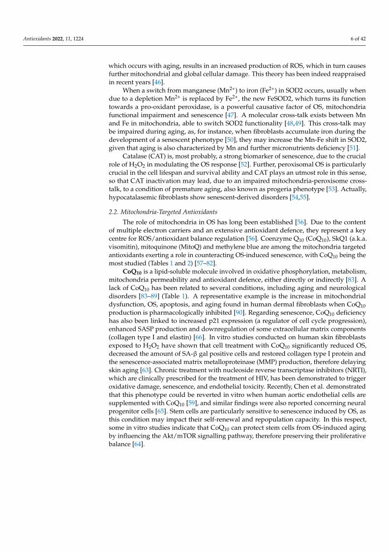
The role of mitochondria in OS has long been established [56]. Due to the contentof multiple electron carriers and an extensive antioxidant defence, they represent a keycentre for ROS/antioxidant balance regulation [56]. Coenzyme Q10 (CoQ10), SkQ1 (a.k.a.visomitin), mitoquinone (MitoQ) and methylene blue are among the mitochondria targetedantioxidants exerting a role in counteracting OS-induced senescence, with CoQ10 being themost studied (Tables 1 and 2) [57–82].
CoQ10 is a lipid-soluble molecule involved in oxidative phosphorylation, metabolism,mitochondria permeability and antioxidant defence, either directly or indirectly [83]. Alack of CoQ10 has been related to several conditions, including aging and neurologicaldisorders [83–89] (Table 1). A representative example is the increase in mitochondrialdysfunction, OS, apoptosis, and aging found in human dermal fibroblasts when CoQ10production is pharmacologically inhibited [90]. Regarding senescence, CoQ10 deficiencyhas also been linked to increased p21 expression (a regulator of cell cycle progression),enhanced SASP production and downregulation of some extracellular matrix components(collagen type I and elastin) [66]. In vitro studies conducted on human skin fibroblastsexposed to H2O2 have shown that cell treatment with CoQ10 significantly reduced OS,decreased the amount of SA-β gal positive cells and restored collagen type I protein andthe senescence-associated matrix metalloproteinase (MMP) production, therefore delayingskin aging [63]. Chronic treatment with nucleoside reverse transcriptase inhibitors (NRTI),which are clinically prescribed for the treatment of HIV, has been demonstrated to triggeroxidative damage, senescence, and endothelial toxicity. Recently, Chen et al. demonstratedthat this phenotype could be reverted in vitro when human aortic endothelial cells aresupplemented with CoQ10 [59], and similar findings were also reported concerning neuralprogenitor cells [65]. Stem cells are particularly sensitive to senescence induced by OS, asthis condition may impact their self-renewal and repopulation capacity. In this respect,some in vitro studies indicate that CoQ10 can protect stem cells from OS-induced agingby influencing the Akt/mTOR signalling pathway, therefore preserving their proliferativebalance [64].
Antioxidants 2022, 11, 1224 7 of 42
Table 1. CoenzymeQ10 and ubiquinol in the prevention of OS-induced senescence.
Ref. Study Design Treatment Form Results Conclusion
Chen et al.,2019 [59]
HAEC chronicallytreated with NRTI
5 µM CoQ10continuously applied
across passagesCoQ10
↓ NRTI-induced senescence↓ ROS
↑Mitochondrial respiration rate
CoQ10 reducescardiovascular side effects of
NRTI treatment
Tarry-Adkinset al., 2013 [58]
Mouse model of lowbirth-weight andcatch-up growth
Post-weaning dietarysupplement CoQ10
↓ Nitrosative and OS↓ DNA damage
↓ Cellular senescence↓Telomere shortening
↓ Apoptosis
CoQ10 prevents cardiacaging and cardiovascular
risk
Ma et al.,2014 [60]
PS-1-mutated ADfibroblasts
Medium with50 µg/mL WS-CoQ10
WS-CoQ10
↓ ROS↑ Cell doubling↓ SIPS
↑ PCNA expression↓MnSOD, p21, p16Ink4A, and Rb
↑ Autophagy
WS-CoQ10inhibits SIPS and improves
autophagy
Xue et al.,2017 [61] Mouse PSCs
Cell treatment with1/10/100 µM CoQ10 for
24/48/72 hCoQ10
↓ Apoptosis↓ SA-β-Gal
↓ ROS and malondialdehyde after72 h treatment
CoQ10 may act as a target inPSC-related pathologies
Wu et al.,2020 [62] ORX mice CoQ10 50 mg/kg/day CoQ10
↓ OS↓ Cell senescence
↑ Osteoblastic bone formation
CoQ10 is anti-osteoporosisand -senescence
Mine et al.,2021 [63]
H2O2-induced SIPS inhuman skin fibroblasts 1 µM and 10 µM CoQ10 CoQ10
↑ Cell viability↓ OS
↓ SA-β-Gal↓ SASP
CoQ10 can contribute toincrease lifespan
Zhang et al.,2015 [64]
D-galactose -inducedaging in MSC
1/10/100 mmol/LCoQ10 for 48 h CoQ10
↓ p-AKT and p-mTOR↓MSC senescence
CoQ10 inhibits MSCsenescence and aging
Velichokovskaet al., 2019 [65] NPCs exposed to ART NP-based delivery of
CoQ10 to mitochondria CoQ10
↓ ROS↑ SIRT3
↑ Cell proliferation↓ SA-β-Gal
↑ Telomere length
ART-induced senescence canbe reversed by NP-CoQ10
Marcheggianiet al., 2021 [66] CoQ10-deprived HDF
5, 10 or 15 µg/mL ofeither CoQ10 or
CoQ10H2for 24 h
CoQ10 orCoQ10H2
↓ SA-β-Gal↓ OS↓ p21
↑ Elastin, collagen type I
CoQ10 or CoQ10H2 preventskin aging and support skin
vitality
Huo et al.,2018 [67]
HUVEC treated withH2O2
24 h in medium with10 µM CoQ10H2
CoQ10H2
↓ SA-β-Gal positive cells↓ SASP↓ ROS
↓ Apoptosis↑ NO and eNOS
↑Mitochondrial function
CoQ10H2delays vascular aging
Yan et al.,2006 [68] SAMP1 mice 250 mg/kg/day lifelong
supplement CoQ10H2
↓ Senescence grading scores↑ Female body weight
= Lifespan= Urinary 8-OHdG andacrolein-lysine adduct
CoQ10H2 decreases cellularsenescence in middle-aged
SAMP1 mice
Olivieri et al.,2013 [69]
Senescent HUVECs inpresence or absence of
LPS
10 µM CoQ10H2 for24 h or 60 days CoQ10H2
↓ LPS-induced NF-kB activation↓ SASP
CoQ10H2 mayprevent aged-induced
endothelial dysfunction
Maruoka et al.,2014 [70] SAMP1 mice
300 mg/kg (Group A)or 30 mg/kg CoQ10H2
(Group B)CoQ10H2
↓ Senescence score at 10 months inGroup A compared to B
↓ OS↑ Antioxidant potential
CoQ10H2reduces senescence and OS
in a dose-dependent manner
Schmelzeret al., 2010 [71]
Middle aged SAMP1mice
500 mg/kg/day ofCoQ10H2 for 6 or
14 monthsCoQ10H2
↓ Senescence grading score↑ PPAR-alpha
CoQ10H2 deceleratesdegeneration in SAMP1 mice
Cirilli et al.,2020 [72]
HUVEC treated withCSE for 24 h 10 µM CoQ10H2
CoQ10H2 andmenaquinone 7
↓ OS↓ Inflammation↓ Apoptosis↓ SA-β-Gal
CoQ10H2 andmenaquinone-7 counteract
CSE-induced damage
Tian et al.,2014 [73] SAMP1 mice Dietary CoQ10H2 (0.3%
w/w) CoQ10H2
↑ PGC-1α, SOD2, IDH2, SIRT1,SIRT3
↑Mitochondrial complex I activity↓ OS
↑ GSH/GSSG ratio
CoQ10H2 protects againstaging progression
Abbreviations: AD: Alzheimer’s Disease; ART: antiretroviral therapy; CoQ10: coenzyme Q10; CoQ10H2: ubiquinol;CSE: cigarette smoke extract; eNOS: endothelial nitric oxide synthase; GSH: reduced glutathione; GSSG: oxidizedglutathione; HAEC: human aortic endothelial cells; HDF: human dermal fibroblasts; HUVEC: human umbilicalvein endothelial cells; IDH2: isocitrate dehydrogenase 2; LPS: lipopolysaccharide; MSC: mesenchymal stemcell; NF-kB: nuclear factor kappa-light-chain-enhancer of activated B cells; NO: nitric oxide; NP: nanoparticle;NPCs: neural progenitor cells; NRTI: nucleoside reverse transcriptase inhibitors; ORX: orchiectomized; OS:oxidative stress; PCNA: proliferating cell nuclear antigen; PGC-1α: peroxisome proliferator-activated receptor γcoactivator 1α; PPAR: peroxisome proliferator-activated receptor; PS-1: presenilin-1; PSCs: pancreatic stellate cells;ROS: reactive oxygen species; SA-β-Gal: senescence-associated β-galactosidase; SAMP1: one of the senescenceaccelerated mice (SAM) strains, which shows shortened life span and early signs of senescence; SASP: senescence-associated secretory phenotype; SIPS: stress induced premature senescence; SIRT: sirtuin; SOD2: superoxidedismutase 2; WS-CoQ10: water-soluble CoQ10; 8-OHdG: 8-hydroxy-2′-deoxyguanosine; ↑: increase; ↓: decrease.

Antioxidants 2022, 11, 1224 8 of 42
Table 2. SkQ1 and MB in the prevention of OS-induced senescence.
Compound Sample Treatment Results Ref.
SkQ1 Podospora anserina, Ceriodaphnia affinis,Drosophila melanogaster, and mouse
Nano- and subnanomolarconcentrations of SkQ1
-Prolonged lifespan-Reduced senescence [74,75]
SkQ1 Wistar and senescence-accelerated rats 250 nmol per kg/day SkQ1 (startingfrom 19 months of age) -Reduced and reversed age-related decline [76]
SkQ1 BALB/c and C57BL/6 mice Lifelong administration of SkQ1 -Decreased cardiomyopathy, fibrosis andcardiac hypertrophy [77]
SkQ1 Senescence-accelerated rats 250 nmol/kg body weight, daily(from 1.5 to 23 months of age) -Reduced Alzheimer’s disease pathology [78]
MB Human IMR90 fibroblasts 10, 100 or 1000 nM of MB for 4 days -Delayed senescence-Improved mitochondrial function [79]
MB Human skin fibroblasts derived fromprogeria patients 100 nM MB
-Effective ROS scavenging-Improved skin fibroblast proliferation
-Delayed senescence[80]
MB Human bone marrow-derived MSCs 200 nM MB -Improved expansion in vigorous MSCs-Improved differentiation in vigorous MSCs [81]
MB Primary rat RGCs 1 µM and 10 µM MB -Stimulated mitochondrial function-Enhanced neuroprotection [82]
Abbreviations: MB: methylene blue; MSCs: mesenchymal stem cells; RGCs: retinal ganglion cells.
In vivo administration of CoQ10 has long been known to improve immune functionsby reducing immunological senescence that characterizes elderly mice [91]. More recently,studies conducted on mouse models of osteoporosis have demonstrated that CoQ10 supple-mentation (50 mg/kg/day) is sufficient to prevent osteoporosis by limiting ROS productionand diminishing cellular senescence, both factors known to contribute to the disease de-velopment [62]. Moreover, CoQ10 may prevent cardiac aging, metabolic syndrome, andcardiovascular disorders when administered post-weaning to a rat model that mimicsthese conditions, and this improvement is mediated by the reduction of ROS and RNS,senescence, and apoptosis [58]. Similar beneficial effects have been obtained in cancer andAlzheimer’s disease (AD), in whose pathogenesis OS plays a predominant role [60,61].Although beneficial, lifelong supplementation with CoQ10 may also be deleterious [92–94].In this respect, results from a study designed to address CoQ10 administration only laterin life showed that old mice subjected to a high CoQ10 diet displayed reduced OS in vari-ous tissues and were more efficient in performing the Morris water maze test comparedto the untreated counterpart [95]. However, no improvements in other psychomotor orcognitive tests suggest that more research is needed to clarify the optimal timing of CoQ10intake [95]. Nevertheless, the introduction of innovative delivery approaches to improveCoQ10 efficiency, such as the use of mitochondria-targeted nanoparticles, may represent apromising strategy to enhance CoQ10 antioxidant activity while limiting the possible sideeffects caused by high-doses administration [65].
CoQ10H2 (or ubiquinol), the reduced form of CoQ10, is even more efficient than CoQ10itself in reverting senescence markers expression, both in vitro and in vivo [66,71]. Thereason for this outperformance could be at least in part explained by a higher CoQ10H2bioavailability at the same concentrations, therefore allowing a more efficient subcellulardelivery [66,71]. For example, a study conducted by Huo et al. has shown that treatmentwith CoQ10H2 of H2O2-induced senescent human umbilical vein endothelial cells (HU-VEC) is effective in reducing SA-β gal, SASP release and ROS production, but enhancednitric oxide (NO) and endothelial NO synthase (eNOS) levels [67]. Diminished inflam-mation, OS-induced senescence and apoptosis have also been observed in the same cellline in other studies [69,72]. In vivo, experiments conducted on SAMP1 mice reportedthat ubiquinol administration at relatively high doses (250–300 mg/kg/day) for at least10 months can reduce senescence grading scores and ROS production, while enhancingantioxidant defences [68,70]. However, no lifespan improvement was detected [68]. Up-regulation of sirtuins 1 and 3 (SIRT1 and SIRT3), SOD2 and isocitrate dehydrogenase 2(IDH2) enzymes, together with a higher reduced to oxidized glutathione (GSH/GSSG)ratio are also described upon dietary CoQ10H2 supplementation in an independent study,thus confirming the role of ubiquinol in protecting against cellular senescence progressionand aging [73]. Finally, these improvements can be further enhanced by the combination of
Antioxidants 2022, 11, 1224 9 of 42
physical exercise and ubiquinol supplementation, as recently reported in in vivo studiescarried out on SAMP8 mouse models [96].
The SkQs are a class of compounds made up by an antioxidant molecule (plasto-quinone), a lipophilic cation and a linker moiety (decane or pentane). The family comprisesSkQ1, SkQR1 and SkQ3, which belong to the mitochondria-targeted plastoquinone deriva-tives with antioxidant activity [97] (Table 2). In particular, SkQ1 and SkQR1 have beenreported to reduce H2O2-induced senescence and apoptosis in vitro and to prevent senes-cence and tissue damage during aging [75]. Moreover, these benefits were achieved alsoin vivo in a wide range of age-related diseases and across species, even in the case of lowdoses administration later in life [74,76,97]. As for CoQ10, SkQ1 given to senescence pronerats at the concentration of 250 µmol/kg/day may be sufficient to prevent the physiologicalage-related deterioration of immunological defences [98]. Finally, AD-related cognitivedecline, behavioural test scores and senescence-associated myocardial disease may improvein murine models upon long-term (lifelong) or limited (between 12 and 18 months of age)intake of SkQ1 [77,78,99–101]. Mechanistically, SkQ1 exerts its antioxidant properties byfatty acid co-mediated uncoupling, through interference with lipoperoxyl radicals and viaregulating the electron flow at the level of mitochondria [102].
Methylene blue (MB) is a well-known mitochondria-targeted antioxidant that hasshown promise in contrasting aging, especially skin aging [103] (Table 2). Methylene blueis reported to be particularly effective in delaying skin cellular senescence and extend-ing fibroblasts lifespan in vitro, as well as in improving mitochondrial functions [79,80].Although not yet fully understood, multiple mechanisms are thought to underlie its antiox-idant function, including Keap/Nrf2 pathway upregulation, MB to MBH2 (the reducedform of MB) cycling in mitochondria, complex IV induction and increased production ofcollagen 2A1 and elastin, two components of the extracellular matrix fundamental for skinpreservation [79,80,104]. Besides skin aging, MB may also prevent senescence and OS inother cell types, such as primary retinal ganglion cells and mesenchymal stem cells (MSCs),but its efficacy on stem cells remains limited to the cellular fraction characterized by a lowerbaseline level of OS [81,82].
Overall, despite promising evidence, results remain unclear. An important limitationis that studies are often performed on a specific cell line or murine model under certainconditions, which often prevent the results from being generalized and/or to be reproduced.In this respect, for example, data on extended fibroblast lifespan are debated, with someevidence showing that they can successfully decelerate aging and prevent senescence whileother studies are inconclusive [105].
2.3. Vitamins
Vitamin A. Preformed vitamin A (all-trans-retinol and its esters) and pro-vitamin A (β-carotene) are essential dietary nutrients that provide a source of retinol, which regulate basicphysiological processes [106,107]. Vitamin A and retinoic acid (a metabolite of all-trans-retinol) administration have been demonstrated to improve AD and age-related attenuationof memory/learning in mouse models, and this is probably due to their immunomodulatoryeffect and the reduction of pro-inflammatory cytokines and chemokines production byastrocytes and microglia, as well as to the promotion of differentiation of neural stemcells and regeneration of neural cells [108–110]. The role of vitamin A in the treatment ofneurodegenerative diseases, such as amyotrophic lateral sclerosis (ALS) and schizophrenia,is currently under investigation [108,111]. Vitamin A has also been studied in associationwith quercetin, a well-known flavonol (see Section 2.7 Flavonoids) [112]. This combinationhas proven capable of reducing rapid senescence-like response induced by acute liverinjury [113].
Vitamin C or ascorbic acid (AA) is a powerful antioxidant that can have beneficialeffects on delaying the aging process and age-related diseases thorough its action on re-dox oxidative and mitochondrial pathways, on the immune system, on inflamm-aging,on endothelial integrity, and on lipoprotein metabolism [114–121]. Supplementation of

Antioxidants 2022, 11, 1224 10 of 42
AA also appears to prevent OS, immunosenescence, telomere attrition, disorganizationof chromatin, and excessive secretion of inflammatory factors, and to prolong life [122].For example, AA has been reported to extend replicative lifespan of human embryonicfibroblasts by restoring age-related decline of mitochondrial function and lowering cellu-lar ROS, therefore reducing mitochondrial and DNA damages with decelerated telomereshortening [123,124]. Moreover, AA was found to have a protective effect also on humanchondrocytes against OS by attenuating the increase of apoptosis, the loss of viability andthe increase of senescence, and therefore hindering the development of osteoarthritis andaging of cartilage [125,126]. In the brain, AA has been increasingly found to promoteseveral beneficial effects on neurodegeneration by direct neuroprotection against OS [116].This vitamin has also been demonstrated to foster anti-senescence and anti-atheroscleroticeffects via an improvement of lipoprotein parameters and microRNA expression throughanti-oxidation and anti-glycation, especially in smokers [127–129]. Finally, a stable AAderivative, 2-O-α-glucopyranosyl-L-ascorbic acid (AA-2G), was also evaluated and com-pared with AA itself for its protective effect against cellular damage and senescence inducedby hydrogen peroxide. The results suggest that the effect of AA-2G is longer-lasting com-pared to that of AA and this derivative might therefore be considered as a more stable formof vitamin C [130].
Vitamin E is a family of fat-soluble vitamins, which comprehends eight organiccompounds with different degrees of antioxidant activity [131]. The impact of vitamin E onthe prevention of chronic diseases is believed to be associated with OS and it has often beenthe subject of several studies. It has been recently observed that a higher consumption ofantioxidants such as vitamin E is able to reduce ROS levels, leading to decreased telomereshortening, decelerating the cellular senescence, and potentially decreasing the risk ofdisease development [132,133].
Vitamin K compounds are a family of fat-soluble vitamins comprising structurallysimilar molecules including two main natural forms: phylloquinone (vitamin K1) andmenaquinones (collectively known as vitamin K2). Besides being responsible for the activa-tion of vitamin K-dependent proteins (VKDPs), which are involved in multiple functionssuch as bone and cardiovascular mineralization, vascular haemostasis, energy metabolism,immune response, brain metabolism, cellular growth, survival, and signalling [134–137],vitamin K appears to suppress the pro-inflammatory cytokines production through anon-carboxylative pathway, by modulating the gene expression of pro-inflammatory mark-ers [138]. Accordingly, warfarin, a vitamin K antagonist, has been found to induce chroniclow-grade inflammation in non-senescent vascular smooth muscle cells and enhance vascu-lar aging and calcification, especially in young patients (<65 years old) [139,140].
2.4. Carotenoids
Carotenoids are naturally occurring lipophilic pigmented molecules found in fruitsand vegetables with important antioxidant properties [141]. Chemically, their polyenestructure, characterized by conjugated double carbon bonds, is at the basis of their abilityto scavenge ROS and free radicals, therefore protecting from OS [141]. Although more than750 carotenoids have been described [142], β-carotene, lycopene, lutein, and zeaxanthinremain the most examined for their implication in human health, with indications of theirinvolvement in several age-related diseases [143–150]. There is evidence that carotenoidsparticipate in the regulation of OS-induced senescence [151,152], and the same is true forparrodienes, which are structurally related to retinoids and carotenoids [153].
β-carotene is the precursor of retinoic acid [154,155]. Although it is generally consid-ered an antioxidant, it can also function as a pro-oxidant compound depending on thecircumstances, which are still not fully understood [156]. In vitro, keratinocytes treatmentwith β-carotene, prior to UVA exposure, prevents the upregulation of MMP-1, MMP-3and MMP-10, therefore suggesting a protective role of β-carotene against OS-inducedsenescence [154].
Antioxidants 2022, 11, 1224 11 of 42
Lutein and zeaxanthin are two macular pigment stereoisomers belonging to the xan-thophyll group of dietary carotenoids [157]. Because of their unique ability to cross theblood-retina barrier, they accumulate in the macula and by virtue of their antioxidant,photoprotective and anti-inflammatory features are involved in the proper eye function-ing [158–160]. A lack of lutein and zeaxanthin is generally associated to a poor cognitiveperformance in elderly [157,161]. Accordingly, improved cognitive functions were observedin elderly patients supplemented for one year with a mixture of lutein and zeaxanthin(12 mg/day), albeit not significant compared to the placebo group [162,163].
There is evidence that OS-induced senescence is involved in the pathogenesis of age-related macular degeneration (AMD), which represents the leading cause of blindnessin aged individuals [158,164,165]. In this respect, Chae et al. documented that luteintreatment protects cells from H2O2-induced senescence by promoting the expression ofantioxidant effectors such as nicotinamide adenine dinucleotide phosphate (NADPH)quinone dehydrogenase 1, heme oxygenase-1 (HO-1) and sirtuins (SIRT1 and SIRT3) [164].Moreover, lutein and zeaxanthin intake, either as supplement or through xanthophyll-enriched foods, might delay AMD thanks to increased antioxidant protection [166]. Finally,data from Sen et al. show that a positive correlation exists between telomere length andxanthophyll carotenoids plasma levels, thus confirming the important role of lutein andzeaxanthin in the context of cellular senescence.
Lycopene is a lipophilic carotenoid naturally found in tomatoes and other red veg-etables and fruits with potent cytoprotective and antioxidant properties [167,168]. Duringaging, lycopene protects from cognitive impairment, insulin resistance and cancer, amongthe others [169–171]. In a study involving 1973 participants, Weber et al. showed thatplasma lycopene levels are significantly different between young and old women, thus sug-gesting that its antioxidant activity is crucial to prevent age-related diseases [161]. Similarly,studies conducted on MSCs demonstrated that cellular pretreatment with lycopene protectsagainst H2O2-induced senescence, enhances antioxidant defences (i.e., improved MnSODactivity and reduced ROS production) and prevents apoptosis through the modulation ofBax and Bak proteins [172]. When used alone, lycopene is known to foster the increase inHO-1, which is detected in dermal fibroblasts after exposure to UVA, thus representing acytoprotective mechanism [173,174]. Moreover, the combination of lycopene with the anti-aging compound nicotinamide mononucleotide (NMN) has proven effective in reducingOS both in vitro and in vivo by enhancing the activity of SOD, CAT, GPx enzymes [175].This effect, combined with the activation of the Kaep1-Nrf2 antioxidant pathway, efficientlyprevents cells to become senescent, therefore confirming the promising role of lycopenein improving the anti-aging effect of already established compounds [175]. These resultsindicate that multiple carotenoids might be responsible for the antioxidant effects reportedin the literature, but more research is needed to clarify the optimal combination of thesesupplements.
2.5. Organosulphur Compounds
Glutathione is a natural tripeptide, that is γ-l-glutamyl-l-cysteinylglycine, harbouringa fundamental role in the regulation of redox homeostasis [176]. Glutathione can existin two forms: reduced glutathione (GSH) and oxidized glutathione (GSSG), which areconverted into each other by the enzymatic activity of GPx (that links two GSH in one GSSGthrough the formation of a disulphide bond) and glutathione reductase (that catalyses thereduction of one GSSG into two GSH to the expenses of NADPH) [177]. Being the mainintracellular antioxidant buffer, both the levels of GSH and GSH/GSSH ratio are tightlycontrolled through a fine regulation of their synthesis, metabolism, transport, and degrada-tion [176,177]. A GSH deficiency has been related to the onset and progression of severaldiseases, including cancer, immunodeficiencies, seizures, neurodegeneration, cardiovascu-lar dysfunctions, and diabetes [178–180]. As GSH levels can be used as a biomarker for theoxidative status of the cell [176,181–183], a reduction in GSH and the GSH/GSSG ratio areoften reported during normal aging and in cellular senescence, both conditions influenced

Antioxidants 2022, 11, 1224 12 of 42
by OS [183–186]. For example, inhibiting or reducing GSH synthesis is sufficient to inducepremature senescence and OS-mediated telomere shortening in HUVEC, and this conditionis not restored by telomerase activity [187]. Similarly, a decreased activity of the enzymeglutamate-cysteine ligase, which is involved in the synthesis of GSH, has been linked tosenescence, ROS production and DNA damage in primary mouse fibroblasts [188]. Ofnote, these detrimental effects are reversed by N-acetylcysteine supplementation, which isknown to increase intracellular GSH levels [188]. Further evidence demonstrates that GSHdeficiency can also trigger senescence through a pathway involving ROS production andErk/p38 regulation, in a mechanism independent from the canonical p53 activation [189].Therapeutically, small extracellular vesicles enriched in the glutathione S-transferase Mu 2(GSTM2) enzyme, which works in conjunction with GSH to reduce OS and detoxify thecell from harmful compounds, can relieve senescence in various tissues when injectedintraperitoneally in old mice [190]. Although reproducible, these results are not alwaysconsistent. Contrary to expectations, Tong et al. reported no reduction in brain GSH levelswhen analysing human postmortem brain samples in elderly subjects compared to youngerones, albeit the lack of data in living tissues represents an important limit of this study [191].Moreover, Barilani et al. recently showed that increased GSH levels accompany MSCsaging [192]. Nevertheless, this mechanism might be a protective strategy to counterbalancethe age-related increase in ROS observed during cellular senescence [191,192].
At brain level, elderly people (>74 years old) are generally characterized by reducedglutathione-S-transferase activity accompanied by slightly lower cerebrospinal fluid (CSF)antioxidant defences compared to younger individuals [193]. These data are consistentwith previous evidence reporting an age-related decline in GSH levels both in the brainand the liver of SAM mice, along with other antioxidant molecules [194]. In humans, thisimpaired glutathione homeostasis might be involved in the pathogenesis of brain disorders,including age-related neurodegeneration [195].
Because GSH is a crucial regulator of oxidative status, it might also represent a promis-ing therapeutic target. Indeed, enrichment analysis research performed on the DrugAgedatabase, a repository of compounds known to extend life, showed that GSH is amongthe most common targets of lifespan prolonging drugs [196]. In this respect, Rebrin et al.demonstrated that the benefits of diets enriched in vitamins and micronutrients should beascribed to increased plasma levels of GSH and improved mitochondria redox homeostasisin a sex and tissue dependent manner [197]. Direct GSH delivery is another therapeuticoption. However, the insufficient bioavailability of GSH remains a limit, and the use ofprodrugs and precursors of GSH have been proposed as an alternative route [198]. Recentdata from Kumar et al. showed that supplementation with glycine and N-acetylcysteineensures the correct GSH balance and extends mice lifespan by 24% [199]. Similarly, the ad-ministration of glutathione precursors (i.e., glycine and cysteine) is sufficient to significantlyincrease GSH levels and reduce OS in aged individuals [200,201].
Overall, these data point to GSH as a key antioxidant regulator involved in OS-inducedsenescence. However, although promising, more research is needed to carefully addressits potential role as biomarker and therapeutic compound in the context of aging andsenescence.
Alliin, allicin, allyl sulphides, allylcysteines and other sulphur-containing com-pounds have long been known for their antioxidant properties [202]. Mainly contained inonion and garlic, they have shown to exert beneficial effects against cardiovascular diseases,cancer, aging, inflammation, OS, and infection, among the others [202,203]. Concerningaging, SAMP8 mice fed for 2 months with a diet containing 2% of aged garlic extract(AGE), which has been reported to have a higher antioxidant activity compared to freshgarlic extract [204], show improved lifespan and learning scores compared to the untreatedcounterpart [205–207]. The improvement in memory functions was then confirmed in vitroby a study conducted on primary hippocampal neurons derived from SAMP10 mice, whosedendrites are increased in length and number upon treatment with S-allylcysteine, themost abundant organosulphur compounds present in AGE [208]. In vivo, 12-week dietary

Antioxidants 2022, 11, 1224 13 of 42
supplementation with S-allylcysteine (0.05% or 0.2%) to 60-week-old wild type mice re-duces senescence, improves mitochondrial functions, and ameliorates both aging and OSbiomarkers [209]. At the molecular level, AGE reduces the production of ROS, increasesglutathione levels, enhances the activity of the main antioxidant enzymes SOD, CAT andGPx, prevents lipid peroxidation and inhibits NF-kB (nuclear factor kappa-light-chain-enhancer of activated B cells) activity [203,210]. Despite encouraging results, discordantevidence emerged from some studies when the molecules contained in the AGE weretested individually. For example, while allicin shows senolytic activity when administeredto breast cancer cells, alliin instead behaves as a pro-senolytic compound in the sameconditions [211]. Still, when used as a whole, garlic extract exerts a strong NO scavengingfunction, reduces MMP-1 expression and ROS levels, inhibits SASP and improves SIRT1activity, thus alleviating UVB-induced senescence in keratinocytes [212]. The combina-tion of the beneficial effects exerted by the different AGE components may explain thisdiscrepancy. For instance, recent evidence has shown that S-1-propenylcysteine, one ofthe AGE components, acts as an anti-inflammatory via stimulation of IL-10 expressionand promotion of macrophage polarization towards an M2c status, which regulates thephagocytosis process of apoptotic cells [213]. According to these results, synergistic effectsmight be achieved by combining anti-inflammatory properties of S-1-propenylcysteinetogether with anti-aging and antioxidant activities reported for the other organosulfurcompounds. Moreover, the administration dosage should be carefully evaluated becausehigh concentrations of antioxidants may instead exacerbate OS. Overall, in line with thewell-known beneficial effects of onion and garlic consumption, it is emerging that var-ious organosulfur compounds commonly found in their extracts can prevent OS, thussupporting their usefulness in counteracting the aging process.
2.6. Nitrogen Non-Protein Compounds
Uric acid (UA) is a by-product of purine metabolism normally found in blood andurine. In the context of OS, although UA is classified as an important antioxidant moleculewhen circulating in the plasma, it exerts a potent pro-oxidant activity once inside the cellor in the form of extracellular crystals, probably due to different environmental interac-tions [214,215]. However, the molecular switch behind this dual role of UA, also defined asthe “uric acid paradox”, remains largely unknown and controversial [216]. Accordingly,chronic serum hyperuricemia positively correlates with inflammation, DNA damage andOS, and has been implicated in the pathogenesis of several disorders, including renal,metabolic, and cardiovascular diseases [214,217,218]. Concerning senescence, several stud-ies have demonstrated a link between UA levels, OS, and cell cycle arrest, both in vitro andin vivo, and improved aging-related functions have been observed following the admin-istration of UA lowering agents [219]. For example, keratinocyte exposure to exogenousUA triggers cellular senescence and OS through a mechanism that is at the basis of theUV-induced damage [220], and a similar pattern has been reported for other cell lines [221].Moreover, xanthine oxidoreductase, an enzyme involved in the production of UA, ROSand RNS, has been shown to promote aging and cellular senescence in vitro as well as inanimal and clinical investigations [222]. Further, in vitro studies have reported an increasedcellular senescence and enhanced ROS production in endothelial progenitor cells culturedin a medium containing UA at high concentrations (10 mg/dL) [223]. Of note, the samedetrimental effects were shown in mice characterized by chronic hyperuricemia [223]. Atthe molecular level, there is evidence that UA triggers OS-induced senescence throughthe inhibition of the enzyme eNOS, which is essential to produce the scavenging moleculeNO [215,223]. This condition triggers an OS imbalance, which promotes cellular senes-cence [215,223]. However, higher plasma UA levels in d-galactose rat models of acceleratedaging were linked to decreased senescence and an increased SOD/(GPx + CAT) enzymaticratio, which is indicative of antioxidant activity, thus confirming the beneficial role of UAwhen considered in the plasma [224].

Antioxidants 2022, 11, 1224 14 of 42
In humans, results from a comparative study conducted on 26 elderly participants and18 controls reported a 2-fold reduction in serum UA levels in aged individuals compared tocontrols, and this pattern was in line with diminished antioxidant defences [225].
Overall, these data show the existence of a correlation between UA and senescence.However, the dual role that UA may play in the context of OS should encourage furtherresearch to better clarify the befits and harms of UA-lowering agents.
2.7. Flavonoids
Flavonoids are a class of polyphenolic secondary metabolites found in plants and areroutinely consumed by humans. Chemically, they are polyphenols with the structure of a15-carbon skeleton (C6–C3–C6) formed by two aromatic rings and one pyran ring [226].Tea, wine, and Chinese herbal plants are the primary sources of flavonoids, as well asleaf vegetables, onion, apples, cherries, berries, soybeans, and citrus fruit [227]. Flavonoidcompounds are divided into six subclasses, flavones, flavonols, flavanones, isoflavones,flavanols, and anthocyanins [228]. Beside the antioxidant activity, flavonoids have anti-inflammatory, vasodilator, anticoagulant, cardioprotective, anti-diabetic, neuroprotective,and anti-obesity activities, which make them of great interest as anti-aging compounds(Table 3) [228–257].
Table 3. Preclinical studies on flavonoids in aging.
Type of Flavonoid Effect Reference
4,4′-dimethoxychalcone - Increases lifespan (yeast, worms, and flies)- Reduces human cell senescence - [229]
Naringenin - Antioxidant effects- Reduces cardiovascular damage- Prolongs lifespan in flies
- [230]
Nobiletin: Rutaceae family- Antioxidant effects - [231]
Quercetin
- Blocks senescence of endothelial cells- Reduces expression of senescence-associated secretion phenotype (SASP)- Enhances health span and lifespan in old mice.- Improves cardiovascular diseases- In combination with dasatinib improves 6-min walking distance, speed, and ability to stand up
- [232]- [233]- [234]- [228]
Fisetin - Blocks cultured senescent fibroblasts in human and animal- Increases lifespan
- [235]- [236]
Apigenin- Reduces SASP - [237]
Theaflavin - Decreases cell senescence- Increases lifespan
- [238]- [239]
Myrecitin - Increases mitochondria metabolism- Reduces neurotoxicity
- [240]- [241]
Rutin- Reduces oxidative stress- Reduces cell senescence- Reduces production of proinflammatory cytokines- Reduces metabolic disorders
- [242]- [243]- [244]
Luteonil - Reduces human senescence cells- Reduces expression of SASP - [245]
Kaempferol - Reduces SASP- Reduces oxidative stress - [229]
Hesperidin - Reduces oxidative stress- Increases antioxidant enzymes
- [246]- [247]
Dyhydromericetin- Reduces oxidative stress- Reduces inflammation- Increases cognitive function- Reduces gut dysfunction
- [248]- [249]- [250]
Epicatechin - Reduces cell senescence- Increases brain function- Reduces skeletal muscle dysfunction
- [251]- [252]- [253]
Genistein- Decreases pro-inflammatory genes expression- Decreases cell senescence- Increases brain cognitive function
- [254]- [255]- [256]
Flavonols. Quercetin is a flavonol known for its antioxidant, anti-inflammatory,antitumor, and senolytic properties [258]. Results obtained on different cell lines showthat treatment with quercetin significantly lowers the levels of ROS and inflammatorycytokines, reduces the expression of SA-β gal, p16 and p53 and markedly increases that ofthe antioxidant enzymes SOD and CAT, regardless of the type of oxidative trigger used

Antioxidants 2022, 11, 1224 15 of 42
to induce senescence [258–262]. In addition to promoting the expression of Nrf2 [263], thebeneficial action of quercetin appears to be mediated by the microRNA-155-5p, which isinvolved in the regulation of SIRT1 and NF-kB [262,264]. Moreover, as aging is associatedwith an inefficient protein-degradation (which is required to protect against OS), the effectof quercetin and its derivatives on the restoration of proteasomal functioning is of interestas rejuvenating strategy [265]. In trials in patients with diabetic kidney disease [266] andidiopathic lung diseases, quercetin induced a reduction in the expression of the agingmarkers p16 and SA-β gal, suggesting an anti-aging effect on kidney cells [267]. Whencombined with dasatinib (a tyrosine kinase inhibitor used as an antitumoral drug), quercetinshowed senolytic activity, improvement of physical function and increased lifespan inmice [268]. Interestingly, as quercetin plus dasatinib treatment reduces intestinal senescenceand inflammation while altering specific microbiota signatures, this optimized senolyticregimen might improve health via reducing intestinal senescence, inflammation, andmicrobial dysbiosis in older subjects [269].
Another promising bioactive flavonol with antioxidant properties is fisetin [270].In vitro cell treatment with fisetin has shown a reduction in senescence, ROS, and apopto-sis [270,271]. In vivo, 6-week oral administration of fisetin drastically reduced senescence,ROS, lipid peroxidation and protein oxidation in a rat model of induced aging, and lifespanextension has been reported in mice [235,272]. This positive outcome is due both to asenolytic activity of fisetin but also to its function as caloric restriction mimetic, which isreported to prolong lifespan [273,274]. However, the timing of fisetin administration seemsto be crucial for obtaining a biological benefit. If on the one hand fisetin is protective whenadministered in the presence of OS, if it is given chronically in physiological conditions, itmay even cause telomere shortening, therefore promoting senescence [275]. For this reason,more studies are needed to better assess the optimal conditions of fisetin intake and itsmechanism of action.
Isoflavonoids. Genistein is a phytoestrogen extracted by soya that is known for itsantioxidant and anti-aging properties, although less potent than other flavonoids such asquercetin and kaempferol [264]. As for other antioxidants, the role of genistein is multiple:it can induce apoptosis acting as a cancer protective compound, but it can also reduceinflammation and OS acting as anti-aging and neurodegenerative protective agent [276].Concerning senescence, genistein alleviates the genotoxicity and the cytotoxicity triggeredby UVB exposure in human dermal fibroblasts [277]. Mechanistically, genistein reducesOS-induced senescence by mitigating the levels of mitochondrial ROS and of the DNAoxidation marker 8-OHdG, as well as by upregulating the SIRT1-FOXO3 axis, which isknown to prevent aging [278].
Flavanols. There is consistency in the literature about the beneficial role of green tea onsenescence-related mechanisms, thanks to its scavenging properties against ROS and RNSand its ability to stimulate autophagy [279–282]. These desirable effects derive from certainmolecules known for their antioxidant role, mainly catechins [281,283]. Even if there are noconclusive results demonstrating the impact of green tea on the human diet, some studiesinvestigated its effects on mice [284]. Catechins supplementation from green tea has beenassociated with a better memory performance and a protective role against DNA oxidativedamage in SAM, independently from the age when the administration of green tea wasstarted [285,286]. These antioxidants have a positive impact also on the brain structure,as murine models fed with green tea show an attenuated brain atrophy compared withSAM drinking pure water, thus suggesting an anti-aging property of these molecules [287].(-)-Epigallocatechin-3-gallate (EGCG) is the most representative flavanol in green tea andits role in contrasting senescence is due to an activation of enzymatic and non-enzymaticantioxidative mechanisms (such as GPx and tocopherol), which are typically reduced inold age [288–292]. Interestingly, EGCG anti-senescence effects can also be observed ata macroscopic level as its supplementation reduces age-related sarcopenia in mice [293].Nonetheless, an excessive amount of green tea has been associated with oxidative damage,underlying the need of further research to set the beneficial dose range [279,294].

Antioxidants 2022, 11, 1224 16 of 42
Flavanones and flavones. Among flavanones, hesperidin is an antioxidant that can betypically found in citrus fruits [295]. Its properties impact positively on cardiomyocytes asit attenuates senescence-related oxidative damage, both independently and in combinationwith other molecules, through the induction of Nrf2 and of GST expression [247,296,297].Citrus juice, which is rich in hesperidin and other flavanones as well as in flavones, an-thocyanins, and other molecules, was reported to reduce ROS levels and reduce SA-β galpositive HUVEC [298]. Citrus fruit also contains another flavanone useful to counteract theeffects of aging on myocardium, which is naringenin [299,300]. A recent study conducted inaging murine models suggests that the antioxidant properties of naringenin deriving fromthe activation of PI3K/Akt/Nrf2 pathway could greatly ameliorate both behavioural andneurological dysfunctions. The authors reported that naringenin administration markedlystimulated the activity of Nrf2 and improved the expression of the antioxidant enzymesHO-1 and NADPH-quinone oxidoreductase 1 [301].
Besides containing flavonones, citrus peels are also rich in flavones. One of these, no-biletin, was demonstrated to attenuate senescence-related cognitive deficits in SAMP8 miceby counteracting amyloid ß accumulation in the brain [302,303]. Flavones and flavononesare also significant components of bergamot juice and they confer anti-aging propertiesthrough the upregulation of SIRT1, Nrf2 and FOXO3 (that are involved in homeostasis,resistance to oxidative damage and overall health respectively), as it was demonstrated inmodels of senescent myocardiocytes and in vivo in mice [304].
Apigenin, also known as 5,7,4′-trihydroxyflavone, is a flavone typically found inparsley, oranges, and chamomile. Its ability to act as a metal chelator, free radical scavenger,and regulator of the main pathways involved in redox homeostasis [i.e., Nrf2, NF-kB,MAPK (mitogen-activated protein kinase) and Akt (a.k.a. protein kinase B] has increased itsinterest as an antioxidant molecule [305]. For example, creams rich in apigenin are used fortheir beneficial effects on skin aging prevention [306,307]. In vitro, human embryonic lungfibroblasts exposed to the pro-senescence stimuli H2O2 or doxorubicin, and subsequentlytreated with apigenin, show reduced SA-β gal activity, cell cycle promotion, increasedlevels of SIRT1, CAT and SOD and reduced expression of the senescence associated p21, p53and p16 proteins compared to the untreated counterpart [308]. Similar results have beenobtained in vivo following administration of apigenin daily for 8 weeks to a d-galactose-induced aging mouse model [309]. Moreover, thanks to its ability to inhibit the SASPand to interfere with the anti-apoptotic pathways, which are generally upregulated insenescent cancer cells, apigenin has been proposed as an adjuvant therapy for tumours,with promising results [237,310,311].
Anthocyanins. Bilberry and mulberry are considered promising nutrients for healthyaging because of their antioxidant properties related to anthocyanins that consist, amongothers, in the increase of SOD activity and AMPK (AMP-activated protein kinase)-mTORautophagy pathway [312]. It has been reported that anthocyanins contrast senescenceas they promote neural stem cells proliferation and diminish aging-related markers andcognitive impairment in mice [313]. The ability of anthocyanins to inhibit β amyloidaggregation is also of interest in therapeutic approaches aimed at slowing down cognitivedecline [314]. In rats, the effect of mulberry extract was observed on the cardiovascularsystem, as it reduced the signs of senescence in endothelial cells [315,316].
Overall, a diet rich in these natural antioxidants may have a significant anti-aging effect.An indirect confirmation of this concept could be deduced by the fact that the MediterraneanDiet, which widely includes both flavanols, flavanones, flavons and anthocyanins, ischaracterized by well-known beneficial effects on health, including a healthy aging as ithinders the pathogenesis of many chronic diseases and extends life expectancy [317,318].
2.8. Non-Flavonoids
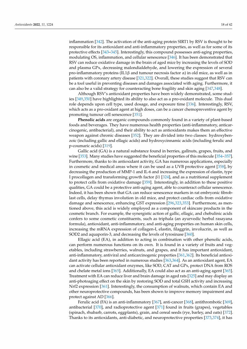
Non-flavonoid antioxidant substances, namely stilbenes (resveratrol), phenolic acids,curcuminoids (curcumin), and lignans [319] could be employed as anti-aging agents, actingagainst OS, inflammation, and cellular senescence (Table 4) [320–332].
Antioxidants 2022, 11, 1224 17 of 42
Table 4. Effects of non-flavonoids treatment in different experimental studies.
Non-Flavonoid Model Effects Reference
Resveratrol
HUVEC cells - Prevention of cells apoptosis, by reducing oxidative damage (↑SOD,↓ROS, and ↓MDA) and inhibiting mitochondrial pathway
[320]
Senescence-accelerated mice - Reduction of oxidative damage in the brain (↑SOD, ↑GSH-Px, and ↓MDA) [321]
Old male mice - Decreasing of inflammation in the liver (↓IL-1β, ↓TNFα, and ↓COX2) [322]
Gallic acid
Rat embryonic fibroblast cells- Reduction of inflammation (↓NF-kB, ↓TNFα, ↓IL-1β, and ↓IL-6)- Reduction of beta-galactosidase activity- Decreasing of ROS production and lipid peroxidation
[323]
UVB-irradiated humanfibroblast cells
- Inhibition of MMP-1 and IL-6 expression, and increasing of procollagentype I
[324]UVB-irradiated hairless mice - Prevention of wrinkle formation, by upregulating procollagen type I and
elastin levels
Ellagic acid D-galactose-treated rats- Attenuation of OS in liver and brain (↑SOD, ↑CAT, ↑GSH-Px)- Amelioration of histopathological changes- Inhibition of inflammation (↓IL-6, ↓IL-1β, ↓TNFα)
[325]
Ferulic acid UVA-irradiated nHDF- Increasing of proliferation and cell cycle- Reduction of OS (↑SOD1, and ↑CAT)- Inhibition of cellular senescence (↓p16)
[326]
p-coumaric acid
Rat chondrocytes - Amelioration of inflammation and cellular senescence, by inhibitingMAPK and NF-kB pathways
[327]
Mice fed with high-fatdiet (HFD)
- Inhibition of ROS production, lipid peroxidation and upregulation ofantioxidants enzymes (↑SOD, ↑CAT, ↑GSH-Px, ↑HO-1)
[328]
Curcumin
Senescence-accelerated mice - Improvement of cognitive deficits, by decreasing OS (↑SOD) andincreasing p-CaMKII and p-NMDAR1 expression
[329]
Mice fed with HFD- Decreasing of OS (↑HO-1)- Reduction of inflammation and vascular aging, by lowering the
accumulation of senescent cells in the aorta and MCP-1 levels in the blood[330]
Lignans
nPC12 cells - Reduction of lipid peroxidation (↓COX-2), and ROS production [331]D-galactose aging mice - Attenuation of OS (↑SOD, ↑GPx)
Old HDFs - Decreasing of senescence markers expression (cyclin D1, p16, p27, p21,caveolin-1), by activating AMPK pathway, and ROS levels
[332]
Abbreviations: AMPK: (AMP-activated protein kinase); CAT: (catalase); GPx: (glutathione peroxidase); GSH-Px:(plasma glutathione peroxidase); HO-1: (heme oxygenase 1); HUVEC: (Human umbilical vein endothelial cell);IL-1β: (interleukin 1 β); IL-6: (interleukin 6); MAPK: (mitogen-activated protein kinase); MCP-1: (monocytechemoattractant protein-1); MDA: (malondialdehyde), COX-2 (cyclooxygenase 2); MMP-1: (matrix metallopro-teinase 1); NF-kB: (nuclear factor kappa B); nHDF: (normal human dermal fibroblasts); nPC12: (neuronallydifferentiated phenchromocytoma cells); p-CaMKII: (p-calcium/calmodulin-dependent kinase II); p-NMDARI:(p-N-methyl-D-aspartate receptor subunit 1); p16: (cyclin-dependent kinase inhibitor 2A); p21: (cyclin-dependentkinase inhibitor 1); p27: (cyclin-dependent kinase inhibitor 1B); SOD: (superoxide dismutase), ROS (reactiveoxygen species); SOD1: (superoxide dismutase 1); TNFα: (tumour necrosis factor); ↑: increase; ↓: decrease.
Stilbenes are a family of natural phenolic compounds found in many plant species ca-pable of acting as antioxidants, anti-inflammatory, antibacterial, and anticanceragents [333,334]. The most important and well-known stilbene is resveratrol (RSV), aphytoalexin found in black grapes, peanuts, blackberries, red wine, and various herbalremedies [335–337]. It has many biological properties, including antioxidant, and anti-inflammatory effects [320,338]. As an antioxidant agent, RSV can scavenge free radicalsand reduce ROS formation, by inhibiting the expression of NADPH and glycogen synthasekinase 3 beta proteins, and upregulating the expression of some antioxidant enzymes, suchas SOD2, CAT, GPx, and thioredoxin [320,339,340]. It can also stimulate the productionof HO-1 by activating Nrf2 [341]. Resveratrol treatment has also been shown to preventor slow down the progression of cardiovascular, neurological, and metabolic disorders,as well as to be promising in the prevention of cancer, viral infection, and pathological
Antioxidants 2022, 11, 1224 18 of 42
inflammation [342]. The activation of the anti-aging protein SIRT1 by RSV is thought to beresponsible for its antioxidant and anti-inflammatory properties, as well as for some of itsprotective effects [343–345]. Interestingly, this compound possesses anti-aging properties,modulating OS, inflammation, and cellular senescence [346]. It has been demonstrated thatRSV can reduce oxidative damage in the brain of aged mice by increasing the levels of SODand plasma GPx, decreasing malondialdehyde, and lowering the expression of severalpro-inflammatory proteins (IL1β and tumour necrosis factor α) in old mice, as well as inpatients with coronary artery disease [321,322]. Overall, these studies suggest that RSV canbe a tool useful in preventing diseases and damages associated with aging. Furthermore, itcan also be a valid strategy for counteracting bone fragility and skin aging [347,348].
Although RSV’s antioxidant properties have been widely demonstrated, some stud-ies [349,350] have highlighted its ability to also act as a pro-oxidant molecule. This dualrole depends upon cell type, used dosage, and exposure time [336]. Interestingly, RSV,which acts as a pro-oxidant agent at high doses, can be a cancer chemopreventive agent bypromoting tumour cell senescence [351].
Phenolic acids are organic compounds commonly found in a variety of plant-basedfoods and beverages. They have numerous health properties (anti-inflammatory, anticar-cinogenic, antibacterial), and their ability to act as antioxidants makes them an effectiveweapon against chronic diseases [352]. They are divided into two classes: hydroxyben-zoic (including gallic and ellagic acids) and hydroxycinnamic acids (including ferulic andp-coumaric acids) [319].
Gallic acid (GA) is a natural substance found in berries, gallnuts, grapes, fruits, andwine [353]. Many studies have suggested the beneficial properties of this molecule [354–357].Furthermore, thanks to its antioxidant activity, GA has numerous applications, especiallyin cosmetic and medical areas where it can be used as a UVB protective agent [358], bydecreasing the production of MMP-1 and IL-6 and increasing the expression of elastin, typeI procollagen and transforming growth factor β1 [324], and as a nutritional supplementto protect cells from oxidative damage [359]. Interestingly, in addition to these positivequalities, GA could be a protective anti-aging agent, able to counteract cellular senescence.Indeed, it has been shown that GA can reduce senescence markers in rat embryonic fibrob-last cells, delay thymus involution in old mice, and protect cardiac cells from oxidativedamage and senescence, enhancing GST expression [296,323,353]. Furthermore, as men-tioned above, this acid is widely employed as a component of skincare products in thecosmetic branch. For example, the synergistic action of gallic, ellagic, and chebulinic acidsconfers to some cosmetic constituents, such as triphala (an ayurvedic herbal rasayanaformula), antioxidant, anti-inflammatory, and anti-aging properties on human skin cells,increasing the mRNA expression of collagen-I, elastin, filaggrin, involucrin, as well asSOD2 and aquaporin-3, and decreasing the levels of tyrosinase [360].
Ellagic acid (EA), in addition to acting in combination with other phenolic acids,can perform numerous functions on its own. It is found in a variety of fruits and veg-etables, including strawberries, walnuts, and grapes, and it has important antioxidant,anti-inflammatory, antiviral and anticarcinogenic properties [361,362]. Its beneficial antioxi-dant activity has been reported in numerous studies [363,364]. As an antioxidant agent, EAcan activate cellular antioxidant enzymes, like SOD, CAT and GPx, protect DNA from ROSand chelate metal ions [365]. Additionally, EA could also act as an anti-aging agent [365].Treatment with EA can reduce liver and brain damage in aged rats [325] and may display ananti-photoaging effect on the skin by restoring SOD and total GSH activity and increasingNrf2 expression [361]. Interestingly, the consumption of walnuts, which contain EA andother neuroprotective compounds, has been shown to improve memory impairment andprotect against AD [366].
Ferulic acid (FA) is an anti-inflammatory [367], anti-cancer [368], antithrombotic [369],antibacterial [370], and radioprotective agent [371] found in fruits (grapes), vegetables(spinach, rhubarb, carrots, eggplants), grain, and cereal seeds (rye, barley, and oats) [372].Thanks to its antioxidants, anti-diabetic, and neuroprotective properties [373,374], it has
Antioxidants 2022, 11, 1224 19 of 42
been shown to prevent type 2 diabetes and AD [375,376] by regulating antioxidant enzymesand caspase activities [326]. Acting as an antioxidant, FA can inhibit the enzymes thatlead to ROS formation, scavenge free radicals, and promote the antioxidant enzymesactivity [372]. This makes FA a compound widely used in cosmetics and in food industry,especially as an anti-aging agent [377]. It has been demonstrated to protect skin from UVradiation through its capacity to reduce the activity of the stress-inducible protein Gadd45α, the expression of MMP-1 and MMP-3 mRNAs, as well as enhancing the levels of theantioxidant enzymes SOD1 and CAT [326,378]. As a result of its anti-aging properties, itcould be an excellent cosmetic component for face masks and antioxidant and protectivecreams. Moreover, it is used in skin-lightening lotions, inhibiting tyrosinase activity andmelanocytic proliferation [372]. Peanuts also contain FA, which may partly explain theirability to prevent aging and cognitive decline [379].
p-coumaric acid (p-CA) is a dietary compound widely found in oranges, apples,grapes, kiwis, onions, potatoes, eggplant, beans, and grains [380,381], which is endowedwith antibacterial [382], anti-diabetic [383], anti-cancer [384], and radioprotective proper-ties [385]. Furthermore, p-CA has analgesic, antipyretic, and anxiolytic effects, as well as theability to inhibit platelet aggregation [381]. Being an antioxidant agent, the treatment withthis acid promotes the expression of Nrf2, with the consequent increased levels of someantioxidant enzymes, including HO-1, SOD, NAD(P)H quinone dehydrogenase 1, CAT, andGPx [328]. Coumaric acid can also slow down the aging processes, due to its antioxidantand anti-inflammatory effects. It has been shown that p-CA can have beneficial effectson skin aging by decreasing collagenase, elastase, and hyaluronidase activity [386], andby reducing the inflammatory response and chondrocytes senescence, inhibiting MAPKand NF-kB signalling pathways [327]. However, although some works have demonstratedthe antioxidant property of p-CA, Pienkowska et al. highlighted that this acid is unableto counteract the premature senescence of human fibroblasts [387]. Consequently, thepresence of contradictory evidence in literature requires further research.
Curcuminoids are natural polyphenolic compounds used as spices and food addi-tives thanks to their aromatic and colouring properties [388]. The numerous beneficialactivities make them potential supportive therapeutics for cancer and inflammatory boweldiseases [389]. Curcumin (CUR), a yellow phenolic pigment, commonly used as a foodspice and herbal remedy, is the most well-known curcuminoid [390]. This compound isknown to have anti-cancer [391], anti-bacterial [392], anti-diabetic [393], and cytoprotec-tive activities [394]. Further, CUR possesses antioxidant properties, through which it canprevent lipid peroxidation, stabilize Nrf2, with the consequent expression of HO-1, andincrease the levels of antioxidant enzymes, such as SOD, GST, GSH and GPx [395]. It hasbeen suggested that this natural compound possesses therapeutic features in several mal-functions, including neurological, cardiological, and metabolic disorders, as well as ulcers,arthritis, acne, and dyspepsia [396]. Curcumin is also thought to be a useful anti-agingagent [395]. Indeed, it has been shown to improve cognitive deficits, suppress vascularaging and inflammation in elderly mice, and attenuate neuronal aging both in vitro andin vivo by downregulating the expression of p16 and p21 and upregulating antioxidant en-zymes, including SOD1, CAT and GPx [329,330,397]. Furthermore, CUR supplementationhas positive benefits on age-related disorders [398–400]. Although an antioxidant action inaging has been widely demonstrated, conflicting studies are present in literature regardingits inability to counteract OS [401], therefore more research is needed to better understandits antioxidant role.
Lignans are found in many plant families and foods, including fruits, vegetables,nuts (sesame), grains, and seeds [402]. In addition to their numerous biological activities(antioxidant, anti-inflammatory, and antitumoral), as well as their ability to protect againstthe onset of chronic and metabolic diseases, lignans and their derivatives are also known toact as anti-aging agents [403]. In fact, they can suppress aging phenotypes in Drosophilaadults, inhibit NADPH oxidase activity and upregulate antioxidant genes, such as SOD1,SOD2, catalase, and DNA repairing genes [221,404]. Further, they can reduce the levels of

Antioxidants 2022, 11, 1224 20 of 42
senescence in old human diploid fibroblasts, activating AMPK pathway [332]. Moreover,lignans molecules can protect tissues and organs against OS, inflammation, and senescenceby acting as neuroprotective and radioprotective agents [405,406].
2.9. Minerals
Despite their tiny amount, micronutrients, i.e., vitamins and minerals, are essentialfor human health, exerting numerous functions, including antioxidant defence rangingfrom genome-related processes, such as DNA replication and repair, to metabolic processesand antioxidant defence [407,408]. Concerning the latter, the structural and functionalroles of a few minerals such as zinc (Zn), selenium (Se), magnesium (Mg) copper (Cu) andmanganese (Mn) is crucial (Table 5) [409–420].
Table 5. Minerals as modulators of OS-induced senescence.
Mineral Sample Treatment/Condition Result Ref.
Zinc Colon cancer lines SW480and SW620 ↓ Zinc
↑ Oxidative stress, cellular proliferation,stress signalling morphological changes,
cell death[409]
Zinc Dermal fibroblast ↑ Zinc ↑ Oxidative stress and DNA damage [410]Zinc HCAECs ↑ Zinc ↑ Senescence [411]
Selenium Bone marrow stromal cells ↑ Selenium ↓ Senescence [412]Selenium Keratinocytes ↑ Selenium ↓ Senescence [413]Selenium Human fibroblasts ↑ Selenium ↓ Senescence [414]Selenium Mice ↓ Selenium ↑ Senescence [415]
Magnesium Endothelial cells ↓Magnesium ↑ Oxidative stress and cell death [416]Magnesium Endothelial cells ↓Magnesium ↑ Pro-inflammatory molecules [417]Magnesium Embryo-hepatocytes ↓Magnesium ↑ Oxidative stress [418]Magnesium Human fibroblasts ↓Magnesium ↑ Telomere shortening [419]Magnesium Rats ↓Magnesium ↑ Age-related diseases [420]
Abbreviations: HCAECs: Human coronary artery endothelial cells; ↑: increase; ↓: decrease.
Zinc, the second most abundant trace mineral in the body after iron, is involved ina wide range of key biological functions exerted through its catalytic role in enzymes,structural function in proteins and other cellular components [421–423]. Importantly, Znexerts antioxidant functions through its catalytic action in Zn- SOD, via the formation andstabilization of sulfhydryl groups in proteins, thus maintaining membrane integrity andprotecting it from oxidation, and through regulation of Zn-binding protein metallothioneinexpression. In this respect, evidence showed that Zn deficiency causes destabilizationof membrane structure and augments OS [424–428]. In addition, Zn suppresses anti-inflammatory responses that would otherwise promote OS [425]. In vitro studies haveshown that Zn deficiency is associated with an increased production of ROS, oxidativedamage to DNA, proteins and lipids, destabilization of membrane structure, dysregula-tion of Zn-binding protein metallothionein [426,429]. For instance, in a colon cancer cellline [409] and dermal fibroblasts [410], Zn dysregulation promoted cellular senescenceactivating stress response and pro-apoptosis pathways. Some Zn-finger proteins andZn-dependent enzymes, such as PATZ1 [430], ZKSCAN3 [431], ZHX3 [432], KLF4 [433]or Zfp637 [434] might be responsible for this Zn-mediated cellular senescence inhibition,reducing ROS production, DNA damage and telomere shortening (Table 6) [430–437]. Insupport of this concept, there is evidence that the downregulation of the Zn-finger proteinZEB2 significantly promotes cell senescence in hepatic stellate cells and dermal fibroblasts,limiting the development of fibrosis [435,436]. Again, the Zn-finger protein 768 has beenfound to be overexpressed in cancer cells, contributing to cell proliferation and repressingsenescence [437]. On the other hand, Zn overload can contribute to augment cellular OSand senescence through different mechanisms not yet well-defined, but possibly related toorganelles dysfunction [411,427,438].
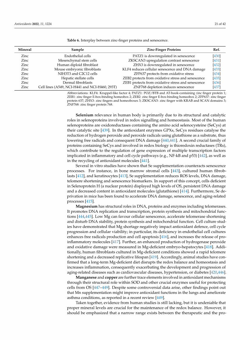
Antioxidants 2022, 11, 1224 21 of 42
Table 6. Interplay between zinc-finger proteins and senescence.
Mineral Sample Zinc-Finger Proteins Ref.
Zinc Endothelial cells PATZ1 is downregulated in senescence [430]Zinc Mesenchymal stem cells ZKSCAN3 upregulation contrast senescence [431]Zinc Human diploid fibroblast ZHX3 is downregulated in senescence [432]Zinc Mouse embryonic fibroblasts KLF4 reduces cellular senescence and DNA damage [433]Zinc NIH3T3 and C2C12 cells ZFP637 protects from oxidative stress [434]Zinc Hepatic stellate cells ZEB2 protects from oxidative stress and senescence [435]Zinc Dermal fibroblasts ZEB1 protects from oxidative stress and senescence [436]Zinc Cell lines (A549, NCI-H441 and NCI-H460, 293T) ZNF768 depletion induces senescence [437]
Abbreviations: KLF4: Kruppel-like factor 4; PATZ1: POZ/BTB and AT-hook-containing zinc finger protein 1;ZEB1: zinc finger E-box-binding homeobox 2; ZEB2: zinc finger E-box-binding homeobox 2; ZFP637: zinc fingerprotein 637; ZHX3: zinc fingers and homeoboxes 3; ZKSCAN3: zinc finger with KRAB and SCAN domains 3;ZNF768: zinc finger protein 768.
Selenium relevance in human body is primarily due to its structural and catalyticroles in selenoproteins involved in redox signalling and homeostasis. Most of the humanselenoproteins are oxidoreductases containing the amino acid selenocysteine (SeCys) attheir catalytic site [439]. In the antioxidant enzymes GPXs, SeCys residues catalyse thereduction of hydrogen peroxide and peroxide radicals using glutathione as a substrate, thuslowering free radicals and consequent DNA damage [440,441]. A second crucial family ofproteins containing SeCys and involved in redox biology is thioredoxin reductases (TRs),which contribute to the regulation of gene expression of multiple transcription factorsimplicated in inflammatory and cell cycle pathways (e.g., NF-kB and p53) [442], as well asin the recycling of antioxidant molecules [441].
Several in vitro studies have shown that Se supplementation counteracts senescenceprocesses. For instance, in bone marrow stromal cells [443], cultured human fibrob-lasts [412], and keratinocytes [413], Se supplementation reduces ROS levels, DNA damage,telomere shortening and senescence biomarkers. In support of this concept, cells deficientin Selenoprotein H (a nuclear protein) displayed high levels of OS, persistent DNA damageand a decreased content in antioxidant molecules (glutathione) [414]. Furthermore, Se de-privation in mice has been found to accelerate DNA damage, senescence, and aging-relatedprocesses [415].
Magnesium has structural roles in DNA, proteins and enzymes including telomerases.It promotes DNA replication and transcription, protein synthesis and mitochondrial func-tions [444,445]. Low Mg can favour cellular senescence, accelerate telomerase shorteningand disturb DNA stability, protein synthesis and mitochondrial function. Cell culture stud-ies have demonstrated that Mg shortage negatively impact antioxidant defence, cell cycleprogression and cellular viability; in particular, its deficiency in endothelial cell culturesenhances free radicals production and cell apoptosis [416], and increases the release of pro-inflammatory molecules [417]. Further, an enhanced production of hydrogenase peroxideand oxidative damage were measured in Mg-deficient embryo-hepatocytes [418]. Addi-tionally, human fibroblasts cultured in Mg-deficient conditions showed a rapid telomereshortening and a decreased replicative lifespan [419]. Accordingly, animal studies have con-firmed that a long-term Mg-deficient diet disrupts the redox balance and homeostasis andincreases inflammation, consequently exacerbating the development and progression ofaging-related diseases such as cardiovascular diseases, hypertension, or diabetes [420,446].
Manganese and copper are further trace elements involved in antioxidant mechanismsthrough their structural role within SOD and other crucial enzymes useful for protectingcells from OS [447–449]. Despite some controversial data arise, other findings point outthat Mn supplementation might improve antioxidant functions in the lungs and ameliorateasthma conditions, as reported in a recent review [449].
Taken together, evidence from human studies is still lacking, but it is undeniable thatproper mineral levels are crucial for the maintenance of the redox balance. However, itshould be emphasized that a narrow range exists between the therapeutic and the pro-

Antioxidants 2022, 11, 1224 22 of 42
oxidative effects of some metals, including Se, Mn, Mg, Cu and Zn; therefore, a cautiousand rational supplement choice must be assessed by the experts in the field.
2.10. Others—Melatonin
Melatonin is a hormone produced and released by the pineal gland with immunomod-ulatory, oncostatic, anti-aging, and endocrine modulator functions [450,451]. Its antioxidantand anti-inflammatory activities are exerted through the suppression of cyclooxygenase2, NLRP3 inflammasome, gasdermin D, TLR-4, NF-kB, and NO release, as well as theconcomitant activation of SIRT1 and Nrf2 free radical scavenging network [452]. For exam-ple, inhibition of sodium nitroprusside-mediated NO release and increased production oftranscripts coding for antioxidant enzymes (i.e., SOD1, GPx1 and CAT) have been reportedupon melatonin treatment in neuroblastoma cells and HUVEC, respectively [451,453]. How-ever, melatonin can also stimulate the release of pro-inflammatory factors depending onthe concurrent conditions, although this response seems to be limited to early treatmentstages and needs to be better investigated [454].
As a reduction in melatonin secretion has been observed during aging [452], severalstudies have explored the role of this hormone in counteracting cellular senescence [451]. Inthis respect, melatonin has been proven capable of reducing oxidative stress and replicativesenescence by enhancing autophagy, activating AMPK/FOXO3 pathways and increasingmitochondrial membrane potential, both in vitro and in vivo [455–457]. Melatonin-induceddecrease in p53, p21 and p16 proteins, together with enhanced SIRT1 activity, have alsobeen reported in the context of H2O2-induced senescence [458,459]. Regarding MSCs, itis known that long term culture stimulates ROS generation, thus promoting oxidativestress-induced senescence [460]. In this context, decreased cellular senescence, preservedself-renewal and activation of antioxidant defence pathways have all been observed af-ter melatonin supplementation [460,461]. In vivo, melatonin intake has been reported todiminish age-derived inflammation and apoptosis and to decrease lipid peroxidation, thio-barbituric acid reactive substances and protein carbonyls, thus counteracting hippocampalsenescence and exerting an anti-aging and antioxidant effect on mice brains [462–465].However, contrasting evidence remains about the effect of melatonin on the regulationof antioxidant enzymes [457,463–465]. Finally, brain oxidative stress amelioration andincreased osteopontin and senescence marker protein-30 have been shown following mela-tonin treatment in the context of vascular demented rats [466]. Altogether, these findingssuggest that, despite encouraging evidence, more research is needed to address the role ofmelatonin in vivo and identify possible side effects.
3. Discussion
The elderly population is growing exponentially in parallel with basic and clinicalresearch discoveries and improvement of the quality of care for those who are sick. Aging isone of the risk factors for chronic diseases, atherosclerosis, cardiovascular diseases, stroke,kidney failure, chronic lung disease, cancers, diabetes, osteoporosis, arthritis, blindness,dementia, and neurodegenerative diseases [467,468]. Lifespans have increased dramaticallyover the last century in large part due to advances in medicine that have nearly eliminatedcertain deadly infectious diseases.
Nutrition is one of the factors that can influence aging. Interviews of older personsreveal that most of them have continuous physical activities, positive thinking, and eathealthy foods such as vegetables, fruits, fishes, and less meat. It was shown that lesscalories [469], good sleep [470], less stress [471], good relationships [472], no smoke and lowalcohol drinking are environmental factors capable of delaying aging, while genetic factorsplay a role for in 25% to 30% of life expectancy [473]. In this context, finding anti-agingdrugs that meet the safety and effectiveness of long-term use has always been an importantstrategy for intervention in the aging field.
Although cellular senescence is crucial for the proper functioning of several physio-logical processes, much scientific evidence has demonstrated that it also plays a leading

Antioxidants 2022, 11, 1224 23 of 42
role in the pathophysiology of aging and age-related diseases [3,474]. Because OS is animportant senescence-triggering stimulus [13], the thorough investigation of existing an-tioxidants and the search for new ones are of major interest. In this narrative review, wehave summarized the potential of the major classes of antioxidants in extending life andpreventing senescence. However, most antioxidants are known to exert a dual effect (bothpro-oxidant and antioxidant), especially based on the doses administered. Accordingly, ithas been reported that high doses of antioxidants promote cellular senescence [156,475,476].In this respect, more research is needed to define the optimal dosage of antioxidants, alsoconsidering the interaction of multiple compounds coming either from the diet or fromsupplements. Currently this remains a limit, as most of the studies investigated a singlecompound on a particular cell line or on a specific animal model, which often preventsgeneralizing the results in a broader context. Besides the dosage, a careful evaluationof the optimal administration window can be also crucial to achieve a clinical benefit asantioxidants efficacy might depend upon OS levels [81]. From a human perspective, notonly dose assessment but also the time of initiation (childhood, adolescence, adulthood,old age) and the duration of the treatment (lifelong, at alternating intervals or for a definedperiod) might affect the clinical outcomes. There are still few studies on the synergy andinterference of multiple antioxidants taken in combination—increasingly fashionable in themodern society—and the benefits and risks of this approach should be carefully evaluated.Similarly, when integrated as supplements, antioxidants may give rise to different effectsdepending upon the time of the day they are taken and individual differences such asphysical activity and lifestyle, which may also greatly vary among subjects. Given theseconsiderations, studies conducted in vitro under limited variable conditions should bereproduced and validated on large-cohorts clinical trials.
Current reports distinguish the existence of senolytic (able to remove senescent cells)or senomorphic (able to modulate senescent cells) substances, which can include someplant-derived antioxidants (e.g., quercein, fisetin) but usually are functional concepts, thedescription of which is outside our review remit, and which should encourage clinicalresearch and nutraceutical application, more than in vitro investigations [266,477–479]. Itis difficult, to date, to indicate if an anti-oxidant vitamin is senolytic or senomorphic, dueto the huge complexity of biological phenomena.
4. Conclusions
Antioxidants are formidable substances, mostly derived from plants, which have beenconsidered so far to be beneficial agents able to address many redox-mediated injuries.Aging is usually considered as a major playground of oxidative stress, but it shouldbe highlighted that ROS are fundamentally signalling molecules and that most of thestress responding mechanisms are tuned by fine modulation of ROS as signalling agents.Therefore, a correct action to address senescence is to find approaches and methods toimprove and promote this modulation. Wise people used to state that equilibria stand onthe fine regulation of pro- and con- hallmarks of xenobiotics. This is also our wish andrecommendation for the future.
Author Contributions: Conceptualization, A.V. and A.P.; writing—original draft preparation, A.V.,S.C., L.I.M.C., E.P., G.B.P., C.S., A.C. and C.B.; writing—review and editing, A.P. and A.V.; supervision,G.R. and A.P. All authors have read and agreed to the published version of the manuscript.
Funding: This research was supported by grants from the Italian Ministry of Health as RicercaCorrente and as RF-2016-02363298.
Institutional Review Board Statement: Not applicable.
Informed Consent Statement: Not applicable.
Data Availability Statement: Data is contained within the article.
Antioxidants 2022, 11, 1224 24 of 42
Acknowledgments: We kindly thank Abdelouahab Bellou (Institute of Sciences in Emergency Medcine, Academy of Medical Sciences, Guangdong Provincial People’s Hospital, Guangzhou, China;Wayne State University School of Medicine, Emergency Department, Detroit, MI, USA) for hisprecious help in revising the manuscript prior to submission.
Conflicts of Interest: The authors declare no conflict of interest.
References1. Hayflick, L.; Moorhead, P.S. The Serial Cultivation of Human Diploid Cell Strains. Exp. Cell Res. 1961, 25, 585–621. [CrossRef]2. Calcinotto, A.; Kohli, J.; Zagato, E.; Pellegrini, L.; Demaria, M.; Alimonti, A. Cellular Senescence: Aging, Cancer, and Injury.
Physiol. Rev. 2019, 99, 1047–1078. [CrossRef] [PubMed]3. Gorgoulis, V.; Adams, P.D.; Alimonti, A.; Bennett, D.C.; Bischof, O.; Bishop, C.; Campisi, J.; Collado, M.; Evangelou, K.; Ferbeyre,
G.; et al. Cellular Senescence: Defining a Path Forward. Cell 2019, 179, 813–827. [CrossRef]4. di Micco, R.; Krizhanovsky, V.; Baker, D.; d’Adda di Fagagna, F. Cellular Senescence in Ageing: From Mechanisms to Therapeutic
Opportunities. Nat. Rev. Mol. Cell Biol. 2021, 22, 75–95. [CrossRef] [PubMed]5. Krizhanovsky, V.; Yon, M.; Dickins, R.A.; Hearn, S.; Simon, J.; Miething, C.; Yee, H.; Zender, L.; Lowe, S.W. Senescence of Activated
Stellate Cells Limits Liver Fibrosis. Cell 2008, 134, 657–667. [CrossRef]6. Sagiv, A.; Krizhanovsky, V. Immunosurveillance of Senescent Cells: The Bright Side of the Senescence Program. Biogerontology
2013, 14, 617–628. [CrossRef]7. Lozano-Torres, B.; Estepa-Fernández, A.; Rovira, M.; Orzáez, M.; Serrano, M.; Martínez-Máñez, R.; Sancenón, F. The Chemistry of
Senescence. Nat. Rev. Chem. 2019, 3, 426–441. [CrossRef]8. Gao, X.; Yu, X.; Zhang, C.; Wang, Y.; Sun, Y.; Sun, H.; Zhang, H.; Shi, Y.; He, X. Telomeres and Mitochondrial Metabolism:
Implications for Cellular Senescence and Age-Related Diseases. Stem Cell Rev. Rep. 2022, 1–13. [CrossRef]9. Kumari, R.; Jat, P. Mechanisms of Cellular Senescence: Cell Cycle Arrest and Senescence Associated Secretory Phenotype. Front.
Cell Dev. Biol. 2021, 9, 645593. [CrossRef]10. Roger, L.; Tomas, F.; Gire, V. Mechanisms and Regulation of Cellular Senescence. Int. J. Mol. Sci. 2021, 22, 13173. [CrossRef]11. Casella, G.; Tsitsipatis, D.; Abdelmohsen, K.; Gorospe, M. MRNA Methylation in Cell Senescence. WIREs RNA 2019, 10, e1547.
[CrossRef] [PubMed]12. Crouch, J.; Shvedova, M.; Thanapaul, R.J.R.S.; Botchkarev, V.; Roh, D. Epigenetic Regulation of Cellular Senescence. Cells 2022, 11,
672. [CrossRef]13. Lettieri-Barbato, D.; Aquilano, K.; Punziano, C.; Minopoli, G.; Faraonio, R. MicroRNAs, Long Non-Coding RNAs, and Circular
RNAs in the Redox Control of Cell Senescence. Antioxidants 2022, 11, 480. [CrossRef]14. Sun, J.; Cheng, B.; Su, Y.; Li, M.; Ma, S.; Zhang, Y.; Zhang, A.; Cai, S.; Bao, Q.; Wang, S.; et al. The Potential Role of M6A RNA
Methylation in the Aging Process and Aging-Associated Diseases. Front. Genet. 2022, 13, 869950. [CrossRef] [PubMed]15. Harries, L.W. Dysregulated RNA Processing and Metabolism: A New Hallmark of Ageing and Provocation for Cellular Senescence.
FEBS J. 2022. [CrossRef]16. Gheitasi, I.; Azizi, A.; Omidifar, N.; Doustimotlagh, A.H. Renoprotective Effects of Origanum Majorana Methanolic L and
Carvacrol on Ischemia/Reperfusion-Induced Kidney Injury in Male Rats. Evid.-Based Complement. Altern. Med. 2020, 2020,9785932. [CrossRef]
17. Gholami, A.; Emadi, F.; Amini, A.; Shokripour, M.; Chashmpoosh, M.; Omidifar, N. Functionalization of Graphene OxideNanosheets Can Reduce Their Cytotoxicity to Dental Pulp Stem Cells. J. Nanomater. 2020, 2020, 6942707. [CrossRef]
18. Liguori, I.; Russo, G.; Curcio, F.; Bulli, G.; Aran, L.; Della-Morte, D.; Gargiulo, G.; Testa, G.; Cacciatore, F.; Bonaduce, D.; et al.Oxidative Stress, Aging, and Diseases. Clin. Interv. Aging 2018, 13, 757–772. [CrossRef]
19. Omidifar, N.; moghadami, M.; Mousavi, S.M.; Hashemi, S.A.; Gholami, A.; Shokripour, M.; Sohrabi, Z. Trends in NaturalNutrients for Oxidative Stress and Cell Senescence. Oxidative Med. Cell. Longev. 2021, 2021, 7501424. [CrossRef]
20. Rangel-Zúñiga, O.A.; Corina, A.; Lucena-Porras, B.; Cruz-Teno, C.; Gómez-Delgado, F.; Jiménez-Lucena, R.; Alcalá-Díaz, J.F.;Haro-Mariscal, C.; Yubero-Serrano, E.M.; Delgado-Lista, J.; et al. TNFA Gene Variants Related to the Inflammatory Status and ItsAssociation with Cellular Aging: From the CORDIOPREV Study. Exp. Gerontol. 2016, 83, 56–62. [CrossRef]
21. Sriram, S.; Yuan, C.; Chakraborty, S.; Tay, W.; Park, M.; Shabbir, A.; Toh, S.-A.; Han, W.; Sugii, S. Oxidative Stress MediatesDepot-Specific Functional Differences of Human Adipose-Derived Stem Cells. Stem Cell Res. Ther. 2019, 10, 141. [CrossRef]
22. Yu, C.; Xiao, J.-H. The Keap1-Nrf2 System: A Mediator between Oxidative Stress and Aging. Oxidative Med. Cell. Longev. 2021,2021, 6635460. [CrossRef]
23. Lopes-Paciencia, S.; Saint-Germain, E.; Rowell, M.-C.; Ruiz, A.F.; Kalegari, P.; Ferbeyre, G. The Senescence-Associated SecretoryPhenotype and Its Regulation. Cytokine 2019, 117, 15–22. [CrossRef] [PubMed]
24. Collado, M.; Serrano, M. Senescence in Tumours: Evidence from Mice and Humans. Nat. Rev. Cancer 2010, 10, 51–57. [CrossRef][PubMed]
25. Narita, M.; Nuñez, S.; Heard, E.; Narita, M.; Lin, A.W.; Hearn, S.A.; Spector, D.L.; Hannon, G.J.; Lowe, S.W. Rb-MediatedHeterochromatin Formation and Silencing of E2F Target Genes during Cellular Senescence. Cell 2003, 113, 703–716. [CrossRef]
Antioxidants 2022, 11, 1224 25 of 42
26. Serrano, M.; Lin, A.W.; McCurrach, M.E.; Beach, D.; Lowe, S.W. Oncogenic Ras Provokes Premature Cell Senescence Associatedwith Accumulation of P53 and P16INK4a. Cell 1997, 88, 593–602. [CrossRef]
27. Chapman, J.; Fielder, E.; Passos, J.F. Mitochondrial Dysfunction and Cell Senescence: Deciphering a Complex Relationship. FEBSLett. 2019, 593, 1566–1579. [CrossRef]
28. Zhu, M.; Meng, P.; Ling, X.; Zhou, L. Advancements in Therapeutic Drugs Targeting of Senescence. Ther. Adv. Chronic Dis. 2020,11, 204062232096412. [CrossRef]
29. Elsallabi, O.; Patruno, A.; Pesce, M.; Cataldi, A.; Carradori, S.; Gallorini, M. Fisetin as a Senotherapeutic Agent: BiopharmaceuticalProperties and Crosstalk between Cell Senescence and Neuroprotection. Molecules 2022, 27, 738. [CrossRef]
30. Coling, D.; Chen, S.; Chi, L.-H.; Jamesdaniel, S.; Henderson, D. Age-Related Changes in Antioxidant Enzymes Related toHydrogen Peroxide Metabolism in Rat Inner Ear. Neurosci. Lett. 2009, 464, 22–25. [CrossRef]
31. Miller, A.-F. Superoxide Dismutases: Ancient Enzymes and New Insights. FEBS Lett. 2012, 586, 585–595. [CrossRef] [PubMed]32. Frye, K.A.; Sendra, K.M.; Waldron, K.J.; Kehl-Fie, T.E. Old Dogs, New Tricks: New Insights into the Iron/Manganese Superoxide
Dismutase Family. J. Inorg. Biochem. 2022, 230, 111748. [CrossRef] [PubMed]33. Treiber, N.; Maity, P.; Singh, K.; Ferchiu, F.; Wlaschek, M.; Scharffetter-Kochanek, K. The Role of Manganese Superoxide Dismutase
in Skin Aging. Derm.-Endocrinol. 2012, 4, 232–235. [CrossRef] [PubMed]34. Fisher, G.J.; Varani, J.; Voorhees, J.J. Looking Older. Arch. Dermatol. 2008, 144, 666–672. [CrossRef] [PubMed]35. Quan, T.; Shao, Y.; He, T.; Voorhees, J.J.; Fisher, G.J. Reduced Expression of Connective Tissue Growth Factor (CTGF/CCN2)
Mediates Collagen Loss in Chronologically Aged Human Skin. J. Investig. Dermatol. 2010, 130, 415–424. [CrossRef] [PubMed]36. Allen, R.G.; Tresini, M.; Keogh, B.P.; Doggett, D.L.; Cristofalo, V.J. Differences in Electron Transport Potential, Antioxidant
Defenses, and Oxidant Generation in Young and Senescent Fetal Lung Fibroblasts (WI-38). J. Cell Physiol 1999, 180, 114–122.[CrossRef]
37. Borlon, C.; Debacq-Chainiaux, F.; Hinrichs, C.; Scharffetter-Kochanek, K.; Toussaint, O.; Wlaschek, M. The Gene ExpressionProfile of Psoralen plus UVA-Induced Premature Senescence in Skin Fibroblasts Resembles a Combined DNA-Damage andStress-Induced Cellular Senescence Response Phenotype. Exp. Gerontol. 2007, 42, 911–923. [CrossRef] [PubMed]
38. Lu, C.-Y.; Lee, H.-C.; Fahn, H.-J.; Wei, Y.-H. Oxidative Damage Elicited by Imbalance of Free Radical Scavenging Enzymes IsAssociated with Large-Scale MtDNA Deletions in Aging Human Skin. Mutat. Res./Fundam. Mol. Mech. Mutagenesis 1999, 423,11–21. [CrossRef]
39. Meewes, C.; Brenneisen, P.; Wenk, J.; Kuhr, L.; Ma, W.; Alikoski, J.; Poswig, A.; Krieg, T.; Scharffetter-Kochanek, K. AdaptiveAntioxidant Response Protects Dermal Fibroblasts from UVA-Induced Phototoxicity. Free Radic. Biol. Med. 2001, 30, 238–247.[CrossRef]
40. Naderi-Hachtroudi, L.; Peters, T.; Brenneisen, P.; Meewes, C.; Hommel, C.; Razi-Wolf, Z.; Schneider, L.A.; Schüller, J.; Wlaschek,M.; Scharffetter-Kochanek, K. Induction of Manganese Superoxide Dismutase in Human Dermal Fibroblasts. Arch. Dermatol.2002, 138, 1473–1479. [CrossRef]
41. Mao, C.; Yuan, J.-Q.; Lv, Y.-B.; Gao, X.; Yin, Z.-X.; Kraus, V.B.; Luo, J.-S.; Chei, C.-L.; Matchar, D.B.; Zeng, Y.; et al. Associationsbetween Superoxide Dismutase, Malondialdehyde and All-Cause Mortality in Older Adults: A Community-Based Cohort Study.BMC Geriatr. 2019, 19, 104. [CrossRef] [PubMed]
42. Mohammedi, K.; Bellili-Muñoz, N.; Marklund, S.L.; Driss, F.; le Nagard, H.; Patente, T.A.; Fumeron, F.; Roussel, R.; Hadjadj, S.;Marre, M.; et al. Plasma Extracellular Superoxide Dismutase Concentration, Allelic Variations in the SOD3 Gene and Risk ofMyocardial Infarction and All-Cause Mortality in People with Type 1 and Type 2 Diabetes. Cardiovasc. Diabetol. 2015, 14, 845.[CrossRef] [PubMed]
43. Martini, H.; Passos, J.F. Cellular Senescence: All Roads Lead to Mitochondria. FEBS J. 2022. [CrossRef] [PubMed]44. Treiber, N.; Maity, P.; Singh, K.; Kohn, M.; Keist, A.F.; Ferchiu, F.; Sante, L.; Frese, S.; Bloch, W.; Kreppel, F.; et al. Accelerated
Aging Phenotype in Mice with Conditional Deficiency for Mitochondrial Superoxide Dismutase in the Connective Tissue. AgingCell 2011, 10, 239–254. [CrossRef]
45. Zhang, Y.; Unnikrishnan, A.; Deepa, S.S.; Liu, Y.; Li, Y.; Ikeno, Y.; Sosnowska, D.; van Remmen, H.; Richardson, A. A New Rolefor Oxidative Stress in Aging: The Accelerated Aging Phenotype in Sod1−Mice Is Correlated to Increased Cellular Senescence.Redox Biol. 2017, 11, 30–37. [CrossRef]
46. Hajam, Y.A.; Rani, R.; Ganie, S.Y.; Sheikh, T.A.; Javaid, D.; Qadri, S.S.; Pramodh, S.; Alsulimani, A.; Alkhanani, M.F.; Harakeh, S.;et al. Oxidative Stress in Human Pathology and Aging: Molecular Mechanisms and Perspectives. Cells 2022, 11, 552. [CrossRef]
47. Ganini, D.; Santos, J.H.; Bonini, M.G.; Mason, R.P. Switch of Mitochondrial Superoxide Dismutase into a Prooxidant Peroxidase inManganese-Deficient Cells and Mice. Cell Chem. Biol. 2018, 25, 413–425.e6. [CrossRef]
48. Naranuntarat, A.; Jensen, L.T.; Pazicni, S.; Penner-Hahn, J.E.; Culotta, V.C. The Interaction of Mitochondrial Iron with ManganeseSuperoxide Dismutase. J. Biol. Chem. 2009, 284, 22633–22640. [CrossRef]
49. Yang, M.; Cobine, P.A.; Molik, S.; Naranuntarat, A.; Lill, R.; Winge, D.R.; Culotta, V.C. The Effects of Mitochondrial IronHomeostasis on Cofactor Specificity of Superoxide Dismutase 2. EMBO J. 2006, 25, 1775–1783. [CrossRef]
50. Killilea, D.W.; Atamna, H.; Liao, C.; Ames, B.N. Iron Accumulation During Cellular Senescence in Human Fibroblasts In Vitro.Antioxid. Redox Signal. 2003, 5, 507–516. [CrossRef]
51. Killilea, D.W.; Maier, J.A.M. A Connection between Magnesium Deficiency and Aging: New Insights from Cellular Studies.Magnes Res. 2008, 21, 77–82. [PubMed]

Antioxidants 2022, 11, 1224 26 of 42
52. Cutler, R.G. Oxidative Stress and Aging: Catalase Is a Longevity Determinant Enzyme. Rejuvenation Res. 2005, 8, 138–140.[CrossRef] [PubMed]
53. Koepke, J.I.; Wood, C.S.; Terlecky, L.J.; Walton, P.A.; Terlecky, S.R. Progeric Effects of Catalase Inactivation in Human Cells. Toxicol.Appl. Pharmacol. 2008, 232, 99–108. [CrossRef] [PubMed]
54. Koepke, J.I.; Nakrieko, K.-A.; Wood, C.S.; Boucher, K.K.; Terlecky, L.J.; Walton, P.A.; Terlecky, S.R. Restoration of PeroxisomalCatalase Import in a Model of Human Cellular Aging. Traffic 2007, 8, 1590–1600. [CrossRef]
55. Wood, C.S.; Koepke, J.I.; Teng, H.; Boucher, K.K.; Katz, S.; Chang, P.; Terlecky, L.J.; Papanayotou, I.; Walton, P.A.; Terlecky, S.R.Hypocatalasemic Fibroblasts Accumulate Hydrogen Peroxide and Display Age-Associated Pathologies. Traffic 2006, 7, 97–107.[CrossRef]
56. Lin, M.T.; Beal, M.F. Mitochondrial Dysfunction and Oxidative Stress in Neurodegenerative Diseases. Nature 2006, 443, 787–795.[CrossRef]
57. Ahmed, E.; Donovan, T.; Yujiao, L.; Zhang, Q. Mitochondrial Targeted Antioxidant in Cerebral Ischemia. J. Neurol. Neurosci. 2015,6, 2. [CrossRef]
58. Tarry-Adkins, J.L.; Blackmore, H.L.; Martin-Gronert, M.S.; Fernandez-Twinn, D.S.; McConnell, J.M.; Hargreaves, I.P.; Giussani,D.A.; Ozanne, S.E. Coenzyme Q10 Prevents Accelerated Cardiac Aging in a Rat Model of Poor Maternal Nutrition and AcceleratedPostnatal Growth. Mol. Metab. 2013, 2, 480–490. [CrossRef]
59. Chen, Y.-F.; Hebert, V.Y.; Stadler, K.; Xue, S.Y.; Slaybaugh, K.; Luttrell-Williams, E.; Glover, M.C.; Krzywanski, D.M.; Dugas,T.R. Coenzyme Q10 Alleviates Chronic Nucleoside Reverse Transcriptase Inhibitor-Induced Premature Endothelial Senescence.Cardiovasc. Toxicol. 2019, 19, 500–509. [CrossRef]
60. Ma, D.; Stokes, K.; Mahngar, K.; Domazet-Damjanov, D.; Sikorska, M.; Pandey, S. Inhibition of Stress Induced PrematureSenescence in Presenilin-1 Mutated Cells with Water Soluble Coenzyme Q10. Mitochondrion 2014, 17, 106–115. [CrossRef]
61. Xue, R.; Yang, J.; Wu, J.; Meng, Q.; Hao, J. Coenzyme Q10 Inhibits the Activation of Pancreatic Stellate Cells throughPI3K/AKT/MTOR Signaling Pathway. Oncotarget 2017, 8, 92300–92311. [CrossRef] [PubMed]
62. Wu, X.; Liang, S.; Zhu, X.; Wu, X.; Dong, Z. CoQ10 Suppression of Oxidative Stress and Cell Senescence Increases Bone Mass inOrchiectomized Mice. Am. J. Transl. Res. 2020, 12, 4314–4325. [PubMed]
63. Mine, Y.; Takahashi, T.; Okamoto, T. Protective Effects of Coenzyme Q10 on Cell Damage Induced by Hydrogen Peroxides inCultured Skin Fibroblasts. J. Clin. Biochem. Nutr. 2021, 69, 20–185. [CrossRef] [PubMed]
64. Zhang, D.; Yan, B.; Yu, S.; Zhang, C.; Wang, B.; Wang, Y.; Wang, J.; Yuan, Z.; Zhang, L.; Pan, J. Coenzyme Q10 Inhibits the Aging ofMesenchymal Stem Cells Induced by D-Galactose through Akt/MTOR Signaling. Oxidative Med. Cell. Longev. 2015, 2015, 867293.[CrossRef] [PubMed]
65. Velichkovska, M.; Surnar, B.; Nair, M.; Dhar, S.; Toborek, M. Targeted Mitochondrial COQ10 Delivery Attenuates Antiretroviral-Drug-Induced Senescence of Neural Progenitor Cells. Mol. Pharm. 2019, 16, 724–736. [CrossRef] [PubMed]
66. Marcheggiani, F.; Kordes, S.; Cirilli, I.; Orlando, P.; Silvestri, S.; Vogelsang, A.; Möller, N.; Blatt, T.; Weise, J.M.; Damiani, E.; et al.Anti-Ageing Effects of Ubiquinone and Ubiquinol in a Senescence Model of Human Dermal Fibroblasts. Free Radic. Biol. Med.2021, 165, 282–288. [CrossRef]
67. Huo, J.; Xu, Z.; Hosoe, K.; Kubo, H.; Miyahara, H.; Dai, J.; Mori, M.; Sawashita, J.; Higuchi, K. Coenzyme Q10 Prevents Senescenceand Dysfunction Caused by Oxidative Stress in Vascular Endothelial Cells. Oxidative Med. Cell. Longev. 2018, 2018, 3181759.[CrossRef]
68. Yan, J.; Fujii, K.; Yao, J.; Kishida, H.; Hosoe, K.; Sawashita, J.; Takeda, T.; Mori, M.; Higuchi, K. Reduced Coenzyme Q10Supplementation Decelerates Senescence in SAMP1 Mice. Exp. Gerontol. 2006, 41, 130–140. [CrossRef]
69. Olivieri, F.; Lazzarini, R.; Babini, L.; Prattichizzo, F.; Rippo, M.R.; Tiano, L.; di Nuzzo, S.; Graciotti, L.; Festa, R.; Brugè, F.; et al.Anti-Inflammatory Effect of Ubiquinol-10 on Young and Senescent Endothelial Cells via MiR-146a Modulation. Free Radic. Biol.Med. 2013, 63, 410–420. [CrossRef]
70. Maruoka, H.; Fujii, K.; Inoue, K.; Kido, S. Long-Term Effect of Ubiquinol on Exercise Capacity and the Oxidative Stress RegulationSystem in SAMP1 Mice. J. Phys. Ther. Sci. 2014, 26, 367–371. [CrossRef]
71. Schmelzer, C.; Kubo, H.; Mori, M.; Sawashita, J.; Kitano, M.; Hosoe, K.; Boomgaarden, I.; Döring, F.; Higuchi, K. Supplementationwith the Reduced Form of Coenzyme Q10 Decelerates Phenotypic Characteristics of Senescence and Induces a PeroxisomeProliferator-Activated Receptor-α Gene Expression Signature in SAMP1 Mice. Mol. Nutr. Food Res. 2010, 54, 805–815. [CrossRef][PubMed]
72. Cirilli, I.; Orlando, P.; Marcheggiani, F.; Dludla, P.V.; Silvestri, S.; Damiani, E.; Tiano, L. The Protective Role of Bioactive Quinonesin Stress-Induced Senescence Phenotype of Endothelial Cells Exposed to Cigarette Smoke Extract. Antioxidants 2020, 9, 1008.[CrossRef] [PubMed]
73. Tian, G.; Sawashita, J.; Kubo, H.; Nishio, S.; Hashimoto, S.; Suzuki, N.; Yoshimura, H.; Tsuruoka, M.; Wang, Y.; Liu, Y.; et al.Ubiquinol-10 Supplementation Activates Mitochondria Functions to Decelerate Senescence in Senescence-Accelerated Mice.Antioxid. Redox Signal. 2014, 20, 2606–2620. [CrossRef] [PubMed]
74. Anisimov, V.N.; Bakeeva, L.E.; Egormin, P.A.; Filenko, O.F.; Isakova, E.F.; Manskikh, V.N.; Mikhelson, V.M.; Panteleeva, A.A.;Pasyukova, E.G.; Pilipenko, D.I.; et al. Mitochondria-Targeted Plastoquinone Derivatives as Tools to Interrupt Execution of theAging Program. 5. SkQ1 Prolongs Lifespan and Prevents Development of Traits of Senescence. Biochemistry 2008, 73, 1329–1342.[CrossRef] [PubMed]

Antioxidants 2022, 11, 1224 27 of 42
75. Skulachev, M.; Antonenko, Y.; Anisimov, V.; Chernyak, B.; Cherepanov, D.; Chistyakov, V.; Egorov, M.; Kolosova, N.; Korshunova,G.; Lyamzaev, K.; et al. Mitochondrial-Targeted Plastoquinone Derivatives. Effect on Senescence and Acute Age-RelatedPathologies. Curr. Drug Targets 2011, 12, 800–826. [CrossRef]
76. Kolosova, N.G.; Stefanova, N.A.; Muraleva, N.A.; Skulachev, V.P. The Mitochondria-Targeted Antioxidant SkQ1 but NotN-Acetylcysteine Reverses Aging-Related Biomarkers in Rats. Aging 2012, 4, 686–694. [CrossRef]
77. Manskikh, V.N.; Gancharova, O.S.; Nikiforova, A.I.; Krasilshchikova, M.S.; Shabalina, I.G.; Egorov, M.V.; Karger, E.M.; Milanovsky,G.E.; Galkin, I.I.; Skulachev, V.P.; et al. Age-Associated Murine Cardiac Lesions Are Attenuated by the Mitochondria-TargetedAntioxidant SkQ1. Histol. Histopathol. 2015, 30, 353–360. [CrossRef]
78. Stefanova, N.A.; Muraleva, N.A.; Skulachev, V.P.; Kolosova, N.G. Alzheimer’s Disease-Like Pathology in Senescence-AcceleratedOXYS Rats Can Be Partially Retarded with Mitochondria-Targeted Antioxidant SkQ1. J. Alzheimer’s Dis. 2013, 38, 681–694.[CrossRef]
79. Atamna, H.; Nguyen, A.; Schultz, C.; Boyle, K.; Newberry, J.; Kato, H.; Ames, B.N. Methylene Blue Delays Cellular Senescenceand Enhances Key Mitochondrial Biochemical Pathways. FASEB J. 2008, 22, 703–712. [CrossRef]
80. Xiong, Z.-M.; O’Donovan, M.; Sun, L.; Choi, J.Y.; Ren, M.; Cao, K. Anti-Aging Potentials of Methylene Blue for Human SkinLongevity. Sci. Rep. 2017, 7, 2475. [CrossRef]
81. Bertolo, A.; Capossela, S.; Fränkl, G.; Baur, M.; Pötzel, T.; Stoyanov, J. Oxidative Status Predicts Quality in Human MesenchymalStem Cells. Stem Cell Res. Ther. 2017, 8, 3. [CrossRef] [PubMed]
82. Daudt, D.R.; Mueller, B.; Park, Y.H.; Wen, Y.; Yorio, T. Methylene Blue Protects Primary Rat Retinal Ganglion Cells from CellularSenescence. Investig. Opthalmol. Vis. Sci. 2012, 53, 4657. [CrossRef] [PubMed]
83. Hargreaves, I.; Heaton, R.A.; Mantle, D. Disorders of Human Coenzyme Q10 Metabolism: An Overview. Int. J. Mol. Sci. 2020, 21,6695. [CrossRef]
84. Yubero, D.; Montero, R.; Martín, M.A.; Montoya, J.; Ribes, A.; Grazina, M.; Trevisson, E.; Rodriguez-Aguilera, J.C.; Hargreaves,I.P.; Salviati, L.; et al. Secondary Coenzyme Q 10 Deficiencies in Oxidative Phosphorylation (OXPHOS) and Non-OXPHOSDisorders. Mitochondrion 2016, 30, 51–58. [CrossRef] [PubMed]
85. Mantle, D. Coenzyme Q10 Supplementation for Diabetes and Its Complications: An Overview. Br. J. Diabetes 2017, 17, 145–148.[CrossRef]
86. Hargreaves, I.; Mantle, D.; Milford, D. Chronic Kidney Disease and Coenzyme Q10 Supplementation. J. Kidney Care 2019, 4, 82–90.[CrossRef]
87. Mantle, D.; Hargreaves, I. Coenzyme Q10 and Degenerative Disorders Affecting Longevity: An Overview. Antioxidants 2019, 8,44. [CrossRef] [PubMed]
88. Emmanuele, V.; López, L.C.; Berardo, A.; Naini, A.; Tadesse, S.; Wen, B.; D’Agostino, E.; Solomon, M.; DiMauro, S.; Quinzii, C.;et al. Heterogeneity of Coenzyme Q10 Deficiency: Patient study and literature review. Arch. Neurol. 2012, 69, 978–983. [CrossRef]
89. Yang, S.; Liu, T.; Li, S.; Zhang, X.; Ding, Q.; Que, H.; Yan, X.; Wei, K.; Liu, S. Comparative Proteomic Analysis of Brains ofNaturally Aging Mice. Neuroscience 2008, 154, 1107–1120. [CrossRef]
90. Marcheggiani, F.; Cirilli, I.; Orlando, P.; Silvestri, S.; Vogelsang, A.; Knott, A.; Blatt, T.; Weise, J.M.; Tiano, L. Modulation ofCoenzyme Q10 Content and Oxidative Status in Human Dermal Fibroblasts Using HMG-CoA Reductase Inhibitor over a BroadRange of Concentrations. From Mitohormesis to Mitochondrial Dysfunction and Accelerated Aging. Aging 2019, 11, 2565–2582.[CrossRef]
91. Bliznakov, E.G. Immunological Senescence in Mice and Its Reversal by Coenzyme Q10. Mech. Ageing Dev. 1978, 7, 189–197.[CrossRef]
92. Sohal, R.S.; Kamzalov, S.; Sumien, N.; Ferguson, M.; Rebrin, I.; Heinrich, K.R.; Forster, M.J. Effect of Coenzyme Q10 Intake onEndogenous Coenzyme Q Content, Mitochondrial Electron Transport Chain, Antioxidative Defenses, and Life Span of Mice. FreeRadic. Biol. Med. 2006, 40, 480–487. [CrossRef] [PubMed]
93. Sumien, N.; Heinrich, K.R.; Shetty, R.A.; Sohal, R.S.; Forster, M.J. Prolonged Intake of Coenzyme Q10 Impairs Cognitive Functionsin Mice. J. Nutr. 2009, 139, 1926–1932. [CrossRef] [PubMed]
94. Mcdonald, S.R.; Sohal, R.S.; Forster, M.J. Concurrent Administration of Coenzyme Q10 and α-Tocopherol Improves Learning inAged Mice. Free Radic. Biol. Med. 2005, 38, 729–736. [CrossRef] [PubMed]
95. Shetty, R.A.; Forster, M.J.; Sumien, N. Coenzyme Q10 Supplementation Reverses Age-Related Impairments in Spatial Learningand Lowers Protein Oxidation. Age 2013, 35, 1821–1834. [CrossRef] [PubMed]
96. Andreani, C.; Bartolacci, C.; Guescini, M.; Battistelli, M.; Stocchi, V.; Orlando, F.; Provinciali, M.; Amici, A.; Marchini, C.; Tiano,L.; et al. Combination of Coenzyme Q10 Intake and Moderate Physical Activity Counteracts Mitochondrial Dysfunctions in aSAMP8 Mouse Model. Oxidative Med. Cell. Longev. 2018, 2018, 8936251. [CrossRef]
97. Skulachev, V.P.; Anisimov, V.N.; Antonenko, Y.N.; Bakeeva, L.E.; Chernyak, B.V.; Erichev, V.P.; Filenko, O.F.; Kalinina, N.I.;Kapelko, V.I.; Kolosova, N.G.; et al. An Attempt to Prevent Senescence: A Mitochondrial Approach. Biochim. Biophys. Acta(BBA)-Bioenerg. 2009, 1787, 437–461. [CrossRef]
98. Obukhova, L.A.; Skulachev, V.P.; Kolosova, N.G. Mitochondria-Targeted Antioxidant SkQ1 Inhibits Age-Dependent Involution ofthe Thymus in Normal and Senescence-Prone Rats. Aging 2009, 1, 389–401. [CrossRef]
99. Stefanova, N.A.; Ershov, N.I.; Kolosova, N.G. Suppression of Alzheimer’s Disease-Like Pathology Progression by Mitochondria-Targeted Antioxidant SkQ1: A Transcriptome Profiling Study. Oxidative Med. Cell. Longev. 2019, 2019, 3984906. [CrossRef]
Antioxidants 2022, 11, 1224 28 of 42
100. Muraleva, N.A.; Stefanova, N.A.; Kolosova, N.G. SkQ1 Suppresses the P38 MAPK Signaling Pathway Involved in Alzheimer’sDisease-Like Pathology in OXYS Rats. Antioxidants 2020, 9, 676. [CrossRef] [PubMed]
101. Loshchenova, P.S.; Sinitsyna, O.I.; Fedoseeva, L.A.; Stefanova, N.A.; Kolosova, N.G. Influence of Antioxidant SkQ1 on Accumula-tion of Mitochondrial DNA Deletions in the Hippocampus of Senescence-Accelerated OXYS Rats. Biochemistry 2015, 80, 596–603.[CrossRef] [PubMed]
102. Ježek, J.; Engstová, H.; Ježek, P. Antioxidant Mechanism of Mitochondria-Targeted Plastoquinone SkQ1 Is Suppressed inAglycemic HepG2 Cells Dependent on Oxidative Phosphorylation. Biochim. Biophys. Acta (BBA)-Bioenerg. 2017, 1858, 750–762.[CrossRef] [PubMed]
103. Xue, H.; Thaivalappil, A.; Cao, K. The Potentials of Methylene Blue as an Anti-Aging Drug. Cells 2021, 10, 3379. [CrossRef][PubMed]
104. Atamna, H.; Atamna, W.; Al-Eyd, G.; Shanower, G.; Dhahbi, J.M. Combined Activation of the Energy and Cellular-DefensePathways May Explain the Potent Anti-Senescence Activity of Methylene Blue. Redox Biol. 2015, 6, 426–435. [CrossRef]
105. Sadowska-Bartosz, I.; Bartosz, G. Effect of Antioxidants on the Fibroblast Replicative Lifespan In Vitro. Oxidative Med. Cell.Longev. 2020, 2020, 6423783. [CrossRef]
106. Dawson, M.I. The Importance of Vitamin A in Nutrition. Curr Pharm Des. 2000, 6, 311–325. [CrossRef]107. Bar-El Dadon, S.; Reifen, R. Vitamin A and the Epigenome. Crit. Rev. Food Sci. Nutr. 2017, 57, 2404–2411. [CrossRef]108. Shudo, K.; Fukasawa, H.; Nakagomi, M.; Yamagata, N. Towards Retinoid Therapy for Alzheimers Disease. Curr. Alzheimer Res.
2009, 6, 302–311. [CrossRef]109. Kitaoka, K.; Shimizu, N.; Ono, K.; Chikahisa, S.; Nakagomi, M.; Shudo, K.; Ishimura, K.; Séi, H.; Yoshizaki, K. The Retinoic Acid
Receptor Agonist Am80 Increases Hippocampal ADAM10 in Aged SAMP8 Mice. Neuropharmacology 2013, 72, 58–65. [CrossRef]110. Fukasawa, H.; Nakagomi, M.; Yamagata, N.; Katsuki, H.; Kawahara, K.; Kitaoka, K.; Miki, T.; Shudo, K. Tamibarotene: A
Candidate Retinoid Drug for Alzheimer’s Disease. Biol. Pharm. Bull. 2012, 35, 1206–1212. [CrossRef]111. Malaspina, A.; Michael-Titus, A.T. Is the Modulation of Retinoid and Retinoid-Associated Signaling a Future Therapeutic Strategy
in Neurological Trauma and Neurodegeneration? J. Neurochem. 2008, 104, 584–595. [CrossRef] [PubMed]112. Li, Y.; Yao, J.; Han, C.; Yang, J.; Chaudhry, M.; Wang, S.; Liu, H.; Yin, Y. Quercetin, Inflammation and Immunity. Nutrients 2016,
8, 167. [CrossRef] [PubMed]113. Fang, J.; Liang, W. ASCs-Derived Exosomes Loaded with Vitamin A and Quercetin Inhibit Rapid Senescence-like Response after
Acute Liver Injury. Biochem. Biophys. Res. Commun. 2021, 572, 125–130. [CrossRef] [PubMed]114. Naidu, K.A. Vitamin C in Human Health and Disease Is Still a Mystery? An Overview. Nutr. J. 2003, 2, 7. [CrossRef]115. Padayatty, S.J.; Katz, A.; Wang, Y.; Eck, P.; Kwon, O.; Lee, J.-H.; Chen, S.; Corpe, C.; Dutta, A.; Dutta, S.K.; et al. Vitamin C as an
Antioxidant: Evaluation of Its Role in Disease Prevention. J. Am. Coll. Nutr. 2003, 22, 18–35. [CrossRef] [PubMed]116. Monacelli, F.; Acquarone, E.; Giannotti, C.; Borghi, R.; Nencioni, A. Vitamin C, Aging and Alzheimer’s Disease. Nutrients 2017,
9, 670. [CrossRef]117. Ammar, M.A.; Ammar, A.A.; Condeni, M.S.; Bell, C.M. Vitamin C for Sepsis and Septic Shock. Am. J. 2021, 28, e649–e679.
[CrossRef]118. Kashiouris, M.G.; L’Heureux, M.; Cable, C.A.; Fisher, B.J.; Leichtle, S.W.; Fowler, A.A. The Emerging Role of Vitamin C as a
Treatment for Sepsis. Nutrients 2020, 12, 292. [CrossRef]119. Teng, J.; Pourmand, A.; Mazer-Amirshahi, M. Vitamin C: The next Step in Sepsis Management? J. Crit. Care 2018, 43, 230–234.
[CrossRef]120. Fritz, H.; Flower, G.; Weeks, L.; Cooley, K.; Callachan, M.; McGowan, J.; Skidmore, B.; Kirchner, L.; Seely, D. Intravenous Vitamin
C and Cancer. Integr. Cancer Ther. 2014, 13, 280–300. [CrossRef]121. van Gorkom, G.N.Y.; Lookermans, E.L.; van Elssen, C.H.M.J.; Bos, G.M.J. The Effect of Vitamin C (Ascorbic Acid) in the Treatment
of Patients with Cancer: A Systematic Review. Nutrients 2019, 11, 977. [CrossRef]122. Uchio, R.; Hirose, Y.; Murosaki, S.; Yamamoto, Y.; Ishigami, A. High Dietary Intake of Vitamin C Suppresses Age-Related Thymic
Atrophy and Contributes to the Maintenance of Immune Cells in Vitamin C-Deficient Senescence Marker Protein-30 KnockoutMice. Br. J. Nutr. 2015, 113, 603–609. [CrossRef] [PubMed]
123. Hwang, W.-S.; Park, S.-H.; Kim, H.-S.; Kang, H.-J.; Kim, M.-J.; Oh, S.-J.; Park, J.-B.; Kim, J.; Kim, S.C.; Lee, J.-Y. Ascorbic AcidExtends Replicative Life Span of Human Embryonic Fibroblast by Reducing DNA and Mitochondrial Damages. Nutr. Res. Pract.2007, 1, 105–112. [CrossRef] [PubMed]
124. Furumoto, K.; Inoue, E.; Nagao, N.; Hiyama, E.; Miwa, N. Age-Dependent Telomere Shortening Is Slowed down by Enrichmentof Intracellular Vitamin C via Suppression of Oxidative Stress. Life Sci. 1998, 63, 935–948. [CrossRef]
125. Chang, Z.; Huo, L.; Li, P.; Wu, Y.; Zhang, P. Ascorbic Acid Provides Protection for Human Chondrocytes against Oxidative Stress.Mol. Med. Rep. 2015, 12, 7086–7092. [CrossRef]
126. Burger, M.; Steinitz, A.; Geurts, J.; Pippenger, B.; Schaefer, D.; Martin, I.; Barbero, A.; Pelttari, K. Ascorbic Acid AttenuatesSenescence of Human Osteoarthritic Osteoblasts. Int. J. Mol. Sci. 2017, 18, 2517. [CrossRef] [PubMed]
127. Kim, S.-M.; Lim, S.-M.; Yoo, J.-A.; Woo, M.-J.; Cho, K.-H. Consumption of High-Dose Vitamin C (1250 Mg per Day) EnhancesFunctional and Structural Properties of Serum Lipoprotein to Improve Anti-Oxidant, Anti-Atherosclerotic, and Anti-AgingEffects via Regulation of Anti-Inflammatory MicroRNA. Food Funct. 2015, 6, 3604–3612. [CrossRef]
Antioxidants 2022, 11, 1224 29 of 42
128. Cho, K.-H. Biomedicinal Implications of High-Density Lipoprotein: Its Composition, Structure, Functions, and Clinical Applica-tions. BMB Rep. 2009, 42, 393–400. [CrossRef]
129. Ferretti, G.; Bacchetti, T.; Nègre-Salvayre, A.; Salvayre, R.; Dousset, N.; Curatola, G. Structural Modifications of HDL andFunctional Consequences. Atherosclerosis 2006, 184, 1–7. [CrossRef]
130. Taniguchi, M.; Arai, N.; Kohno, K.; Ushio, S.; Fukuda, S. Anti-Oxidative and Anti-Aging Activities of 2-O-α-Glucopyranosyl-L-Ascorbic Acid on Human Dermal Fibroblasts. Eur. J. Pharm. 2012, 674, 126–131. [CrossRef]
131. Burton, G.W.; Traber, M.G. Vitamin E: Antioxidant Activity, Biokinetics, and Bioavailability. Annu. Rev. Nutr. 1990, 10, 357–382.[CrossRef] [PubMed]
132. Shen, J.; Gammon, M.D.; Terry, M.B.; Wang, Q.; Bradshaw, P.; Teitelbaum, S.L.; Neugut, A.I.; Santella, R.M. Telomere Length,Oxidative Damage, Antioxidants and Breast Cancer Risk. Int. J. Cancer 2009, 124, 1637–1643. [CrossRef] [PubMed]
133. Corina, A.; Rangel-Zúñiga, O.A.; Jiménez-Lucena, R.; Alcalá-Díaz, J.F.; Quintana-Navarro, G.; Yubero-Serrano, E.M.; López-Moreno, J.; Delgado-Lista, J.; Tinahones, F.; Ordovás, J.M.; et al. Low Intake of Vitamin E Accelerates Cellular Aging in PatientsWith Established Cardiovascular Disease: The CORDIOPREV Study. J. Gerontol. Ser. A 2019, 74, 770–777. [CrossRef]
134. Shearer, M.J.; Newman, P. Recent Trends in the Metabolism and Cell Biology of Vitamin K with Special Reference to Vitamin KCycling and MK-4 Biosynthesis. J. Lipid Res. 2014, 55, 345–362. [CrossRef] [PubMed]
135. Kaisar, M.A.; Prasad, S.; Cucullo, L. Protecting the BBB Endothelium against Cigarette Smoke-Induced Oxidative Stress UsingPopular Antioxidants: Are They Really Beneficial? Brain Res. 2015, 1627, 90–100. [CrossRef]
136. Berendsen, A.; Santoro, A.; Pini, E.; Cevenini, E.; Ostan, R.; Pietruszka, B.; Rolf, K.; Cano, N.; Caille, A.; Lyon-Belgy, N.; et al. AParallel Randomized Trial on the Effect of a Healthful Diet on Inflammageing and Its Consequences in European Elderly People:Design of the NU-AGE Dietary Intervention Study. Mech. Ageing Dev. 2013, 134, 523–530. [CrossRef]
137. Santoro, A.; Pini, E.; Scurti, M.; Palmas, G.; Berendsen, A.; Brzozowska, A.; Pietruszka, B.; Szczecinska, A.; Cano, N.; Meunier, N.;et al. Combating Inflammaging through a Mediterranean Whole Diet Approach: The NU-AGE Project’s Conceptual Frameworkand Design. Mech. Ageing Dev. 2014, 136–137, 3–13. [CrossRef]
138. Ohsaki, Y.; Shirakawa, H.; Miura, A.; Giriwono, P.E.; Sato, S.; Ohashi, A.; Iribe, M.; Goto, T.; Komai, M. Vitamin K Suppressesthe Lipopolysaccharide-Induced Expression of Inflammatory Cytokines in Cultured Macrophage-like Cells via the Inhibition ofthe Activation of Nuclear Factor KB through the Repression of IKKα/β Phosphorylation. J. Nutr. Biochem. 2010, 21, 1120–1126.[CrossRef]
139. Wei, N.; Lu, L.; Zhang, H.; Gao, M.; Ghosh, S.; Liu, Z.; Qi, J.; Wang, J.; Chen, J.; Huang, H. Warfarin Accelerates Aortic Calcificationby Upregulating Senescence-Associated Secretory Phenotype Maker Expression. Oxidative Med. Cell. Longev. 2020, 2020, 2043762.[CrossRef]
140. Nakano-Kurimoto, R.; Ikeda, K.; Uraoka, M.; Nakagawa, Y.; Yutaka, K.; Koide, M.; Takahashi, T.; Matoba, S.; Yamada, H.; Okigaki,M.; et al. Replicative Senescence of Vascular Smooth Muscle Cells Enhances the Calcification through Initiating the OsteoblasticTransition. Am. J. Physiol. Heart Circ. Physiol. 2009, 297, H1673-84. [CrossRef]
141. Young, A.; Lowe, G. Carotenoids—Antioxidant Properties. Antioxidants 2018, 7, 28. [CrossRef]142. Ross, A.C.; Caballero, B.H.; Cousins, R.J.; Tucker, K.L.; Ziegler, T.R. Modern Nutrition in Health and Disease, 11th ed.; Wolters
Kluwer Health Adis (ESP): Alphen aan den Rijn, The Netherlands, 2012.143. Mezzomo, N.; Ferreira, S.R.S. Carotenoids Functionality, Sources, and Processing by Supercritical Technology: A Review. J. Chem.
2016, 2016, 3164312. [CrossRef]144. Rafi, M.M.; Kanakasabai, S.; Gokarn, S.V.; Krueger, E.G.; Bright, J.J. Dietary Lutein Modulates Growth and Survival Genes in
Prostate Cancer Cells. J. Med. Food 2015, 18, 173–181. [CrossRef] [PubMed]145. Cao, W.; Zeng, F.; Li, B.; Lin, J.; Liang, Y.; Chen, Y. Higher Dietary Carotenoid Intake Associated with Lower Risk of Hip Fracture
in Middle-Aged and Elderly Chinese: A Matched Case-Control Study. Bone 2018, 111, 116–122. [CrossRef] [PubMed]146. Akbaraly, T.N.; Fontbonne, A.; Favier, A.; Berr, C. Plasma Carotenoids and Onset of Dysglycemia in an Elderly Population.
Diabetes Care 2008, 31, 1355–1359. [CrossRef] [PubMed]147. Leermakers, E.T.; Darweesh, S.K.; Baena, C.P.; Moreira, E.M.; Melo van Lent, D.; Tielemans, M.J.; Muka, T.; Vitezova, A.;
Chowdhury, R.; Bramer, W.M.; et al. The Effects of Lutein on Cardiometabolic Health across the Life Course: A Systematic Reviewand Meta-Analysis1,2. Am. J. Clin. Nutr. 2016, 103, 481–494. [CrossRef]
148. Sandmann, G. Carotenoids of Biotechnological Importance. Adv. Biochem. Eng. Biotechnol. 2014, 148, 449–467. [CrossRef]149. Milani, A.; Basirnejad, M.; Shahbazi, S.; Bolhassani, A. Carotenoids: Biochemistry, Pharmacology and Treatment. Br. J. Pharmacol.
2017, 174, 1290–1324. [CrossRef]150. Ames, B.N. Prolonging Healthy Aging: Longevity Vitamins and Proteins. Proc. Natl. Acad. Sci. USA 2018, 115, 10836–10844.
[CrossRef] [PubMed]151. Mária, J.; Ingrid, Ž. Effects of Bioactive Compounds on Senescence and Components of Senescence Associated Secretory
Phenotypes in Vitro. Food Funct. 2017, 8, 2394–2418. [CrossRef]152. Chisté, R.C.; Freitas, M.; Mercadante, A.Z.; Fernandes, E. Carotenoids Inhibit Lipid Peroxidation and Hemoglobin Oxidation, but
Not the Depletion of Glutathione Induced by ROS in Human Erythrocytes. Life Sci. 2014, 99, 52–60. [CrossRef] [PubMed]153. Flori, E.; Mastrofrancesco, A.; Kovacs, D.; Ramot, Y.; Briganti, S.; Bellei, B.; Paus, R.; Picardo, M. 2,4,6-Octatrienoic Acid Is a
Novel Promoter of Melanogenesis and Antioxidant Defence in Normal Human Melanocytes via PPAR-γ Activation. Pigment CellMelanoma Res. 2011, 24, 618–630. [CrossRef] [PubMed]

Antioxidants 2022, 11, 1224 30 of 42
154. Wertz, K.; Seifert, N.; Hunziker, P.B.; Riss, G.; Wyss, A.; Lankin, C.; Goralczyk, R. β-Carotene Inhibits UVA-Induced MatrixMetalloprotease 1 and 10 Expression in Keratinocytes by a Singlet Oxygen-Dependent Mechanism. Free Radic. Biol. Med. 2004, 37,654–670. [CrossRef] [PubMed]
155. Fiedor, J.; Burda, K. Potential Role of Carotenoids as Antioxidants in Human Health and Disease. Nutrients 2014, 6, 466–488.[CrossRef]
156. Ribeiro, D.; Freitas, M.; Silva, A.M.S.; Carvalho, F.; Fernandes, E. Antioxidant and Pro-Oxidant Activities of Carotenoids andTheir Oxidation Products. Food Chem. Toxicol. 2018, 120, 681–699. [CrossRef]
157. Feeney, J.; O’Leary, N.; Moran, R.; O’Halloran, A.M.; Nolan, J.M.; Beatty, S.; Young, I.S.; Kenny, R.A. Plasma Lutein and ZeaxanthinAre Associated With Better Cognitive Function Across Multiple Domains in a Large Population-Based Sample of Older Adults:Findings from The Irish Longitudinal Study on Aging. J. Gerontol. Ser. A 2017, 72, 1431–1436. [CrossRef]
158. Li, L.H.; Lee, J.C.-Y.; Leung, H.H.; Lam, W.C.; Fu, Z.; Lo, A.C.Y. Lutein Supplementation for Eye Diseases. Nutrients 2020, 12, 1721.[CrossRef]
159. Jia, Y.-P.; Sun, L.; Yu, H.-S.; Liang, L.-P.; Li, W.; Ding, H.; Song, X.-B.; Zhang, L.-J. The Pharmacological Effects of Lutein andZeaxanthin on Visual Disorders and Cognition Diseases. Molecules 2017, 22, 610. [CrossRef]
160. Stringham, J.M.; Johnson, E.J.; Hammond, B.R. Lutein across the Lifespan: From Childhood Cognitive Performance to the AgingEye and Brain. Curr. Dev. Nutr. 2019, 3, nzz066. [CrossRef]
161. Weber, D.; Kochlik, B.; Demuth, I.; Steinhagen-Thiessen, E.; Grune, T.; Norman, K. Plasma Carotenoids, Tocopherols andRetinol-Association with Age in the Berlin Aging Study II. Redox Biol. 2020, 32, 101461. [CrossRef]
162. Lindbergh, C.A.; Renzi-Hammond, L.M.; Hammond, B.R.; Terry, D.P.; Mewborn, C.M.; Puente, A.N.; Miller, L.S. Lutein andZeaxanthin Influence Brain Function in Older Adults: A Randomized Controlled Trial. J. Int. Neuropsychol. Soc. 2018, 24, 77–90.[CrossRef] [PubMed]
163. Mewborn, C.M.; Lindbergh, C.A.; Hammond, B.R.; Renzi-Hammond, L.M.; Miller, L.S. The Effects of Lutein and ZeaxanthinSupplementation on Brain Morphology in Older Adults: A Randomized, Controlled Trial. J. Aging Res. 2019, 2019, 3709402.[CrossRef] [PubMed]
164. Chae, S.; Park, S.; Park, G. Lutein Protects Human Retinal Pigment Epithelial Cells from Oxidative Stress-induced CellularSenescence. Mol. Med. Rep. 2018, 18, 5182–5190. [CrossRef] [PubMed]
165. Sommerburg, O.; Keunen, J.E.E.; Bird, A.C.; van Kuijk, F.J.G.M. Fruits and Vegetables That Are Sources for Lutein and Zeaxanthin:The Macular Pigment in Human Eyes. Br. J. Ophthalmol. 1998, 82, 907–910. [CrossRef] [PubMed]
166. Carpentier, S.; Knaus, M.; Suh, M. Associations between Lutein, Zeaxanthin, and Age-Related Macular Degeneration: AnOverview. Crit. Rev. Food Sci. Nutr. 2009, 49, 313–326. [CrossRef]
167. Paiva, S.A.R.; Russell, R.M. β-Carotene and Other Carotenoids as Antioxidants. J. Am. Coll. Nutr. 1999, 18, 426–433. [CrossRef]168. Leh, H.E.; Lee, L.K. Lycopene: A Potent Antioxidant for the Amelioration of Type II Diabetes Mellitus. Molecules 2022, 27, 2335.
[CrossRef]169. Chen, P.; Zhang, W.; Wang, X.; Zhao, K.; Negi, D.S.; Zhuo, L.; Qi, M.; Wang, X.; Zhang, X. Lycopene and Risk of Prostate Cancer.
Medicine 2015, 94, e1260. [CrossRef]170. Li, J.; Zhang, Y.; Zeng, X.; Cheng, Y.; Tang, L.; Hong, D.; Yang, X. Lycopene Ameliorates Insulin Resistance and Increases Muscle
Capillary Density in Aging via Activation of SIRT1. J. Nutr. Biochem. 2022, 99, 108862. [CrossRef]171. Chen, D.; Huang, C.; Chen, Z. A Review for the Pharmacological Effect of Lycopene in Central Nervous System Disorders. Biomed.
Pharmacother. 2019, 111, 791–801. [CrossRef]172. Kim, J.Y.; Lee, J.-S.; Han, Y.-S.; Lee, J.H.; Bae, I.; Yoon, Y.M.; Kwon, S.M.; Lee, S.H. Pretreatment with Lycopene Attenuates
Oxidative Stress-Induced Apoptosis in Human Mesenchymal Stem Cells. Biomol. Ther. 2015, 23, 517–524. [CrossRef] [PubMed]173. Yeh, S.-L.; Hu, M.-L.; Huang, C.-S. Lycopene Enhances UVA–Induced DNA Damage and Expression of Heme Oxygenase–1 in
Cultured Mouse Embryo Fibroblasts. Eur. J. Nutr. 2005, 44, 365–370. [CrossRef] [PubMed]174. Fernández-García, E. Photoprotection of Human Dermal Fibroblasts against Ultraviolet Light by Antioxidant Combinations
Present in Tomato. Food Funct. 2014, 5, 285–290. [CrossRef]175. Liu, X.; DILXAT, T.; Shi, Q.; Qiu, T.; Lin, J. The Combination of Nicotinamide Mononucleotide and Lycopene Prevents Cognitive
Impairment and Attenuates Oxidative Damage in D-Galactose Induced Aging Models via Keap1-Nrf2 Signaling. Gene 2022, 822,146348. [CrossRef]
176. Gaucher, C.; Boudier, A.; Bonetti, J.; Clarot, I.; Leroy, P.; Parent, M. Glutathione: Antioxidant Properties Dedicated to Nanotech-nologies. Antioxidants 2018, 7, 62. [CrossRef] [PubMed]
177. Lv, H.; Zhen, C.; Liu, J.; Yang, P.; Hu, L.; Shang, P. Unraveling the Potential Role of Glutathione in Multiple Forms of Cell Death inCancer Therapy. Oxidative Med. Cell. Longev. 2019, 2019, 3150145. [CrossRef]
178. Wu, G.; Fang, Y.-Z.; Yang, S.; Lupton, J.R.; Turner, N.D. Glutathione Metabolism and Its Implications for Health. J. Nutr. 2004, 134,489–492. [CrossRef]
179. Traverso, N.; Ricciarelli, R.; Nitti, M.; Marengo, B.; Furfaro, A.L.; Pronzato, M.A.; Marinari, U.M.; Domenicotti, C. Role ofGlutathione in Cancer Progression and Chemoresistance. Oxidative Med. Cell. Longev. 2013, 2013, 972913. [CrossRef]
180. Staal, F.J.T.; Ela, S.W.; Roederer, M.; Anderson, M.T.; Herzenberg, L.A.; Herzenberg, L.A. Glutathione Deficiency and HumanImmunodeficiency Virus Infection. Lancet 1992, 339, 909–912. [CrossRef]

Antioxidants 2022, 11, 1224 31 of 42
181. Waris, S.; Patel, A.; Ali, A.; Mahmood, R. Acetaldehyde-Induced Oxidative Modifications and Morphological Changes in IsolatedHuman Erythrocytes: An in Vitro Study. Environ. Sci. Pollut. Res. 2020, 27, 16268–16281. [CrossRef]
182. Rusu, M.E.; Georgiu, C.; Pop, A.; Mocan, A.; Kiss, B.; Vostinaru, O.; Fizesan, I.; Stefan, M.-G.; Gheldiu, A.-M.; Mates, L.; et al.Antioxidant Effects of Walnut (Juglans regia L.) Kernel and Walnut Septum Extract in a D-Galactose-Induced Aging Model and inNaturally Aged Rats. Antioxidants 2020, 9, 424. [CrossRef] [PubMed]
183. Morin, D.; Long, R.; Panel, M.; Laure, L.; Taranu, A.; Gueguen, C.; Pons, S.; Leoni, V.; Caccia, C.; Vatner, S.F.; et al. Hsp22Overexpression Induces Myocardial Hypertrophy, Senescence and Reduced Life Span through Enhanced Oxidative Stress. FreeRadic. Biol. Med. 2019, 137, 194–200. [CrossRef] [PubMed]
184. Armeni, T.; Ercolani, L.; Urbanelli, L.; Magini, A.; Magherini, F.; Pugnaloni, A.; Piva, F.; Modesti, A.; Emiliani, C.; Principato,G. Cellular Redox Imbalance and Changes of Protein S-Glutathionylation Patterns Are Associated with Senescence Induced byOncogenic H-Ras. PLoS ONE 2012, 7, e52151. [CrossRef] [PubMed]
185. Redondo, J.; Sarkar, P.; Kemp, K.; Heesom, K.J.; Wilkins, A.; Scolding, N.J.; Rice, C.M. Dysregulation of Mesenchymal StromalCell Antioxidant Responses in Progressive Multiple Sclerosis. Stem Cells Transl. Med. 2018, 7, 748–758. [CrossRef] [PubMed]
186. Huang, C.; Gao, J.; Wei, T.; Shen, W. Angiotensin II-Induced Erythrocyte Senescence Contributes to Oxidative Stress. RejuvenationRes. 2022, 25, 30–38. [CrossRef]
187. Kurz, D.J.; Decary, S.; Hong, Y.; Trivier, E.; Akhmedov, A.; Erusalimsky, J.D. Chronic Oxidative Stress Compromises TelomereIntegrity and Accelerates the Onset of Senescence in Human Endothelial Cells. J. Cell Sci. 2004, 117, 2417–2426. [CrossRef][PubMed]
188. Chen, Y.; Johansson, E.; Fan, Y.; Shertzer, H.G.; Vasiliou, V.; Nebert, D.W.; Dalton, T.P. Early Onset Senescence Occurs WhenFibroblasts Lack the Glutamate–Cysteine Ligase Modifier Subunit. Free Radic. Biol. Med. 2009, 47, 410–418. [CrossRef]
189. Probin, V.; Wang, Y.; Zhou, D. Busulfan-Induced Senescence Is Dependent on ROS Production Upstream of the MAPK Pathway.Free Radic. Biol. Med. 2007, 42, 1858–1865. [CrossRef]
190. Fafián-Labora, J.A.; Rodríguez-Navarro, J.A.; O’Loghlen, A. Small Extracellular Vesicles Have GST Activity and AmeliorateSenescence-Related Tissue Damage. Cell Metab. 2020, 32, 71–86.e5. [CrossRef]
191. Tong, J.; Fitzmaurice, P.S.; Moszczynska, A.; Mattina, K.; Ang, L.-C.; Boileau, I.; Furukawa, Y.; Sailasuta, N.; Kish, S.J. DoGlutathione Levels Decline in Aging Human Brain? Free Radic. Biol. Med. 2016, 93, 110–117. [CrossRef]
192. Barilani, M.; Lovejoy, C.; Piras, R.; Abramov, A.Y.; Lazzari, L.; Angelova, P.R. Age-related Changes in the Energy of HumanMesenchymal Stem Cells. J. Cell. Physiol. 2022, 237, 1753–1767. [CrossRef] [PubMed]
193. Martin-de-Pablos, A.; Córdoba-Fernández, A.; Fernández-Espejo, E. Analysis of Neurotrophic and Antioxidant Factors Related toMidbrain Dopamine Neuronal Loss and Brain Inflammation in the Cerebrospinal Fluid of the Elderly. Exp. Gerontol. 2018, 110,54–60. [CrossRef] [PubMed]
194. Liu, J.; Mori, A. Age-Associated Changes in Superoxide Dismutase Activity, Thiobarbituric Acid Reactivity and ReducedGlutathione Level in the Brain and Liver in Senescence Accelerated Mice (SAM): A Comparison with DdY Mice. Mech. AgeingDev. 1993, 71, 23–30. [CrossRef]
195. Iskusnykh, I.Y.; Zakharova, A.A.; Pathak, D. Glutathione in Brain Disorders and Aging. Molecules 2022, 27, 324. [CrossRef]196. Barardo, D.; Thornton, D.; Thoppil, H.; Walsh, M.; Sharifi, S.; Ferreira, S.; Anžic, A.; Fernandes, M.; Monteiro, P.; Grum, T.; et al.
The DrugAge Database of Aging-Related Drugs. Aging Cell 2017, 16, 594–597. [CrossRef]197. Rebrin, I.; Zicker, S.; Wedekind, K.J.; Paetau-Robinson, I.; Packer, L.; Sohal, R.S. Effect of Antioxidant-Enriched Diets on
Glutathione Redox Status in Tissue Homogenates and Mitochondria of the Senescence-Accelerated Mouse. Free Radic. Biol. Med.2005, 39, 549–557. [CrossRef]
198. Homma, T.; Fujii, J. Application of Glutathione as Anti-Oxidative and Anti-Aging Drugs. Curr. Drug Metab. 2015, 16, 560–571.[CrossRef]
199. Kumar, P.; Osahon, O.W.; Sekhar, R.V. GlyNAC (Glycine and N-Acetylcysteine) Supplementation in Mice Increases Length of Lifeby Correcting Glutathione Deficiency, Oxidative Stress, Mitochondrial Dysfunction, Abnormalities in Mitophagy and NutrientSensing, and Genomic Damage. Nutrients 2022, 14, 1114. [CrossRef]
200. Sekhar, R.V.; Patel, S.G.; Guthikonda, A.P.; Reid, M.; Balasubramanyam, A.; Taffet, G.E.; Jahoor, F. Deficient Synthesis ofGlutathione Underlies Oxidative Stress in Aging and Can Be Corrected by Dietary Cysteine and Glycine Supplementation. Am. J.Clin. Nutr. 2011, 94, 847–853. [CrossRef]
201. Sekhar, R.V. GlyNAC Supplementation Improves Glutathione Deficiency, Oxidative Stress, Mitochondrial Dysfunction, Inflamma-tion, Aging Hallmarks, Metabolic Defects, Muscle Strength, Cognitive Decline, and Body Composition: Implications for HealthyAging. J. Nutr. 2021, 151, 3606–3616. [CrossRef]
202. Ansary, J.; Forbes-Hernández, T.Y.; Gil, E.; Cianciosi, D.; Zhang, J.; Elexpuru-Zabaleta, M.; Simal-Gandara, J.; Giampieri, F.;Battino, M. Potential Health Benefit of Garlic Based on Human Intervention Studies: A Brief Overview. Antioxidants 2020, 9, 619.[CrossRef] [PubMed]
203. El-Saber Batiha, G.; Magdy Beshbishy, A.; G. Wasef, L.; Elewa, Y.H.A.; A. Al-Sagan, A.; Abd El-Hack, M.E.; Taha, A.E.; M.Abd-Elhakim, Y.; Prasad Devkota, H. Chemical Constituents and Pharmacological Activities of Garlic (Allium Sativum L.): AReview. Nutrients 2020, 12, 872. [CrossRef] [PubMed]
204. Elosta, A.; Slevin, M.; Rahman, K.; Ahmed, N. Aged Garlic Has More Potent Antiglycation and Antioxidant Properties Comparedto Fresh Garlic Extract in Vitro. Sci. Rep. 2017, 7, 39613. [CrossRef] [PubMed]
Antioxidants 2022, 11, 1224 32 of 42
205. Moriguchi, T.; Saito, H.; Nishiyama, N. Aged Garlic Extract Prolongs Longevity and Improves Spatial Memory Deficit inSenescence-Accelerated Mouse. Biol. Pharm. Bull. 1996, 19, 305–307. [CrossRef] [PubMed]
206. Moriguchi, T.; Takashina, K.; Chu, P.; Saito, H.; Nishiyama, N. Prolongation of Life Span and Improved Learning in the SenescenceAccelerated Mouse Produced by Aged Garlic Extract. Biol. Pharm. Bull. 1994, 17, 1589–1594. [CrossRef] [PubMed]
207. Nishimura, H.; Higuchi, O.; Tateshita, K.; Tomobe, K.; Okuma, Y.; Nomura, Y. Antioxidative Activity and Ameliorative Effects ofMemory Impairment of Sulfur-Containing Compounds in Allium Species. BioFactors 2006, 26, 135–146. [CrossRef] [PubMed]
208. Hashimoto, M.; Nakai, T.; Masutani, T.; Unno, K.; Akao, Y. Improvement of Learning and Memory in Senescence-AcceleratedMice by S-Allylcysteine in Mature Garlic Extract. Nutrients 2020, 12, 1834. [CrossRef]
209. Chen, P.; Chang, C.; Lin, W.; Nagabhushanam, K.; Ho, C.; Pan, M. S-Allylcysteine Ameliorates Aging Features via RegulatingMitochondrial Dynamics in Naturally Aged C57BL/6J Mice. Mol. Nutr. Food Res. 2022, 66, 2101077. [CrossRef]
210. Borek, C. Antioxidant Health Effects of Aged Garlic Extract. J. Nutr. 2001, 131, 1010S–1015S. [CrossRef]211. Deltcheva, E.; Chylinski, K.; Sharma, C.M.; Gonzales, K.; Chao, Y.; Pirzada, Z.A.; Eckert, M.R.; Vogel, J.; Charpentier, E. CRISPR
RNA Maturation by Trans-Encoded Small RNA and Host Factor RNase III. Nature 2011, 471, 602–607. [CrossRef]212. Kim, H. Protective Effect of Garlic on Cellular Senescence in UVB-Exposed HaCaT Human Keratinocytes. Nutrients 2016, 8, 464.
[CrossRef] [PubMed]213. Miki, S.; Suzuki, J.; Takashima, M.; Ishida, M.; Kokubo, H.; Yoshizumi, M. S-1-Propenylcysteine Promotes IL-10-Induced M2c
Macrophage Polarization through Prolonged Activation of IL-10R/STAT3 Signaling. Sci. Rep. 2021, 11, 22469. [CrossRef][PubMed]
214. Roumeliotis, S.; Roumeliotis, A.; Dounousi, E.; Eleftheriadis, T.; Liakopoulos, V. Dietary Antioxidant Supplements and Uric Acidin Chronic Kidney Disease: A Review. Nutrients 2019, 11, 1911. [CrossRef] [PubMed]
215. Sánchez-Lozada, L.G. The Pathophysiology of Uric Acid on Renal Diseases. Contrib. Neprhol. 2018, 192, 17–24. [CrossRef]216. Kang, D.-H.; Ha, S.-K. Uric Acid Puzzle: Dual Role as Anti-Oxidantand Pro-Oxidant. Electrolytes Blood Press. 2014, 12, 1.
[CrossRef]217. Vazirpanah, N.; Radstake, T.; Broen, J. Inflamm-Ageing and Senescence in Gout: The Tale of an Old King’s Disease. Curr. Aging
Sci. 2015, 8, 186–201. [CrossRef]218. Goldberg, E.L.; Dixit, V.D. Drivers of Age-Related Inflammation and Strategies for Healthspan Extension. Immunol. Rev. 2015,
265, 63–74. [CrossRef]219. Fan, Y.; Xia, J.; Jia, D.; Zhang, M.; Zhang, Y.; Huang, G.; Wang, Y. Mechanism of Ginsenoside Rg1 Renal Protection in a Mouse
Model of D-Galactose-Induced Subacute Damage. Pharm. Biol. 2016, 54, 1815–1821. [CrossRef]220. Cheong, K.; Lee, A. Guanine Deaminase Stimulates Ultraviolet-Induced Keratinocyte Senescence in Seborrhoeic Keratosis via
Guanine Metabolites. Acta Derm. Venereol. 2020, 100, 1–10. [CrossRef]221. Yu, M.-A.; Sánchez-Lozada, L.G.; Johnson, R.J.; Kang, D.-H. Oxidative Stress with an Activation of the Renin-Angiotensin System
in Human Vascular Endothelial Cells as a Novel Mechanism of Uric Acid-Induced Endothelial Dysfunction. J. Hypertens 2010, 28,1234–1242. [CrossRef]
222. Battelli, M.G.; Bortolotti, M.; Bolognesi, A.; Polito, L. Pro-Aging Effects of Xanthine Oxidoreductase Products. Antioxidants 2020,9, 839. [CrossRef] [PubMed]
223. Chen, I.-C.; Kuo, C.-S.; Wu, C.-C.; Tsai, H.-Y.; Lin, C.-P.; Li, S.-Y.; Chou, R.-H.; Huang, P.-H.; Chen, J.-W.; Lin, S.-J. ChronicHyperuricemia Impairs Blood Flow Recovery in the Ischemic Hindlimb through Suppression of Endothelial Progenitor Cells.Oncotarget 2018, 9, 9285–9298. [CrossRef] [PubMed]
224. Mladenov, M.; Gokik, M.; Hadzi-Petrushev, N.; Gjorgoski, I.; Jankulovski, N. The Relationship Between Antioxidant Enzymesand Lipid Peroxidation in Senescent Rat Erythrocytes. Physiol. Res. 2015, 64, 891. [CrossRef] [PubMed]
225. Park, K.H.; Shin, D.G.; Kim, J.R.; Cho, K.H. Senescence-Related Truncation and Multimerization of Apolipoprotein A-I inHigh-Density Lipoprotein With an Elevated Level of Advanced Glycated End Products and Cholesteryl Ester Transfer Activity. J.Gerontol. Ser. A: Biomed. Sci. Med. Sci. 2010, 65, 600–610. [CrossRef] [PubMed]
226. Panche, A.N.; Diwan, A.D.; Chandra, S.R. Flavonoids: An Overview. J. Nutr. Sci. 2016, 5, e47. [CrossRef] [PubMed]227. Khan, H.; Belwal, T.; Efferth, T.; Farooqi, A.A.; Sanches-Silva, A.; Vacca, R.A.; Nabavi, S.F.; Khan, F.; Prasad Devkota, H.; Barreca,
D.; et al. Targeting Epigenetics in Cancer: Therapeutic Potential of Flavonoids. Crit. Rev. Food Sci. Nutr. 2021, 61, 1616–1639.[CrossRef]
228. Rufino, A.T.; Costa, V.M.; Carvalho, F.; Fernandes, E. Flavonoids as Antiobesity Agents: A Review. Med. Res. Rev. 2021, 41,556–585. [CrossRef]
229. Kim, J.M.; Lee, E.K.; Kim, D.H.; Yu, B.P.; Chung, H.Y. Kaempferol Modulates Pro-Inflammatory NF-KB Activation by SuppressingAdvanced Glycation Endproducts-Induced NADPH Oxidase. Age 2010, 32, 197–208. [CrossRef]
230. Hua, Y.Q.; Zeng, Y.; Xu, J.; Xu, X. le Naringenin Alleviates Nonalcoholic Steatohepatitis in Middle-Aged Apoe−/−mice: Role ofSIRT1. Phytomedicine 2021, 81, 153412. [CrossRef]
231. Chattopadhyay, D.; Sen, S.; Chatterjee, R.; Roy, D.; James, J.; Thirumurugan, K. Context- and Dose-Dependent Modulatory Effectsof Naringenin on Survival and Development of Drosophila Melanogaster. Biogerontology 2016, 17, 383–393. [CrossRef]
232. Zhu, Y.; Tchkonia, T.; Pirtskhalava, T.; Gower, A.C.; Ding, H.; Giorgadze, N.; Palmer, A.K.; Ikeno, Y.; Hubbard, G.B.; Lenburg, M.;et al. The Achilles’ Heel of Senescent Cells: From Transcriptome to Senolytic Drugs. Aging Cell 2015, 14, 644–658. [CrossRef][PubMed]
Antioxidants 2022, 11, 1224 33 of 42
233. Ogrodnik, M.; Evans, S.A.; Fielder, E.; Victorelli, S.; Kruger, P.; Salmonowicz, H.; Weigand, B.M.; Patel, A.D.; Pirtskhalava, T.;Inman, C.L.; et al. Whole-body Senescent Cell Clearance Alleviates Age-related Brain Inflammation and Cognitive Impairment inMice. Aging Cell 2021, 20, e13296. [CrossRef] [PubMed]
234. Dookun, E.; Passos, J.F.; Arthur, H.M.; Richardson, G.D. Therapeutic Potential of Senolytics in Cardiovascular Disease. Cardiovasc.Drugs Ther. 2022, 36, 187–196. [CrossRef] [PubMed]
235. Yousefzadeh, M.J.; Zhu, Y.; McGowan, S.J.; Angelini, L.; Fuhrmann-Stroissnigg, H.; Xu, M.; Ling, Y.Y.; Melos, K.I.; Pirtskhalava,T.; Inman, C.L.; et al. Fisetin Is a Senotherapeutic That Extends Health and Lifespan. EBioMedicine 2018, 36, 18–28. [CrossRef][PubMed]
236. Chen, T.; Shen, L.; Yu, J.; Wan, H.; Guo, A.; Chen, J.; Long, Y.; Zhao, J.; Pei, G. Rapamycin and Other Longevity-PromotingCompounds Enhance the Generation of Mouse Induced Pluripotent Stem Cells. Aging Cell 2011, 10, 908–911. [CrossRef]
237. Perrott, K.M.; Wiley, C.D.; Desprez, P.-Y.; Campisi, J. Apigenin Suppresses the Senescence-Associated Secretory Phenotype andParacrine Effects on Breast Cancer Cells. Geroscience 2017, 39, 161–173. [CrossRef]
238. Cai, Q.; Ji, S.; Li, M.; Zheng, S.; Zhou, X.; Guo, H.; Deng, S.; Zhu, J.; Li, D.; Xie, Z. Theaflavin-Regulated Imd Condensates ControlDrosophila Intestinal Homeostasis and Aging. iScience 2021, 24, 102150. [CrossRef]
239. Xiao, Y.-Z.; Yang, M.; Xiao, Y.; Guo, Q.; Huang, Y.; Li, C.-J.; Cai, D.; Luo, X.-H. Reducing Hypothalamic Stem Cell SenescenceProtects against Aging-Associated Physiological Decline. Cell Metab. 2020, 31, 534–548.e5. [CrossRef]
240. Büchter, C.; Ackermann, D.; Havermann, S.; Honnen, S.; Chovolou, Y.; Fritz, G.; Kampkötter, A.; Wätjen, W. Myricetin-MediatedLifespan Extension in Caenorhabditis Elegans Is Modulated by DAF-16. Int. J. Mol. Sci. 2013, 14, 11895–11914. [CrossRef]
241. Jung, H.-Y.; Lee, D.; Ryu, H.G.; Choi, B.-H.; Go, Y.; Lee, N.; Lee, D.; Son, H.G.; Jeon, J.; Kim, S.-H.; et al. Myricetin ImprovesEndurance Capacity and Mitochondrial Density by Activating SIRT1 and PGC-1α. Sci. Rep. 2017, 7, 6237. [CrossRef]
242. Chattopadhyay, D.; Thirumurugan, K. Longevity-Promoting Efficacies of Rutin in High Fat Diet Fed Drosophila Melanogaster.Biogerontology 2020, 21, 653–668. [CrossRef] [PubMed]
243. Li, T.; Chen, S.; Feng, T.; Dong, J.; Li, Y.; Li, H. Rutin Protects against Aging-Related Metabolic Dysfunction. Food Funct. 2016, 7,1147–1154. [CrossRef] [PubMed]
244. Yu, X.-L.; Li, Y.-N.; Zhang, H.; Su, Y.-J.; Zhou, W.-W.; Zhang, Z.-P.; Wang, S.-W.; Xu, P.-X.; Wang, Y.-J.; Liu, R.-T. Rutin InhibitsAmylin-Induced Neurocytotoxicity and Oxidative Stress. Food Funct. 2015, 6, 3296–3306. [CrossRef] [PubMed]
245. Burton, M.D.; Rytych, J.L.; Amin, R.; Johnson, R.W. Dietary Luteolin Reduces Proinflammatory Microglia in the Brain of SenescentMice. Rejuvenation Res. 2016, 19, 286–292. [CrossRef]
246. Sun, K.; Xiang, L.; Ishihara, S.; Matsuura, A.; Sakagami, Y.; Qi, J. Anti-Aging Effects of Hesperidin on Saccharomyces Cerevisiaevia Inhibition of Reactive Oxygen Species and UTH1 Gene Expression. Biosci. Biotechnol. Biochem. 2012, 76, 640–645. [CrossRef]
247. Elavarasan, J.; Velusamy, P.; Ganesan, T.; Ramakrishnan, S.K.; Rajasekaran, D.; Periandavan, K. Hesperidin-Mediated Expressionof Nrf2 and Upregulation of Antioxidant Status in Senescent Rat Heart. J. Pharm. Pharmacol. 2012, 64, 1472–1482. [CrossRef]
248. Fan, X.; Zeng, Y.; Fan, Z.; Cui, L.; Song, W.; Wu, Q.; Gao, Y.; Yang, D.; Mao, X.; Zeng, B.; et al. Dihydromyricetin PromotesLongevity and Activates the Transcription Factors FOXO and AOP in Drosophila. Aging 2021, 13, 460–476. [CrossRef]
249. Qian, J.; Wang, X.; Cao, J.; Zhang, W.; Lu, C.; Chen, X. Dihydromyricetin Attenuates D-Galactose-Induced Brain Aging of Micevia Inhibiting Oxidative Stress and Neuroinflammation. Neurosci. Lett. 2021, 756, 135963. [CrossRef]
250. Martínez-Coria, H.; Mendoza-Rojas, M.X.; Arrieta-Cruz, I.; López-Valdés, H.E. Preclinical Research of Dihydromyricetin for BrainAging and Neurodegenerative Diseases. Front. Pharmacol. 2019, 10, 1334. [CrossRef]
251. Ramirez-Sanchez, I.; Mansour, C.; Navarrete-Yañez, V.; Ayala-Hernandez, M.; Guevara, G.; Castillo, C.; Loredo, M.; Bustamante,M.; Ceballos, G.; Villarreal, F.J. (−)-Epicatechin Induced Reversal of Endothelial Cell Aging and Improved Vascular Function:Underlying Mechanisms. Food Funct. 2018, 9, 4802–4813. [CrossRef]
252. Si, H.; Wang, X.; Zhang, L.; Parnell, L.D.; Ahmed, B.; LeRoith, T.; Ansah, T.-A.; Zhang, L.; Li, J.; Ordovás, J.M.; et al. DietaryEpicatechin Improves Survival and Delays Skeletal Muscle Degeneration in Aged Mice. FASEB J. 2019, 33, 965–977. [CrossRef][PubMed]
253. Navarrete-Yañez, V.; Garate-Carrillo, A.; Rodriguez, A.; Mendoza-Lorenzo, P.; Ceballos, G.; Calzada-Mendoza, C.; Hogan, M.C.;Villarreal, F.; Ramirez-Sanchez, I. Effects of (−)-Epicatechin on Neuroinflammation and Hyperphosphorylation of Tau in theHippocampus of Aged Mice. Food Funct. 2020, 11, 10351–10361. [CrossRef] [PubMed]
254. Lee, K.Y.; Kim, J.-R.; Choi, H.C. Genistein-Induced LKB1–AMPK Activation Inhibits Senescence of VSMC through AutophagyInduction. Vasc. Pharmacol. 2016, 81, 75–82. [CrossRef] [PubMed]
255. Kim, J.M.; Uehara, Y.; Choi, Y.J.; Ha, Y.M.; Ye, B.H.; Yu, B.P.; Chung, H.Y. Mechanism of Attenuation of Pro-Inflammatory AngII-Induced NF-KB Activation by Genistein in the Kidneys of Male Rats during Aging. Biogerontology 2011, 12, 537–550. [CrossRef]
256. Bonet-Costa, V.; Herranz-Pérez, V.; Blanco-Gandía, M.; Mas-Bargues, C.; Inglés, M.; Garcia-Tarraga, P.; Rodriguez-Arias, M.;Miñarro, J.; Borras, C.; Garcia-Verdugo, J.M.; et al. Clearing Amyloid-β through PPARγ/ApoE Activation by Genistein Is aTreatment of Experimental Alzheimer’s Disease. J. Alzheimer’s Dis. 2016, 51, 701–711. [CrossRef] [PubMed]
257. Fan, X.; Fan, Z.; Yang, Z.; Huang, T.; Tong, Y.; Yang, D.; Mao, X.; Yang, M. Flavonoids—Natural Gifts to Promote Health andLongevity. Int. J. Mol. Sci. 2022, 23, 2176. [CrossRef]
258. Zhang, J.; Hong, Y.; Liuyang, Z.; Li, H.; Jiang, Z.; Tao, J.; Liu, H.; Xie, A.; Feng, Y.; Dong, X.; et al. Quercetin PreventsRadiation-Induced Oral Mucositis by Upregulating BMI-1. Oxidative Med. Cell. Longev. 2021, 2021, 2231680. [CrossRef]

Antioxidants 2022, 11, 1224 34 of 42
259. Smith, M.J.; Fowler, M.; Naftalin, R.J.; Siow, R.C.M. UVA Irradiation Increases Ferrous Iron Release from Human Skin Fibroblastand Endothelial Cell Ferritin: Consequences for Cell Senescence and Aging. Free Radic. Biol. Med. 2020, 155, 49–57. [CrossRef]
260. Li, S.; Cao, H.; Shen, D.; Jia, Q.; Chen, C.; Xing, S. Quercetin Protects against Ox-LDL-induced Injury via Regulation of ABCAl,LXR-α and PCSK9 in RAW264.7 Macrophages. Mol. Med. Rep. 2018, 18, 799–806. [CrossRef]
261. Sohn, E.-J.; Kim, J.M.; Kang, S.-H.; Kwon, J.; An, H.J.; Sung, J.-S.; Cho, K.A.; Jang, I.-S.; Choi, J.-S. Restoring Effects of NaturalAnti-Oxidant Quercetin on Cellular Senescent Human Dermal Fibroblasts. Am. J. Chin. Med. 2018, 46, 853–873. [CrossRef]
262. Zoico, E.; Nori, N.; Darra, E.; Tebon, M.; Rizzatti, V.; Policastro, G.; de Caro, A.; Rossi, A.P.; Fantin, F.; Zamboni, M. SenolyticEffects of Quercetin in an in Vitro Model of Pre-Adipocytes and Adipocytes Induced Senescence. Sci. Rep. 2021, 11, 23237.[CrossRef] [PubMed]
263. Wei, Y.; Fu, J.; Wu, W.; Ma, P.; Ren, L.; Yi, Z.; Wu, J. Quercetin Prevents Oxidative Stress-Induced Injury of Periodontal LigamentCells and Alveolar Bone Loss in Periodontitis. Drug Des. Dev. Ther. 2021, 15, 3509–3522. [CrossRef] [PubMed]
264. Zefzoufi, M.; Fdil, R.; Bouamama, H.; Gadhi, C.; Katakura, Y.; Mouzdahir, A.; Sraidi, K. Effect of Extracts and Isolated CompoundsDerived from Retama Monosperma (L.) Boiss. on Anti-Aging Gene Expression in Human Keratinocytes and Antioxidant Activity.J. Ethnopharmacol. 2021, 280, 114451. [CrossRef] [PubMed]
265. Chondrogianni, N.; Kapeta, S.; Chinou, I.; Vassilatou, K.; Papassideri, I.; Gonos, E.S. Anti-Ageing and Rejuvenating Effects ofQuercetin. Exp. Gerontol. 2010, 45, 763–771. [CrossRef] [PubMed]
266. Hickson, L.J.; Langhi Prata, L.G.P.; Bobart, S.A.; Evans, T.K.; Giorgadze, N.; Hashmi, S.K.; Herrmann, S.M.; Jensen, M.D.; Jia, Q.;Jordan, K.L.; et al. Corrigendum to ‘Senolytics Decrease Senescent Cells in Humans: Preliminary Report from a Clinical Trial ofDasatinib plus Quercetin in Individuals with Diabetic Kidney Disease’ EBioMedicine 47 (2019) 446–456. EBioMedicine 2020, 52,102595. [CrossRef] [PubMed]
267. Abharzanjani, F.; Hemmati, M. Protective Effects of Quercetin and Resveratrol on Aging Markers in Kidney under High GlucoseCondition: In Vivo and in Vitro Analysis. Mol. Biol. Rep. 2021, 48, 5435–5442. [CrossRef]
268. Xu, M.; Pirtskhalava, T.; Farr, J.N.; Weigand, B.M.; Palmer, A.K.; Weivoda, M.M.; Inman, C.L.; Ogrodnik, M.B.; Hachfeld, C.M.;Fraser, D.G.; et al. Senolytics Improve Physical Function and Increase Lifespan in Old Age. Nat. Med. 2018, 24, 1246–1256.[CrossRef]
269. Saccon, T.D.; Nagpal, R.; Yadav, H.; Cavalcante, M.B.; de Nunes, A.D.C.; Schneider, A.; Gesing, A.; Hughes, B.; Yousefzadeh, M.;Tchkonia, T.; et al. Senolytic Combination of Dasatinib and Quercetin Alleviates Intestinal Senescence and Inflammation andModulates the Gut Microbiome in Aged Mice. J. Gerontol. Ser. A 2021, 76, 1895–1905. [CrossRef]
270. Molagoda, I.M.N.; Kavinda, M.H.D.; Choi, Y.H.; Lee, H.; Kang, C.-H.; Lee, M.-H.; Lee, C.-M.; Kim, G.-Y. Fisetin ProtectsHaCaT Human Keratinocytes from Fine Particulate Matter (PM2.5)-Induced Oxidative Stress and Apoptosis by Inhibiting theEndoplasmic Reticulum Stress Response. Antioxidants 2021, 10, 1492. [CrossRef]
271. Giri, S.; Takada, A.; Paudel, D.; Yoshida, K.; Furukawa, M.; Kuramitsu, Y.; Matsushita, K.; Abiko, Y.; Furuichi, Y. An in VitroSenescence Model of Gingival Epithelial Cell Induced by Hydrogen Peroxide Treatment. Odontology 2022, 110, 44–53. [CrossRef]
272. Singh, S.; Garg, G.; Singh, A.K.; Bissoyi, A.; Rizvi, S.I. Fisetin, a Potential Caloric Restriction Mimetic, Attenuates SenescenceBiomarkers in Rat Erythrocytes. Biochem. Cell Biol. 2019, 97, 480–487. [CrossRef] [PubMed]
273. Mitchell, S.J.; Bernier, M.; Mattison, J.A.; Aon, M.A.; Kaiser, T.A.; Anson, R.M.; Ikeno, Y.; Anderson, R.M.; Ingram, D.K.; de Cabo,R. Daily Fasting Improves Health and Survival in Male Mice Independent of Diet Composition and Calories. Cell Metab. 2019, 29,221–228.e3. [CrossRef]
274. Singh, S.; Garg, G.; Singh, A.K.; Tripathi, S.S.; Rizvi, S.I. Fisetin, a Potential Caloric Restriction Mimetic, Modulates IonicHomeostasis in Senescence Induced and Naturally Aged Rats. Arch. Physiol. Biochem. 2022, 128, 51–58. [CrossRef] [PubMed]
275. Boesten, D.M.P.H.J.; de Vos-Houben, J.M.J.; Timmermans, L.; den Hartog, G.J.M.; Bast, A.; Hageman, G.J. Accelerated Agingduring Chronic Oxidative Stress: A Role for PARP-1. Oxidative Med. Cell. Longev. 2013, 2013, 680414. [CrossRef] [PubMed]
276. Mas-Bargues, C.; Borrás, C.; Viña, J. Genistein, a Tool for Geroscience. Mech. Ageing Dev. 2022, 204, 111665. [CrossRef] [PubMed]277. Wang, Y.N.; Wu, W.; Chen, H.C.; Fang, H. Genistein Protects against UVB-Induced Senescence-like Characteristics in Human
Dermal Fibroblast by P66Shc down-Regulation. J. Dermatol. Sci. 2010, 58, 19–27. [CrossRef]278. Zhang, H.; Pang, X.; Yu, H.; Zhou, H. Genistein Suppresses Ox-LDL-elicited Oxidative Stress and Senescence in HUVECs through
the SIRT1-p66shc-Foxo3a Pathways. J. Biochem. Mol. Toxicol. 2022, 36, e22939. [CrossRef]279. Prasanth, M.; Sivamaruthi, B.; Chaiyasut, C.; Tencomnao, T. A Review of the Role of Green Tea (Camellia Sinensis) in Antipho-
toaging, Stress Resistance, Neuroprotection, and Autophagy. Nutrients 2019, 11, 474. [CrossRef]280. Kochman, J.; Jakubczyk, K.; Antoniewicz, J.; Mruk, H.; Janda, K. Health Benefits and Chemical Composition of Matcha Green Tea:
A Review. Molecules 2020, 26, 85. [CrossRef]281. Cabrera, C.; Artacho, R.; Giménez, R. Beneficial Effects of Green Tea—A Review. J. Am. Coll Nutr 2006, 25, 79–99. [CrossRef]282. Levine, B.; Kroemer, G. Biological Functions of Autophagy Genes: A Disease Perspective. Cell 2019, 176, 11–42. [CrossRef]
[PubMed]283. Li, F.; Wang, Y.; Li, D.; Chen, Y.; Qiao, X.; Fardous, R.; Lewandowski, A.; Liu, J.; Chan, T.-H.; Dou, Q.P. Perspectives on the Recent
Developments with Green Tea Polyphenols in Drug Discovery. Expert Opin. Drug. Discov. 2018, 13, 643–660. [CrossRef] [PubMed]284. Khan, N.; Mukhtar, H. Tea Polyphenols in Promotion of Human Health. Nutrients 2018, 11, 39. [CrossRef] [PubMed]
Antioxidants 2022, 11, 1224 35 of 42
285. Unno, K.; Takabayashi, F.; Yoshida, H.; Choba, D.; Fukutomi, R.; Kikunaga, N.; Kishido, T.; Oku, N.; Hoshino, M. DailyConsumption of Green Tea Catechin Delays Memory Regression in Aged Mice. Biogerontology 2007, 8, 89–95. [CrossRef][PubMed]
286. Unno, K.; Ishikawa, Y.; Takabayashi, F.; Sasaki, T.; Takamori, N.; Iguchi, K.; Hoshino, M. Daily Ingestion of Green Tea Catechinsfrom Adulthood Suppressed Brain Dysfunction in Aged Mice. Biofactors 2008, 34, 263–271. [CrossRef]
287. Unno, K.; Takabayashi, F.; Kishido, T.; Oku, N. Suppressive Effect of Green Tea Catechins on Morphologic and FunctionalRegression of the Brain in Aged Mice with Accelerated Senescence (SAMP10). Exp. Gerontol. 2004, 39, 1027–1034. [CrossRef]
288. Hsu, Y.-W.; Chen, W.-K.; Tsai, C.-F. Senescence-Mediated Redox Imbalance in Liver and Kidney: Antioxidant RejuvenatingPotential of Green Tea Extract. Int. J. Environ. Res. Public Health 2021, 19, 260. [CrossRef] [PubMed]
289. Kishido, T.; Unno, K.; Yoshida, H.; Choba, D.; Fukutomi, R.; Asahina, S.; Iguchi, K.; Oku, N.; Hoshino, M. Decline in GlutathionePeroxidase Activity Is a Reason for Brain Senescence: Consumption of Green Tea Catechin Prevents the Decline in Its Activityand Protein Oxidative Damage in Ageing Mouse Brain. Biogerontology 2007, 8, 423–430. [CrossRef]
290. Srividhya, R.; Jyothilakshmi, V.; Arulmathi, K.; Senthilkumaran, V.; Kalaiselvi, P. Attenuation of Senescence-induced OxidativeExacerbations in Aged Rat Brain by (−)-epigallocatechin-3-gallate. Int. J. Dev. Neurosci. 2008, 26, 217–223. [CrossRef]
291. Marrazzo, P.; Angeloni, C.; Freschi, M.; Lorenzini, A.; Prata, C.; Maraldi, T.; Hrelia, S. Combination of Epigallocatechin Gallateand Sulforaphane Counteracts In Vitro Oxidative Stress and Delays Stemness Loss of Amniotic Fluid Stem Cells. Oxidative Med.Cell. Longev. 2018, 2018, 5263985. [CrossRef]
292. Shin, J.-H.; Jeon, H.-J.; Park, J.; Chang, M.-S. Epigallocatechin-3-Gallate Prevents Oxidative Stress-Induced Cellular Senescence inHuman Mesenchymal Stem Cells via Nrf2. Int. J. Mol. Med. 2016, 38, 1075–1082. [CrossRef] [PubMed]
293. Chang, Y.-C.; Liu, H.-W.; Chan, Y.-C.; Hu, S.-H.; Liu, M.-Y.; Chang, S.-J. The Green Tea Polyphenol Epigallocatechin-3-GallateAttenuates Age-Associated Muscle Loss via Regulation of MiR-486-5p and Myostatin. Arch. Biochem. Biophys. 2020, 692, 108511.[CrossRef] [PubMed]
294. Mao, X.; Gu, C.; Chen, D.; Yu, B.; He, J. Oxidative Stress-Induced Diseases and Tea Polyphenols. Oncotarget 2017, 8, 81649–81661.[CrossRef] [PubMed]
295. Chanet, A.; Milenkovic, D.; Manach, C.; Mazur, A.; Morand, C. Citrus Flavanones: What Is Their Role in CardiovascularProtection? J. Agric. Food Chem. 2012, 60, 8809–8822. [CrossRef]
296. Chularojmontri, L.; Gerdprasert, O.; Wattanapitayakul, S.K. Pummelo Protects Doxorubicin-Induced Cardiac Cell Death byReducing Oxidative Stress, Modifying Glutathione Transferase Expression, and Preventing Cellular Senescence. Evid.-BasedComplement. Altern. Med. 2013, 2013, 254835. [CrossRef]
297. Mulero, J.; Bernabé, J.; Cerdá, B.; García-Viguera, C.; Moreno, D.A.; Albaladejo, M.D.; Avilés, F.; Parra, S.; Abellán, J.; Zafrilla, P.Variations on Cardiovascular Risk Factors in Metabolic Syndrome after Consume of a Citrus-Based Juice. Clin. Nutr. 2012, 31,372–377. [CrossRef]
298. Buachan, P.; Chularojmontri, L.; Wattanapitayakul, S. Selected Activities of Citrus Maxima Merr. Fruits on Human EndothelialCells: Enhancing Cell Migration and Delaying Cellular Aging. Nutrients 2014, 6, 1618–1634. [CrossRef]
299. Testai, L.; Piragine, E.; Piano, I.; Flori, L.; da Pozzo, E.; Miragliotta, V.; Pirone, A.; Citi, V.; di Cesare Mannelli, L.; Brogi, S.; et al. TheCitrus Flavonoid Naringenin Protects the Myocardium from Ageing-Dependent Dysfunction: Potential Role of SIRT1. OxidativeMed. Cell. Longev. 2020, 2020, 4650207. [CrossRef]
300. da Pozzo, E.; Costa, B.; Cavallini, C.; Testai, L.; Martelli, A.; Calderone, V.; Martini, C. The Citrus Flavanone Naringenin ProtectsMyocardial Cells against Age-Associated Damage. Oxidative Med. Cell. Longev. 2017, 2017, 9536148. [CrossRef]
301. Zhang, Y.; Liu, B.; Chen, X.; Zhang, N.; Li, G.; Zhang, L.-H.; Tan, L.-Y. Naringenin Ameliorates Behavioral Dysfunction andNeurological Deficits in a D-Galactose-Induced Aging Mouse Model Through Activation of PI3K/Akt/Nrf2 Pathway. RejuvenationRes. 2017, 20, 462–472. [CrossRef]
302. Nakajima, A.; Aoyama, Y.; Nguyen, T.-T.L.; Shin, E.-J.; Kim, H.-C.; Yamada, S.; Nakai, T.; Nagai, T.; Yokosuka, A.; Mimaki, Y.;et al. Nobiletin, a Citrus Flavonoid, Ameliorates Cognitive Impairment, Oxidative Burden, and Hyperphosphorylation of Tau inSenescence-Accelerated Mouse. Behav. Brain Res. 2013, 250, 351–360. [CrossRef] [PubMed]
303. Nakajima, A.; Aoyama, Y.; Shin, E.-J.; Nam, Y.; Kim, H.-C.; Nagai, T.; Yokosuka, A.; Mimaki, Y.; Yokoi, T.; Ohizumi, Y.; et al.Nobiletin, a Citrus Flavonoid, Improves Cognitive Impairment and Reduces Soluble Aβ Levels in a Triple Transgenic MouseModel of Alzheimer’s Disease (3XTg-AD). Behav. Brain Res. 2015, 289, 69–77. [CrossRef] [PubMed]
304. da Pozzo, E.; de Leo, M.; Faraone, I.; Milella, L.; Cavallini, C.; Piragine, E.; Testai, L.; Calderone, V.; Pistelli, L.; Braca, A.; et al.Antioxidant and Antisenescence Effects of Bergamot Juice. Oxidative Med. Cell. Longev. 2018, 2018, 9395804. [CrossRef]
305. Kashyap, P.; Shikha, D.; Thakur, M.; Aneja, A. Functionality of Apigenin as a Potent Antioxidant with Emphasis on Bioavailability,Metabolism, Action Mechanism and in Vitro and in Vivo Studies: A Review. J. Food Biochem. 2022, 46, e13950. [CrossRef]
306. Choi, S.; Youn, J.; Kim, K.; Joo, D.H.; Shin, S.; Lee, J.; Lee, H.K.; An, I.-S.; Kwon, S.; Youn, H.J.; et al. Apigenin Inhibits UVA-InducedCytotoxicity in Vitro and Prevents Signs of Skin Aging in Vivo. Int. J. Mol. Med. 2016, 38, 627–634. [CrossRef]
307. Salehi, B.; Venditti, A.; Sharifi-Rad, M.; Kregiel, D.; Sharifi-Rad, J.; Durazzo, A.; Lucarini, M.; Santini, A.; Souto, E.; Novellino, E.;et al. The Therapeutic Potential of Apigenin. Int. J. Mol. Sci. 2019, 20, 1305. [CrossRef] [PubMed]
308. Li, B.S.; Zhu, R.Z.; Lim, S.-H.; Seo, J.H.; Choi, B.-M. Apigenin Alleviates Oxidative Stress-Induced Cellular Senescence viaModulation of the SIRT1-NAD+-CD38 Axis. Am. J. Chin. Med. 2021, 49, 1235–1250. [CrossRef] [PubMed]

Antioxidants 2022, 11, 1224 36 of 42
309. Sang, Y.; Zhang, F.; Wang, H.; Yao, J.; Chen, R.; Zhou, Z.; Yang, K.; Xie, Y.; Wan, T.; Ding, H. Apigenin Exhibits Protective Effectsin a Mouse Model of D-Galactose-Induced Aging via Activating the Nrf2 Pathway. Food Funct. 2017, 8, 2331–2340. [CrossRef]
310. Cháirez-Ramírez, M.H.; de la Cruz-López, K.G.; García-Carrancá, A. Polyphenols as Antitumor Agents Targeting Key Players inCancer-Driving Signaling Pathways. Front. Pharmacol. 2021, 12, 710304. [CrossRef] [PubMed]
311. Sharma, M.; Hunter, K.D.; Fonseca, F.P.; Radhakrishnan, R. Emerging Role of Cellular Senescence in the Pathogenesis of OralSubmucous Fibrosis and Its Malignant Transformation. Head Neck 2021, 43, 3153–3164. [CrossRef] [PubMed]
312. Li, J.; Zhao, R.; Zhao, H.; Chen, G.; Jiang, Y.; Lyu, X.; Wu, T. Reduction of Aging-Induced Oxidative Stress and Activation ofAutophagy by Bilberry Anthocyanin Supplementation via the AMPK–MTOR Signaling Pathway in Aged Female Rats. J. Agric.Food Chem. 2019, 67, 7832–7843. [CrossRef] [PubMed]
313. Gao, J.; Wu, Y.; He, D.; Zhu, X.; Li, H.; Liu, H.; Liu, H. Anti-Aging Effects of Ribes Meyeri Anthocyanins on Neural Stem Cells andAging Mice. Aging 2020, 12, 17738–17753. [CrossRef] [PubMed]
314. Shih, P.-H.; Chan, Y.-C.; Liao, J.-W.; Wang, M.-F.; Yen, G.-C. Antioxidant and Cognitive Promotion Effects of Anthocyanin-RichMulberry (Morus Atropurpurea L.) on Senescence-Accelerated Mice and Prevention of Alzheimer’s Disease. J. Nutr. Biochem.2010, 21, 598–605. [CrossRef] [PubMed]
315. Lee, G.; Hoang, T.; Jung, E.; Jung, S.; Han, S.; Chung, M.; Chae, S.; Chae, H. Anthocyanins Attenuate Endothelial Dysfunctionthrough Regulation of Uncoupling of Nitric Oxide Synthase in Aged Rats. Aging Cell 2020, 19, e13279. [CrossRef]
316. Parzonko, A.; Oswit, A.; Bazylko, A.; Naruszewicz, M. Anthocyans-Rich Aronia Melanocarpa Extract Possesses Ability to ProtectEndothelial Progenitor Cells against Angiotensin II Induced Dysfunction. Phytomedicine 2015, 22, 1238–1246. [CrossRef]
317. Tosti, V.; Bertozzi, B.; Fontana, L. Health Benefits of the Mediterranean Diet: Metabolic and Molecular Mechanisms. J. Gerontol.Ser. A 2018, 73, 318–326. [CrossRef]
318. Gantenbein, K.V.; Kanaka-Gantenbein, C. Mediterranean Diet as an Antioxidant: The Impact on Metabolic Health and OverallWellbeing. Nutrients 2021, 13, 1951. [CrossRef]
319. Singla, R.K.; Dubey, A.K.; Garg, A.; Sharma, R.K.; Fiorino, M.; Ameen, S.M.; Haddad, M.A.; Al-Hiary, M. Natural Polyphenols:Chemical Classification, Definition of Classes, Subcategories, and Structures. J. AOAC Int. 2019, 102, 1397–1400. [CrossRef]
320. Liu, Y.; Chen, X.; Li, J. Resveratrol Protects against Oxidized Low-Density Lipoprotein-Induced Human Umbilical Vein EndothelialCell Apoptosis via Inhibition of Mitochondrial-Derived Oxidative Stress. Mol. Med. Rep. 2017, 15, 2457–2464. [CrossRef]
321. Liu, G.-S.; Zhang, Z.-S.; Yang, B.; He, W. Resveratrol Attenuates Oxidative Damage and Ameliorates Cognitive Impairment in theBrain of Senescence-Accelerated Mice. Life Sci. 2012, 91, 872–877. [CrossRef]
322. Tung, B.T.; Rodríguez-Bies, E.; Talero, E.; Gamero-Estévez, E.; Motilva, V.; Navas, P.; López-Lluch, G. Anti-Inflammatory Effect ofResveratrol in Old Mice Liver. Exp. Gerontol. 2015, 64, 1–7. [CrossRef] [PubMed]
323. Rahimifard, M.; Baeeri, M.; Bahadar, H.; Moini-Nodeh, S.; Khalid, M.; Haghi-Aminjan, H.; Mohammadian, H.; Abdollahi, M.Therapeutic Effects of Gallic Acid in Regulating Senescence and Diabetes; an In Vitro Study. Molecules 2020, 25, 5875. [CrossRef][PubMed]
324. Hwang, E.; Park, S.-Y.; Lee, H.J.; Lee, T.Y.; Sun, Z.; Yi, T.H. Gallic Acid Regulates Skin Photoaging in UVB-Exposed Fibroblast andHairless Mice. Phytother. Res. 2014, 28, 1778–1788. [CrossRef] [PubMed]
325. Chen, P.; Chen, F.; Zhou, B. Antioxidative, Anti-Inflammatory and Anti-Apoptotic Effects of Ellagic Acid in Liver and Brain ofRats Treated by D-Galactose. Sci. Rep. 2018, 8, 1465. [CrossRef] [PubMed]
326. Hahn, H.J.; Kim, K.B.; Bae, S.; Choi, B.G.; An, S.; Ahn, K.J.; Kim, S.Y. Pretreatment of Ferulic Acid Protects Human DermalFibroblasts against Ultraviolet A Irradiation. Ann. Dermatol. 2016, 28, 740. [CrossRef]
327. Huang, X.; You, Y.; Xi, Y.; Ni, B.; Chu, X.; Zhang, R.; You, H. P-Coumaric Acid Attenuates IL-1β-Induced Inflammatory Responsesand Cellular Senescence in Rat Chondrocytes. Inflammation 2020, 43, 619–628. [CrossRef]
328. Shen, Y.; Song, X.; Li, L.; Sun, J.; Jaiswal, Y.; Huang, J.; Liu, C.; Yang, W.; Williams, L.; Zhang, H.; et al. Protective Effects ofP-Coumaric Acid against Oxidant and Hyperlipidemia-an in Vitro and in Vivo Evaluation. Biomed. Pharmacother. 2019, 111,579–587. [CrossRef]
329. Sun, C.Y.; Qi, S.S.; Zhou, P.; Cui, H.R.; Chen, S.X.; Dai, K.Y.; Tang, M.L. Neurobiological and Pharmacological Validity of Curcuminin Ameliorating Memory Performance of Senescence-Accelerated Mice. Pharmacol. Biochem. Behav. 2013, 105, 76–82. [CrossRef]
330. Takano, K.; Tatebe, J.; Washizawa, N.; Morita, T. Curcumin Inhibits Age-Related Vascular Changes in Aged Mice Fed a High-FatDiet. Nutrients 2018, 10, 1476. [CrossRef]
331. Yu, S.; Lin, S.; Yu, Y.; Chien, M.; Su, K.; Lin, C.; Way, T.; Yiang, G.; Lin, C.; Chan, D.; et al. Isochaihulactone Protects PC12 Cellagainst H2O2 Induced Oxidative Stress and Exerts the Potent Anti-Aging Effects in D-Galactose Aging Mouse Model. ActaPharmacol. Sin. 2010, 31, 1532–1540. [CrossRef]
332. Jang, H.-J.; Yang, K.E.; Oh, W.K.; Lee, S.-I.; Hwang, I.-H.; Ban, K.-T.; Yoo, H.-S.; Choi, J.-S.; Yeo, E.-J.; Jang, I.-S. Nectandrin B-Mediated Activation of the AMPK Pathway Prevents Cellular Senescence in Human Diploid Fibroblasts by Reducing IntracellularROS Levels. Aging 2019, 11, 3731–3749. [CrossRef] [PubMed]
333. Chong, J.; Poutaraud, A.; Hugueney, P. Metabolism and Roles of Stilbenes in Plants. Plant Sci. 2009, 177, 143–155. [CrossRef]334. Flamini, R.; de Rosso, M. High-Resolution Mass Spectrometry and Biological Properties of Grapevine and Wine Stilbenoids. Stud.
Nat. Prod. Chem. 2019, 61, 175–210.
Antioxidants 2022, 11, 1224 37 of 42
335. Griñán-Ferré, C.; Bellver-Sanchis, A.; Izquierdo, V.; Corpas, R.; Roig-Soriano, J.; Chillón, M.; Andres-Lacueva, C.; Somogyvári, M.;Soti, C.; Sanfeliu, C.; et al. The Pleiotropic Neuroprotective Effects of Resveratrol in Cognitive Decline and Alzheimer’s DiseasePathology: From Antioxidant to Epigenetic Therapy. Ageing Res. Rev. 2021, 67, 101271. [CrossRef]
336. Li, B.; Hou, D.; Guo, H.; Zhou, H.; Zhang, S.; Xu, X.; Liu, Q.; Zhang, X.; Zou, Y.; Gong, Y.; et al. Resveratrol Sequentially InducesReplication and Oxidative Stresses to Drive P53-CXCR2 Mediated Cellular Senescence in Cancer Cells. Sci. Rep. 2017, 7, 208.[CrossRef]
337. Baur, J.A.; Sinclair, D.A. Therapeutic Potential of Resveratrol: The in Vivo Evidence. Nat. Rev. Drug Discov. 2006, 5, 493–506.[CrossRef]
338. Lu, X.; Ma, L.; Ruan, L.; Kong, Y.; Mou, H.; Zhang, Z.; Wang, Z.; Wang, J.M.; Le, Y. Resveratrol Differentially ModulatesInflammatory Responses of Microglia and Astrocytes. J. Neuroinflamm. 2010, 7, 46. [CrossRef]
339. Carrizzo, A.; Forte, M.; Damato, A.; Trimarco, V.; Salzano, F.; Bartolo, M.; Maciag, A.; Puca, A.A.; Vecchione, C. AntioxidantEffects of Resveratrol in Cardiovascular, Cerebral and Metabolic Diseases. Food Chem. Toxicol. 2013, 61, 215–226. [CrossRef]
340. Simão, F.; Matté, A.; Pagnussat, A.S.; Netto, C.A.; Salbego, C.G. Resveratrol Prevents CA1 Neurons against Ischemic Injury byParallel Modulation of Both GSK-3β and CREB through PI3-K/Akt Pathways. Eur. J. Neurosci. 2012, 36, 2899–2905. [CrossRef]
341. Chen, C.-Y.; Jang, J.-H.; Li, M.-H.; Surh, Y.-J. Resveratrol Upregulates Heme Oxygenase-1 Expression via Activation of NF-E2-Related Factor 2 in PC12 Cells. Biochem. Biophys. Res. Commun. 2005, 331, 993–1000. [CrossRef]
342. Singh, A.P.; Singh, R.; Verma, S.S.; Rai, V.; Kaschula, C.H.; Maiti, P.; Gupta, S.C. Health Benefits of Resveratrol: Evidence fromClinical Studies. Med. Res. Rev. 2019, 39, 1851–1891. [CrossRef] [PubMed]
343. Cao, W.; Dou, Y.; Li, A. Resveratrol Boosts Cognitive Function by Targeting SIRT1. Neurochem. Res. 2018, 43, 1705–1713. [CrossRef]344. Ohtsu, A.; Shibutani, Y.; Seno, K.; Iwata, H.; Kuwayama, T.; Shirasuna, K. Advanced Glycation End Products and Lipopolysaccha-
rides Stimulate Interleukin-6 Secretion via the RAGE/TLR4-NF-κB-ROS Pathways and Resveratrol Attenuates These Inflamma-tory Responses in Mouse Macrophages. Exp. Ther. Med. 2017, 14, 4363–4370. [CrossRef]
345. Zhang, N.; Li, Z.; Xu, K.; Wang, Y.; Wang, Z. Resveratrol Protects against High-Fat Diet Induced Renal Pathological Damage andCell Senescence by Activating SIRT1. Biol. Pharm. Bull. 2016, 39, 1448–1454. [CrossRef] [PubMed]
346. Li, Y.-R.; Li, S.; Lin, C.-C. Effect of Resveratrol and Pterostilbene on Aging and Longevity. BioFactors 2018, 44, 69–82. [CrossRef][PubMed]
347. Ali, D.; Chen, L.; Kowal, J.M.; Okla, M.; Manikandan, M.; AlShehri, M.; AlMana, Y.; AlObaidan, R.; AlOtaibi, N.; Hamam, R.; et al.Resveratrol Inhibits Adipocyte Differentiation and Cellular Senescence of Human Bone Marrow Stromal Stem Cells. Bone 2020,133, 115252. [CrossRef] [PubMed]
348. Subedi, L.; Lee, T.H.; Wahedi, H.M.; Baek, S.-H.; Kim, S.Y. Resveratrol-Enriched Rice Attenuates UVB-ROS-Induced Skin Agingvia Downregulation of Inflammatory Cascades. Oxidative Med. Cell. Longev. 2017, 2017, 8379539. [CrossRef] [PubMed]
349. Kilic Eren, M.; Kilincli, A.; Eren, Ö. Resveratrol Induced Premature Senescence Is Associated with DNA Damage Mediated SIRT1and SIRT2 Down-Regulation. PLoS ONE 2015, 10, e0124837. [CrossRef] [PubMed]
350. Martins, L.A.M.; Coelho, B.P.; Behr, G.; Pettenuzzo, L.F.; Souza, I.C.C.; Moreira, J.C.F.; Borojevic, R.; Gottfried, C.; Guma, F.C.R.Resveratrol Induces Pro-Oxidant Effects and Time-Dependent Resistance to Cytotoxicity in Activated Hepatic Stellate Cells. CellBiochem. Biophys. 2014, 68, 247–257. [CrossRef]
351. Heiss, E.H.; Schilder, Y.D.C.; Dirsch, V.M. Chronic Treatment with Resveratrol Induces Redox Stress- and Ataxia Telangiectasia-Mutated (ATM)-Dependent Senescence in P53-Positive Cancer Cells. J. Biol. Chem. 2007, 282, 26759–26766. [CrossRef]
352. Chandrasekara, A. Phenolic Acids. In Encyclopedia of Food Chemistry; Elsevier: Amsterdam, The Netherlands, 2019; pp. 535–545.353. Guo, L.; Cao, J.; Wei, T.; Li, J.; Feng, Y.; Wang, L.; Sun, Y.; Chai, Y. Gallic Acid Attenuates Thymic Involution in the D-Galactose
Induced Accelerated Aging Mice. Immunobiology 2020, 225, 151870. [CrossRef] [PubMed]354. Liao, C.-C.; Chen, S.-C.; Huang, H.-P.; Wang, C.-J. Gallic Acid Inhibits Bladder Cancer Cell Proliferation and Migration via
Regulating Fatty Acid Synthase (FAS). J. Food Drug Anal. 2018, 26, 620–627. [CrossRef] [PubMed]355. Punithavathi, V.R.; Stanely Mainzen Prince, P.; Kumar, M.R.; Selvakumari, C.J. Protective Effects of Gallic Acid on Hepatic Lipid
Peroxide Metabolism, Glycoprotein Components and Lipids in Streptozotocin-Induced Type II Diabetic Wistar Rats. J. Biochem.Mol. Toxicol. 2011, 25, 68–76. [CrossRef] [PubMed]
356. Szwajgier, D.; Borowiec, K.; Pustelniak, K. The Neuroprotective Effects of Phenolic Acids: Molecular Mechanism of Action.Nutrients 2017, 9, 477. [CrossRef]
357. Moghtaderi, H.; Sepehri, H.; Delphi, L.; Attari, F. Gallic Acid and Curcumin Induce Cytotoxicity and Apoptosis in Human BreastCancer Cell MDA-MB-231. BioImpacts 2018, 8, 185–194. [CrossRef]
358. Gao, J.; Hu, J.; Hu, D.; Yang, X. A Role of Gallic Acid in Oxidative Damage Diseases: A Comprehensive Review. Nat. Prod.Commun. 2019, 14, 1934578X1987417. [CrossRef]
359. Dludla, P.; Nkambule, B.; Jack, B.; Mkandla, Z.; Mutize, T.; Silvestri, S.; Orlando, P.; Tiano, L.; Louw, J.; Mazibuko-Mbeje, S.Inflammation and Oxidative Stress in an Obese State and the Protective Effects of Gallic Acid. Nutrients 2018, 11, 23. [CrossRef]
360. Varma, S.R.; Sivaprakasam, T.O.; Mishra, A.; Kumar, L.M.S.; Prakash, N.S.; Prabhu, S.; Ramakrishnan, S. Protective Effects ofTriphala on Dermal Fibroblasts and Human Keratinocytes. PLoS ONE 2016, 11, e0145921. [CrossRef]
361. Baek, B.; Lee, S.H.; Kim, K.; Lim, H.-W.; Lim, C.-J. Ellagic Acid Plays a Protective Role against UV-B-Induced Oxidative Stress byup-Regulating Antioxidant Components in Human Dermal Fibroblasts. Korean J. Physiol. Pharmacol. 2016, 20, 269. [CrossRef]
362. Vattem, D.A.; Shetty, K. Biological Functionality of Ellagic Acid: A Review. J. Food Biochem. 2005, 29, 234–266. [CrossRef]
Antioxidants 2022, 11, 1224 38 of 42
363. Hwang, J.M.; Cho, J.S.; Kim, T.H.; Lee, Y.I. Ellagic Acid Protects Hepatocytes from Damage by Inhibiting Mitochondrial Productionof Reactive Oxygen Species. Biomed. Pharmacother. 2010, 64, 264–270. [CrossRef] [PubMed]
364. Uzar, E.; Alp, H.; Cevik, M.U.; Fırat, U.; Evliyaoglu, O.; Tufek, A.; Altun, Y. Ellagic Acid Attenuates Oxidative Stress on Brainand Sciatic Nerve and Improves Histopathology of Brain in Streptozotocin-Induced Diabetic Rats. Neurol. Sci. 2012, 33, 567–574.[CrossRef] [PubMed]
365. Baeeri, M.; Momtaz, S.; Navaei-Nigjeh, M.; Niaz, K.; Rahimifard, M.; Ghasemi-Niri, S.F.; Sanadgol, N.; Hodjat, M.; Sharifzadeh,M.; Abdollahi, M. Molecular Evidence on the Protective Effect of Ellagic Acid on Phosalone-Induced Senescence in Rat EmbryonicFibroblast Cells. Food Chem. Toxicol. 2017, 100, 8–23. [CrossRef] [PubMed]
366. Muthaiyah, B.; Essa, M.M.; Lee, M.; Chauhan, V.; Kaur, K.; Chauhan, A. Dietary Supplementation of Walnuts Improves MemoryDeficits and Learning Skills in Transgenic Mouse Model of Alzheimer’s Disease. J. Alzheimer’s Dis. 2014, 42, 1397–1405. [CrossRef]
367. Ou, L.; Kong, L.-Y.; Zhang, X.-M.; Niwa, M. Oxidation of Ferulic Acid by Momordica Charantia Peroxidase and RelatedAnti-Inflammation Activity Changes. Biol. Pharm. Bull. 2003, 26, 1511–1516. [CrossRef]
368. Zhang, X.; Lin, D.; Jiang, R.; Li, H.; Wan, J.; Li, H. Ferulic Acid Exerts Antitumor Activity and Inhibits Metastasis in Breast CancerCells by Regulating Epithelial to Mesenchymal Transition. Oncol. Rep. 2016, 36, 271–278. [CrossRef] [PubMed]
369. Hong, Q.; Ma, Z.-C.; Huang, H.; Wang, Y.-G.; Tan, H.-L.; Xiao, C.-R.; Liang, Q.-D.; Zhang, H.-T.; Gao, Y. Antithrombotic Activitiesof Ferulic Acid via Intracellular Cyclic Nucleotide Signaling. Eur. J. Pharmacol. 2016, 777, 1–8. [CrossRef]
370. Borges, A.; Ferreira, C.; Saavedra, M.J.; Simões, M. Antibacterial Activity and Mode of Action of Ferulic and Gallic Acids AgainstPathogenic Bacteria. Microb. Drug Resist. 2013, 19, 256–265. [CrossRef]
371. Wagle, S.; Sim, H.-J.; Bhattarai, G.; Choi, K.-C.; Kook, S.-H.; Lee, J.-C.; Jeon, Y.-M. Supplemental Ferulic Acid Inhibits TotalBody Irradiation-Mediated Bone Marrow Damage, Bone Mass Loss, Stem Cell Senescence, and Hematopoietic Defect in Mice byEnhancing Antioxidant Defense Systems. Antioxidants 2021, 10, 1209. [CrossRef]
372. Zdunska, K.; Dana, A.; Kolodziejczak, A.; Rotsztejn, H. Antioxidant Properties of Ferulic Acid and Its Possible Application. Ski.Pharmacol. Physiol. 2018, 31, 332–336. [CrossRef]
373. Balasubashini, M.S.; Rukkumani, R.; Viswanathan, P.; Menon, V.P. Ferulic Acid Alleviates Lipid Peroxidation in Diabetic Rats.Phytother. Res. 2004, 18, 310–314. [CrossRef] [PubMed]
374. Ren, Z.; Zhang, R.; Li, Y.; Li, Y.; Yang, Z.; Yang, H. Ferulic Acid Exerts Neuroprotective Effects against CerebralIschemia/Reperfusion-Induced Injury via Antioxidant and Anti-Apoptotic Mechanisms In Vitro and In Vivo. Int. J.Mol. Med. 2017, 40, 1444–1456. [CrossRef]
375. Meng, G.; Meng, X.; Ma, X.; Zhang, G.; Hu, X.; Jin, A.; Zhao, Y.; Liu, X. Application of Ferulic Acid for Alzheimer’s Disease:Combination of Text Mining and Experimental Validation. Front. Neuroinform. 2018, 12, 31. [CrossRef] [PubMed]
376. Narasimhan, A.; Chinnaiyan, M.; Karundevi, B. Ferulic Acid Exerts Its Antidiabetic Effect by Modulating Insulin-SignallingMolecules in the Liver of High-Fat Diet and Fructose-Induced Type-2 Diabetic Adult Male Rat. Appl. Physiol. Nutr. Metab. 2015,40, 769–781. [CrossRef] [PubMed]
377. Stompor-Goracy, M.; Machaczka, M. Recent Advances in Biological Activity, New Formulations and Prodrugs of Ferulic Acid.Int. J. Mol. Sci. 2021, 22, 12889. [CrossRef]
378. Kim, S.; Kim, J.; Lee, Y.I.; Jang, S.; Song, S.Y.; Lee, W.J.; Lee, J.H. Particulate Matter-induced Atmospheric Skin Aging Is Aggravatedby UVA and Inhibited by a Topical L-Ascorbic Acid Compound. Photodermatol. Photoimmunol. Photomed. 2022, 38, 123–131.[CrossRef]
379. Igarashi, K.; Kurata, D. Effect of High-Oleic Peanut Intake on Aging and Its Hippocampal Markers in Senescence-AcceleratedMice (SAMP8). Nutrients 2020, 12, 3461. [CrossRef]
380. Manach, C.; Scalbert, A.; Morand, C.; Rémésy, C.; Jiménez, L. Polyphenols: Food Sources and Bioavailability. Am. J. Clin. Nutr.2004, 79, 727–747. [CrossRef]
381. Pei, K.; Ou, J.; Huang, J.; Ou, S. P-Coumaric Acid and Its Conjugates: Dietary Sources, Pharmacokinetic Properties and BiologicalActivities. J. Sci. Food Agric. 2016, 96, 2952–2962. [CrossRef]
382. Lou, Z.; Wang, H.; Rao, S.; Sun, J.; Ma, C.; Li, J. P-Coumaric Acid Kills Bacteria through Dual Damage Mechanisms. Food Control2012, 25, 550–554. [CrossRef]
383. Amalan, V.; Vijayakumar, N.; Indumathi, D.; Ramakrishnan, A. Antidiabetic and Antihyperlipidemic Activity of P-Coumaric Acidin Diabetic Rats, Role of Pancreatic GLUT 2: In Vivo Approach. Biomed. Pharmacother. 2016, 84, 230–236. [CrossRef] [PubMed]
384. Nasr Bouzaiene, N.; Kilani Jaziri, S.; Kovacic, H.; Chekir-Ghedira, L.; Ghedira, K.; Luis, J. The Effects of Caffeic, Coumaric andFerulic Acids on Proliferation, Superoxide Production, Adhesion and Migration of Human Tumor Cells in Vitro. Eur. J. Pharmacol.2015, 766, 99–105. [CrossRef] [PubMed]
385. Kook, S.-H.; Cheon, S.-R.; Kim, J.-H.; Choi, K.-C.; Kim, M.-K.; Lee, J.-C. Dietary Hydroxycinnamates Prevent Oxidative Damagesto Liver, Spleen, and Bone Marrow Cells in Irradiation-Exposed Mice. Food Sci. Biotechnol. 2017, 26, 279–285. [CrossRef] [PubMed]
386. Widowati, W.; Fauziah, N.; Herdiman, H.; Afni, M.; Afifah, E.; Kusuma, H.S.W.; Nufus, H.; Arumwardana, S.; Rihibiha, D.D.Antioxidant and Anti Aging Assays of Oryza Sativa Extracts, Vanillin and Coumaric Acid. J. Nat. Remedies 2016, 16, 88. [CrossRef]
387. Pienkowska, N.; Bartosz, G.; Pichla, M.; Grzesik-Pietrasiewicz, M.; Gruchala, M.; Sadowska-Bartosz, I. Effect of Antioxidants onthe H2O2-Induced Premature Senescence of Human Fibroblasts. Aging 2020, 12, 1910–1927. [CrossRef]
388. Morita, H.; Abe, I.; Noguchi, H. Plant Type III PKS. In Comprehensive Natural Products II; Elsevier: Amsterdam, The Netherlands,2010; pp. 171–225.
Antioxidants 2022, 11, 1224 39 of 42
389. Amalraj, A.; Pius, A.; Gopi, S.; Gopi, S. Biological Activities of Curcuminoids, Other Biomolecules from Turmeric and TheirDerivatives—A Review. J. Tradit. Complement. Med. 2017, 7, 205–233. [CrossRef]
390. Tsuda, T. Curcumin as a Functional Food-Derived Factor: Degradation Products, Metabolites, Bioactivity, and Future Perspectives.Food Funct. 2018, 9, 705–714. [CrossRef]
391. Tomeh, M.; Hadianamrei, R.; Zhao, X. A Review of Curcumin and Its Derivatives as Anticancer Agents. Int. J. Mol. Sci. 2019, 20,1033. [CrossRef]
392. Adamczak, A.; Ozarowski, M.; Karpinski, T.M. Curcumin, a Natural Antimicrobial Agent with Strain-Specific Activity. Pharma-ceuticals 2020, 13, 153. [CrossRef]
393. Nabavi, S.; Thiagarajan, R.; Rastrelli, L.; Daglia, M.; Sobarzo-Sanchez, E.; Alinezhad, H.; Nabavi, S. Curcumin: A Natural Productfor Diabetes and Its Complications. Curr. Top. Med. Chem. 2015, 15, 2445–2455. [CrossRef]
394. Donatus, I.A.; Sardjoko; Vermeulen, N.P.E. Cytotoxic and Cytoprotective Activities of Curcumin. Biochem. Pharmacol. 1990, 39,1869–1875. [CrossRef]
395. Zia, A.; Farkhondeh, T.; Pourbagher-Shahri, A.M.; Samarghandian, S. The Role of Curcumin in Aging and Senescence: MolecularMechanisms. Biomed. Pharmacother. 2021, 134, 111119. [CrossRef] [PubMed]
396. Rahmani, A.; Alsahli, M.; Aly, S.; Khan, M.; Aldebasi, Y. Role of Curcumin in Disease Prevention and Treatment. Adv. Biomed. Res.2018, 7, 38. [CrossRef] [PubMed]
397. Lee, J.; Kim, Y.S.; Kim, E.; Kim, Y.; Kim, Y. Curcumin and Hesperetin Attenuate D-Galactose-Induced Brain Senescence In Vitroand In Vivo. Nutr. Res. Pract. 2020, 14, 438. [CrossRef]
398. Lee, S.-J.; Chandrasekran, P.; Mazucanti, C.H.; O’Connell, J.F.; Egan, J.M.; Kim, Y. Dietary Curcumin Restores Insulin Homeostasisin Diet-Induced Obese Aged Mice. Aging 2022, 14, 225–239. [CrossRef]
399. Santos-Parker, J.R.; Lubieniecki, K.L.; Rossman, M.J.; van Ark, H.J.; Bassett, C.J.; Strahler, T.R.; Chonchol, M.B.; Justice, J.N.; Seals,D.R. Curcumin Supplementation and Motor-Cognitive Function in Healthy Middle-Aged and Older Adults. Nutr. Healthy Aging2018, 4, 323–333. [CrossRef]
400. Tavakol, S.; Zare, S.; Hoveizi, E.; Tavakol, B.; Rezayat, S.M. The Impact of the Particle Size of Curcumin Nanocarriers andthe Ethanol on Beta_1-Integrin Overexpression in Fibroblasts: A Regenerative Pharmaceutical Approach in Skin Repair andAnti-Aging Formulations. DARU J. Pharm. Sci. 2019, 27, 159–168. [CrossRef]
401. Schiborr, C.; P. Eckert, G.; Weissenberger, J.; E. Muller, W.; Schwamm, D.; Grune, T.; Rimbach, G.; Frank, J. Cardiac OxidativeStress and Inflammation Are Similar in SAMP8 and SAMR1 Mice and Unaltered by Curcumin and Ginkgo Biloba Extract Intake.Curr. Pharm. Biotechnol. 2010, 11, 861–867. [CrossRef]
402. Yoder, S.C.; Lancaster, S.M.; Hullar, M.A.J.; Lampe, J.W. Gut Microbial Metabolism of Plant Lignans. In Diet-Microbe Interactions inthe Gut; Elsevier: Amsterdam, The Netherlands, 2015; pp. 103–117.
403. Adolphe, J.L.; Whiting, S.J.; Juurlink, B.H.J.; Thorpe, L.U.; Alcorn, J. Health Effects with Consumption of the Flax LignanSecoisolariciresinol Diglucoside. Br. J. Nutr. 2010, 103, 929–938. [CrossRef]
404. Le, T.D.; Nakahara, Y.; Ueda, M.; Okumura, K.; Hirai, J.; Sato, Y.; Takemoto, D.; Tomimori, N.; Ono, Y.; Nakai, M.; et al. SesaminSuppresses Aging Phenotypes in Adult Muscular and Nervous Systems and Intestines in a Drosophila Senescence-AcceleratedModel. Eur. Rev. Med. Pharm. Sci. 2019, 23, 1826–1839. [CrossRef]
405. Sowndhararajan, K.; Deepa, P.; Kim, M.; Park, S.J.; Kim, S. An Overview of Neuroprotective and Cognitive EnhancementProperties of Lignans from Schisandra Chinensis. Biomed. Pharmacother. 2018, 97, 958–968. [CrossRef] [PubMed]
406. Velalopoulou, A.; Chatterjee, S.; Pietrofesa, R.; Koziol-White, C.; Panettieri, R.; Lin, L.; Tuttle, S.; Berman, A.; Koumenis, C.;Christofidou-Solomidou, M. Synthetic Secoisolariciresinol Diglucoside (LGM2605) Protects Human Lung in an Ex Vivo Model ofProton Radiation Damage. Int. J. Mol. Sci. 2017, 18, 2525. [CrossRef] [PubMed]
407. Shenkin, A. Micronutrients in Health and Disease. Postgrad. Med. J. 2006, 82, 559–567. [CrossRef] [PubMed]408. Mehri, A. Trace Elements in Human Nutrition (II)—An Update. Int. J. Prev. Med. 2020, 11, 2. [CrossRef]409. Rudolf, E.; Rudolf, K. Low Zinc Environment Induces Stress Signaling, Senescence and Mixed Cell Death Modalities in Colon
Cancer Cells. Apoptosis 2015, 20, 1651–1665. [CrossRef]410. Rudolf, E.; Cervinka, M. Stress Responses of Human Dermal Fibroblasts Exposed to Zinc Pyrithione. Toxicol. Lett. 2011, 204,
164–173. [CrossRef]411. Malavolta, M.; Costarelli, L.; Giacconi, R.; Basso, A.; Piacenza, F.; Pierpaoli, E.; Provinciali, M.; Ogo, O.A.; Ford, D. Changes in
Zn Homeostasis during Long Term Culture of Primary Endothelial Cells and Effects of Zn on Endothelial Cell Senescence. Exp.Gerontol. 2017, 99, 35–45. [CrossRef]
412. Legrain, Y.; Touat-Hamici, Z.; Chavatte, L. Interplay between Selenium Levels, Selenoprotein Expression, and ReplicativeSenescence in WI-38 Human Fibroblasts. J. Biol. Chem. 2014, 289, 6299–6310. [CrossRef]
413. Jobeili, L.; Rousselle, P.; Béal, D.; Blouin, E.; Roussel, A.-M.; Damour, O.; Rachidi, W. Selenium Preserves Keratinocyte Stemnessand Delays Senescence by Maintaining Epidermal Adhesion. Aging 2017, 9, 2302–2315. [CrossRef]
414. Wu, R.T.Y.; Cao, L.; Chen, B.P.C.; Cheng, W.-H. Selenoprotein H Suppresses Cellular Senescence through Genome Maintenanceand Redox Regulation. J. Biol. Chem. 2014, 289, 34378–34388. [CrossRef]
415. Wu, R.T.; Cao, L.; Mattson, E.; Witwer, K.W.; Cao, J.; Zeng, H.; He, X.; Combs, G.F.; Cheng, W. Opposing Impacts on Healthspanand Longevity by Limiting Dietary Selenium in Telomere Dysfunctional Mice. Aging Cell 2017, 16, 125–135. [CrossRef] [PubMed]
Antioxidants 2022, 11, 1224 40 of 42
416. Dickens, B.F.; Weglicki, W.B.; Li, Y.-S.; Mak, I.T. Magnesium Deficiency in Vitro Enhances Free Radical-Induced IntracellularOxidation and Cytotoxicity in Endothelial Cells. FEBS Lett. 1992, 311, 187–191. [CrossRef]
417. Wolf, F.I.; Trapani, V.; Simonacci, M.; Ferré, S.; Maier, J.A.M. Magnesium Deficiency and Endothelial Dysfunction: Is OxidativeStress Involved? Magnes Res. 2008, 21, 58–64. [PubMed]
418. Yang, Y.; Wu, Z.; Chen, Y.; Qiao, J.; Gao, M.; Yuan, J.; Nie, W.; Guo, Y. Magnesium Deficiency Enhances Hydrogen PeroxideProduction and Oxidative Damage in Chick Embryo Hepatocyte In Vitro. BioMetals 2006, 19, 71–81. [CrossRef]
419. Killilea, D.W.; Ames, B.N. Magnesium Deficiency Accelerates Cellular Senescence in Cultured Human Fibroblasts. Proc. Natl.Acad. Sci. USA 2008, 105, 5768–5773. [CrossRef] [PubMed]
420. Blache, D.; Devaux, S.; Joubert, O.; Loreau, N.; Schneider, M.; Durand, P.; Prost, M.; Gaume, V.; Adrian, M.; Laurant, P.; et al.Long-Term Moderate Magnesium-Deficient Diet Shows Relationships between Blood Pressure, Inflammation and Oxidant StressDefense in Aging Rats. Free Radic. Biol. Med. 2006, 41, 277–284. [CrossRef]
421. Chasapis, C.T.; Loutsidou, A.C.; Spiliopoulou, C.A.; Stefanidou, M.E. Zinc and Human Health: An Update. Arch. Toxicol. 2012, 86,521–534. [CrossRef]
422. Cousins, R.J.; Blanchard, R.K.; Moore, J.B.; Cui, L.; Green, C.L.; Liuzzi, J.P.; Cao, J.; Bobo, J.A. Regulation of Zinc Metabolism andGenomic Outcomes. J. Nutr. 2003, 133, 1521S–1526S. [CrossRef] [PubMed]
423. Maret, W. Zinc Biochemistry: From a Single Zinc Enzyme to a Key Element of Life. Adv. Nutr. 2013, 4, 82–91. [CrossRef]424. Choi, S.; Liu, X.; Pan, Z. Zinc Deficiency and Cellular Oxidative Stress: Prognostic Implications in Cardiovascular Diseases. Acta
Pharmacol. Sin. 2018, 39, 1120–1132. [CrossRef]425. Lee, S.R. Critical Role of Zinc as Either an Antioxidant or a Prooxidant in Cellular Systems. Oxidative Med. Cell. Longev. 2018,
2018, 9156285. [CrossRef] [PubMed]426. Marreiro, D.; Cruz, K.; Morais, J.; Beserra, J.; Severo, J.; de Oliveira, A. Zinc and Oxidative Stress: Current Mechanisms.
Antioxidants 2017, 6, 24. [CrossRef]427. Yu, K.-N.; Yoon, T.-J.; Minai-Tehrani, A.; Kim, J.-E.; Park, S.J.; Jeong, M.S.; Ha, S.-W.; Lee, J.-K.; Kim, J.S.; Cho, M.-H. Zinc Oxide
Nanoparticle Induced Autophagic Cell Death and Mitochondrial Damage via Reactive Oxygen Species Generation. Toxicol. Vitr.2013, 27, 1187–1195. [CrossRef] [PubMed]
428. O’Dell, B.L. Role of Zinc in Plasma Membrane Function. J. Nutr. 2000, 130, 1432S–1436S. [CrossRef] [PubMed]429. Eide, D.J. The Oxidative Stress of Zinc Deficiency. Metallomics 2011, 3, 1124. [CrossRef] [PubMed]430. Cho, J.H.; Kim, M.J.; Kim, K.J.; Kim, J.-R. POZ/BTB and AT-Hook-Containing Zinc Finger Protein 1 (PATZ1) Inhibits Endothelial
Cell Senescence through a P53 Dependent Pathway. Cell Death Differ. 2012, 19, 703–712. [CrossRef]431. Hu, H.; Ji, Q.; Song, M.; Ren, J.; Liu, Z.; Wang, Z.; Liu, X.; Yan, K.; Hu, J.; Jing, Y.; et al. ZKSCAN3 Counteracts Cellular Senescence
by Stabilizing Heterochromatin. Nucleic Acids Res. 2020, 48, 6001–6018. [CrossRef]432. Igata, T.; Tanaka, H.; Etoh, K.; Hong, S.; Tani, N.; Koga, T.; Nakao, M. Loss of the Transcription Repressor ZHX3 Induces
Senescence-Associated Gene Expression and Mitochondrial-Nucleolar Activation. PLoS ONE 2022, 17, e0262488. [CrossRef]433. Liu, C.; la Rosa, S.; Hagos, E.G. Oxidative DNA Damage Causes Premature Senescence in Mouse Embryonic Fibroblasts Deficient
for Krüppel-like Factor 4. Mol. Carcinog. 2015, 54, 889–899. [CrossRef]434. Gao, B.; Li, K.; Wei, Y.-Y.; Zhang, J.; Li, J.; Zhang, L.; Gao, J.-P.; Li, Y.-Y.; Huang, L.-G.; Lin, P.; et al. Zinc Finger Protein
637 Protects Cells against Oxidative Stress-Induced Premature Senescence by MTERT-Mediated Telomerase Activity and TelomereMaintenance. Cell Death Dis. 2014, 5, e1334. [CrossRef]
435. Yang, J.; Lu, Y.; Yang, P.; Chen, Q.; Wang, Y.; Ding, Q.; Xu, T.; Li, X.; Li, C.; Huang, C.; et al. MicroRNA-145 Induces the Senescenceof Activated Hepatic Stellate Cells through the Activation of P53 Pathway by ZEB2. J. Cell. Physiol. 2019, 234, 7587–7599.[CrossRef]
436. Yi, Y.; Xie, H.; Xiao, X.; Wang, B.; Du, R.; Liu, Y.; Li, Z.; Wang, J.; Sun, L.; Deng, Z.; et al. Ultraviolet A Irradiation InducesSenescence in Human Dermal Fibroblasts by Down-Regulating DNMT1 via ZEB1. Aging 2018, 10, 212–228. [CrossRef] [PubMed]
437. Poirier, A.; Gagné, A.; Laflamme, P.; Marcoux, M.; Orain, M.; Plante, S.; Joubert, D.; Joubert, P.; Laplante, M. ZNF768 ExpressionAssociates with High Proliferative Clinicopathological Features in Lung Adenocarcinoma. Cancers 2021, 13, 4136. [CrossRef]
438. Salazar, G. NADPH Oxidases and Mitochondria in Vascular Senescence. Int. J. Mol. Sci. 2018, 19, 1327. [CrossRef] [PubMed]439. Reich, H.J.; Hondal, R.J. Why Nature Chose Selenium. ACS Chem. Biol. 2016, 11, 821–841. [CrossRef] [PubMed]440. Alehagen, U.; Opstad, T.B.; Alexander, J.; Larsson, A.; Aaseth, J. Impact of Selenium on Biomarkers and Clinical Aspects Related
to Ageing. A Review. Biomolecules 2021, 11, 1478. [CrossRef]441. Hariharan, S.; Dharmaraj, S. Selenium and Selenoproteins: It’s Role in Regulation of Inflammation. Inflammopharmacology 2020,
28, 667–695. [CrossRef]442. Selenius, M.; Rundlöf, A.-K.; Olm, E.; Fernandes, A.P.; Björnstedt, M. Selenium and the Selenoprotein Thioredoxin Reductase in
the Prevention, Treatment and Diagnostics of Cancer. Antioxid. Redox Signal. 2010, 12, 867–880. [CrossRef]443. Ebert, R.; Ulmer, M.; Zeck, S.; Meissner-Weigl, J.; Schneider, D.; Stopper, H.; Schupp, N.; Kassem, M.; Jakob, F. Selenium
Supplementation Restores the Antioxidative Capacity and Prevents Cell Damage in Bone Marrow Stromal Cells In Vitro. Stemcells 2006, 24, 1226–1235. [CrossRef]
444. Hartwig, A. Role of Magnesium in Genomic Stability. Mutat. Res./Fundam. Mol. Mech. Mutagenesis 2001, 475, 113–121. [CrossRef]445. Maguire, D.; Neytchev, O.; Talwar, D.; McMillan, D.; Shiels, P. Telomere Homeostasis: Interplay with Magnesium. Int. J. Mol. Sci.
2018, 19, 157. [CrossRef]
Antioxidants 2022, 11, 1224 41 of 42
446. Mazur, A.; Maier, J.A.M.; Rock, E.; Gueux, E.; Nowacki, W.; Rayssiguier, Y. Magnesium and the Inflammatory Response: PotentialPhysiopathological Implications. Arch. Biochem. Biophys. 2007, 458, 48–56. [CrossRef] [PubMed]
447. Altobelli, G.G.; van Noorden, S.; Balato, A.; Cimini, V. Copper/Zinc Superoxide Dismutase in Human Skin: Current Knowledge.Front. Med. 2020, 7, 183. [CrossRef] [PubMed]
448. Kinnula, V.L.; Crapo, J.D. Superoxide Dismutases in the Lung and Human Lung Diseases. Am. J. Respir. Crit. Care Med. 2003, 167,1600–1619. [CrossRef] [PubMed]
449. Zajac, D. Mineral Micronutrients in Asthma. Nutrients 2021, 13, 4001. [CrossRef]450. Majidinia, M.; Reiter, R.J.; Shakouri, S.K.; Yousefi, B. The Role of Melatonin, a Multitasking Molecule, in Retarding the Processes
of Ageing. Ageing Res. Rev. 2018, 47, 198–213. [CrossRef]451. Lahiri, D.K.; Ghosh, C. Interactions between Melatonin, Reactive Oxygen Species, and Nitric Oxide. Ann. N. Y. Acad. Sci. 1999,
893, 325–330. [CrossRef]452. Hardeland, R. Aging, Melatonin, and the Pro- and Anti-Inflammatory Networks. Int. J. Mol. Sci. 2019, 20, 1223. [CrossRef]453. Huang, H.; Liu, X.; Chen, D.; Lu, Y.; Li, J.; Du, F.; Zhang, C.; Lu, L. Melatonin Prevents Endothelial Dysfunction in SLE by
Activating the Nuclear Receptor Retinoic Acid-Related Orphan Receptor-α. Int. Immunopharmacol. 2020, 83, 106365. [CrossRef]454. Radogna, F.; Diederich, M.; Ghibelli, L. Melatonin: A Pleiotropic Molecule Regulating Inflammation. Biochem. Pharmacol. 2010, 80,
1844–1852. [CrossRef]455. Lee, J.H.; Yoon, Y.M.; Song, K.; Noh, H.; Lee, S.H. Melatonin Suppresses Senescence-derived Mitochondrial Dysfunction in
Mesenchymal Stem Cells via the HSPA1L–Mitophagy Pathway. Aging Cell 2020, 19, e13111. [CrossRef] [PubMed]456. Chen, Z.; Zhao, C.; Liu, P.; Huang, H.; Zhang, S.; Wang, X. Anti-Apoptosis and Autophagy Effects of Melatonin Protect Rat
Chondrocytes against Oxidative Stress via Regulation of AMPK/Foxo3 Pathways. CARTILAGE 2021, 13, 1041S–1053S. [CrossRef][PubMed]
457. Wu, K.K. Control of Mesenchymal Stromal Cell Senescence by Tryptophan Metabolites. Int. J. Mol. Sci. 2021, 22, 697. [CrossRef]458. Cai, B.; Ma, W.; Bi, C.; Yang, F.; Zhang, L.; Han, Z.; Huang, Q.; Ding, F.; Li, Y.; Yan, G.; et al. Long Noncoding RNA H19 Mediates
Melatonin Inhibition of Premature Senescence of C-Kit+ Cardiac Progenitor Cells by Promoting MiR-675. J. Pineal Res. 2016, 61,82–95. [CrossRef]
459. Zhou, L.; Chen, X.; Liu, T.; Gong, Y.; Chen, S.; Pan, G.; Cui, W.; Luo, Z.-P.; Pei, M.; Yang, H.; et al. Melatonin Reverses H2O2-Induced Premature Senescence in Mesenchymal Stem Cells via the SIRT1-Dependent Pathway. J. Pineal Res. 2015, 59, 190–205.[CrossRef] [PubMed]
460. Liao, N.; Shi, Y.; Zhang, C.; Zheng, Y.; Wang, Y.; Zhao, B.; Zeng, Y.; Liu, X.; Liu, J. Antioxidants Inhibit Cell Senescence andPreserve Stemness of Adipose Tissue-Derived Stem Cells by Reducing ROS Generation during Long-Term In Vitro Expansion.Stem Cell Res. Ther. 2019, 10, 306. [CrossRef]
461. Shuai, Y.; Liao, L.; Su, X.; Yu, Y.; Shao, B.; Jing, H.; Zhang, X.; Deng, Z.; Jin, Y. Melatonin Treatment Improves Mesenchymal StemCells Therapy by Preserving Stemness during Long-Term In Vitro Expansion. Theranostics 2016, 6, 1899–1917. [CrossRef]
462. Puig, Á.; Rancan, L.; Paredes, S.D.; Carrasco, A.; Escames, G.; Vara, E.; Tresguerres, J.A.F. Melatonin Decreases the Expressionof Inflammation and Apoptosis Markers in the Lung of a Senescence-Accelerated Mice Model. Exp. Gerontol. 2016, 75, 1–7.[CrossRef]
463. Parisotto, E.B.; Vidal, V.; García-Cerro, S.; Lantigua, S.; Wilhelm Filho, D.; Sanchez-Barceló, E.J.; Martínez-Cué, C.; Rueda, N.Chronic Melatonin Administration Reduced Oxidative Damage and Cellular Senescence in the Hippocampus of a Mouse Modelof Down Syndrome. Neurochem. Res. 2016, 41, 2904–2913. [CrossRef]
464. Okatani, Y.; Wakatsuki, A.; Reiter, R.J.; Miyahara, Y. Melatonin Reduces Oxidative Damage of Neural Lipids and Proteins inSenescence-Accelerated Mouse. Neurobiol. Aging 2002, 23, 639–644. [CrossRef]
465. Sumsuzzman, D.M.; Khan, Z.A.; Choi, J.; Hong, Y. Differential Role of Melatonin in Healthy Brain Aging: A Systematic Reviewand Meta-Analysis of the SAMP8 Model. Aging 2021, 13, 9373–9397. [CrossRef] [PubMed]
466. Bin-Jaliah, I.; Sakr, H. Melatonin Ameliorates Brain Oxidative Stress and Upregulates Senescence Marker Protein-30 andOsteopontin in a Rat Model of Vascular Dementia. Physiol. Int. 2018, 105, 38–52. [CrossRef] [PubMed]
467. Singh, P.P.; Demmitt, B.A.; Nath, R.D.; Brunet, A. The Genetics of Aging: A Vertebrate Perspective. Cell 2019, 177, 200–220.[CrossRef] [PubMed]
468. López-Otín, C.; Blasco, M.A.; Partridge, L.; Serrano, M.; Kroemer, G. The Hallmarks of Aging. Cell 2013, 153, 1194–1217. [CrossRef][PubMed]
469. Redman, L.M.; Smith, S.R.; Burton, J.H.; Martin, C.K.; Il’yasova, D.; Ravussin, E. Metabolic Slowing and Reduced OxidativeDamage with Sustained Caloric Restriction Support the Rate of Living and Oxidative Damage Theories of Aging. Cell Metab.2018, 27, 805–815.e4. [CrossRef] [PubMed]
470. Mazzotti, D.R.; Guindalini, C.; dos Moraes, W.A.S.; Andersen, M.L.; Cendoroglo, M.S.; Ramos, L.R.; Tufik, S. Human Longevity IsAssociated with Regular Sleep Patterns, Maintenance of Slow Wave Sleep, and Favorable Lipid Profile. Front. Aging Neurosci.2014, 6, 134. [CrossRef]
471. Epel, E.S.; Lithgow, G.J. Stress Biology and Aging Mechanisms: Toward Understanding the Deep Connection Between Adaptationto Stress and Longevity. J. Gerontol. Ser. A Biol. Sci. Med. Sci. 2014, 69, S10–S16. [CrossRef]

Antioxidants 2022, 11, 1224 42 of 42
472. Jenkinson, C.E.; Dickens, A.P.; Jones, K.; Thompson-Coon, J.; Taylor, R.S.; Rogers, M.; Bambra, C.L.; Lang, I.; Richards, S.H. IsVolunteering a Public Health Intervention? A Systematic Review and Meta-Analysis of the Health and Survival of Volunteers.BMC Public Health 2013, 13, 773. [CrossRef]
473. Young, R.D. Validated Living Worldwide Supercentenarians, Living and Recently Deceased: February 2018. Rejuvenation Res.2018, 21, 67–69. [CrossRef]
474. Hernandez-Segura, A.; Nehme, J.; Demaria, M. Hallmarks of Cellular Senescence. Trends Cell Biol. 2018, 28, 436–453. [CrossRef]475. Lyublinskaya, O.; Kornienko, J.; Ivanova, J.; Pugovkina, N.; Alekseenko, L.; Lyublinskaya, E.; Tyuryaeva, I.; Smirnova, I.;
Grinchuk, T.; Shorokhova, M.; et al. Induction of Premature Cell Senescence Stimulated by High Doses of Antioxidants IsMediated by Endoplasmic Reticulum Stress. Int. J. Mol. Sci. 2021, 22, 11851. [CrossRef] [PubMed]
476. Kornienko, J.S.; Smirnova, I.S.; Pugovkina, N.A.; Ivanova, J.S.; Shilina, M.A.; Grinchuk, T.M.; Shatrova, A.N.; Aksenov, N.D.;Zenin, V.V.; Nikolsky, N.N.; et al. High Doses of Synthetic Antioxidants Induce Premature Senescence in Cultivated MesenchymalStem Cells. Sci. Rep. 2019, 9, 1296. [CrossRef]
477. Lee, B.P.; Harries, L.W. Senotherapeutic Drugs: A New Avenue for Skincare? Plast. Reconstr. Surg. 2021, 148, 21S–26S. [CrossRef]478. Wissler Gerdes, E.O.; Zhu, Y.; Tchkonia, T.; Kirkland, J.L. Discovery, Development, and Future Application of Senolytics: Theories
and Predictions. FEBS J. 2020, 287, 2418–2427. [CrossRef] [PubMed]479. Kirkland, J.L.; Tchkonia, T. Senolytic Drugs: From Discovery to Translation. J. Intern. Med. 2020, 288, 518–536. [CrossRef]
[PubMed]
Related Documents


![Unique Oxidative Stress Markers … · Unique Oxidative Stress Markers ... anti-Acrolein [ACR], mAb (5F6) ... antioxidants such as Vitamin E.](https://static.cupdf.com/doc/110x72/5ad2ff3f7f8b9a05208d5d5c/unique-oxidative-stress-markers-oxidative-stress-markers-anti-acrolein-acr.jpg)