doi:10.1093/brain/awh622 Brain (2005), 128, 2224–2239 REVIEW ARTICLE The rises and falls of disconnection syndromes Marco Catani and Dominic H. ffytche Centre for Neuroimaging Sciences, Institute of Psychiatry, De Crespigny Park, London, UK Correspondence to: Marco Catani, Centre for Neuroimaging Sciences, PO 89, Institute of Psychiatry, De Crespigny Park, London, UK E-mail: [email protected] In a brain composed of localized but connected specialized areas, disconnection leads to dysfunction. This simple formulation underlay a range of 19th century neurological disorders, referred to collectively as dis- connection syndromes. Although disconnectionism fell out of favour with the move against localized brain theories in the early 20th century, in 1965, an American neurologist brought disconnection to the fore once more in a paper entitled, ‘Disconnexion syndromes in animals and man’. In what was to become the manifesto of behavioural neurology, Norman Geschwind outlined a pure disconnectionist framework which revolution- ized both clinical neurology and the neurosciences in general. For him, disconnection syndromes were higher function deficits that resulted from white matter lesions or lesions of the association cortices, the latter acting as relay stations between primary motor, sensory and limbic areas. From a clinical perspective, the work reawakened interest in single case studies by providing a useful framework for correlating lesion locations with clinical deficits. In the neurosciences, it helped develop contemporary distributed network and connec- tionist theories of brain function. Geschwind’s general disconnectionist paradigm ruled clinical neurology for 20 years but in the late 1980s, with the re-emergence of specialized functional roles for association cortex, the orbit of its remit began to diminish and it became incorporated into more general models of higher dysfunction. By the 1990s, textbooks of neurology were devoting only a few pages to classical disconnection theory. Today, new techniques to study connections in the living human brain allow us, for the first time, to test the classical formulation directly and broaden it beyond disconnections to include disorders of hyperconnectivity. In this review, on the 40th anniversary of Geschwind’s publication, we describe the changing fortunes of disconnection theory and adapt the general framework that evolved from it to encompass the entire spectrum of higher function disorders in neurology and psychiatry. Keywords: white matter fibre pathways; visual agnosia; diffusion tensor tractography; apraxia; aphasia Received May 13, 2005. Revised July 10, 2005. Accepted July 26, 2005. Advance Access publication September 1, 2005 Introduction As originally outlined by Wernicke in his associationist the- ory, higher brain functions were the product of associative connections between cortical areas storing motor and sensory images. It followed that disorders of higher function resulted from a disconnecting breakdown of associative connections through white matter lesions (Wernicke, 1874). Today, this disconnection paradigm is still to be found within the neurology clinic and outside it within ‘functional’ disorders as diverse as schizophrenia (Bullmore et al., 1997), autism (Frith, 2001) and dyslexia (Demonet et al., 2004), where disconnect- ing ‘lesions’ remain inferred rather than demonstrable. However, it was not always so. For the first half of the 20th century, function in general was thought to relate to the brain as an equipotential whole, cortical connections, disconnections and the location of lesions becoming an irrele- vance. One man is credited with the re-emergence of the disconnection paradigm, and 2005 is the 40th anniversary of the publication that founded the neo-associationist school. Norman Geschwind’s ‘Disconnexion syndromes in animals and man’, published in Brain in two parts for editorial con- venience although, in effect, a single monograph, outlined a general theory of higher brain function founded on what today might be called distributed brain networks. The impor- tance of the paper is demonstrated by the exponential increase in citations from 1965 to 1985, at one time the paper being cited once every 5 days (Absher and Benson, 1993). # The Author (2005). Published by Oxford University Press on behalf of the Guarantors of Brain. All rights reserved. For Permissions, please email: [email protected] by guest on January 18, 2016 http://brain.oxfordjournals.org/ Downloaded from

Welcome message from author
This document is posted to help you gain knowledge. Please leave a comment to let me know what you think about it! Share it to your friends and learn new things together.
Transcript
doi:10.1093/brain/awh622 Brain (2005), 128, 2224–2239
REV IEW ARTICLE
The rises and falls of disconnection syndromes
Marco Catani and Dominic H. ffytche
Centre for Neuroimaging Sciences, Institute of Psychiatry, De Crespigny Park, London, UK
Correspondence to: Marco Catani, Centre for Neuroimaging Sciences, PO 89, Institute of Psychiatry,De Crespigny Park, London, UKE-mail: [email protected]
In a brain composed of localized but connected specialized areas, disconnection leads to dysfunction. Thissimple formulation underlay a range of 19th century neurological disorders, referred to collectively as dis-connection syndromes. Although disconnectionism fell out of favour with the move against localized braintheories in the early 20th century, in 1965, an American neurologist brought disconnection to the fore oncemore in a paper entitled, ‘Disconnexion syndromes in animals and man’. In what was to become the manifestoof behavioural neurology, Norman Geschwind outlined a pure disconnectionist framework which revolution-ized both clinical neurology and the neurosciences in general. For him, disconnection syndromes were higherfunction deficits that resulted from white matter lesions or lesions of the association cortices, the latter actingas relay stations between primary motor, sensory and limbic areas. From a clinical perspective, the workreawakened interest in single case studies by providing a useful framework for correlating lesion locationswith clinical deficits. In the neurosciences, it helped develop contemporary distributed network and connec-tionist theories of brain function. Geschwind’s general disconnectionist paradigm ruled clinical neurology for20 years but in the late 1980s, with the re-emergence of specialized functional roles for association cortex, theorbit of its remit began to diminish and it became incorporated into more general models of higher dysfunction.By the 1990s, textbooks of neurology were devoting only a few pages to classical disconnection theory. Today,new techniques to study connections in the living human brain allow us, for the first time, to test the classicalformulation directly and broaden it beyond disconnections to include disorders of hyperconnectivity. In thisreview, on the 40th anniversary of Geschwind’s publication, we describe the changing fortunes of disconnectiontheory and adapt the general framework that evolved from it to encompass the entire spectrum of higherfunction disorders in neurology and psychiatry.
Keywords: white matter fibre pathways; visual agnosia; diffusion tensor tractography; apraxia; aphasia
Received May 13, 2005. Revised July 10, 2005. Accepted July 26, 2005. Advance Access publication September 1, 2005
IntroductionAs originally outlined by Wernicke in his associationist the-
ory, higher brain functions were the product of associative
connections between cortical areas storing motor and sensory
images. It followed that disorders of higher function resulted
from a disconnecting breakdown of associative connections
through white matter lesions (Wernicke, 1874). Today,
this disconnection paradigm is still to be found within the
neurology clinic and outside it within ‘functional’ disorders as
diverse as schizophrenia (Bullmore et al., 1997), autism (Frith,
2001) and dyslexia (Demonet et al., 2004), where disconnect-
ing ‘lesions’ remain inferred rather than demonstrable.
However, it was not always so. For the first half of the
20th century, function in general was thought to relate to
the brain as an equipotential whole, cortical connections,
disconnections and the location of lesions becoming an irrele-
vance. One man is credited with the re-emergence of the
disconnection paradigm, and 2005 is the 40th anniversary
of the publication that founded the neo-associationist school.
Norman Geschwind’s ‘Disconnexion syndromes in animals
and man’, published in Brain in two parts for editorial con-
venience although, in effect, a single monograph, outlined a
general theory of higher brain function founded on what
today might be called distributed brain networks. The impor-
tance of the paper is demonstrated by the exponential increase
in citations from 1965 to 1985, at one time the paper being
cited once every 5 days (Absher and Benson, 1993).
# The Author (2005). Published by Oxford University Press on behalf of the Guarantors of Brain. All rights reserved. For Permissions, please email: [email protected]
by guest on January 18, 2016http://brain.oxfordjournals.org/
Dow
nloaded from
Behavioural neurology, cognitive neurology, neuropsychiatry
and neuropsychology share at least one thing in common—
they each owe much to Geschwind.
Geschwind’s disconnection framework was both general
and specific, giving it immediate clinical appeal. On the
one hand, it helped classify higher dysfunction into a limited
set of syndromes and on the other hand, it allowed a cus-
tomized neuroanatomical account of a given patient’s deficit
pattern. For some higher dysfunctions our contemporary
accounts remain largely as Geschwind left them, and for
others, explanations have moved on with the recognition
of specialized functional roles in association cortex. Yet, it
is not the neurological details of Geschwind’s model that have
been his legacy—it has been the ambition of an overarching
clinical theory, the return of clinicopathological correlations
to neurology and the facilitation of a discourse between clinic
and neuroscience laboratory.
Geschwind’s theory, and that of Wernicke’s school before
him, was founded on the anatomy of connections, the knowl-
edge of which was derived from human post mortem dis-
sections and, for Geschwind, studies of the monkey brain.
More recently, the advent of novel techniques for tracing
connections non-invasively, such as diffusion tensor imaging
(Le Bihan and Breton, 1985; Moseley et al., 1990; Basser et al.,
1994) and tractography (Conturo et al., 1999; Jones et al.,
1999; Mori et al., 1999; Basser et al., 2000; Poupon et al., 2000)
allow us to study connections in the living human brain, both
in normal subjects and patients with neurological and psy-
chiatric disorders. Other developments in electrophysiology
and neuroimaging are focusing attention on connections by
studying the implied connectivity of brain regions revealed by
statistical correlation. For the first time we are in a position,
not only to correlate disconnecting lesions with their clinical
symptoms in vivo, thus testing the classical disconnectionist
paradigm directly, but also to correlate clinical symptoms
with increased connectivity between brain regions. In what
follows we celebrate Geschwind’s contribution with a review
of disconnection theory and its syndromes before, during
and after him, outlining the ebb and flow of their fortunes
over the last two centuries. In the final section we update the
framework that evolved from Geschwind, incorporating novel
connectivity evidence in the spirit of the neurological revo-
lution of 1965.
Disconnection syndromes beforeGeschwind—the classicalassociationist eraIn the late 19th century a new paradigm emerged with which
to understand normal brain function and its disorders. The
paradigm was based on two central tenets: that of the locali-
zation of function in discrete cortical areas and that of
connections between such cortical areas through white matter
association pathways. Although the idea of localized
functions in specific parts of the brain predates Franz Joseph
Gall (1758–1828), it is to him that credit is due for recognizing
the specific functional importance of the cortex (Zola-
Morgan, 1995). Often forgotten as a widely acclaimed
neuroanatomist, Gall established the basic division between
grey and white matter, recognizing that white matter was
made up of ascending and descending conducting fibres origi-
nating from or projecting to the cortex. Gall developed a
system of organology in which functional variations were
correlated with the size of specific cortical regions (cortical
organs) both across and within species. Of course, his use of
external cranial features as an indirect measure of cortical
organ size was flawed and the system fell into disrepute. In
England Gall’s ideas evolved into phrenology; in France, they
had an important influence on the later clinicopathological
correlation studies of Jean-Baptiste Bouillaud (1796–1881),
Paul Broca (1824–80) and Jean Martin Charcot (1825–93).
The credit for the detailed anatomy of white matter path-
ways falls to Theodore Meynert (1833–92), the Professor of
Psychiatry in Vienna, who, like Gall, was a neuroanatomist
of international repute. He was the first to recognize the
important functional role played by fibres connecting differ-
ent parts of a single hemisphere, which he termed association
fibres. He was also the first to classify white matter fibres into
three groups. The first group consisted of projection fibres,
the ascending or descending pathways arising and terminating
in the cortex, the second of commissural fibres which con-
nected cortex in both hemispheres and the third of association
fibres which connected cortical regions within a hemisphere.
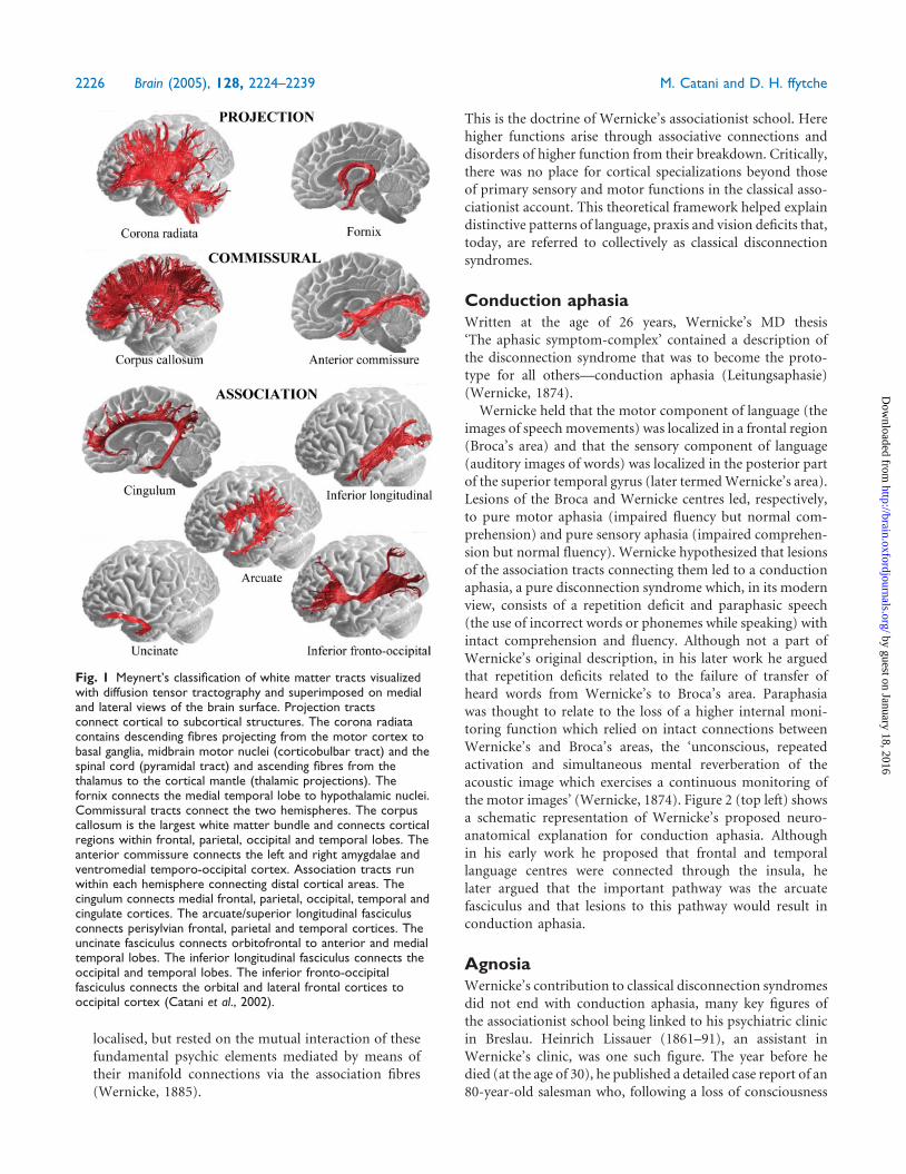
Figure 1 shows tractography reconstructions of the major
white matter tracts of the human brain as classified by
Meynert (Catani et al., 2002).
Although Meynert used his neuroanatomical findings
to develop a theory of psychological function which had
profound influence on the early development of psychiatry,
including Freud’s early work, it is Karl Wernicke (1848–1904)
who is considered the father of disconnection theory.
Wernicke conceived the brain as a mosaic-like arrangement
of areas containing ‘fundamental psychic elements’ or
‘memory images’ related to motor acts and sensory experi-
ences. These memory images areas were localized in primary
sensory and motor areas according to the following principle:
the acoustic images find their abode within the cortical
terminals of the acoustic nerve; the visual images,
within the cortical endings of the optic nerve; and
the olfactory images in that of the olfactory nerve;
and so on. Likewise the motor images or movement-
representation could be located in the cortical sites of
the motor nerve origins. (Wernicke, 1885)
Perhaps in an attempt to distance himself from phrenological
theory, Wernicke was adamant that the higher functions
were not localized in specific regions but were the result of
associative connections between motor and sensory memory
image areas. Thus, for Wernicke
Any higher psychic process, exceeding these mere
primary assumptions, could not, I reasoned, be
Disconnection syndromes Brain (2005), 128, 2224–2239 2225
by guest on January 18, 2016http://brain.oxfordjournals.org/
Dow
nloaded from
localised, but rested on the mutual interaction of these
fundamental psychic elements mediated by means of
their manifold connections via the association fibres
(Wernicke, 1885).
This is the doctrine of Wernicke’s associationist school. Here
higher functions arise through associative connections and
disorders of higher function from their breakdown. Critically,
there was no place for cortical specializations beyond those
of primary sensory and motor functions in the classical asso-
ciationist account. This theoretical framework helped explain
distinctive patterns of language, praxis and vision deficits that,
today, are referred to collectively as classical disconnection
syndromes.
Conduction aphasiaWritten at the age of 26 years, Wernicke’s MD thesis
‘The aphasic symptom-complex’ contained a description of
the disconnection syndrome that was to become the proto-
type for all others—conduction aphasia (Leitungsaphasie)
(Wernicke, 1874).
Wernicke held that the motor component of language (the
images of speech movements) was localized in a frontal region
(Broca’s area) and that the sensory component of language
(auditory images of words) was localized in the posterior part
of the superior temporal gyrus (later termed Wernicke’s area).
Lesions of the Broca and Wernicke centres led, respectively,
to pure motor aphasia (impaired fluency but normal com-
prehension) and pure sensory aphasia (impaired comprehen-
sion but normal fluency). Wernicke hypothesized that lesions
of the association tracts connecting them led to a conduction
aphasia, a pure disconnection syndrome which, in its modern
view, consists of a repetition deficit and paraphasic speech
(the use of incorrect words or phonemes while speaking) with
intact comprehension and fluency. Although not a part of
Wernicke’s original description, in his later work he argued
that repetition deficits related to the failure of transfer of
heard words from Wernicke’s to Broca’s area. Paraphasia
was thought to relate to the loss of a higher internal moni-
toring function which relied on intact connections between
Wernicke’s and Broca’s areas, the ‘unconscious, repeated
activation and simultaneous mental reverberation of the
acoustic image which exercises a continuous monitoring of
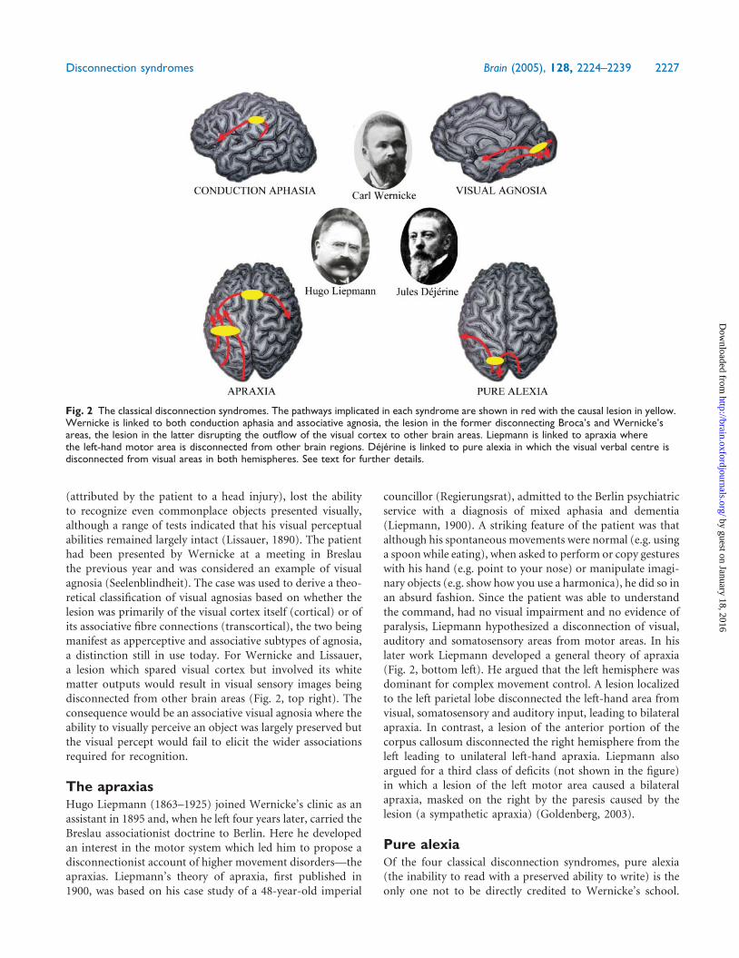
the motor images’ (Wernicke, 1874). Figure 2 (top left) shows
a schematic representation of Wernicke’s proposed neuro-
anatomical explanation for conduction aphasia. Although
in his early work he proposed that frontal and temporal
language centres were connected through the insula, he
later argued that the important pathway was the arcuate
fasciculus and that lesions to this pathway would result in
conduction aphasia.
AgnosiaWernicke’s contribution to classical disconnection syndromes
did not end with conduction aphasia, many key figures of
the associationist school being linked to his psychiatric clinic
in Breslau. Heinrich Lissauer (1861–91), an assistant in
Wernicke’s clinic, was one such figure. The year before he
died (at the age of 30), he published a detailed case report of an
80-year-old salesman who, following a loss of consciousness
Fig. 1 Meynert’s classification of white matter tracts visualizedwith diffusion tensor tractography and superimposed on medialand lateral views of the brain surface. Projection tractsconnect cortical to subcortical structures. The corona radiatacontains descending fibres projecting from the motor cortex tobasal ganglia, midbrain motor nuclei (corticobulbar tract) and thespinal cord (pyramidal tract) and ascending fibres from thethalamus to the cortical mantle (thalamic projections). Thefornix connects the medial temporal lobe to hypothalamic nuclei.Commissural tracts connect the two hemispheres. The corpuscallosum is the largest white matter bundle and connects corticalregions within frontal, parietal, occipital and temporal lobes. Theanterior commissure connects the left and right amygdalae andventromedial temporo-occipital cortex. Association tracts runwithin each hemisphere connecting distal cortical areas. Thecingulum connects medial frontal, parietal, occipital, temporal andcingulate cortices. The arcuate/superior longitudinal fasciculusconnects perisylvian frontal, parietal and temporal cortices. Theuncinate fasciculus connects orbitofrontal to anterior and medialtemporal lobes. The inferior longitudinal fasciculus connects theoccipital and temporal lobes. The inferior fronto-occipitalfasciculus connects the orbital and lateral frontal cortices tooccipital cortex (Catani et al., 2002).
2226 Brain (2005), 128, 2224–2239 M. Catani and D. H. ffytche
by guest on January 18, 2016http://brain.oxfordjournals.org/
Dow
nloaded from
(attributed by the patient to a head injury), lost the ability
to recognize even commonplace objects presented visually,
although a range of tests indicated that his visual perceptual
abilities remained largely intact (Lissauer, 1890). The patient
had been presented by Wernicke at a meeting in Breslau
the previous year and was considered an example of visual
agnosia (Seelenblindheit). The case was used to derive a theo-
retical classification of visual agnosias based on whether the
lesion was primarily of the visual cortex itself (cortical) or of
its associative fibre connections (transcortical), the two being
manifest as apperceptive and associative subtypes of agnosia,
a distinction still in use today. For Wernicke and Lissauer,
a lesion which spared visual cortex but involved its white
matter outputs would result in visual sensory images being
disconnected from other brain areas (Fig. 2, top right). The
consequence would be an associative visual agnosia where the
ability to visually perceive an object was largely preserved but
the visual percept would fail to elicit the wider associations
required for recognition.
The apraxiasHugo Liepmann (1863–1925) joined Wernicke’s clinic as an
assistant in 1895 and, when he left four years later, carried the
Breslau associationist doctrine to Berlin. Here he developed
an interest in the motor system which led him to propose a
disconnectionist account of higher movement disorders—the
apraxias. Liepmann’s theory of apraxia, first published in
1900, was based on his case study of a 48-year-old imperial
councillor (Regierungsrat), admitted to the Berlin psychiatric
service with a diagnosis of mixed aphasia and dementia
(Liepmann, 1900). A striking feature of the patient was that
although his spontaneous movements were normal (e.g. using
a spoon while eating), when asked to perform or copy gestures
with his hand (e.g. point to your nose) or manipulate imagi-
nary objects (e.g. show how you use a harmonica), he did so in
an absurd fashion. Since the patient was able to understand
the command, had no visual impairment and no evidence of
paralysis, Liepmann hypothesized a disconnection of visual,
auditory and somatosensory areas from motor areas. In his
later work Liepmann developed a general theory of apraxia
(Fig. 2, bottom left). He argued that the left hemisphere was
dominant for complex movement control. A lesion localized
to the left parietal lobe disconnected the left-hand area from
visual, somatosensory and auditory input, leading to bilateral
apraxia. In contrast, a lesion of the anterior portion of the
corpus callosum disconnected the right hemisphere from the
left leading to unilateral left-hand apraxia. Liepmann also
argued for a third class of deficits (not shown in the figure)
in which a lesion of the left motor area caused a bilateral
apraxia, masked on the right by the paresis caused by the
lesion (a sympathetic apraxia) (Goldenberg, 2003).
Pure alexiaOf the four classical disconnection syndromes, pure alexia
(the inability to read with a preserved ability to write) is the
only one not to be directly credited to Wernicke’s school.
Fig. 2 The classical disconnection syndromes. The pathways implicated in each syndrome are shown in red with the causal lesion in yellow.Wernicke is linked to both conduction aphasia and associative agnosia, the lesion in the former disconnecting Broca’s and Wernicke’sareas, the lesion in the latter disrupting the outflow of the visual cortex to other brain areas. Liepmann is linked to apraxia wherethe left-hand motor area is disconnected from other brain regions. Dejerine is linked to pure alexia in which the visual verbal centre isdisconnected from visual areas in both hemispheres. See text for further details.
Disconnection syndromes Brain (2005), 128, 2224–2239 2227
by guest on January 18, 2016http://brain.oxfordjournals.org/
Dow
nloaded from
In Paris during the 1880s, the associationist theory of language
was being taught by Charcot using a model consisting of a
network of specialized areas (the ‘bell’ diagram; Brais, 1993),
which included a visual centre for words. However, it was
Jules Dejerine (1849–1917) who proposed the first disconnec-
tionist account of pure alexia and identified the angular gyrus
as the visual centre for words. His theory was based on the
combined evidence of two case reports, the first published in
1891, the second in the following year. The first report
described a 63-year-old sailor admitted to the Bicetre hospital
in the outskirts of Paris, where Dejerine was Head of Clinical
Neurology, who lost the ability to both read and write fol-
lowing a left angular gyrus stroke (Dejerine, 1891).The second
described a 68-year-old man with right hemianopia who lost
the ability to read but whose writing was intact for 4 years
until 6 days before his death (Dejerine, 1892). The patient had
evidence of a mixture of lesions of different ages in the left
hemisphere:
recent lesions due to red softening in the inferior pari-
etal lobe and the angular gyrus. Old lesions—atrophical
yellow spots—localized in the lingual lobe, fusiform
lobe the cuneus and the tip of the occipital lobe as well
as a strongly marked atrophia of the optical radiations
in the fold of the corpus callosum. (Dejerine, 1892)
Based on these findings, Dejerine proposed that a ‘visual
verbal centre’, whose function was the storage of visual images
of words, was located in the left angular gyrus. According
to Dejerine, the alexia with agraphia of the first patient was
related to a lesion of this area. In contrast, the pure alexia of
the second was related to the longstanding left central white
matter occipital lesion which both destroyed optic radiation
inputs to left visual areas and disconnected left and right
visual areas from the visual verbal centre. The patient’s
acute angular gyrus lesion damaged the visual verbal centre
itself, adding agraphia to the clinical picture. Figure 2 (bottom
right) illustrates Dejerine’s model of pure alexia with a single
lesion affecting connections of left and right visual cortices to
the left angular gyrus and visual inputs to left hemispheric
visual cortex (inputs not shown).
The fall of the classical eraAlthough Dejerine’s model of pure alexia was strongly
influenced by the associationist school and its disconnection
syndromes, in proposing a centre in the angular gyrus
specialized for the visual representations of words, Dejerine
broke Wernicke’s cardinal rule on two counts. First, his model
suggested that higher functions could be located in cortex
rather than as the result of connections. Second, he gave
his region specialized for higher visual function (in this
case word images) a distinct anatomical locus outside visual
cortex. For Wernicke, these developments constituted a
retrograde step towards phrenology, a move away from the
associationist doctrine of higher functions as the result of
connections. In his view, visual areas only stored images of
single letters with alexia resulting from a disconnection
between visual areas and more anterior temporal and frontal
areas. Shortly before his death, Wernicke wrote an explicit
critique of Dejerine’s view (Bub et al., 1993).
Despite Wernicke’s opposition to higher functional centres,
the allure of cortical specialization proved difficult to resist.
Regions specialized for higher functions began to appear
in many accounts of clinical deficits, the view being strength-
ened by developments in anatomy with Campbell’s and
Brodmann’s division of the cortex into discrete cytoarchi-
tectonic regions (see ffytche and Catani, 2005 for a review).
By 1934, Kleist, ironically one of Wernicke’s successors in
Breslau, had published a detailed cortical map of higher
functions which had become difficult to distinguish from
phrenological maps of the previous century (Luria, 1980).
This return to narrow localizationism was easy to criticize
and the early 20th century saw the emergence of a holistic,
anti-localizationist, anti-associationist approach to higher
function that was to dominate until Geschwind. This move-
ment was led amongst others, by Henry Head in England,
who referred disparagingly to the classical school as diagram
makers, Von Monakow in Switzerland, Kurt Goldstein in
Germany and Karl Lashley in America (Geschwind, 1964).
By 1965, localization theory, disconnection syndromes and
any interest in clinicopathological correlations had been
relegated to little more than footnotes in the history of
neurology.
Geschwind’s neo-associationismIt was the work of Myers and Sperry in the 1950s that began
the shift away from pure holism which was to culminate
in Geschwind’s revived associationism. Their experiments
on the consequences of callosal section, first in animals and
then in patients with epilepsy revealed unequivocal evidence
for hemispheric localization of brain functions. The work
reawakened neurological interest in clinicopathological cor-
relations in general and led Geschwind, then in his 30s and
working in the aphasia research section of the Boston Veter-
ans Administration Hospital, to ‘re-examine the older clinical
literature and to re-assess our patients with disturbances of
the higher functions’ (Geschwind, 1965a). The outcome was
his 1965 publication which was to become a manifesto for the
neo-associationist school. In the following section we provide
a brief summary of Geschwind’s 1965 paper, a work which, in
total, spanned 116 unillustrated pages of Brain, focusing on
those areas in which he extended the classical associationist
account.
The neuroanatomical and phylogeneticfoundations of Geschwind’s theoryGeschwind added two new components to the classical view:
a forgotten principle—Flechsig’s rule—and a comparative
phylogenetic perspective to the development of associations.
Paul Flechsig (1847–1929), Professor of Psychiatry in Leipzig,
had performed detailed studies of cortical myelination at
2228 Brain (2005), 128, 2224–2239 M. Catani and D. H. ffytche
by guest on January 18, 2016http://brain.oxfordjournals.org/
Dow
nloaded from

different stages of human brain development (Fig. 3). One
consequence of this work was the emergence of a general
pattern of connections in which ‘no long association system
is known which connects two primordial zones that are to
be regarded as sensory centres’ (Flechsig, 1901). Flechsig’s
primordial zones were the cortical regions fully myelinated
at birth and corresponding to primary sensory areas. For
Flechsig, all connections between such areas were indirect
as they passed through their respective surrounding mantles
of association cortex. Although Flechsig’s anatomical obser-
vations applied to sensory cortex only, Geschwind generalized
the rule to include motor cortex and interhemispheric con-
nections (Geschwind, 1965a, b). The rule formed the corner-
stone of his theory, implying that association cortex acted, in
effect, as an obligatory relay station between different brain
regions and that a lesion to it would serve to disconnect them.
The second novel component of Geschwind’s theory
related to the phylogeny of Flechsig’s rule and the light this
shed on the evolution of higher functions. For Geschwind, the
rule was one that had emerged in primate species:
in the lower mammals connections between regions of
the cortex may arise directly from the primary receptive
or motor areas. As one moves up the phylogenetic
scale, these connections come to be made between
newly developed regions of cortex interspersed between
the older zones. These regions are called association
cortex . . . all intercortical long connections (whether in
or between hemispheres) are made by way of these
association areas and not between the primary motor
or receptive areas. (Geschwind, 1965a)
The implication was that the evolution of association cortex
underlay the evolution of higher function, the trend continu-
ing with the evolution of non-human primates to man where
the key anatomical development was the emergence of a
higher-order association area in the human parietal lobe.
This new area allowed a uniquely human pattern of connec-
tions between sense modalities that did not depend on the
limbic system:
While connections between primary receptive regions
and limbic structures are powerful in subhuman forms,
intermodal connections between vision, audition and
somesthesis are probably weak in these animals. . . . In
man the situation changes with the development of the
association areas of the human inferior parietal lobule,
situated at the junction of the older association
areas attached to the visual, somesthetic, and auditory
regions. It is speculated that this new ‘association area
of association areas’ now frees man from the dominant
pattern of sensory-limbic associations and permits
cross-modal associations involving non-limbic
modalities. (Geschwind, 1965a)
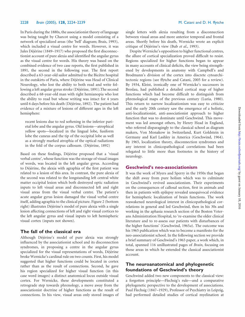
Figure 4 summarizes Geschwind’s evolutionary perspective.
In the rabbit primary sensory cortices of different modalities
are connected both directly and through the limbic system. In
the monkey, with the evolution of Flechsig’s rule, the limbic
system continues to play an important role in connecting
different sensory modalities; however, connections to the
limbic cortex now arise from the mantle of association cortex
surrounding primary sensory areas. In man, intermodality
connections are freed from the limbic system through
the development of the inferior parietal lobe (the angular
and supramarginal gyri), an area connecting visual, auditory
and somatosensory association areas. For Geschwind, the
area and its multisensory connections played a particular
role in language development:
it is only in man that associations between two non-
limbic stimuli are readily formed and it is this ability
which underlies the learning of names of objects. . . .The angular gyrus is important in the process of
associating a heard name to a seen or felt object, it is
probably also important for associations in the reverse
direction. A name passes through Wernicke’s area, then
via the angular gyrus arouses associations in the other
parts of the brain. It is probably thus that Wernicke’s
area attains it essential importance in comprehension
i.e. the arousal of association. (Geschwind, 1965a)
In a sense, by highlighting the importance of the angular gyrus
in language, Geschwind was returning to Dejerine’s model.
However, the role of the angular gyrus was very different
for Dejerine and Geschwind. Dejerine viewed the region as
one storing visual memories of letters and words, whereas
Geschwind viewed it as having a more general function in
Fig. 3 Flechsig’s myelogenetic map of human cortex. The numbering of each region refers to its chronological order of development.Those shown in colour are myelinated at birth and constitute his primordial zones (Flechsig, 1901).
Disconnection syndromes Brain (2005), 128, 2224–2239 2229
by guest on January 18, 2016http://brain.oxfordjournals.org/
Dow
nloaded from
forming multimodality associations, a prerequisite function
for language and semantics.
Geschwind’s disconnection syndromesFor Wernicke and his school, disconnection and its syn-
dromes had implied a white matter lesion to the association
tracts connecting two areas. For Geschwind, basing his argu-
ment on Flechsig’s rule, disconnection syndromes went
beyond this to imply a lesion of association cortex itself,
particularly that in the parietal lobe. In Geschwind’s 1965
model, even a pure lesion of association cortex could cause
a disconnection syndrome, little distinction being made
between such lesions and those restricted to white matter
tracts.
lesions of association cortex, if extensive enough, act to
disconnect primary receptive or motor areas from other
regions of the cortex in the same or in the opposite
hemisphere. . . . Thus a ‘disconnexion lesion’ will be
a large lesion either of association cortex or of the
white matter leading from this association cortex.
(Geschwind, 1965a)
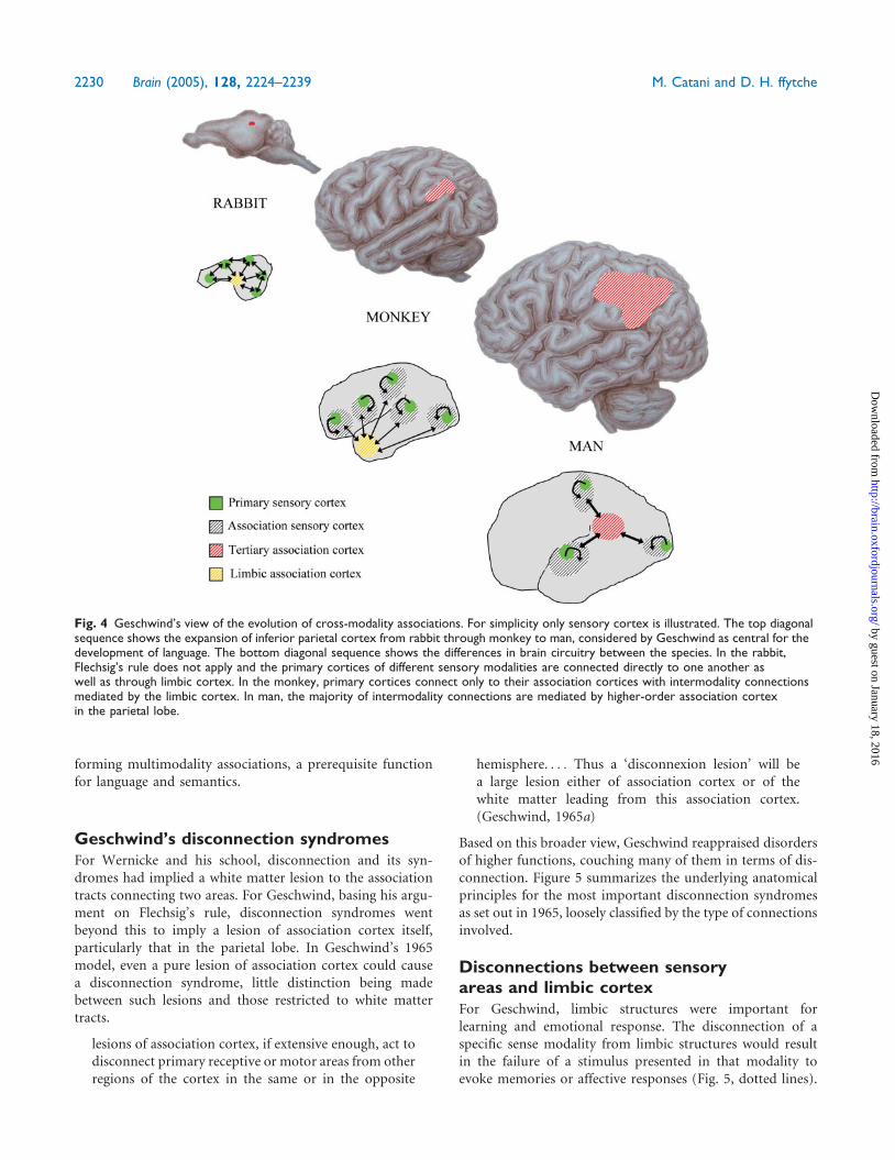
Based on this broader view, Geschwind reappraised disorders
of higher functions, couching many of them in terms of dis-
connection. Figure 5 summarizes the underlying anatomical
principles for the most important disconnection syndromes
as set out in 1965, loosely classified by the type of connections
involved.
Disconnections between sensoryareas and limbic cortexFor Geschwind, limbic structures were important for
learning and emotional response. The disconnection of a
specific sense modality from limbic structures would result
in the failure of a stimulus presented in that modality to
evoke memories or affective responses (Fig. 5, dotted lines).
Fig. 4 Geschwind’s view of the evolution of cross-modality associations. For simplicity only sensory cortex is illustrated. The top diagonalsequence shows the expansion of inferior parietal cortex from rabbit through monkey to man, considered by Geschwind as central for thedevelopment of language. The bottom diagonal sequence shows the differences in brain circuitry between the species. In the rabbit,Flechsig’s rule does not apply and the primary cortices of different sensory modalities are connected directly to one another aswell as through limbic cortex. In the monkey, primary cortices connect only to their association cortices with intermodality connectionsmediated by the limbic cortex. In man, the majority of intermodality connections are mediated by higher-order association cortexin the parietal lobe.
2230 Brain (2005), 128, 2224–2239 M. Catani and D. H. ffytche
by guest on January 18, 2016http://brain.oxfordjournals.org/
Dow
nloaded from
Lesions causing such syndromes were located in limbic
association cortex (e.g. temporal pole, parahippocampal
gyrus and insula) and less commonly, sensory association
cortex or the white matter tracts connecting the two. In
man, such sensory–limbic disconnection occurred in the
somatosensory and auditory but not visual systems. Dis-
connection of somatosensory cortex from the limbic lobe
resulted in pain asymbolia (no response to pain in the
presence of normal tactile discriminatory function). Dis-
connection of auditory cortex from the limbic lobe resulted
in a range of symptoms (verbal learning impairment,
euphoria and symptom denial) commonly found in patients
with Wernicke’s aphasia. In the visual system, the develop-
ment of an indirect connecting pathway between visual
and limbic structures through the inferior parietal lobe
meant that symptoms commonly seen in non-human pri-
mates with visual–limbic disconnections did not occur in
man (Geschwind, 1965a).
Disconnections between sensoryareas and Wernicke’s areaDisconnection of Wernicke’s area from specific sensory areas
would lead to modality-specific language deficits, caused
either by a lesion of direct connections between Wernicke’s
and sensory areas or by a lesion of indirect connections
through the angular gyrus (Fig. 5, solid lines). Geschwind
distinguished four such syndromes. The first consisted of
tactile aphasia (the inability to name a held object in the
presence of preserved speech and naming in other sense
modalities), today referred to as tactile anomia. The second
consisted of pure word deafness, a syndrome originally
described by Liepmann, an inability to understand spoken
words in the presence of preserved hearing (Liepmann,
1898). The third consisted of pure alexia for which Geschwind
used Dejerine’s explanatory model. The fourth consisted of
modality-specific agnosias (inabilities to recognize objects
in the presence of intact elementary sensation). Geschwind
focused on visual agnosia, his account differing from the
classical Lissauer–Wernicke model in its emphasis on lan-
guage. For Geschwind, both perception and language were
intact in visual agnosia, the deficit arising from a failure of
their communication. He argued that visual agnosia was a
highly isolated disturbance of naming, the result of a dis-
connection of visual areas from the inferior parietal lobe,
and hence indirectly to Wernicke’s area, with the con-
sequent inability of a sensory percept to arouse language
associations.
Fig. 5 Geschwind’s disconnection syndromes. The pathways implicated in the principle syndromes described by Geschwind, classifiedinto three types: sensory–limbic disconnection syndromes (dotted lines), sensory–motor disconnection syndromes (dashed lines);sensory–Wernicke’s area disconnection syndromes (solid lines). See text for further details.
Disconnection syndromes Brain (2005), 128, 2224–2239 2231
by guest on January 18, 2016http://brain.oxfordjournals.org/
Dow
nloaded from
Disconnections between sensory areasand motor cortexIn the left hemisphere, disconnections of the hand motor
cortex from posterior sensory areas or Broca’s area from
Wernicke’s area resulted in apraxia and conduction aphasia
respectively (Fig. 5, dashed line). Geschwind’s account of
apraxia adhered faithfully to Liepmann’s classical explana-
tion. His account of conduction aphasia added to Wernicke’s
model in that he suspected the preserved ability of patients
with conduction aphasia to produce semantically related
words when asked to repeat was related to indirect connec-
tions through the angular gyrus (a semantic detour)
(Geschwind, 1965b).
Disconnection between the hemispheresFor convenience, Fig. 5 has illustrated Geschwind’s dis-
connection syndromes from the perspective of the left hemi-
sphere; however, his 1965 paper emphasized the importance
of interhemispheric disconnection, although it did not con-
stitute a separate ‘callosal’ disconnection syndrome. The role
of callosal disconnection in his model was the same as that put
forward by Dejerine and Liepmann in their respective models
of pure alexia and apraxia.
Disconnection syndromesafter GeschwindIn a sense, Geschwind became for the 20th century what
Wernicke was for the 19th century, a generation of neuro-
logists influenced by his approach, among them Marsel
Mesulam, Antonio Damasio, Frank Benson and Albert
Galaburda, all of whom passed through Geschwind’s neuro-
logy department in Boston (Damasio and Galaburda 1985;
Mesulam, 1985b). (Geschwind and Wernicke also share in
common the fact that their family origins were <150 miles
apart in what is now southern Poland and that both died
prematurely in their 50s.) He was not simply Wernicke’s,
Liepmann’s and Dejerine’s translator across centuries and
languages, his own contribution being to enrich the classical
associationist view with the idea of association cortex as an
obligatory relay, the theory of hierarchies of associations
within the inferior parietal lobe and the importance of this
region in the phylogeny and ontogeny of language. However,
Geschwind’s 1965 framework held most, if not all, disorders
of higher function to be related to disconnection and although
the theory did not fall after its publication, with time it
became integrated into a more general model in which the
specializations of association cortex were given equal footing
with corticocortical connections (see below). This evolution
to a more general account took two separate but closely
related paths, both delineated in Geschwind’s 1965 Brain
paper. One path used as its starting point brain lesion loca-
tion, capitalizing on the emerging imaging techniques of com-
puterized tomography, PET and SPET scanning. This was the
path taken by Damasio, leading to important insights into the
neural mechanisms of perception (Damasio, 1985), language
(Damasio and Damasio, 1983; Damasio and Geschwind,
1984) and emotion (Damasio, 1999, Damasio et al., 2000).
The second path used as its starting point the connections of
brain regions, capitalizing on emerging techniques for tracing
neural connections, primate neurophysiology and computa-
tional theory. This was the path taken by Mesulam, leading to
important insights into the dynamic brain networks under-
lying memory, attention and language (Mesulam, 1990b), the
neural mechanisms of perception (Mesulam, 1998), neglect
(Mesulam, 1981), aphasia (Mesulam, 2001) and cholinergic
pathways (Mesulam, 1990a; Selden et al., 1998). Although
Geschwind never returned to the grand perspective of his
1965 theory, over the next 20 years he continued to work
on clinicopathological correlations from both of these per-
spectives with members of his school, extending the remit of
his 1965 model beyond classical neurology into neurodevel-
opmental disorders, such as dyslexia. Together with his 1965
paper, this corpus of work founded behavioural neurology,
strongly influenced the development of related disciplines,
such as neuropsychiatry and neuropsychology and prepared
the ground for neural network and connectionist theories in
the cognitive sciences.
The contemporary neuroanatomicalbasis of higher brain functionsGeschwind’s 1965 anatomical framework for higher function
and its disorders, although correct in many respects, also had
weaknesses which he himself and members of his school
amended in later years as new evidence became available.
Although admitting it a simplification, Geschwind held
in 1965 that association cortex was a little more than a homo-
geneous relay station between primary sensory and motor
areas with no specialized roles of its own. That this homo-
geneous view might be wrong became increasingly apparent
in the 1960s and 1970s with anatomical and neurophysiologi-
cal work in the monkey and clinical evidence in man and by
1985, both Mesulam and Damasio had incorporated
specific functional roles for association cortex into their neu-
roanatomical accounts of higher function (Damasio, 1985;
Mesulam, 1985a). The functional subdivision of association
cortex was later confirmed in man using functional imaging
techniques (Zeki et al., 1991). Such cortical specialization
is more complex than envisaged by classical localizationist
theory. What were once considered localized centres (e.g.
Broca’s and Wernicke’s areas) are today held to be extended
territories composed of many specialized cortical subregions
serving different but related functions (see for example
Cannestra et al., 2000; Boatman, 2004; Damasio et al., 2004;
Hickok and Poeppel, 2004 for evidence in the language
domain). A further weakness of Geschwind’s 1965 position
was the predominantly feed forward, serial nature of his
account. Information passed from posterior sensory cortices,
through hierarchies of association cortices to limbic and ante-
rior frontal cortices in a serial fashion with little attention
2232 Brain (2005), 128, 2224–2239 M. Catani and D. H. ffytche
by guest on January 18, 2016http://brain.oxfordjournals.org/
Dow
nloaded from
being paid to the functional implications of feed-back and
parallel pathways. The contributions to higher function
made by these anatomical features were later emphasized
by Damasio in his retroactivation model (Damasio, 1989;
Damasio and Damasio, 1994) and Mesulam in his large-
scale network model (Mesulam, 1990b). The presence in
man of parallel, bidirectional and distributed processing for
higher functions has recently been confirmed (for example,
in the domain of language see Matsumoto et al., 2004; Bitan
et al., 2005; Catani et al., 2005). The contemporary frame-
work for clinicopathological correlations that evolved from
Geschwind’s 1965 model thus consists of a network of
multiple specialized cortical areas, grouped into territories
and connected through parallel, bidirectional pathways. In
this model, two key elements underlie higher function deficits:
(i) the loss of specialized cortical function and (ii) damage
to connecting pathways. Of these, in man, the former is better
understood than the latter as brain lesion studies and
functional imaging have helped advance our understanding
of cortical specialization and its related deficits. In contrast,
our anatomical knowledge of connecting pathways has, until
recently, relied on primate tracing studies (Mesulam, 2005).
An important consequence of this limitation is that it has
allowed for hypothetical post hoc disconnection explanations
of higher function deficits. For a given lesion, the absence of
a predicted deficit (or for a given deficit, the absence of a
predicted lesion) is amenable to explanation by any number
of alternative pathways making it difficult to test or falsify the
disconnectionist account.
The advent of new techniques is beginning to redress the
imbalance between our knowledge of the cortex and that of
its connections. Tractography, although lacking the anato-
mical precision of post mortem tracing, is helping advance
our understanding of human white matter anatomy and may
have an important contribution to make in testing the dis-
connection paradigm. Other techniques exploring mathe-
matical ‘connections’ between areas in electrophysiological
and neuroimaging data provide complementary evidence
on the functional roles of such connections. Together these
techniques allow us to broaden the remit of the contemporary
clinicopathological correlation framework beyond disorders
of disconnection and cortical deficit to include disorders
of hyperconnection and cortical hyperfunction. In the final
part of this review we accommodate the evidence of these
emerging techniques within the existing framework, illustrat-
ing how we envisage the updated framework can be used
clinically with specific tractography-derived examples.
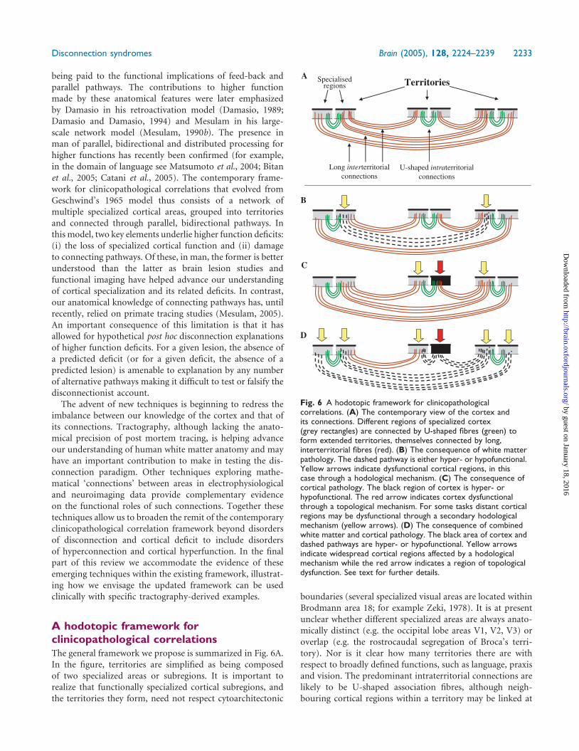
A hodotopic framework forclinicopathological correlationsThe general framework we propose is summarized in Fig. 6A.
In the figure, territories are simplified as being composed
of two specialized areas or subregions. It is important to
realize that functionally specialized cortical subregions, and
the territories they form, need not respect cytoarchitectonic
boundaries (several specialized visual areas are located within
Brodmann area 18; for example Zeki, 1978). It is at present
unclear whether different specialized areas are always anato-
mically distinct (e.g. the occipital lobe areas V1, V2, V3) or
overlap (e.g. the rostrocaudal segregation of Broca’s terri-
tory). Nor is it clear how many territories there are with
respect to broadly defined functions, such as language, praxis
and vision. The predominant intraterritorial connections are
likely to be U-shaped association fibres, although neigh-
bouring cortical regions within a territory may be linked at
TerritoriesSpecialised regions
U-shaped intraterritorial connections
Long interterritorial connections
B
C
A
D
Fig. 6 A hodotopic framework for clinicopathologicalcorrelations. (A) The contemporary view of the cortex andits connections. Different regions of specialized cortex(grey rectangles) are connected by U-shaped fibres (green) toform extended territories, themselves connected by long,interterritorial fibres (red). (B) The consequence of white matterpathology. The dashed pathway is either hyper- or hypofunctional.Yellow arrows indicate dysfunctional cortical regions, in thiscase through a hodological mechanism. (C) The consequence ofcortical pathology. The black region of cortex is hyper- orhypofunctional. The red arrow indicates cortex dysfunctionalthrough a topological mechanism. For some tasks distant corticalregions may be dysfunctional through a secondary hodologicalmechanism (yellow arrows). (D) The consequence of combinedwhite matter and cortical pathology. The black area of cortex anddashed pathways are hyper- or hypofunctional. Yellow arrowsindicate widespread cortical regions affected by a hodologicalmechanism while the red arrow indicates a region of topologicaldysfunction. See text for further details.
Disconnection syndromes Brain (2005), 128, 2224–2239 2233
by guest on January 18, 2016http://brain.oxfordjournals.org/
Dow
nloaded from
their border by intracortical fibres (not shown in the figure).
Tractography evidence suggests that territories are connected
by multiple parallel long association fibre pathways (Catani
et al., 2005). Although the anatomical details of these intert-
erritorial connections are yet to be worked out, existing data
suggest that they connect specific specialized subregions in
different territories in parallel.
What are the functional consequences of pathology in this
updated model? Here we introduce a new terminology to
allow us to extend the model beyond classical neurological
disorders of white matter disconnections and cortical deficits
to include those involving white matter hyperconnectivity
and cortical hyperfunction. According to the new termino-
logy, a topological mechanism (from the Greek topos = place)
refers generally to a dysfunction of the cortex irrespective of
whether the dysfunction is one of deficit, hyperfunction or
a combination of the two. Topologically-related dysfunctions
range from deficits related to the loss of a specialized cortical
region, e.g. prosopagnosia from lesions of face-specialized
cortex in the fusiform gyrus (Sergent et al., 1992), to specific
positive symptoms, such as face hallucinations related to
the hyperexcitibility and spontaneous activation of face-
specialized cortex (ffytche et al., 1998). A hodological mecha-
nism (from the Greek hodos = road or path) refers generally
to dysfunction related to connecting pathways, irrespective of
whether the dysfunction is one of disconnection, hypercon-
nection or a combination of the two. Hodologically-related
dysfunctions range from classical disconnection syndromes
such as the conduction aphasia of pure subcortical lesions
(Naeser et al., 1982) to the combination of frontofrontal
hyperconnectivity with frontal disconnection from other
brain regions in autism (Courchesne and Pierce, 2005).
Figure 6B–D illustrates the different patterns of cortical
dysfunction that occur as the result of pathologies within
this hodotopic framework. The first pattern relates to a
pathology of white matter pathways, such as might be caused
by localized vascular, neoplastic or demyelinating lesions
leading to disconnection or to hyperconnectivity, e.g. through
a failure to prune connections (Fig. 6B). Distant cortical
regions connected by the affected pathway are dysfunctional
as the consequence of pure hodological effects (the yellow
arrows in the figure). For some tasks this dysfunction may
simply reflect the failure or excess of transfer of outputs from
one area to another; however, for tasks requiring the simul-
taneous cooperation of cortical regions (e.g. synchronous
bimanual coordination) one can consider the function itself
to be distributed (a functional loop), the lesion disrupting or
enhancing the function as a whole. Whether for a serial or
distributed task, the dysfunction of connected regions may
only be apparent for those tasks requiring both connected
areas, the function of each area individually being normal
when they form a part of a different task network (a dynamic
diaschisis; Price et al., 2001). Cortical pathology causes a
different pattern dysfunction (Fig. 6C). Here cortical lesions
or hyperactivity lead to dysfunction through a topological
mechanism (red arrow). The dysfunction may go beyond
the affected cortical site to include cortical regions connected
to it. These effects arise through a hodological mechanism
but differ from those described above in that the white matter
is not directly affected. One may wish to refer to these remote
effects as secondary hodological (yellow arrows). As for pure
hodologically based dysfunction, these remote effects will
demonstrate a dynamic diaschisis, only being apparent for
tasks normally requiring both regions. Both neuroimaging
(Kempler et al., 1988; Price et al., 2001) and clinical
(De Renzi and Vignolo, 1962) studies of the language network
have found evidence for these remote functional effects. Focal
pure cortical lesions are rare and include superficial vascular
or neoplastic lesions which spare long associative fibres
(whether or not such lesions encroach superficial white matter
makes little difference to the ensuing pattern of deficit).
An equivalent pattern of deficits will also arise in a range
of localized neurodegenerative disorders, such as posterior
cortical atrophy, primary progressive aphasia and fronto-
temporal dementia. Pure topological hyperactivity occurs
in conditions such as sensory deafferentation (Burke, 2002)
and focal seizures. The third type of dysfunction involves both
hodological and topological mechanisms (Fig. 6D). In relation
to deficits, this is the pattern most likely to be encountered
clinically and would typically be caused by a stroke, tumour or
brain injury. Here the lesion involves both cortical and sub-
cortical structures with superficial and deep white matter
affected. In this case, combined topological (red arrow)
and hodological effects (yellow arrows) produce widespread
cortical dysfunction. Auditory hallucinations in schizo-
phrenia provide an example of combined hodological and
topological hyperfunction, with increased activation of
Broca’s, Wernicke’s and Geschwind’s territory (Lennox
et al., 2000; Shergill et al., 2000) and indirect, diffusion tensor
tractography evidence of increased anatomical connectivity
between these regions (Hubl et al., 2004).
We acknowledge that the hodotopic framework is an
oversimplification and merely follows the now 40-year tradi-
tion of incorporating new anatomical and functional details
into Geschwind’s scheme as they become available. However,
we hope it adds clinically useful features to existing models,
in particular, extending them beyond classical neurological
deficits and disconnections to encompass a broader range of
disorders. Of course, its main clinical usefulness will come
not from the generalizations outlined above, but from its
application to specific functional domains. In some domains,
our understanding of cortical and white matter anatomy is
already sufficient to provide a sketch of the type of account
we envisage. The following three examples help illustrate the
contribution we hope the hodotopic framework will eventu-
ally make to the classification and assessment of patients with
disorders of higher function.
Language network disordersFigure 7 shows our recent tractography reconstruction of the
parallel perisylvian language network. One pathway connects
2234 Brain (2005), 128, 2224–2239 M. Catani and D. H. ffytche
by guest on January 18, 2016http://brain.oxfordjournals.org/
Dow
nloaded from

Broca’s and Wernicke’s territory directly (the long segment)
and corresponds to the classical arcuate fasciculus (shown in
red). An additional parallel indirect pathway between Broca’s
and Wernicke’s territory passes through the inferior parietal
lobe, a region we named after Geschwind (Catani et al., 2005).
This indirect connection consists of a posterior segment con-
necting temporal and parietal cortex (shown in yellow) and an
anterior segment connecting parietal and frontal cortex
(shown in green). The cortical terminations of these pathways
are shown superimposed on a parasagittal anisotropy image,
the three projection zones corresponding to Broca’s territory
(inferior part of the precentral gyrus and posterior part of the
middle and inferior frontal gyri), Geschwind’s territory
(angular and supramarginal gyri) and Wernicke’s territory
(posterior part of the superior and middle temporal gyri).
Although evidence is not yet available, it is likely that each
of these connecting pathways plays a different functional role,
the direct pathway being involved in phonologically based
language functions such as repetition, the indirect pathway
being involved in semantically based language functions, such
as auditory comprehension (posterior segment) and vocaliza-
tion of semantic content (anterior segment) (Catani et al.,
2005). From the perspective of our hodotopic model, lesions
affecting different territories, white matter segments or their
combination would be expected to cause different types of
language deficit as illustrated by the following scenarios in
relation to a lesion in the inferior parietal lobe. Two scenarios
result from a pure hodological mechanism: (i) If the lesion is
purely subcortical, affecting the long segment only, we would
expect to find a classical conduction aphasia with a repetition
deficit in the presence of normal auditory comprehension and
verbal fluency. (ii) If the subcortical lesion affects both direct
and indirect pathways we would expect to find a global apha-
sia despite intact cortex (Naeser et al., 2005). In contrast,
a cortical lesion encroaching on Geschwind’s territory
would be expected to produce a pattern of deficit which
varied depending on which cortical subregions are affected:
(i) If involving only the anterior portions of Geschwind’s
territory (the cortical endstation of the anterior segment),
the syndrome will be one of non-fluent aphasia with spared
repetition and comprehension (see Basso et al., 1985 for an
example of this deficit pattern with a retrorolandic lesion).
(ii) If involving all of Geschwind’s territory (the cortical end-
station of both anterior and posterior segments), the deficit
will be one of a mixed transcortical aphasia with normal
repetition but both reduced verbal fluency and comprehen-
sion. (iii) The same lesion extending into the deep white
matter would present with a global aphasia with impaired
repetition, fluency and comprehension. Hyperfunction invol-
ving different territories, segments and their combination
would also be expected to cause a heterogeneity of positive
symptoms with, for example, hyperfunction in the indirect
pathway causing semantically based symptoms and hyper-
function in the direct pathway causing disorders of excessive
repetition (e.g. the echolalia of autism). Although only a few
studies have been performed to test this prediction, there is
already evidence of specific indirect pathway hyperfunction
in schizophrenic patients with auditory hallucinations
(Lennox et al., 2000; Hubl et al., 2004).
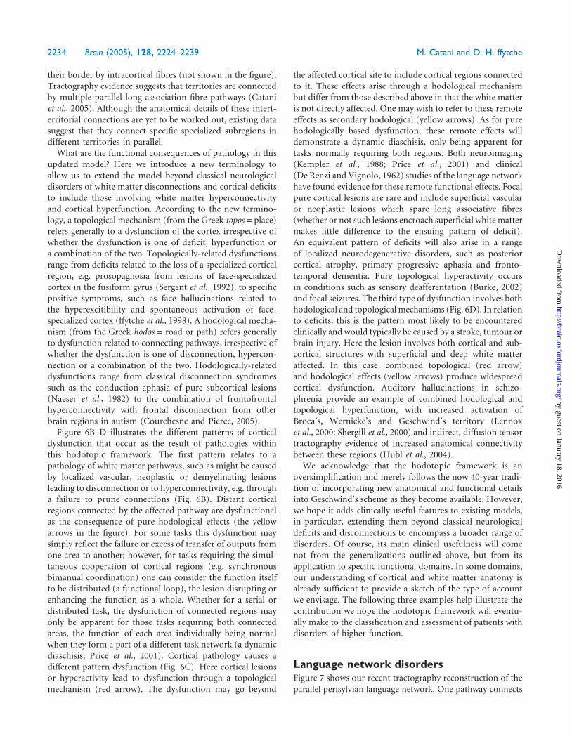
Praxis network disordersFigure 8 illustrates the frontoparietal network thought to
underlie praxis. The contemporary view of praxis derived
from work in the monkey is of multiple functions related
to different body parts (e.g. the trunk, face or limb) and
motor acts (e.g. reaching/grasping, motor sequencing and
posture in relation to the limb) (Rizzolatti et al., 1998).
Each of these praxic functions has distinct cortical loci and
connecting circuitry. At least three sets of fibre connections
equivalent to those underlying praxis in the monkey can be
identified in the human brain. The dorsolateral frontoparietal
Fig. 7 The parallel perisylvian language network. Top panel:a 3D reconstruction of the direct and indirect perisylvianpathways in the left hemisphere (lateral view) derived from anaverage brain. The long segment fibres (red) connect Broca’s andWernicke’s territories. The anterior segment fibres (green)connect Broca’s and Geschwind’s territories. The posteriorsegment fibres (yellow) connect Wernicke’s and Geschwind’sterritories. Bottom panel: a parasagittal section through thediffusion tensor anisotropy volume with fibre tracts coregistered.The terminal portions of all three segments are displayed in red.Adapted from Catani et al. (2005).
Disconnection syndromes Brain (2005), 128, 2224–2239 2235
by guest on January 18, 2016http://brain.oxfordjournals.org/
Dow
nloaded from
set connects the dorsal motor and premotor cortex to the
superior parietal lobe (green fibres); the ventrolateral fron-
toparietal set connects the ventrolateral motor and premotor
cortex to the inferior parietal lobe (red fibres) and the medial
frontoparietal set connects the medial frontal lobe to the
medial parietal lobe (precuneus) (yellow fibres). From the
perspective of our hodotopic model, a lesion involving the
cortical territories and white matter connections involved in
praxis will have different effects depending on its cortical
location and subcortical extension. If the lesion is restricted
to superior parietal cortex, its consequence will depend on
which praxic subfunctions within the territory are affected. If
the lesion extends into white matter, additional abnormalities
would be expected (see Leiguarda and Marsden, 2000 for
a review of clinicopathological correlations in apraxia). The
hyperfunctional and hyperconnectivity disorders of this
network are at present unclear.
Visual network disordersAlthough Geschwind’s 1965 model held all agnosias as dis-
connections between visual and language areas, today visual
agnosia is often considered a family of disorders, each related
to a specific deficit of visual form perception (e.g. faces or
objects). This cortical deficit account is partly a translation to
the domain of visual form perception of the model used
to explain deficits of other visual attributes, such as colour
(achromatopsia) or motion (akinetopsia) (Damasio, 1985;
Zeki, 1990, 1991) and has been applied to prosopagnosia
and alexia (Sergent et al., 1992; Leff et al., 2001). Whether
both accounts of the associative agnosias (cortical deficit or
disconnection) are correct remains to be established; what is
certain is that the white matter connections of visual areas are
complex and the contribution they make to visual perception
and its deficits is not fully understood. Tractography has
recently helped delineate occipitotemporal connections in
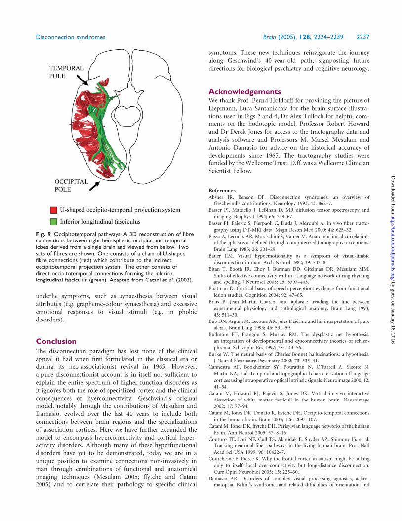
the human brain (Catani et al., 2003), shown in Fig. 9. As
in the monkey (Tusa and Ungerleider, 1985), visually
specialized cortical areas in the human brain are connected
by chains of U-shaped fibres, a subset of which forms the
occipitotemporal projection system. Here we have illustrated
a chain on the lateral surface (red fibres) but equivalent chains
are found on the ventral surface. Recent tractography
evidence confirms the existence in man of a parallel, direct
occipitotemporal pathway (the inferior longitudinal fascic-
ulus, green fibres) connecting prestriate cortex to medial
temporal structures (the hippocampus, parahippocampal
gyrus and amygdala) (Catani et al., 2003). Although the dif-
ferent functions of these two sets of connections is unclear,
it seems likely that they differ, one possibility being that the
indirect system relates to visual perceptual qualities and the
direct with emotional qualities and visual memory. From
the perspective of our hodotopic model, a ventral or lateral
temporal cortical lesion would lead to specific deficits related
to the cortical specializations lost, an extension into medial
white matter would lead, in addition, to visual hypoemotion-
ality (a deficit of visually evoked emotions with preserved
emotional responses to non-visual stimuli; see Bauer, 1982
for an example) or visual amnesia (a deficit of registering
novel visual experiences in short term memory with the pre-
served ability to register non-visual images; see Ross, 1980
for an example). Hyperfunction of specialized visual cortical
areas is associated with hallucinations of specific visual
attributes (ffytche et al., 1998; ffytche and Howard, 1999).
The consequences of hyperconnectivity within the indirect
or direct occipitotemporal pathways are unclear but may
Fig. 8 Praxic frontoparietal circuitry. A 3D reconstruction of thefibre connections between parietal and frontal lobes in the lefthemisphere derived from a single brain and shown from the left(top panel) and top of the brain (bottom panel). The pathwayshave been defined using a two-region of interest approach(Catani et al., 2002). One pair of regions was used to identifydorsolateral fibres connecting the superior parietal lobe anddorsal motor and premotor cortex (green fibres). Another pair ofregions was used to identify ventrolateral fibres connecting theinferior parietal lobe and ventrolateral motor and premotorcortex. The third pair of regions was used to identify dorsomedialfibres connecting the medial parietal lobe with the medialfrontal lobe. The dotted white line is the central sulcus.
2236 Brain (2005), 128, 2224–2239 M. Catani and D. H. ffytche
by guest on January 18, 2016http://brain.oxfordjournals.org/
Dow
nloaded from
underlie symptoms, such as synaesthesia between visual
attributes (e.g. grapheme-colour synaesthesia) and excessive
emotional responses to visual stimuli (e.g. in phobic
disorders).
ConclusionThe disconnection paradigm has lost none of the clinical
appeal it had when first formulated in the classical era or
during its neo-associationist revival in 1965. However,
a pure disconnectionist account is in itself not sufficient to
explain the entire spectrum of higher function disorders as
it ignores both the role of specialized cortex and the clinical
consequences of hyerconnectivity. Geschwind’s original
model, notably through the contributions of Mesulam and
Damasio, evolved over the last 40 years to include both
connections between brain regions and the specializations
of association cortices. Here we have further expanded the
model to encompass hyperconnectivity and cortical hyper-
activity disorders. Although many of these hyperfunctional
disorders have yet to be demonstrated, today we are in a
unique position to examine connections non-invasively in
man through combinations of functional and anatomical
imaging techniques (Mesulam 2005; ffytche and Catani
2005) and to correlate their pathology to specific clinical
symptoms. These new techniques reinvigorate the journey
along Geschwind’s 40-year-old path, signposting future
directions for biological psychiatry and cognitive neurology.
AcknowledgementsWe thank Prof. Bernd Holdorff for providing the picture of
Liepmann, Luca Santanicchia for the brain surface illustra-
tions used in Figs 2 and 4, Dr Alex Tulloch for helpful com-
ments on the hodotopic model, Professor Robert Howard
and Dr Derek Jones for access to the tractography data and
analysis software and Professors M. Marsel Mesulam and
Antonio Damasio for advice on the historical accuracy of
developments since 1965. The tractography studies were
funded by the Wellcome Trust. D.ff. was a Wellcome Clinician
Scientist Fellow.
References
Absher JR, Benson DF. Disconnection syndromes: an overview of
Geschwind’s contributions. Neurology 1993; 43: 862–7.
Basser PJ, Mattiello J, LeBihan D. MR diffusion tensor spectroscopy and
imaging. Biophys J 1994; 66: 259–67.
Basser PJ, Pajevic S, Pierpaoli C, Duda J, Aldroubi A. In vivo fiber tracto-
graphy using DT-MRI data. Magn Reson Med 2000; 44: 625–32.
Basso A, Lecours AR, Moraschini S, Vanier M. Anatomoclinical correlations
of the aphasias as defined through computerized tomography: exceptions.
Brain Lang 1985; 26: 201–29.
Bauer RM. Visual hypoemotionality as a symptom of visual-limbic
disconnection in man. Arch Neurol 1982; 39: 702–8.
Bitan T, Booth JR, Choy J, Burman DD, Gitelman DR, Mesulam MM.
Shifts of effective connectivity within a language network during rhyming
and spelling. J Neurosci 2005; 25: 5397–403.
Boatman D. Cortical bases of speech perception: evidence from functional
lesion studies. Cognition 2004; 92: 47–65.
Brais B. Jean Martin Charcot and aphasia: treading the line between
experimental physiology and pathological anatomy. Brain Lang 1993;
45: 511–30.
Bub DN, Arguin M, Lecours AR. Jules Dejerine and his interpretation of pure
alexia. Brain Lang 1993; 45: 531–59.
Bullmore ET, Frangou S, Murray RM. The dysplastic net hypothesis:
an integration of developmental and dysconnectivity theories of schizo-
phrenia. Schizophr Res 1997; 28: 143–56.
Burke W. The neural basis of Charles Bonnet hallucinations: a hypothesis.
J Neurol Neurosurg Psychiatry 2002; 73: 535–41.
Cannestra AF, Bookheimer SY, Pouratian N, O’Farrell A, Sicotte N,
Martin NA, et al. Temporal and topographical characterization of language
cortices using intraoperative optical intrinsic signals. Neuroimage 2000; 12:
41–54.
Catani M, Howard RJ, Pajevic S, Jones DK. Virtual in vivo interactive
dissection of white matter fasciculi in the human brain. Neuroimage
2002; 17: 77–94.
Catani M, Jones DK, Donato R, ffytche DH. Occipito-temporal connections
in the human brain. Brain 2003; 126: 2093–107.
Catani M, Jones DK, ffytche DH. Perisylvian language networks of the human
brain. Ann Neurol 2005; 57: 8–16.
Conturo TE, Lori NF, Cull TS, Akbudak E, Snyder AZ, Shimony JS, et al.
Tracking neuronal fiber pathways in the living human brain. Proc Natl
Acad Sci USA 1999; 96: 10422–7.
Courchesne E, Pierce K. Why the frontal cortex in autism might be talking
only to itself: local over-connectivity but long-distance disconnection.
Curr Opin Neurobiol 2005; 15: 225–30.
Damasio AR. Disorders of complex visual processing agnosias, achro-
matopsia, Balint’s syndrome, and related difficulties of orientation and
Fig. 9 Occipitotemporal pathways. A 3D reconstruction of fibreconnections between right hemispheric occipital and temporallobes derived from a single brain and viewed from below. Twosets of fibres are shown. One consists of a chain of U-shapedfibre connections (red) which contribute to the indirectoccipitotemporal projection system. The other consists ofdirect occipitotemporal connections forming the inferiorlongitudinal fasciculus (green). Adapted from Catani et al. (2003).
Disconnection syndromes Brain (2005), 128, 2224–2239 2237
by guest on January 18, 2016http://brain.oxfordjournals.org/
Dow
nloaded from
construction. In: Mesulam MM, editor. Principles of behavioural neuro-
logy. Vol. 1. Philadelphia: Davis; 1985. p. 259–88.
Damasio AR. Time-locked multiregional retroactivation: a systems-level
proposal for the neural substrates of recall and recognition. Cognition
1989; 33: 25–62.
Damasio AR. The feeling of what happens: body and emotion in the making of
consciousness. New York: Harcourt Brace; 1999.
Damasio AR, Damasio H. The anatomic basis of pure alexia. Neurology 1983;
33: 1573–83.
Damasio AR, Geschwind N. The neural basis of language. Annu Rev Neurosci
1984; 7: 127–47.
Damasio AR, Galaburda A. Norman Geschwind. Arch Neurol 1985; 42:
500–4.
Damasio AR, Damasio H. Cortical systems for retrieval of concrete
knowledge: the convergence zone framework. In: Koch C, editor. Large-
scale neuronal theories of the brain. Cambridge MA: MIT Press; 1994.
p. 61–74.
Damasio AR, Grabowski TJ, Bechara A, Damasio H, Ponto LLB, Parvizi J, et al.
Subcortical and cortical brain activity during the feeling of self-generated
emotions. Nat Neurosci 2000; 3: 1049–56.
Damasio H, Tranel D, Grabowski T, Adolphs R, Damasio A. Neural systems
behind word and concept retrieval. Cognition 2004; 92: 179–229.
De Renzi E, Vignolo LA. The token test: a sensitive test to detect receptive
disturbances in aphasics. Brain 1962; 85: 665–78.
Dejerine J. Sur un cas de cecite verbale avec agraphie suivi d’autopsie.
Mem Soc Biol 1891; 3: 197–201.
Dejerine J. Contibution a l’etude anatomo-pathologique et clinique des
differentes varietes de cecite-verbale. Mem Soc Biol 1892; 4: 61–90.
Demonet JF, Taylor MJ, Chaix Y. Developmental dyslexia. Lancet 2004; 363:
1451–60.
ffytche DH, Howard RJ, Brammer MJ, David A, Woodruff P, Williams S.
The anatomy of conscious vision: an fMRI study of visual hallucinations.
Nat Neurosci 1998; 1: 738–42.
ffytche DH, Howard RJ. The perceptual consequences of visual loss: positive
pathologies of vision. Brain 1999; 122: 1247–60.
ffytche DH, Catani M. Beyond localisation: from hodology to function. Philos
Trans R Soc Lond B Biol Sci 2005; 360: 767–79.
Flechsig P. Developmental (myelogenetic) localisation of the cerebral cortex
in the human subject. Lancet 1901; 2: 1027–9.
Frith U. Mind blindness and the brain in autism. Neuron 2001; 32: 969–79.
Geschwind N. The paradoxical position of Kurt Goldstein in the history of
aphasia. Cortex 1964; 214–224. Reprinted in: Devinsky O, Schacter SC,
editors. Norman Geschwind: selected publications on language, behavior
and epilepsy. Boston: Butterworth–Heinemann; 1997. p. 53–61.
Geschwind N. Disconnexion syndromes in animals and man. I. Brain 1965a;
88: 237–94.
Geschwind N. Disconnexion syndromes in animals and man. II. Brain 1965b;
88: 585–644.
Goldenberg G. Apraxia and beyond: life and work of Hugo Liepmann. Cortex
2003; 39: 509–24.
Hickok G, Poeppel D. Dorsal and ventral streams: a framework for under-
standing aspects of the functional anatomy of language. Cognition 2004;
92: 67–99.
Hubl D, Koenig T, Strik W, Federspiel A, Kreis R, Boesch C, et al. Pathways
that make voices: white matter changes in auditory hallucinations. Arch
Gen Psychiatry 2004; 61: 658–68.
Jones DK, Simmons A, Williams SC, Horsfield MA. Non-invasive assessment
of axonal fiber connectivity in the human brain via diffusion tensor MRI.
Magn Reson Med 1999; 42: 37–41.
Kempler D, Metter EJ, Jackson CA, Hanson WR, Riege WH, Mazziotta JC,
et al. Disconnection and cerebral metabolism. The case of conduction
aphasia. Arch Neurol 1988; 45: 275–9.
Le Bihan D, Breton E. Imagerie de diffusion in-vivo par resonance
magnetique nucleaire. C R Acad Sci 1985; 301: 1109–12.
Leff AP, Crewes H, Plant GT, Scott SK, Kennard C, Wise RJS. The functional
anatomy of single-word reading in patients with hemianopic and pure
alexia. Brain 2001; 124: 510–21.
Leiguarda RC, Marsden CD. Limb apraxias: higher-order disorders of
sensorimotor integration. Brain 2000; 123: 860–79.
Lennox BR, Park SBG, Medley I, Morris PG, Jones PB. The functional
anatomy of auditory hallucinations in schizophrenia. Psychiatry Res
2000; 100: 13–20.
Liepmann H. Ein Fall von reiner Sprachtaubheit. Breslau: Schletter; 1898.
Liepmann H. Das Krankheitsbild der Apraxie (motorische Asymbolie) auf
Grund eines Falles von einseitiger Apraxie. Monatssch Psychia Neurol
1900; 8: 15–44, 102–32, 182–97.
Lissauer H. Ein Fall von Seelenblindheit nebst einem Beitrage zur
Theorie derselben. Arch Psychiat Nervenkr 1890; 21: 222–70.
Luria AR. Higher cortical functions in man. New York: Basic Books Inc.; 1980.
Matsumoto R, Nair DR, LaPresto E, Najm I, Bingaman W, Shibasaki H, et al.
Functional connectivity in the human language system: a cortico-cortical
evoked potential study. Brain 2004; 127: 2316–30.
Mesulam MM. A cortical network for directed attention and unilateral
neglect. Ann Neurol 1981; 10: 309–25.
Mesulam MM. Patterns in behavioral neuroanatomy: association areas,
the limbic system and hemispheric specialisation. In: Mesulam M, editor.
Principles of behavioral neurology. Philadelphia: F.A. Davis Co.; 1985a:
1–70.
Mesulam MM. Norman Geschwind, 1926-1984. Ann Neurol 1985b;
18: 98–100.
Mesulam MM. Human brain cholinergic pathways. Prog Brain Res 1990a;
84: 231–41.
Mesulam MM. Large-scale neurocognitive networks and distributed
processing for attention, language, and memory. Ann Neurol 1990b; 28:
597–613.
Mesulam MM. From sensation to cognition. Brain 1998; 121: 1013–52.
Mesulam MM. Primary progressive aphasia. Ann Neurol 2001; 49:
425–32.
Mesulam MM. Imaging connectivity in the human cerebral cortex: the next
frontier? Ann Neurol 2005; 57: 5–7.
Mori S, Crain BJ, Chacko VP, van Zijl PC. Three-dimensional tracking
of axonal projections in the brain by magnetic resonance imaging. Ann
Neurol 1999; 45: 265–9.
Moseley ME, Cohen Y, Kucharczyk J, Mintorovitch J, Asgari HS,
Wendland MF, et al. Diffusion-weighted MR imaging of anisotropic
water diffusion in cat central nervous system. Radiology 1990; 176:
439–45.
Naeser MA, Alexander MP, Helm-Estabrooks N, Levine HL, Laughlin SA,
Geschwind N. Aphasia with predominantly subcortical lesion sites:
description of three capsular/putaminal aphasia syndromes. Arch Neurol
1982; 39: 2–14.
Naeser MA, Martin PI, Nicholas M, Baker EH, Seekins H, Helm-Estabrooks
N, et al. Improved naming after TMS treatments in a chronic, global
aphasia patient—case report. Neurocase 2005; 11: 182–93.
Poupon C, Clark CA, Frouin V, Regis J, Bloch I, Le Bihan D, et al.
Regularization of diffusion-based direction maps for the tracking of
brain white matter fascicles. Neuroimage 2000; 12: 184–95.
Price CJ, Warburton EA, Moore CJ, Frackowiak RS, Friston KJ. Dynamic
diaschisis: anatomically remote and context-sensitive human brain lesions.
J Cogn Neurosci 2001; 13: 419–29.
Rizzolatti G, Luppino G, Matelli M. The organization of the cortical motor
system: new concepts. Electroencephalogr Clin Neurophysiol 1998; 106:
283–96.
Ross ED. Sensory-specific and fractional disorders of recent memory in
man. I. Isolated loss of visual recent memory. Arch Neurol 1980; 37:
193–200.
Selden NR, Gitelman DR, Salamon-Murayama N, Parrish TB, Mesulam MM.
Trajectories of cholinergic pathways within the cerebral hemispheres of the
human brain. Brain 1998; 121: 2249–57.
Sergent J, Ohta S, Macdonald B. Functional neuroanatomy of face and object
processing. Brain 1992; 115: 15–36.
Shergill SS, Brammer MJ, Williams SCR, Murray RM, McGuire PK.
Mapping auditory hallucinations in schizophrenia using functional
magnetic resonance imaging. Arch Gen Psychiatry 2000; 57: 1033–38.
2238 Brain (2005), 128, 2224–2239 M. Catani and D. H. ffytche
by guest on January 18, 2016http://brain.oxfordjournals.org/
Dow
nloaded from
Tusa RJ, Ungerleider LG. The inferior longitudinal fasciculus: a reexamination
in humans and monkeys. Ann Neurol 1985; 18: 583–91.
Wernicke K. The aphasia symptom-complex. 1874. Breslau, Cohn and
Weigert. Translated in: Eling P, editor. Reader in the history of aphasia.
Vol. 4. Amsterdam: John Benjamins; 1994. p. 69–89.
Wernicke K. Some new studies on aphasia. Fortschr Med 1885; 824–30.
Translated in: Eling P, editor. Reader in the history of aphasia. Vol. 4.
Amsterdam: John Benjamins; 1994. p. 90–98.
Zeki S. A century of cerebral achromatopsia. Brain 1990; 113: 1721–77.
Zeki S. Cerebral akinetopsia (visual motion blindness)—a review. Brain 1991;
114: 811–24.
Zeki S, Watson JDG, Lueck CJ, Friston KJ, Kennard C, Frackowiak RSJ.
A direct demonstration of functional specialization in human visual cortex.
J Neurosci 1991; 11: 641–9.
Zeki SM. Uniformity and diversity of structure and function in rhesus
monkey prestriate visual cortex. J Physiol 1978; 277: 273–90.
Zola-Morgan S. Localization of brain function: the legacy of Franz Joseph Gall
(1758–1828). Annu Rev Neurosci 1995; 18: 359–83.
Disconnection syndromes Brain (2005), 128, 2224–2239 2239
by guest on January 18, 2016http://brain.oxfordjournals.org/
Dow
nloaded from
Related Documents