The Quality of The Quality of Image and Image and Radiation Risk Radiation Risk in mammography in mammography Carlo Maccia Carlo Maccia Medical Physicist Medical Physicist CAATS 43 Bd du Maréchal Joffre – Bourg-La-Reine – FRANCE XI. National Turkish Medical Physics Congress 14-18 November 2007 - Antalya

The Quality of Image and Radiation Risk in mammography Carlo Maccia Medical Physicist CAATS 43 Bd du Maréchal Joffre – Bourg-La-Reine – FRANCE XI. National.
Dec 19, 2015
Welcome message from author
This document is posted to help you gain knowledge. Please leave a comment to let me know what you think about it! Share it to your friends and learn new things together.
Transcript
The Quality of Image The Quality of Image and Radiation Risk and Radiation Risk in mammographyin mammography
Carlo MacciaCarlo MacciaMedical PhysicistMedical Physicist
CAATS 43 Bd du Maréchal Joffre – Bourg-La-Reine – FRANCE
XI. National Turkish Medical Physics Congress 14-18 November 2007 - Antalya
IntroductionIntroduction
Subject matter : mammography (scope is Subject matter : mammography (scope is breast cancer screening)breast cancer screening)
The physics of the imaging systemThe physics of the imaging system How to maintain the image quality while How to maintain the image quality while
complying with dose requirementscomplying with dose requirements Main features of quality controlMain features of quality control

ContentsContents
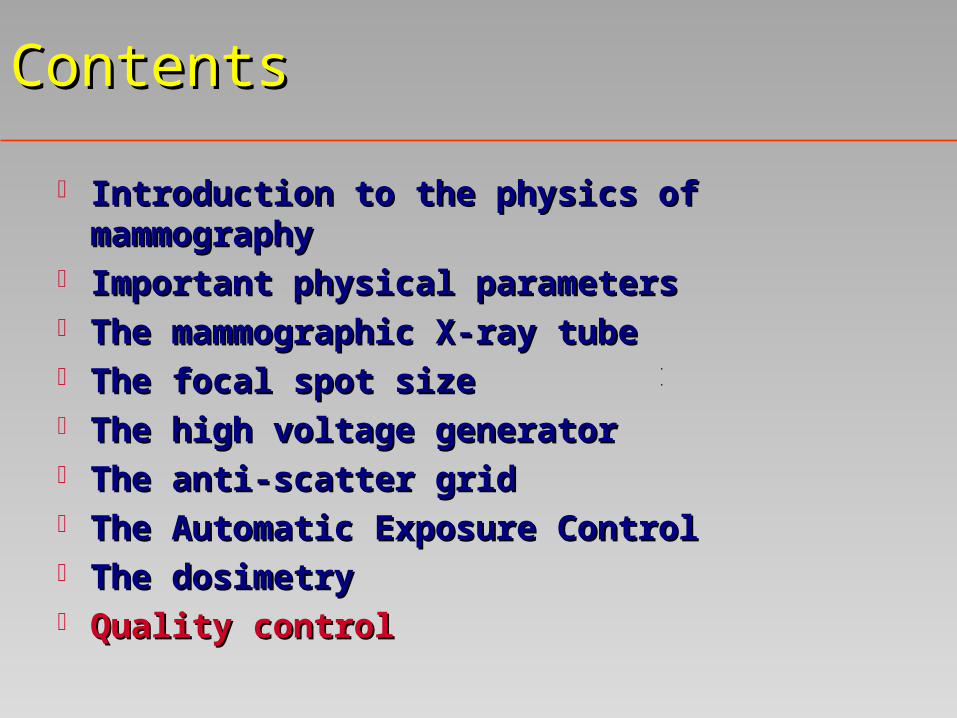
Introduction to the physics of mammographyIntroduction to the physics of mammography Important physical parametersImportant physical parameters The mammographic X-ray tubeThe mammographic X-ray tube The focal spot sizeThe focal spot size The high voltage generatorThe high voltage generator The anti-scatter grid The anti-scatter grid The Automatic Exposure ControlThe Automatic Exposure Control The dosimetryThe dosimetry Quality controlQuality control
Introduction to the physics of Introduction to the physics of mammographymammography
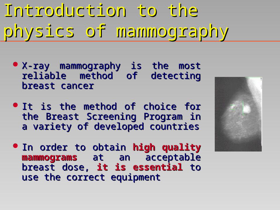
X-ray mammography is the most reliable X-ray mammography is the most reliable method of detecting breast cancermethod of detecting breast cancer
It is the method of choice for the Breast It is the method of choice for the Breast Screening Program in a variety of Screening Program in a variety of developed countriesdeveloped countries
In order In order to obtain to obtain high quality high quality mammogramsmammograms at an acceptable breast at an acceptable breast dose, dose, it is essentialit is essential to use the correct to use the correct equipmentequipment

Main components of the mammographic Main components of the mammographic imaging systemimaging system
A mammographic X-ray tubeA mammographic X-ray tube A device for compressing the breastA device for compressing the breast An anti-scatter gridAn anti-scatter grid A mammographic image receptor (film, A mammographic image receptor (film,
photostimulable plate, flat panel)photostimulable plate, flat panel) An automatic Exposure Control SystemAn automatic Exposure Control System

Main variables of the mammographic Main variables of the mammographic imaging systemimaging system
ContrastContrast: capability of the system to make visible : capability of the system to make visible small differences in soft tissue densitysmall differences in soft tissue density
SharpnessSharpness: capability of the system to make visible : capability of the system to make visible small details small details (calcifications (calcifications down to 0.1 mmdown to 0.1 mm))
DoseDose: the female breast is a very radiosensitive : the female breast is a very radiosensitive organ and there is a risk of carcinogenesis organ and there is a risk of carcinogenesis associated with the technique associated with the technique
NoiseNoise: determines how far the dose can be reduced : determines how far the dose can be reduced given the task of identifying a particular object given the task of identifying a particular object against the backgroundagainst the background

The contrastThe contrast
Linear attenuation coefficientsLinear attenuation coefficients for different for different types of breast tissue types of breast tissue are similar in magnitudeare similar in magnitude and the soft tissue contrast can be quite small and the soft tissue contrast can be quite small
The contrast must be made The contrast must be made as high as possibleas high as possible by by imaging with a low photon energy (hence imaging with a low photon energy (hence increasing breast dose)increasing breast dose)
In practice, to avoid a high breast dose, a In practice, to avoid a high breast dose, a compromisecompromise must be made between the must be made between the requirements of low dose and high contrastrequirements of low dose and high contrast

Variation of contrast with photon Variation of contrast with photon energyenergy
10 20 30 40 50 Energy (keV)
1.0
0.1
0.01
0.001
Ca5 (PO4)3 OH Calcification
of 0.1mm
Glandular tissue of 1mm
Con
tras
t
•The contrast decreasesby a factor of 6 between15 and 30 keV
•The glandular tissuecontrast falls below 0.1for energies above 27
keV

Contributors to the total unsharpness in Contributors to the total unsharpness in the imagethe image
ReceptorReceptor unsharpness: unsharpness: ((screen-film combination) screen-film combination) can be as small as 0.1 - 0.15 mm (full width at half can be as small as 0.1 - 0.15 mm (full width at half maximum of the point response function) with a maximum of the point response function) with a limiting value as high as 20 line pairs per mmlimiting value as high as 20 line pairs per mm
Geometric unsharpnessGeometric unsharpness: focal spot size and imaging : focal spot size and imaging geometry must be chosen so that the overall geometry must be chosen so that the overall unsharpness reflects the performance capability of the unsharpness reflects the performance capability of the screenscreen
Patient movementPatient movement

The breast doseThe breast dose
Dose decreases rapidly with depth in tissue due to Dose decreases rapidly with depth in tissue due to the low energy X-ray spectrum usedthe low energy X-ray spectrum used
Relevant quantityRelevant quantity: : The The average glandular doseaverage glandular dose ((AGDAGD) related to the tissues which are believed to ) related to the tissues which are believed to be the most sensitive to radiation-induced be the most sensitive to radiation-induced carcinogenesiscarcinogenesis
The breast doseThe breast dose
The breast doseThe breast dose is affected by: is affected by: the breast composition and thicknessthe breast composition and thickness the photon energythe photon energy the sensitivity of the image receptorthe sensitivity of the image receptor
The breast composition The breast composition has a significant has a significant influenceinfluence on the dose on the dose
The area of the compressed breast The area of the compressed breast has a small has a small influenceinfluence on the dose on the dose
the mean path of the photons < breast dimensionsthe mean path of the photons < breast dimensions majority of the interactions are photoelectricmajority of the interactions are photoelectric

Variation of mean glandular dose with Variation of mean glandular dose with photon energyphoton energy
10 20 30 40 (keV)
20
10
2
1
0.2
8 cm
Mea
n G
lan
du
lar
Dos
e (a
rb. U
nit
s) •The figure demonstrates the rapid increase in dosewith decreasing photon energyand increasing breast thickness
•For the 8 cm thick breast thereis a dose increase of a factor of 30between photon energies of 17.5 and 30 keV
•At 20 keV there is a dose increase of a factor of 17 betweenthicknesses of 2 an 8 cm
2 cm

Contributors to the image noiseContributors to the image noise
1) the quantum mottle 1) the quantum mottle
2) the properties of the image receptor 2) the properties of the image receptor
3) the film development and display systems 3) the film development and display systems
N.B. :N.B. : both quantum mottle and film granularity both quantum mottle and film granularity contribute significantly to the total image noise for contribute significantly to the total image noise for screen-film-mammography screen-film-mammography

ContentsContents
Introduction to the physics of mammographyIntroduction to the physics of mammography Important physical parametersImportant physical parameters The mammographic X-ray tubeThe mammographic X-ray tube The focal spot sizeThe focal spot size The high voltage generatorThe high voltage generator The anti-scatter grid The anti-scatter grid The Automatic Exposure ControlThe Automatic Exposure Control The dosimetryThe dosimetry Quality controlQuality control

Contradictory objectives for the spectrum Contradictory objectives for the spectrum of a mammographic X-ray tubeof a mammographic X-ray tube
The The ideal ideal X-ray spectrumX-ray spectrum for mammography for mammography is a compromise between :is a compromise between :
to achieve a high contrast and high signal to to achieve a high contrast and high signal to noise ratio (noise ratio (low photon energylow photon energy))
to keep the breast dose ALARA (to keep the breast dose ALARA (high high photon energyphoton energy))

The X-ray spectrum in mammographyThe X-ray spectrum in mammography
In a practice using a screen-In a practice using a screen-film, it may not be possible to film, it may not be possible to vary the SNR because the film vary the SNR because the film may become over or under-may become over or under-exposedexposed
The figure gives the The figure gives the conventional mammographic conventional mammographic spectrum produced by a Mo spectrum produced by a Mo target and a Mo filtertarget and a Mo filter 10 15 20 25 30
15
10
5
Energy (keV)
Nu
mb
er o
f p
hot
ons
(arb
itra
ry n
orm
alis
atio
n)
X-ray spectrum at 30 kV for an X-ray tube with a Mo target and a 0.03 mm Mo filter

MMain features of the X-ray spectrum in ain features of the X-ray spectrum in mammography mammography
Characteristic X-ray lines at 17.4 and 19.6 keV Characteristic X-ray lines at 17.4 and 19.6 keV and the heavy attenuation above 20 keV (position and the heavy attenuation above 20 keV (position of the of the MoMo K-edge) K-edge)
Reasonably close to the energies optimal for Reasonably close to the energies optimal for imaging breast of small to medium thicknessimaging breast of small to medium thickness
A higher energy spectrum is obtained by replacing A higher energy spectrum is obtained by replacing the the MoMo filter with a material of higher atomic filter with a material of higher atomic number with its K-edge at a higher energy (number with its K-edge at a higher energy (RhRh, , PdPd))
WW can also be used as target material can also be used as target material

Options for an optimum X-ray Options for an optimum X-ray spectrum in mammographyspectrum in mammography
Several scientific works have demonstrated that Several scientific works have demonstrated that contrast is better for the Mo/Mo target/filter contrast is better for the Mo/Mo target/filter combinationscombinations
This advantage decreases with increasing breast This advantage decreases with increasing breast thicknessthickness
Using Rh/Rh for target/filter combination brings a Using Rh/Rh for target/filter combination brings a substantial dose saving for bigger breasts while substantial dose saving for bigger breasts while keeping an acceptable contrast levelkeeping an acceptable contrast level
Options for an optimum X-ray Options for an optimum X-ray spectrum in mammographyspectrum in mammography
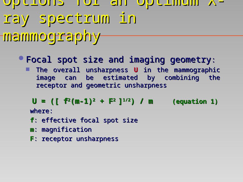
Focal spot size and imaging geometryFocal spot size and imaging geometry:: The overall unsharpness The overall unsharpness UU in the mammographic image in the mammographic image
can be estimated by combining the receptor and can be estimated by combining the receptor and geometric unsharpness geometric unsharpness
U = ([ fU = ([ f22(m-1)(m-1)22 + F + F2 2 ]]1/21/2) / m) / m (equation 1) (equation 1)
where:where:
ff: effective focal spot size: effective focal spot size
mm: magnification: magnification
FF: receptor unsharpness: receptor unsharpness
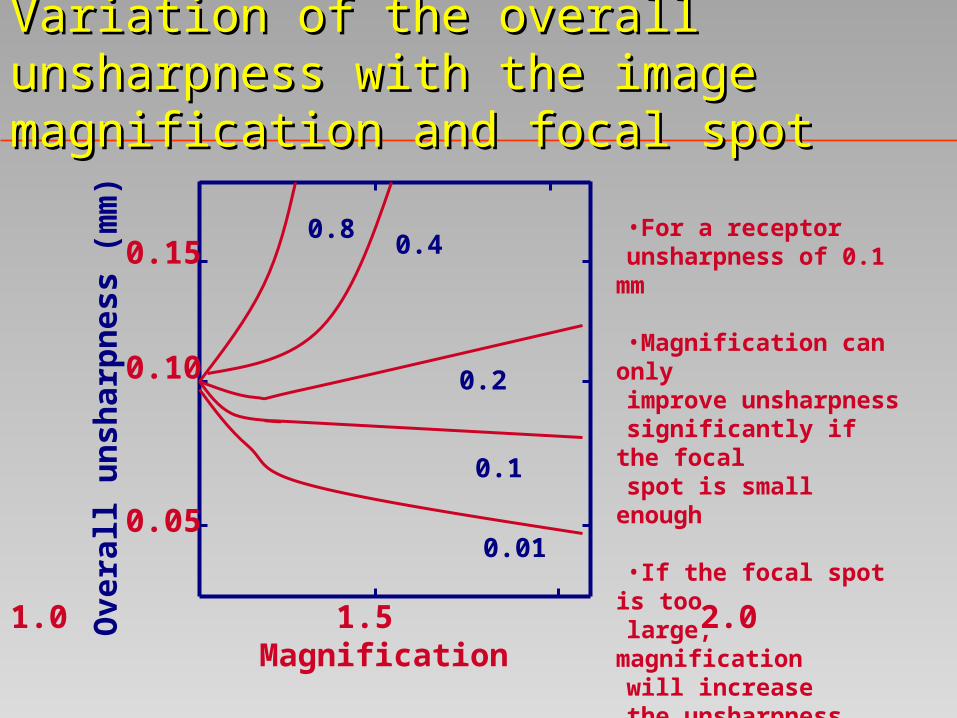
Variation of the overall unsharpness with the Variation of the overall unsharpness with the image magnification and focal spotimage magnification and focal spot
1.0 1.5 2.0Magnification
0.15
0.10
0.05
0.8
Ove
rall
un
shar
pn
ess
(mm
) •For a receptor unsharpness of 0.1 mm
•Magnification can only improve unsharpnesssignificantly if the focal spot is small enough
•If the focal spot is too large, magnification will increase the unsharpness
0.4
0.2
0.1
0.01

ContentsContents
Introduction to the physics of mammographyIntroduction to the physics of mammography Important physical parametersImportant physical parameters The mammographic X-ray tubeThe mammographic X-ray tube The focal spot sizeThe focal spot size The high voltage generatorThe high voltage generator The anti-scatter grid The anti-scatter grid The Automatic Exposure ControlThe Automatic Exposure Control The dosimetryThe dosimetry Quality controlQuality control
The focal spot sizeThe focal spot size
Ideally, for the screening unit a single-focus X-ray Ideally, for the screening unit a single-focus X-ray tube with a 0.3 focal stube with a 0.3 focal spot is recommendedpot is recommended
For general mammography purposes, a dual focus For general mammography purposes, a dual focus X-ray tube with an additional fine focus (0.1) to X-ray tube with an additional fine focus (0.1) to be used for magnification techniques exclusively be used for magnification techniques exclusively is requiredis required
The size of the focal spot The size of the focal spot should be verifiedshould be verified ( (star star pattern, slit camera or pinhole methodpattern, slit camera or pinhole method) ) yearly or yearly or when resolution decays rapidlywhen resolution decays rapidly

Target/filter combinationTarget/filter combination
The window of the X-ray tube should be beryllium The window of the X-ray tube should be beryllium (not glass) with a maximum thickness of 1 mm(not glass) with a maximum thickness of 1 mm
The typical target/filter combinations nowadays The typical target/filter combinations nowadays available are: available are:
Mo + 30 Mo + 30 m Mom Mo Mo + 25 Mo + 25 m Mom Mo W + 60 W + 60 m Mom Mo W + 50 W + 50 m Rhm Rh W + 40 W + 40 m Pdm Pd Rh + 25 Rh + 25 m Rhm Rh

X-ray tube filtrationX-ray tube filtration
Total permanent filtrationTotal permanent filtration 0.5 mm of Al or 0.5 mm of Al or
0.03 mm of Mo (recommended by 0.03 mm of Mo (recommended by ICRP 34)ICRP 34)
The The beam qualitybeam quality is defined by the HVL is defined by the HVL
A better index of the A better index of the beam qualitybeam quality is the total is the total
filtration which can be related to the HVL using filtration which can be related to the HVL using
published datapublished data
State-of-the-art specifications State-of-the-art specifications for screen-film mammographyfor screen-film mammography
A nearly constant potential waveform with a ripple A nearly constant potential waveform with a ripple not greater than that produced by a 6-pulse not greater than that produced by a 6-pulse rectification systemrectification system
The tube voltage range should be The tube voltage range should be 25 - 35 kV25 - 35 kV The tube current The tube current should be at least 100 mA on should be at least 100 mA on
broad focus and 50 mA on fine focusbroad focus and 50 mA on fine focus.. The range of tube current exposure time product The range of tube current exposure time product
(mAs) (mAs) should be at least 5 - 800 mAsshould be at least 5 - 800 mAs It should be possible to repeat exposures at the It should be possible to repeat exposures at the
highest loadings at intervals highest loadings at intervals < 30 seconds< 30 seconds

ContentsContents
Introduction to the physics of mammographyIntroduction to the physics of mammography Important physical parametersImportant physical parameters The mammographic X-ray tubeThe mammographic X-ray tube The focal spot sizeThe focal spot size The high voltage generatorThe high voltage generator The anti-scatter gridThe anti-scatter grid The Automatic Exposure ControlThe Automatic Exposure Control The dosimetryThe dosimetry Quality controlQuality control

Why an anti-scatter grid ?Why an anti-scatter grid ?
Effects of scatter may significantly degrade the Effects of scatter may significantly degrade the contrast of the image and the need forcontrast of the image and the need for an efficient an efficient anti-scatter anti-scatter devicedevice is evident is evident
The effect is quantified by the :The effect is quantified by the :
Contrast Degradation Factor (CDF)Contrast Degradation Factor (CDF) : : CDF=1/(1+S/P)CDF=1/(1+S/P)
where: where: S/P :S/P : ratio of the scattered to primary ratio of the scattered to primary radiation amountsradiation amounts
Calculated values of CDF: 0.76 and 0.48 for breast Calculated values of CDF: 0.76 and 0.48 for breast thickness of 2 and 8 cm respectively thickness of 2 and 8 cm respectively [Dance et al.][Dance et al.]

The anti-scatter gridThe anti-scatter grid
Two types of anti-scatter grids available:Two types of anti-scatter grids available: stationary gridstationary grid: : with high line density (e.g. 80 lines/cm) with high line density (e.g. 80 lines/cm)
and an aluminium interspace materialand an aluminium interspace material moving gridmoving grid:: with about 30 lines/cm with paper or with about 30 lines/cm with paper or
cotton fiber interspacecotton fiber interspace
The performance of the anti-scatter grid can be The performance of the anti-scatter grid can be expressed in termsexpressed in terms of of the contrast improvementthe contrast improvement (CIF)(CIF) and and Bucky factorsBucky factors (BF)(BF)

The anti-scatter grid: performance The anti-scatter grid: performance indexesindexes
The The CIFCIF relates the contrast with the grid to that without relates the contrast with the grid to that without the grid while the grid while
The The BFBF gives the increase in dose associated with the use gives the increase in dose associated with the use of gridof grid
CIF and BF values for the Philips moving gridCIF and BF values for the Philips moving grid
Breast Thickness (cm)
CIF BF
2 1.25 1.68
4 1.38 1.85
6 1.54 2.06
8 1.68 2.24

Automatic exposure control device Automatic exposure control device (AEC)(AEC)
The system should produce a stable optical The system should produce a stable optical density (OD density (OD variation of less than variation of less than 0.2 0.2 ) in spite ) in spite of a wide range of mAsof a wide range of mAs
Hence the system should be fitted with an AEC Hence the system should be fitted with an AEC located after the film receptor to allow for quite located after the film receptor to allow for quite different breast characteristicsdifferent breast characteristics
The detector should be movable to cover The detector should be movable to cover different anatomical sites on the breast and the different anatomical sites on the breast and the system should be adaptable to at least three film-system should be adaptable to at least three film-screen combinationsscreen combinations
ContentsContents
Introduction to the physics of mammographyIntroduction to the physics of mammography Important physical parametersImportant physical parameters The mammographic X-ray tubeThe mammographic X-ray tube The focal spot sizeThe focal spot size The high voltage generatorThe high voltage generator The anti-scatter grid The anti-scatter grid The Automatic Exposure ControlThe Automatic Exposure Control The dosimetryThe dosimetry Quality controlQuality control

Breast dosimetry in screen-film Breast dosimetry in screen-film mammographymammography
There exists a small risk of radiation induced There exists a small risk of radiation induced cancer associated with X-ray examination of the cancer associated with X-ray examination of the breast breast
Achieving an image quality at the lowest possible Achieving an image quality at the lowest possible dose is therefore requireddose is therefore required
Hence breast dosimetry Hence breast dosimetry The Average Glandular Dose (The Average Glandular Dose (AGDAGD) is the ) is the
dosimetry quantity generally recommended for risk dosimetry quantity generally recommended for risk assessmentassessment
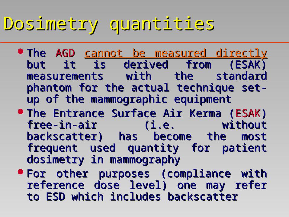
Dosimetry quantitiesDosimetry quantities
The The AGDAGD cannot be measured directly cannot be measured directly but it but it is derived from (ESAK) measurements with is derived from (ESAK) measurements with the standard phantom for the actual technique the standard phantom for the actual technique set-up of the mammographic equipmentset-up of the mammographic equipment
The Entrance Surface Air Kerma (The Entrance Surface Air Kerma (ESAKESAK) ) free-in-air (i.e. without backscatter) has free-in-air (i.e. without backscatter) has become the most frequent used quantity for become the most frequent used quantity for patient dosimetry in mammographypatient dosimetry in mammography
For other purposes (compliance with For other purposes (compliance with reference dose level) one may refer to ESD reference dose level) one may refer to ESD which includes backscatterwhich includes backscatter

Dosimetry quantitiesDosimetry quantities
ESAK can be determined by:ESAK can be determined by: a TLD dosemeter calibrated in terms of air kerma free-in-air a TLD dosemeter calibrated in terms of air kerma free-in-air
at a HVL as close as possible to 0.4 mm Al at a HVL as close as possible to 0.4 mm Al with a standard with a standard phantomphantom
a TLD dosemeter calibrated in terms of air kerma free-in-air a TLD dosemeter calibrated in terms of air kerma free-in-air at a HVL as close as possible to 0.4 mm Al at a HVL as close as possible to 0.4 mm Al stuck to the stuck to the patient skinpatient skin (appropriate backsactter factor should be applied (appropriate backsactter factor should be applied to Entrance Surface Dose measured with the TLD to express to Entrance Surface Dose measured with the TLD to express ESAK)ESAK)
NoteNote : due to low kV used the TLD is seen on the image : due to low kV used the TLD is seen on the image a radiation dosemeter with a dynamic range covering at least a radiation dosemeter with a dynamic range covering at least
0.5 to 100 mGy (better than 0.5 to 100 mGy (better than 10% accuracy) 10% accuracy)


How have IQ and dose standards been How have IQ and dose standards been developed in European guidelines ?developed in European guidelines ?
Digital should not be worse than film-screen Digital should not be worse than film-screen systemssystems
IQ : Contrast detail measurements using CDMAM IQ : Contrast detail measurements using CDMAM test objecttest object
Dose : breasts simulated with PMMADose : breasts simulated with PMMA

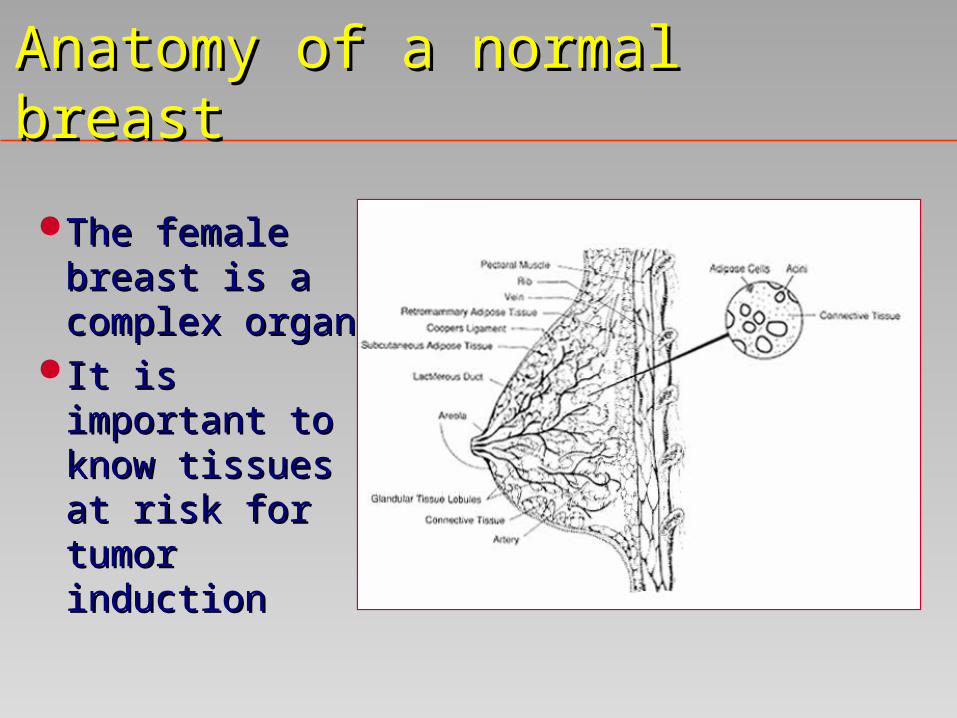
Anatomy of a normal breastAnatomy of a normal breast
The female breast The female breast is a complex organis a complex organ
It is important to It is important to know tissues at know tissues at risk for tumor risk for tumor inductioninduction
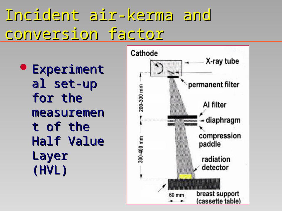
Incident air-kerma and conversion Incident air-kerma and conversion factorfactor
Experimental Experimental set-up for the set-up for the measurement measurement of the Half of the Half Value Layer Value Layer (HVL) (HVL)

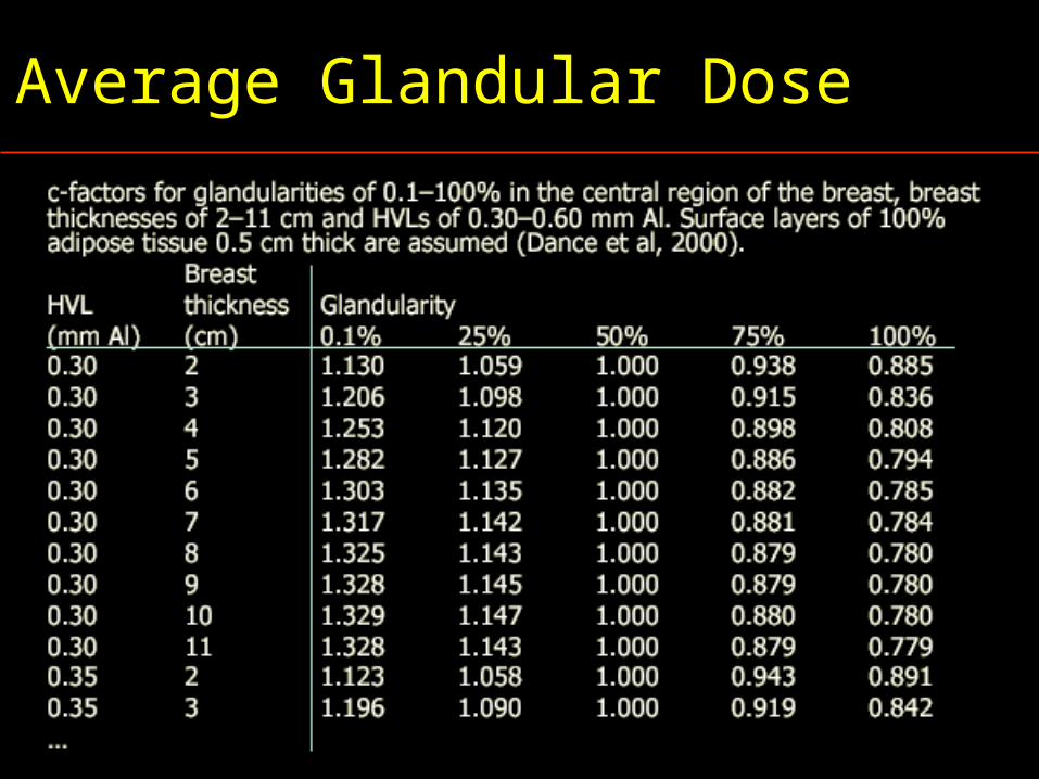
AGD = K.g.c.sAGD = K.g.c.s where :where :
k =k = Entrance Surface Air Kerma Entrance Surface Air Kerma g =g = incident air-kerma conversion factor for incident air-kerma conversion factor for
breast thicknesses (50% water, 50% breast thicknesses (50% water, 50% fat) fat)
c =c = glandularity factor glandularity factor s =s = x-ray spectrum correction factorx-ray spectrum correction factor
Average Glandular DoseAverage Glandular Dose
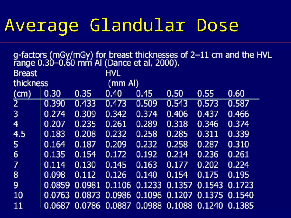
Average Glandular DoseAverage Glandular Dose
Average Glandular DoseAverage Glandular Dose
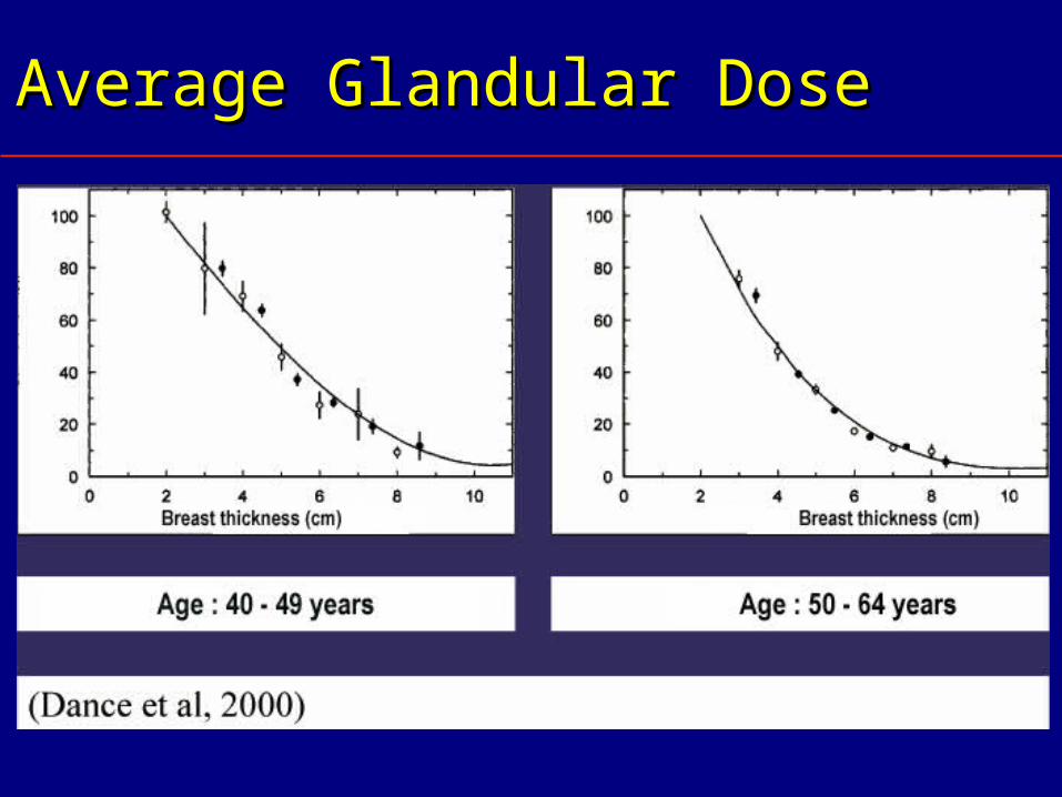
Average Glandular DoseAverage Glandular Dose


ContentsContents
Introduction to the physics of mammographyIntroduction to the physics of mammography Important physical parametersImportant physical parameters The mammographic X-ray tubeThe mammographic X-ray tube The focal spot sizeThe focal spot size The high voltage generatorThe high voltage generator The anti-scatter grid The anti-scatter grid The Automatic Exposure ControlThe Automatic Exposure Control The dosimetryThe dosimetry Quality controlQuality control

Why Quality Control ?Why Quality Control ?
BSS requires Quality Assurance for medical BSS requires Quality Assurance for medical exposuresexposures
Principles established by WHO, (ICRP for dose), Principles established by WHO, (ICRP for dose), guidelines prepared by EC, PAHO,…guidelines prepared by EC, PAHO,…
A Quality Control program should ensure:A Quality Control program should ensure: The best image qualityThe best image quality With the least dose to the breastWith the least dose to the breast Hence regular check of important parametersHence regular check of important parameters
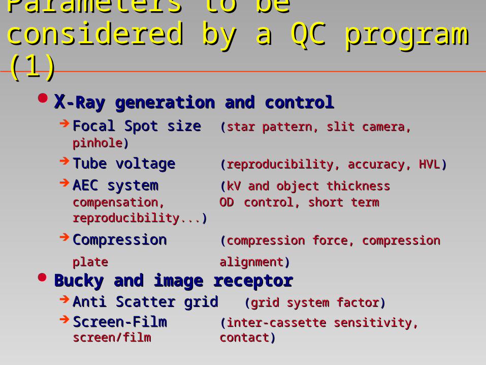
Parameters to be considered by a QC Parameters to be considered by a QC program (1)program (1)
XX-Ray generation and control-Ray generation and control Focal Spot sizeFocal Spot size ((star pattern, slit camera, pinholestar pattern, slit camera, pinhole))
Tube voltage Tube voltage ((reproducibility, accuracy, HVLreproducibility, accuracy, HVL))
AEC system AEC system ((kV and object thickness kV and object thickness
compensation, compensation, OD OD control, short term control, short term reproducibilityreproducibility......))
Compression Compression ((compression force, compression plate compression force, compression plate
alignmentalignment)) Bucky and image receptorBucky and image receptor
Anti Scatter grid Anti Scatter grid ((grid system factorgrid system factor))
Screen-FilmScreen-Film ((inter-cassette sensitivity, screen/film inter-cassette sensitivity, screen/film contactcontact))
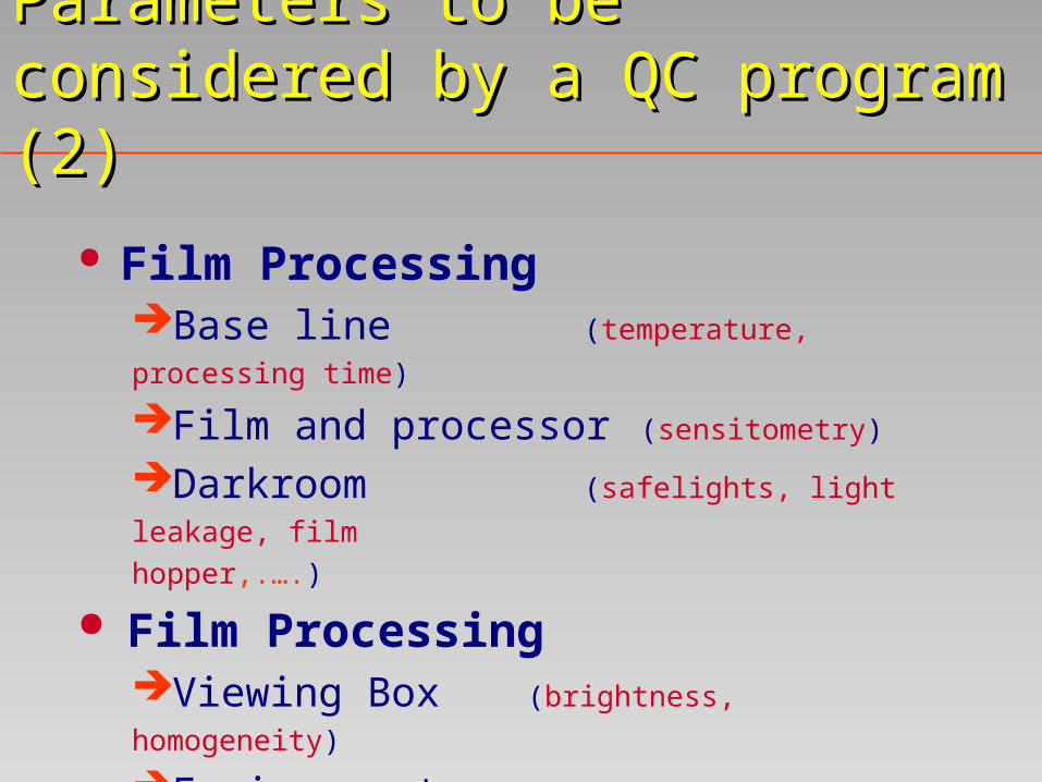
Film ProcessingBase line (temperature, processing time)
Film and processor (sensitometry)
Darkroom (safelights, light leakage, film
hopper,.….)
Film ProcessingViewing Box (brightness, homogeneity)
Environment
Parameters to be considered by a QC Parameters to be considered by a QC program (2)program (2)

System PropertiesReference Dose (entrance surface dose)
Image Quality (spatial resolution,
image contrast, noise
threshold contrast
visibility, exposure time)
Parameters to be considered by a QC Parameters to be considered by a QC program (3)program (3)

SummarySummary
To achieve the best image quality while To achieve the best image quality while keeping the breast dose at the ALARA level keeping the breast dose at the ALARA level is the final goal to be reached when is the final goal to be reached when consistently using a film-screen or digital consistently using a film-screen or digital mammography equipment.mammography equipment.
Implementing a well defined QC protocol Implementing a well defined QC protocol can effectively contribute to the achievement can effectively contribute to the achievement of such a goal.of such a goal.
Related Documents