The Proliferation and Expansion of Retinal Stem Cells Require Functional Pax6 Shunbin Xu a,* , Mary E. Sunderland b,* , Brenda L.K. Coles a , Angela Kam c , Tamara Holowacz a , Ruth Ashery-Padan e , Till Marquardt f,g , Roderick R. McInnes a,d,h , and Derek van der Kooy a aDepartment of Medical Genetics and Microbiology, University of Toronto, ON M5S 1A8, Canada bInstitute of Medical Science, University of Toronto, ON M5S 1A8, Canada cDepartment of Surgery, University of Toronto, ON M5S 1A8, Canada dDepartment of Pediatrics, University of Toronto, ON M5S 1A8, Canada eTel Aviv University, Tel Aviv, Israel fThe Salk Institute, La Jolla, USA gMax-Planck Institute, 37077 Gottingen, Germany hPrograms in Developmental Biology and Genetics, Research Institute, Hospital for Sick Children, Toronto, ON M5G 1X8, Canada Abstract Retinal stem cells (RSCs) exist as rare pigmented ciliary epithelial cells in adult mammalian eyes. We hypothesized that RSCs are at the top of the retinal cell lineage. Thus, genes expressed early in embryonic development to establish the retinal field in forebrain neuroectoderm may play important roles in RSCs. Pax6, a paired domain and homeodomain-containing transcription factor, is one of the earliest genes expressed in the eye field and is considered a master control gene for retinal and eye development. Here, we demonstrate that Pax6 is enriched in RSCs. Inactivation of Pax6 in vivo results in loss of competent RSCs as assayed by the failure to form clonal RSC spheres from the optic vesicles of conventional Pax6 knockout embryos and from the ciliary epithelial cells of adult Pax6 conditional knockout mice. In vitro clonal inactivation of Pax6 in adult RSCs results in a serious proliferation defect, suggesting that Pax6 is required for the proliferation and expansion of RSCs. Keywords retinal stem cell; ciliary epithelia; Pax6 Correspondence: Derek van der Kooy, [email protected]. Tel: 416-978-1960; Fax: 416-978-3844 or Shunbin Xu, [email protected]. Tel: 312-563-3554; Fax: 312-563-3571. * Present addresses: Rush University Medical Center, Department of Ophthalmology and Neurological Sciences, 1735 W. Harrison St. Ste 318, Chicago, IL 60612 (SX); Arizona State University, School of Life Sciences, Tempe, AZ 85287-4601 (MES). Publisher's Disclaimer: This is a PDF file of an unedited manuscript that has been accepted for publication. As a service to our customers we are providing this early version of the manuscript. The manuscript will undergo copyediting, typesetting, and review of the resulting proofbefore it is published in its final citable form. Please note that during the production process errors may be discovered which could affect the content, and all legal disclaimers that apply to the journal pertain. NIH Public Access Author Manuscript Dev Biol. Author manuscript; available in PMC 2008 April 15. Published in final edited form as: Dev Biol. 2007 April 15; 304(2): 713–721. NIH-PA Author Manuscript NIH-PA Author Manuscript NIH-PA Author Manuscript

Welcome message from author
This document is posted to help you gain knowledge. Please leave a comment to let me know what you think about it! Share it to your friends and learn new things together.
Transcript

The Proliferation and Expansion of Retinal Stem Cells RequireFunctional Pax6
Shunbin Xua,*, Mary E. Sunderlandb,*, Brenda L.K. Colesa, Angela Kamc, TamaraHolowacza, Ruth Ashery-Padane, Till Marquardtf,g, Roderick R. McInnesa,d,h, and Derek vander KooyaaDepartment of Medical Genetics and Microbiology, University of Toronto, ON M5S 1A8, Canada
bInstitute of Medical Science, University of Toronto, ON M5S 1A8, Canada
cDepartment of Surgery, University of Toronto, ON M5S 1A8, Canada
dDepartment of Pediatrics, University of Toronto, ON M5S 1A8, Canada
eTel Aviv University, Tel Aviv, Israel
fThe Salk Institute, La Jolla, USA
gMax-Planck Institute, 37077 Gottingen, Germany
hPrograms in Developmental Biology and Genetics, Research Institute, Hospital for Sick Children, Toronto,ON M5G 1X8, Canada
AbstractRetinal stem cells (RSCs) exist as rare pigmented ciliary epithelial cells in adult mammalian eyes.We hypothesized that RSCs are at the top of the retinal cell lineage. Thus, genes expressed early inembryonic development to establish the retinal field in forebrain neuroectoderm may play importantroles in RSCs. Pax6, a paired domain and homeodomain-containing transcription factor, is one ofthe earliest genes expressed in the eye field and is considered a master control gene for retinal andeye development. Here, we demonstrate that Pax6 is enriched in RSCs. Inactivation of Pax6 invivo results in loss of competent RSCs as assayed by the failure to form clonal RSC spheres fromthe optic vesicles of conventional Pax6 knockout embryos and from the ciliary epithelial cells ofadult Pax6 conditional knockout mice. In vitro clonal inactivation of Pax6 in adult RSCs results ina serious proliferation defect, suggesting that Pax6 is required for the proliferation and expansion ofRSCs.
Keywordsretinal stem cell; ciliary epithelia; Pax6
Correspondence: Derek van der Kooy, [email protected]. Tel: 416-978-1960; Fax: 416-978-3844 or Shunbin Xu,[email protected]. Tel: 312-563-3554; Fax: 312-563-3571.*Present addresses: Rush University Medical Center, Department of Ophthalmology and Neurological Sciences, 1735 W. Harrison St.Ste 318, Chicago, IL 60612 (SX); Arizona State University, School of Life Sciences, Tempe, AZ 85287-4601 (MES).Publisher's Disclaimer: This is a PDF file of an unedited manuscript that has been accepted for publication. As a service to our customerswe are providing this early version of the manuscript. The manuscript will undergo copyediting, typesetting, and review of the resultingproofbefore it is published in its final citable form. Please note that during the production process errors may be discovered which couldaffect the content, and all legal disclaimers that apply to the journal pertain.
NIH Public AccessAuthor ManuscriptDev Biol. Author manuscript; available in PMC 2008 April 15.
Published in final edited form as:Dev Biol. 2007 April 15; 304(2): 713–721.
NIH
-PA Author Manuscript
NIH
-PA Author Manuscript
NIH
-PA Author Manuscript
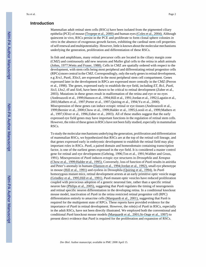
IntroductionMammalian adult retinal stem cells (RSCs) have been isolated from the pigmented ciliaryepithelia (PCE) of mouse (Tropepe et al., 2000) and human eyes (Coles et al., 2004). Althoughquiescent in vivo, RSCs persist in the PCE and proliferate to form clonal sphere colonies invitro in the absence of exogenous growth factors, exhibiting the cardinal stem cell propertiesof self renewal and multipotentiality. However, little is known about the molecular mechanismsunderlying the generation, proliferation and differentiation of these RSCs.
In fish and amphibians, many retinal precursor cells are located in the ciliary margin zone(CMZ) and continuously add new neurons and Muller glial cells to the retina in adult animals(Johns, 1977;Wetts and Fraser, 1988). Cells in CMZ are spatially ordered with respect to thedevelopment, with stem cells being most peripheral and differentiating retinal progenitor cells(RPCs) more central in the CMZ. Correspondingly, only the early genes in retinal development,e.g Xrx1, Pax6, XSix3, are expressed in the most peripheral stem cell compartment. Genesexpressed later in the development in RPCs are expressed more centrally in the CMZ (Perronet al., 1998). The genes, expressed early to establish the eye field, including ET, Rx1, Pax6,Six3, Lhx2, tll and Six6, have been shown to be critical to retinal development (Zuber et al.,2003). Mutations in these genes result in malformation of the retina and eye or no eyes(Andreazzoli et al., 1999;Hanson et al., 1994;Hill et al., 1991;Jordan et al., 1992;Lagutin et al.,2003;Mathers et al., 1997;Porter et al., 1997;Quiring et al., 1994;Yu et al., 2000).Misexpression of these genes can induce ectopic retinal or eye tissues (Andreazzoli et al.,1999;Bernier et al., 2000;Chow et al., 1999;Halder et al., 1995;Loosli et al., 1999;Mathers etal., 1997;Oliver et al., 1996;Zuber et al., 2003). All of these studies suggest that the earlyexpressed eye field genes may have important functions in the regulation of retinal stem cells.However, the roles of these genes in RSCs have not been fully studied, especially in mammalianRSCs.
To study the molecular mechanisms underlying the generation, proliferation and differentiationof mammalian RSCs, we hypothesized that RSCs are at the top of the retinal cell lineage, andthat genes expressed early in embryonic development to establish the retinal field may playimportant roles in RSCs. Pax6, a paired domain and homeodomain containing transcriptionfactor, is one of the earliest genes expressed in the eye field. It is considered a master controlgene for retinal and eye development (Gehring, 1996;Ton et al., 1991;Walther and Gruss,1991). Misexpression of Pax6 induces ectopic eye structures in Drosophila and Xenopus(Chow et al., 1999;Halder et al., 1995). Conversely, loss of function of Pax6 results in aniridiaand Peter’s anomaly in humans (Hanson et al., 1994;Jordan et al., 1992), small eye phenotypein mouse (Hill et al., 1991) and eyeless in Drosophila (Quiring et al., 1994). In Pax6homozygous mutant mice, retinal development arrests at an early primitive optic vesicle stage(Grindley et al., 1995;Hill et al., 1991). Pax6 mutant optic vesicles have reduced proliferationcoupled with precocious adoption of a generic neuronal fate, rather than a specific retinalneuron fate (Philips et al., 2005), suggesting that Pax6 regulates the timing of neurogenesisand retinal specific neuron differentiation in the developing retina. In a conditional knockoutmouse model, inactivation of Pax6 in the retina restricted retinal progenitor cell (RPC)differentiation entirely to amacrine cells (Marquardt et al., 2001), suggesting that Pax6 isrequired for the multipotent state of RPCs. These reports have provided evidence for theimportance of Pax6 in retinal development. However, the role(s) of Pax6 in RSCs, especiallyin the adult RSCs, have not been directly illustrated. We employed both the conventional andconditional Pax6 knockout mouse models (Marquardt et al., 2001;St-Onge et al., 1997) topresent direct evidence that Pax6 is required for the proliferation and expansion of RSCs.
Xu et al. Page 2
Dev Biol. Author manuscript; available in PMC 2008 April 15.
NIH
-PA Author Manuscript
NIH
-PA Author Manuscript
NIH
-PA Author Manuscript

Experimental ProceduresAnimals
The Z/EG reporter mice (Novak et al., 2000) were provided by Drs. Andras Nagy and CorrineLobe. The Pax6LacZ/LacZ (St-Onge et al., 1996;St-Onge et al., 1997) and α–Cre;Pax6loxP/loxP
mice (Marquardt, 2003;Marquardt et al., 2001) kindly were provided by Dr. Peter Gruss.
ImmunohistochemistryPax6, Syntaxin (HPC-1), Rhodopsin (1D4) and nestin antibodies were purchased fromDevelopmental Hybridoma Bank, Sigma and Chemicon respectively. Immunohistochemistrywas performed as previously described (Tropepe et al., 2000).
RNA preparation and RT-PCRTotal RNA was isolated using RNeasy kit (Qiagen). RT-PCR was performed using OneStepRT-PCR kit (Qiagen). Primer sequences: Pax6: Fw1:5′-TCACAGCGGAGTGAATCAGC-3′and Rev1:5′-TATCGTTGGTACAGACCCCCTC-3′; Fw2:5′-CGGAGTGAATCAGCTTGGT-G-3′ and Rev2:5′-GTTGGTACAGACCCCCTCGG-3′.GAPDH: Fw:5′-TGCACCACCAACTGCTTAGC–3′ and Rev: 5′-TGGATGCAGGGATGATGTTC-3′. 18s rRNA: Fw:5′-GTAACCCGTTGAACCCCATT-3′;Rev: 5′-CCATCCAATCGGTAGTAGCG-3′. 50 ng of total RNA or 1/4 of the RNA from asingle sphere was used in the first round Pax6 amplification with Fw1/Rev1. The reactionprogram was 50 °C for 30 minute for RT, 95 °C for 15 minutes to inactivate RT, followed by25 cycles of 95°C for 30 seconds, 60 °C for 1 minute, and 72 °C for 1 minute and then 72 °Cfor 10 minutes. 2μl of 1:10 dilutant of the first round amplification product was used in Pax6nested PCR with primers Fw2/Rev2. The primers encompass the alternatively spliced exon5a(Glaser et al., 1992;Ton et al., 1991;Walther and Gruss, 1991). The amplicons are 367 basepairs (bp) (without exon5a) and 409 bp (with exon5a).
Embryonic day (E)14 and adult RSC primary cultureThe dissection and primary culture were performed as described previously (Tropepe et al.,2000). Briefly, for adult RSC isolation: adult mice were sacrificed by cervical dislocation. Eyeswere removed and placed in oxygenated artificial cerebral spinal fluid (aCSF: 124mM NaCl,5 mM KCl, 1.3 mM MgCl2, 2mM CaCl2, 26 mM NaHCO3, and 10 mM D-glucose, pH 7.4).Each eye was bisected and the ciliary body was isolated as a thin strips and placed in dispase(Invitrogen) for 10 minutes at 37°C, followed by trypsin/hyaluronidase treatment [1.33 mg/mL trypsin (Sigma), 0.67 mg/mL hyaluronidase (Sigma) and 0.2 mg/mL kynurenic acid(Sigma) in aCSF modified with high Mg2+ (3.2 mM MgCl2) and low Ca2+ (0.1 mM CaCl2)]at 37°C for 15 minutes. Ciliary epithelial cells were scraped off from the strips and collectedin serum free medium (SFM) with trypsin inhibitor (1 mg/mL. Roche Diagnostics). The cellswere triturated 60 times to a single cell suspension. Cells were resuspended in SFM with FGF2(10 ng/mL) and heparin (2μg/mL), plated at a low clonal density (10 cellsμL) and incubatedat 37°C in a CO2 (5%) incubator (Thermo Electron Co) for 7 days.
For E14 embryos: the eyes were carefully removed and placed in dispase (Invitrogen) at 37°C for 30 seconds. RPE and neural retina (NR) were separated, collected in SFM and trituratedabout 60 times to achieve a single cell suspension. The cells were spun down and resuspendedin SFM with FGF2 (10 ng/mL) and heparin (2μg/mL). The NR cells were plated at 10cellsμL. All RPE cells from each animal were plated in 4 wells of a 24-well plate and the totalnumbers of RSCs were assessed as the total number of RSC spheres formed. In Pax6homozygous knockout (Pax6LacZ/LacZ) embryos, retinal development was aborted at theprimitive optic vesicle stage, which consisted of a thin epithelial sheet, without the thickening
Xu et al. Page 3
Dev Biol. Author manuscript; available in PMC 2008 April 15.
NIH
-PA Author Manuscript
NIH
-PA Author Manuscript
NIH
-PA Author Manuscript

of the inner neuroretinal layers and the differentiation of the RPE layer. To evaluate the RSCstatus in these Pax6LacZ/LacZ embryos, each entire primitive optic vesicle carefully wasdissected, dissociated into single cells, and plated in 4 wells of a 24-well plate. Cells wereincubated at 37°C in a CO2 (5%) incubator for 7 days.
Three and four independent experiments were performed at the adult and E14 time points,respectively.
Fluorescent Activated Cell Sorting (FACS)The ciliary epithelial cells of adult Pax6lacZ/wt or wild type littermates were isolated anddissociated into single cells in 200μL of SFM. The cells were separated into two aliquots; onewas treated with 100μM of a fluorescent β-gal substrate: 5′-chloromethylfluorescein di-β-D-galactopyranoside (CMFDG) (Invitrogen), the other was used as negative control. Bothaliquots were incubated at 37 °C for 25′. The cells were spun down and resuspended in500μl of SFM+FGF2 (10 ng/ml)+ heparin (2 mg/ml). Propidium iodide (PI) (1.5μM) was addedto assay for live/dead cells. Total live cells were sorted by the green fluorescence frombreakdown product of CMFDG, plated at clonal density (<10 cellsμl) and incubated at 37 °Cfor 1 week.
Retroviral infectionBi-cistronic pMXIE-Cre-EGFP virus was constructed by inserting a nlsCre cassette in front ofthe IRES-EGFP in a replication-incompetent retroviral construct pMXIE-EGFP (Hitoshi et al.,2002). Ciliary epithelial cells from adult Pax6flox/flox or Pax6wt/wt animals were isolated anddissociated to single cells and plated in fibronectin-coated 24-well plates at 10 cells/ul.Retrovirus (pMXIE-Cre-EGFP or pMXIE-EGFP) was added 18-24 hours after plating(m.o.i=10). The infected cells (GFP+) were counted under the fluorescent microscope(Olympus) every 24 hours. For differentiation, differentiation medium (SFM+FGF2+heparin+1% Fetal Bovine Serum (FBS)) was added 24 hours after viral infection and was changedevery 4 days. After 21 days, cells were fixed with 4% paraformaldehyde. Immunostaining withthe amacrine cell marker, HPC-1, or the rod photoreceptor marker, 1D4, was performed.
ResultsPax6 is expressed in the adult retinal stem cell niche- the ciliary epithelia
To study the role of Pax6 in RSCs, we first asked how Pax6 is expressed in the adult RSCniche – the pigmented ciliary epithelia. It has been shown that Pax6 is expressed in the ciliaryepithelia and the epithelia of the iris (Marquardt et al., 2001). By immunohistochemistry andconfocal microscopy, Pax6 was detected in both layers of the ciliary epithelia (Fig.1A).However, Pax6 is expressed at a much higher level in the outer layer (arrows in Fig.1A), wherepigmented RSCs are found (Tropepe et al., 2000), than in the inner layer (arrowheads in Fig.1A). Pax6 expression was further confirmed by RT-PCR on RNA isolated from ciliary epitheliaand clonally derived RSC spheres from adult mice (Fig.1B). As controls, Pax6 expression alsowas assessed and detected in adult neural retina (NR), forebrain neural stem cell (NSC) spheres(Reynolds and Weiss, 1996), embryonic stem (ES) cell-derived NSC spheres (ESDS) (Tropepeet al., 2000;Tropepe et al., 2001) and ES cells. The primers in the RT-PCR assays encompassthe alternatively spliced exon5a (Glaser et al., 1992;Ton et al., 1991;Walther and Gruss,1991). Although both forms of Pax6 transcripts, with or without exon5a (+5a or -5a), wereexpressed, the ratios of -5a transcript to +5a transcript were different in different tissues (Fig1C). As in the neural retina, the long transcript with exon5a (+5a) was predominant in RSCsand in the ciliary epithelium (Fig 1 B & C), while the short transcript (-5a) was predominantin NSC spheres. The ESDS and undifferentiated ES cells expressed almost exclusively theshort form. The alternatively spliced exon5a encodes a 14 amino acid insertion in the paired
Xu et al. Page 4
Dev Biol. Author manuscript; available in PMC 2008 April 15.
NIH
-PA Author Manuscript
NIH
-PA Author Manuscript
NIH
-PA Author Manuscript

domain, which changes the binding specificity of the paired domain (Epstein et al., 1994;Glaseret al., 1992) and may contribute to the different functions of Pax6 in different tissues (Azumaet al., 2005;Dominguez et al., 2004).
Selection for Pax6 enriches for retinal stem cellsTo further investigate if Pax6 is expressed in RSCs and its role as a possible intrinsic regulatingfactor for RSCs, we employed the knockout/knockin mouse model (St-Onge et al., 1997), inwhich the Pax6 gene, from the start codon through the entire paired domain, was replaced byβ-galactosidase (β-gal) gene. Therefore, the β-gal expression is under the control of theendogenous Pax6 promoters and can be used as a marker for Pax6 expression. In theheterozygous Pax6LacZ/wt ciliary epithelial cells, the wild type allele provides the Pax6function, while the mutant allele with the β-gal gene provides a marker for Pax6 expression.Taking advantage of this, the ciliary epithelial cells from adult Pax6lacZ/wt mice were treatedwith a fluorescent β-gal substrate CMFDG and were sorted into three non-overlapping portionsby the intensity of their fluorescence: R1, R2 and R3 (Fig.2 A&B). Subsequent clonal RSCsphere assays showed that cells with low or no Pax6 expression (R1 and R2) did not give riseto any RSC spheres (Fig.2 A&B and table I). Only the cells with high Pax6 expression (R3)gave rise to clonal RSC spheres at a frequency of 1/76, at least a 6.6-fold enrichment of RSCs,compared to the 1/500 frequency seen in non-sorted ciliary epithelial cells (Tropepe et al.,2000). This result suggests that high levels of Pax6 expression are seen in RSCs and thatselection for Pax6 enriches for RSCs. The power of enrichment of RSCs by high levels ofPax6 expression might be underestimated here, because the heterozygous Pax6LacZ/wt cellsused in this experiment have only one functional Pax6 allele. We have observed a 56%reduction of sphere formation in the adult Pax6LacZ/wt ciliary epithelial cells (fig. 2C) and asimilar reduction in E14 embryonic heterozygous RSCs (Fig. 2D), compared to the wild type.Therefore, we predict a higher RSC enrichment in the wild type cells based on their Pax6expression.
Inactivation of Pax6 function in vivo results in loss of competent retinal stem cells atembryonic day 14 (E14)
To study the role of Pax6 in the early development of RSCs, we performed clonal RSC sphereassays on embryonic day (E) 14 Pax6LacZ mice (Fig.2D). At this stage in wild type mice, RSCsreside in the peripheral retinal pigmented epithelium (RPE), which includes the presumptivepigmented ciliary epithelia where the adult RSCs reside (Tropepe et al., 2000). InPax6LacZ/LacZ embryos, retinal development arrests at the early optic vesicle stage (Grindleyet al., 1995;Hill et al., 1991). To evaluate the total RSC population of these embryos, weharvested the entire rudimentary optic vesicles from Pax6LacZ/LacZ embryos and performedclonal RSC sphere assays. No RSC spheres were derived from Pax6LacZ/LacZ optic vesiclecells. In Pax6LacZ/wt heterozygote, we found a 58% reduction in the number of RSC spherescompared to the wild type. These results strongly suggest that loss of Pax6 function led to acomplete loss of competent RSCs in Pax6 knockout embryos and that a decreased level ofPax6 in the heterozygote resulted in a reduction in RSC population. Thus, Pax6 is required forthe generation of clonal RSC spheres.
The RSC spheres are composed by a few stem cells, but the majority of the cells in the spheresare progenitor cells. The failure to form RSC spheres could be a result of defects in either thestem cells or the progenitor cells or both. At E14, the neural retina contains RPCs, which canproliferate and form primary PRC spheres, which, however, cannot be clonally passagedbecause of their limited self-renewal ability (Tropepe et al., 2000). A clonal RPC sphere assayon the heterozygote and wild type controls showed a slight, but not significant decrease in thenumbers of RPC from the heterozygote when compared to the wild type [3.6+/- 0.7, (n=12) inthe heterozygote versus 4.9 +/- 1.0 (n=9) per 5,000 cells in the wild type, p=0.27 (Kruskal-
Xu et al. Page 5
Dev Biol. Author manuscript; available in PMC 2008 April 15.
NIH
-PA Author Manuscript
NIH
-PA Author Manuscript
NIH
-PA Author Manuscript

Wallis test)]. This suggests that the effect of inactivation of Pax6 on clonal RSC sphereformation can not be explained fully by its effect on RPCs and that inactivation of Pax6 mayhave a direct effect on the proliferation of RSCs.
Pax6 is required for RSC sphere formation in adult animalsThe Pax6LacZ/LacZ conventional knockout mice die soon after birth. Therefore, to directly studythe role of Pax6 in RSCs in adult mice, we employed the α-Cre; Pax6loxP/loxP conditionalknockout mouse model (Marquardt et al., 2001), in which the endogenous Pax6 gene wasreplaced by a modified Pax6 allele, Pax6loxP, in which exons 4-6 are flanked by two loxP sites.The expression of Cre recombinase is under the control of the Pax6 retinal specific regulatoryelement, α promoter (Kammandel et al., 1999), which targets the expression of Pax6 to theperipheral neuroretina, ciliary epithelia and the iris. A RSC sphere assay on the adultconditional knockout mice (Fig.3A) revealed that there were 23% (p<0.05) and 72% (p<0.05)decreases in the numbers of clonal RSC spheres derived from the heterozygote and conditionalknockout, respectively, compared to wild type This suggests that inactivation of Pax6 resultsin a negative effect on the competence of RSCs to form RSC spheres and possibly themaintenance of RSCs in adult animals.
It has been shown that the α-promoter activity has a dorsal expressional gap at E13.5(Kammandel et al., 1999;Marquardt et al., 2001). If this expression gap persists in the adultmice, Cre recombinase in the α-Cre transgenic animals would fail to be expressed in the gapregion, and hence, Pax6 function wouldn’t be inactivated in the gap. Therefore, wehypothesized that the spheres from the α-Cre;Pax6loxP/loxP mice might have been derived fromthe RSCs that escaped Cre excision. To confirm the α-promoter controlled expression in adultmice, we generated α-Cre;Z/EG double transgenic mice. The Z/EG line (Novak et al., 2000)is a Cre reporter line: Cre excision removes the lacZ gene and activates the expression of thesecond reporter, enhanced green fluorescent protein (EGFP). The α-Cre;Z/EG doubletransgenic mice showed that the α promoter not only has the dorsal expression gap, but also aventral gap in the adult mice that extended into the adult peripheral retina and ciliary epithelia(Fig. 3B). To further confirm our hypothesis that the residual spheres from the α-Cre;Pax6loxP/loxP mice were derived from the RSCs that have escaped Cre excision, weperformed RT-PCR on the RNA samples from these spheres. Indeed, all of the spheres derivedfrom the α-Cre;Pax6loxP/loxP mice were actually Pax6 positive (figure 3C). Therefore, no RSCspheres were derived from true Pax6 knockout ciliary epithelial cells in adult mice, suggestingthat Pax6 is required for clonal RSC sphere formation.
Inactivation of Pax6 inhibits the proliferation of retinal stem cellsTo study the mechanism(s) of Pax6 on the maintenance of RSCs, we infected Pax6loxP/loxP
and wild type adult ciliary epithelial cells in vitro with a bi-cistronic Cre-EGFP retrovirus(pMXIE-Cre-EGFP) (Fig.4A), which expresses both Cre recombinase and EGFP in theinfected cells. Since, only RSCs in the adult ciliary epithelial cells divide and only the dividingcells can be infected with retrovirus, we could use EGFP expression to trace the infected clonalRSCs in which Pax6 function is inactivated by the Cre recombinase. Four days after viralinfection, the infected cells still retained a retinal precursor phenotype (Fig. 4B). However, theaverage number of progeny in each GFP positive RSC clone derived from a single RSC wasreduced by 65% in Cre infected Pax6loxP/loxP clones compared to the Cre infected wild type(Pax6wt/wt) RSC clones (Fig.4C). 91% of the Cre virus-infected Pax6loxP/loxP RSC clones hadonly one or two cells/clone (Fig.4D). Only one of these Pax6-/- clones had 4 GFP-positivecells, and none of the clones contained more than 4 GFP positive cells. In contrast, 43% of thewild type infected clones had more than 4 progeny, including 2 clones (~7.6%) with 10 or moreGFP positive cells. We did not observe decreases in RSC clone size when using the controlretrovirus (pMXIE-EGFP) in Pax6loxP/loxP or Pax6wt/wt RSCs (4.8+/- 0.6 and 4.1 +/- 0.5 cells/
Xu et al. Page 6
Dev Biol. Author manuscript; available in PMC 2008 April 15.
NIH
-PA Author Manuscript
NIH
-PA Author Manuscript
NIH
-PA Author Manuscript

clone, respectively). The Cre-EGFP virus-infected RSCs survived as well as the control virus(pMXIE-EGFP) infected cells. Indeed, we observed more clones in the pMXIE-Cre-GFPinfected Pax6loxP/loxP cells than in the wild type cells (total numbers of clones observed: inwild type cells: n=35 with the Cre-EGFP virus and n=65 with the EGFP virus; inPax6loxP/loxP cells: n=55 with the Cre-EGFP virus and n=26 with the EGFP virus). Therefore,the smaller number of cells per clone in pMXIE-Cre-EGFP infected Pax6 mutant RSCs cannotbe due to more cell death of RSCs. The best explanation for this result is that inactivation ofPax6 inhibits the proliferation of RSCs.
Previously, Marquardt et al showed that Pax6 inactivation results in the exclusive generationof amacrine cells in the retina (Marquardt et al., 2001). To determine the function of Pax6 onfate determination in adult RSCs and their progeny, we differentiated retrovirus-infected RSCcolonies on laminin coated slides. The results showed that inactivation of Pax6 by retroviralexpression of Cre-EGFP in Pax6loxP/loxP RSCs resulted in differentiation almost exclusivelyinto amacrine cells using syntaxin as a marker (97.2 +/- 2.8% versus 12.8 +/- 1.1% in wild typeinfected RSC colonies) (Table 2 and figure 4E), consistent with the previous observation byMarquardt et al in α–Cre;Pax6loxP/loxP animals in vivo. In contrast, only 0.95 +/- 0.9% ofinfected cells are rhodopsin (1D4) positive photoreceptors amongst the Pax6loxP/loxP cellscompared to 21.8% +/- 1.7% in wild type infected RSC colonies. Our data suggest thatinactivation of Pax6 produces two effects: inhibiting the proliferation of RSCs, and separately,directing the differentiation of RSC progeny to an amacrine cell fate.
DiscussionMammalian adult RSCs exist as rare pigmented ciliary epithelial cells. They are quiescent invivo, but they persist through their adult lives, maintaining their capability to self renew anddifferentiate into mature retinal cell types (Coles et al., 2004;Tropepe et al., 2000). Previously,we demonstrated that the numbers of retinal progenitors negatively regulate the numbers ofRSCs by a cell non-autonomous mechanism in vivo (Coles et al., 2006;Tropepe et al., 2000).Here, we reveal directly that Pax6 is required for the proliferation and thus the expansion ofRSCs. In vivo, this serious proliferation defect in Pax6-/- RSCs would preclude the expansionof the first RSCs by symmetrical divisions in the embryonic optic cup, as well as inhibit theexpansion of the retinal progenitor populations through asymmetric proliferation of RSCs.
Our result showed that Pax6 is highly expressed in the retinal stem cell niche- the pigmentedciliary epithelia. Previously, Pax6 has been shown to be expressed in the ciliary epithelia (Daset al., 2005;Marquardt et al., 2001). By immunostaining and confocal microscopy, the presentreport reveals that Pax6 expression is not equal in the two layers of the ciliary epithelia (Fig1). Pax6 is expressed at a much higher level in the outer layer than the inner layer of the ciliaryepithelia. The inner layer of the ciliary epithelia is nonpigmented and is continuous with theneural retina, the outer layer is pigmented and continuous with the RPE. It is the outerpigmented layer where the RSCs reside (Tropepe et al., 2000). Therefore, the results suggestthat high level Pax6 expression co-exists with RSCs in the outer pigmented ciliary epithelia.Our FACS sorting and subsequent clonal sphere assay further showed that only the cells withthe highest levels of Pax6 expression can give rise to RSC spheres, suggesting that Pax6 ishighly enriched in RSCs and that high levels of Pax6 expression are hallmarks of the retinalstem cells.
Clonal RSC sphere assays on both the embryonic retina of the conventional knockout miceand adult retina of the conditional knockout mice revealed that Pax6 expression is not only ahallmark of the retinal stem cells, but also that Pax6 is required for their stemness or competenceto form RSC spheres (Fig 2D & 3A). In Pax6 homozygous knockout (Pax6LacZ/LacZ) embryos,retinal development is arrested at a primitive optic vesicle stage. Although some RPE markers
Xu et al. Page 7
Dev Biol. Author manuscript; available in PMC 2008 April 15.
NIH
-PA Author Manuscript
NIH
-PA Author Manuscript
NIH
-PA Author Manuscript

are expressed in these primitive optic vesicles, no fully differentiated RPE is developed in thePax6 knockout embryo (Grindley et al., 1996; Baumer et al., 2003;Philips et al, 2005).However, the mere absence of the pigment could not be the reason for the loss of RSCcompetence. Previously, we have shown that similar numbers of RSC spheres can be derivedfrom pigmented and non-pigmented animals (Tropepe et al., 2000).
Furthermore, the experiments employing in vitro inactivation of Pax6 by the Cre-EGFPretroviral infection showed that the underlying mechanism is a requirement for Pax6 in theproliferation and expansion of retinal stem cells (Fig 4). Our in vitro inactivation of Pax6experiment supports that the inability of adult retinal stem cells to form spheres is the result ofa decrease in their ability to proliferate. Previously, Marquardt et al showed a reducedproliferation of RPCs in the Pax6 conditional knockout retina at E12.5-18.5 (Marquardt et al.,2001). While we were preparing this manuscript, Philips et al (Philips et al., 2005) also reportedreduced proliferation of RPCs in Pax6-/- optic vesicle at E10.5. We believe that Pax6 playsroles in both the proliferation and differentiation of RSCs and RPCs. Our present study is thefirst report on the function of Pax6 in retinal stem cells and our results uncovered a novel aspectof Pax6 function that it plays important roles in the proliferation of retinal stem cells, otherthan in the retinal progenitor cells.
Our finding that Pax6 is required for the expansion of RSCs contrasts with the function ofPax6 in the cortical neural stem cells, where inactivation of Pax6 results in increasedproliferation of neural stem cells and overexpression of Pax6 in cortical precursors led todecreased proliferation (Estivill-Torrus et al., 2002;Haubst et al., 2004;Heins et al., 2002).However, in neural progenitor cells, Pax6 appeared to have similar functions as in retinalprogenitors (Philips et al., 2005) to promotes proliferation by preventing the progenitor cellsfrom exiting the cell cycles and premature differentiation (Quinn et al., 2006). Pax6 carriesmultiple functional domains. It is suggested that the selective use of the paired domain (PDand PD5a) and homeodomain of the gene may contribute to the region-specific differences ofPax6 function (Dominguez et al., 2004;Haubst et al., 2004;Heins et al., 2002). It appears thatboth the paired domain and the homeodomain are important for the regulation of proliferationand cell fate determination in the retina (Favor et al., 2001;Haubst et al., 2004), while thehomeodomain plays no role in these functions in the telencephalon (Haubst et al., 2004). Thetwo different isoforms of paired domains, PD and PD5a, also impose different effects to theoverall function of Pax6 in different tissues. With the insertion of exon 5a, PD5a appears toenhance the proliferation rather than the cell fate specification in both the telencephalon inmouse (Haubst et al., 2004) and the eyes in vertebrates (Azuma et al., 2005;Singh et al.,2002) and in Drosophila (Dominguez et al., 2004). The relative predominant expression of the+5a transcript encoding PD5a in the RSCs and retina (Fig 1) may, to some extent, explain theproliferative deficits we observed in the RSCs in the mouse models, because both isoforms(PD & PD5a) of the Pax6 are inactivated in these mouse models (Marquardt et al., 2001;St-Onge et al., 1997). The cross-interactions between PD/PD5a (Epstein et al., 1994;Epstein etal., 1994), homeodomain (Mishra et al., 2002;Singh et al., 2000) and the C terminus of Pax6protein (Singh et al., 2001;Singh et al., 2002) modulate the overall functions of Pax6 in differenttissue at different developmental stages. More detailed studies on these cross-interactions andthe target genes of Pax6 and Pax6(+5a) will be critical to the full understanding of the differentfunctions of Pax6 in the regulation of the proliferation and cell fate determination in differenttissues.
In our in vitro inactivation experiment, under stem cell culture condition (non-differentiating),the RSCs with Pax6 inactivated by the Cre recombinase retain retinal precursor phenotype andcontinue to express nestin. Under differentiating condition for 21 days, however, theydifferentiated almost exclusively to an amacrine cell phenotype characterized by the expressionof amacrine cell specific marker, syntaxin, consistent with the observation by Marquardt et al
Xu et al. Page 8
Dev Biol. Author manuscript; available in PMC 2008 April 15.
NIH
-PA Author Manuscript
NIH
-PA Author Manuscript
NIH
-PA Author Manuscript

(2001) in the conditional knockout retina that inactivation of Pax6 result in loss of themultipotency of the RPCs in the developing neural retina, biasing the differentiation of RPCsalmost exclusively to amacrine cells.
In Sey/Sey (Pax6-/-) embryos, most of the eye field transcription factors, e.g. Rx1, Six3, Six6and Lhx2 are expressed in the rudimentary optic vesicle (Jean et al., 1999;Mathers et al.,1997;Oliver et al., 1995;Porter et al., 1997). Other reports show that retinal markers, such asTyrp2, Mitf and CHX10 are expressed (Baumer et al., 2003;Grindley et al., 1995) in Pax6mutants, arguing that RSCs and progenitor cells can be formed in the absence of Pax6. Thepresent data suggest that even if the first RSCs are generated in the embryonic optic vesicle,the presence of a serious proliferation deficit in Pax6-/- RSC will limit the expansion of theRSC pool by decreasing symmetrical divisions and limit the increase of the RPC pool bydecreasing the asymmetrical division of RSCs.
The present study reveals that an intrinsic factor, Pax6 is required for and acts cellautonomously in retinal stem cells. Other early genes involved in eye field specification, e.g.ET, Rx1,Six6 Six3, Lhx2 and tll (Hollemann et al., 1998;Jean et al., 1999;Li et al., 1997;Matherset al., 1997;Oliver et al., 1995;Porter et al., 1997;Zuber et al., 2003) also may play importantroles in retinal stem cells. It will be important to study the different roles and interactions ofthese early genes in the generation, proliferation and differentiation of RSCs. Furthermore,searching for the upstream and downstream targets of these key players regulating RSCs willopen new directions in the improved manipulation of these stem cells and their use in thetreatment of retinal degeneration.
Acknowledgements
This work was supported by grants from NIH, the Canadian Institute of Health Research, the Canadian GeneticDiseases Network, and the Stem Cell Network of Canada.
ReferencesAndreazzoli M, Gestri G, Angeloni D, Menna E, Barsacchi G. Role of Xrx1 in Xenopus eye and anterior
brain development. Development 1999;126:2451–60. [PubMed: 10226004]Azuma N, Tadokoro K, Asaka A, Yamada M, Yamaguchi Y, Handa H, Matsushima S, Watanabe T,
Kohsaka S, Kida Y, Shiraishi T, Ogura T, Shimamura K, Nakafuku M. The Pax6 isoform bearing analternative spliced exon promotes the development of the neural retinal structure. Hum Mol Genet2005;14:735–45. [PubMed: 15677484]
Baumer N, Marquardt T, Stoykova A, Spieler D, Treichel D, Ashery-Padan R, Gruss P. Retinal pigmentedepithelium determination requires the redundant activities of Pax2 and Pax6. Development2003;130:2903–15. [PubMed: 12756174]
Bernier G, Panitz F, Zhou X, Hollemann T, Gruss P, Pieler T. Expanded retina territory by midbraintransformation upon overexpression of Six6 (Optx2) in Xenopus embryos. Mech Dev 2000;93:59–69.[PubMed: 10781940]
Chow RL, Altmann CR, Lang RA, Hemmati-Brivanlou A. Pax6 induces ectopic eyes in a vertebrate.Development 1999;126:4213–22. [PubMed: 10477290]
Coles BL, Angenieux B, Inoue T, Del Rio-Tsonis K, Spence JR, McInnes RR, Arsenijevic Y, van derKooy D. Facile isolation and the characterization of human retinal stem cells. Proc Natl Acad Sci US A 2004;101:15772–7. [PubMed: 15505221]
Coles BL, Horsford DJ, McInnes RR, van der Kooy D. Loss of retinal progenitor cells leads to an increasein the retinal stem cell population in vivo. Eur J Neurosci 2006;23:75–82. [PubMed: 16420417]
Das AV, James J, Rahnenfuhrer J, Thoreson WB, Bhattacharya S, Zhao X, Ahmad I. Retinal propertiesand potential of the adult mammalian ciliary epithelium stem cells. Vision Res 2005;45:1653–66.[PubMed: 15792841]
Xu et al. Page 9
Dev Biol. Author manuscript; available in PMC 2008 April 15.
NIH
-PA Author Manuscript
NIH
-PA Author Manuscript
NIH
-PA Author Manuscript

Dominguez M, Ferres-Marco D, Gutierrez-Avino FJ, Speicher SA, Beneyto M. Growth and specificationof the eye are controlled independently by Eyegone and Eyeless in Drosophila melanogaster. Nat Genet2004;36:31–9. [PubMed: 14702038]
Epstein J, Cai J, Glaser T, Jepeal L, Maas R. Identification of a Pax paired domain recognition sequenceand evidence for DNA-dependent conformational changes. J Biol Chem 1994;269:8355–61.[PubMed: 8132558]
Epstein JA, Glaser T, Cai J, Jepeal L, Walton DS, Maas RL. Two independent and interactive DNA-binding subdomains of the Pax6 paired domain are regulated by alternative splicing. Genes Dev1994;8:2022–34. [PubMed: 7958875]
Estivill-Torrus G, Pearson H, van Heyningen V, Price DJ, Rashbass P. Pax6 is required to regulate thecell cycle and the rate of progression from symmetrical to asymmetrical division in mammaliancortical progenitors. Development 2002;129:455–66. [PubMed: 11807037]
Favor J, Peters H, Hermann T, Schmahl W, Chatterjee B, Neuhauser-Klaus A, Sandulache R. Molecularcharacterization of Pax6(2Neu) through Pax6(10Neu): an extension of the Pax6 allelic series and theidentification of two possible hypomorph alleles in the mouse Mus musculus. Genetics2001;159:1689–700. [PubMed: 11779807]
Gehring WJ. The master control gene for morphogenesis and evolution of the eye. Genes Cells 1996;1:11–5. [PubMed: 9078363]
Glaser T, Walton DS, Maas RL. Genomic structure, evolutionary conservation and aniridia mutations inthe human PAX6 gene. Nat Genet 1992;2:232–9. [PubMed: 1345175]
Grindley JC, Davidson DR, Hill RE. The role of Pax-6 in eye and nasal development. Development1995;121:1433–42. [PubMed: 7789273]
Halder G, Callaerts P, Gehring WJ. Induction of ectopic eyes by targeted expression of the eyeless genein Drosophila. Science 1995;267:1788–92. [PubMed: 7892602]
Hanson IM, Fletcher JM, Jordan T, Brown A, Taylor D, Adams RJ, Punnett HH, van Heyningen V.Mutations at the PAX6 locus are found in heterogeneous anterior segment malformations includingPeters’ anomaly. Nat Genet 1994;6:168–73. [PubMed: 8162071]
Haubst N, Berger J, Radjendirane V, Graw J, Favor J, Saunders GF, Stoykova A, Gotz M. Moleculardissection of Pax6 function: the specific roles of the paired domain and homeodomain in braindevelopment. Development 2004;131:6131–40. [PubMed: 15548580]
Heins N, Malatesta P, Cecconi F, Nakafuku M, Tucker KL, Hack MA, Chapouton P, Barde YA, GotzM. Glial cells generate neurons: the role of the transcription factor Pax6. Nat Neurosci 2002;5:308–15. [PubMed: 11896398]
Hill RE, Favor J, Hogan BL, Ton CC, Saunders GF, Hanson IM, Prosser J, Jordan T, Hastie ND, vanHeyningen V. Mouse small eye results from mutations in a paired-like homeobox-containing gene.Nature 1991;354:522–5. [PubMed: 1684639]
Hitoshi S, Alexson T, Tropepe V, Donoviel D, Elia AJ, Nye JS, Conlon RA, Mak TW, Bernstein A, vander Kooy D. Notch pathway molecules are essential for the maintenance, but not the generation, ofmammalian neural stem cells. Genes Dev 2002;16:846–58. [PubMed: 11937492]
Hollemann T, Bellefroid E, Pieler T. The Xenopus homologue of the Drosophila gene tailless has afunction in early eye development. Development 1998;125:2425–32. [PubMed: 9609825]
Jean D, Bernier G, Gruss P. Six6 (Optx2) is a novel murine Six3-related homeobox gene that demarcatesthe presumptive pituitary/hypothalamic axis and the ventral optic stalk. Mech Dev 1999;84:31–40.[PubMed: 10473118]
Johns PR. Growth of the adult goldfish eye. III. Source of the new retinal cells. J Comp Neurol1977;176:343–57. [PubMed: 915042]
Jordan T, Hanson I, Zaletayev D, Hodgson S, Prosser J, Seawright A, Hastie N, van Heyningen V. Thehuman PAX6 gene is mutated in two patients with aniridia. Nat Genet 1992;1:328–32. [PubMed:1302030]
Kammandel B, Chowdhury K, Stoykova A, Aparicio S, Brenner S, Gruss P. Distinct cis-essential modulesdirect the time-space pattern of the Pax6 gene activity. Dev Biol 1999;205:79–97. [PubMed:9882499]
Xu et al. Page 10
Dev Biol. Author manuscript; available in PMC 2008 April 15.
NIH
-PA Author Manuscript
NIH
-PA Author Manuscript
NIH
-PA Author Manuscript

Lagutin OV, Zhu CC, Kobayashi D, Topczewski J, Shimamura K, Puelles L, Russell HR, McKinnon PJ,Solnica-Krezel L, Oliver G. Six3 repression of Wnt signaling in the anterior neuroectoderm isessential for vertebrate forebrain development. Genes Dev 2003;17:368–79. [PubMed: 12569128]
Li H, Tierney C, Wen L, Wu JY, Rao Y. A single morphogenetic field gives rise to two retina primordiaunder the influence of the prechordal plate. Development 1997;124:603–15. [PubMed: 9043075]
Loosli F, Winkler S, Wittbrodt J. Six3 overexpression initiates the formation of ectopic retina. GenesDev 1999;13:649–54. [PubMed: 10090721]
Marquardt T. Transcriptional control of neuronal diversification in the retina. Prog Retin Eye Res2003;22:567–77. [PubMed: 12892642]
Marquardt T, Ashery-Padan R, Andrejewski N, Scardigli R, Guillemot F, Gruss P. Pax6 is required forthe multipotent state of retinal progenitor cells. Cell 2001;105:43–55. [PubMed: 11301001]
Mathers PH, Grinberg A, Mahon KA, Jamrich M. The Rx homeobox gene is essential for vertebrate eyedevelopment. Nature 1997;387:603–7. [PubMed: 9177348]
Mishra R, Gorlov IP, Chao LY, Singh S, Saunders GF. PAX6, paired domain influences sequencerecognition by the homeodomain. J Biol Chem 2002;277:49488–94. [PubMed: 12388550]
Novak A, Guo C, Yang W, Nagy A, Lobe CG. Z/EG, a double reporter mouse line that expresses enhancedgreen fluorescent protein upon Cre-mediated excision. Genesis 2000;28:147–55. [PubMed:11105057]
Oliver G, Loosli F, Koster R, Wittbrodt J, Gruss P. Ectopic lens induction in fish in response to the murinehomeobox gene Six3. Mech Dev 1996;60:233–9. [PubMed: 9025075]
Oliver G, Mailhos A, Wehr R, Copeland NG, Jenkins NA, Gruss P. Six3, a murine homologue of thesine oculis gene, demarcates the most anterior border of the developing neural plate and is expressedduring eye development. Development 1995;121:4045–55. [PubMed: 8575305]
Perron M, Kanekar S, Vetter ML, Harris WA. The genetic sequence of retinal development in the ciliarymargin of the Xenopus eye. Dev Biol 1998;199:185–200. [PubMed: 9698439]
Philips GT, Stair CN, Young Lee H, Wroblewski E, Berberoglu MA, Brown NL, Mastick GS. Precociousretinal neurons: Pax6 controls timing of differentiation and determination of cell type. Dev Biol2005;279:308–21. [PubMed: 15733660]
Porter FD, Drago J, Xu Y, Cheema SS, Wassif C, Huang SP, Lee E, Grinberg A, Massalas JS, BodineD, Alt F, Westphal H. Lhx2, a LIM homeobox gene, is required for eye, forebrain, and definitiveerythrocyte development. Development 1997;124:2935–44. [PubMed: 9247336]
Quinn JC, Molinek M, Martynoga BS, Zaki PA, Faedo A, Bulfone A, Hevner RF, West JD, Price DJ.Pax6 controls cerebral cortical cell number by regulating exit from the cell cycle and specifies corticalcell identity by a cell autonomous mechanism. Dev Biol. 2006
Quiring R, Walldorf U, Kloter U, Gehring WJ. Homology of the eyeless gene of Drosophila to the Smalleye gene in mice and Aniridia in humans. Science 1994;265:785–9. [PubMed: 7914031]
Reynolds BA, Weiss S. Clonal and population analyses demonstrate that an EGF-responsive mammalianembryonic CNS precursor is a stem cell. Dev Biol 1996;175:1–13. [PubMed: 8608856]
Singh S, Chao LY, Mishra R, Davies J, Saunders GF. Missense mutation at the C-terminus of PAX6negatively modulates homeodomain function. Hum Mol Genet 2001;10:911–8. [PubMed: 11309364]
Singh S, Mishra R, Arango NA, Deng JM, Behringer RR, Saunders GF. Iris hypoplasia in mice that lackthe alternatively spliced Pax6(5a) isoform. Proc Natl Acad Sci U S A 2002;99:6812–5. [PubMed:11983873]
Singh S, Stellrecht CM, Tang HK, Saunders GF. Modulation of PAX6 homeodomain function by thepaired domain. J Biol Chem 2000;275:17306–13. [PubMed: 10747901]
St-Onge L, Furth PA, Gruss P. Temporal control of the Cre recombinase in transgenic mice by atetracycline responsive promoter. Nucleic Acids Res 1996;24:3875–7. [PubMed: 8871571]
St-Onge L, Sosa-Pineda B, Chowdhury K, Mansouri A, Gruss P. Pax6 is required for differentiation ofglucagon-producing alpha-cells in mouse pancreas. Nature 1997;387:406–9. [PubMed: 9163426]
Ton CC, Hirvonen H, Miwa H, Weil MM, Monaghan P, Jordan T, van Heyningen V, Hastie ND, Meijers-Heijboer H, Drechsler M, et al. Positional cloning and characterization of a paired box- andhomeobox-containing gene from the aniridia region. Cell 1991;67:1059–74. [PubMed: 1684738]
Xu et al. Page 11
Dev Biol. Author manuscript; available in PMC 2008 April 15.
NIH
-PA Author Manuscript
NIH
-PA Author Manuscript
NIH
-PA Author Manuscript

Tropepe V, Coles BL, Chiasson BJ, Horsford DJ, Elia AJ, McInnes RR, van der Kooy D. Retinal stemcells in the adult mammalian eye. Science 2000;287:2032–6. [PubMed: 10720333]
Tropepe V, Hitoshi S, Sirard C, Mak TW, Rossant J, van der Kooy D. Direct neural fate specificationfrom embryonic stem cells: a primitive mammalian neural stem cell stage acquired through a defaultmechanism. Neuron 2001;30:65–78. [PubMed: 11343645]
Walther C, Gruss P. Pax-6, a murine paired box gene, is expressed in the developing CNS. Development1991;113:1435–49. [PubMed: 1687460]
Wetts R, Fraser SE. Multipotent precursors can give rise to all major cell types of the frog retina. Science1988;239:1142–5. [PubMed: 2449732]
Yu RT, Chiang MY, Tanabe T, Kobayashi M, Yasuda K, Evans RM, Umesono K. The orphan nuclearreceptor Tlx regulates Pax2 and is essential for vision. Proc Natl Acad Sci U S A 2000;97:2621–5.[PubMed: 10706625]
Zuber ME, Gestri G, Viczian AS, Barsacchi G, Harris WA. Specification of the vertebrate eye by anetwork of eye field transcription factors. Development 2003;130:5155–67. [PubMed: 12944429]
Xu et al. Page 12
Dev Biol. Author manuscript; available in PMC 2008 April 15.
NIH
-PA Author Manuscript
NIH
-PA Author Manuscript
NIH
-PA Author Manuscript

Fig 1. Pax6 is expressed in the adult ciliary epithelia and RSC spheresA. confocal image of Pax6 immunostaining of a ciliary process; Anti-Pax6 Monoclonalantibody stains the nuclei of the positive cells. White arrowheads show the less intensely stainedthe ciliary epithelial cells of the inner layer; black arrows indicate the more intensely stainedciliary epithelial cells in the outer layer. The arrowheads and arrows mark the epithelial cellson one side of the ciliary process. B. RT-PCR of Pax6 gene expression. Pax6 is detected inclonally derived RSC spheres (RSC) and ciliary epithelia (CE) of adult mice. N ctl: negativecontrol (amplification without reverse transcription). Amplicons encompass the alternativelyspliced exon 5a (Ton et al., 1991;Walther and Gruss, 1991). The amplification products withor without alternatively spliced exon 5a were labeled as +5a and -5a. C. The ratio of the +5aand −5a transcripts normalized by the control housekeeping gene GAPDH.
Xu et al. Page 13
Dev Biol. Author manuscript; available in PMC 2008 April 15.
NIH
-PA Author Manuscript
NIH
-PA Author Manuscript
NIH
-PA Author Manuscript

Fig 2. Pax6 is enriched in retinal stem cells and required for the generation of clonal RSC spheresA. (top) Profiles of the non-treated wild type and CMFDG-treated adult Pax6lacZ/wt cells sortedby propidium iodide (PI) and forward scattered count (FSC). The rectangles show the livesingle cells. The numbers in the rectangles are percentages of live cells. (bottom) Profiles ofthe live single cells sorted by the CMFDG reaction elicited fluorescence. R1, R2 and R3indicate the three non-overlapping portions we collected for the clonal RSC sphere assay intable I; B. Examples of the cells from R1, R2 and R3 portions under light microscopy (top)and fluorescent microscopy (bottom). Note that only cells from R3 portion have significantfluorescence elicited from CMFDG/β-galactosidase reaction; the dashed circle in the R2bottom panel marks the R2 cell with very weak fluorescence; C. Clonal RSC sphere assays onadult Pax6lacZ/wt (het) and wild type (wt) mice; D. Clonal RSC sphere assays on embryonicday 14 (E14) Pax6LacZ mice. ko: Pax6LacZ/LacZ (n=9). het: Pax6LacZ/wt (n=16); wt: wild typelittermates (n=13).
Xu et al. Page 14
Dev Biol. Author manuscript; available in PMC 2008 April 15.
NIH
-PA Author Manuscript
NIH
-PA Author Manuscript
NIH
-PA Author Manuscript

Fig 3. Pax6 is required for RSC sphere formation in adult animalsA. RSC sphere assays in the Pax6 conditional knockout mouse model (Marquardt et al.,2001). ko: α-Cre +/-; Pax6loxP/loxP (n=4). het: α-Cre +/-; Pax6loxP/wt (n=4). wt: α-Cre -/-;Pax6wt/wt (n=4). B. whole mount fluorescent images of adult α-Cre, Z/EG mouse eyes showingthe α-promoter-driven expression has gaps (marked by white brackets) as seen from dorsal(left) and ventral (right) views of the retina. Anterior is on the left. C. RT-PCR of Pax6 on theclonal RSC spheres derived from adult α-Cre; Pax6loxP/loxP ciliary epithelial cells. The sameprimers, encompassing the alternatively spliced exon 5a were used. 1: wild type single RSCsphere; 2-6: RSC spheres derived from α-Cre; Pax6loxP/loxP CE cells, 7: negative control(amplification without reverse transcription).
Xu et al. Page 15
Dev Biol. Author manuscript; available in PMC 2008 April 15.
NIH
-PA Author Manuscript
NIH
-PA Author Manuscript
NIH
-PA Author Manuscript

Fig 4. Inactivation of Pax6 inhibits the proliferation of RSCs in vitroA. pMXIE-Cre-EGFP (top) and pMXIE-EGFP (bottom) retroviral constructs. PCMV: CMVpromoter; nlsCre: Cre recombinase with a nuclear localization signal (nls); EGFP: enhancedgreen fluorescent protein; IRES: internal ribosomal entry site; pA: polyA signal. B. Cre-EGFPretroviral infection of adult Pax6loxP/loxP and wild type RSCs. a-h: Pax6 is inactivated in theCre-EGFP infected Pax6loxP/loxP RSCs (arrows in a-d), but not the wild type RSCs (arrowheadsin e-h). a,e: GFP channel showing the infected cells; b,f: Pax6 immunostaining; c,g: Hoechstnuclear staining; d,h: merged images. i-p: RSCs infected with Cre-EGFP retrovirus retainprogenitor identity with neural progenitor marker nestin expression. i,m: GFP channel showingthe infected cells; j,n: immunostaining of nestin. k,o: Hoechst nuclear staining; l,p: mergedimages. GFP expression marks the infected RSCs and their progeny. C: Average number ofprogeny in each clone derived from single viral infected adult RSCs 4 days after retroviralinfection in vitro. Data represent means +/- SEMs. D. Distribution of different sized clonesderived from single virus-infected RSCs 4 days after Cre-EGFP retroviral infection of wildtype or Pax6loxP/loxP ciliary epithelial cells. E. Immunohistochemistry of differentiated RSCsfrom Pax6loxP/loxP animals 21 day after pMXIE-Cre-EGFP viral infection with syntaxinmonoclonal antibody. Arrowheads showed two infected cells, which are expressing theamacrine cell marker syntaxin. Arrows pointed two non-infected cells, which were syntaxinnegative.
Xu et al. Page 16
Dev Biol. Author manuscript; available in PMC 2008 April 15.
NIH
-PA Author Manuscript
NIH
-PA Author Manuscript
NIH
-PA Author Manuscript

NIH
-PA Author Manuscript
NIH
-PA Author Manuscript
NIH
-PA Author Manuscript
Xu et al. Page 17
Table 1Summary of the clonal RSC sphere assays after the FACS sorting
R1 R2 R3
Cells Collected 5089 15,396 832Spheres Formed 0 0 11Frequency 0 0 1/76
Dev Biol. Author manuscript; available in PMC 2008 April 15.

NIH
-PA Author Manuscript
NIH
-PA Author Manuscript
NIH
-PA Author Manuscript
Xu et al. Page 18
Table 2Differentiation assays on the pMXIE-Cre-EGFP retroviral infected RSCs
Cell type Pax6Flox/flox Pax6wt/wt
Amacrine cells (syntaxin positive) 97.2±2.8 12.8±1.1Rod photoreceptors (1D4 positive) 0.95±0.9 21.8±1.7
Dev Biol. Author manuscript; available in PMC 2008 April 15.
Related Documents