Effects of Elevated Pax6 Expression and Genetic Background on Mouse Eye Development Simon A. Chanas 1,2 , J. Martin Collinson 3 , Thaya Ramaesh 1,4,5 , Natalie Dorà 3 , Dirk A. Kleinjan 6 , Robert E. Hill 6 , and John D. West 1 1 Division of Reproductive and Developmental Sciences, Genes and Development Group, University of Edinburgh, Edinburgh, Scotland, United Kingdom 3 School of Medical Sciences, College of Life Sciences and Medicine, University of Aberdeen, Institute of Medical Sciences, Aberdeen, Scotland, United Kingdom 4 Department of Clinical and Surgical Sciences, Ophthalmology Section, University of Edinburgh, Princess Alexandra Eye Pavilion, Royal Infirmary of Edinburgh, Edinburgh, Scotland, United Kingdom 6 Medical and Developmental Genetics Section, MRC Human Genetics Unit, Edinburgh, Scotland, United Kingdom. Abstract Purpose—To analyze the effects of Pax6 overexpression and its interaction with genetic background on eye development. Methods—Histologic features of eyes from hemizygous PAX77 +/− transgenic (high Pax6 gene dose) and wild-type mice were compared on different genetic backgrounds. Experimental PAX77 +/− ↔wild-type and control wild-type↔wild-type chimeras were analyzed to investigate the causes of abnormal eye development in PAX77 +/− mice. Results—PAX77 +/− mice showed an overlapping but distinct spectrum of eye abnormalities to Pax6 +/− heterozygotes (low Pax6 dose). Some previously reported PAX77 +/− eye abnormalities did not occur on all three genetic backgrounds examined. Several types of eye abnormalities occurred in the experimental PAX77 +/− ↔wild-type chimeras, and they occurred more frequently in chimeras with higher contributions of PAX77 +/− cells. Groups of RPE cells intruded into the optic nerve sheath, indicating that the boundary between the retina and optic nerve may be displaced. Both PAX77 +/− and wild-type cells were involved in this ingression and in retinal folds, suggesting that neither effect was cell-autonomous. Cell-autonomous effects included failure of PAX77 +/− and wild-type cells to mix normally and overrepresentation of PAX77 +/− in the lens epithelium and RPE. Conclusions—The extent of PAX77 +/− eye abnormalities depended on PAX77 +/− genotype, genetic background, and stochastic variation. Chimera analysis identified two types of cell- autonomous effects of the PAX77 +/− genotype. Abnormal cell mixing between PAX77 +/− and Copyright © Association for Research in Vision and Ophthalmology Corresponding author: John D. West, Division of Reproductive and Developmental Sciences, Genes and Development Group, University of Edinburgh, Hugh Robson Building, George Square, Edinburgh EH8 9XD, Scotland, UK; [email protected].. 2 Present affiliation: Division of Cancer Studies, University of Birmingham, Cancer Research UK Institute for Cancer Studies, Birmingham, United Kingdom 5 Present affiliation: Division of Reproductive and Developmental Sciences, Centre for Reproductive Biology, University of Edinburgh, Queen’s Medical Research Institute, Edinburgh Scotland, United Kingdom. Disclosure: S.A. Chanas, None; J.M. Collinson, None; T. Ramaesh, None; N. Dorà, None; D.A. Kleinjan, None; R.E. Hill, None; J.D. West, None Europe PMC Funders Group Author Manuscript Invest Ophthalmol Vis Sci. Author manuscript; available in PMC 2010 March 01. Published in final edited form as: Invest Ophthalmol Vis Sci. 2009 September ; 50(9): 4045–4059. doi:10.1167/iovs.07-1630. Europe PMC Funders Author Manuscripts Europe PMC Funders Author Manuscripts

Welcome message from author
This document is posted to help you gain knowledge. Please leave a comment to let me know what you think about it! Share it to your friends and learn new things together.
Transcript
Effects of Elevated Pax6 Expression and Genetic Background onMouse Eye Development
Simon A. Chanas1,2, J. Martin Collinson3, Thaya Ramaesh1,4,5, Natalie Dorà3, Dirk A.Kleinjan6, Robert E. Hill6, and John D. West11Division of Reproductive and Developmental Sciences, Genes and Development Group,University of Edinburgh, Edinburgh, Scotland, United Kingdom3School of Medical Sciences, College of Life Sciences and Medicine, University of Aberdeen,Institute of Medical Sciences, Aberdeen, Scotland, United Kingdom4Department of Clinical and Surgical Sciences, Ophthalmology Section, University of Edinburgh,Princess Alexandra Eye Pavilion, Royal Infirmary of Edinburgh, Edinburgh, Scotland, UnitedKingdom6Medical and Developmental Genetics Section, MRC Human Genetics Unit, Edinburgh, Scotland,United Kingdom.
AbstractPurpose—To analyze the effects of Pax6 overexpression and its interaction with geneticbackground on eye development.
Methods—Histologic features of eyes from hemizygous PAX77+/− transgenic (high Pax6 genedose) and wild-type mice were compared on different genetic backgrounds. ExperimentalPAX77+/−↔wild-type and control wild-type↔wild-type chimeras were analyzed to investigatethe causes of abnormal eye development in PAX77+/− mice.
Results—PAX77+/− mice showed an overlapping but distinct spectrum of eye abnormalities toPax6+/− heterozygotes (low Pax6 dose). Some previously reported PAX77+/− eye abnormalitiesdid not occur on all three genetic backgrounds examined. Several types of eye abnormalitiesoccurred in the experimental PAX77+/−↔wild-type chimeras, and they occurred more frequentlyin chimeras with higher contributions of PAX77+/− cells. Groups of RPE cells intruded into theoptic nerve sheath, indicating that the boundary between the retina and optic nerve may bedisplaced. Both PAX77+/− and wild-type cells were involved in this ingression and in retinal folds,suggesting that neither effect was cell-autonomous. Cell-autonomous effects included failure ofPAX77+/− and wild-type cells to mix normally and overrepresentation of PAX77+/− in the lensepithelium and RPE.
Conclusions—The extent of PAX77+/− eye abnormalities depended on PAX77+/− genotype,genetic background, and stochastic variation. Chimera analysis identified two types of cell-autonomous effects of the PAX77+/− genotype. Abnormal cell mixing between PAX77+/− and
Copyright © Association for Research in Vision and Ophthalmology
Corresponding author: John D. West, Division of Reproductive and Developmental Sciences, Genes and Development Group,University of Edinburgh, Hugh Robson Building, George Square, Edinburgh EH8 9XD, Scotland, UK; [email protected] affiliation: Division of Cancer Studies, University of Birmingham, Cancer Research UK Institute for Cancer Studies,Birmingham, United Kingdom5Present affiliation: Division of Reproductive and Developmental Sciences, Centre for Reproductive Biology, University ofEdinburgh, Queen’s Medical Research Institute, Edinburgh Scotland, United Kingdom.
Disclosure: S.A. Chanas, None; J.M. Collinson, None; T. Ramaesh, None; N. Dorà, None; D.A. Kleinjan, None; R.E. Hill, None;J.D. West, None
Europe PMC Funders GroupAuthor ManuscriptInvest Ophthalmol Vis Sci. Author manuscript; available in PMC 2010 March 01.
Published in final edited form as:Invest Ophthalmol Vis Sci. 2009 September ; 50(9): 4045–4059. doi:10.1167/iovs.07-1630.
Europe PM
C Funders A
uthor Manuscripts
Europe PM
C Funders A
uthor Manuscripts
wild-type cells suggests altered expression of cell surface adhesion molecules. Some phenotypicdifferences between PAX77+/−↔wild-type and Pax6+/−↔wild-type chimeras may reflectdifferences in the levels of PAX77+/− and Pax6+/− contributions to chimeric lenses.
The eye develops from three major sources of cells. The surface ectoderm of the headproduces the lens and the corneal, limbal, and conjunctival epithelia. The neuroectodermproduces the neural retina and retinal pigment epithelium (RPE), and extensions of boththese tissues cover the ciliary body, ciliary process, and iris. The periocular mesenchyme,comprising mesoderm and neural crest–derived mesectoderm cells, produces the choroid,sclera, corneal stroma, corneal endoderm, and stroma of the ciliary body, ciliary process,and iris.1
The transcription factor Pax6 is essential for normal eye development in vertebrates.2-6During development Pax6 is expressed in the developing lens; conjunctival, limbal andcorneal epithelia; neural retina; RPE; ciliary body; and iris.4,5 After birth, Pax6 isdownregulated in many eye tissues, but expression continues in the amacrine cells of theretina and the lens and in the conjunctival, limbal, and corneal epithelia.5,7,8
Homozygous Pax6−/− mice, with two nonfunctional alleles, die at birth with no eyes or noseand with brain abnormalities.5,9-11 In some cases they have abnormal dentition but thepenetrance of this effect is highly dependent on the genetic background.12 Human PAX6−/−
homozygotes or compound heterozygotes are rare and the condition is lethal, causinganophthalmia with severe craniofacial and central nervous system defects.13
Heterozygous Pax6+/− mice and PAX6+/− humans produce low levels of Pax6 and are viableand fertile but have a range of eye abnormalities.2 In Pax6+/− mice, abnormal eyedevelopment commonly results in small eyes, iris hypoplasia, cataracts, a thin cornealepithelium with fewer cell layers, corneal opacity, failure of the lens to separate completelyfrom the corneal epithelium, and glaucoma. Other developmental abnormalities, includingretinal dysplasia, coloboma, abnormal cell accumulation in the vitreous, and adhesionsbetween the lens and cornea (keratolenticular strands) or between the iris and cornea(iridocorneal synechia), may also occur.10,14-18 Adult Pax6+/− mice also show progressivecorneal deterioration. The corneal epithelium is thin and fragile and goblet cells accumulate;the stroma becomes vascularized from the periphery and infiltrated with inflammatory cells.16,19
Human PAX6+/− heterozygotes usually have normal-sized eyes but otherwise show a rangeof abnormalities similar to those in Pax6+/− mice. Clinical conditions associated withPAX6+/− heterozygosity include aniridia (absent or hypoplastic iris), Peters’ anomaly(keratolenticular adhesions with loss of posterior cornea), iridolenticular adhesions,iridocorneal adhesions, cataract, corneal opacity, congenital nystagmus, and fovealhypoplasia.2,3,13,20,21 Postnatal changes include early-onset glaucoma, cornealvascularization, corneal infiltration by inflammatory cells (autosomal dominant keratitis),and accumulation of goblet cells in the corneal epithelium. The corneal epithelium is notmaintained adequately, and this is thought to involve a deficiency of limbal stem cells.22
Eye development seems to be unusually sensitive to Pax6 dosage and both PAX6 geneduplication in humans23 and experimentally induced high expression levels in the mouse24can also cause eye abnormalities. PAX77+/− transgenic mice are hemizygous for five to sixcopies of a human PAX6 yeast artificial chromosome (YAC) inserted at a single locus.24The YAC contains the human PAX6 gene with all the enhancer and promoter elements. Theamino acid sequence of human PAX6 is identical with mouse Pax6 and the human PAX6regulatory elements are fully functional in mouse, so that the transgene is expressed in anappropriate tissue-specific manner and fully functional.24
Chanas et al. Page 2
Invest Ophthalmol Vis Sci. Author manuscript; available in PMC 2010 March 01.
Europe PM
C Funders A
uthor Manuscripts
Europe PM
C Funders A
uthor Manuscripts
The extra copies of PAX6 can compensate for the Pax6 deficiencies in both Pax6−/− andPax6+/− mice. On a wild-type Pax6+/+ genetic background, however, this overexpression ofPax6 in PAX77+/− mice results in eye abnormalities, which overlap with those seen inPax6+/− mice. These hemizygous PAX77+/− Pax6+/+ mice often had microphthalmia, smallcorneas; flat irides; abnormal, small or absent ciliary bodies; small abnormal lenses; andabnormal photoreceptors, but the phenotype was variable.
Pax6 has multiple pleiotropic roles in cell proliferation, migration, adhesion, andsignaling25 and may act cell-autonomously or nonautonomously. Experimental studies haveidentified several primary cell-autonomous roles for Pax6 that underlie the eye abnormalitiesin homozygous Pax6−/− mice.6,26-30 Some of the eye abnormalities in heterozygousPax6+/− mice are caused by nonautonomous effects and may be secondary to primary affectsof low Pax6 in the surface ectoderm and lens.14,15 Nonautonomous effects probably alsounderlie some of the defects caused by abnormal migration of neural crest cells18 but cell-autonomous effects may also be involved because Pax6 appears to be expressed transientlyin the corneal stroma,17 and chimera experiments indicate that Pax6 acts cell-autonomouslyin this tissue.28
Analysis of mouse chimeras has been used successfully to distinguish between the cell-autonomous and nonautonomous effects of Pax6−/− and Pax6+/− genotypes on eyedevelopment.14,26-28 One important observation to emerge from studies of Pax6+/−↔wild-type chimeras was that Pax6+/− cells were excluded from the lens epithelium ofPax6+/−↔wild-type chimeras by embryonic day (E)16.5. Although Pax6+/− cellscontributed to tissues of the anterior segment, the chimeras did not show iris hypoplasia orother anterior segment abnormalities that occur in nonchimeric Pax6+/− mice. This exclusionof Pax6+/− cells from the lens epithelium is interpreted as a cell-autonomous effect, and itproduces an almost entirely wild-type lens. It was further proposed that the largely wild-typelens cells exerted a nonautonomous effect on the anterior segment and prevented theformation of anterior segment abnormalities.14 One of the purposes of this study was todetermine whether PAX77+/− cells are similarly excluded from the lens.
In the present study, we first examined the range of eye abnormalities caused by thePAX77+/− genotype on different genetic backgrounds. We then analyzed fetalPAX77+/−↔wild-type chimeras to investigate the underlying developmental causes of theseeye abnormalities and distinguish between cell-autonomous and nonautonomous effects ofelevated Pax6 expression in the eye. The use of these chimeras to demonstrate a cell-autonomous effect of increased Pax6 expression in the neurocortex has been reportedelsewhere.31 Both low levels of Pax6 in Pax6+/− heterozygotes and high levels in PAX77+/−
transgenics cause lens abnormalities, and so we specifically wanted to use chimeras to testthe hypothesis that PAX77+/− cells would be depleted or excluded from the lens epithelium,like Pax6+/− cells in our Pax6+/−↔wild-type chimeras.
Materials and MethodsMice
Transgenic PAX77 mice on an outbred CD1 background were a kind gift from Veronica vanHeyningen (MRC Human Genetics Unit, Edinburgh) and were produced in her laboratory.24 The PAX77 transgene was maintained on an outbred albino CD1A background(homozygous Gpi1a/a, CD1 strain) and also crossed to inbred CBA/Ca mice. Outbred CD1and inbred CBA/Ca, BALB/c, and A/J strain mice were obtained from Bantin & Kingman(Aldbrough, UK) and Harlan Olac (Bicester, UK). The mice were bred and maintained inthe Medical Faculty Animal Area at the University of Edinburgh. Animal work was
Chanas et al. Page 3
Invest Ophthalmol Vis Sci. Author manuscript; available in PMC 2010 March 01.
Europe PM
C Funders A
uthor Manuscripts
Europe PM
C Funders A
uthor Manuscripts

performed in accordance with U.K. institutional guidelines, Home Office regulations, andthe ARVO Statement for the Use of Animals in Ophthalmic and Vision Research.
Chimera ProductionTwo series of E16.5 chimeric fetuses (series SCA and SCB) were generated by aggregationof eight-cell-stage embryos, as described previously,32 based on the original method ofTarkowski.33 One of each pair of aggregated embryos was hemizygous (Tg+/−) for theTgN(Hbb-b1) reiterated β-globin transgenic sequence that can be identified by DNA in situhybridization on histologic sections and acts as a lineage marker.34,35 The aggregatedembryos also differed at the albino locus (Tyr) and for Gpi1, encoding glucose phosphateisomerase. Each series produced PAX77+/−↔wild-type (PAX77−/−) experimental chimerasand wild-type (PAX77−/−)↔wild-type (PAX77−/−) control chimeras. The genotypecombinations for the experimental chimeras were PAX77+/−, Gpi1a/a, Tyrc/c,Tg−/−↔PAX77−/−, Gpi1b/b, Tyr+/+, and Tg+/− in series SCA and PAX77+/−, Gpi1b/b, Tyr+/+,Tg+/−↔PAX77−/−, Gpi1a/a, Tyrc/c, and Tg−/− in series SCB.
For series SCA, crosses between CD1A females and CD1A-PAX77+/− males producedPAX77+/− and PAX77−/− eight-cell stage embryos, all of which were Gpi1a/a, Tyrc/c, andTg−/−. These embryos were aggregated to PAX77−/−, Gpi1b/b, Tyr+/+, and Tg+/− eight-cellembryos produced from crosses between (C57BL/6 × CBA/Ca)F1 females (Gpi1b/b, Tyr+/+,and Tg+/+) and males from stock BTC that were Gpi1b/b, pigmented (Tyr+/+), andhomozygous for the transgenic lineage marker (Tg+/+) on a largely (C57BL/6 × CBA/Ca)F1genetic background. In PAX77+/−↔wild-type experimental chimeras from series SCA, thetransgenic lineage marker was carried by the wild-type embryo.
The PAX77+/− transgene was backcrossed for five generations onto the CBA/Ca inbredstrain to produce series SCB chimeras. In this series, the embryos from the PAX77+/− ×PAX77−/− cross were positively labeled with the β-globin marker transgene. For series SCB,eight-cell embryos were obtained from crosses between CBA-PAX77+/− females and BTCmales to produce PAX77+/− and PAX77−/− embryos, all of which were Gpi1b/b, Tyr+/+, andTg+/−. These embryos were aggregated to PAX77−/−, Gpi1+/+, Tyrc/c, and Tg−/− embryosproduced from intercrosses between (BALB/c × A/J)F1 females and (BALB/c × A/J)F1males.
For each series, chimeras were produced as described previously,32 except that embryoswere cultured in either M1636 or KSOM37,38 culture medium and handled in either M239or KSOM-H40 handling medium. Females were superovulated and mated to appropriatemales. Eight-cell stage embryos were collected, denuded of their zonae pellucidae, andaggregated in the combinations described herein. After overnight culture, the E3.5aggregated embryos were surgically transferred to the uteri of E2.5 pseudopregnant Gpi1c/c
females (hybrid stock CF132) and recovered at E16.5 (which was timed with respect to therecipient female).
Analysis of ChimerasThe recipient females were killed at E16.5, and the fetuses were dissected into cold PBS.GPI1 electrophoresis was performed on limb and tail tissue, and scanning densitometry wasused to estimate the global chimeric composition as percentage of GPI1-A and percentage ofGPI1-B, as described previously.32 Any maternal contamination would have beenrepresented as a GPI1-C band, which was excluded from the analysis. Heads were fixed andembedded in paraffin wax for analysis of the compositions of specific eye tissues by DNA-DNA in situ hybridization to detect the β-globin transgenic lineage marker.35 The fetaltorsos were digested to obtain DNA for PCR genotyping to distinguish between control
Chanas et al. Page 4
Invest Ophthalmol Vis Sci. Author manuscript; available in PMC 2010 March 01.
Europe PM
C Funders A
uthor Manuscripts
Europe PM
C Funders A
uthor Manuscripts
wild-type (PAX77−/−)↔wild-type (PAX77−/−) and experimental PAX77+/−↔wild-type(PAX77−/−) chimeras.
For DNA-DNA in situ hybridization, fetal heads were sectioned coronally at 7 μm andmounted on slides coated with 3-aminopropyltriethoxysilane (Sigma-Aldrich, Poole, UK)and subjected to in situ hybridization with a digoxigenin labeled probe for the β-globintransgene, as described previously35 with the amendment that, after dewaxing, the slideswere placed in 10 mM sodium citrate and microwaved at full power four times for 5 minuteseach before being allowed to cool for 30 minutes. Hybridized probe was detected bydiaminobenzidine (DAB) staining for peroxidase labeled antibody (polyclonal sheep anti-digoxigenin Fab fragments conjugated to horseradish peroxidase; Roche, Welwyn GardenCity, UK). The slides were counterstained with hematoxylin, and the percentage oftransgene-positive cells in each of the selected tissues was estimated in the chimeric embryoheads by counting nuclei with and without a hybridization signal under phase contrast lightmicroscopy. Typically between 200 and 500 nuclei were scored as Tg-positive or -negative.Not all Tg+/− nuclei produce the expected hybridization signal, because only part of thenucleus may be included in the section.35 Consequently, the percentage Tg-positive cellswas corrected with a tissue-specific correction factor determined by the percentage of Tg-positive cells present in eye tissues of E16.5 Tg+/−-positive control fetuses from (C57BL/6 ×CBA/Ca) × BTC crosses.
For PAX77+/−↔wild-type experimental chimeras in series SCA the global PAX77+/−
contribution was estimated as the percentage GPI1-A, and the PAX77+/− contributions tospecific eye tissues were estimated from the corrected percentage of Tg-negative cells (oruncorrected percentage of albino cells for the RPE). Contributions to specific tissues wereevaluated by comparing the corrected percentage Tg-negative/GPI1-A ratios (observed/expected ratios) for experimental and control chimeras. Underrepresentation of PAX77+/−
cells in specific tissue would produce a significantly lower observed/expected ratio inexperimental chimeras than control chimeras. For PAX77+/−↔wild-type experimentalchimeras in series SCB the global PAX77+/− contribution was estimated as percentage ofGPI1-B and the PAX77+/− contributions to specific eye tissues were estimated from thecorrected percentage of Tg-positive cells (or uncorrected percentage of pigmented cells inthe RPE). For this series of chimeras, underrepresentation of PAX77+/− cells in a specifictissue would produce a significantly lower corrected percentage Tg-positive/GPI1-B ratio(observed/expected ratio) in experimental chimeras than control chimeras.
HistologyTwelve-week-old mice were killed by cervical dislocation and the eyes were removed,weighed, and fixed in Bouin’s solution overnight at 4°C. After fixation, the lens wasremoved from most eyes via a hole cut in the posterior to facilitate sectioning. (If left intact,the brittle lenses often shatter during sectioning, and fragments can obscure other tissues.)The eyes were dehydrated in alcohols and embedded in paraffin wax and 7-μm microtomesections were cut along the anterior-posterior axis. Fetuses were decapitated, and heads werefixed in 3:1 ethanol/acetic acid overnight at 4°C, dehydrated, and embedded in paraffin wax,and 7-μm coronal sections were cut. Sections were mounted onto slides and stained withhematoxylin and eosin according to standard histologic procedures. For PAS staining, theslides were treated with periodic acid and then stained with Schiff’s reagent.
ImmunohistochemistryEmbryos were fixed in 4% wt/vol paraformaldehyde, dehydrated, and embedded in paraffinwax. The 7-μm sections were dewaxed and rehydrated. Antigen retrieval andimmunohistochemistry was performed simultaneously on control and transgenic sections as
Chanas et al. Page 5
Invest Ophthalmol Vis Sci. Author manuscript; available in PMC 2010 March 01.
Europe PM
C Funders A
uthor Manuscripts
Europe PM
C Funders A
uthor Manuscripts
described in Collinson et al.28 Pax6/PAX6 staining levels in PAX77+/− and PAX77−/−
(wild-type) eyes were quantified by a modification of the protocol described in Song et al.41To control for variability of immunostaining efficiency between sections and samples,AP2α staining was used as an internal control, and Pax6 levels were expressed as a ratiorelative to AP2α labeling intensity. Wax sections of E14.5 eyes were rehydrated, andantigen retrieval performed by boiling in 0.01 M citrate buffer (pH 6) 20 minutes. Thesections were simultaneously double-labeled for Pax6 and AP2α with the PAX6 (1:40dilution) and 3b5 (1:40 dilution) monoclonal antibodies, respectively, obtained from theDevelopmental Studies Hybridoma Bank (developed under the auspices of the NationalInstitute of Child Health and Human Development, Bethesda, MD, and maintained by theUniversity of Iowa, Iowa City). Secondary antibodies were Alexa Fluor-488 labeled goatanti-mouse IgG1 for Pax6 and Alexa Fluor-594 labeled goat anti-mouse IgG2b for AP2α(1:300 dilution for both; Invitrogen, Paisley, UK). PAX77+/− and wild-type samples wereprocessed identically and immunostained side-by-side. After mounting, fluorescencephotographs were taken in monochrome using a camera (QICAM Fast1394) with identicalexposure settings within commercial software (Volocity 4; ImproVision, Coventry, UK).Both Pax6 and AP2α images were captured from each field, and underexposed to avoidburnout. Images were exported as unprocessed .tif files into image-analysis software(Photoshop 7; Adobe Systems, San Jose, CA) and the Pax6 and AP2α fluorescence intensityof sections of the lens epithelium was measured using the histogram tool (Luminosity;Adobe Systems). Pax6 and AP2α luminosities were measured at identical sites on each lensepithelium, and a mean Pax6/AP2α ratio determined for each lens. The increase in Pax6protein levels in PAX77+/− lenses was then calculated by dividing the mean Pax6/AP2αvalue for PAX77+/− lenses by the mean Pax6/AP2α value for wild-type lenses, separatelyfor each genetic background.
PAX77 Genotyping by PCR Analysis of Genomic DNAPCR was performed on genomic DNA from digested mouse tail tips or fetal torsos. Tissueswere digested overnight at 55°C in 1× digestion buffer (10 mM Tris [pH 7.8], 50 mM KCl, 5mM MgCl2, 0.45% Igepal, 0.45% Tween-20, and 0.1 mg/mL gelatin) with 0.6 mg/mLfreshly prepared proteinase K added. The following day, the proteinase K was inactivated byheating the tubes at 100°C for 5 to 10 minutes. The tubes were then briefly centrifuged at12,000g to pellet precipitates, and the solution was removed to a fresh tube. Genomic DNAwas stored at −20°C until required.
Two separate pairs of primers were used to amplify a 282-bp region of transgenic humanPAX6 and a 398-bp region of mouse Pax6 which can be resolved using agarose gelelectrophoresis. The sequences of the primers used to amplify the mouse PAX6 region were5′-GAGGGTTTCCTGGATCTGG-3′ and 5′-CGCAAATACACCTTTGCTCA-3′ for theforward and reverse primers, respectively. The sequences of the primers used to amplify thehuman PAX6 transgene were 5′-CACGGTTTACTGGGTCTGG-3′ and 5′-CCGTGTGCCTCAACCGTA-3′ for the forward and reverse primers, respectively. Avolume of 1 μL of genomic DNA was added to a PCR reaction of 50-μL total volumecontaining 1× Taq buffer, 1.5 mM MgCl2, 200 ng of each primer, 10 nM of each dNTP, and2.5 units of Taq DNA polymerase (Roche). The reactions were subjected to 30 cycles of95°C for 1 minute, 57.5°C for 45 seconds, and 72°C for 45 seconds. A volume of 2 μL 6×DNA loading buffer (0.25% bromophenol blue, 15% Ficoll 400; Roche) was added to 12 μLof each PCR reaction, and the DNA was subjected to gel electrophoresis in a 2% agarose gelcontaining 1× TAE (40 mM Tris [unbuffered], 0.2 mM EDTA, and 0.1% acetic acid) and0.6 μg/mL ethidium bromide. Electrophoresis proceeded at 100 V for between 1 and 2hours. The DNA was then viewed by placing the gel under UV light with a wavelength of320 nm. Both human PAX6 and mouse Pax6 DNA fragments were amplified from the DNA
Chanas et al. Page 6
Invest Ophthalmol Vis Sci. Author manuscript; available in PMC 2010 March 01.
Europe PM
C Funders A
uthor Manuscripts
Europe PM
C Funders A
uthor Manuscripts

of mice carrying the PAX77 transgene, whereas only the mouse Pax6 fragment wasamplified from wild-type mice and control chimeras.
ResultsReduced Eye Size Caused by Interactions between Genetic Background and PAX77Genotype
Microphthalmia was the most noticeable external phenotype in our founder PAX77+/−
transgenic mice, and this characteristic was retained when PAX77+/− was maintained on asimilar outbred CD1 genetic background. Overall, 4/29 (14%) of these CD1-PAX77+/−
mice, whose genotype was confirmed by PCR, were anophthalmic and the others were allseverely microphthalmic. On this genetic background, the wet mass of PAX77+/− eyesranged from 1.2 to 11.3 mg. Figure 1A shows that the mean mass was reduced to only 22%of the wild-type mean at 12 weeks—a highly significant difference.
CD1-PAX77+/− males were crossed to inbred CBA/Ca females to produce (CBA/Ca ×CD1)F1-PAX77+/− (hereafter abbreviated to F1-PAX77+/−) mice. On this F1 hybrid geneticbackground, there was no significant difference in eye size and the mean mass of PAX77+/−
eyes was 95% of the wild-type mean. The eye size was reduced after further backcrosses toinbred CBA/Ca mice to produce partially congenic CBA-PAX77+/− mice, and by the fifth-generation (N5) the mean mass of PAX77+/− eyes was significantly reduced to 60% of thewild-type mean (Fig. 1A). Thus, both the penetrance and expressivity of the PAX77+/−
microphthalmia phenotype was dependent on genetic background; the expressivity differedon CBA and CD1 genetic backgrounds and on an F1 genetic background the phenotype wasrarely penetrant. (The overlap between F1 and F1-PAX77+/− eye sizes was demonstrated bythe observation that at 12 weeks, 38 of 43 F1-PAX77+/− eyes were heavier than the lightestwild-type F1 eye, and 19 of 43 F1-PAX77+/− eyes exceeded the wild-type F1 median eyemass.) Analysis of variance showed a highly significant interaction between genotype andgenetic background for the mean eye mass of each mouse (P < 0.001), confirming that theeffect of the PAX77+/− genotype depends on the genetic background.
The wet mass of the CBA-PAX77+/− eyes ranged from 5.0 to 31.4 mg and varied not onlyamong mice, but also between eyes in individual mice (Fig. 1B). The percentage coefficientof variation of eye mass was below 20% for all three groups of wild-type mice (7.2%,10.5%, and 4.4% for CD1, F1, and CBA genetic backgrounds, respectively) but was higherin CBA-PAX77+/− (48.2%) and CD1-PAX77+/− (39.6%) than in F1-PAX77+/− (18.28%)mice. The within-mouse differences in wet mass between left and right eyes (Fig. 1B) wereconsistently low in wild-type mice on all three genetic backgrounds and F1-PAX77+/− micebut statistically significant for CD1- and CBA-PAX77+/− mice. Neither the left nor the righteyes were consistently heavier. The differences between eyes were greater for CBA-PAX77+/− than CD1-PAX77+/−, but this is partly because CD1-PAX77+/− eyes were muchsmaller and partly because mice with no eyes or eyes that were too small to weigh wereexcluded so the CD1-PAX77+/− variation is underestimated. The percentage differenceprovides a relative measure of variation that is less affected by differences in overall meaneyemass (Fig. 1C) and this shows larger percentage differences between left and right eyesin CBA-PAX77+/− and CD1-PAX77+/− than in F1-PAX77+/− mice.
Histologic Evaluation of Genetic Background Effects on Adult PAX77+/− Eye AbnormalitiesFigures 2A–D show representative normal histologic features in adult wild-type CBA mouseeyes. Histologic examination of the lens and the vitreous was not performed because thelenses were removed from all the wild-type eyes (see the Materials and Methods section).Histologic features were entirely normal in wild-type eyes on all three genetic backgrounds
Chanas et al. Page 7
Invest Ophthalmol Vis Sci. Author manuscript; available in PMC 2010 March 01.
Europe PM
C Funders A
uthor Manuscripts
Europe PM
C Funders A
uthor Manuscripts
except that outbred CD1 mice are albino (Tyrc/c) and some CD1 eyes had no outer nuclearlayer in the neural retina. The latter abnormality is characteristic of mice homozygous forthe common Pde6brd1 allele, which causes retinal degeneration. This allele is absent fromthe inbred CBA/Ca strain that we used and the histology of the outer nuclear layer wasentirely normal. The major effects of the genetic background on the range of PAX77+/−
morphologic abnormalities are summarized in Table 1 and are described in the followingsections.
F1-PAX77+/− mice, whose genotype was verified by PCR, displayed a less severe eyephenotype than CD1-PAX77+/− or CBA-PAX77+/− mice. Histologic analysis of 14 F1-PAX77+/− eyes, mostly with lenses removed, revealed several consistent abnormalities thatwere absent from wild-type F1 littermates. F1-PAX77+/− eyes had smaller corneas thannormal (Figs. 2A, 2E) but there were no other obvious corneal abnormalities (Fig. 2G). Theiris contained cystlike structures (11/14 cases), and the ciliary body was disorganized andsmall compared with that in their wild-type littermates (Fig. 2F). The retinal pigmentedepithelium (RPE) appeared normal, but the neural retina was sometimes disorganized,particularly in the outer nuclear layer (ONL); Figure 2H shows a minor abnormality.
Histologic analysis of eight CBA-PAX77+/− eyes revealed a variable phenotype that wasintermediate between that of CD1-PAX77+/− and F1-PAX77+/− mice (Table 1). The cornealdiameter of the CBA-PAX77+/− eyes was usually disproportionately reduced, relative to theeye diameter, compared with that of wild-type CBA eyes (Figs. 2I–2E), but the cornealepithelium thickness was not significantly affected (Figs. 2C, 2K). In contrast to Pax6+/−
heterozygotes,16 no goblet cells were identified in PAX77+/− corneal epithelia stained withhematoxylin and eosin (Figs. 2G, 2K, 2N) or PAS (Fig. 2R and data not shown). The iriswas thickened and cystic (Fig. 2J) and the ciliary bodies were either absent or disorganizedand small (Fig. 2J). The retina appeared thinner (6/8 eyes) and the inner and outer nuclearlayers were disorganized (4/8 eyes) and often contained rosettes (Fig. 2L). Lenses were leftin situ in some (3/8 eyes) CBA-PAX77+/− eyes to allow histologic examination. Lenseswere small relative to the size of the eyes, and all contained cataracts. The anterior chamberand the vitreous usually contained eosinophilic material (7/8 eyes; Fig. 2L).
Inspection of the intact CD1-PAX77+/− eyes with a dissecting microscope showed that theyall had lens opacities (cataracts). Histologic analysis of 13 CD1-PAX77+/− eyes revealed amore severe phenotype than either CBA-PAX77+/− or F1-PAX77+/− eyes (Table 1), whichincluded severe microphthalmia and significant morphologic abnormalities in several eyetissues (Figs. 2M–T). Corneal diameters were small but not disproportionately so in thesmallest CD1-PAX77+/− eyes. In all CD1-PAX77+/− eyes, the iris was malformed, thickerthan both wild-type and CBA-PAX77+/− irides, and displayed adhesions and sometimescystlike cavities (7/13; Fig. 2N). The ciliary bodies were disorganized in all eyes, and somehad a persistent pupillary membrane (Figs. 2O, 2Q, 2R). CD1-PAX77+/− lenses, left in situfor histology, were small and often vacuolated (Fig. 2S), and the lens capsule, demonstratedby PAS staining, was thickened, wrinkled (6/13), and adherent to the adjacent tissues (Fig.2R). Densely packed cells with epithelial morphology formed multiple layers beneath thecapsule in all CD1-PAX77+/− eyes examined (implying subcapsular epithelial cellproliferation; Fig. 2Q) and were also sometimes seen between differentiated lens fibers.Retinas showed several abnormalities (Figs. 2P, 2S, 2T); most were dysplastic (10/13) andcontained rosettes (7/13), and some were folded (4/13) and/or adherent to the lens capsule(10/13), but they were still arranged in recognizable layers. Occasional absence of the outernuclear layer was attributed to homozygosity for a retinal degeneration allele (e.g., Pde6brd1)rather the PAX77 genotype, because it also occurred in some control, wild-type CD1animals. Sometimes the vitreous cavity was not detectable (3/13), as the retina adhered tothe entire posterior lens capsule (Fig. 2M). In one of these eyes, the anterior chamber was
Chanas et al. Page 8
Invest Ophthalmol Vis Sci. Author manuscript; available in PMC 2010 March 01.
Europe PM
C Funders A
uthor Manuscripts
Europe PM
C Funders A
uthor Manuscripts
also absent, because the iris was adherent to the cornea and anterior lens capsule. Lessfrequently, some CD1-PAX77+/− eyes contained an ectopic ball of retinal cells, possiblyretinal ganglion cells, between the retina and lens (Fig. 2T). The anterior chamber and thevitreous often contained eosinophilic material (Figs. 2N, 2O) similar to that seen in CBA-PAX77+/− eyes.
Genetic Background Effects on Fetal PAX77+/− Eye AbnormalitiesEye histology was examined in coronal sections of the heads of E16.5 fetuses on the twomost extreme genetic backgrounds (CD1 and F1) to compare wild-type CD1 (n = 6; Figs.3A, 3B), wild-type F1 (n = 6; Fig. 3C), CD1-PAX77+/− (n = 8; Figs. 3D–F), and F1-PAX77+/− (n = 6; Figs. 3G–I) eyes. Although the anterior segment appeared similar in wild-type embryos on both genetic backgrounds, the vitreous showed some differences. In theE16.5 wild-type F1 mice, most of the hyaloid system had disappeared from the vitreous, andthe blood vessels were mainly located on the surface of the lens and on the inner surface ofthe retina, but persistent small clumps of cells were detected in some wild-type CD1 eyes(4/6 eyes; Fig. 3B).
Although the size of the PAX77+/− eyes and lens was not markedly affected at E16.5,comparison of CD1-PAX77+/− and F1-PAX77+/− fetal eyes showed that genetic backgrounddifferences were already apparent by this stage of development. Lens vacuoles occurred insome of the eyes on both backgrounds (8/14). In the F1-PAX77+/− eyes, the vitreousappeared normal, but the neuroblastic layers were slightly disorganized in two of six eyes,and ingression of RPE cells was observed in the optic nerve sheath at the optic disc in some(3/6), whereas the other eyes showed a sharp boundary between the RPE and the optic nerveat the disc. Pigment cells were detected further proximally in the optic nerve sheath in oneF1-PAX77+/− eye (Figs. 3H, 3I). Neural retinal and vitreous abnormalities were moreprominent in the CD1-PAX77+/− eyes but RPE abnormalities were difficult to see on analbino CD1 background. Most strikingly, CD1-PAX77+/− eyes showed various degrees ofretinal folding (4/8). These folds were not restricted to any portion of the retina and could beobserved along the dorsal, ventral, and nasal axes (Figs. 3D, 3E). Sometimes a retinal foldwas in contact with the posterior surface of the lens. The posterior lens cells were epithelial-like, suggesting that they may have been displaced from their normal anterior location.Furthermore, the CD1-PAX77+/− eyes sometimes contained a clump of retinal cellsposterior to the lens (3/8 cases; Fig. 3F) similar to that seen in some adult PAX77+/− eyes. Insome (3/8) of the CD1-PAX77+/− eyes, the anterior chamber angle was obstructed withcells, but this was not seen in F1-PAX77+/− eyes.
Pax6 Expression in PAX77+/− EyesPrevious studies have shown that Pax6 expression is increased in PAX77+/− transgenic miceon a CD1 genetic background.24,31 Immunohistochemical staining suggested that Pax6protein levels were also elevated in PAX77+/− fetal eye tissues on a CBA/Ca inbredbackground (Figs. 4A, 4B). For example, Pax6 staining was stronger in the proximal RPE ofE14.5 PAX77+/− eyes than in wild-type eyes, where it is downregulated.5
It was possible that the variation in severity of the PAX77 eye phenotype may reflectdifferences in levels of PAX6 over-expression in ocular tissues among the mice on differentgenetic backgrounds. Initial investigation by Western blot on fetal eyes did not reveal cleardifferences in Pax6 expression between PAX77+/− mice and wild-types on any geneticbackground (data not shown). This is presumably because the transgenic eyes haveproportionately less Pax6-expressing tissue (smaller lens and smaller thinner retina) and thiswould offset any increase in Pax6 level per cell in those tissues. Pax6 immunostaining levelsin the E14.5 lens epithelium were therefore compared by using a modification of the
Chanas et al. Page 9
Invest Ophthalmol Vis Sci. Author manuscript; available in PMC 2010 March 01.
Europe PM
C Funders A
uthor Manuscripts
Europe PM
C Funders A
uthor Manuscripts
semiquantitative method described by Song et al.41 (see the Materials and Methods section),with AP2α expression used as an internal control. This analysis is presented in Figures 4Cand 4D and more fully in Supplementary Table S1, and shows that for all three geneticbackgrounds Pax6/PAX6 levels in the PAX77+/− mice were slightly (20%–29%) butsignificantly higher than in the wild-types. Although the severities of the abnormal PAX77phenotypes on different genetic backgrounds increased in the rank order F1 < CBA/Ca <CD1, the rank order for normalized Pax6 staining levels in PAX77 lenses was F1 < CD1 <CBA/Ca. There was no significant difference in Pax6 protein levels between CD1-PAX77+/− and F1-PAX77+/− lenses (t-test: P = 0.43, n = 8), which represent the twoextremes of the PAX77 eye phenotypes. Analysis of variance confirmed that the normalizedPax6/PAX6 levels were significantly higher in PAX77 than wild-type lenses (P < 0.0001)but, in contrast to the results for eye mass (discussed earlier), there was no significantinteraction between genotype and genetic background for Pax6 staining (P = 0.53). Theseresults do not support the hypothesis that the more severe eye abnormalities in CD1-PAX77+/− eyes reflect higher Pax6 protein levels than in F1-PAX77+/− or CBA-PAX77+/−
eyes.
Morphologic Abnormalities in Fetal PAX77+/−↔Wild-Type ChimerasTwo series of E16.5 experimental PAX77+/−↔wild-type chimeras were produced, eachwith their own set of PAX77−/−↔wild-type control chimeras, as described in the Materialsand Methods section. CD1A × CD1-PAX77+/− embryos were incorporated into series SCAchimeras (genotype combination of SCA experimental chimeras: PAX77+/−, Gpi1a/a, Tyrc/c,Tg−/− ↔PAX77−/−, Gpi1b/b, Tyr+/+, and Tg+/−). CBA-PAX77+/− × BTC embryos were usedfor series SCB (genotype combination of SCB experimental chimeras: PAX77+/−, Gpi1b/b,Tyr+/+, Tg+/−↔PAX77−/−, Gpi1a/a, Tyrc/c, and Tg−/−). Eight experimental chimeras (sevenSCA plus one SCB) and seven control chimeras (five SCA plus two SCB) were analyzed.The global PAX77+/− contribution (or PAX77−/− contribution in control chimeras) of cellsfrom the PAX77+/− × wild-type embryo used to produce the chimera was estimated as themean percentage of GPI1-A in several nonocular tissues for series SCA and the percentageof GPI1-B for series SCB (see the Materials and Methods section). The contributions to thelens, corneal epithelium and corneal stroma were estimated as a corrected percentage of Tg-negative cells for SCA chimeras and corrected percentage of Tg-positive cells for SCBchimeras. The contribution to the RPE was estimated as the percentage of albino cells forseries SCA and the percentage of pigmented cells for SCB chimeras.
Fetal experimental PAX77+/−↔wild-type chimeric eyes displayed a few morphologicabnormalities, some of which were similar to those seen in E16.5 PAX77+/− transgenicfetuses. No gross abnormalities were seen in the cornea, lens, or nasal epithelium but retinalabnormalities occurred relatively frequently (Table 2). The frequency of abnormalitiescorrelated significantly with the global composition of the chimeras (P = 0.032 by Kendall’scorrelation), such that chimeras with high overall proportions of PAX77+/− cells tended tohave more abnormalities (Table 2).
Retinal Dysgenesis in Fetal PAX77+/−↔Wild-Type ChimerasNine of the 16 eyes from the experimental chimeras displayed retinal dysgenesis in the formof large retinal folds similar to those in E16.5 PAX77+/− nonchimeric fetuses. These retinalovergrowths occurred only in chimeras with >40% PAX77+/− cells overall (Table 2) andwere restricted to the nasal side of the retina. Spatial analysis of the distribution of wild-typeand PAX77+/− cells in the dysmorphic retinas suggests both cell-autonomous effects andnonautonomous effects are involved. In some cases, the folds were composed either entirelyof PAX77+/− or entirely of wild-type cells but in other cases the folds contained patches ofboth PAX77+/− and wild-type cells (Fig. 5A–C). The presence of both cell types in the folds
Chanas et al. Page 10
Invest Ophthalmol Vis Sci. Author manuscript; available in PMC 2010 March 01.
Europe PM
C Funders A
uthor Manuscripts
Europe PM
C Funders A
uthor Manuscripts

suggests that this is a non–cell-autonomous defect resulting from Pax6 overexpression or asecondary defect caused by some other affected tissue.
In some cases clusters of wild-type or PAX77+/− cells were organized into small vesicles ornests of cells of the same genotype. Figure 5D shows a small vesicle of wild-type (Tg+/−)cells within a PAX77+/− domain in the retina. One possibility is that overexpression of Pax6in PAX77+/− cells causes a cell-autonomous effect on expression of cell surface adhesionmolecules. This could cause reduced cell mixing, resulting in the formation of coherentclones of daughter cells and/or sorting-out of neighboring wild-type and PAX77+/− cells.
A small ectopic ball of retinal cells occurred between the lens and nasal side of the retina inone eye of PAX77+/−↔wild-type experimental chimera SCA12 (Fig. 5E). This chimera hadthe highest global proportion of PAX77+/− cells (87%) and the ectopic ball of cells was alsomostly PAX77+/−, but it did contain a few Tg-positive wild-type cells. The presence of bothcell types suggests that this is a non–cell-autonomous defect caused by overexpression ofPax6 in PAX77+/− tissues but further examples are needed to determine whether PAX77+/−
cells are overrepresented. Similar ectopic balls of cells occurred in some nonchimeric, adult,and fetal CD1-PAX77+/− eyes (Figs. 2T, 3F) but not in the wild-type eyes.
Sorting-Out of PAX77+/− and Wild-Type Cells in Other Ocular Tissues in ChimerasIn the anterior segment of control chimeras, Tg-positive and -negative cells contributedfreely to all tissues and were fairly evenly distributed (Fig. 5F). In contrast, cells in thedeveloping ciliary bodies were segregated into patches of like genotype in 6 of 16 eyes fromexperimental PAX77+/−↔wild-type chimeras (Figs. 5G–I; Table 2). In these cases, the near-distal domain of the neural retina that is fated to become the ciliary body was largelycomposed of Tg-negative PAX77+/− cells, but had balls or buds of Tg-positive wild-typecells projecting from the inner edge, facing the lens (Figs. 5H, 5I). This observation showsthat PAX77+/− cells can contribute to the early stages of ciliary body specification andsuggests that Pax6 dosage could modulate cell surface adhesion properties in a cell-autonomous way that leads to reduced cell mixing and sorting-out of the cells in chimeras.
Cell segregation in the iris pigment epithelium (IPE) occurred in 6 of 16 eyes from theexperimental chimeras (Table 2) with PAX77+/− cells ectopically bulging from a largelywild-type iris pigment epithelium (Fig. 5G). Balls of PAX77+/− cells were also occasionallyobserved in the trabecular meshwork of a few of these eyes, which may indicate segregationon the basis of Pax6 dosage in this tissue too (Fig. 5J). However, it is possible that thesewere balls of dislodged PAX77+/− IPE cells, for which the link to the rest of the IPE hadbeen lost or was not visible.
Segregation of PAX77+/− and Wild-Type Cells in Nasal Tissues of ChimerasA clump of serous gland tubules, on each side of the nasal region, expresses Pax6 (Fig. 5K)and is connected to and forms part of the nasal epithelia. Frequently, in thePAX77+/−↔wild-type chimeras, PAX77−/− cells were segregated to the smaller peripheraltubules, whereas the larger central tubules were very predominantly PAX77+/− (Fig 5L).This segregation is incomplete and could be partly a consequence of clonal growth of thetubules. However, it occurred much more frequently in experimental PAX77+/−↔wild-typechimeras (11/16) than control chimeras (1/14), and this difference was highly significant byFisher’s exact test (P = 0.0008). If the two genetically distinct cell populations showed agreater tendency to clump together in larger patches during development of experimentalchimeras than in wild-type chimeras, it may cause tubules to develop from cells of a singlegenotype more frequently.
Chanas et al. Page 11
Invest Ophthalmol Vis Sci. Author manuscript; available in PMC 2010 March 01.
Europe PM
C Funders A
uthor Manuscripts
Europe PM
C Funders A
uthor Manuscripts
Abnormal Intrusion of PAX77+/− Cells into the Optic Nerve Sheath in ChimerasCoherent groups of RPE cells intruded into the sheath of the optic nerve in most (14/16) ofthe eyes of experimental PAX77−/−↔wild-type chimeras (Figs. 5M–O; Table 2). Thisinvolved both PAX77+/− and PAX77−/− RPE cells, implying that it was not a cellautonomous effect. Although single RPE cells also sometimes occurred in the distal opticnerve sheath in control PAX77−/−↔wild-type chimeras, groups of RPE cells were neverseen. This ingression of RPE cells into the optic nerve sheath was also noted in nonchimericfetal PAX77+/− eyes (Figs. 3H, 3I).
Elevated Contributions of PAX77+/− Cells to the Lens Epithelium and RPE in FetalChimeras
The analysis of chimeras discussed earlier indicates that PAX77+/− cells contributed to allocular and nasal tissues that were examined in the experimental PAX77+/−↔wild-typechimeras in series SCA and SCB. The qualitative observations showed that, unlike Pax6+/−
cells,14 PAX77+/− cells were not excluded from the lens epithelium of chimeras by E16.5.Quantitative analysis of four eye tissues was undertaken to test whether PAX77+/− cellswere at least depleted from the lens epithelium of chimeras.
For PAX77+/−↔wild-type experimental chimeras in series SCA the PAX77+/− contributionsto specific eye tissues were estimated from the observed corrected percentage of Tg-negative cells (or observed percentage of albino cells for the RPE). This was divided by thecontribution expected from the global estimate of the percentage of GPI1-A. Low observed/expected (corrected percentage of Tg-negative/percentage of GPI1-A) ratios suggestunderrepresentation of PAX77+/− cells in the specific tissue. Observed/expected ratios forfour eye tissues from experimental chimeras in series SCA were compared to the equivalentratios for the control chimeras. The results (Fig. 6) showed that PAX77+/− cells were notunderrepresented in the lens epithelium but were slightly but significantly overrepresented.The mean observed/expected contribution ratio for the lens epithelia in the 14 eyes fromexperimental SCA chimeras was 1.41 ± 0.14 versus 0.98 ± 0.08 for the 10 control chimericeyes (P = 0.03). PAX77+/− cells also showed a small but significant overrepresentation inthe RPE (1.52 ± 0.19 vs. 1.03 ± 0.05; P = 0.04). PAX77+/− cells were neither significantlyoverrepresented nor underrepresented in the corneal epithelium and corneal stroma.Addition of results for the SCB chimeras made no difference in the conclusions (Fig. 6B).For chimeras in series SCB, observed contributions were estimated as the correctedpercentage of Tg-positive cells or observed percentage of pigmented RPE cells and theexpected contribution was estimated as the percentage of GPI1-B. These quantitative resultssuggest that elevated Pax6 expression in PAX77+/− cells has a cell-autonomous effect inboth the lens epithelium and RPE although, in each case, the effect is small.
DiscussionGenetic Background and Stochastic Effects on the PAX77+/− Phenotype
The original report of elevated Pax6 expression in PAX77+/− transgenic mice on a wild-type(Pax6+/+) background24 described a variable phenotype that included microphthalmia andother eye abnormalities. We found a similar range of abnormalities but showed that thePAX77+/− phenotype is critically dependent on the genetic background. Penetrance andexpressivity of different phenotypes increased in the order F1-PAX77+/− < CBA-PAX77+/−
< CD1-PAX77+/−. Eye mass correlated well with the severity of the morphologicabnormalities and provided a sensitive quantitative measure of both the severity and thevariability of the phenotype. Semiquantitative analysis of Pax6 protein levels provided noevidence that the differences in PAX77 eye phenotypes on different genetic backgroundswere caused by differences in Pax6 protein levels in PAX77+/− eyes.
Chanas et al. Page 12
Invest Ophthalmol Vis Sci. Author manuscript; available in PMC 2010 March 01.
Europe PM
C Funders A
uthor Manuscripts
Europe PM
C Funders A
uthor Manuscripts

PAX77+/− eye size was originally described as varying from almost normal to severemicrophthalmia.24 but when we obtained CD1-PAX77+/− mice in 2001, the phenotype wasvery severe, and no eyes were nearly normal in size. This apparent increase in severity withtime may reflect selection for a permissive genetic background, that produces a moreconsistently severe phenotype, in the outbred CD1 strain as a byproduct of choosing micewith an unambiguous phenotype for breeding. Genetic background affects the penetrance ofsome homozygous Pax6−/− phenotypes12 and may also explain the greater range ofabnormal eye phenotypes reported for heterozygous Pax6Sey/+ mice on a CD1 geneticbackground18 than reported previously for Pax6Sey/+ heterozygotes. This suggests that thegene pool of the outbred CD1 strain contains alleles of modifier genes that influence theexpressivity and penetrance of various abnormal ocular phenotypes in Pax6Sey/+ orPAX77+/− mice. Selection for a severe phenotype would then select for combinations ofmodifier genes that were permissive for many abnormal PAX77+/− phenotypes.
Within-mouse variation in eye mass was greater on inbred CBA/Ca and outbred CD1genetic backgrounds than the F1 hybrid background. Genetic differences do not cause thisvariation directly but could affect the occurrence of stochastic variation. Eye size is,therefore, likely to be affected by three factors: PAX77+/− genotype, genetic background,and stochastic variation. This finding highlights the importance of standardizing the geneticbackground of mice when comparing the phenotypic effects of different mutants ortransgenes.
Differences between Phenotypes Caused by High and Low Pax6 ExpressionAlthough both Pax6+/− mice with low Pax6 levels and PAX77+/− mice with elevated Pax6expression cause microphthalmia and an overlapping spectrum of other eye abnormalities,there are significant differences in the abnormalities produced by the two genotypes (Table3). Abnormalities of CBA-PAX77+/− corneas are reported in more detail elsewhere.42
Possible Mechanisms Underlying Effects of Pax6 Overexpression in PAX77+/− TissuesAbsence of Pax6 may either reduce or enhance proliferation, depending on the tissue,25 andthe same may be true of overexpression. Indeed, we found PAX77+/− cells to beoverrepresented in the RPE and lens epithelium, whereas PAX77+/− cells were depleted insome regions of the brain.31 Pax6 overexpression slowed proliferation in rabbit cornealepithelial cells45,46 but increased retinal cell proliferation.47 Reduced cell proliferationmay affect eye size and/or corneal diameter. Increased cell proliferation could contribute tothe retinal dysplasia in PAX77+/− eyes and provide a competitive advantage for PAX77+/−
cells in the lens epithelium and/or RPE of PAX77+/−↔wild-type chimeras, where PAX77+/−
cells were enriched.
Some PAX77+/− eye abnormalities may be due to a disruption of the ratio between Pax6 andother factors, with which it interacts to control gene expression.48 However, the extent towhich Pax6 protein levels are elevated in each eye tissue is difficult to predict. According toSchedl et al.,24 PAX77+/− mice have five to six copies of the human PAX6 gene andexpress the equivalent of 5 to 8 copies of human PAX6 mRNA in both severely and mildlyaffected eyes. The increase in Pax6 levels in fetal PAX77+/− lenses was less than predictedfrom the gene copy numbers. This finding is consistent with those in studies of fetal brain31and adult cornea42 and probably reflects negative autoregulation of Pax6.31
Alternative splicing49-52 and promoter usage53,54 to produce different isoforms causesanother complication. Pax6(5a) contains 14 more amino acids than the predominant Pax6isoform, which disrupts its ability to bind to DNA. Overexpression of either the Pax6 orPax6(5a) isoform in different systems has highlighted effects of specific isoforms on gene
Chanas et al. Page 13
Invest Ophthalmol Vis Sci. Author manuscript; available in PMC 2010 March 01.
Europe PM
C Funders A
uthor Manuscripts
Europe PM
C Funders A
uthor Manuscripts
expression, cell biology, and eye development.55-61 The ratio of Pax6 to Pax6(5a) proteinisoforms differs in wild-type and PAX77+/− brains.31 If the isoform ratio varies with Pax6genotype and among different eye tissues, the consequences for eye development would bedifficult to predict.
Cell-Autonomous Effect of Pax6 Overexpression in the PAX77+/− Lens EpitheliumPAX77+/− cells showed a small but significant overrepresentation in the lens epithelium,implying that the PAX77+/− genotype has a cell-autonomous effect. The type of effectdiffers from that seen in Pax6+/−↔wild-type chimeras where Pax6+/− cells were excludedfrom the lens epithelium by E16.5.14 The reciprocal cell-autonomous effects of Pax6+/− andPAX77+/− in chimeras suggest that the probability of lens epithelial cell survival depends onthe level of Pax6 expression. For example, an effect of Pax6 level on differential celladhesion may explain the chimera observations if cell adhesiveness increased in the orderPax6+/− < wild-type < PAX77+/−. If the two populations of lens epithelial cells in chimerastended to sort out so that less adhesive cells surrounded more adhesive cells,62,63 the lessadhesive cells would preferentially move to the periphery and differentiate into fiber cells.
A more direct effect of Pax6 expression on epithelial cell proliferation and/or lens fiber celldifferentiation is probably also involved. Fiber cell differentiation is characterized by adecline in Pax6 levels and upregulation of the transcription factor cMaf, which regulatesexpression of lens crystallins.6,64-67 Overexpression of Pax6 in the lenses of transgenicmice causes low levels of cMaf,58 incomplete fiber elongation, and cataracts. Thus, highPax6 levels in PAX77+/− lens epithelial cells could impair or delay their differentiation intofiber cells and explain both the occurrence of apparently undifferentiated lens epithelial cellsamong lens fibers in PAX77+/− lenses (Fig. 2Q) and the enrichment for PAX77+/− cells inthe PAX77+/−↔wild-type lens epithelium. The depletion of Pax6+/− cells from the lensepithelium of Pax6+/−↔wild-type chimeras14 could similarly be explained by low Pax6levels causing early upregulation of cMaf and precocious differentiation of Pax6+/− lensepithelial cells into fiber cells.
Cell-Autonomous and Nonautonomous Effects of Pax6 in Other PAX77+/− Eye TissuesCell-autonomous effects were implicated by the overrepresentation of PAX77+/− cells in theRPE and abnormal cell mixing in several other tissues. Normally, Pax6 expression isdownregulated in the RPE during fetal development5 but downregulation appears to bedelayed in PAX77+/− eyes (Figs. 4A, 4B). This delay might increase proliferation ofPAX77+/− RPE cells, resulting in their overrepresentation in chimeras. PAX77+/− and wild-type cells failed to mix normally in several tissues of PAX77+/−↔wild-type chimeras andtended to sort out or form vesicles or buds of cells of a single genotype, which were not seenin control chimeras. This observation implies that differences in Pax6 levels in PAX77+/−
and wild-type cells act cell autonomously to reduce cell mixing between cells of differentgenotypes, probably by altering expression of cell surface adhesion molecules.
Intrusion of RPE cells into the optic nerve sheath and folding of the neural retina seem to benonautonomous effects because both wild-type and PAX77+/− cells were involved. Intrusionof RPE cells into the sheath of the optic nerve has been reported previously in Pax2−/−
embryos68 and the position of the optic cup/optic stalk boundary depends on expression ofPax2 and Pax6, which negatively regulate each other. Overexpression of Pax6 or a delay inits downregulation could cause the optic cup/optic stalk boundary to be displacedproximally. Convoluted overgrowth of retinal tissue occurs in the absence of alens6,26,30,69-71 and in the presence of a small lens in the aphakia mutant.72 Thus, theretinal overgrowth in fetal PAX77+/−↔wild-type chimeras could be secondary to a lens
Chanas et al. Page 14
Invest Ophthalmol Vis Sci. Author manuscript; available in PMC 2010 March 01.
Europe PM
C Funders A
uthor Manuscripts
Europe PM
C Funders A
uthor Manuscripts
abnormality, but analysis of later stage chimeras may be necessary to demonstrate anycorrelation.
Differences between Pax6+/−↔Wild-Type and PAX77+/−↔Wild-Type ChimerasOverall, PAX77+/−↔wild-type chimeras were less severely affected than homozygousPax6−/−↔wild-type chimeras26-28 but had more abnormalities than Pax6+/−↔wild-typechimeras14 (Table 4). The only effect seen in our Pax6+/−↔wild-type chimeras was a cell-autonomous exclusion of Pax6+/− cells in the lens epithelium by E16.5, producing an almostentirely wild-type lens. As suggested previously,14 normalization of the lens genotype mayhave secondary effects elsewhere in the eye so that no morphologic abnormalities ornonautonomous effects of the Pax6+/− genotype were apparent. In contrast, PAX77+/− cellswere overrepresented in the lens epithelia of PAX77+/−↔wild-type chimeras, and thesechimeras showed several morphologic abnormalities as well as other cell-autonomous andnonautonomous effects (Table 4).
Supplementary MaterialRefer to Web version on PubMed Central for supplementary material.
AcknowledgmentsThe authors thank Veronica van Heyningen for providing a founder stock of PAX77 mice; Jean Flockhart, AilsaTravers, Julie Thomson, and Matthew Sharp for assistance with chimera production; Jane Quinn and MichaelMolinek for technical advice; Denis Doogan, Maureen Ross and Mark Fisken for expert mouse husbandry; RobElton for statistical advice; and David Price for helpful discussion.
Supported by Wellcome Trust Grant 065035 (JDW).
References1. Cvekl A, Tamm ER. Anterior eye development and ocular mesenchyme: new insights from mouse
models and human diseases. Bioessays. 2004; 26:374–386. [PubMed: 15057935]
2. Hill RE, Favor J, Hogan BLM, et al. Mouse small eye results from mutations in a paired-likehomeobox-containing gene. Nature. 1991; 354:522–525. [PubMed: 1684639]
3. Ton CCT, Hirvonen H, Miwa H, et al. Positional cloning and characterization of a paired box-containing and homeobox-containing gene from the aniridia region. Cell. 1991; 67:1059–1074.[PubMed: 1684738]
4. Walther C, Gruss P. Pax-6, a murine paired box gene, is expressed in the developing CNS.Development. 1991; 113:1435–1449. [PubMed: 1687460]
5. Grindley JC, Davidson DR, Hill RE. The role of Pax-6 in eye and nasal development. Development.1995; 121:1433–1442. [PubMed: 7789273]
6. Ashery-Padan R, Marquardt T, Zhou XL, Gruss P. Pax6 activity in the lens primordium is requiredfor lens formation and for correct placement of a single retina in the eye. Genes Dev. 2000;14:2701–2711. [PubMed: 11069887]
7. Koroma BM, Yang JM, Sundin OH. The Pax-6 homeobox gene is expressed throughout the cornealand conjunctival epithelia. Invest Ophthalmol Vis Sci. 1997; 38:108–120. [PubMed: 9008636]
8. Macdonald R, Wilson SW. Distribution of Pax6 protein during eye development suggests discreteroles in proliferative and differentiated visual cells. Dev Genes Evol. 1997; 206:363–369.
9. Hogan BM, Horsburgh G, Cohen J, Hetherington CM, Fisher G, Lyon MF. Small eyes (Sey): ahomozygous lethal mutation on chromosome 2 which affects the differentiation of both lens andnasal placodes in the mouse. J Embryol Exp Morphol. 1986; 97:95–110. [PubMed: 3794606]
10. Hogan BLM, Hirst EMA, Horsburgh G, Hetherington CM. Small eye (Sey): a mouse model for thegenetic analysis of craniofacial abnormalities. Development. 1988; 103(suppl):115–119. [PubMed:3250848]
Chanas et al. Page 15
Invest Ophthalmol Vis Sci. Author manuscript; available in PMC 2010 March 01.
Europe PM
C Funders A
uthor Manuscripts
Europe PM
C Funders A
uthor Manuscripts
11. Schmahl W, Knoedlseder M, Favor J, Davidson D. Defects in neuronal migration and thepathogenesis of cortical malformations are associated with Small eye (Sey) in the mouse, a pointmutation of the Pax-6-locus. Acta Neuropathol. 1993; 86:126–135. [PubMed: 8213068]
12. Quinn JC, West JD, Kaufman MH. Genetic background effects on dental and other craniofacialabnormalities in homozygous small eye (Pax6Sey/Pax6Sey) mice. Anat Embryol. 1997; 196:311–321. [PubMed: 9363853]
13. Glaser T, Jepeal L, Edwards JG, Young SR, Favor J, Maas R. PAX6 gene dosage effects in afamily with congenital cataracts, aniridia, anophthalmia and central nervous system defects. NatGenet. 1994; 7:463–471. (published correction in Nat Genet. 1995;8:203). [PubMed: 7951315]
14. Collinson JM, Quinn JC, Buchanan MA, et al. Primary defects in the lens underlie complexanterior segment abnormalities of the Pax6 heterozygous eye. Proc Nat Acad Sci USA. 2001;98:9688–9693. [PubMed: 11481423]
15. van Raamsdonk CD, Tilghman SM. Dosage requirement and allelic expression of PAX6 duringlens placode formation. Development. 2000; 127:5439–5448. [PubMed: 11076764]
16. Ramaesh T, Collinson JM, Ramaesh K, Kaufman MH, West JD, Dhillon B. Corneal abnormalitiesin Pax6+/− small eye mice mimic human aniridia-related keratopathy. Invest Ophthalmol Vis Sci.2003; 44:1871–1878. [PubMed: 12714618]
17. Baulmann DC, Ohlmann A, Flugel-Koch C, Goswami S, Cvekl A, Tamm ER. Pax6 heterozygouseyes show defects in chamber angle differentiation that are associated with a wide spectrum ofother anterior eye segment abnormalities. Mech Dev. 2002; 118:3–17. [PubMed: 12351165]
18. Kanakubo S, Nomura T, Yamamura KI, Miyazaki JI, Tamai M, Osumi N. Abnormal migration anddistribution of neural crest cells in Pax6 heterozygous mutant eye, a model for human eyediseases. Genes Cells. 2006; 11:919–933. [PubMed: 16866875]
19. Davis J, Duncan MK, Robison WG, Piatigorsky J. Requirement for Pax6 in cornealmorphogenesis: a role in adhesion. J Cell Sci. 2003; 116:2157–2167. [PubMed: 12692153]
20. Hanson IM, Fletcher JM, Jordan T, et al. Mutations at the PAX6 locus are found in heterogeneousanterior segment malformations including Peters’ anomaly. Nat Genet. 1994; 6:168–173.[PubMed: 8162071]
21. Hanson I, Churchill A, Love J, et al. Missense mutations in the most ancient residues of the PAX6paired domain underlie a spectrum of human congenital eye malformations. Hum Mol Genet.1999; 8:165–172. [PubMed: 9931324]
22. Nishida K, Kinoshita S, Ohashi Y, Kuwayama Y, Yamamoto S. Ocular surface abnormalities inaniridia. Am J Ophthalmol. 1995; 120:368–375. [PubMed: 7661209]
23. Aalfs CM, Fantes JA, WennigerPrick L, et al. Tandem duplication of 11p12–p13 in a child withborderline development delay and eye abnormalities: dose effect of the PAX6 gene product? Am JMed Genet. 1997; 73:267–271. [PubMed: 9415682]
24. Schedl A, Ross A, Lee M, et al. Influence of Pax6 gene dosage on development: overexpressioncauses severe eye abnormalities. Cell. 1996; 86:71–82. [PubMed: 8689689]
25. Simpson TI, Price DJ. Pax6: a pleiotropic player in development. Bioessays. 2002; 24:1041–1051.[PubMed: 12386935]
26. Quinn JC, West JD, Hill RE. Multiple functions for Pax6 in mouse eye and nasal development.Genes Dev. 1996; 10:435–446. [PubMed: 8600027]
27. Collinson JM, Hill RE, West JD. Different roles for Pax6 in the optic vesicle and facial epitheliummediate early morphogenesis of the murine eye. Development. 2000; 127:945–956. [PubMed:10662634]
28. Collinson JM, Quinn JC, Hill RE, West JD. The roles of Pax6 in the cornea, retina, and olfactoryepithelium of the developing mouse embryo. Dev Biol. 2003; 255:303–312. [PubMed: 12648492]
29. Davis-Silberman N, Kalich T, Oron-Karni V, et al. Genetic dissection of Pax6 dosage requirementsin the developing mouse eye. Hum Mol Genet. 2005; 14:2265–2276. [PubMed: 15987699]
30. Li S, Goldowitz D, Swanson DJ. The requirement of Pax6 for postnatal eye development: evidencefrom experimental mouse chimeras. Invest Ophthalmol Vis Sci. 2007; 48:3292–3300. [PubMed:17591901]
31. Manuel M, Georgala PA, Carr CB, et al. Controlled overexpression of Pax6 in vivo negativelyauto-regulates the Pax6 locus, causing cell-autonomous defects of late cortical progenitor
Chanas et al. Page 16
Invest Ophthalmol Vis Sci. Author manuscript; available in PMC 2010 March 01.
Europe PM
C Funders A
uthor Manuscripts
Europe PM
C Funders A
uthor Manuscripts
proliferation with little effect on cortical arealization. Development. 2007; 134:545–555.[PubMed: 17202185]
32. West JD, Flockhart JH. Genotypically unbalanced diploid↔ diploid foetal mouse chimaeras:possible relevance to human confined mosaicism. Genet Res. 1994; 63:87–99. [PubMed: 8026741]
33. Tarkowski AK. Mouse chimaeras developed from fused eggs. Nature. 1961; 190:857–860.[PubMed: 13775333]
34. Lo CW, Coulling M, Kirby C. Tracking of mouse cell lineage using microinjected DNAsequences: analysis using genomic Southern blotting and tissue-section in situ hybridizations.Differentiation. 1987; 35:37–44. [PubMed: 3428512]
35. Keighren M, West JD. Analysis of cell ploidy in histological sections of mouse tissues by DNA-DNA in situ hybridization with digoxygenin labelled probes. Histochem J. 1993; 25:30–44.[PubMed: 8432662]
36. Whittingham DG. Culture of mouse ova. J Reprod Fert. 1971; 14(suppl):7–21.
37. Lawitts, JA.; Biggers, JD. Culture of preimplantation embryos. In: Wasserman, PM.; DePamphilis,ML., editors. Guide to Techniques in Mouse Development: Methods in Enzymology. AcademicPress Inc; San Diego: 1993. p. 153-164.
38. Erbach GT, Lawitts JA, Papaioannou VE, Biggers JD. Differential growth of the mousepreimplantation embryo in chemically defined media. Biol Reprod. 1994; 50:1027–1033.[PubMed: 8025158]
39. Quinn P, Barros C, Whittingham DG. Preservation of hamster oocytes to assay the fertilizingcapacity of human spermatozoa. J Reprod Fert. 1982; 66:161–168.
40. Summers MC, Bhatnagar PR, Lawitts JA, Biggers JD. Fertilization in vitro of mouse ova frominbred and outbred strains: complete preimplantation embryo development in glucose-supplemented KSOM. Biol Reprod. 1995; 53:431–437. [PubMed: 7492697]
41. Song N, Schwab KR, Patterson LT, et al. Pygopus 2 has a crucial, Wnt pathway-independentfunction in lens induction. Development. 2007; 134:1873–1885. [PubMed: 17428831]
42. Dorà N, Ou J, Kucerova R, Parisi I, West JD, Collinson JM. PAX6 dosage effects on cornealdevelopment, growth and wound healing. Dev Dyn. 2008; 237:1295–1306. [PubMed: 18386822]
43. Clayton RM. Problems of differentiation in the vertebrate lens. Curr Top Dev Biol. 1970; 5:115–180. [PubMed: 4118743]
44. Roberts RC. Small eyes-a new dominant eye mutant in the mouse. Genet Res. 1967; 9:121–122.
45. Li T, Lu L. Epidermal growth factor-induced proliferation requires down-regulation of Pax6 incorneal epithelial cells. J Biol Chem. 2005; 280:12988–12995. [PubMed: 15659382]
46. Ouyang J, Shen YC, Yeh LK, et al. Pax6 overexpression suppresses cell proliferation and retardsthe cell cycle in corneal epithelial cells. Invest Ophthalmol Vis Sci. 2006; 47:2397–2407.[PubMed: 16723449]
47. Azuma N, Tadokoro K, Asaka A, et al. The Pax6 isoform bearing an alternative spliced exonpromotes the development of the neural retinal structure. Hum Mol Genet. 2005; 14:735–745.[PubMed: 15677484]
48. Chauhan BK, Yang Y, Cveklova K, Cvekl A. Functional interactions between alternatively splicedforms of Pax6 in crystallin gene regulation and in haploinsufficiency. Nuc Acid Res. 2004;32:1696–1709.
49. Epstein JA, Glaser T, Cai JX, Jepeal L, Walton DS, Maas RL. Two independent and interactiveDNA-binding subdomains of the Pax6 paired domain are regulated by alternative splicing. GenesDev. 1994; 8:2022–2034. [PubMed: 7958875]
50. Richardson J, Cvekl A, Wistow G. Pax-6 is essential for lens-specific expression of ζ-crystallin.Proc Natl Acad Sci USA. 1995; 92:4676–4680. [PubMed: 7753863]
51. Jaworski C, Sperbeck S, Graham C, Wistow G. Alternative splicing of Pax6 in bovine eye andevolutionary conservation of intron sequences. Biochem Biophys Res Commun. 1997; 240:196–202. [PubMed: 9367909]
52. Kozmik Z, Czerny T, Busslinger M. Alternatively spliced insertions in the paired domain restrictthe DNA sequence specificity of Pax6 and Pax8. EMBO J. 1997; 16:6793–6803. [PubMed:9362493]
Chanas et al. Page 17
Invest Ophthalmol Vis Sci. Author manuscript; available in PMC 2010 March 01.
Europe PM
C Funders A
uthor Manuscripts
Europe PM
C Funders A
uthor Manuscripts
53. Kim J, Lauderdale JD. Analysis of Pax6 expression using a BAC transgene reveals the presence ofa paired-less isoform of Pax6 in the eye and olfactory bulb. Dev Biol. 2006; 292:486–505.[PubMed: 16464444]
54. Kleinjan DA, Seawright A, Mella S, et al. Long-range downstream enhancers are essential forPax6 expression. Dev Biol. 2006; 299:563–581. [PubMed: 17014839]
55. Chauhan BK, Reed NA, Yang Y, et al. A comparative cDNA microarray analysis reveals aspectrum of genes regulated by Pax6 in mouse lens. Genes Cells. 2002; 7:1267–1283. [PubMed:12485166]
56. Chauhan BK, Reed NA, Zhang WJ, Duncan MK, Kilimann MW, Cvekl A. Identification of genesdownstream of Pax6 in the mouse lens using cDNA microarrays. J Biol Chem. 2002; 277:11539–11548. [PubMed: 11790784]
57. Duncan MK, Kozmik Z, Cveklova K, Piatigorsky J, Cvekl A. Overexpression of PAX6(5a) in lensfiber cells results in cataract and upregulation of α5β1 integrin expression. J Cell Sci. 2000;113:3173–3185. [PubMed: 10954416]
58. Duncan MK, Xie LK, David LL, et al. Ectopic Pax6 expression disturbs lens fiber celldifferentiation. Invest Ophthalmol Vis Sci. 2004; 45:3589–3598. [PubMed: 15452066]
59. Leconte L, Lecoin L, Martin P, Saule S. Pax6 interacts with cVax and Tbx5 to establish thedorsoventral boundary of the developing eye. J Biol Chem. 2004; 279:47272–47277. [PubMed:15322073]
60. Duncan MK, Haynes JI, Cvekl A, Piatigorsky J. Dual roles for Pax-6: a transcriptional repressor oflens fiber cell-specific β-crystallin genes. Mol Cell Biol. 1998; 18:5579–5586. [PubMed:9710641]
61. Kralova J, Czerny T, Spanielova H, Ratajova V, Kozmik Z. Complex regulatory element withinthe γE- and γF-crystallin enhancers mediates Pax6 regulation and is required for induction byretinoic acid. Gene. 2002; 286:271–282. [PubMed: 11943482]
62. Steinberg MS. Does differential adhesion govern self assembly processes in histogenesis?—equilibrium configurations and the emergence of a hierarchy among populations of embryoniccells. J Exp Zool. 1970; 173:395–434. [PubMed: 5429514]
63. Steinberg MS, Takeichi M. Experimental specification of cell sorting, tissue spreading, andspecific spatial patterning by quantitative differences in cadherin expression. Proc Natl Acad SciUSA. 1994; 91:206–209. [PubMed: 8278366]
64. Kawauchi S, Takahashi S, Nakajima O, et al. Regulation of lens fiber cell differentiation bytranscription factor c-Maf. J Biol Chem. 1999; 274:19254–19260. [PubMed: 10383433]
65. Wigle JT, Chowdhury K, Gruss P, Oliver G. Prox1 function is crucial for mouse lens-fibreelongation. Nat Genet. 1999; 21:318–322. [PubMed: 10080188]
66. Sakai M, Serria MS, Ikeda H, Yoshida K, Imaki J, Nishi S. Regulation of c-maf gene expressionby Pax6 in cultured cells. Nuc Acid Res. 2001; 29:1228–1237.
67. Ring BZ, Cordes SP, Overbeek PA, Barsh GS. Regulation of mouse lens fiber cell developmentand differentiation by the Maf gene. Development. 2000; 127:307–317. [PubMed: 10603348]
68. Schwarz M, Cecconi F, Bernier G, et al. Spatial specification of mammalian eye territories byreciprocal transcriptional repression of Pax2 and Pax6. Development. 2000; 127:4325–4334.[PubMed: 11003833]
69. Breitman ML, Clapoff S, Rossant J, et al. Genetic ablation: targeted expression of a toxin genescauses microphthalmia in transgenic mice. Science. 1987; 238:1563–1565. [PubMed: 3685993]
70. Kaur S, Key B, Stock J, McNeish JD, Akeson R, Potter SS. Targeted ablation of alpha-crystallin-synthesizing cells produces lens-deficient eyes in transgenic mice. Development. 1989; 105:613–619. [PubMed: 2612368]
71. Harrington L, Klintworth GK, Secor TE, Breitman ML. Developmental analysis of ocularmorphogenesis in αA-crystallin/diphtheria toxin transgenic mice undergoing ablation of the lens.Dev Biol. 1991; 148:508–516. [PubMed: 1743399]
72. Zwaan J, Webster EH. Localisation of keratin in the cells of the cornea in aphakia and normalmouse. Exp Eye Res. 1985; 40:127–133. [PubMed: 2579838]
Chanas et al. Page 18
Invest Ophthalmol Vis Sci. Author manuscript; available in PMC 2010 March 01.
Europe PM
C Funders A
uthor Manuscripts
Europe PM
C Funders A
uthor Manuscripts
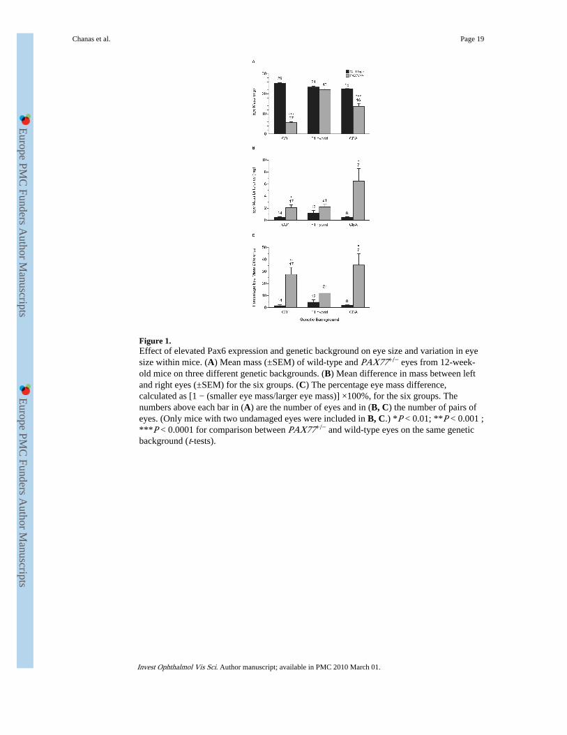
Figure 1.Effect of elevated Pax6 expression and genetic background on eye size and variation in eyesize within mice. (A) Mean mass (±SEM) of wild-type and PAX77+/− eyes from 12-week-old mice on three different genetic backgrounds. (B) Mean difference in mass between leftand right eyes (±SEM) for the six groups. (C) The percentage eye mass difference,calculated as [1 − (smaller eye mass/larger eye mass)] ×100%, for the six groups. Thenumbers above each bar in (A) are the number of eyes and in (B, C) the number of pairs ofeyes. (Only mice with two undamaged eyes were included in B, C.) *P < 0.01; **P < 0.001 ;***P < 0.0001 for comparison between PAX77+/− and wild-type eyes on the same geneticbackground (t-tests).
Chanas et al. Page 19
Invest Ophthalmol Vis Sci. Author manuscript; available in PMC 2010 March 01.
Europe PM
C Funders A
uthor Manuscripts
Europe PM
C Funders A
uthor Manuscripts
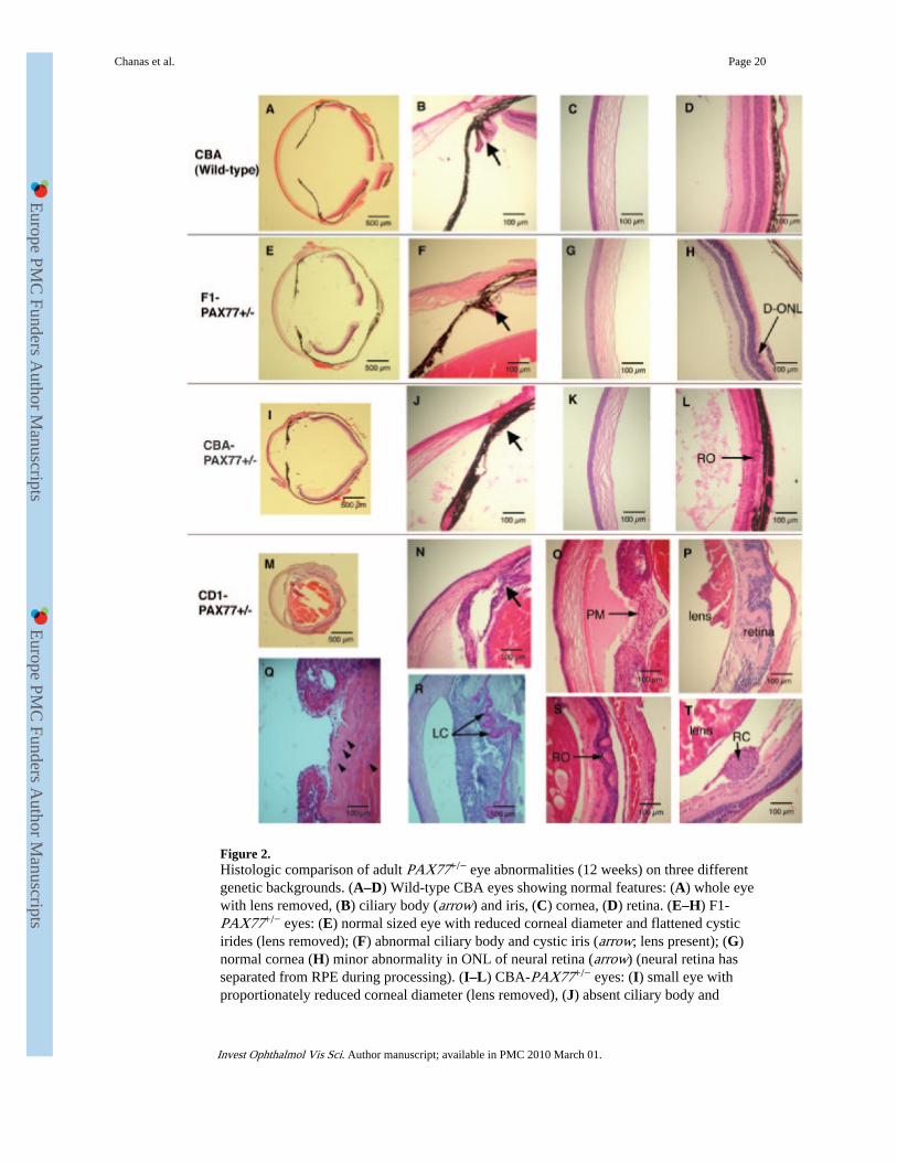
Figure 2.Histologic comparison of adult PAX77+/− eye abnormalities (12 weeks) on three differentgenetic backgrounds. (A–D) Wild-type CBA eyes showing normal features: (A) whole eyewith lens removed, (B) ciliary body (arrow) and iris, (C) cornea, (D) retina. (E–H) F1-PAX77+/− eyes: (E) normal sized eye with reduced corneal diameter and flattened cysticirides (lens removed); (F) abnormal ciliary body and cystic iris (arrow; lens present); (G)normal cornea (H) minor abnormality in ONL of neural retina (arrow) (neural retina hasseparated from RPE during processing). (I–L) CBA-PAX77+/− eyes: (I) small eye withproportionately reduced corneal diameter (lens removed), (J) absent ciliary body and
Chanas et al. Page 20
Invest Ophthalmol Vis Sci. Author manuscript; available in PMC 2010 March 01.
Europe PM
C Funders A
uthor Manuscripts
Europe PM
C Funders A
uthor Manuscripts

thickened, cystic iris (arrow), (K) cornea with normal epithelium, (L) eosinophilic materialin the vitreous and thin retina with rosettes (RO, arrow). (M–T) Albino CD1-PAX77+/−
eyes: (M) severely microphthalmic and disorganized eye (lens intact), (N) absent ciliarybody and thickened, cystic iris (arrow) and eosinophilic staining in the anterior chamber (O)persistent pupillary membrane (PM, arrow) and eosinophilic staining in the anteriorchamber, (P) abnormal and folded retina, (Q) lens epithelium is multi-layered (threearrowheads on left) and there appears to be undifferentiated lens epithelial cells among thelens fibers (arrowhead on right), (R) extensively folded lens capsule (arrows; stained withPAS), (S) vacuolated lens and abnormal retina with rosettes (RO, arrow), (T) ectopic ball ofretinal cells (RC, arrow) between retina and lens. D-ONL, disorganized outer nuclear layer;LC, lens capsule; PM, pupillary membrane; RC, retinal cells; RO, rosette.
Chanas et al. Page 21
Invest Ophthalmol Vis Sci. Author manuscript; available in PMC 2010 March 01.
Europe PM
C Funders A
uthor Manuscripts
Europe PM
C Funders A
uthor Manuscripts

Figure 3.Histologic analysis of E16.5 fetal mouse eyes on two genetic backgrounds. (A, B) Wild-type, albino CD1 eyes showing normal histology but with persistent clumps of cells (arrow)in the vitreous in (B). (C) Wild-type pigmented F1 eye showing normal histology. (D–F)CD1-PAX77+/− eyes with abnormal retinal folding (D, E) and ectopic ball (arrow) of cells(F). (D, arrow) A retinal fold in contact with the posterior surface of the lens in a region thatcontains cells with anterior lens epithelial morphology. (G–I) F1-PAX77+/− eye showingnormal morphology (G) but ingression of pigmented RPE cells into the optic nerve sheath(arrow in H; enlarged in I, arrow). C, clump of cells; PC, pigmented cells; RC, ectopic ballof retinal cells. Scale bar, 100 μm.
Chanas et al. Page 22
Invest Ophthalmol Vis Sci. Author manuscript; available in PMC 2010 March 01.
Europe PM
C Funders A
uthor Manuscripts
Europe PM
C Funders A
uthor Manuscripts
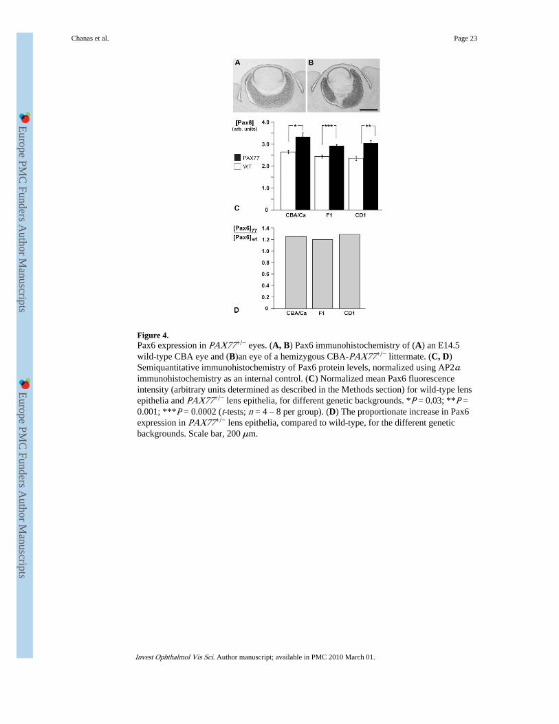
Figure 4.Pax6 expression in PAX77+/− eyes. (A, B) Pax6 immunohistochemistry of (A) an E14.5wild-type CBA eye and (B)an eye of a hemizygous CBA-PAX77+/− littermate. (C, D)Semiquantitative immunohistochemistry of Pax6 protein levels, normalized using AP2αimmunohistochemistry as an internal control. (C) Normalized mean Pax6 fluorescenceintensity (arbitrary units determined as described in the Methods section) for wild-type lensepithelia and PAX77+/− lens epithelia, for different genetic backgrounds. *P = 0.03; **P =0.001; ***P = 0.0002 (t-tests; n = 4 – 8 per group). (D) The proportionate increase in Pax6expression in PAX77+/− lens epithelia, compared to wild-type, for the different geneticbackgrounds. Scale bar, 200 μm.
Chanas et al. Page 23
Invest Ophthalmol Vis Sci. Author manuscript; available in PMC 2010 March 01.
Europe PM
C Funders A
uthor Manuscripts
Europe PM
C Funders A
uthor Manuscripts

Figure 5.Abnormalities in E16.5 PAX77+/−↔wild-type chimeras. Retinal abnormalities in E16.5PAX77+/−↔wild-type SCA chimeras: Retinal folds (arrows) could be composed largely ofPAX77+/− (Tg-negative) cells (A) or almost entirely of wild-type (Tg+/−) cells (B), ormixtures of cells of both genotypes (C). (D) Example of a small vesicle of wild-type (Tg+/−)cells (arrow) within PAX77+/− domain of retina. (E) Detached group of retinal cells (arrow)between the lens and retina of chimera SCA12. Several wild-type (Tg-positive) cells werepresent in the ectopic clump of predominantly PAX77+/− (Tg-negative) cells. Compositionof iris, ciliary body, and lens in E16.5 fetal chimeras: (F) SCA control chimera withapparently random distribution of pigmented and Tg+ cells in different eye tissues. SCAexperimental chimeras: (G) contribution of PAX77+/−, Tg−/− and PAX77−/−, Tg+/− cells toall tissues (including the lens) and a bud of PAX77+/−, Tyrc/c, and Tg−/− cells (arrow)projecting from the otherwise pigmented iris pigment epithelium. (H, I) Buds of Tg-positivewild-type (PAX77−/−, Tg+/−) cells (arrows) in developing ciliary bodies that are otherwisePAX77+/−, and Tg−/−. (J) Putative segregation of Tg-negative, PAX77+/− cells (arrow)within the trabecular meshwork of an experimental SCA chimera. Serous gland: (K) Pax6immunohistochemical staining of nonchimeric serous gland. (L) Partial segregation of Tg-negative, PAX77+/− cells to larger central serous gland tubules (nasal region) and Tg-positive, wild-type cells to smaller tubules (arrows). Ingression of RPE cells into the opticnerve heads of experimental chimeras: (M) Control SCA chimera showing a sharp boundarybetween pigmented cells of the RPE and the optic nerve. (N, O) Ingression of pigmentedcells (arrow) into the peripheral sheath of the optic nerve in experimental SCA and SCBchimeras. Because of the composition of the chimeras, the pigmented cells were PAX77+/−
in the SCB chimera shown in (N) but wild-type in the SCA chimera shown in (O). Scalebar: (A, B, C, L) 100 μm; (D, F–K, M–O) 50 μm; (E) 30 μm.
Chanas et al. Page 24
Invest Ophthalmol Vis Sci. Author manuscript; available in PMC 2010 March 01.
Europe PM
C Funders A
uthor Manuscripts
Europe PM
C Funders A
uthor Manuscripts
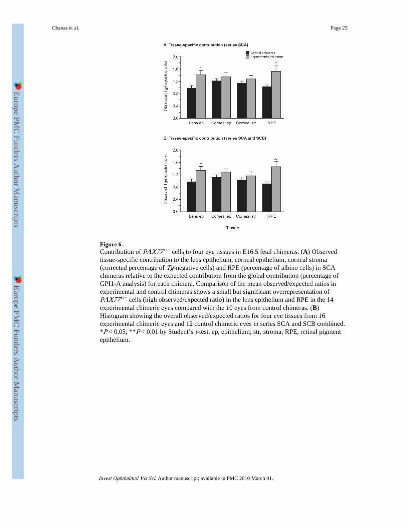
Figure 6.Contribution of PAX77+/− cells to four eye tissues in E16.5 fetal chimeras. (A) Observedtissue-specific contribution to the lens epithelium, corneal epithelium, corneal stroma(corrected percentage of Tg-negative cells) and RPE (percentage of albino cells) in SCAchimeras relative to the expected contribution from the global contribution (percentage ofGPI1-A analysis) for each chimera. Comparison of the mean observed/expected ratios inexperimental and control chimeras shows a small but significant overrepresentation ofPAX77+/− cells (high observed/expected ratio) in the lens epithelium and RPE in the 14experimental chimeric eyes compared with the 10 eyes from control chimeras. (B)Histogram showing the overall observed/expected ratios for four eye tissues from 16experimental chimeric eyes and 12 control chimeric eyes in series SCA and SCB combined.*P < 0.05; **P < 0.01 by Student’s t-test. ep, epithelium; str, stroma; RPE, retinal pigmentepithelium.
Chanas et al. Page 25
Invest Ophthalmol Vis Sci. Author manuscript; available in PMC 2010 March 01.
Europe PM
C Funders A
uthor Manuscripts
Europe PM
C Funders A
uthor Manuscripts

Europe PM
C Funders A
uthor Manuscripts
Europe PM
C Funders A
uthor Manuscripts
Chanas et al. Page 26
TAB
LE 1
Maj
or E
ffec
ts o
f th
e G
enet
ic B
ackg
roun
d on
PA
X77
+/−
Eye
Abn
orm
aliti
es
Gen
etic
Bac
kgro
und
Eye
Abn
orm
alit
yC
D1
CB
AF
1
Adu
lt
M
ean
perc
enta
ge o
f w
ild-t
ype
eye
mas
s22
%60
%95
%
M
icro
phth
alm
ia+
+−
A
bsen
t or
diso
rgan
ized
cili
ary
bodi
es+
++
C
ystic
iris
++
+
A
bnor
mal
ret
ina
++
+
Sm
all c
orne
a di
amet
er±
*+
+
Sm
all l
ens
++
ND
C
atar
act
++
ND
T
hick
ened
iris
++
−
E
osin
ophi
lic s
tain
ing
in a
nter
ior
cham
ber
++
−
E
ctop
ic r
etin
al c
ells
pos
teri
or to
lens
+N
DN
D
Pe
rsis
tent
pup
illar
y m
embr
ane
+−
−
Fetu
s (E
16.5
)
In
gres
sion
of
RPE
cel
ls in
to o
ptic
ner
ve s
heat
hN
D†
ND
+
R
etin
al f
oldi
ng+
ND
−
E
ctop
ic r
etin
al c
ells
pos
teri
or to
lens
+N
D−
ND
, not
don
e (e
.g. b
ecau
se le
nses
wer
e re
mov
ed f
rom
mos
t F1-
PAX
77+
/− a
nd C
BA
-PA
X77
+/−
eye
s be
fore
em
bedd
ing
for
hist
olog
y).
* Cor
neal
dia
met
er w
as n
ot o
bvio
usly
dis
prop
ortio
nate
ly r
educ
ed in
the
smal
lest
CD
1-PA
X77
+/−
eye
s.
† CD
1-PA
X77
+/−
mic
e ar
e al
bino
so
any
RPE
cel
ls in
the
optic
ner
ve s
heat
h w
ould
be
less
obv
ious
.
Invest Ophthalmol Vis Sci. Author manuscript; available in PMC 2010 March 01.
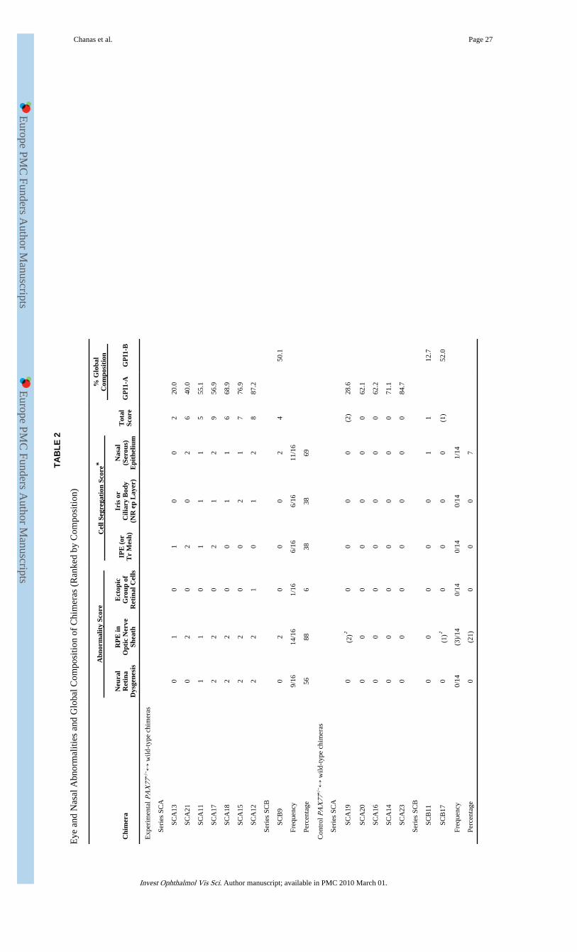
Europe PM
C Funders A
uthor Manuscripts
Europe PM
C Funders A
uthor Manuscripts
Chanas et al. Page 27
TAB
LE 2
Eye
and
Nas
al A
bnor
mal
ities
and
Glo
bal C
ompo
sitio
n of
Chi
mer
as (
Ran
ked
by C
ompo
sitio
n)
Abn
orm
alit
y Sc
ore
Cel
l Seg
rega
tion
Sco
re*
% G
loba
lC
ompo
siti
on
Chi
mer
aN
eura
lR
etin
aD
ysge
nesi
s
RP
E in
Opt
ic N
erve
Shea
th
Ect
opic
Gro
up o
fR
etin
al C
ells
IPE
(or
Tr
Mes
h)
Iris
or
Cili
ary
Bod
y(N
R e
p L
ayer
)
Nas
al(S
erou
s)E
pith
eliu
m
Tot
alSc
ore
GP
I1-A
GP
I1-B
Exp
erim
enta
l PA
X77
+/−↔
wild
-typ
e ch
imer
as
Se
ries
SC
A
SCA
130
10
10
02
20.0
SCA
210
20
20
26
40.0
SCA
111
10
11
15
55.1
SCA
172
20
21
29
56.9
SCA
182
20
01
16
68.9
SCA
152
20
02
17
76.9
SCA
122
21
01
28
87.2
Se
ries
SC
B
SCB
90
20
00
24
50.1
Fr
eque
ncy
9/16
14/1
61/
166/
166/
1611
/16
Pe
rcen
tage
5688
638
3869
Con
trol
PA
X77
+/−↔
wild
-typ
e ch
imer
as
Se
ries
SC
A
SCA
190
(2)†
00
00
(2)
28.6
SCA
200
00
00
00
62.1
SCA
160
00
00
00
62.2
SCA
140
00
00
00
71.1
SCA
230
00
00
00
84.7
Se
ries
SC
B
SCB
110
00
00
11
12.7
SCB
170
(1)†
00
00
(1)
52.0
Fr
eque
ncy
0/14
(3)/
140/
140/
140/
141/
14
Pe
rcen
tage
0(2
1)0
00
7
Invest Ophthalmol Vis Sci. Author manuscript; available in PMC 2010 March 01.

Europe PM
C Funders A
uthor Manuscripts
Europe PM
C Funders A
uthor Manuscripts
Chanas et al. Page 28T
he to
tal a
bnor
mal
ity/s
egre
gatio
n sc
ore
in P
AX
77+
/−↔
wild
-typ
e ch
imer
as s
how
ed a
sig
nifi
cant
cor
rela
tion
(Ken
dall
τ =
0.6
18; P
= 0
.032
) w
ith th
e gl
obal
com
posi
tion
(% G
PI1-
A o
r %
GPI
1-B
dep
endi
ng w
hich
is p
rodu
ced
by th
e PA
X77
+/−
cel
ls).
IPE
, iri
s pi
gmen
tep
ithel
ium
; RG
Cs,
ret
inal
gan
glio
n ce
lls R
PE, r
etin
al p
igm
ent e
pith
eliu
m; T
r m
esh,
trab
ecul
ar m
eshw
ork;
NR
ep
laye
r, e
pith
elia
l lay
er o
f ir
is a
nd c
iliar
y bo
dy th
at e
xten
ds f
rom
the
neur
al r
etin
a.
* Abn
orm
ality
or
cell
segr
egat
ion
scor
e: 0
, abs
ent;
1, p
rese
nt u
nila
tera
lly; 2
, pre
sent
bila
tera
lly.
† Onl
y in
divi
dual
RPE
cel
ls w
ere
pres
ent i
n th
e op
tic n
erve
she
ath
of c
ontr
ol c
him
eras
.
Invest Ophthalmol Vis Sci. Author manuscript; available in PMC 2010 March 01.
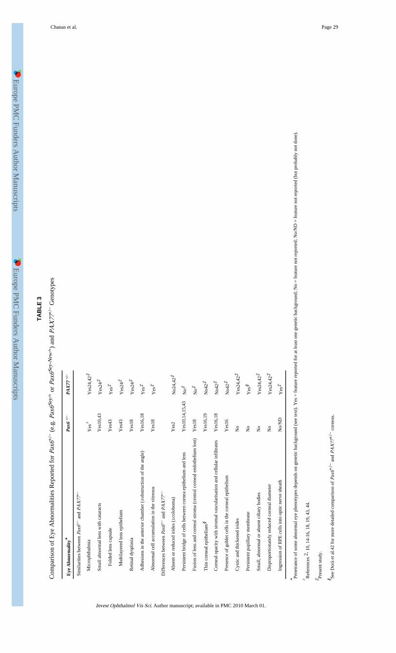
Europe PM
C Funders A
uthor Manuscripts
Europe PM
C Funders A
uthor Manuscripts
Chanas et al. Page 29
TAB
LE 3
Com
pari
son
of E
ye A
bnor
mal
ities
Rep
orte
d fo
r Pa
x6+
/− (
e.g.
Pax
6Sey/
+ o
r Pa
x6Se
y-N
ew/+
) an
d PA
X77
+/−
Gen
otyp
es
Eye
Abn
orm
alit
y*P
ax6
+/−
PA
X77
+/−
Sim
ilari
ties
betw
een
Pax6
+/−
and
PA
X77
+/−
M
icro
phth
alm
iaY
es†
Yes
24,4
2‡
Sm
all a
bnor
mal
lens
with
cat
arac
tsY
es10
,43
Yes
24‡
Fold
ed le
ns c
apsu
leY
es43
Yes
‡
Mul
tilay
ered
lens
epi
thel
ium
Yes
43Y
es24
‡
R
etin
al d
yspl
asia
Yes
18Y
es24
‡
A
dhes
ions
in th
e an
teri
or c
ham
ber
(±ob
stru
ctio
n of
the
angl
e)Y
es16
,18
Yes
‡
A
bnor
mal
cel
l acc
umul
atio
n in
the
vitr
eous
Yes
18Y
es‡
Dif
fere
nces
bet
wee
n Pa
x6+
/− a
nd P
AX
77+
/−
A
bsen
t or
redu
ced
irid
es (
±co
lobo
ma)
Yes
2N
o24,
42‡
Pe
rsis
tent
bri
dge
of c
ells
bet
wee
n co
rnea
epi
thel
ium
and
lens
Yes
10,1
4,15
,43
No‡
Fu
sion
of
lens
and
cor
neal
str
oma
(cen
tral
cor
neal
end
othe
lium
lost
)Y
es18
No‡
T
hin
corn
eal e
pith
eliu
m§
Yes
16,1
9N
o42‡
C
orne
al o
paci
ty w
ith s
trom
al v
ascu
lari
satio
n an
d ce
llula
r in
filtr
ates
Yes
16,1
8N
o42‡
Pr
esen
ce o
f go
blet
cel
ls in
the
corn
eal e
pith
eliu
mY
es16
No4
2‡
C
ystic
and
thic
kene
d ir
ides
No
Yes
24,4
2‡
Pe
rsis
tent
pup
illar
y m
embr
ane
No
Yes
§
Sm
all,
abno
rmal
or
abse
nt c
iliar
y bo
dies
No
Yes
24,4
2‡
D
ispr
opor
tiona
tely
red
uced
cor
neal
dia
met
erN
oY
es24
,42‡
In
gres
sion
of
RPE
cel
ls in
to o
ptic
ner
ve s
heat
hN
o/N
DY
es‡
* Pene
tran
ce o
f so
me
abno
rmal
eye
phe
noty
pes
depe
nds
on g
enet
ic b
ackg
roun
d (s
ee te
xt).
Yes
= f
eatu
re r
epor
ted
for
at le
ast o
ne g
enet
ic b
ackg
roun
d; N
o =
fea
ture
not
rep
orte
d; N
o/N
D =
fea
ture
not
rep
orte
d (b
ut p
roba
bly
not d
one)
.
† Ref
eren
ces
2, 1
0, 1
4-16
, 18,
19,
43,
44.
‡ Pres
ent s
tudy
.
§ See
Dor
à et
al.4
2 fo
r m
ore
deta
iled
com
pari
son
of P
ax6+
/− a
nd P
AX
77+
/− c
orne
as.
Invest Ophthalmol Vis Sci. Author manuscript; available in PMC 2010 March 01.

Europe PM
C Funders A
uthor Manuscripts
Europe PM
C Funders A
uthor Manuscripts
Chanas et al. Page 30
TAB
LE 4
Com
pari
son
of E
ffec
ts o
n E
16.5
Fet
al E
yes
in P
ax6+
/−↔
Wild
-Typ
e an
d PA
X77
+/−↔
Wild
-Typ
e C
him
eras
Tis
sue
Pax
6+/− ↔
Wild
-Typ
e C
him
eras
(Col
linso
n et
al.1
4)P
AX
77+/
− ↔W
ild-T
ype
Fet
al C
him
eras
(Pre
sent
Stu
dy)
Mor
phol
ogic
al a
bnor
mal
ities
O
ptic
ner
ve/r
etin
a bo
unda
ryN
one
RPE
cel
ls in
opt
ic n
erve
she
ath
N
eura
l ret
ina
Non
eR
etin
al d
ysge
nesi
s an
d ec
topi
c ba
lls o
f R
GC
s
Prob
ably
non
auto
nom
ous
effe
cts
O
ptic
ner
ve/r
etin
a bo
unda
ryN
one
PAX
77+
/− a
nd w
ild-t
ype
RPE
cel
ls in
ON
S
N
eura
l ret
ina
Non
ePA
X77
+/−
and
wild
-typ
e ce
lls in
ret
inal
fol
ds (
also
w
ild-t
ype
cells
am
ong
ecto
pic
balls
of
PAX
77+
/−
R
GC
s*)
Cel
l-au
tono
mou
s ef
fect
s
L
ens
epith
eliu
mPa
x6+
/− c
ells
exc
lude
d†PA
X77
+/−
cel
ls o
verr
epre
sent
ed
R
PEN
one
PAX
77+
/− c
ells
ove
rrep
rese
nted
N
eura
l ret
ina,
iris
pig
men
t epi
thel
ium
,
ci
liary
bod
y an
d ir
is N
R e
p la
yer
Non
eSo
me
segr
egat
ion
of P
AX
77+
/− a
nd w
ild-t
ype
cells
NR
ep
laye
r, e
pith
elia
l lay
er o
f ir
is a
nd c
iliar
y bo
dy th
at e
xten
ds f
rom
the
neur
al r
etin
a; O
NS,
opt
ic n
erve
she
ath;
RG
Cs,
ret
inal
gan
glio
n ce
lls; R
PE, r
etin
al p
igm
ent e
pith
eliu
m.
* Furt
her
wor
k is
nee
ded
to d
eter
min
e w
heth
er th
e ef
fect
is c
ell-
auto
nom
ous
or n
onau
tono
mou
s.
† Pax6
+/−
cel
ls w
ere
excl
uded
fro
m th
e le
ns e
pith
eliu
m in
the
firs
t stu
dy o
f Pa
x6+
/−↔
wild
-typ
e ch
imer
as.1
4 A
mor
e re
cent
stu
dy s
how
ed th
at P
ax6+
/− c
ells
cou
ld c
ontr
ibut
e to
the
lens
epi
thel
ium
of
Pax6
+/−
↔w
ild-t
ype
chim
eras
,30
but i
t was
unc
lear
whe
ther
they
wer
e de
plet
ed b
ecau
se th
e co
ntri
butio
n of
Pax
6+/−
cel
ls w
as n
ot q
uant
ifie
d.
Invest Ophthalmol Vis Sci. Author manuscript; available in PMC 2010 March 01.
Related Documents


