The Plasma Membrane Proton Pump PMA-1 Is Incorporated into Distal Parts of the Hyphae Independently of the Spitzenkörper in Neurospora crassa Rosa A. Fajardo-Somera, a Barry Bowman, b Meritxell Riquelme a Department of Microbiology, Center for Scientific Research and Higher Education of Ensenada (CICESE), Ensenada, Baja California, Mexico a ; Department of Molecular, Cell & Developmental Biology, University of California, Santa Cruz, California, USA b Most models for fungal growth have proposed a directional traffic of secretory vesicles to the hyphal apex, where they temporar- ily aggregate at the Spitzenkörper before they fuse with the plasma membrane (PM). The PM H -translocating ATPase (PMA-1) is delivered via the classical secretory pathway (endoplasmic reticulum [ER] to Golgi) to the cell surface, where it pumps H out of the cell, generating a large electrochemical gradient that supplies energy to H -coupled nutrient uptake systems. To charac- terize the traffic and delivery of PMA-1 during hyphal elongation, we have analyzed by laser scanning confocal microscopy (LSCM) strains of Neurospora crassa expressing green fluorescent protein (GFP)-tagged versions of the protein. In conidia, PMA-1-GFP was evenly distributed at the PM. During germination and germ tube elongation, PMA-1-GFP was found all around the conidial PM and extended to the germ tube PM, but fluorescence was less intense or almost absent at the tip. Together, the data indicate that the electrochemical gradient driving apical nutrient uptake is generated from early developmental stages. In mature hyphae, PMA-1-GFP localized at the PM at distal regions (>120 m) and in completely developed septa, but not at the tip, indicative of a distinct secretory route independent of the Spitzenkörper occurring behind the apex. O ne of the main features of filamentous fungi is their apical mode of growth. In filamentous fungi, cell wall growth and exocytosis are linked processes that involve the highly polarized traffic of cell wall-building secretory vesicles to apical areas, where they deliver proteins and lipids. One of the most widely accepted models for fungal growth, the vesicle supply center for fungal morphogenesis, postulates a unidirectional traffic of vesicles to the hyphal apex, where they aggregate temporarily at an apical structure, the Spitzenkörper, prior to fusion with the apical plasma membrane (PM) by the process of exocytosis (1). How- ever, some PM proteins, such as the H -ATPases, essential for hyphal growth, have been previously predicted to be absent or inactive at the hyphal apex (2–4), suggesting the existence of ves- icle delivery routes other than the above and independent of the Spitzenkörper that reach nonapical regions of the hyphal PM. H - ATPases are involved in pumping protons out of the cell, gener- ating a large electrochemical gradient and supplying energy to H -coupled nutrient uptake systems (5). This electrochemical gradient has been studied in several fungal species by diverse methods, including vibrating probes, microelectrodes, and pH in- dicators. The results showed that current normally flows inward at the hyphal apical regions and flows outward at distal areas (3, 4, 6–9). PMA1, the H -ATPase-encoding gene in Saccharomyces cerevisiae (SCRG_01016 [10]), has a single homolog in Neurospora crassa (NCU01680 [11, 12]). In S. cerevisiae, Pma1p is one of the most abundant proteins of the cell surface (25 to 50%), while in N. crassa, it constitutes about 5 to 10% of the PM total protein con- tent (13, 14). In S. cerevisiae, Pma1p is delivered to the cell surface via the classical endoplasmic reticulum (ER) to the Golgi secretory path- way defined by the SEC genes (15–17), where specialized proteins ensure the efficient transport of Pma1p through the secretory pathway. For instance, Lst1p (Sec24p homolog) is involved in the export of Pma1p from the ER. Together with Sec23p, Lst1p is necessary for the efficient packing of Pma1p into ER-derived COPII vesicles (18, 19). Furthermore, Ast1p and Ast2p participate in the transport of Pma1p from Golgi to the PM (20). Upon arrival to the PM, Pma1p is very stable (with a half-life of 12 h [21]). Lipid rafts have a role in sorting Pma1p to the PM. Upon lipid raft disruption, Pma1p is missorted to the vacuole, where it is de- graded (22). In elo3 and lcb1 mutants, defective in lipid rafts, mutant pma1-7 is not degraded in vacuoles. Instead, it is delivered to the PM (23, 24). As part of an ongoing project to characterize the relationship between the secretory pathway and hyphal growth in filamentous fungi, we have analyzed the biogenesis and trafficking of the PM H -translocating ATPase in N. crassa by fusing the gene pma-1 to gfp and studying the location of PMA-1 in living cells during veg- etative development. MATERIALS AND METHODS Culture conditions. N. crassa cells were grown on Vogel’s minimal me- dium agar (VMM [25]) supplemented with histidine (0.1 mg/ml) when needed. Liquid cultures were grown in Vogel’s complete medium (VCM; 1 Vogel’s salts, 1.5% sucrose, 0.5% yeast extract, 0.5% Casamino Acids) at 30°C and 200 rpm for 16 h. For transformation, conidia were spread directly on plates containing FGS medium (0.05% fructose, 0.05% glu- cose, 2% sorbose) or mixed with top agar (1 Vogel’s salts, 1 M sorbitol, Received 28 January 2013 Accepted 28 May 2013 Published ahead of print 31 May 2013 Address correspondence to Meritxell Riquelme, [email protected]. Supplemental material for this article may be found at http://dx.doi.org/10.1128 /EC.00328-12. Copyright © 2013, American Society for Microbiology. All Rights Reserved. doi:10.1128/EC.00328-12 August 2013 Volume 12 Number 8 Eukaryotic Cell p. 1097–1105 ec.asm.org 1097 on March 27, 2016 by guest http://ec.asm.org/ Downloaded from

Welcome message from author
This document is posted to help you gain knowledge. Please leave a comment to let me know what you think about it! Share it to your friends and learn new things together.
Transcript
The Plasma Membrane Proton Pump PMA-1 Is Incorporated intoDistal Parts of the Hyphae Independently of the Spitzenkörper inNeurospora crassa
Rosa A. Fajardo-Somera,a Barry Bowman,b Meritxell Riquelmea
Department of Microbiology, Center for Scientific Research and Higher Education of Ensenada (CICESE), Ensenada, Baja California, Mexicoa; Department of Molecular, Cell &Developmental Biology, University of California, Santa Cruz, California, USAb
Most models for fungal growth have proposed a directional traffic of secretory vesicles to the hyphal apex, where they temporar-ily aggregate at the Spitzenkörper before they fuse with the plasma membrane (PM). The PM H�-translocating ATPase (PMA-1)is delivered via the classical secretory pathway (endoplasmic reticulum [ER] to Golgi) to the cell surface, where it pumps H� outof the cell, generating a large electrochemical gradient that supplies energy to H�-coupled nutrient uptake systems. To charac-terize the traffic and delivery of PMA-1 during hyphal elongation, we have analyzed by laser scanning confocal microscopy(LSCM) strains of Neurospora crassa expressing green fluorescent protein (GFP)-tagged versions of the protein. In conidia,PMA-1-GFP was evenly distributed at the PM. During germination and germ tube elongation, PMA-1-GFP was found all aroundthe conidial PM and extended to the germ tube PM, but fluorescence was less intense or almost absent at the tip. Together, thedata indicate that the electrochemical gradient driving apical nutrient uptake is generated from early developmental stages. Inmature hyphae, PMA-1-GFP localized at the PM at distal regions (>120 �m) and in completely developed septa, but not at thetip, indicative of a distinct secretory route independent of the Spitzenkörper occurring behind the apex.
One of the main features of filamentous fungi is their apicalmode of growth. In filamentous fungi, cell wall growth and
exocytosis are linked processes that involve the highly polarizedtraffic of cell wall-building secretory vesicles to apical areas, wherethey deliver proteins and lipids. One of the most widely acceptedmodels for fungal growth, the vesicle supply center for fungalmorphogenesis, postulates a unidirectional traffic of vesicles tothe hyphal apex, where they aggregate temporarily at an apicalstructure, the Spitzenkörper, prior to fusion with the apicalplasma membrane (PM) by the process of exocytosis (1). How-ever, some PM proteins, such as the H�-ATPases, essential forhyphal growth, have been previously predicted to be absent orinactive at the hyphal apex (2–4), suggesting the existence of ves-icle delivery routes other than the above and independent of theSpitzenkörper that reach nonapical regions of the hyphal PM. H�-ATPases are involved in pumping protons out of the cell, gener-ating a large electrochemical gradient and supplying energy toH�-coupled nutrient uptake systems (5). This electrochemicalgradient has been studied in several fungal species by diversemethods, including vibrating probes, microelectrodes, and pH in-dicators. The results showed that current normally flows inward atthe hyphal apical regions and flows outward at distal areas (3, 4,6–9).
PMA1, the H�-ATPase-encoding gene in Saccharomycescerevisiae (SCRG_01016 [10]), has a single homolog in Neurosporacrassa (NCU01680 [11, 12]). In S. cerevisiae, Pma1p is one of themost abundant proteins of the cell surface (25 to 50%), while in N.crassa, it constitutes about 5 to 10% of the PM total protein con-tent (13, 14).
In S. cerevisiae, Pma1p is delivered to the cell surface via theclassical endoplasmic reticulum (ER) to the Golgi secretory path-way defined by the SEC genes (15–17), where specialized proteinsensure the efficient transport of Pma1p through the secretorypathway. For instance, Lst1p (Sec24p homolog) is involved in the
export of Pma1p from the ER. Together with Sec23p, Lst1p isnecessary for the efficient packing of Pma1p into ER-derivedCOPII vesicles (18, 19). Furthermore, Ast1p and Ast2p participatein the transport of Pma1p from Golgi to the PM (20). Upon arrivalto the PM, Pma1p is very stable (with a half-life of �12 h [21]).Lipid rafts have a role in sorting Pma1p to the PM. Upon lipid raftdisruption, Pma1p is missorted to the vacuole, where it is de-graded (22). In �elo3 and �lcb1 mutants, defective in lipid rafts,mutant pma1-7 is not degraded in vacuoles. Instead, it is deliveredto the PM (23, 24).
As part of an ongoing project to characterize the relationshipbetween the secretory pathway and hyphal growth in filamentousfungi, we have analyzed the biogenesis and trafficking of the PMH�-translocating ATPase in N. crassa by fusing the gene pma-1 togfp and studying the location of PMA-1 in living cells during veg-etative development.
MATERIALS AND METHODSCulture conditions. N. crassa cells were grown on Vogel’s minimal me-dium agar (VMM [25]) supplemented with histidine (0.1 mg/ml) whenneeded. Liquid cultures were grown in Vogel’s complete medium (VCM;1� Vogel’s salts, 1.5% sucrose, 0.5% yeast extract, 0.5% Casamino Acids)at 30°C and 200 rpm for 16 h. For transformation, conidia were spreaddirectly on plates containing FGS medium (0.05% fructose, 0.05% glu-cose, 2% sorbose) or mixed with top agar (1� Vogel’s salts, 1 M sorbitol,
Received 28 January 2013 Accepted 28 May 2013
Published ahead of print 31 May 2013
Address correspondence to Meritxell Riquelme, [email protected].
Supplemental material for this article may be found at http://dx.doi.org/10.1128/EC.00328-12.
Copyright © 2013, American Society for Microbiology. All Rights Reserved.
doi:10.1128/EC.00328-12
August 2013 Volume 12 Number 8 Eukaryotic Cell p. 1097–1105 ec.asm.org 1097
on March 27, 2016 by guest
http://ec.asm.org/
Dow
nloaded from

1% agar, 1� FGS) and spread onto 3 petri dishes containing FGS mediumsupplemented with hygromycin (300 �g/ml).
For confocal microscopy, transformants of N. crassa expressing greenfluorescent protein (GFP) and/or red fluorescent protein (RFP) were rou-tinely grown on VMM overnight at 30°C. For imaging, the inverted agarblock method was used as previously described (27). Strains used or gen-erated in this study are listed in Table 1.
Recombinant DNA techniques and plasmid constructions. Sincepma-1 is an essential gene, PMA-1-GFP, under the control of the ccg-1promoter, was targeted to the his-3 locus. Standard methods were usedfor cloning procedures to fuse GFP to the carboxy terminus of PMA-1(28). PCR was performed in a Bio-Rad thermal cycler using PlatinumPfu polymerase (Invitrogen) according to the manufacturer’s instruc-tions. The predicted pma-1 gene (3,077 bp; NCU01680.4) was ampli-fied from N. crassa genomic DNA by PCR using custom-designedprimers. Forward primer PMA-1-XbaI-F at the 5= end of the genecontained an introduced XbaI restriction site; reverse primer PMA-1-PacI-R at the 3= end of the gene sequence, excluding the stop codon,contained a PacI restriction site (Table 2). The amplified and purifiedPCR product was digested with XbaI and PacI and cloned into XbaI-and PacI-digested plasmid pMF272 (NCBI GenBank accession num-ber AY598428 [29]), yielding plasmid pMR001. All plasmids werepropagated in Escherichia coli strain DH5� supercompetent cells (In-vitrogen). Sequencing of plasmid pMR001 was carried out at the CoreInstrumentation Facility of the Institute for Integrative Genome Biol-ogy at the University of California, Riverside.
To overcome potential mislocalization of GFP-tagged PMA-1 due tooverexpression driven by the ccg-1 promoter, we constructed a strain inwhich the 3= end of the endogenous pma-1 was fused to the gfp gene, usingfusion PCR (see Fig. S1 in the supplemental material) (M. Riquelme, E. L.Bredeweg, O. Callejas-Negrete, R. W. Roberson, S. Ludwig, A. Beltrán-Aguilar, S. Seiler, and M. Freitag, unpublished data). Primer pairsORFS54-F P1–ORFS55-R P2 and ORFS56-F P3–ORFS57-R P4 (Table 2)were used to amplify 1 kb of the pma-1 open reading frame (ORF) and 3=untranslated region (UTR), respectively, from N. crassa genomic DNA.The cassette gfp-hph was amplified using primers 10XGlyF P7 and loxPRP8 (Table 2) from plasmid pCCG-1::Gly::GFP (30). Both fragments (ORFor 3= UTR) and the cassette gfp-hph were fused by PCR using primers pairsORFS54-F P1– hph SMR P6 (Table 2) to amplify 1 kb of the ORF fused togfp-hph and hph SMF P5–ORFS57-R P4 (Table 2) to amplify hph fused to1 kb of the 3= UTR. PCRs were performed in a Lab Line thermal cycler
TABLE 1 Plasmids and strains used or generated in this study
Plasmid or strain Characteristics or genotypeReference orsource
PlasmidspMF272 Pccg-1::sgfp� 29pZERO-GFP Pccg-1::10Xgly::gfp�::loxp�::hph�::loxp� 30pMR001 Pccg-1:: pma-1::sgfp� This study
StrainsN1 mata FGSC#988N150 matA FGSC#9013SMRP277 mata sad-2�::hph� FGSC#20680SMRP278 matA sad-2�::hph� 32N39 matA fl FGSC#4317N40 mata fl FGSC#4347VMA-1-RFP matA vma-1-rfp� FGSC#10169SMRP24 matA his-3 mus-51�::bar� FGSC#9717SMRP25 mata mus-51�::bar� FGSC#9718TESM001-3a matA his-3�::Pccg-1::pma-1::sgfp� This studyTRAF6-5a mata native promoter pma-1::sgfp� This studyTRAF9-1a mata native promoter pmb::sgfp� This studyNRAF6-11 mata native promoter pma-1::sgfp� This studyNRAF9-1 mata native promoter pmb::sgfp� This study
a Heterokaryon.
TABLE 2 Oligonucleotides used or generated in this study
Oligonucleotide Sequence Source or reference
PMA-1-Xba-F 5=- AATCTAGACGCCAATGGCGGACCACTC-3= This studyPMA-1-PacI-R 5=-CGTTAATTAATTGCGACTTCTCATGCTGAG-3= This studyORFS45-F P1 5=-CGAGCCGGGTGCTTTTGCTCCCTA-3= This studyORFS46-R P2 5=-TCCGCCTCCGCCTCCGCCGCCTCCGCCACCCATGATACCCGTTCC-3= This studyORFS47-F P3 5=-TATACGAAGTTATGGATCCGAGCTCGGGGGATGGTATAATGGAAGG-3= This studyORFS48-R P4 5=-CGCTTTCCGTCCTTCTGCAACCAC-3= This studyORFS54-F P1 5=-GCCCGGCTCTGAGGTCTACGAC-3= This studyORFS55-R P2 5=-CCGCCTCCGCCTCCGCCGCCTCCGCCTTGCGACTTCTCATGCTGAG-3= This studyORFS56-F P3 5=-GCTATACGAAGTTATGGATCCGAGCTCGGCGATATAATGATTTTCC-3= This studyORFS57-R P4 5=-CCACAAACCAGAAATAACCGTGCC-3= This studyORFS87-F P1seq 5=-CCTCCCCGTCACGATACCTAC-3= This studyORFS88-F P2seq 5=-CCGTATCTGGATCTTCTCCTTC-3= This studyORFS89-F P1seq 5=-GTCATTTTCGTCATCAACCTG-3= This studyORFS90-F P2seq 5=-CAAGGCTTCACCGCGTTCATGC-3= This studyhph SMR P6 5=-TCGCCTCGCTCCAGTCAATGACC-3= Riquelme et al., unpublishedhph SMF P5 5=-AAAAAGCCTGAACTCACCGCGACG-3= Riquelme et al., unpublished10XGlyF P7 5=-GGCGGAGGCGGCGGAGGCGGAGGCGGAGG-3= Riquelme et al., unpublishedloxPR P8 5=-CGAGCTCGGATCCATAACTTCGTATAGCA-3= Riquelme et al., unpublishedOMF1651-R 5=-CGGCTGAAGCACTGCACGCCG-3= Freitag labMRp10 F 5=-AGAGACAAGAAAATTACCCCCTTCTT-3= 31MRp11 R 5=-AACTACAACAGCCACAACGTCTATATC-3= 31MRp12 F 5=-ATAATGAACGGAAGGTAGTTGTAGAAAG-3= 31MRp13 R 5=-ATGGATATAATGTGGCTGTTGAAAG-3= 31pMF272F 5=-CAAATCAACACAACACTCAAACCA-3= 31pMF272 R 5=-GGCACCACCCCGGTGAACAGCTCC-3= 31
Fajardo-Somera et al.
1098 ec.asm.org Eukaryotic Cell
on March 27, 2016 by guest
http://ec.asm.org/
Dow
nloaded from
using TaKaRa DNA polymerase (TaKaRa Inc.). pmb, the basic amino acidpermease-encoding gene (NCU05168.4), was gfp tagged using the samestrategy with primer pairs ORFS45-F P1 and ORFS46-R P2 and ORFS47-FP3 and ORFS48-R P4.
Neurospora genetic methods. Transformation of N. crassa strainFGSC#9717 conidia (his-3; �mus-51 matA) with plasmid pMR001 (DraIlinearized) was carried out by electroporation on a Bio-Rad Gene Pulser(25-�F capacitance, 1.5-kV voltage, and 600-� resistance). his-3� pro-totrophic transformants showing fluorescence were selected (TESM003).The correct integration of the recombinant cassette in the selected trans-formant, TESM001-3, was verified by PCR using primers MRp10 F,MRp11 R, MRp12 F, and MRp13 R as previously reported (31).
For transformation with DNA fragments obtained from fusion PCR,conidia of N. crassa strain FGSC#9718 (�mus-51 mata) were used. Hy-gromycin (300 �g/ml)-resistant heterokaryon transformants (PMA-1-GFP or PMB-GFP) showing fluorescence were selected (TRAF6-5 andTRAF9-1, respectively).
To obtain homokaryon strains expressing PMA-1-GFP or PMB-GFP,we crossed N. crassa heterokaryon strains TRAF6-5 and TRAF9-1 with N.crassa �sad-2 (matA [32]). Hygromycin (300 �g/ml)-resistant coloniesthat showed fluorescence were selected (NRAF6-11 and NRAF9-1).
Correct integration in NRAF6-11 and NRAF9-1 strains was verified byPCR using primer pairs ORFS87-F P1 seq/OMF1651-R and ORFS88-F P2seq/hph SMR P6 and primer pairs ORFS89-F P1 seq/OMF1651-R andORFS90-F P2 seq/hph SMR P6, respectively (see Fig. S2 in the supplemen-tal material). Mycelium for genomic DNA extraction was grown at 28°Cfor 7 days on liquid medium with no shaking and no light, filtered, sub-merged in liquid nitrogen, and lyophilized. DNA was extracted with theDNeasy plant extraction kit (Qiagen).
Sucrose gradient and Western blot analysis. Liquid Vogel’s completemedium (VCM) was inoculated with NRAF6-11 (1 � 106 conidia/ml)and incubated for 16 h at 30°C and 200 rpm. Mycelia were filtered byvacuum, triturated with liquid nitrogen, and homogenized with 1 volumeof glass beads (0.5 mm; Sigma) and 2 volumes of 33 mM phosphate bufferpH 8.2 (38) in a Braun MSK homogenizer for 4 pulses for 30 s each time inthe presence of CO2 flow as the coolant. The homogenate was centrifugedat 5,000 � g for 5 min at 4°C. The total extract was loaded onto a sucrosegradient as described previously (33). Density (Abbe refractometer; CarlZeiss, Göttingen, Germany) and absorbance (280 nm, 6505 UV-visible[UV-Vis] spectrophotometer; Jenway) were determined for each fraction.
For Western blot analysis, 30 �l of each fraction was loaded per lane.The samples were heated at 37°C for 30 min and run in a 7% SDS-poly-acrylamide gel. Anti-GFP (1:500; Santa Cruz Biotechnology) and goatanti-mouse IgG conjugated to horseradish peroxidase (1:1000; Roche)were used as primary and secondary antibodies, respectively.
Fluorescence microscopy and image processing. Cells expressingGFP were imaged using an inverted Zeiss laser scanning confocal micro-scope (LSM-510; Carl Zeiss) and an Olympus FluoView FV1000 confocalmicroscope (Olympus, Japan), both fitted with an argon ion laser with aGFP filter set with a 488-nm excitation wavelength and a 505- to 550-nmemission wavelength and an He/Ne1 laser with an RFP filter set with a543-nm excitation wavelength and a 560-nm emission wavelength. A100� (PH3), 1.3-numerical aperture (NA) Plan-Neofluar oil immersionobjective was used for LSM-510. For FV1000, a 60�, 1.42-NA Plan-Apo-chromat oil immersion objective was used. A photomultiplier moduleallowed us to combine the confocal with phase-contrast or differentialinterference contrast (DIC) to provide us with a simultaneous view of thefluorescently labeled proteins and the entire cell. The imaging parametersused produced no detectable background signal from any source otherthan from GFP. Time-lapse imaging was performed at scan intervals of 1.5to 30 s for periods up to 40 min and captured with a combination of lowenergy, high attenuation, and a less concentrated excitation laser beamcaused by the low-NA objective, to avoid photobleaching during repeti-tive imaging. Confocal images were captured using LSM-510 (version 3.2;Carl Zeiss) or Olympus Fluoview (version 4.0a; Olympus) software and
evaluated with an LSM-510 image examiner (version 3.2) or OlympusFluoview viewer (version 4.0a), respectively. Some of the time series wereconverted into animation movies using the same software.
To analyze the distribution of the PMA-1-GFP fluorescence, a recon-struction was made with Adobe Photoshop CS5 for 20 hyphae, taking 10frames for each hypha along its longitudinal axis. The first captured frameincluded the hyphal apex up to the region starting to show fluorescence;the next 9 frames included the region behind the first frame to the regionwhere fluorescence could be observed in the PM. After incubation for 5 h,germinated conidia of strains expressing PMA-1-GFP (under the controlof native or ccg-1 promoters) were imaged and the fluorescence of thePMA-1–GFP driven by Pccg-1 was measured from the base of the conid-ium to the region of the germ tube PM where fluorescence was no longerobserved.
For fluorescence recovery after photobleaching (FRAP) analysis, a re-gion of interest was selected, and 500 repetitions at 90% laser intensitywere applied during a time series sequence acquisition. Triplicate experi-ments were performed. For fluorescence intensity measurements beforeand after photobleaching, ImageJ (National Institutes of Health, USA)software was used. FRAPAnalyser (University of Luxembourg) was usedto obtain fluorescence recovery plots and fitted curves. All analyses weresubmitted to a background subtraction. Nonadjacent unbleached PM re-gions were used as reference controls.
Cells stained with FM4-64 [N-(3-triethylammoniumpropyl)-4-(6-(4(diethylamino) phenyl) hexatrienyl) pyridinium dibromide; MolecularProbes] were imaged with the argon ion laser (excitation wavelength, 514nm, and emission wavelength, 670 nm).
For each treatment, agar blocks containing hyphae were inverted ontoa coverslip containing a 10-�l drop of the corresponding solution. Cellswere imaged 5 to 10 min after that.
Inhibitors and fluorescent dyes. An inhibitor of the classicalER-Golgi secretory pathway, brefeldin A (BFA; Sigma), was added togrowing hyphae at a final concentration of 200 �g/ml in VMM from a20-mg/ml stock solution in dimethyl sulfoxide (DMSO). FM4-64 wasused at a final concentration of 10 �M in VMM. Cytoskeleton inhibitorswere used to disturb the Spitzenkörper. Stock solutions of cytochalasin A(Sigma; 1 mg/ml) and latrunculin A (Sigma; 400 �g/ml), both actin in-hibitors, were prepared in DMSO and diluted in VMM to working con-centrations of 50 �g/ml and 20 �g/ml, respectively. A stock solution ofmethyl 1-(butylcarbamoyl)-2-benzimidazolecarbamate (benomyl; Sig-
FIG 1 PMA-1-GFP expression under the control of the pma-1 native pro-moter in conidia and germlings of N. crassa. (A) In conidia, PMA-1-GFP wasevenly distributed along the PM; (B to F) in young germlings, ranging from 7to 49 �m in length, PMA-1-GFP accumulated all along the PM, although withless intensity at the apex. Bars � 5 �m.
Distribution of H�-ATPase in Neurospora crassa
August 2013 Volume 12 Number 8 ec.asm.org 1099
on March 27, 2016 by guest
http://ec.asm.org/
Dow
nloaded from

ma; 500 �g/ml), a microtubule inhibitor, was prepared in ethanol anddiluted in VMM to a working concentration of 100 �g/ml.
RESULTSSelection of strains expressing PMA-1-GFP. Strains showing flu-orescence were selected by screening prototrophic transformantsby laser scanning confocal microscopy (LSCM). The integrationof the pma-1-gfp cassette at the his-3 locus was confirmed by PCR,yielding the predicted 3.2-kb amplicon with primers MRp10 F andMRp11 R and a 2.1-kb amplicon with primers MRp12 F andMRp13 R (data not shown). The correct integration of the pma-1-gfp-hph and pmb-gfp-hph cassettes was confirmed by PCR withprimers sets ORFS87-F P1seq–OMF1651-R and ORFS88-FP2seq–OMF1651-R to pma-1 and with primers sets ORFS89-FP1seq–OMF1651-R and ORFS90-F P2seq–OMF1651-R to pmb,obtaining the expected 1.5-kb and 2.1-kb amplicons (data notshown). Strains TESM003, NRAF6-11, and NRAF9-1 showingstable fluorescence were selected for subsequent analyses.
Although the strain expressing PMA-GFP under the control ofthe endogenous promoter grew slower than the correspondingparental strain (see Fig. S3 in the supplemental material), itshowed wild-type hyphal morphology. pma-1 is an essential gene;therefore, the viability of the strain with the replacement of thenative gene with a pma-1-gfp version indicated that the recombi-nant protein was functional. Therefore, all subsequent micros-copy analyses were conducted using the strain in which PMA-1-GFP was expressed under the control of the native promoter.
The molecular mass of N. crassa PMA-1 is 99.88 kDa (920amino acids), but when the protein is tagged with GFP, the mo-lecular mass increases to 126.8 kDa, as confirmed by Westernblotting (see Fig. S4B in the supplemental material). We used asucrose gradient to identify PMA-1-GFP-containing vesicles ac-cording to their buoyant densities and to compare the buoyantdensities with those of previously reported markers of the secre-tory pathway. PMA-1-GFP was detected in fractions 7 to 19, cor-responding to densities ranging between 1.103 and 1.213 g/ml.The strongest signal was observed for high-density fractions 15and 16 (see Fig. S4A and B), corresponding to 1.155 and 1.158g/ml. This indicated that PMA-1 travels in high-density vesiclesand is found at the PM (34, 35).
PMA-1-GFP is absent in the apical plasma membrane. PMA-1-GFP was practically absent at the apex of the PM of germlings.The distribution of PMA-1-GFP was analyzed during the differentgermination phases, previously established (36, 37): phase I,conidia during hydration; phase II, germ tube emergence; phaseIII, germ tube elongation, and phase IV, mature hyphae. In non-germinated conidia growing isotropically during phase I, PMA-1-
GFP fluorescence was evenly distributed throughout the PM (Fig.1A). In the next phases of germination (II and III), expression ofPMA-1-GFP was found all around the conidial PM and extendedto the germ tube PM, although at the tip the fluorescence was lessintense or almost absent (Fig. 1B to F).
In mature hyphae, PMA-1-GFP was localized at the PM atdistal hyphal regions and in already completed septa. Mature hy-phae are subdivided in three regions: region I, or hyphal apex (thefirst 1 to 5 �m); region II, or subapical region (5 to 20 �m); andregion III, or distal region (�20 �m) (36). In mature hyphae,PMA-1-GFP was found in the PM at far distal regions (�120 �mfrom the apex) (Fig. 2A). PMA-1-GFP did not accumulate at thePM in hyphal region I or II or the first portion of region III (Fig. 2Aand B). In distal region III, PMA-1-GFP could be found in thelumen of tubular endomembranes (Fig. 2B). These tubular endo-membranes partially colocalized with the vacuolar H�-translocat-ing ATPase VMA-1 (Fig. 3). In distal regions, we observed PMA-1-GFP in the lumen of globular vacuoles, whereas VMA-1-RFPwas found at the membrane of the globular vacuoles (Fig. 3A toD). Furthermore, we observed small vesicular carriers in germ-lings and in apical and distal regions of hyphae (Fig. 1 and 2).These small vesicles seemed to fuse with the PM, and they did notcolocalize with FM4-64 (see Movies S1 and S2 in the supplementalmaterial).
In distal regions, PMA-1-GFP was also found in the PM ofmature septa (Fig. 2D, 3, and 4J). To investigate whether PMA-1-GFP participates in septum formation, the cells were stained withthe vital dye FM4-64. FM4-64 accumulated at the septum duringits formation, when no PMA-1-GFP could be observed (Fig. 4A toC; see also Movie S1 in the supplemental material). FM4-64 was
FIG 2 Distribution of PMA-1-GFP in a mature hypha of N. crassa. (A) Reconstruction of a growing hypha imaged by LSCM; (B to D) magnifications of boxesin panel A. (B) At the hyphal subapex (�50 �m from the tip), PMA-1-GFP localized at individual vesicles (arrows); (C) behind the subapex, PMA-1-GFPlocalized at the tubular endomembrane system; (D) in distal areas (�120 �m under the native promoter), PMA-1-GFP localized at the PM of septa. Bars � 50�m (A) and 20 �m (B to D).
FIG 3 PMA-1-GFP localizes in the lumens of both tubular and sphericalvacuoles, whereas VMA-1-RFP is found in the vacuolar membrane. (A and B)PMA-GFP; (C and D) VMA-1-RFP; (E and F) merge. The left column showsthe distal region (spherical vacuoles), and the right column shows the apicalregion (tubular vacuoles). Bar � 20 �m.
Fajardo-Somera et al.
1100 ec.asm.org Eukaryotic Cell
on March 27, 2016 by guest
http://ec.asm.org/
Dow
nloaded from

visible at septa after 6 min (Fig. 4E), whereas PMA-1-GFP was notvisible at septa until 39 min later (Fig. 4J).
FRAP is a widely used technique to assess the mobility of afluorescently tagged protein (38, 39). FRAP analysis was con-ducted to analyze the dynamics of PMA-1-GFP incorporationinto the PM and discern whether its delivery occurs from vesiclesarriving from the cytoplasm or via lateral diffusion of PMA-1-GFPalready incorporated into the lipid bilayer in adjacent regions.After exposure to high-intensity laser irradiation, fluorescence inthe PM disappeared and started gradually reappearing until it wasalmost fully reestablished (Fig. 5). Different photobleaching testswere conducted. First, three small different regions of interest(ROIs) of the PM were selected. This allowed us to analyze FRAPin the photobleached regions and also iFRAP (inverse FRAP) inthe unbleached regions contiguous to the photobleached regions(Fig. 5A). Second, a large ROI including the whole span of the PMwithin a hyphal compartment was selected (Fig. 5B). Finally, anROI including the cytoplasm contained within a hyphal compart-ment was selected (Fig. 5C). Fluorescence recovery values wereplotted, and variables, such as the mobile fraction (Mf; fraction offluorescent proteins that diffuse into the bleached region) and the
recovery half-time (t1/2), were calculated. Low t1/2 values (28 to 50s) were obtained for ROIs including the PM, whereas high t1/2
values (550 s) were obtained for ROIs that included the cytoplasm(Fig. 5), indicating that PMA-1-GFP moved from the cytoplasmto the PM. In all cases, the Mf had similar values (81 to 87% [Fig.5A to C]). iFRAP revealed loss of fluorescence in unbleached re-gions of the PM (Mf � 45%; t1/2 � 49 s [Fig. 5A]), indicatingadditional lateral diffusion of PMA-1-GFP. Fluorescence in theregions where iFRAP was analyzed did not fully recover, althoughit gradually reached an intensity that coincided with the fluores-cence intensity of adjacent bleached regions (not shown).
PMA-1-GFP is excluded from the Spitzenkörper. Most hy-potheses about fungal growth suggest a unidirectional traffic ofvesicles to the hyphal apex and a temporary aggregation at theSpitzenkörper, followed by fusion with the apical PM by the pro-cess of exocytosis (40). Clearly, PMA-1-GFP did not accumulatein the Spitzenkörper (Fig. 6A and E; see also Movie S1 in thesupplemental material). We observed that PMA-1-GFP was in-corporated at the PM in distal areas. To rule out the possibility thatthe presence of a functional Spitzenkörper could conceivably pre-clude PMA-1 localization at the tip, several cytoskeleton inhibi-tors shown earlier to disturb Spitzenkörper integrity were used.When either the microtubular or the actin cytoskeleton was dis-rupted, and no Spitzenkörper was visible, PMA-1-GFP was stilllocalized at the PM and absent at the apex (Fig. 6B to D and G toK). Cytochalasin A and latrunculin A disturbed the FM4-64-stained Spitzenkörper, while benomyl had a less drastic effect butstill reduced the Spitzenkörper size and stability, as previouslyshown (41). PMA-1-GFP was found closer to apical regions of thePM in all cases, presumably because the cytoskeleton inhibitorsperturb cell polarization.
In addition, during branch emergence it was observed that thetubular vacuoles are present in the branch but keep a certain dis-tance from the tip. Also, PMA-1-GFP started to localize in the PMof the base of the branch only when it was also localized in the PMof the main hyphae (see Movies S1 and S2 in the supplementalmaterial).
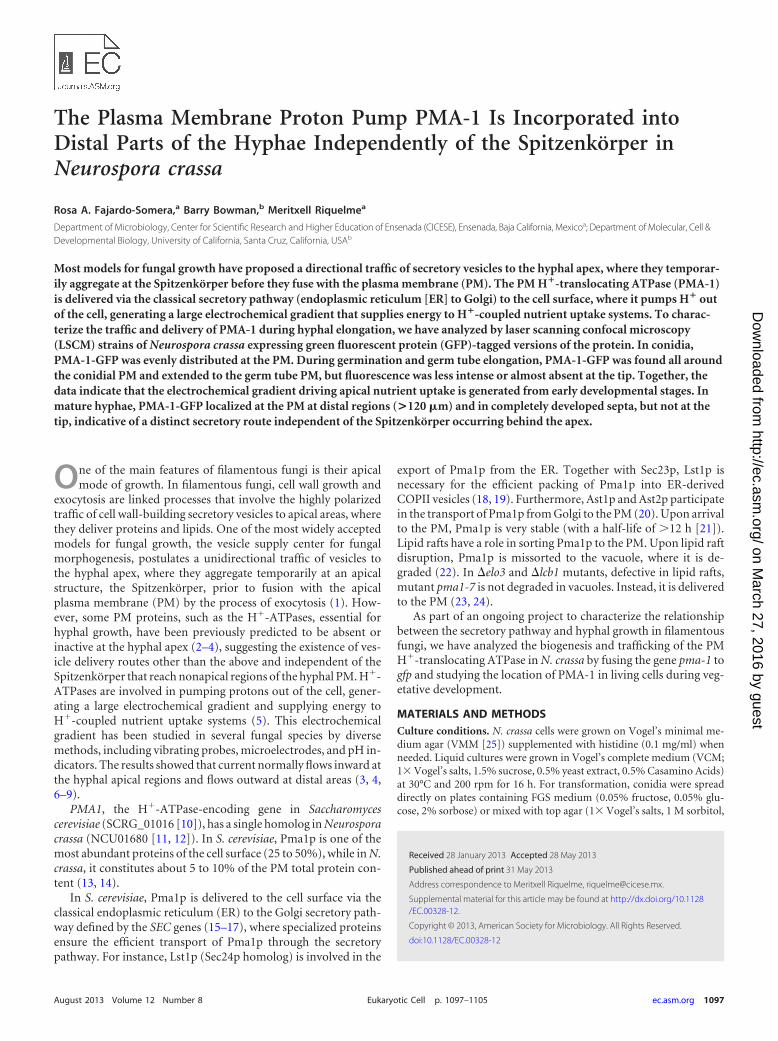
Brefeldin A affects the distribution of PMA-1-GFP. BFA is aninhibitor of the classical ER-Golgi secretory pathway, traditionallyused to block vesicle budding from the Golgi. N. crassa expressingPMA-1-GFP was exposed to BFA (200 �g/ml) and analyzed byLSCM. After 7 min of exposure, growth ceased and PMA-1-GFPstarted to accumulate at putative “brefeldin bodies” in subapicaland distal regions (Fig. 7A, arrows). After 30 min of exposure,hyphal tips started to swell and small vesicles began to accumulatein apical regions (Fig. 7B). After 90 min, PMA-1-GFP started toaccumulate in the PM of apical regions (Fig. 7C). Approximately 2h later, the tips were swollen, and PMA-1-GFP was distributedthroughout the PM all along the hypha, including the apical andsubapical regions (Fig. 7D). From that point on, hyphae began torecover and to display polarized growth (data not shown).
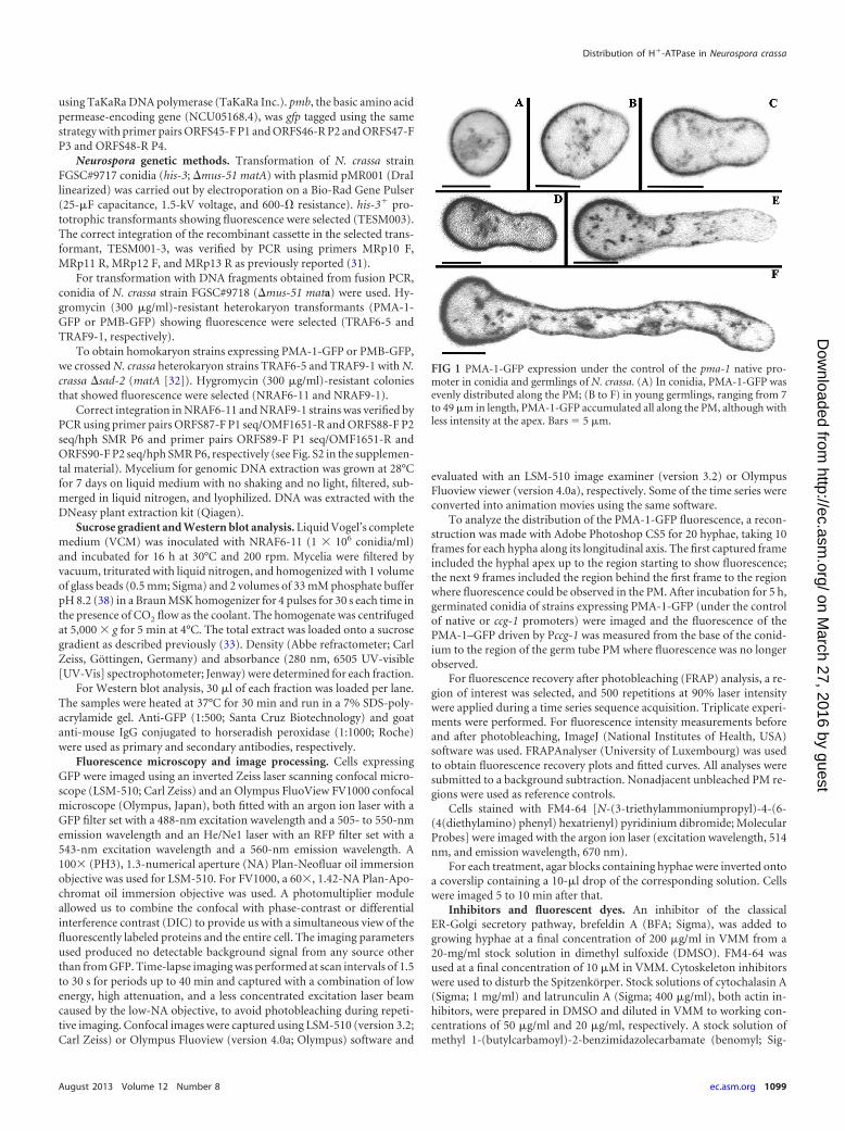
To elucidate whether the tubular endomembranes observed inapical regions which become vesicular when exposed to BFA arepart of the endosomal pathway, we used FRAP analysis in spher-ical vacuoles located near the subapical region. Spherical vacuolesnear subapical regions were exposed to high-intensity laser irradi-ation in the presence or absence of BFA. In the absence of BFA(Fig. 8A to D), the fluorescence in the spherical vacuoles started torecover after 5 s (Fig. 8C), and it was fully recovered at 3 min 50 s(Fig. 8D). However, in the presence of BFA (200 �g/ml) (Fig. 8E to
FIG 4 PMA-1-GFP localized at the PM of already-formed septa. (A to C) NoGFP fluorescence could be seen at the nascent septa stained with FM4-64 (10�M). (D to G) After 6 min, septa stained with FM4-64 seemed to be com-pleted, whereas no PMA-1-GFP could be still observed. (G to I) After 14 min25 s, PMA-1-GFP started to faintly accumulate at completed septa. (J to L)After 45 min 6 s, PMA-1-GFP was clearly observed at completed septa. Bar �20 �m.
Distribution of H�-ATPase in Neurospora crassa
August 2013 Volume 12 Number 8 ec.asm.org 1101
on March 27, 2016 by guest
http://ec.asm.org/
Dow
nloaded from

H), fluorescence at vacuoles was not recovered after 4 min 30 s(Fig. 8H).
DISCUSSIONPMA-1 is excluded from the apical region. Traffic of PMA-1-GFP provides insight into one of the pathways that delivers pro-teins from the Golgi to the cell surface in fungal hyphae.
Our studies demonstrate that H�-ATPase is differentially dis-tributed along the fungal cell surface in mature hyphae. PMA-1-GFP was found at the cell surface of subapical and distal parts butnot at the apex. These results confirm earlier predictions that theH�-ATPase is deficient or inactive at the apex but abundant dis-tally (9). Protons are expelled distally by the proton-translocatingATPase and enter the apical region by a number of transporters,including those carrying potassium and phosphate ions as previ-ously reported (9). Furthermore, the distribution of PMA-1-GFPin the PM is in agreement with the intracellular pH gradient in N.
crassa. Analysis of intracellular pH using bromocresol green (a pHindicator) showed that apical zones, devoid of PMA-1, have anacid pH, whereas distal regions, in which PMA-1 is abundant,have a basic pH (8). A similar cytochemical gradient was observedin other filamentous fungi (6).
Some proteins, such as transporter or symporter proteins, needprotons for their function, suggesting that these proteins may usethe electrochemical gradient created by the H�-ATPase. In S.cerevisiae, the arginine/H� transporter Can1p, like Pma1p, is alsofound in all regions of the PM (42). However, Pma1p and Can1poccupy different PM microdomains, which confirmed the exis-tence of protein compartmentation within the PM in yeast (42).Protein compartmentation probably also exists within the PM ofN. crassa. In, N. crassa the basic amino acid permease PMB, thehomolog of Can1p, tagged with GFP displayed a distribution sim-
FIG 5 FRAP analysis of PMA-1-GFP fluorescence at the plasma membrane. (A)Three small different ROIs were photobleached (dashed boxes). Black arrowheadsindicate photobleached ROIs for which FRAP analysis was conducted, and grayarrowheads show ROIs selected for iFRAP analysis. Bar � 20 �m. (B) A large ROIalong the plasma membrane (dashed box) was photobleached. Bar � 10 �m. (C)An ROI including the cytoplasmic region contained within a hyphal compartmentand a small ROI in the PM were selected. Black arrowheads indicate ROIs forwhich FRAP analysis was conducted. Bar � 10 �m. In each case, localization ofPMA-1-GFP before and after photobleaching is shown. Graphs show normalizedplots of fluorescence recovery values and fitted curves. Mf and t1/2 values areshown in each analysis. (D) Scheme showing lateral diffusion of a protein withinthe PM lipid bilayer versus exchange from an endomembrane to the PM.
FIG 6 Effects of cytoskeletal inhibitors on PMA-1-GFP localization. (A to D)Hyphae at low magnification; (E to L) PMA-1-GFP fluorescence at high mag-nification; (M to T) FM4-64 staining. For each treatment, a hyphal tip (top)and a hyphal distal region (bottom) are shown. Untreated cells (A, E, and F)showed PMA-1-GFP fluorescence at tubular endomembranes in subapicalregions and at the PM in distal regions. A prominent Spitzenkörper was ob-served stained by FM4-64 at hyphal apices. Hyphae treated with cytochalasin Ashowed initial swelling of tips followed by dichotomous branching (B, G, andO) and abnormal PMA-1-GFP distribution (B, G, and H); fluorescence couldno longer be observed in tubular endomembranes, and at the PM, it extendedtoward subapical regions (B and G). No Spitzenkörper was present at the tips(O). Hyphae treated with latrunculin A displayed swollen tips (C, I, and Q).PMA-1-GFP was observed at the PM near subabipal regions (C and I), and noSpitzenkörper was present at the apices (Q). In addition, PMA-1-GFP and FM4-64 colocalized fluorescence could be observed accumulating cortically, be-low the cell surface in subapical and distal regions, indicative of impairedendocytosis (I, J, Q, and R). Hyphae treated with benomyl showed distortedmorphology (D). PMA-1-GFP fluorescence at the subapical tubular endo-membranes was more prominent (K), and at the PM, it extended towardsubapical regions (D). The Spitzenkörper appeared reduced in size and integ-rity (S). In all cases, PMA-1-GFP continued to be localized in the PM in distalregions (F, H, J, and L). Cytochalasin A was used at 100 �g/ml, latrunculin A at 20�g/ml, and benomyl at 50 �g/ml. Bars � 10 �m (A to D) and 20 �m (E to T).
Fajardo-Somera et al.
1102 ec.asm.org Eukaryotic Cell
on March 27, 2016 by guest
http://ec.asm.org/
Dow
nloaded from
ilar to that of PMA-1 (see Fig. S5 in the supplemental material).Coexpression of PMA-1 and PMB tagged with different fluores-cent markers and a higher-resolution microscopic analysis areneeded to discern whether these two proteins occupy different PMmicrodomains in the range of several hundred nanometers.
Exocytosis occurs in distal regions of hyphae. Even thoughPMA-1-GFP was found in cell fractions of a density correspond-ing to that of ER and macrovesicles (35), it did not accumulate atthe Spitzenkörper, where there is the highest concentration ofsecretory vesicles according to electron micrographs of hyphal tipsof N. crassa (36). Instead, PMA-1-GFP localizes in subapical re-gions and is somehow directly delivered to the PM.
FRAP analysis revealed rates of fluorescence recovery higherfor cytoplasmic exchange than for lateral diffusion, suggestingthat PMA-1 is incorporated to the PM primarily by membrane-cytoplasmic exchange. However, lateral diffusion also occurs, ascould be inferred from the iFRAP results. FRAP analysis suggestscontinuous exocytosis of PMA-1 to the PM in distal regions. SomeSNARE proteins and components of the exocyst complex(AoSec3) which are involved in exocytosis events have been local-ized in the septum and PM in Aspergillus oryzae (43, 44). In A.oryzae, it has been shown that transport proteins are expressedprimarily in distal regions. For instance, AoUapC and AoGap1,two amino acid permeases, did not accumulate at the Spitzenkör-per. They were observed in lateral PM and septa (43, 45). Somemechanisms of exocytosis, such as kiss-and-run (K&R) mecha-
nisms, do not involve growth of the PM (46–48). During K&R,proteins are released but the vesicles do not fully collapse; i.e., thevesicle membrane does not fuse completely with the PM and doesnot contribute to membrane expansion (49). While K&R mecha-nisms have not been explored or identified in yeast or filamentousfungi, the collective evidence suggests that they may exist. Intui-tively, in growing hyphae, if all the vesicular membrane exocyto-sed at areas other than the tip contributed to an increase in thehyphal surface area, endocytosis would have to occur at regions ofthe hyphae other than the subapical collar suggested by severalauthors (50–53). Clearly, future studies are needed to quantita-tively analyze the contribution of each of these processes to cellgrowth.
PMA-1 recycling involves the endosomal pathway. After ashort exposure to BFA, PMA-1-GFP delivery to the PM was notapparently blocked in N. crassa hyphae. In addition, PMA-1-GFPaccumulated at small structures visible in apical and subapicalregions, similarly to the brefeldin bodies or “brefeldin organelles”observed in Aspergillus nidulans and S. cerevisiae cells expressingfluorescently labeled Golgi markers PHOSBP, HypBSec7, and Sec7p,respectively, when exposed to BFA (54–56). As mentioned earlier,Pma1p is very stable in the PM; therefore, the fact that we still seeit in the PM after short exposure to BFA could indicate that even ifno additional PMA-1-GFP arrives at the PM, the protein existingat the time the drug was applied has not been yet recycled. Uponlonger exposure to BFA, PMA-1-GFP was found at the PM inapical regions, indicating that somehow the apex lost its “polar”markers and PMA-1 was missorted to all regions of the PM. What
FIG 7 Effects of brefeldin A (200 �g/ml) on N. crassa mature hyphae express-ing PMA-1-GFP. (A) After 7 min of exposure, PMA-1-GFP began to accumu-late at putative brefeldin bodies (arrows in subapical and distal regions). (Band C) After 30 to 90 min of exposure, the tip began to swell and PMA-1-GFPstarted to accumulate at the PM of the apex. (D) After 2 h of exposure, PMA-1-GFP accumulated throughout the PM all along the hypha, including theapex and subapex. Bars � 20 �m.
FIG 8 FRAP analysis in spherical vacuoles near the subapical regions of N.crassa hyphae treated with BFA (200 �g/ml). (A to D) FRAP in untreatedhyphae; (E to H) FRAP in hyphae exposed to BFA. (A and E) Fluorescence ina spherical vacuole before photobleaching; (B and F) photobleaching of fluo-rescence in a spherical vacuole; (C) fluorescence recovery in a spherical vacu-ole of an untreated hypha 5 s after photobleaching; (D) fluorescence recoveredcompletely after 3 min 50 s; (G and H) fluorescence of a spherical vacuole in ahypha exposed to BFA did not recover after 4 min 30 s. Bars � 20 �m.
Distribution of H�-ATPase in Neurospora crassa
August 2013 Volume 12 Number 8 ec.asm.org 1103
on March 27, 2016 by guest
http://ec.asm.org/
Dow
nloaded from
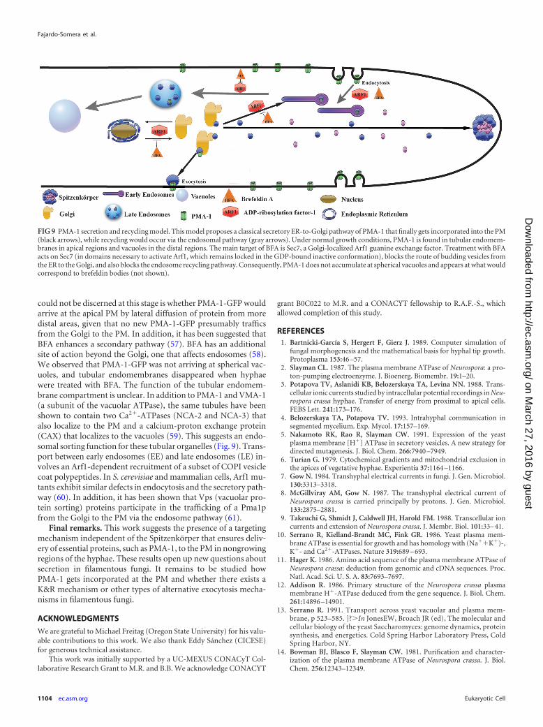
could not be discerned at this stage is whether PMA-1-GFP wouldarrive at the apical PM by lateral diffusion of protein from moredistal areas, given that no new PMA-1-GFP presumably trafficsfrom the Golgi to the PM. In addition, it has been suggested thatBFA enhances a secondary pathway (57). BFA has an additionalsite of action beyond the Golgi, one that affects endosomes (58).We observed that PMA-1-GFP was not arriving at spherical vac-uoles, and tubular endomembranes disappeared when hyphaewere treated with BFA. The function of the tubular endomem-brane compartment is unclear. In addition to PMA-1 and VMA-1(a subunit of the vacuolar ATPase), the same tubules have beenshown to contain two Ca2�-ATPases (NCA-2 and NCA-3) thatalso localize to the PM and a calcium-proton exchange protein(CAX) that localizes to the vacuoles (59). This suggests an endo-somal sorting function for these tubular organelles (Fig. 9). Trans-port between early endosomes (EE) and late endosomes (LE) in-volves an Arf1-dependent recruitment of a subset of COPI vesiclecoat polypeptides. In S. cerevisiae and mammalian cells, Arf1 mu-tants exhibit similar defects in endocytosis and the secretory path-way (60). In addition, it has been shown that Vps (vacuolar pro-tein sorting) proteins participate in the trafficking of a Pma1pfrom the Golgi to the PM via the endosome pathway (61).
Final remarks. This work suggests the presence of a targetingmechanism independent of the Spitzenkörper that ensures deliv-ery of essential proteins, such as PMA-1, to the PM in nongrowingregions of the hyphae. These results open up new questions aboutsecretion in filamentous fungi. It remains to be studied howPMA-1 gets incorporated at the PM and whether there exists aK&R mechanism or other types of alternative exocytosis mecha-nisms in filamentous fungi.
ACKNOWLEDGMENTS
We are grateful to Michael Freitag (Oregon State University) for his valu-able contributions to this work. We also thank Eddy Sánchez (CICESE)for generous technical assistance.
This work was initially supported by a UC-MEXUS CONACyT Col-laborative Research Grant to M.R. and B.B. We acknowledge CONACYT
grant B0C022 to M.R. and a CONACYT fellowship to R.A.F.-S., whichallowed completion of this study.
REFERENCES1. Bartnicki-García S, Hergert F, Gierz J. 1989. Computer simulation of
fungal morphogenesis and the mathematical basis for hyphal tip growth.Protoplasma 153:46 –57.
2. Slayman CL. 1987. The plasma membrane ATPase of Neurospora: a pro-ton-pumping electroenzyme. J. Bioenerg. Biomembr. 19:1–20.
3. Potapova TV, Aslanidi KB, Belozerskaya TA, Levina NN. 1988. Trans-cellular ionic currents studied by intracellular potential recordings in Neu-rospora crassa hyphae. Transfer of energy from proximal to apical cells.FEBS Lett. 241:173–176.
4. Belozerskaya TA, Potapova TV. 1993. Intrahyphal communication insegmented mycelium. Exp. Mycol. 17:157–169.
5. Nakamoto RK, Rao R, Slayman CW. 1991. Expression of the yeastplasma membrane [H�] ATPase in secretory vesicles. A new strategy fordirected mutagenesis. J. Biol. Chem. 266:7940 –7949.
6. Turian G. 1979. Cytochemical gradients and mitochondrial exclusion inthe apices of vegetative hyphae. Experientia 37:1164 –1166.
7. Gow N. 1984. Transhyphal electrical currents in fungi. J. Gen. Microbiol.130:3313–3318.
8. McGillviray AM, Gow N. 1987. The transhyphal electrical current ofNeurospora crassa is carried principally by protons. J. Gen. Microbiol.133:2875–2881.
9. Takeuchi G, Shmidt J, Caldwell JH, Harold FM. 1988. Transcellular ioncurrents and extension of Neurospora crassa. J. Membr. Biol. 101:33– 41.
10. Serrano R, Kielland-Brandt MC, Fink GR. 1986. Yeast plasma mem-brane ATPase is essential for growth and has homology with (Na��K�)-,K�- and Ca2�-ATPases. Nature 319:689 – 693.
11. Hager K. 1986. Amino acid sequence of the plasma membrane ATPase ofNeurospora crassa: deduction from genomic and cDNA sequences. Proc.Natl. Acad. Sci. U. S. A. 83:7693–7697.
12. Addison R. 1986. Primary structure of the Neurospora crassa plasmamembrane H�-ATPase deduced from the gene sequence. J. Biol. Chem.261:14896 –14901.
13. Serrano R. 1991. Transport across yeast vacuolar and plasma mem-brane, p 523–585. ]?�In JonesEW, Broach JR (ed), The molecular andcellular biology of the yeast Saccharomyces: genome dynamics, proteinsynthesis, and energetics. Cold Spring Harbor Laboratory Press, ColdSpring Harbor, NY.
14. Bowman BJ, Blasco F, Slayman CW. 1981. Purification and character-ization of the plasma membrane ATPase of Neurospora crassa. J. Biol.Chem. 256:12343–12349.
FIG 9 PMA-1 secretion and recycling model. This model proposes a classical secretory ER-to-Golgi pathway of PMA-1 that finally gets incorporated into the PM(black arrows), while recycling would occur via the endosomal pathway (gray arrows). Under normal growth conditions, PMA-1 is found in tubular endomem-branes in apical regions and vacuoles in the distal regions. The main target of BFA is Sec7, a Golgi-localized Arf1 guanine exchange factor. Treatment with BFAacts on Sec7 (in domains necessary to activate Arf1, which remains locked in the GDP-bound inactive conformation), blocks the route of budding vesicles fromthe ER to the Golgi, and also blocks the endosome recycling pathway. Consequently, PMA-1 does not accumulate at spherical vacuoles and appears at what wouldcorrespond to brefeldin bodies (not shown).
Fajardo-Somera et al.
1104 ec.asm.org Eukaryotic Cell
on March 27, 2016 by guest
http://ec.asm.org/
Dow
nloaded from

15. Brada D, Schekman R. 1988. Coincident localization of secretory andplasma membrane proteins in organelles of the yeast secretory pathway. J.Bacteriol. 170:2775–2783.
16. Holcomb CL, Hansen WJ, Etcheverry T, Shekman R. 1988. Secretoryvesicles externalize the major plasma membrane ATPase in yeast. J. CellBiol. 106:641– 648.
17. Chang A, Slayman CW. 1991. Maturation of the yeast plasma membrane[H�] ATPase involves phosphorylation during intracellular transport. J.Cell Biol. 115:289 –295.
18. Roberg KJ, Crotwell M, Espenshade P, Gimeno R, Kaiser CA. 1999.LST1 is a SEC24 homologue used for selective export of the plasma mem-brane ATPase from the endoplasmic reticulum. J. Cell Biol. 145:659 – 672.
19. Shimoni Y, Kurihara T, Ravazzola M, Amherdt M, Orci L, Schekman R.2000. Lst1p and Sec24p cooperate in sorting of the plasma membraneATPase into COPII vesicles in Saccharomyces cerevisiae. J. Cell Biol. 151:973–984.
20. Chang A, Fink GR. 1995. Targeting of the yeast plasma membrane [H�]ATPase: a novel gene AST1 prevents mislocalization of mutant ATPase tothe vacuole. J. Cell Biol. 128:39 – 49.
21. Benito B, Moreno E, Lagunas R. 1991. Half-life of the plasma membraneATPase and its activating system in resting yeast cells. Biochim. Biophys.Acta 1063:265–268.
22. Stack JH, Horazdovsky B, Emr SD. 1995. Receptor-mediated proteinsorting to the vacuole in yeast: roles for a protein kinase, a lipid kinase andGTP-binding proteins. Annu. Rev. Cell Dev. Biol. 11:1–33.
23. Bagnat M, Chang A, Simons K. 2001. Plasma membrane proton ATPasePma1p requires raft association for surface delivery in yeast. Mol. Biol.Cell 12:4129 – 4138.
24. Eisenkolb M, Zenzmaier C, Leitner E, Schneiter R. 2002. A specificstructural requirements for ergosterol in long-chain fatty acid synthesismutants important for maintaining raft domains in yeast. Mol. Biol. Cell13:4414 – 4428.
25. Vogel HJ. 1956. A convenient growth medium for Neurospora (mediumN). Microbiol. Gen. Bull. 13:42– 43.
26. Reference deleted.27. Hickey PC, Jacobson D, Read ND, Glass NL. 2002. Live-cell imaging of
vegetative hyphal fusion in Neurospora crassa. Fungal Genet. Biol. 37:109 –119.
28. Sambrook J, Fritsch EF, Maniatis T. 1989. Molecular cloning: a labora-tory manual, 2nd ed. Cold Spring Harbor Laboratory Press, Cold SpringHarbor, NY.
29. Freitag M, Hickey PC, Raju NB, Selker EU, Read ND. 2004. GFP as atool to analyze the organization, dynamics and function of nuclei andmicrotubules in Neurospora crassa. Fungal Genet. Biol. 41:897–910.
30. Honda S, Selker EU. 2009. Tools for fungal proteomics: multifunctionalNeurospora vectors for gene replacement, protein expression and proteinpurification. Genetics 1:11–23.
31. Riquelme M, Bartnicki-García S, González-Prieto JM, Sánchez-León E,Verdín-Ramos J, Beltrán-Aguilar A, Freitag M. 2007. Spitzenkörperlocalization and intracellular traffic of green fluorescent protein-labeledCHS-3 and CHS-6 chitin synthases in living hyphae of Neurospora crassa.Eukaryot. Cell 6:1853–1864.
32. Shiu PK, Zickler D, Raju NB, Ruprich-Robert G, Metzenberg RL. 2006.SAD-2 is required for meiotic silencing by unpaired DNA and perinuclearlocalization of SAD-1 RNA-directed RNA polymerase. Proc. Natl. Acad.Sci. U. S. A. 103:2243–2248.
33. Richthammer C, Enseleit M, Sanchez-Leon E, März S, Heilig Y,Riquelme M, Seiler S. 2012. RHO1 and RHO2 share partially overlappingfunctions in the regulation of cell wall integrity and hyphal polarity inNeurospora crassa. Mol. Microbiol. 85:716 –733.
34. Leal-Morales CA, Bracker CE, Bartnicki-García S. 1994. Subcellularlocalization, abundance and stability of chitin synthetases 1 and 2 fromSaccharomyces cerevisiae. Microbiology 140:2207–2216.
35. Verdín J, Bartnicki-García S, Riquelme M. 2009. Functional stratifica-tion of the Spitzenkörper of Neurospora crassa. Mol. Microbiol. 74:1044 –1053.
36. Riquelme M, Roberson RW, McDaniel DP, Bartnicki-Garcia S. 2002.The effects of ropy-1 mutation on cytoplasmic organization and intracel-lular motility in mature hyphae of Neurospora crassa. Fungal Genet. Biol.37:171–179.
37. Araujo-Palomares CL, Castro-Longoria E, Riquelme M. 2007. Ontogenyof the Spitzenkörper in germlings of Neurospora crassa. Fungal Genet.Biol. 44:492–503.
38. Lippincott-Schwartz J, Snapp E, Kenworthy A. 2001. Studying proteindynamics in living cells. Nat. Rev. Mol. Cell Biol. 2:444 – 456.
39. Goehring NW, Chowdhury D, Hyman AA, Grill SW. 2010. FRAPanalysis of membrane-associated proteins: lateral diffusion and mem-brane-cytoplasmic exchange. Biophys. J. 99:2443–2452.
40. Bartnicki-García S. 2006. Chitosomes: past, present and future. FEMSYeast Res. 6:957–965.
41. Riquelme M, Gierz G, Bartnicki-García S. 2000. Dynein and dynactindeficiencies affect the formation and function of the Spitzenköper anddistort hyphal morphogenesis of Neurospora crassa. Microbiology 146:1743–1752.
42. Malínská K, Malínský J, Opekarová M, Tanner W. 2003. Visualizationof protein compartmentation within the plasma membrane of living yeastcells. Mol. Biol. Cell 11:4427– 4436.
43. Hayakawa Y, Ishikawa E, Shoji JY, Nakano H, Kitamoto K. 2011.Septum-directed secretion in the filamentous fungus Aspergillus oryzae.Mol. Microbiol. 1:40 –55.
44. Kuratsu M, Taura A, Shoji JY, Kikuchi S, Arioka M, Kitamoto K. 2007.Systematic analysis of SNARE localization in the filamentous fungusAspergillus oryzae. Fungal Genet. Biol. 44:1310 –1323.
45. Masai K, Maruyama J, Sakamoto K, Nakajima H, Akita O, Kitamoto K.2006. Square-plate culture method allows detection of differential geneexpression and screening of novel, region-specific genes in Aspergillusoryzae. Appl. Microbiol. Biotechnol. 71:881– 891.
46. Alés E, Tabares L, Poyato JM, Valero V, Lindau M, Alvarez de ToledoG. 1999. High calcium concentrations shift the mode of exocytosis to thekiss-and-run mechanism. Nat. Cell Biol. 1:40 – 44.
47. Henkel AW, Meiri H, Horstmann H, Lindau M, Almers W. 2000.Rhythmic opening and closing of vesicles during constitutive exo- andendocytosis in chromaffin cells. EMBO J. 19:84 –93.
48. Lollike K, Borregaard N, Lindau M. 1998. Capacitance flickers andpseudoflickers of small granules, measured in the cell-attached configura-tion. Biophys. J. 75:53–59.
49. Keighron J, Ewing AG, Cans AS. 2012. Analytical tools to monitorexocytosis: a focus on new fluorescent probes and methods. Analyst 137:1755–1763.
50. Delgado-Álvarez DL, Callejas-Negrete OA, Gómez N, Freitag M, Rob-erson RW, Smith LG, Mouriño-Pérez RR. 2010. Visualization of F-actinlocalization and dynamics with live cell markers in Neurospora crassa.Fungal Genet. Biol. 47:573–586.
51. Upadhyay S, Shaw BD. 2008. The role of actin, fimbrin, and endocy-tosis in growth of hyphae in Aspergillus nidulans. Mol. Microbiol. 68:690 –705.
52. Araujo-Bazán L, Peñalva MA, Espeso EA. 2008. Preferential localizationof the endocytic internalization machinery to hyphal tips underlies polar-ization of the actin cytoskeleton in Aspergillus nidulans. Mol. Microbiol.67:891–905.
53. Taheri-Talesh N, Horio T, Araujo-Bazán L, Dou X, Espeso EA, PeñalvaMA, Osmani SA, Oakley BR. 2008. The tip growth apparatus of Asper-gillus nidulans. Mol. Biol. Cell 19:1439 –1449.
54. Pantazopoulou A, Peñalva MA. 2009. Organization and dynamics of theAspergillus nidulans Golgi during apical extension and mitosis. Mol. Biol.Cell 20:4335– 4347.
55. Rambourg A, Clermont Y, Jackson CL, Kepes F. 1995. Effects of brefel-din A on the three-dimensional structure of the Golgi apparatus in a sen-sitive strain of Saccharomyces cerevisiae. Anat. Rec. 241:1–9.
56. Deitz SB, Rambourg A, Kepes F, Franzusoff A. 2000. Sec7p directs thetransitions required for yeast Golgi biogenesis. Traffic 1:172–183.
57. Klausner RD, Donaldson JG, Lippincot-Schwartz J. 1992. Brefeldin A:insights into the control of membrane traffic and organelle structure. J.Cell Biol. 116:1071–1080.
58. Nebenführ A, Ritzenthaler C, Robinson DG. 2002. Brefeldin A: deci-phering an enigmatic inhibitor of secretion. Plant Physiol. 3:1102–1108.
59. Bowman BJ, Draskovic M, Freitag M, Bowman EJ. 2009. Structure anddistribution of organelles and cellular location of calcium transporters inNeurospora crassa. Eukaryot. Cell 8:1845–1855.
60. Gaynor EC, Chen CY, Emr SD, Graham TR. 1998. ARF is required formaintenance of yeast Golgi and endosome structure and function. Mol.Biol. Cell 9:653– 670.
61. Luo W, Chang A. 2000. An endosome-to-plasma membrane pathwayinvolved in trafficking of a mutant plasma membrane ATPase in yeast.Mol. Biol. Cell 11:579 –592.
Distribution of H�-ATPase in Neurospora crassa
August 2013 Volume 12 Number 8 ec.asm.org 1105
on March 27, 2016 by guest
http://ec.asm.org/
Dow
nloaded from
Related Documents