Progress in Retinal and Eye Research 24 (2005) 39–73 The optic nerve head as a biomechanical structure: a new paradigm for understanding the role of IOP-related stress and strain in the pathophysiology of glaucomatous optic nerve head damage Claude F. Burgoyne a,b, , J. Crawford Downs a,b , Anthony J. Bellezza a,b , J.-K. Francis Suh b , Richard T. Hart b a LSU Eye Center, Louisiana State University Health Sciences Center, 2020 Gravier Street, Suite B, New Orleans, LA 70112, USA b Department of Biomedical Engineering, Tulane University, New Orleans, LA, USA Abstract We propose here a conceptual framework for understanding the optic nerve head (ONH) as a biomechanical structure. Basic principles of biomechanical engineering are used to propose a central role for intraocular pressure (IOP)-related stress and strain in the physiology of ONH aging and the pathophysiology of glaucomatous damage. Our paradigm suggests that IOP-related stress and strain (1) are substantial within the load-bearing connective tissues of the ONH even at low levels of IOP and (2) underlie both ONH aging and the two central pathophysiologies of glaucomatous damage—mechanical failure of the connective tissues of the lamina cribrosa, scleral canal wall, and peripapillary sclera, and axonal compromise within the lamina cribrosa by a variety of mechanisms. Modeling the ONH as a biomechanical structure generates a group of testable hypotheses regarding the central mechanisms of glaucomatous damage and provides a logic for classifying the principal components of the susceptibility of an individual ONH to a given level of IOP. r 2004 Elsevier Ltd. All rights reserved. Contents 1. Introduction .............................................................................. 41 2. The ONH as a biomechanical structure ........................................................... 42 3. The relationship between IOP and IOP-related stress ................................................. 44 4. The relationship between stress, strain, material properties, and mechanical behavior .......................... 44 5. The effects of IOP-related connective tissue strain on the connective tissues, astrocytes, and axons depend on the state of the connective tissues ...................................................................... 45 5.1. Load-bearing connective tissues ............................................................ 46 5.1.1. Connective tissues undamaged (within their elastic limits) ................................... 46 5.1.2. Connective tissues damaged (undergoing some phase of mechanical failure) ...................... 47 ARTICLE IN PRESS www.elsevier.com/locate/prer 1350-9462/$ - see front matter r 2004 Elsevier Ltd. All rights reserved. doi:10.1016/j.preteyeres.2004.06.001 Corresponding author. LSU Eye Center, 2020 Gravier Street, Suite B, New Orleans, LA 70112, USA. Tel.: +1-504-412-1200x1306; fax: +1-504- 412-1315. E-mail address: [email protected] (C.F. Burgoyne).

Welcome message from author
This document is posted to help you gain knowledge. Please leave a comment to let me know what you think about it! Share it to your friends and learn new things together.
Transcript

ARTICLE IN PRESS
1350-9462/$ - se
doi:10.1016/j.pr
�Correspond412-1315.
E-mail addr
Progress in Retinal and Eye Research 24 (2005) 39–73
www.elsevier.com/locate/prer
The optic nerve head as a biomechanical structure: a new paradigmfor understanding the role of IOP-related stress and strain in thepathophysiology of glaucomatous optic nerve head damage
Claude F. Burgoynea,b,�, J. Crawford Downsa,b, Anthony J. Bellezzaa,b,J.-K. Francis Suhb, Richard T. Hartb
aLSU Eye Center, Louisiana State University Health Sciences Center, 2020 Gravier Street, Suite B, New Orleans, LA 70112, USAbDepartment of Biomedical Engineering, Tulane University, New Orleans, LA, USA
Abstract
We propose here a conceptual framework for understanding the optic nerve head (ONH) as a biomechanical structure. Basic
principles of biomechanical engineering are used to propose a central role for intraocular pressure (IOP)-related stress and strain in
the physiology of ONH aging and the pathophysiology of glaucomatous damage. Our paradigm suggests that IOP-related stress and
strain (1) are substantial within the load-bearing connective tissues of the ONH even at low levels of IOP and (2) underlie both ONH
aging and the two central pathophysiologies of glaucomatous damage—mechanical failure of the connective tissues of the lamina
cribrosa, scleral canal wall, and peripapillary sclera, and axonal compromise within the lamina cribrosa by a variety of mechanisms.
Modeling the ONH as a biomechanical structure generates a group of testable hypotheses regarding the central mechanisms of
glaucomatous damage and provides a logic for classifying the principal components of the susceptibility of an individual ONH to a
given level of IOP.
r 2004 Elsevier Ltd. All rights reserved.
Contents
1. Introduction . . . . . . . . . . . . . . . . . . . . . . . . . . . . . . . . . . . . . . . . . . . . . . . . . . . . . . . . . . . . . . . . . . . . . . . . . . . . . . 41
2. The ONH as a biomechanical structure . . . . . . . . . . . . . . . . . . . . . . . . . . . . . . . . . . . . . . . . . . . . . . . . . . . . . . . . . . . 42
3. The relationship between IOP and IOP-related stress . . . . . . . . . . . . . . . . . . . . . . . . . . . . . . . . . . . . . . . . . . . . . . . . . 44
4. The relationship between stress, strain, material properties, and mechanical behavior . . . . . . . . . . . . . . . . . . . . . . . . . . 44
5. The effects of IOP-related connective tissue strain on the connective tissues, astrocytes, and axons depend on the state
of the connective tissues . . . . . . . . . . . . . . . . . . . . . . . . . . . . . . . . . . . . . . . . . . . . . . . . . . . . . . . . . . . . . . . . . . . . . . 45
5.1. Load-bearing connective tissues . . . . . . . . . . . . . . . . . . . . . . . . . . . . . . . . . . . . . . . . . . . . . . . . . . . . . . . . . . . . 46
5.1.1. Connective tissues undamaged (within their elastic limits) . . . . . . . . . . . . . . . . . . . . . . . . . . . . . . . . . . . 46
5.1.2. Connective tissues damaged (undergoing some phase of mechanical failure) . . . . . . . . . . . . . . . . . . . . . . 47
e front matter r 2004 Elsevier Ltd. All rights reserved.
eteyeres.2004.06.001
ing author. LSU Eye Center, 2020 Gravier Street, Suite B, New Orleans, LA 70112, USA. Tel.: +1-504-412-1200x1306; fax: +1-504-
ess: [email protected] (C.F. Burgoyne).
ARTICLE IN PRESSC.F. Burgoyne et al. / Progress in Retinal and Eye Research 24 (2005) 39–7340
5.2. Retinal ganglion cell axons . . . . . . . . . . . . . . . . . . . . . . . . . . . . . . . . . . . . . . . . . . . . . . . . . . . . . . . . . . . . . . . 47
5.2.1. Connective tissues undamaged. . . . . . . . . . . . . . . . . . . . . . . . . . . . . . . . . . . . . . . . . . . . . . . . . . . . . . . 47
5.2.2. Connective tissues damaged . . . . . . . . . . . . . . . . . . . . . . . . . . . . . . . . . . . . . . . . . . . . . . . . . . . . . . . . 47
5.3. Astrocytes and endothelial cells . . . . . . . . . . . . . . . . . . . . . . . . . . . . . . . . . . . . . . . . . . . . . . . . . . . . . . . . . . . . 48
5.3.1. Connective tissues undamaged. . . . . . . . . . . . . . . . . . . . . . . . . . . . . . . . . . . . . . . . . . . . . . . . . . . . . . . 48
5.3.2. Connective tissues damaged . . . . . . . . . . . . . . . . . . . . . . . . . . . . . . . . . . . . . . . . . . . . . . . . . . . . . . . . 48
6. Effects of IOP-related connective tissue strain on the volume flow of blood and nutrient diffusion . . . . . . . . . . . . . . . . . 48
7. Two principal pathophysiologies underlie IOP-related damage to the ONH . . . . . . . . . . . . . . . . . . . . . . . . . . . . . . . . . 48
7.1. Mechanical failure within the load-bearing connective tissues . . . . . . . . . . . . . . . . . . . . . . . . . . . . . . . . . . . . . . . 48
7.2. Damage to the axons by a variety of mechanisms . . . . . . . . . . . . . . . . . . . . . . . . . . . . . . . . . . . . . . . . . . . . . . . 48
8. ONH biomechanics: experimental studies. . . . . . . . . . . . . . . . . . . . . . . . . . . . . . . . . . . . . . . . . . . . . . . . . . . . . . . . . . 50
8.1. Compliance testing the ONH surface in normal monkeys . . . . . . . . . . . . . . . . . . . . . . . . . . . . . . . . . . . . . . . . . 50
8.2. The lamina cribrosa and scleral canal deform following acute elevations of IOP . . . . . . . . . . . . . . . . . . . . . . . . . 50
8.2.1. The lamina cribrosa and scleral canal wall act like an expandable trampoline at low levels of IOP,
with the canal expanding and the lamina thinning and more tautly stretched as IOP is elevated
from 0 to 10mm Hg. . . . . . . . . . . . . . . . . . . . . . . . . . . . . . . . . . . . . . . . . . . . . . . . . . . . . . . . . . . . . . 51
8.2.2. The lamina cribrosa bows posteriorly following acute elevation of IOP from 10 to 30mm Hg . . . . . . . . . 51
8.3. Damage to the ONH connective tissues occurs early in experimental glaucoma . . . . . . . . . . . . . . . . . . . . . . . . . . 52
8.3.1. Early onset of hypercompliance of the ONH surface in experimental glaucoma . . . . . . . . . . . . . . . . . . . . 52
8.3.2. Relative stiffness rather than hypercompliance in optic nerve transection eyes . . . . . . . . . . . . . . . . . . . . . 52
8.3.3. Early onset of permanent posterior deformation of the ONH surface in experimental glaucoma . . . . . . . . 53
8.3.4. Permanent posterior deformation of the anterior scleral canal wall and lamina cribrosa underlie
the onset of CSLT-detected ONH surface change in early experimental glaucoma . . . . . . . . . . . . . . . . . . 53
8.3.5. Hypercompliance of the anterior scleral canal wall and lamina cribrosa accompanies the onset
of CSLT-detected ONH surface change in early experimental glaucoma . . . . . . . . . . . . . . . . . . . . . . . . . 53
8.4. The viscoelastic material properties of posterior sclera are altered in early glaucoma monkey eyes. . . . . . . . . . . . . 54
8.4.1. Elastic and viscoelastic material properties of normal rabbit and monkey sclera . . . . . . . . . . . . . . . . . . . 54
8.4.2. Viscoelastic material properties of posterior sclera are altered in early glaucoma monkeys eyes . . . . . . . . . 54
8.5. IOP-related stress and strain markedly diminish patency of the anterior laminar capillaries in normal and early
glaucoma monkey eyes at high IOP . . . . . . . . . . . . . . . . . . . . . . . . . . . . . . . . . . . . . . . . . . . . . . . . . . . . . . . . . 56
9. Finite element modeling of the ONH. . . . . . . . . . . . . . . . . . . . . . . . . . . . . . . . . . . . . . . . . . . . . . . . . . . . . . . . . . . . . 57
9.1. Idealized models. . . . . . . . . . . . . . . . . . . . . . . . . . . . . . . . . . . . . . . . . . . . . . . . . . . . . . . . . . . . . . . . . . . . . . . 59
9.2. Continuum and micro finite element models . . . . . . . . . . . . . . . . . . . . . . . . . . . . . . . . . . . . . . . . . . . . . . . . . . . 59
9.2.1. Posterior scleral shell surface. . . . . . . . . . . . . . . . . . . . . . . . . . . . . . . . . . . . . . . . . . . . . . . . . . . . . . . . 60
9.2.2. Peripapillary scleral thickness . . . . . . . . . . . . . . . . . . . . . . . . . . . . . . . . . . . . . . . . . . . . . . . . . . . . . . . 60
9.2.3. Posterior scleral thickness . . . . . . . . . . . . . . . . . . . . . . . . . . . . . . . . . . . . . . . . . . . . . . . . . . . . . . . . . . 61
9.2.4. Construction of a composite posterior scleral shell finite element model . . . . . . . . . . . . . . . . . . . . . . . . . 62
9.2.5. Construction of a continuum ONH finite element model for each connected, surfaced, 3D geometry. . . . . 62
9.2.6. Micro finite element modeling . . . . . . . . . . . . . . . . . . . . . . . . . . . . . . . . . . . . . . . . . . . . . . . . . . . . . . . 62
10. Clinical and research implications of the biomechanical paradigm . . . . . . . . . . . . . . . . . . . . . . . . . . . . . . . . . . . . . . . . 63
10.1. Implication 1 . . . . . . . . . . . . . . . . . . . . . . . . . . . . . . . . . . . . . . . . . . . . . . . . . . . . . . . . . . . . . . . . . . . . . . . . . 63
10.2. Implication 2 . . . . . . . . . . . . . . . . . . . . . . . . . . . . . . . . . . . . . . . . . . . . . . . . . . . . . . . . . . . . . . . . . . . . . . . . . 64
10.3. Implication 3 . . . . . . . . . . . . . . . . . . . . . . . . . . . . . . . . . . . . . . . . . . . . . . . . . . . . . . . . . . . . . . . . . . . . . . . . . 66
10.4. Implication 4 . . . . . . . . . . . . . . . . . . . . . . . . . . . . . . . . . . . . . . . . . . . . . . . . . . . . . . . . . . . . . . . . . . . . . . . . . 66
10.5. Implication 5 . . . . . . . . . . . . . . . . . . . . . . . . . . . . . . . . . . . . . . . . . . . . . . . . . . . . . . . . . . . . . . . . . . . . . . . . . 67
11. Summary . . . . . . . . . . . . . . . . . . . . . . . . . . . . . . . . . . . . . . . . . . . . . . . . . . . . . . . . . . . . . . . . . . . . . . . . . . . . . . . . 69
12. Future directions . . . . . . . . . . . . . . . . . . . . . . . . . . . . . . . . . . . . . . . . . . . . . . . . . . . . . . . . . . . . . . . . . . . . . . . . . . . 69
Acknowledgements . . . . . . . . . . . . . . . . . . . . . . . . . . . . . . . . . . . . . . . . . . . . . . . . . . . . . . . . . . . . . . . . . . . . . . . . . . . . . 70
References . . . . . . . . . . . . . . . . . . . . . . . . . . . . . . . . . . . . . . . . . . . . . . . . . . . . . . . . . . . . . . . . . . . . . . . . . . . . . . . . . . . 70
ARTICLE IN PRESSC.F. Burgoyne et al. / Progress in Retinal and Eye Research 24 (2005) 39–73 41
1. Introduction
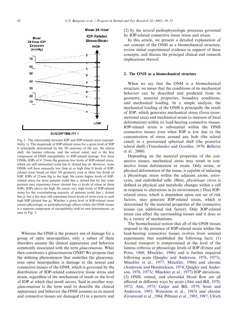
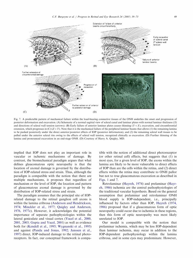
The clinical hallmarks of a glaucomatous opticneuropathy are glaucomatous excavation of the tissuesof the optic nerve head (ONH)—a progressive posteriordisplacement of the surface of the ONH and progressiveexcavation of the prelaminar tissues beneath theanteriormost aspect of the scleral canal, the anteriorscleral ring (Fig. 1, left) (Ernest and Potts, 1968)—andglaucomatous visual field loss, which most commonlystarts as a nasal step and progresses through an arcuatescotoma to full hemifield loss. These clinical hallmarksdistinguish a glaucomatous optic neuropathy from themany other optic neuropathies in which damage to theretinal ganglion cell axons, either at the nerve head orwithin the orbital optic nerve and chiasm, leads toretinal ganglion cell death. Although exceptions exist(Quigley and Anderson, 1976, 1977c; Minckler et al.,1977; Minckler, 1986), ischemic, inflammatory, andcompressive damage to the nerve head, orbital opticnerve, or optic chiasm usually results in pallor andatrophy of the nerve head but little or no excavation ofthe remaining rim tissue (Fig. 1, right). In addition, inthese entities the pattern of axon damage within theoptic nerve (Anderson and Hendrickson, 1974), and asdetected by visual field testing (Anderson and Hen-drickson, 1974), is usually different from that ofglaucomatous optic neuropathy.The mechanisms of glaucomatous damage to the
tissues of the ONH remain controversial (Fechtner andWeinreb, 1994; Schumer and Podos, 1994). For the past30 years, discussion has focused on how the retinalganglion cell axons are damaged within the laminacribrosa, and controversy has centered on whetherintraocular pressure (IOP) (the mechanical hypothesis)or ONH blood supply (the vascular hypothesis) isresponsible for ONH axonal damage in this disease.Like other investigators (Van Buskirk and Cioffi,
1992, 1993; Van Buskirk, 1994), we believe thatseparating the effects of IOP from the effects of ONHblood flow in the pathophysiology of glaucomatousdamage to the ONH is artificial. In place of the‘‘mechanical vs. vascular’’ paradigm (which concen-trates on the mechanism of axonal insult only), we
Fig. 1. Progressive deformation and excavation of the tissues of the ONH b
glaucomatous optic neuropathy (left). Axonal damage resulting from an epis
reprinted from Quigley, H.A., Anderson, D.R., 1977. Cupping of the opti
Otolaryngol. 83, 755–762, Copyright, 1977, with permission from American
propose the larger concept of the ONH as a biomecha-nical structure. This paradigm suggests that the influ-ence of IOP-related connective tissue stress (force/crosssectional area) and strain (local deformation) is centralto both the physiology and pathophysiology of all thethree ONH tissue types—(1) connective (load-bearingconnective tissues of the peripapillary sclera, scleralcanal wall, and lamina cribrosa), (2) axonal (retinalganglion cell axons), and (3) cellular (astrocytes, glialcells, endothelial cells, and pericytes, along with theirbasement membranes)—as well as the volume flow ofblood that nourishes them.Within this paradigm, it is hypothesized that the
ONH connective tissues are exposed to substantial levelsof IOP-related stress/strain at normal levels of IOP,which further increase as IOP is elevated (Fig. 2)(Greene, 1980; Bellezza et al., 2000). Stresses and strainsat a given level of IOP are physiologic or pathophysio-logic depending upon the response of the tissues thatexperience them (Fig. 3). Physiologic stress/straininduces a broad spectrum of changes in both theconnective tissues and blood vessels that are central tonormal aging. Pathophysiologic stress/strain inducespathologic changes in cell synthesis and tissue micro-architecture (Fig. 3) that underlie the two governingpathophysiologies in glaucoma: (1) mechanical failure ofthe load-bearing connective tissues of the ONH, and (2)progressive damage to the adjacent axons by a variety ofmechanisms (Fig. 4).The paradigm proposes that the two pathophysiolo-
gies of glaucoma develop and progress independently,albeit with interactions, during each stage of theneuropathy (Fig. 4). However, the progression of theneuropathy within an individual ONH is very muchinfluenced by the physiologic age of the tissues in whichit occurs.Thus, although the levels of IOP-related stress and
strain are central to the principal physiologic andpathophysiologic processes (Fig. 3) within eachtissue, it is the interplay between (1) the resultantconnective tissue damage, (2) axonal compromise, and(3) the physiologic age of the tissues (Fig. 4) thatdetermines how a particular ONH comes to lookglaucomatous.
eneath the anterior rim of the scleral canal is the clinical hallmark of a
ode of AION, right, usually results only in ONH pallor. AION images
c disc in ischemic optic neuropathy. Trans. Am. Acad. Ophthalmol.
Academy of Ophthalmology.
ARTICLE IN PRESS
Fig. 2. The relationship between IOP and IOP-related stress (suscept-
ibility 1). The magnitude of IOP-related stress for a given level of IOP
is principally determined by the 3D anatomy of the eye, the scleral
shell, the lamina cribrosa, and the scleral canal, and is the first
component of ONH susceptibility to IOP-related damage. For most
ONHs, IOPs of 6–21mm Hg generate low levels of IOP-related stress,
which are still substantial (solid line b, dotted line d). However, some
ONHs will have unusually low (line a) or high (line f) levels of IOP-
related stress based on their 3D geometry even at these low levels of
IOP. IOPs of 22mm Hg to the high 30s create higher levels of IOP-
related stress for most patients (solid line e, dotted line h), but some
patients may experience lower (dotted line c) levels of stress at these
IOPs. IOPs above the high 30s create very high levels of IOP-related
stress for the overwhelming majority of patients (solid line i, dotted
line j), but a few may still experience lower levels of stress even at such
high IOP (dotted line g). Whether a given level of IOP-related stress
creates physiologic or pathophysiologic effects within the ONH tissues
is a separate component of susceptibility with its own determinants, as
seen in Fig. 3.
C.F. Burgoyne et al. / Progress in Retinal and Eye Research 24 (2005) 39–7342
Whereas the ONH is the primary site of damage for agroup of optic neuropathies, only a subset of thesedisorders assume the clinical appearance and behaviorcommonly associated with the term glaucomatous. Whatthen constitutes a glaucomatous ONH? We propose thatthe defining phenomenon that underlies the glaucoma-tous optic neuropathies is damage to the neural andconnective tissues of the ONH, which is governed by thedistribution of IOP-related connective tissue stress andstrain, regardless of the mechanism of insult or the levelof IOP at which that insult occurs. Said in another way,glaucomatous is the term used to describe the clinicalappearance and behavior the ONH assumes as its neuraland connective tissues are damaged (1) in a pattern and
(2) by the several pathophysiologic processes governedby IOP-related connective tissue stress and strain.In this article, we present a detailed explanation of
our concept of the ONH as a biomechanical structure,review initial experimental evidence in support of theseconcepts, and discuss the principal clinical and researchimplications thereof.
2. The ONH as a biomechanical structure
When we say that the ONH is a biomechanicalstructure, we mean that the conditions of its mechanicalbehavior can be described and predicted from itsgeometry, material properties, boundary conditions,and mechanical loading. In a simple analysis, themechanical loading of the ONH is principally the resultof IOP, which generates mechanical stress (force/cross-sectional area) and mechanical strain (a measure of localdeformation) within its load-bearing connective tissues.IOP-related stress is substantial within the ONHconnective tissues even when IOP is low due to theconcentration of stress around any hole (the scleralcanal) in a pressurized spherical shell (the posteriorscleral shell) (Timoshenko and Goodier, 1970; Bellezzaet al., 2000).Depending on the material properties of the con-
nective tissues, mechanical stress may result in con-nective tissue deformation or strain. Only strain, orphysical deformation of the tissue, is capable of inducinga physiologic stress within the adjacent axons, astro-cytes, and endothelial cells. (Here, physiologic stress isdefined as physical and metabolic changes within a cellin response to alterations in its environment.) Thus IOP-related stress, which is dependent upon one set of riskfactors, may generate IOP-related strain, which isdetermined by the material properties of the connectivetissues (an additional risk factor). Only IOP-relatedstrain can affect the surrounding tissues and it does soby a variety of mechanisms.The biomechanical notion that all of the ONH tissues
respond to the presence of IOP-related strain within theload-bearing connective tissues evolves from seminalexperiments that established the following facts: (1)Axonal transport is compromised at the level of thelamina cribrosa at physiologic levels of IOP (Ernest andPotts, 1968; Minckler, 1986) and is further impairedfollowing acute (Quigley and Anderson, 1976, 1977c;Minckler et al., 1977; Minckler, 1986) and chronic(Anderson and Hendrickson, 1974; Quigley and Ander-son, 1976, 1977c; Minckler et al., 1977) IOP elevations.(2) ONH, retinal, and choroidal blood flow are allaffected in different ways by acute (Alm and Bill, 1970,1972; Alm, 1973; Geijer and Bill, 1979; Sossi andAnderson, 1983; Weinstein et al., 1983) and chronic(Grunwald et al., 1984; Pillunat et al., 1985, 1987; Ulrich
ARTICLE IN PRESS
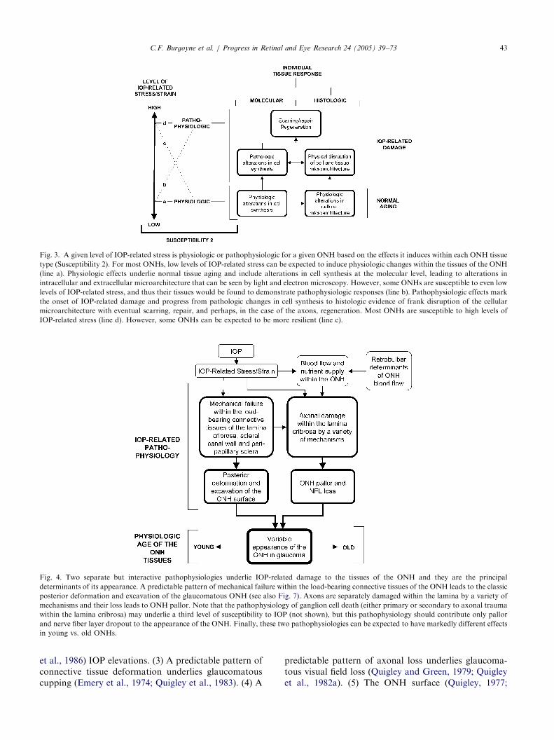
Fig. 3. A given level of IOP-related stress is physiologic or pathophysiologic for a given ONH based on the effects it induces within each ONH tissue
type (Susceptibility 2). For most ONHs, low levels of IOP-related stress can be expected to induce physiologic changes within the tissues of the ONH
(line a). Physiologic effects underlie normal tissue aging and include alterations in cell synthesis at the molecular level, leading to alterations in
intracellular and extracellular microarchitecture that can be seen by light and electron microscopy. However, some ONHs are susceptible to even low
levels of IOP-related stress, and thus their tissues would be found to demonstrate pathophysiologic responses (line b). Pathophysiologic effects mark
the onset of IOP-related damage and progress from pathologic changes in cell synthesis to histologic evidence of frank disruption of the cellular
microarchitecture with eventual scarring, repair, and perhaps, in the case of the axons, regeneration. Most ONHs are susceptible to high levels of
IOP-related stress (line d). However, some ONHs can be expected to be more resilient (line c).
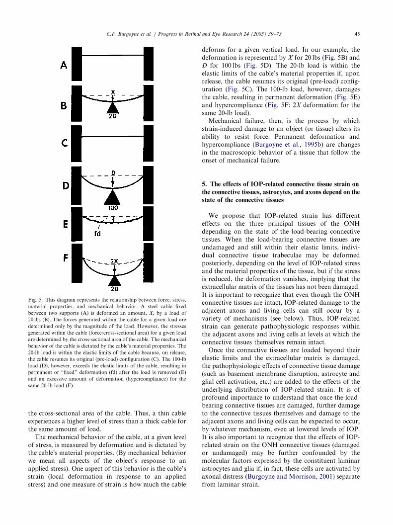
Fig. 4. Two separate but interactive pathophysiologies underlie IOP-related damage to the tissues of the ONH and they are the principal
determinants of its appearance. A predictable pattern of mechanical failure within the load-bearing connective tissues of the ONH leads to the classic
posterior deformation and excavation of the glaucomatous ONH (see also Fig. 7). Axons are separately damaged within the lamina by a variety of
mechanisms and their loss leads to ONH pallor. Note that the pathophysiology of ganglion cell death (either primary or secondary to axonal trauma
within the lamina cribrosa) may underlie a third level of susceptibility to IOP (not shown), but this pathophysiology should contribute only pallor
and nerve fiber layer dropout to the appearance of the ONH. Finally, these two pathophysiologies can be expected to have markedly different effects
in young vs. old ONHs.
C.F. Burgoyne et al. / Progress in Retinal and Eye Research 24 (2005) 39–73 43
et al., 1986) IOP elevations. (3) A predictable pattern ofconnective tissue deformation underlies glaucomatouscupping (Emery et al., 1974; Quigley et al., 1983). (4) A
predictable pattern of axonal loss underlies glaucoma-tous visual field loss (Quigley and Green, 1979; Quigleyet al., 1982a). (5) The ONH surface (Quigley, 1977;

ARTICLE IN PRESSC.F. Burgoyne et al. / Progress in Retinal and Eye Research 24 (2005) 39–7344
Zeimer et al., 1981; Pederson and Herschler, 1982;Zeimer and Ogura, 1989; Coleman et al., 1991;Burgoyne et al., 1994, 1995a; Quigley and Pease, 1996;Park and Hong, 1998) and lamina cribrosa (Levy andCrapps, 1984; Yan et al., 1994) are compliant structures,bowing posteriorly and returning to baseline followingacute elevations of IOP. (Compliance is here defined asthe ease of deformation in response to an applied load.)(6) ONH surface hypercompliance (defined as apathologically exaggerated ease of deformation inresponse to an applied load) occurs early in experi-mental glaucoma (Burgoyne et al., 1995b), whichsuggests that damage to the connective tissues occursearly in the pathophysiology of IOP-related damage tothe ONH. (7) Profound changes in the extracellularmatrix of the load-bearing tissues occur in aging(Hernandez et al., 1989; Repka and Quigley, 1989) andin glaucoma (Minckler and Spaeth, 1981; Tengrothand Ammitzboll, 1984; Hernandez et al., 1990; Morri-son et al., 1990), and IOP-induced alterations in thesynthetic activities of the cells associated with thesetissues may underlie these changes (Hernandez et al.,1994a, b; Clark et al., 1995).
3. The relationship between IOP and IOP-related stress
The connective tissues of the peripapillary sclera, thelamina cribrosa, and the scleral canal wall bear theforces generated by the IOP and are thus the load-bearing tissues of the ONH (Zeimer, 1995). For agiven ONH, IOP-related force has a predictabledistribution and leads to predictable levels of IOP-related stress. It is fundamental to our concept thatIOP-related stress within the connective tissues of theONH is not only substantial even at low levels of IOP,but is also increased as IOP increases. We hypothesizethat (1) the magnitude and distribution of IOP-relatedstress within a given ONH for a given level of IOP isprimarily determined by the three-dimensional (3D)anatomy of the eye, the scleral shell, the lamina cribrosa,and the scleral canal (Greene, 1980; Bellezza et al.,2000); and (2) the mechanical response of the load-bearing tissues to a given level of IOP-related stress(strain) is determined by their material properties(Zeimer, 1995).To begin the study of the relationship between IOP,
IOP-related stress, and IOP-related strain within theconnective tissues of the ONH (Yan et al., 1994; Dongqiand Zeqin, 1999; Bellezza et al., 2000), we are buildingfinite element models of the scleral canal as a hole in apressurized spherical shell. To our knowledge, Greene(1980) was the first to predict that IOP-related stresswithin the peripapillary sclera would be further magni-fied due to the concentration of stress around any holein a pressurized spherical shell.
Our initial finite element models (Bellezza et al., 2000)demonstrated IOP-generated stress of approximately10–17 times IOP within the tissues of the scleral shell atpoints distant from the ONH, 30 times IOP within theconnective tissues of the peripapillary sclera, 30–100times IOP within the scleral canal wall, and 50–180times IOP within the laminar beams, depending on the3D geometry of these tissues.
4. The relationship between stress, strain, material
properties, and mechanical behavior
The extracellular matrix of the sclera and laminacribrosa resists tensile, compressive, and shear stresses(Morrison, 1995; Zeimer, 1995; Hernandez and Gong,1996). (Tensile stress generates elongation of an objectalong the applied loading axis; compressive stressgenerates compression of an object along the appliedloading axis; shear stress skews the shape of an object bypulling parallel planes either in opposite directions or bydifferent relative magnitudes). Within the laminarbeams, fibrils of collagen types I and III and elastinfibers are longitudinally oriented within a denseproteoglycan matrix (Hernandez et al., 1987; Morrisonet al., 1988, 1989b; Hernandez, 1992). At the insertion ofthe peripheral laminar beams into the scleral canal wall(the laminar insertion zone), elastin fibers from thebeams insert into a circle of elastin that rings the scleralcanal (Quigley et al., 1991a, b; Hernandez, 1992).Mechanical testing of soft tissues has demonstrated
that collagen and elastin (but principally collagen) resisttensile stress (Vogel, 1974) and the proteoglycan matrixresists compressive stress (Larrabee, 1986). Collagenfibers first straighten (substantially) and then elongate(minimally) to resist a uniaxially applied load (loadapplied in a single direction) (Kenedi et al., 1965;Broom, 1978). Tensile stiffness and strength arehypothesized to be manifestations of collagen cross-linking and fiber diameter, as well as interactionsbetween collagen and the glycosaminoglycans (GAGs)of the extracellular matrix (Vogel, 1980; Oxlund andAndreassen, 1980). Tissue elasticity, defined as resump-tion of initial configuration after removal of an appliedload, is due to collagen and elastin (Oxlund et al., 1988).Thus, the composition of the extracellular matrix, interms of relative amounts and types of collagen, elastin,GAGs, and other constituents, determines the materialproperties of a tissue and, consequently, its behavior inresponse to applied force.Consider a steel cable of a given cross-sectional area
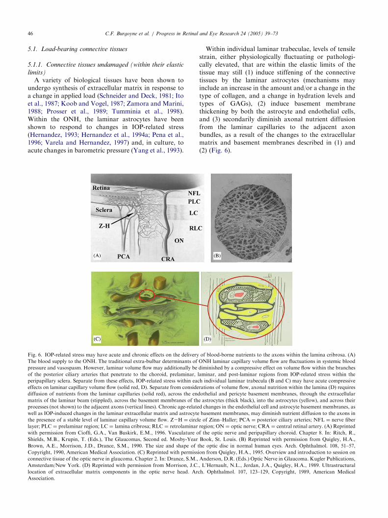
suspended tautly between two supports (Fig. 5A). Theforces generated within the cable for a given loadare determined only by the magnitude of the load.However, the stress (force/cross-sectional area) gener-ated within the cable for a given load is determined by
ARTICLE IN PRESS
Fig. 5. This diagram represents the relationship between force, stress,
material properties, and mechanical behavior. A steel cable fixed
between two supports (A) is deformed an amount, X, by a load of
20 lbs (B). The forces generated within the cable for a given load are
determined only by the magnitude of the load. However, the stresses
generated within the cable (force/cross-sectional area) for a given load
are determined by the cross-sectional area of the cable. The mechanical
behavior of the cable is dictated by the cable’s material properties. The
20-lb load is within the elastic limits of the cable because, on release,
the cable resumes its original (pre-load) configuration (C). The 100-lb
load (D), however, exceeds the elastic limits of the cable, resulting in
permanent or ‘‘fixed’’ deformation (fd) after the load is removed (E)
and an excessive amount of deformation (hypercompliance) for the
same 20-lb load (F).
C.F. Burgoyne et al. / Progress in Retinal and Eye Research 24 (2005) 39–73 45
the cross-sectional area of the cable. Thus, a thin cableexperiences a higher level of stress than a thick cable forthe same amount of load.The mechanical behavior of the cable, at a given level
of stress, is measured by deformation and is dictated bythe cable’s material properties. (By mechanical behaviorwe mean all aspects of the object’s response to anapplied stress). One aspect of this behavior is the cable’sstrain (local deformation in response to an appliedstress) and one measure of strain is how much the cable
deforms for a given vertical load. In our example, thedeformation is represented by X for 20 lbs (Fig. 5B) andD for 100 lbs (Fig. 5D). The 20-lb load is within theelastic limits of the cable’s material properties if, uponrelease, the cable resumes its original (pre-load) config-uration (Fig. 5C). The 100-lb load, however, damagesthe cable, resulting in permanent deformation (Fig. 5E)and hypercompliance (Fig. 5F: 2X deformation for thesame 20-lb load).Mechanical failure, then, is the process by which
strain-induced damage to an object (or tissue) alters itsability to resist force. Permanent deformation andhypercompliance (Burgoyne et al., 1995b) are changesin the macroscopic behavior of a tissue that follow theonset of mechanical failure.
5. The effects of IOP-related connective tissue strain on
the connective tissues, astrocytes, and axons depend on the
state of the connective tissues
We propose that IOP-related strain has differenteffects on the three principal tissues of the ONHdepending on the state of the load-bearing connectivetissues. When the load-bearing connective tissues areundamaged and still within their elastic limits, indivi-dual connective tissue trabeculae may be deformedposteriorly, depending on the level of IOP-related stressand the material properties of the tissue, but if the stressis reduced, the deformation vanishes, implying that theextracellular matrix of the tissues has not been damaged.It is important to recognize that even though the ONHconnective tissues are intact, IOP-related damage to theadjacent axons and living cells can still occur by avariety of mechanisms (see below). Thus, IOP-relatedstrain can generate pathophysiologic responses withinthe adjacent axons and living cells at levels at which theconnective tissues themselves remain intact.Once the connective tissues are loaded beyond their
elastic limits and the extracellular matrix is damaged,the pathophysiologic effects of connective tissue damage(such as basement membrane disruption, astrocyte andglial cell activation, etc.) are added to the effects of theunderlying distribution of IOP-related strain. It is ofprofound importance to understand that once the load-bearing connective tissues are damaged, further damageto the connective tissues themselves and damage to theadjacent axons and living cells can be expected to occur,by whatever mechanism, even at lowered levels of IOP.It is also important to recognize that the effects of IOP-related strain on the ONH connective tissues (damagedor undamaged) may be further confounded by themolecular factors expressed by the constituent laminarastrocytes and glia if, in fact, these cells are activated byaxonal distress (Burgoyne and Morrison, 2001) separatefrom laminar strain.
ARTICLE IN PRESSC.F. Burgoyne et al. / Progress in Retinal and Eye Research 24 (2005) 39–7346
5.1. Load-bearing connective tissues
5.1.1. Connective tissues undamaged (within their elastic
limits)
A variety of biological tissues have been shown toundergo synthesis of extracellular matrix in response toa change in applied load (Schneider and Deck, 1981; Itoet al., 1987; Koob and Vogel, 1987; Zamora and Marini,1988; Prosser et al., 1989; Tumminia et al., 1998).Within the ONH, the laminar astrocytes have beenshown to respond to changes in IOP-related stress(Hernandez, 1993; Hernandez et al., 1994a; Pena et al.,1996; Varela and Hernandez, 1997) and, in culture, toacute changes in barometric pressure (Yang et al., 1993).
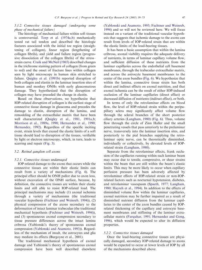
Fig. 6. IOP-related stress may have acute and chronic effects on the delivery
The blood supply to the ONH. The traditional extra-bulbar determinants of
pressure and vasospasm. However, laminar volume flow may additionally be
of the posterior ciliary arteries that penetrate to the choroid, prelaminar, l
peripapillary sclera. Separate from these effects, IOP-related stress within eac
effects on laminar capillary volume flow (solid red, D). Separate from conside
diffusion of nutrients from the laminar capillaries (solid red), across the end
matrix of the laminar beam (stippled), across the basement membranes of th
processes (not shown) to the adjacent axons (vertical lines). Chronic age-relate
well as IOP-induced changes in the laminar extracellular matrix and astrocyte
the presence of a stable level of laminar capillary volume flow. Z2H ¼ circl
layer; PLC ¼ prelaminar region; LC ¼ lamina cribrosa; RLC ¼ retrolaminar
with permission from Cioffi, G.A., Van Buskirk, E.M., 1996. Vasculature o
Shields, M.B., Krupin, T. (Eds.), The Glaucomas, Second ed. Mosby-Year
Brown, A.E., Morrison, J.D., Drance, S.M., 1990. The size and shape of t
Copyright, 1990, American Medical Association. (C) Reprinted with permiss
connective tissue of the optic nerve in glaucoma. Chapter 2. In: Drance, S.M.,
Amsterdam/New York. (D) Reprinted with permission from Morrison, J.C.
location of extracellular matrix components in the optic nerve head. Ar
Association.
Within individual laminar trabeculae, levels of tensilestrain, either physiologically fluctuating or pathologi-cally elevated, that are within the elastic limits of thetissue may still (1) induce stiffening of the connectivetissues by the laminar astrocytes (mechanisms mayinclude an increase in the amount and/or a change in thetype of collagen, and a change in hydration levels andtypes of GAGs), (2) induce basement membranethickening by both the astrocyte and endothelial cells,and (3) secondarily diminish axonal nutrient diffusionfrom the laminar capillaries to the adjacent axonbundles, as a result of the changes to the extracellularmatrix and basement membranes described in (1) and(2) (Fig. 6).
of blood-borne nutrients to the axons within the lamina cribrosa. (A)
ONH laminar capillary volume flow are fluctuations in systemic blood
diminished by a compressive effect on volume flow within the branches
aminar, and post-laminar regions from IOP-related stress within the
h individual laminar trabecula (B and C) may have acute compressive
rations of volume flow, axonal nutrition within the lamina (D) requires
othelial and pericyte basement membranes, through the extracellular
e astrocytes (thick black), into the astrocytes (yellow), and across their
d changes in the endothelial cell and astrocyte basement membranes, as
basement membranes, may diminish nutrient diffusion to the axons in
e of Zinn–Haller; PCA ¼ posterior ciliary arteries; NFL ¼ nerve fiber
region; ON ¼ optic nerve; CRA ¼ central retinal artery. (A) Reprinted
f the optic nerve and peripapillary choroid. Chapter 8. In: Ritch, R.,
Book, St. Louis. (B) Reprinted with permission from Quigley, H.A.,
he optic disc in normal human eyes. Arch. Ophthalmol. 108, 51–57,
ion from Quigley, H.A., 1995. Overview and introduction to session on
Anderson, D.R. (Eds.) Optic Nerve in Glaucoma. Kugler Publications,
, L’Hernault, N.L., Jerdan, J.A., Quigley, H.A., 1989. Ultrastructural
ch. Ophthalmol. 107, 123–129, Copyright, 1989, American Medical
ARTICLE IN PRESSC.F. Burgoyne et al. / Progress in Retinal and Eye Research 24 (2005) 39–73 47
5.1.2. Connective tissues damaged (undergoing some
phase of mechanical failure)
The histology of mechanical failure within soft tissuesis controversial. Torp et al. (1974a,b) mechanicallytested rat tail tendon and described the histologicfeatures associated with the initial toe region (straigh-tening of collagen), linear region (lengthening ofcollagen fibrils), and yield and failure region (progres-sive dissociation of the collagen fibrils) of the stress–strain curve. Craik and McNeil (1965) described changesin the trichrome staining pattern of collagen (from greento red) and the onset of fracture lines within collagenseen by light microscopy in human skin stretched tofailure. Quigley et al. (1991b) reported disruption ofboth collagen and elastin in the laminar insertion sites ofhuman and monkey ONHs with early glaucomatousdamage. They hypothesized that the disruption ofcollagen may have preceded the change in elastin.Based on these observations, we hypothesize that
IOP-related disruption of collagen is the earliest stage ofconnective tissue damage in glaucoma and precedes thedamage to elastin, disruption of the lamina, andremodeling of the extracellular matrix that have beenwell characterized (Quigley et al., 1981, 1991a,b;Morrison et al., 1989a, 1990; Hernandez et al., 1990;Hernandez, 1992). Regardless of the specific incitingevent, strain levels that exceed the elastic limits of a softtissue should lead to disruption of the tissues, verifiableby light or electron microscopy, which, in turn, leads toscarring and repair (Fig. 3).
5.2. Retinal ganglion cell axons
5.2.1. Connective tissues undamaged
IOP-related damage to the axons that occurs while theconnective tissues are within their elastic limits canresult from a variety of mechanisms (Fig. 4). Theprincipal effect should be ONH pallor due to axon loss,without excavation of the ONH surface, because, bydefinition, the connective tissues are within their elasticlimits and still able to resist IOP-related load. Theprincipal mechanisms may include (1) axonal ischemiathrough a variety of mechanisms (the traditionalvascular hypothesis (Fechtner and Weinreb, 1994)), (2)physical compression of the axons secondary to thedeformation of intact laminar trabeculae (the traditionalmechanical hypothesis (Fechtner and Weinreb, 1994)),and (3) spontaneous axonal compression secondary totissue pressure differences across the intact laminacribrosa (Yablonski’s theory of spontaneous axonalcompression (Yablonski and Asamoto, 1993)). Regard-less of the mechanism of insult, the astrocytes and gliamay mediate its effects (Burgoyne et al., 2001).The traditional mechanical hypothesis of axonal
damage and Yablonski’s theory of spontaneous axonalcompression have been well described elsewhere
(Yablonski and Asamoto, 1993; Fechtner and Weinreb,1994) and so will not be reviewed here. We will focusinstead on a variant of the traditional vascular hypoth-esis that suggests that ischemic damage to the axons canresult from levels of IOP-related strain that are withinthe elastic limits of the load-bearing tissues.It has been a basic assumption that within the lamina
cribrosa, axonal viability requires the adequate deliveryof nutrients, in terms of laminar capillary volume flow,and sufficient diffusion of these nutrients from thelaminar capillaries across the endothelial cell basementmembranes, through the trabecular extracellular matrix,and across the astrocyte basement membranes to thecenter of the axon bundles (Fig. 6). We hypothesize thatwithin the lamina, connective tissue strain has bothdirect and indirect effects on axonal nutrition, and thataxonal ischemia can be the result of either IOP-inducedocclusion of the laminar capillaries (direct effect) ordecreased diffusion of nutrients (indirect effect), or both.In terms of only the retrolaminar effects on blood
flow, the level of IOP-related strain within the peripa-pillary sclera may significantly affect volume flowthrough the scleral branches of the short posteriorciliary arteries (Langham, 1980) (Fig. 6). Thus, volumeflow through the circle of Zinn Haller and the smallpenetrating vessels that pass anteriorly to the prelaminarnerve, transversely into the laminar insertion sites, andposteriorly to the pial branches supplying the retro-laminar optic nerve, can be theoretically diminished,individually or collectively, by elevated levels of IOP-related strain (Langham, 1980).Separate from the retrolaminar effects, frank occlu-
sion of the capillaries running within each laminar beammay occur due to tensile, compressive, or shear strainswithin the beam that are still within the beam’s elasticlimits. This may be more likely to occur when capillaryperfusion pressure has been adversely affected byretrolaminar effects of IOP-related strain or non-IOP-related factors such as nocturnal hypotension, diabetes,and retrolaminar vasospasm (Spaeth, 1977; Langham,1980; Hayreh et al., 1994). In addition to the effects ofdiminished volume flow within the laminar capillaries,axonal nutrition may be further impaired as a result ofdiminished nutrient diffusion from the laminar capil-laries to the center of the axon bundles caused by IOP-related thickening of the capillary and astrocyte base-ment membranes and stiffening of the laminar extra-cellular matrix (Farquhar, 1991; Hernandez and Gong,1996), which would be expected to alter its diffusionproperties.
5.2.2. Connective tissues damaged
Once the load-bearing connective tissues are physi-cally damaged, secondary IOP-related damage to axonswould be expected to occur at lower levels of IOP by allof the mechanisms described above.
ARTICLE IN PRESSC.F. Burgoyne et al. / Progress in Retinal and Eye Research 24 (2005) 39–7348
5.3. Astrocytes and endothelial cells
5.3.1. Connective tissues undamaged
Elevated levels of IOP-related strain can be expectedto induce synthesis of extracellular matrix by thelaminar astrocytes, as outlined above, which alters thematerial properties of the adjacent laminar trabeculae.Additionally, the laminar astrocytes and laminar capil-lary endothelial cells may thicken their basementmembranes as a secondary response to the physicaldistortion induced by laminar deformation. Thickeningof the basement membranes of the laminar capillaryendothelial cells in response to elevated levels of IOP-related stress was reported in a poster by Hernandez’sgroup at the 1997 ARVO meeting (Pena et al., 1997).Basement membranes of the laminar trabeculae havealso been noted to thicken with age (Hernandez et al.,1989).
5.3.2. Connective tissues damaged
Laminar trabecular basement membrane thickening isa common finding in published descriptions of thepathology of glaucomatous damage to the ONH tissues(Hernandez et al., 1990; Morrison et al., 1990). Physicaldisruption of both astrocyte and endothelial basementmembranes may be an important stimulus to the onsetof scarring and repair within the laminar trabeculae.
6. Effects of IOP-related connective tissue strain on the
volume flow of blood and nutrient diffusion
Before it reaches the lamina cribrosa, the volumeflow of blood at a given level of IOP is principallyinfluenced by systemic blood pressure and vasospasm.Additionally, it has been hypothesized that IOP-relatedstrain within the peripapillary sclera exerts a compres-sive effect on volume flow within the branches ofthe posterior ciliary arteries that penetrate to thechoroid, prelaminar, laminar, and post-laminar regions(Langham, 1980).Separate from these effects, IOP-related strain within
each individual laminar trabecula may have acutecompressive effects on laminar capillary volume flow.Axonal nutrition within the lamina requires diffusion ofnutrients from the laminar capillaries, across theendothelial and pericyte basement membranes, throughthe extracellular matrix of the laminar beam, across thebasement membranes of the astrocytes, into the astro-cytes, and across their processes to the adjacent axons.Chronic age-related changes in the endothelial cell andastrocyte basement membranes, as well as IOP-inducedchanges in the laminar extracellular matrix and astro-cyte basement membranes, may diminish nutrientdiffusion to the axons even in the presence of a stablelevel of laminar capillary volume flow.
7. Two principal pathophysiologies underlie IOP-related
damage to the ONH
7.1. Mechanical failure within the load-bearing
connective tissues
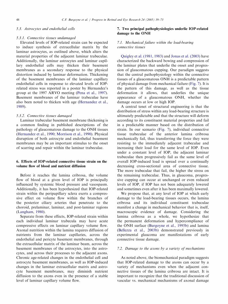
Quigley et al. (1981, 1983) and Jonas et al. (2003) havecharacterized the backward bowing and compression ofthe laminar plates that underlie the onset and progres-sion of glaucomatous cupping. Our paradigm suggeststhat the central pathophysiology within the connectivetissues of a glaucomatous ONH is a predictable patternof physical damage from mechanical failure (Fig. 7). It isthe pattern of this damage, as well as the tissuedeformation it allows, that underlies the uniqueappearance of a glaucomatous ONH, whether thedamage occurs at low or high IOP.A central tenet of structural engineering is that the
distribution of stress within any load-bearing structure isultimately predictable and that the structure will deformaccording to its constituent material properties and failin a predictable manner based on the distribution ofstrain. In our scenario (Fig. 7), individual connectivetissue trabeculae of the anterior lamina cribrosamechanically fail, thus transferring the force they wereresisting to the immediately adjacent trabeculae andincreasing their load for the same level of IOP. Evenunder a constant level of IOP, the adjacent laminartrabeculae then progressively fail as the same level ofoverall IOP-induced load is spread over a continuallydecreasing cross-sectional area of connective tissue.The more trabeculae that fail, the higher the stress onthe remaining trabeculae. Thus, in glaucoma, progres-sive cupping can occur at unchanged or even reducedlevels of IOP, if IOP has not been adequately loweredand sometimes even after it has been maximally lowered.We propose that, at any level of IOP at which early
damage to the load-bearing tissues occurs, the laminacribrosa and its individual constituent trabeculaemanifest a change in mechanical behavior that is, itself,macroscopic evidence of damage. Considering thelamina cribrosa as a whole, we hypothesize thatthe permanent deformation and hypercompliance ofthe ONH surface (Burgoyne et al., 1995b) and lamina(Bellezza et al., 2003b) demonstrated previously inexperimental glaucoma are manifestations of earlyconnective tissue damage.
7.2. Damage to the axons by a variety of mechanisms
As noted above, the biomechanical paradigm suggeststhat IOP-related damage to the axons can occur by avariety of mechanisms even when the adjacent con-nective tissues of the lamina cribrosa are intact. It isimportant to recognize that the traditional discussion ofvascular vs. mechanical mechanisms of axonal damage
ARTICLE IN PRESS
Fig. 7. A predictable pattern of mechanical failure within the load-bearing connective tissues of the ONH underlies the onset and progression of
posterior deformation and excavation. (A) Schematic of a normal sagittal view of scleral canal and laminar plates with normal laminar thickness (X)
and directions of scleral wall tension (arrows). (B) Early failure of anterior laminar plates causes thinning ðYoX Þ, excavation, and circumferential
extension, which progresses in (C) ðZoY Þ. Note that it is the mechanical failure of the peripheral laminar beams that allows (1) the remaining lamina
to be pushed posteriorly under the direct anterior/posterior effects of IOP (posterior deformation), and (2) the remaining scleral wall tissues to be
pulled under the anterior scleral rim owing to the effects of scleral wall tension, recognized clinically as excavation. (D) Further thinning of the
lamina and pronounced excavation in an end-stage ONH. (D) Courtesy of Harry A. Quigley, MD.
C.F. Burgoyne et al. / Progress in Retinal and Eye Research 24 (2005) 39–73 49
implied that IOP does not play an important role invascular or ischemic mechanisms of damage. Bycontrast, the biomechanical paradigm argues that whatdefines glaucomatous optic neuropathy is that thelocation of axonal damage is governed by the distribu-tion of IOP-related stress and strain. Thus, although theparadigm is compatible with the notion that there aremultiple mechanisms, it proposes that regardless ofmechanism or the level of IOP, the location and patternof glaucomatous axonal damage is governed by thedistribution of IOP-related stress and strain.The paradigm assumes that the principal site of IOP-
related damage to the retinal ganglion cell axons iswithin the lamina cribrosa (Anderson and Hendrickson,1974; Minckler et al., 1977; Quigley and Anderson,1976, 1977c). However, it acknowledges the potentialimportance of separate pathophysiologies within thelateral geniculate and visual cortex (Yucel et al., 2000,2001, 2003; Gupta and Yucel, 2001) as well as evidenceboth for (Kendell et al., 1995; Wygnanski et al., 1995)and against (Panda and Jonas, 1992; Janssen et al.,1997) direct, IOP-induced damage to the retinal photo-receptors. In fact, our conceptual framework is compa-
tible with the notion of additional direct photoreceptor(or other retinal cell) effects, but suggests that (1) inmost eyes, for a given level of IOP, the axons within thelamina are likely to be more vulnerable to direct effectsof IOP than are the cells within the retina, and (2) directeffects within the retina may contribute to ONH pallorbut not to true glaucomatous excavation as described inFigs. 1 and 7.Retrolaminar (Hayreh, 1974) and prelaminar (Hayr-
eh, 1986) ischemia are the central pathophysiologies ofthe traditional vascular hypothesis. Based on the generalassumption that prelaminar and retrolaminar ONHblood supply is IOP-independent, i.e., principallyinfluenced by factors other than IOP, Hayreh (1974,1986) proposed that if a glaucomatous form of opticneuropathy could occur due to ischemia in these regions,then this form of optic neuropathy was most likelyunrelated to IOP.Our model is compatible with the notion that
prelaminar ischemia, which may be less IOP-dependentthan laminar ischemia, may occur in addition to theIOP-dependent pathophysiology within the laminacribrosa, and in some eyes may predominate. However,

ARTICLE IN PRESSC.F. Burgoyne et al. / Progress in Retinal and Eye Research 24 (2005) 39–7350
as pointed out above, prelaminar ischemia alone,whether or not it is IOP-induced, should principallycause pallor of the ONH tissues and not glaucomatousexcavation. Therefore, prelaminar ischemia alone isunlikely to explain the excavation of a glaucomatousONH.Additionally, Hayreh (1974) has argued that primary
ischemia to the retrolaminar optic nerve alone couldcause optic neuropathy indistinguishable from humanglaucoma. However, most investigators believe thatischemic or compressive lesions of the retrolaminar opticnerve seldom lead to true glaucomatous excavation ofthe ONH surface (Quigley and Anderson, 1977a; Trobeet al., 1980). As noted above, our concept is compatiblewith the notion that primary retrolaminar ischemia cancause non-IOP-related damage to the axons. Further-more, in some eyes, retrolaminar ischemia may becapable of altering the material properties of the load-bearing connective tissues such that the onset andprogression of mechanical failure occur at normal levelsof IOP.
8. ONH biomechanics: experimental studies
8.1. Compliance testing the ONH surface in normal
monkeys
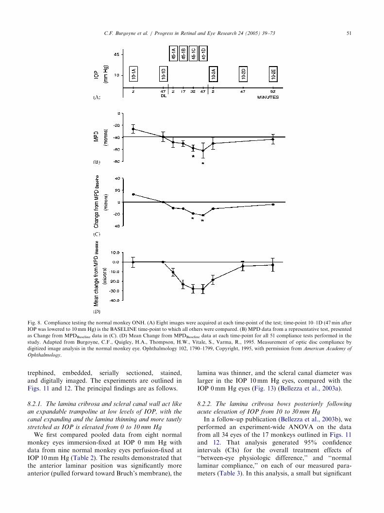
A number of investigators have previously shown thatacute increases in IOP cause posterior deformation ofthe surface of the ONH relative to the peripapillaryretina (Zeimer and Ogura, 1989; Coleman et al., 1991;Quigley and Pease, 1996; Morgan et al., 2002). Weperformed initial studies in the laboratories of Dr. HarryQuigley using a video camera to image the monkeyONH surface during acute IOP elevations (Burgoyneet al., 1994, 1995a). Fig. 8A explains the protocoland data for a single compliance test. The position ofthe ONH surface at each time point of a compliancetest is characterized by the image analysis-basedparameter mean position of the disc (MPD) (Burgoyneet al., 1995a). In these studies, 51 compliance testswere performed on 28 normal eyes of 18 monkeys(Burgoyne et al., 1995a). Fig. 8D presents the meanchange from MPDBaseline at each time point of thecompliance test for all 51 tests. The results suggest thatthe normal monkey ONH exhibits a posterior deforma-tion of approximately 30mm during acute IOP eleva-tions from 10 to 45mm Hg.More recently, we examined ONH surface compliance
using confocal scanning laser tomographic (CSLT)imaging (Heickell et al., 2001). In these studies, a totalof 156 compliance tests were performed on 48 normaleyes in 30 monkeys using three separate protocols.CSLT images were obtained using a 10� and/or 15� and/or 20� scan angle at various times after IOP was raised
from 10 to 30 and/or to 45mm Hg. At each time point,six images were analyzed to calculate MPD, whichexpressed the overall position of the optic disc surface atthat time point. Statistical analysis (ANOVA) wasperformed to evaluate differences in the amounts ofdeformation in individual eyes at different IOPs, atdifferent compliance-testing sessions, and in the twoeyes of individual monkeys under the same testingconditions.In this group of eyes, posterior deformation of the
surface of the ONH ranged from 15 to 86 mm as early as10min after IOP was increased from 10 to 30mm Hg(Table 1). When IOP was increased from 30 to 45mmHg in a subset of these eyes, most showed additionaldeformation. Of the 12 eyes for which both 15� and 20�
images were obtained during the same compliance test,seven showed larger amounts of deformation in the 20�
images (Fig. 9). Of the 18 monkeys tested in both eyes,12 showed some differences and four showed substantialdifferences between the two eyes.From these studies, we concluded that in the normal
monkey eye, the surface of the ONH deforms rapidly inresponse to increased IOP, sometimes in as little as10min. The amount of deformation varies from oneindividual to another and even within the two eyes ofindividual monkeys. Increasing the scan angle from 15�
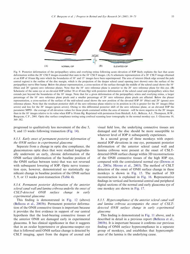
to 20� frequently increases the amount of deformationdetected, suggesting that, in addition to the ONH, theperipapillary sclera may be involved in the deformationin some eyes (Fig. 9).
8.2. The lamina cribrosa and scleral canal deform
following acute elevations of IOP
Deformation of the lamina cribrosa following acuteIOP elevation has been described in human cadaver eyes(Quigley and Green, 1979; Yan et al., 1994; Albon et al.,2000). To more fully characterize the response of theanterior scleral canal and lamina cribrosa to acute IOPelevation, we performed measurements of anteriorlaminar position and thickness, scleral canal diameterat Bruch’s membrane and the anterior laminar insertion,and anterior scleral canal wall geometry in every sixth4-mm serial sagittal section of the ONH from both eyesof 17 monkeys (Fig. 10). Under pentobarbital anesthe-sia, both eyes of four normal monkeys (two normaleyes) and five monkeys with early glaucoma (one normaland one early glaucoma eye) were cannulated, and theIOP set to 10mm Hg in one normal eye and 30 or 45mmHg in the other (normal or early glaucoma) eye. After15–80min in which the mean blood pressure (diastolicplus one third the diastolic/systolic difference) wasmaintained as high as possible (80–100mm Hg), eachmonkey was perfusion-fixed with 1L of paraformalde-hyde and 6L of glutaraldehyde pressurized to150–200mm Hg. The ONH tissues from each eye were
ARTICLE IN PRESS
Fig. 8. Compliance testing the normal monkey ONH. (A) Eight images were acquired at each time-point of the test; time-point 10–1D (47min after
IOP was lowered to 10mm Hg) is the BASELINE time-point to which all others were compared. (B) MPD data from a representative test, presented
as Change from MPDBaseline data in (C). (D) Mean Change from MPDBaseline data at each time-point for all 51 compliance tests performed in the
study. Adapted from Burgoyne, C.F., Quigley, H.A., Thompson, H.W., Vitale, S., Varma, R., 1995. Measurement of optic disc compliance by
digitized image analysis in the normal monkey eye. Ophthalmology 102, 1790–1799, Copyright, 1995, with permission from American Academy of
Ophthalmology.
C.F. Burgoyne et al. / Progress in Retinal and Eye Research 24 (2005) 39–73 51
trephined, embedded, serially sectioned, stained,and digitally imaged. The experiments are outlined inFigs. 11 and 12. The principal findings are as follows.
8.2.1. The lamina cribrosa and scleral canal wall act like
an expandable trampoline at low levels of IOP, with the
canal expanding and the lamina thinning and more tautly
stretched as IOP is elevated from 0 to 10 mm Hg
We first compared pooled data from eight normalmonkey eyes immersion-fixed at IOP 0 mm Hg withdata from nine normal monkey eyes perfusion-fixed atIOP 10mm Hg (Table 2). The results demonstrated thatthe anterior laminar position was significantly moreanterior (pulled forward toward Bruch’s membrane), the
lamina was thinner, and the scleral canal diameter waslarger in the IOP 10mm Hg eyes, compared with theIOP 0mm Hg eyes (Fig. 13) (Bellezza et al., 2003a).
8.2.2. The lamina cribrosa bows posteriorly following
acute elevation of IOP from 10 to 30 mm Hg
In a follow-up publication (Bellezza et al., 2003b), weperformed an experiment-wide ANOVA on the datafrom all 34 eyes of the 17 monkeys outlined in Figs. 11and 12. That analysis generated 95% confidenceintervals (CIs) for the overall treatment effects of‘‘between-eye physiologic difference,’’ and ‘‘normallaminar compliance,’’ on each of our measured para-meters (Table 3). In this analysis, a small but significant

ARTICLE IN PRESS
Table 1
Monkey characteristics and early IOP-30 mm Hg deformation data
Study
no.
Monkey
no.
Weight
(kg)
Axial length (mm)a Disc area (mm2)b MPDBaseline ðmmÞc Magnitude of early
IOP-30 deformation ðmmÞusing 15� scan angled
OD OS OD OS OD OS OD OS
1 1 5.5 18.64 1.03 +38 �68
2 12.0 20.16 1.26 �104 �27
3 6.4 21.35 1.88 �156 �34
2 4 11.4 21.02 1.48 �100 �38
5 9.2 20.34 1.53 �115 �42
6 7.8 20.19 1.79 �17 �37
7 6.5 21.99 1.90 �126 +2
8 5.5 21.09 1.60 +74 �36
9 3.1 19.57 1.31 �154 �9
10 12.4 20.38 1.46 +10 �16
11 10.9 21.10 1.84 �83 �15
12 11.5 20.90 1.71 +26 �86
3 13 6.7 19.21 19.33 1.49 1.33 �139 �164 �36 �45
14 6.0 20.87 20.80 1.59 1.76 �127 �132 �10 �8
15 7.5 21.39 21.25 1.77 1.47 +35 +40 �37 �46
16 6.8 20.33 20.22 1.93 1.77 �174 �188 �50 �22
17 6.8 20.75 20.75 1.70 1.80 �151 �146 �35 �33
18 5.7 20.47 20.47 1.36 1.40 �132 �114 �10 �22
19 6.0 19.92 19.63 1.54 1.52 �104 �125 �34 �49
20 6.8 19.74 19.81 1.23 1.23 �86 �102 �64 �54
21 7.5 21.79 21.54 1.92 1.94 �119 �115 �60 �41
22 5.8 20.61 20.51 1.62 1.75 �144 �153 �40 �35
23 6.0 20.40 20.44 1.73 1.43 �165 �132 �9 �51
24 7.2 20.06 18.71 1.18 1.03 �41 �25 �44 �43
25 8.3 21.61 21.52 1.65 1.47 �28 �28 �48 �21
26 8.3 20.72 20.64 1.21 1.26 �121 �93 �39 �41
27 7.8 21.10 20.84 1.93 1.65 �201 �168 0 �37
28 5.7 20.30 20.15 1.26 1.27 �123 �99 �42 �42
29 5.6 21.05 21.00 1.60 1.65 �74 �41 �38 �36
30 5.0 20.93 20.80 2.27 2.14 �211 �194 +2 �11
Reprinted with permission from Heickell, A.G., Bellezza, A.J., Thompson, H.W., Burgoyne, C.F., 2001. Optic disc surface compliance testing using
confocal scanning laser tomography in the normal monkey eye. J. Glaucoma 10, 369–382.aMean axial length at the baseline observation point for the three or five test days.bMean value of the parameter Disc Area within the 15� baseline observation point images for the three or five test days.cMean value of the parameter MPD within the 15� baseline observation point images for the three or five test days.dBold type indicates values statistically significantly different from corresponding baseline observation point value (Po0:05, ANOVA).
C.F. Burgoyne et al. / Progress in Retinal and Eye Research 24 (2005) 39–7352
treatment effect of 12–19mm for normal laminarcompliance exceeded the 95% CI for physiologicbetween-eye differences. The relatively small effect islikely a result of low IOP elevations (from 10 to 30 mmHg) of short duration (15min) in three of the fourmonkeys.
8.3. Damage to the ONH connective tissues occurs early
in experimental glaucoma
8.3.1. Early onset of hypercompliance of the ONH
surface in experimental glaucoma
In a second series of experiments performed in Dr.Quigley’s laboratories, monkeys were tested for com-pliance as normals and then on multiple occasions after
the onset of experimental glaucoma or optic nervetransection in one eye (Burgoyne et al., 1995b). In bothlongitudinally and cross-sectionally studied eyes, ONHsurface compliance increased significantly 1–2 weeksafter the onset of experimental glaucoma (Fig. 14), andthe increase persisted through the 9–12 week evaluation.However, compliance was again reduced during weeks13–18 and beyond week 18 following IOP elevation.
8.3.2. Relative stiffness rather than hypercompliance in
optic nerve transection eyes
In striking contrast to the experimental glaucomaeyes, the optic nerve transection eyes showed no earlyperiod of hypercompliance. Instead, the transection eyes
ARTICLE IN PRESS
Fig. 9. Posterior deformation of the peripapillary sclera and overlying retina following acute elevation of IOP likely explains the fact that acute
deformation within the 20� CSLT images exceeded that seen in the 10� CSLT images. (A) A schematic representation of a 20� CSLT image obtained
at an IOP of 10mm Hg onto which the boundaries of 10� and 15� images have been superimposed. The area of interest (black edge around the pale
central region) is the outline of the disc margin, which is the projection of the deeper scleral canal opening (not shown) onto the surface of the
peripapillary nerve fiber hump. Below the planar representation, a cross-section of the surface through the middle of the scleral canal shows the 10�
(blue) and 20� (green) zero reference planes. Note that the 10� zero reference plane is anterior to the 20� zero reference plane for this eye. (B)
Schematic of the same eye at an elevated IOP (either 30 or 45mm Hg) with posterior deformation of the scleral canal and peripapillary sclera that
extends just beyond the boundaries of the 10� image. Note that for a given deformation of the peripapillary sclera and overlying retina, a larger
percentage of the 10� zero reference plane pixels and a smaller percentage of 20� zero reference plane pixels are affected. Below the planar
representation, a cross-section of the surface of the mid-scleral canal shows the new position of the elevated IOP 10� (blue) and 20� (green) zero
reference planes. Note that the resultant posterior shift of the zero reference plane relative to its position in (A) is greater for the 10� images (blue
arrow) and less for the 20� images (green arrow). Owing to this differential posterior shift of the zero reference plane, at an elevated IOP the
parameter MPD—the average of all elevation values for those pixels contained within the area of interest—will be more negative in the 20� images
than in the 10� images relative to its value when IOP is 10mm Hg. Reprinted with permission from Heickell, A.G., Bellezza, A.J., Thompson, H.W.,
Burgoyne, C.F., 2001. Optic disc surface compliance testing using confocal scanning laser tomography in the normal monkey eye. J. Glaucoma 10,
369–382.
C.F. Burgoyne et al. / Progress in Retinal and Eye Research 24 (2005) 39–73 53
progressed to qualitatively less movement of the disc 5,9, and 13 weeks following transection (Fig. 14).
8.3.3. Early onset of permanent posterior deformation of
the ONH surface in experimental glaucoma
Separate from a change in optic disc compliance, theglaucomatous optic discs that were studied longitudin-ally underwent an early, chronic deformation of theONH surface (deformation of the baseline position ofthe ONH surface between tests) that was not reversedwith subsequent lowering of IOP. Optic nerve transec-tion eyes, however, demonstrated no statistically sig-nificant change in baseline position of the ONH surface5, 9, or 13 weeks post-transection (Table 4).
8.3.4. Permanent posterior deformation of the anterior
scleral canal wall and lamina cribrosa underlie the onset of
CSLT-detected ONH surface change in early
experimental glaucoma
This finding is demonstrated in Fig. 12 (above)(Bellezza et al., 2003b). Permanent posterior deforma-tion of the ONH connective tissues is important becauseit provides the first evidence in support of our centralhypothesis that the load-bearing connective tissues ofthe anterior ONH are damaged early in experimentalglaucoma. It has clinical significance in that it suggeststhat in an ocular hypertensive or glaucoma-suspect eyethat is followed until ONH surface change is detected byCSLT imaging, apart from the degree of axonal and
visual field loss, the underlying connective tissues aredamaged and the disc should be more susceptible towhatever level of IOP it subsequently experiences.In a second group of three monkeys with experi-
mental IOP elevations in one eye, permanent posteriordeformation of the anterior scleral canal wall andlamina cribrosa were present at the onset of CSLT-detected ONH surface change within 3D reconstructionsof the ONH connective tissues of the high IOP eye,compared with the contralateral normal eye (Downs etal., 2003a; Hirons et al., 2003). The method of CSLTdetection of the onset of ONH surface change in thesemonkeys is shown in Fig. 15. The method of 3Dreconstruction is explained in Fig. 16. Representativefindings in vertical and horizontal central and peripheraldigital sections of the normal and early glaucoma eye ofone monkey are shown in Fig. 17.
8.3.5. Hypercompliance of the anterior scleral canal wall
and lamina cribrosa accompanies the onset of CSLT-
detected ONH surface change in early experimental
glaucoma
This finding is demonstrated in Fig. 11 above, and isdescribed in detail in a previous report (Bellezza et al.,2003b). It is important because it confirms our previousfinding of ONH surface hypercompliance in a separategroup of monkeys, and establishes that hypercompli-ance of the lamina is the underlying cause.
ARTICLE IN PRESS
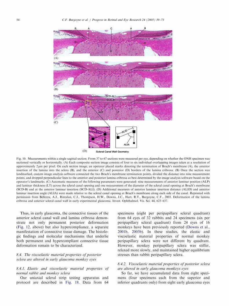
Fig. 10. Measurements within a single sagittal section. From 37 to 67 sections were measured per eye, depending on whether the ONH specimen was
sectioned vertically or horizontally. (A) Each composite section image consists of four to six individual overlapping images taken at a resolution of
approximately 2mm per pixel. On each section image, an operator placed marks denoting the termination of Bruch’s membrane (A), the anteriorinsertion of the lamina into the sclera (B), and the anterior (C) and posterior (D) borders of the lamina cribrosa. (B) Once the section was
landmarked, custom image analysis software connected the two Bruch’s membrane termination points, divided the distance into nine measurement
points, and dropped perpendicular lines to the anterior and posterior lamina cribrosa as best determined by the image analysis software based on the
operator’s landmarks. (C) Automatic measures of the following parameters were generated: nine measurements of anterior laminar position (ALP)
and laminar thickness (LT) across the scleral canal opening and one measurement of the diameter of the scleral canal opening at Bruch’s membrane
(SCD-B) and at the anterior laminar insertion (SCD-ALI). (D) Additional measures of anterior laminar insertion distance (ALID) and anterior
laminar insertion angle (ALIA) were made relative to the scleral canal opening at Bruch’s membrane along each side of the canal. Reprinted with
permission from Bellezza, A.J., Rintalan, C.J., Thompson, H.W., Downs, J.C., Hart, R.T., Burgoyne, C.F., 2003. Deformation of the lamina
cribrosa and anterior scleral canal wall in early experimental glaucoma. Invest. Ophthalmol. Vis. Sci. 44, 623–637.
C.F. Burgoyne et al. / Progress in Retinal and Eye Research 24 (2005) 39–7354
Thus, in early glaucoma, the connective tissues of theanterior scleral canal wall and lamina cribrosa demon-strate not only permanent posterior deformation(Fig. 12, above) but also hypercompliance, a separatemanifestation of connective tissue damage. The histolo-gic findings and molecular mechanisms that underlieboth permanent and hypercompliant connective tissuedeformation remain to be characterized.
8.4. The viscoelastic material properties of posterior
sclera are altered in early glaucoma monkey eyes
8.4.1. Elastic and viscoelastic material properties of
normal rabbit and monkey sclera
Our uniaxial scleral strip testing apparatus andprotocol are described in Fig. 18. Data from 64
specimens (eight per peripapillary scleral quadrant)from 64 eyes of 32 rabbits and 24 specimens (six perperipapillary scleral quadrant) from 24 eyes of 16monkeys have been previously reported (Downs et al.,2001b, 2003b). In these studies, the elastic andviscoelastic material properties of normal monkeyperipapillary sclera were not different by quadrant.However, monkey peripapillary sclera was stiffer,relaxed more slowly, and maintained higher equilibriumstresses than rabbit peripapillary sclera.
8.4.2. Viscoelastic material properties of posterior sclera
are altered in early glaucoma monkeys eyes
So far, we have accumulated data from eight speci-mens (four specimens each from the superior andinferior quadrants only) from eight early glaucoma eyes
ARTICLE IN PRESS
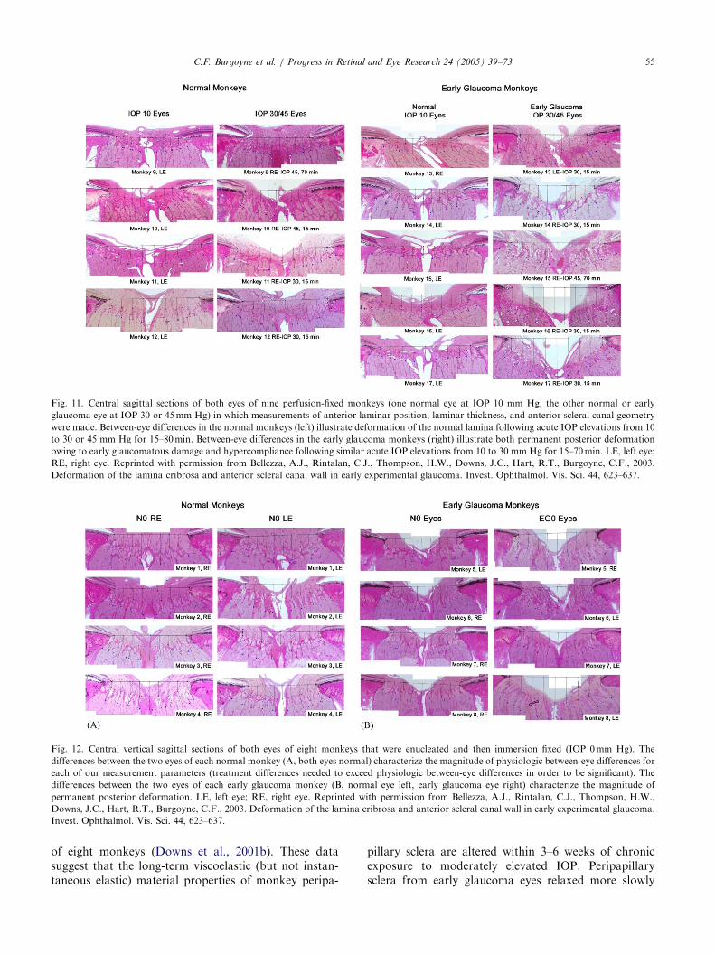
Fig. 11. Central sagittal sections of both eyes of nine perfusion-fixed monkeys (one normal eye at IOP 10 mm Hg, the other normal or early
glaucoma eye at IOP 30 or 45mm Hg) in which measurements of anterior laminar position, laminar thickness, and anterior scleral canal geometry
were made. Between-eye differences in the normal monkeys (left) illustrate deformation of the normal lamina following acute IOP elevations from 10
to 30 or 45 mm Hg for 15–80min. Between-eye differences in the early glaucoma monkeys (right) illustrate both permanent posterior deformation
owing to early glaucomatous damage and hypercompliance following similar acute IOP elevations from 10 to 30 mm Hg for 15–70min. LE, left eye;
RE, right eye. Reprinted with permission from Bellezza, A.J., Rintalan, C.J., Thompson, H.W., Downs, J.C., Hart, R.T., Burgoyne, C.F., 2003.
Deformation of the lamina cribrosa and anterior scleral canal wall in early experimental glaucoma. Invest. Ophthalmol. Vis. Sci. 44, 623–637.
Fig. 12. Central vertical sagittal sections of both eyes of eight monkeys that were enucleated and then immersion fixed (IOP 0mm Hg). The
differences between the two eyes of each normal monkey (A, both eyes normal) characterize the magnitude of physiologic between-eye differences for
each of our measurement parameters (treatment differences needed to exceed physiologic between-eye differences in order to be significant). The
differences between the two eyes of each early glaucoma monkey (B, normal eye left, early glaucoma eye right) characterize the magnitude of
permanent posterior deformation. LE, left eye; RE, right eye. Reprinted with permission from Bellezza, A.J., Rintalan, C.J., Thompson, H.W.,
Downs, J.C., Hart, R.T., Burgoyne, C.F., 2003. Deformation of the lamina cribrosa and anterior scleral canal wall in early experimental glaucoma.
Invest. Ophthalmol. Vis. Sci. 44, 623–637.
C.F. Burgoyne et al. / Progress in Retinal and Eye Research 24 (2005) 39–73 55
of eight monkeys (Downs et al., 2001b). These datasuggest that the long-term viscoelastic (but not instan-taneous elastic) material properties of monkey peripa-
pillary sclera are altered within 3–6 weeks of chronicexposure to moderately elevated IOP. Peripapillarysclera from early glaucoma eyes relaxed more slowly
ARTICLE IN PRESS
Table 2
Lamina cribrosa position and thickness by IOP groupa
Region Anterior laminar position Laminar thickness
IOP 10mm Hg IOP 0mm Hg IOP 10mm Hg IOP 0mm Hg
Overall 116� 2 184� 2 195� 2 264� 2
Central 161� 4 267� 6 223� 4 268� 4
Superior 102� 2 151� 4 179� 2 273� 4
Inferior 106� 4 158� 4 186� 2 270� 2
Nasal 109� 4 181� 4 189� 2 258� 4
Temporal 102� 2 162� 4 199� 2 252� 2
Reprinted with permission from Bellezza, A.J., Rintalan, C.J., Thompson, H.W., Downs, J.C., Hart, R.T., Burgoyne, C.F., 2003. Anterior scleral
canal geometry in pressurised (IOP 10) and nonpressurized (IOP 0) normal monkey eyes. Br. J. Ophthalmol. 87, 1284–1290 (http://
bjo.bmjjournals.com/).aValues are means � 95% confidence intervals in mm.
Fig. 13. Representative middle sagittal section images from four representative IOP 0mm Hg (immersion-fixed) and four representative IOP 10mm
Hg (perfusion-fixed) ONHs from normal monkey eyes. Note that within the IOP 0 eyes (left), the lamina cribrosa appears ‘‘sagged’’ posteriorly
relative to the ‘‘taut,’’ more anteriorly positioned lamina in the IOP 10 eyes (right). OD, right eye; OS, left eye. Reprinted with permission from
Bellezza, A.J., Rintalan, C.J., Thompson, H.W., Downs, J.C., Hart, R.T., Burgoyne, C.F., 2003. Anterior scleral canal geometry in pressurised (IOP
10) and non-pressurised (IOP 0) normal monkey eyes. Br. J. Ophthalmol. 87, 1284–1290 (http://bjo.bmjjournals.com/).
C.F. Burgoyne et al. / Progress in Retinal and Eye Research 24 (2005) 39–7356
and maintained higher equilibrium stresses than that ofnormal eyes.
8.5. IOP-related stress and strain markedly diminish
patency of the anterior laminar capillaries in normal and
early glaucoma monkey eyes at high IOP
To test the hypothesis that moderate, short-term IOPelevations diminish the patency of capillaries in thelamina cribrosa, we qualitatively evaluated anterior andposterior laminar and retrolaminar capillary patency inthe high-resolution digital images of peripheral and
central sections from both the low and high IOP eye ofeach of the nine perfusion-fixed monkeys described inFig. 11, above.At the time of enucleation, all nine of the high IOP
eyes, but none of the low IOP eyes, contained smallamounts of residual blood within the retinal and vortexvessels. Within the digital section images (Fig. 19), theposterior laminar and retrolaminar capillaries werewidely patent in all low and high IOP eyes. However,anterior laminar capillary patency was markedly dimin-ished in all nine high IOP eyes, compared to thecontralateral low IOP eyes. We concluded that anterior

ARTICLE IN PRESS
Table 3
Magnitude of experiment-wide effects
Parameter ðmmÞ Immersion-fixed monkey eyes Perfusion-fixed monkey eyes Hypercompliance alone
Intramonkey
physiologic
between-eye
difference N0-
RE–N0-LE
Plastic
deformation
EG0–N0
Normal
laminar
compliance
N30/
45–N10(N)
Permanent
deformation with
early glaucomatous
hypercompliance
EG30/45–N10(EG)
Difference between
[EG30/45–N10(EG)]
and [EG0–N0]
Anterior laminar position 1–17a
62–77 10–23 132–145 55–83
Laminar thickness 6–15 50–60 3–11 56–64 �4–14
Vertical scleral canal diameter
Bruch’s membrane �51–52 33–129 �69–65 88–198 �41–69
Anterior laminar insertion �109–7 31–139 �96–56 135–259 �4–228
Horizontal scleral canal
diameter
Bruch’s membrane NA NA �46–40 96–182 NA
Anterior laminar insertion NA NA �61–12 23–96 NA
Pairs of values in bold print denote significant differences between the compared groups. N0-RE and N0-LE, normal right and left eyes of normal
monkeys, immersion-fixed at IOP 0mm Hg; N0 and EG0, normal and early glaucoma eyes of early glaucoma monkeys, immersion-fixed at IOP
0mm Hg; N10(N), normal eye of normal monkey, perfusion-fixed at IOP 10mm Hg; N30/45, normal eye of normal monkey, perfusion-fixed at IOP
30 or 45 mm Hg; N10(EG), normal eye of early glaucoma monkey, perfusion-fixed at IOP 10 mm Hg; EG 30/45, early glaucoma eye of early
glaucoma monkey, perfusion-fixed at IOP 30 or 45 mm Hg; NA, not available. Reprinted with permission from Bellezza, A.J., Rintalan, C.J.,
Thompson, H.W., Downs, J.C., Hart, R.T., Burgoyne, C.F., 2003. Deformation of the lamina cribrosa and anterior scleral canal wall in early
experimental glaucoma. Invest. Ophthalmol. Vis. Sci. 44, 623–637.aValues are 95% confidence intervals in mm of the differences between the compared groups of eyes.
C.F. Burgoyne et al. / Progress in Retinal and Eye Research 24 (2005) 39–73 57
laminar capillary patency was markedly diminished innormal and early glaucoma monkey eyes perfusion-fixedfollowing moderate, short-term IOP elevations.Previous studies using microspheres (Alm, 1973;
Geijer and Bill, 1979) have suggested that volume flowwithin the prelaminar and anterior laminar capillarybeds is preferentially diminished once perfusion pressure(defined as the mean blood pressure minus the IOP) isless than 30mm Hg. Although the clinical significance ofour findings remains to be determined, they are inagreement with these previous reports and suggest thatwithin the tissues of the normal and early glaucomamonkey ONH, blood flow within the anterior laminarcapillaries may be most susceptible to fluctuations inocular perfusion pressure.
9. Finite element modeling of the ONH
We are building continuum and micro finite elementmodels of the load-bearing connective tissues of theONH so as to model and predict the stresses and strainsthey experience. The finite element method is anengineering tool used to study structures with complexgeometries and material properties (Fig. 20). In the finiteelement method, the digital geometry of the structurebeing modeled is ‘‘discretized’’ into multiple, small,regularly shaped building blocks (the finite elements—
see Fig. 20). The mechanical behavior of the totalstructure is then calculated from the combined behaviorof these constitutive elements. As applied to the laminacribrosa and scleral canal, our finite element models willconsist of a computational model that captures theimportant aspects of their geometry, material properties,and boundary conditions.Because each element of the model is individually
treated (i.e., programmed to possess its own shape andmaterial properties), computing requirements dictatethat structures be modeled first using a ‘‘continuum’’approach, in which the digital geometry of the laminacribrosa, for example, is broken into relatively largerepresentative volume elements that include multiplelaminar beams (Fig. 21). For each continuum element,the stiffness is found from the volume fraction andpredominant orientation of the beams contained withinthe element. Model development advances from con-tinuum to micro finite element modeling by graduallydiscretizing the large continuum elements into theirconstituent voxels or groups of voxels (Fig. 21). Inrelated voxel-based finite element models of trabecularbone, the individual voxels within each trabecular strutthemselves become elements, thus generating modelswith 10–100 million elements, requiring 20,000 CPUhours (6 weeks) of supercomputer time to characterizethe stresses and strains within each element of the model(Quigley et al., 1982a).
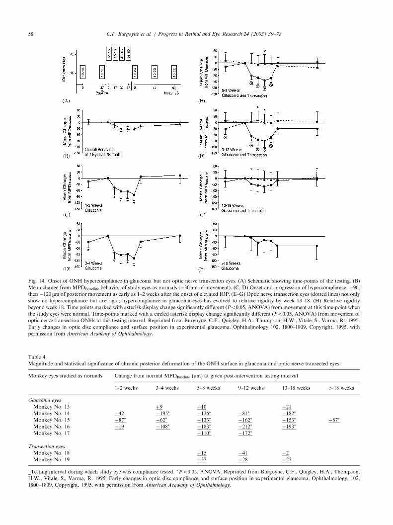
ARTICLE IN PRESS
Fig. 14. Onset of ONH hypercompliance in glaucoma but not optic nerve transection eyes. (A) Schematic showing time-points of the testing. (B)
Mean change from MPDBaseline behavior of study eyes as normals (�30mm of movement). (C, D) Onset and progression of hypercompliance; �90,then �120mm of posterior movement as early as 1–2 weeks after the onset of elevated IOP. (E–G) Optic nerve transection eyes (dotted lines) not onlyshow no hypercompliance but are rigid; hypercompliance in glaucoma eyes has evolved to relative rigidity by week 13–18. (H) Relative rigidity
beyond week 18. Time points marked with asterisk display change significantly different (Po0:05, ANOVA) from movement at this time-point whenthe study eyes were normal. Time-points marked with a circled asterisk display change significantly different (Po0:05, ANOVA) from movement ofoptic nerve transection ONHs at this testing interval. Reprinted from Burgoyne, C.F., Quigley, H.A., Thompson, H.W., Vitale, S., Varma, R., 1995.
Early changes in optic disc compliance and surface position in experimental glaucoma. Ophthalmology 102, 1800–1809, Copyright, 1995, with
permission from American Academy of Ophthalmology.
Table 4
Magnitude and statistical significance of chronic posterior deformation of the ONH surface in glaucoma and optic nerve transected eyes
Monkey eyes studied as normals Change from normal MPDBaseline ðmmÞ at given post-intervention testing interval
1–2 weeks 3–4 weeks 5–8 weeks 9–12 weeks 13–18 weeks 418 weeks
Glaucoma eyes
Monkey No. 13 þ9 �10 �21
Monkey No. 14 �42 �195� �126� �81� �182�
Monkey No. 15 �87� �62� �133� �162� �153� �87�
Monkey No. 16 �19 �108� �183� �212� �193�
Monkey No. 17 �110� �172�
Transection eyes
Monkey No. 18 �15 �41 �2
Monkey No. 19 �37 �28 �27
_Testing interval during which study eye was compliance tested. �Po0:05, ANOVA. Reprinted from Burgoyne, C.F., Quigley, H.A., Thompson,H.W., Vitale, S., Varma, R. 1995. Early changes in optic disc compliance and surface position in experimental glaucoma. Ophthalmology, 102,
1800–1809, Copyright, 1995, with permission from American Academy of Ophthalmology.
C.F. Burgoyne et al. / Progress in Retinal and Eye Research 24 (2005) 39–7358

ARTICLE IN PRESS
Fig. 15. Post-laser IOP, Change from MPDBaseline, and MPDBaselinedata from both eyes of a representative early glaucoma monkey. IOP,
Change from MPDBaseline (surface compliance following an acute IOP
elevation from 10 to 30mm Hg), and MPDBaseline � 95% confidence
interval (surface deformation) are plotted for both the normal
(triangles) and early glaucoma eyes (circles) at each post-laser imaging
session. For all three parameters, the horizontal lines represent the
upper and lower boundaries of the 95% CI for the mean of the three
pre-laser, normal compliance-testing sessions (dashed lines, normal
eye; solid lines, early glaucoma eye). This monkey was killed (Sac) at
the onset of confocal scanning laser tomography-detected ONH
surface change, which was defined as the onset of permanent posterior
deformation of the ONH surface (MPDBaseline � 95% CI beneath the
normal pre-laser range) at two consecutive post-laser imaging sessions.
However, in this monkey, because the MPDBaseline data at week 7 post-
laser were only questionably significant, the experimental glaucoma
eye was followed for one additional imaging session. Reprinted with
permission from Bellezza, A.J., Rintalan, C.J., Thompson, H.W.,
Downs, J.C., Hart, R.T., Burgoyne, C.F., 2003. Deformation of the
lamina cribrosa and anterior scleral canal wall in early experimental
glaucoma. Invest. Ophthalmol. Vis. Sci. 44, 623–637.
C.F. Burgoyne et al. / Progress in Retinal and Eye Research 24 (2005) 39–73 59
The principal goal of building finite element models isto acquire the ability to estimate the mechanicalresponse of the ONH connective tissue structure, i.e.,its deformed shape, as well as its constitutive stressesand strains, under a variety of loading conditions. In thecase of the normal and early glaucoma ONH, we willrefine and validate our models’ predictions of laminardeformation using the rigorous histologic studies out-lined previously (Bellezza et al., 2003a,b), and thedeformation within the 3D reconstructions of a new
group of normal and early glaucoma monkeys that havebeen perfusion-fixed at varying levels of IOP (Table 5).Demonstration of the accuracy of the models’ predic-tions—a key component of the refinement and valida-tion process—will serve as evidence that engineeringprinciples can be used to understand and predict ONHconnective tissue behavior in normal, early, andmoderate glaucoma monkey eyes.
9.1. Idealized models
Our initial modeling used finite element models from13 digital 3D geometries, representing the posteriorscleral shell of 13 idealized human eyes (Fig. 22)(Bellezza et al., 2000). The purpose of this study wasto assess the effect of the size and shape of the scleralcanal, as well as the thickness of the peripapillary sclera,on the level of IOP-related stress for a given level ofIOP. Within these models, larger scleral canal diameter,elongation of the canal, and thinning of the sclera allincreased IOP-related stress for a given level of IOP. Forall models, maximum IOP-related stress ranged from 6times IOP (posterior sclera) to 122 times IOP (laminartrabeculae). For each model, maximum IOP-relatedstress was highest within the laminar trabecular region,and decreased progressively through the laminar inser-tion, peripapillary scleral, and posterior scleral regions.Varying the inner radius had little effect on themaximum IOP-related stress within the scleral canal.These initial finite element models suggest that IOP-
related stress within the load-bearing connective tissuesof the ONH is substantial even at low levels of IOP,which supports our concept that the ONH is a ‘‘high-stress’’ environment that requires strong connectivetissues to withstand the stresses generated by evennormal levels of IOP. These data additionally suggestthat scleral canal size/shape and scleral thickness areprincipal determinants of the magnitude of IOP-relatedstress within the connective tissues of the ONH for agiven level of IOP. However, models that incorporatephysiologic scleral canal and laminar geometries, a morerefined finite element model meshwork, and non-isotropic material properties are needed to improveclinical relevance.
9.2. Continuum and micro finite element models
Continuum and micro finite element modeling of theONH requires construction of 3D digital geometriesthat more accurately represent the 3D architecture ofthe structure being modeled. We are currently con-structing representative geometries for the posteriorscleral shell, peripapillary retina, scleral canal, andlamina cribrosa that are based on the actual anatomyof individual monkey eyes.
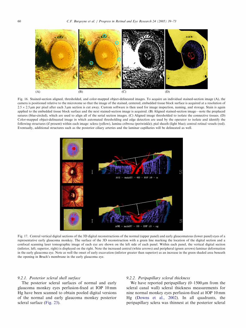
ARTICLE IN PRESS
Fig. 16. Stained-section aligned, thresholded, and color-mapped object-delineated images. To acquire an individual stained-section image (A), the
camera is positioned relative to the microtome so that the image of the stained, centered, embedded tissue block surface is acquired at a resolution of
2:5 2:5mm per pixel after each 3mm section is cut away. Custom software is then used for image inspection, naming, and storage. Stain is againapplied to the embedded tissue block surface and the next stained-section image is acquired. (B) Aligned stained-section image—note the preplaced
sutures (blue-circled), which are used to align all of the serial section images. (C) Aligned image thresholded to isolate the connective tissues. (D)
Color-mapped object-delineated image in which automated thresholding and edge detection are used by the operator to isolate and identify the
following structures (if present) within each image: sclera (yellow), lamina cribrosa (periwinkle); pial sheath (light blue); central retinal vessels (red).
Eventually, additional structures such as the posterior ciliary arteries and the laminar capillaries will be delineated as well.
Fig. 17. Central vertical digital sections of the 3D digital reconstructions of the normal (upper panel) and early glaucomatous (lower panel) eyes of a
representative early glaucoma monkey. The surface of the 3D reconstruction with a green line marking the location of the digital section and a
confocal scanning laser tomographic image of each eye are shown on the left side of each panel. Within each panel, the vertical digital section
(inferior, left; superior, right) is displayed on the right. Note the increased central (white arrows) and peripheral (green arrows) laminar deformation
in the early glaucoma eye. Note as well the onset of early excavation (inferior greater than superior) as an increase in the green shaded area beneath
the opening in Bruch’s membrane in the early glaucoma eye.
C.F. Burgoyne et al. / Progress in Retinal and Eye Research 24 (2005) 39–7360
9.2.1. Posterior scleral shell surface
The posterior scleral surfaces of normal and earlyglaucoma monkey eyes perfusion-fixed at IOP 10mmHg have been scanned to obtain pooled digital versionsof the normal and early glaucoma monkey posteriorscleral surface (Fig. 23).
9.2.2. Peripapillary scleral thickness
We have reported peripapillary (0–1500mm from thescleral canal wall) scleral thickness measurements fornine normal monkey eyes perfusion-fixed at IOP 10mmHg (Downs et al., 2002). In all quadrants, theperipapillary sclera was thinnest at the posterior scleral

ARTICLE IN PRESS
Fig. 18. Uniaxial elastic and viscoelastic strength of materials testing. (A) A specimen cutter was constructed to obtain a single, uniform, dumbbell-
shaped specimen from each eye for testing. (B–D) Testing was performed within a custom testing chamber that maintained the specimen in a 37�C,
100% humidity environment throughout the test. (D) An extensometer was employed to measure strain (local deformation) directly from the central
portion of the specimen rather than using grip-to-grip displacement to calculate strain indirectly. (E) Time plot of a three-stage tensile test in which
the preconditioning, stress-relaxation, and tensile ramp to failure stages are depicted. (F) Standard stress relaxation response of biologic tissue (stage
two) from which viscoelastic material property parameters are calculated: instantaneous modulus (measure of tissue’s instantaneous resistance to
deformation or stiffness), relaxation time constants (how quickly the stress relaxes in response to constant deformation), and equilibrium modulus
(measure of long term tissue stiffness after the material has fully stress relaxed). (A), (D), and (E) Reprinted with permission from Downs, J.C., Suh,
J.-K.F., Thomas, K.A., Bellezza, A.J., Burgoyne, C.F., Hart, R.T., 2003. Viscoelastic characterization of peripapillary sclera: material properties by
quadrant in rabbit and monkey eyes. J. Biomech. Eng. 125, 124–131, Copyright, 2003, American Society of Mechanical Engineers (ASME).
Fig. 19. Histologic sections from the superior scleral canal of both eyes of a normal monkey perfusion fixed with one eye at IOP 10mm Hg (middle
left and above) and one eye at IOP 45mm Hg (middle right and below). Higher magnification demonstrates patent prelaminar, laminar, and
retrolaminar capillaries in the IOP 10 eye (middle left). However, in the IOP 45 eye (middle right), the prelaminar and anterior laminar capillaries do
not appear patent, with only the posterior laminar and retrolaminar capillaries open.
C.F. Burgoyne et al. / Progress in Retinal and Eye Research 24 (2005) 39–73 61
canal opening (201mm—nasal; 201mm—temporal;240mm—inferior; and 249mm—superior) and progres-sively thickened to a maximum (326mm—nasal;415mm—superior; 420mm—temporal; and 422mm—inferior) 600–1000mm from the posterior scleral canalopening (Fig. 24).
9.2.3. Posterior scleral thickness
We have reported posterior scleral thickness datafrom both eyes of eight perfusion-fixed monkeys (fournormal IOP 10/30 or 45mm Hg monkeys and four earlyglaucoma IOP 10/30 or 45 mm Hg monkeys) (Downset al., 2001a). In the perfusion-fixed normal monkey
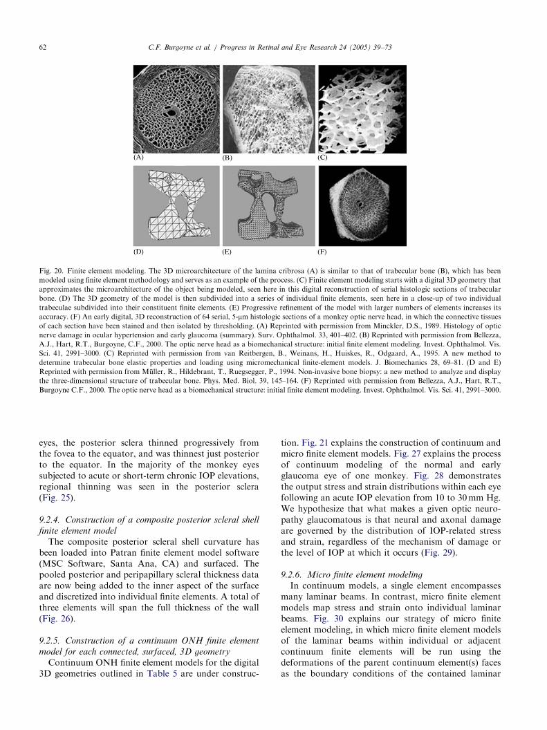
ARTICLE IN PRESS
Fig. 20. Finite element modeling. The 3D microarchitecture of the lamina cribrosa (A) is similar to that of trabecular bone (B), which has been
modeled using finite element methodology and serves as an example of the process. (C) Finite element modeling starts with a digital 3D geometry that
approximates the microarchitecture of the object being modeled, seen here in this digital reconstruction of serial histologic sections of trabecular
bone. (D) The 3D geometry of the model is then subdivided into a series of individual finite elements, seen here in a close-up of two individual
trabeculae subdivided into their constituent finite elements. (E) Progressive refinement of the model with larger numbers of elements increases its
accuracy. (F) An early digital, 3D reconstruction of 64 serial, 5-mm histologic sections of a monkey optic nerve head, in which the connective tissuesof each section have been stained and then isolated by thresholding. (A) Reprinted with permission from Minckler, D.S., 1989. Histology of optic
nerve damage in ocular hypertension and early glaucoma (summary). Surv. Ophthalmol. 33, 401–402. (B) Reprinted with permission from Bellezza,
A.J., Hart, R.T., Burgoyne, C.F., 2000. The optic nerve head as a biomechanical structure: initial finite element modeling. Invest. Ophthalmol. Vis.
Sci. 41, 2991–3000. (C) Reprinted with permission from van Reitbergen, B., Weinans, H., Huiskes, R., Odgaard, A., 1995. A new method to
determine trabecular bone elastic properties and loading using micromechanical finite-element models. J. Biomechanics 28, 69–81. (D and E)
Reprinted with permission from Muller, R., Hildebrant, T., Ruegsegger, P., 1994. Non-invasive bone biopsy: a new method to analyze and display
the three-dimensional structure of trabecular bone. Phys. Med. Biol. 39, 145–164. (F) Reprinted with permission from Bellezza, A.J., Hart, R.T.,
Burgoyne C.F., 2000. The optic nerve head as a biomechanical structure: initial finite element modeling. Invest. Ophthalmol. Vis. Sci. 41, 2991–3000.
C.F. Burgoyne et al. / Progress in Retinal and Eye Research 24 (2005) 39–7362
eyes, the posterior sclera thinned progressively fromthe fovea to the equator, and was thinnest just posteriorto the equator. In the majority of the monkey eyessubjected to acute or short-term chronic IOP elevations,regional thinning was seen in the posterior sclera(Fig. 25).
9.2.4. Construction of a composite posterior scleral shell
finite element model
The composite posterior scleral shell curvature hasbeen loaded into Patran finite element model software(MSC Software, Santa Ana, CA) and surfaced. Thepooled posterior and peripapillary scleral thickness dataare now being added to the inner aspect of the surfaceand discretized into individual finite elements. A total ofthree elements will span the full thickness of the wall(Fig. 26).
9.2.5. Construction of a continuum ONH finite element
model for each connected, surfaced, 3D geometry
Continuum ONH finite element models for the digital3D geometries outlined in Table 5 are under construc-
tion. Fig. 21 explains the construction of continuum andmicro finite element models. Fig. 27 explains the processof continuum modeling of the normal and earlyglaucoma eye of one monkey. Fig. 28 demonstratesthe output stress and strain distributions within each eyefollowing an acute IOP elevation from 10 to 30mm Hg.We hypothesize that what makes a given optic neuro-pathy glaucomatous is that neural and axonal damageare governed by the distribution of IOP-related stressand strain, regardless of the mechanism of damage orthe level of IOP at which it occurs (Fig. 29).
9.2.6. Micro finite element modeling
In continuum models, a single element encompassesmany laminar beams. In contrast, micro finite elementmodels map stress and strain onto individual laminarbeams. Fig. 30 explains our strategy of micro finiteelement modeling, in which micro finite element modelsof the laminar beams within individual or adjacentcontinuum finite elements will be run using thedeformations of the parent continuum element(s) facesas the boundary conditions of the contained laminar

ARTICLE IN PRESS
Fig. 21. Continuum and micro finite element model (FEM) construction. (A) The delineated scleral canal wall and lamina are isolated from the voxel
geometry of a normal eye. The scleral canal wall alone is isolated in (B), and surfaced in (C) using Patran FEM pre-processing software. Then, the
volume within this surface (the intracanalicular space) is filled with 315 finite elements (D). For each constituent element (E and F), material
properties are assigned based on the density and predominant orientation of the laminar beams within that element (G). To accomplish this, custom
software queries the 3D voxel geometry for information on the presence or absence of connective tissue voxels within the volume of each element (F
and G) and casts rays through the element volume to determine the predominant orientation of the laminar beams therein. The material properties of
each continuum finite element are a function of the connective tissue volume fraction and the predominant orientation of the connective tissue beams.
For the continuum models of the normal and early glaucoma eyes, we will use the uniaxial viscoelastic material properties for posterior monkey
sclera that we have previously characterized. In contrast to the large elements of the continuum models, our micro FEMs will use individual voxels or
groups of voxels as elements (H).
Table 5
Scheduled 3D reconstructions
Monkey status Number of monkeys at each IOP (mm Hg)
10/10a 10/30 10/45
Normal 4 3 3
Early glaucoma 3 3 3
Moderate glaucoma 4 4 4
aIOPs at which the two eyes of a given monkey will be set prior to
perfusion fixation.
C.F. Burgoyne et al. / Progress in Retinal and Eye Research 24 (2005) 39–73 63
beams. In this manner, micro finite element models ofareas of interest within the ONH, as suggested by thecontinuum modeling of the same ONH, will be run,making both the computational processing and analysisof results more efficient.Initial results from a micro finite element model
yielded average von Mises stresses that were manytimes those seen in the parent continuum element, withlarge stress and strain concentrations and gradientsseen within individual laminar beams (Fig. 31). Microfinite element modeling of the microarchitecture ofthe connective tissue of the lamina cribrosa will be usedto predict the mechanical environment of the connectivetissue beams, containing astrocytes, and adjacent
axons at physiologic and pathophysiologic levelsof IOP.
10. Clinical and research implications of the
biomechanical paradigm
10.1. Implication 1
For an individual eye, there are at least threecomponents of susceptibility to a given level of 24-hIOP exposure. The first component (Fig. 2) is deter-mined by the relationship between the magnitude of theIOP and the magnitude of the IOP-related stress withinthe load-bearing ONH connective tissues of thatparticular eye (Bellezza et al., 2000). The secondcomponent (Fig. 3) evolves from the larger issue ofhow a given ONH tolerates a given level of IOP-relatedstress. This second component of susceptibility isprincipally determined by the relationship between thelevel of IOP-related stress and (1) the laminar andprelaminar axonal nutrient supply and (2) the materialproperties of the individual lamina cribrosa andperipapillary sclera. The third component of suscept-ibility (not shown) evolves from the retinal ganglioncell and ONH astrocyte and glial cell response topathophysiologic stress within the axon (by whatever
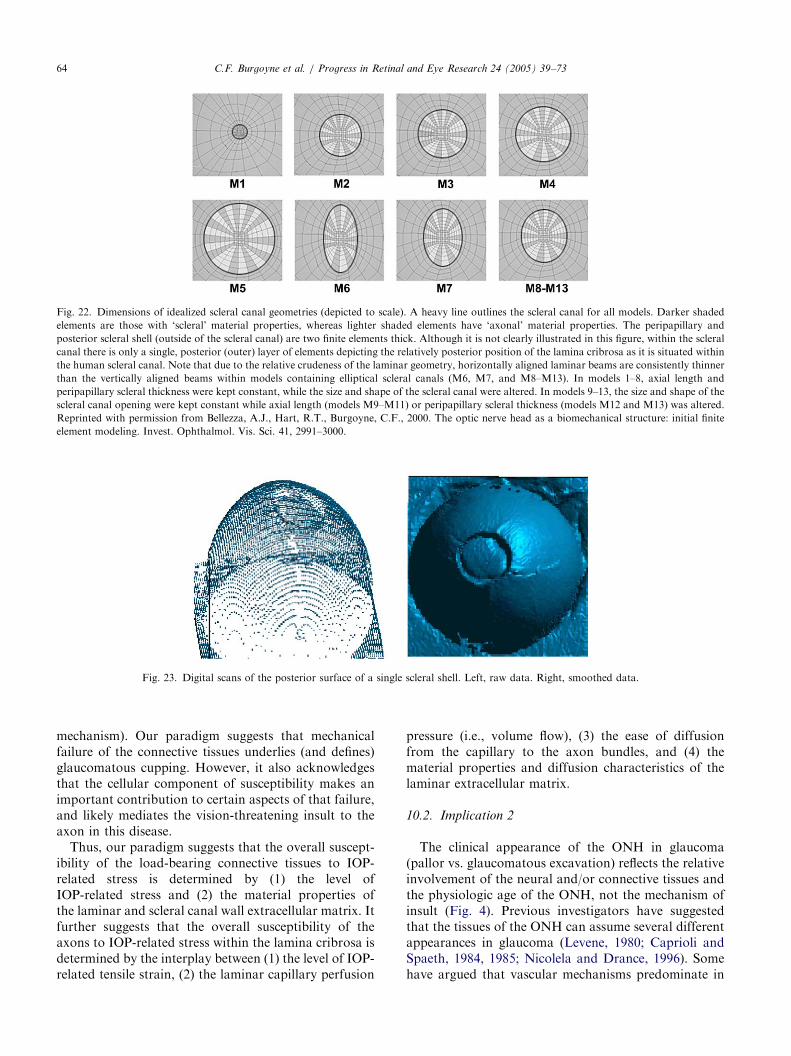
ARTICLE IN PRESS
Fig. 22. Dimensions of idealized scleral canal geometries (depicted to scale). A heavy line outlines the scleral canal for all models. Darker shaded
elements are those with ‘scleral’ material properties, whereas lighter shaded elements have ‘axonal’ material properties. The peripapillary and
posterior scleral shell (outside of the scleral canal) are two finite elements thick. Although it is not clearly illustrated in this figure, within the scleral
canal there is only a single, posterior (outer) layer of elements depicting the relatively posterior position of the lamina cribrosa as it is situated within
the human scleral canal. Note that due to the relative crudeness of the laminar geometry, horizontally aligned laminar beams are consistently thinner
than the vertically aligned beams within models containing elliptical scleral canals (M6, M7, and M8–M13). In models 1–8, axial length and
peripapillary scleral thickness were kept constant, while the size and shape of the scleral canal were altered. In models 9–13, the size and shape of the
scleral canal opening were kept constant while axial length (models M9–M11) or peripapillary scleral thickness (models M12 and M13) was altered.
Reprinted with permission from Bellezza, A.J., Hart, R.T., Burgoyne, C.F., 2000. The optic nerve head as a biomechanical structure: initial finite
element modeling. Invest. Ophthalmol. Vis. Sci. 41, 2991–3000.
Fig. 23. Digital scans of the posterior surface of a single scleral shell. Left, raw data. Right, smoothed data.
C.F. Burgoyne et al. / Progress in Retinal and Eye Research 24 (2005) 39–7364
mechanism). Our paradigm suggests that mechanicalfailure of the connective tissues underlies (and defines)glaucomatous cupping. However, it also acknowledgesthat the cellular component of susceptibility makes animportant contribution to certain aspects of that failure,and likely mediates the vision-threatening insult to theaxon in this disease.Thus, our paradigm suggests that the overall suscept-
ibility of the load-bearing connective tissues to IOP-related stress is determined by (1) the level ofIOP-related stress and (2) the material properties ofthe laminar and scleral canal wall extracellular matrix. Itfurther suggests that the overall susceptibility of theaxons to IOP-related stress within the lamina cribrosa isdetermined by the interplay between (1) the level of IOP-related tensile strain, (2) the laminar capillary perfusion
pressure (i.e., volume flow), (3) the ease of diffusionfrom the capillary to the axon bundles, and (4) thematerial properties and diffusion characteristics of thelaminar extracellular matrix.
10.2. Implication 2
The clinical appearance of the ONH in glaucoma(pallor vs. glaucomatous excavation) reflects the relativeinvolvement of the neural and/or connective tissues andthe physiologic age of the ONH, not the mechanism ofinsult (Fig. 4). Previous investigators have suggestedthat the tissues of the ONH can assume several differentappearances in glaucoma (Levene, 1980; Caprioli andSpaeth, 1984, 1985; Nicolela and Drance, 1996). Somehave argued that vascular mechanisms predominate in
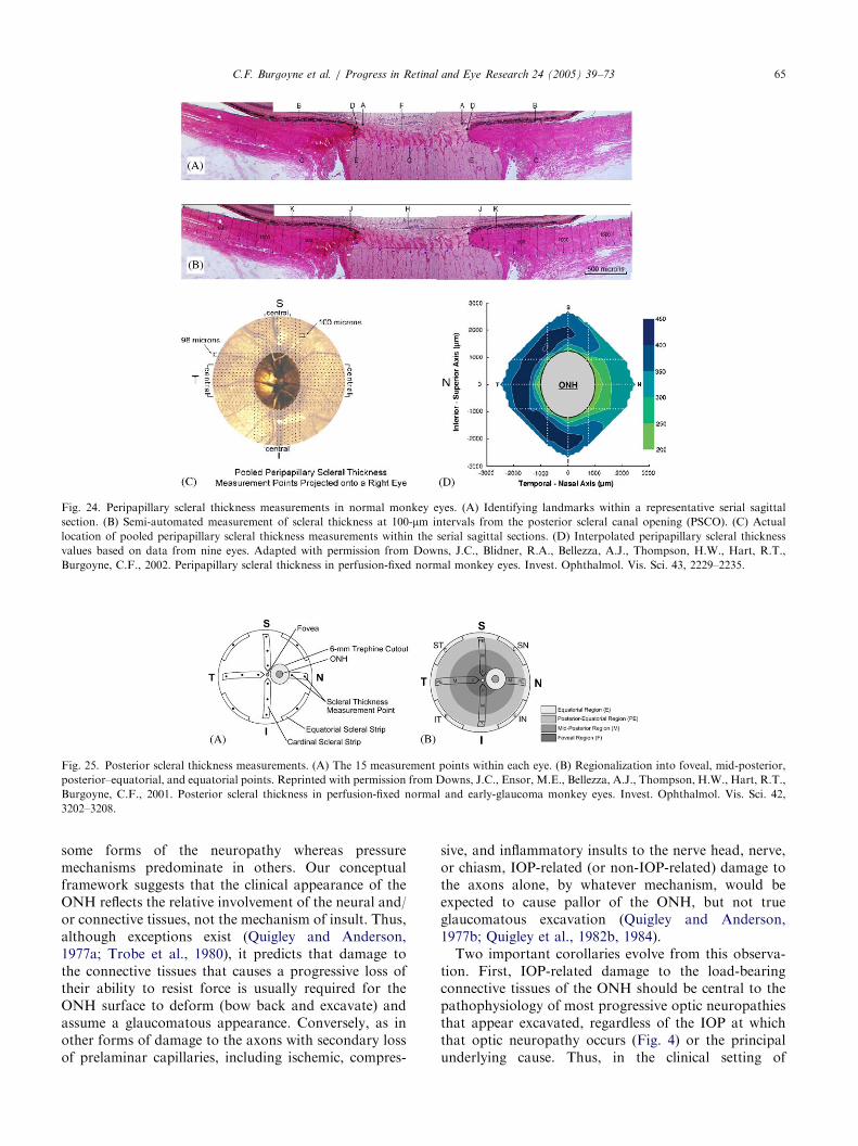
ARTICLE IN PRESS
Fig. 24. Peripapillary scleral thickness measurements in normal monkey eyes. (A) Identifying landmarks within a representative serial sagittal
section. (B) Semi-automated measurement of scleral thickness at 100-mm intervals from the posterior scleral canal opening (PSCO). (C) Actuallocation of pooled peripapillary scleral thickness measurements within the serial sagittal sections. (D) Interpolated peripapillary scleral thickness
values based on data from nine eyes. Adapted with permission from Downs, J.C., Blidner, R.A., Bellezza, A.J., Thompson, H.W., Hart, R.T.,
Burgoyne, C.F., 2002. Peripapillary scleral thickness in perfusion-fixed normal monkey eyes. Invest. Ophthalmol. Vis. Sci. 43, 2229–2235.
Fig. 25. Posterior scleral thickness measurements. (A) The 15 measurement points within each eye. (B) Regionalization into foveal, mid-posterior,
posterior–equatorial, and equatorial points. Reprinted with permission from Downs, J.C., Ensor, M.E., Bellezza, A.J., Thompson, H.W., Hart, R.T.,
Burgoyne, C.F., 2001. Posterior scleral thickness in perfusion-fixed normal and early-glaucoma monkey eyes. Invest. Ophthalmol. Vis. Sci. 42,
3202–3208.
C.F. Burgoyne et al. / Progress in Retinal and Eye Research 24 (2005) 39–73 65
some forms of the neuropathy whereas pressuremechanisms predominate in others. Our conceptualframework suggests that the clinical appearance of theONH reflects the relative involvement of the neural and/or connective tissues, not the mechanism of insult. Thus,although exceptions exist (Quigley and Anderson,1977a; Trobe et al., 1980), it predicts that damage tothe connective tissues that causes a progressive loss oftheir ability to resist force is usually required for theONH surface to deform (bow back and excavate) andassume a glaucomatous appearance. Conversely, as inother forms of damage to the axons with secondary lossof prelaminar capillaries, including ischemic, compres-
sive, and inflammatory insults to the nerve head, nerve,or chiasm, IOP-related (or non-IOP-related) damage tothe axons alone, by whatever mechanism, would beexpected to cause pallor of the ONH, but not trueglaucomatous excavation (Quigley and Anderson,1977b; Quigley et al., 1982b, 1984).Two important corollaries evolve from this observa-
tion. First, IOP-related damage to the load-bearingconnective tissues of the ONH should be central to thepathophysiology of most progressive optic neuropathiesthat appear excavated, regardless of the IOP at whichthat optic neuropathy occurs (Fig. 4) or the principalunderlying cause. Thus, in the clinical setting of

ARTICLE IN PRESSC.F. Burgoyne et al. / Progress in Retinal and Eye Research 24 (2005) 39–7366
progressive ONH excavation by whatever mechanism,consideration should be given to aggressively loweringIOP so as to reduce IOP-related strain within theconnective tissues, and an appropriate work-up for non-IOP-related causes should then be performed if theclinical indications for a non-IOP-related cause warrantfurther evaluation.Second, glaucomatous cupping may not be the only
form of IOP-related optic neuropathy. Because IOP-related strain can adversely influence volume flowand diffusion at all levels of IOP, it may also contributeto some progressive optic neuropathies principallycharacterized by pallor. Our model predicts the existenceof a subset of patients with elevated IOP who developvisual field loss that is accompanied by a pale but notexcavated ONH (Fig. 32). In these patients, our model
Fig. 26. Representative posterior scleral shell geometry. The actual
computational model will be three elements thick (as opposed to the
two-element-thick sclera shown above) and will possess a pooled
surface curvature and thickness based on histologic measurements of a
group of normal and early glaucoma monkey eyes (see Figs. 23–25).
Reprinted with permission from Bellezza, A.J., Hart, R.T., Burgoyne,
C.F., 2000. The optic nerve head as a biomechanical structure: initial
finite element modeling. Invest. Ophthalmol. Vis. Sci. 41, 2991–3000.
Fig. 27. Representative continuum finite element models for the normal eye
these continuum models, the histologic boundaries of the principal connective
filled, then discretized into relatively large elements as depicted in Fig. 21. T
models on the right contain just those elements within the scleral canal of e
suggests that elevated IOP generates a level of IOP-relatedstrain that is within the elastic limits of the individualONH’s load-bearing connective tissues; thus no excava-tion occurs. However, the IOP-related strain still causesaxonal compromise by one or more of the mechanismsoutlined above. Less obviously, our paradigm suggeststhat when IOP is not measurably elevated and ONHpallor exceeds excavation, an extensive work-up for non-IOP-related causes should be performed and IOP low-ering might be additionally considered as a means ofincreasing perfusion to the axons within the prelaminar,laminar, and retrolaminar regions of the ONH.
10.3. Implication 3
There is a logic for lowering IOP to both protect theload-bearing connective tissues and enhance ONHperfusion. The logic for lowering IOP to protect theconnective tissues is apparent from the extensivediscussions above and from Fig. 7. However, ourparadigm suggests that, at all ages and at all stages ofIOP-related damage, delivery of nutrients to the ONHaxons can be adversely affected both by retrobulbardeterminants of ONH perfusion (e.g., blood pressureand vasospasm) and by the additional negative effects ofelevated levels of IOP-related strain within the ONHconnective tissues (Fig. 6).
10.4. Implication 4
Progression of ONH damage following IOP loweringis still most likely the result of inadequate IOP lowering.The continued damage to some ONHs following somedegree of IOP lowering is often cited as evidence thatfactors other than IOP are always most important in this
(upper) and early glaucoma eye (lower) of an individual monkey. In
tissue structures have been isolated within the 3D geometries, surfaced,
he models on the left contain two elements of peripapillary sclera. The
ach eye.
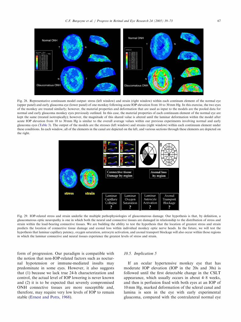
ARTICLE IN PRESS
Fig. 28. Representative continuum model output: stress (left window) and strain (right window) within each continum element of the normal eye
(upper panel) and early glaucoma eye (lower panel) of one monkey following acute IOP elevation from 10 to 30mm Hg. In this exercise, the two eyes
of the monkey are treated similarly; however, the material properties and deformation that are used as input to the models are the pooled data for
normal and early glaucoma monkey eyes previously outlined. In this case, the material properties of each continuum element of the normal eye are
kept the same (treated isotropically); however, the magnitude of this shared value is altered until the laminar deformation within the model after
acute IOP elevation from 10 to 30mm Hg is similar to the overall average values within our previous experiments involving normal and early
glaucoma eyes (Table 3). The output of the models are the stresses (left window) and strains (right window) within each continuum element under
these conditions. In each window, all of the elements in the canal are depicted on the left, and various sections through these elements are depicted on
the right.
Fig. 29. IOP-related stress and strain underlie the multiple pathophysiologies of glaucomatous damage. Our hypothesis is that, by definition, a
glaucomatous optic neuropathy is one in which both the neural and connective tissues are damaged in relationship to the distribution of stress and
strain within the load-bearing connective tissues. We are building the ability to test the hypothesis that the location of greatest stress and strain
predicts the location of connective tissue damage and axonal loss within individual monkey optic nerve heads. In the future, we will test the
hypothesis that laminar capillary patency, oxygen saturation, astrocyte activation, and axonal transport blockage will also occur within those regions
in which the laminar connective and neural tissues experience the greatest levels of stress and strain.
C.F. Burgoyne et al. / Progress in Retinal and Eye Research 24 (2005) 39–73 67
form of progression. Our paradigm is compatible withthe notion that non-IOP-related factors such as noctur-nal hypotension or immune-mediated insults maypredominate in some eyes. However, it also suggeststhat (1) because we lack true 24-h characterization andcontrol, the actual level of IOP lowering is never knownand (2) it is to be expected that severely compromisedONH connective tissues are more susceptible and,therefore, may require very low levels of IOP to remainstable (Ernest and Potts, 1968).
10.5. Implication 5
If an ocular hypertensive monkey eye that hasmoderate IOP elevation (IOP in the 20s and 30s) isfollowed until the first detectable change in the CSLTappearance, which usually occurs in about 4–8 weeks,and then is perfusion fixed with both eyes at an IOP of10mm Hg, marked deformation of the scleral canal andlamina is seen in the eye with early experimentalglaucoma, compared with the contralateral normal eye
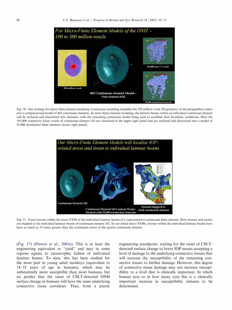
ARTICLE IN PRESS
Fig. 30. Our strategy for micro finite element modeling. Continuum modeling simplifies the 250 million voxel 3D geometry of the peripapillary sclera
into a computational model of 465 continuum elements. In micro finite element modeling, the laminar beams within an individual continuum element
will be surfaced and discretized into elements, with the remaining continuum model being used to establish their boundary conditions. Here the
195,000 connective tissue voxels of continuum element 142 are visualized in the upper right panel and are surfaced and discretized into a model of
55,000 tetrahedral finite elements (lower right panel).
Fig. 31. Focal stresses within the micro FEM of the individual laminar beams of a representative continuum finite element. Here stresses and strains
are mapped to the individual laminar beams of continuum element 142. In our initial micro FEMs, stresses within the individual laminar beams have
been as much as 33 times greater than the continuum stress of the parent continuum element.
C.F. Burgoyne et al. / Progress in Retinal and Eye Research 24 (2005) 39–7368
(Fig. 17) (Downs et al., 2003a). This is at least theengineering equivalent to ‘‘yield’’ and may in someregions equate to catastrophic failure of individuallaminar beams. To date, this has been studied forthe most part in young adult monkeys (equivalent to18–35 years of age in humans), which may besubstantially more susceptible than most humans, butwe predict that the onset of CSLT-detected ONHsurface change in humans will have the same underlyingconnective tissue correlates. Thus, from a purely
engineering standpoint, waiting for the onset of CSLT-detected surface change to lower IOP means accepting alevel of damage to the underlying connective tissues thatwill increase the susceptibility of the remaining con-nective tissues to further damage. However, this degreeof connective tissue damage may not increase suscept-ibility to a level that is clinically important. In whichhuman eyes or in how many eyes this is a clinicallyimportant increase in susceptibility remains to bedetermined.

ARTICLE IN PRESS
Fig. 32. Pallor exceeds excavation in the elevated IOP form of the neuropathy, as shown in the ONH photo (left) and 10-2 visual field (right) of the
right eye of a 42-year-old patient with a long history of chronically elevated IOP. In this particular eye, there had been substantial reversal of cupping
following a successful glaucoma filtration surgery. Although the amount of remaining rim tissue is substantial, both pallor and visual field loss are
profound.
C.F. Burgoyne et al. / Progress in Retinal and Eye Research 24 (2005) 39–73 69
11. Summary
The principal appeal of modeling the ONH as abiomechanical structure is that it provides a conceptualframework that explains several heretofore confusingaspects of the disease. First, our theory of a predictablecascade of IOP-induced mechanical failure within theload-bearing connective tissues provides the first testablehypotheses regarding the specific events underlying theonset and progression of glaucomatous excavation.Second, the biomechanical notion that IOP-related
stress within the sclera is magnified within andimmediately around the scleral canal (Greene, 1980;Bellezza et al., 2000), and that this elevated level of stresscentrally determines the physiologic and pathophysio-logic milieu, provides a new logic for understanding howa glaucomatous form of damage to the ONH can occurat all levels of IOP depending on the unique suscept-ibility of an individual ONH.Third, the notions of elevated levels of IOP-related
stress within the peripapillary sclera and IOP-inducedbasement membrane and connective tissue changeswithin the laminar trabeculae allow our model tohypothesize a series of interactions between IOP, IOP-related stress, IOP-related strain, ONH volume bloodflow, and the delivery of blood-borne nutrients to theaxons within the lamina cribrosa. This hypothesissuggests that vascular mechanisms of damage to theONH are not necessarily IOP-independent and proposesa logic for understanding the complicated interactionbetween these three important factors—IOP, volumeblood flow, and nutrient delivery—within the tissues ofthe ONH.Fourth, the concept of mechanical failure suggests a
biomechanical explanation for the clinical notion that
progression of ONH damage after lowering of IOP ismost likely caused by inadequate pressure reduction.Our model suggests that diurnally (24-h) confirmedlevels of low IOP may be necessary in some eyes becauseonce the load-bearing connective tissues are focallydamaged, stresses are higher within the remainingtissues bearing load.Fifth, our paradigm suggests a logic for characterizing
the multiple effects of IOP-related strain on theindividual tissues of the visual system (Fig. 2). Althoughthe precise steps within each staging pathway will beunique to each tissue, the general progression frommolecular changes, to histologic evidence of alterationand damage, to histologic evidence of repair and/orregeneration should have equivalent manifestations inall of the individually affected tissues.
12. Future directions
Our goal is to build a science that predicts thesusceptibility of the connective tissues of an individualONH to a given level of IOP. Eventually, our modelswill identify the principal determinants of the level ofIOP-related stress for a given level of IOP. Then, ifclinical measures of these parameters can be developed,which may eventually be achieved with micro magneticresonance imaging (Sadun et al., 2002), finite elementmodeling of the ONH connective tissues within indivi-dual eyes may become a standard clinical test todetermine target IOPs for eyes that are at risk fordamage.We are beginning to extend our studies to the
associated susceptibilities of the astrocytes and retinal
ganglion cell axons within the ONH. We are building cell

ARTICLE IN PRESSC.F. Burgoyne et al. / Progress in Retinal and Eye Research 24 (2005) 39–7370
and tissue culture systems to characterize the short- andlong-term molecular response of astrocytes to stress andstrain. Eventually, we hope to use probes for astrocyteactivation and axonal distress in order to map thelocations of these phenomena within our 3D connectivetissue reconstructions relative to the levels of connectivetissue stress and strain at varying stages of glaucoma-tous damage.We will soon begin studies designed to characterize
the biochemical and histologic alterations that underliechanges in the material properties of the posterior sclerain early glaucoma. Our ultimate goal will be to describethe histologic and ultrastructural changes of mechanicalfailure within the peripapillary sclera, laminar trabecu-lae, and scleral canal wall in early, moderate, and severeglaucomatous damage.Finally, our 3D reconstructions contain the posterior
ciliary arteries, their principal branches to the pre-laminar,laminar, and post-laminar ONH, and the capillarieswithin each laminar beam. We will eventually model theeffects of IOP-related stress and strain, aging, and thestage of glaucomatous damage on volume flow within thisblood supply and the diffusion of nutrients from thelaminar capillaries through the laminar extracellularmatrix, astrocyte basement membrane, and astrocyte cellprocesses into the center of the axon bundles.
Acknowledgements
The concepts contained herein were initially formu-lated during the two years the first author (CFB)spent as a fellow in the laboratories of Harry Quigley.Dr. Quigley’s work and ideas have deeply influencedtheir subsequent development. Refinement of theseconcepts has followed important conversations withRosario Hernandez, Art Neufeld, Abe Clark, RanZeimer, Ross Ethier, Jost Jonas, Bal Chauhan, BobWeinreb, Doug Anderson, and especially Jack Cioffiand John Morrison. Yeni Yucel was kind enough tocritically read the manuscript before submission.This work was supported in part by USPHS grants
R01EY011610 (CFB) and P30EY002377 (LSU EyeCenter core grant) from the National Eye Institute,National Institutes of Health, Bethesda, Maryland; agrant from the American Health Assistance Founda-tion, Rockville, Maryland (CFB); a grant from TheWhitaker Foundation, Rosslyn, Virginia (CFB); aCareer Development Award (CFB) and an unrestricteddepartmental grant (LSU Eye Center) from Research toPrevent Blindness, Inc., New York, New York; and agraduate student stipend from the Board of Regents,State of Louisiana (AJB).None of the authors have a financial or proprietary
interest in any drug, process, or instrument mentioned inthis article.
References
Albon, J., Purslow, P.P., Karwatowski, W.S., Easty, D.L., 2000. Age
related compliance of the lamina cribrosa in human eyes. Br. J.
Ophthalmol. 84, 318–323.
Alm, A., 1973. Ocular and optic nerve blood flow at normal and
increased intraocular pressures in monkeys (Macaca irus): a study
with radioactively labeled microspheres including flow determina-
tion in brain and some other tissues. Exp. Eye Res. 15, 15–29.
Alm, A., Bill, A., 1970. Blood flow and oxygen extraction in the cat
uvea at normal and high intraocular pressures. Acta Physiol.
Scand. 80, 19–28.
Alm, A., Bill, A., 1972. The oxygen supply to the retina. II. Effects of
high intraocular pressure and of increased arterial carbon dioxide
tension on uveal and retinal blood flow in cats. Acta Physiol.
Scand. 84, 306–319.
Anderson, D.R., Hendrickson, A., 1974. Effect of intraocular pressure
on rapid axoplasmic transport in monkey optic nerve. Invest.
Ophthalmol. 13, 771–783.
Bellezza, A.J., Hart, R.T., Burgoyne, C.F., 2000. The optic nerve head
as a biomechanical structure: initial finite element modeling. Invest.
Ophthalmol. Vis. Sci. 41, 2991–3000.
Bellezza, A.J., Rintalan, C.J., Thompson, H.W., Downs, J.C., Hart,
R.T., Burgoyne, C.F., 2003a. Anterior scleral canal geometry in
pressurised (IOP 10) and non-pressurised (IOP 0) normal monkey
eyes. Br. J. Ophthalmol. 87, 1284–1290.
Bellezza, A.J., Rintalan, C.J., Thompson, H.W., Downs, J.C., Hart,
R.T., Burgoyne, C.F., 2003b. Deformation of the lamina cribrosa
and anterior scleral canal wall in early experimental glaucoma.
Invest. Ophthalmol. Vis. Sci. 44, 623–637.
Broom, N.D., 1978. Simultaneous morphological and stress–strain
studies of the fibrous components in wet heart valve leaflet tissue.
Connect. Tissue Res. 6, 37–50.
Burgoyne, C.F., Morrison, J.C., 2001. The anatomy and pathophy-
siology of the optic nerve head in glaucoma. J. Glaucoma 10
(Suppl. 1), S16–S18.
Burgoyne, C.F., Varma, R., Quigley, H.A., Vitale, S., Pease, M.E.,
Lenane, P.L., 1994. Global and regional detection of induced optic
disc change by digitized image analysis. Arch. Ophthalmol. 112,
261–268.
Burgoyne, C.F., Quigley, H.A., Thompson, H.W., Vitale, S., Varma,
R., 1995a. Measurement of optic disc compliance by digitized image
analysis in the normal monkey eye. Ophthalmology 102, 1790–1799.
Burgoyne, C.F., Quigley, H.A., Thompson, H.W., Vitale, S., Varma,
R., 1995b. Early changes in optic disc compliance and surface
position in experimental glaucoma. Ophthalmology 102,
1800–1809.
Caprioli, J., Spaeth, G.L., 1984. Comparison of visual field defects in
the low-tension glaucomas with those in the high-tension glauco-
mas. Am. J. Ophthalmol. 97, 730–737.
Caprioli, J., Spaeth, G.L., 1985. Comparison of the optic nerve head in
high- and low-tension glaucoma. Arch. Ophthalmol. 103,
1145–1149.
Clark, A.F., Browder, S., Steely, H.T., Wilson, K., Cantu-Crouch, D.,
McCartney, M.D., 1995. Cell biology of the human lamina
cribrosa. In: Drance, S.M., Anderson, D.R. (Eds.), Optic
Nerve in Glaucoma. Kugler Publications, Amsterdam/New York,
pp. 79–105.
Coleman, A.L., Quigley, H.A., Vitale, S., Dunkelberger, G., 1991.
Displacement of the optic nerve head by acute changes in
intraocular pressure in monkey eyes. Ophthalmology 98, 35–40.
Craik, J.E., McNeil, I.R.R., 1965. Histological studies of stressed skin.
In: Kenedi, R.M. (Ed.), Biomechanics and Related Biomechanical
Engineering Topics. Pergamon Press, London, pp. 159–164.
Dongqi, H., Zeqin, R., 1999. A biomathematical model for pressure-
dependent lamina cribrosa behavior. J. Biomech. 32, 579–584.
ARTICLE IN PRESSC.F. Burgoyne et al. / Progress in Retinal and Eye Research 24 (2005) 39–73 71
Downs, J.C., Ensor, M.E., Bellezza, A.J., Thompson, H.W., Hart,
R.T., Burgoyne, C.F., 2001a. Posterior scleral thickness in
perfusion-fixed normal and early-glaucoma monkey eyes. Invest.
Ophthalmol. Vis. Sci. 42, 3202–3208.
Downs, J.C., Thomas, K.A., Hart, R.T., Burgoyne, C.F., 2001b.
Elastic and viscoelastic material properties of peripapillary sclera in
normal and early glaucomatous monkey eyes. ARVO Abstract
#4461, Invest. Ophthalmol. Vis. Sci. 42 (Suppl.), S831.
Downs, J.C., Blidner, R.A., Bellezza, A.J., Thompson, H.W., Hart,
R.T., Burgoyne, C.F., 2002. Peripapillary scleral thickness in
perfusion-fixed normal monkey eyes. Invest. Ophthalmol. Vis. Sci.
43, 2229–2235.
Downs, J.C., Bellezza, A.J., Reynaud, J.F., Hirons, B.A., Zhou, P.,
Burgoyne, C.F., 2003a. Plastic deformation of the lamina cribrosa
within digital three-dimensional (3D) reconstructions of early
glaucomatous monkey optic nerve heads. ARVO Abstract #4796,
Association for Research in Vision and Ophthalmology, Fort
Lauderdale, Florida, May 4–9, 2003. [For text, see http://
www.arvo.org.]
Downs, J.C., Suh, J.K., Thomas, K.A., Bellezza, A.J., Burgoyne, C.F.,
Hart, R.T., 2003b. Viscoelastic characterization of peripapillary
sclera: material properties by quadrant in rabbit and monkey eyes.
J. Biomech. Eng. 125, 124–131.
Emery, J.M., Landis, D., Paton, D., Boniuk, M., Craig, J.M., 1974.
The lamina cribrosa in normal and glaucomatous human eyes.
Trans. Am. Acad. Ophthalmol. Otolaryngol. 78, OP290–OP297.
Ernest, J.T., Potts, A.M., 1968. Pathophysiology of the distal portion
of the optic nerve. I. Tissue pressure relationships. Am. J.
Ophthalmol. 66, 373–387.
Farquhar, M.G., 1991. The glomerular basement membrane: a
selective macromolecular filter. In: Hay, E.D. (Ed.), Cell Biology
of Extracellular Matrix, second ed. Plenum, New York, pp.
365–418.
Fechtner, R.D., Weinreb, R.N., 1994. Mechanisms of optic nerve
damage in primary open angle glaucoma. Surv. Ophthalmol. 39,
23–42.
Geijer, C., Bill, A., 1979. Effects of raised intraocular pressure on
retinal, prelaminar, laminar, and retrolaminar optic nerve blood
flow in monkeys. Invest. Ophthalmol. Vis. Sci. 18, 1030–1042.
Greene, P.R., 1980. Mechanical considerations in myopia: relative
effects of accommodation, convergence, intraocular pressure, and
the extraocular muscles. Am. J. Optom. Physiol. Opt. 57, 902–914.
Grunwald, J.E., Riva, C.E., Stone, R.A., Keates, E.U., Petrig, B.L.,
1984. Retinal autoregulation in open-angle glaucoma. Ophthal-
mology 91, 1690–1694.
Gupta, N., Yucel, Y.H., 2001. Glaucoma and the brain. J. Glaucoma
10 (Suppl. 1), S28–S29.
Hayreh, S.S., 1974. Pathogenesis of cupping of the optic disc. Br. J.
Ophthalmol. 58, 863–876.
Hayreh, S.S., 1986. Pathogenesis of optic nerve head changes in
glaucoma. Semin. Ophthalmol. 1, 1–13.
Hayreh, S.S., Zimmerman, M.B., Podhajsky, P., Alward, W.L.M.,
1994. Nocturnal arterial hypotension and its role in optic nerve
head and ocular ischemic disorders. Am. J. Ophthalmol. 117,
603–624.
Heickell, A.G., Bellezza, A.J., Thompson, H.W., Burgoyne, C.F.,
2001. Optic disc surface compliance testing using confocal scanning
laser tomography in the normal monkey eye. J. Glaucoma 10,
369–382.
Hernandez, M.R., 1992. Ultrastructural immunocytochemical analysis
of elastin in the human lamina cribrosa: changes in elastic fibers in
primary open-angle glaucoma. Invest. Ophthalmol. Vis. Sci. 33,
2891–2903.
Hernandez, M.R., 1993. Basic sciences in clinical glaucoma: extra-
cellular matrix macromolecules of the lamina cribrosa: a pressure-
sensitive connective tissue. J. Glaucoma 2, 50–57.
Hernandez, M.R., Gong, H., 1996. Extracellular matrix of the
trabecular meshwork and optic nerve head. In: Ritch, R., Shields,
M.B., Krupin, T. (Eds.), The Glaucomas: Basic Sciences, vol. 1,
second ed. Mosby-Year Book, Inc., St. Louis, pp. 213–249.
Hernandez, M.R., Luo, X.X., Igoe, F., Neufeld, A.H., 1987.
Extracellular matrix of the human lamina cribrosa. Am. J.
Ophthalmol. 104, 567–576.
Hernandez, M.R., Luo, X.X., Andrzejewska, W., Neufeld, A.H., 1989.
Age-related changes in the extracellular matrix of the human optic
nerve head. Am. J. Ophthalmol. 107, 476–484.
Hernandez, M.R., Andrzejewska, W.M., Neufeld, A.H., 1990.
Changes in the extracellular matrix of the human optic nerve head
in primary open-angle glaucoma. Am. J. Ophthalmol. 109,
180–188.
Hernandez, M.R., Yang, J., Ye, H., 1994a. Activation of elastin
mRNA expression in human optic nerve heads with primary open-
angle glaucoma. J. Glaucoma 3, 214–225.
Hernandez, M.R., Ye, H., Roy, S., 1994b. Collagen type IV gene
expression in human optic nerve heads with primary open-angle
glaucoma. Exp. Eye Res. 59, 41–52.
Hirons, B.A., Reynaud, J.F., Bellezza, A.J., Zhou, P., Downs, J.C.,
Burgoyne, C.F., 2003. Visualization of digital three-dimensional
(3D) reconstructions of the connective tissues of the optic nerve
head. ARVO Abstract #3872, Association for Research in Vision
and Ophthalmology, Fort Lauderdale, Florida, May 4–9, 2003.
[For text, see http://www.arvo.org.]
Ito, H., Kwan, C.Y., Daniel, E.E., 1987. Elastin and elastase-like
enzyme change in aorta of rat with malignant hypertension. Exp.
Mol. Pathol. 47, 26–36.
Janssen, P., Naskar, R., Moore, S., Thanos, S., Thiel, H.J., 1997.
Evidence for glaucoma-induced horizontal cell alterations in the
human retina. Ger. J. Ophthalmol. 5, 378–385.
Jonas, J.B., Berenshtein, E., Holbach, L., 2003. Anatomic relationship
between lamina cribrosa, intraocular space, and cerebrospinal fluid
space. Invest. Ophthalmol. Vis. Sci. 44, 5189–5195.
Kendell, K.R., Quigley, H.A., Kerrigan, L.A., Pease, M.E., Quigley,
E.N., 1995. Primary open-angle glaucoma is not associated
with photoreceptor loss. Invest. Ophthalmol. Vis. Sci. 36,
200–205.
Kenedi, R.M., Gibson, T., Daly, C.H., 1965. Bioengineering studies of
the human skin: the effects of unidirectional tension. In: Jackson,
S.F., Harkness, R.D., Partridge, S.M., Tristram, G.R. (Eds.),
Structure and Function of Connective and Skeletal Tissue. Butter-
worths, London, pp. 388–395.
Koob, T.J., Vogel, K.G., 1987. Proteoglycan synthesis in organ
cultures from regions of bovine tendon subjected to different
mechanical forces. Biochem. J. 246, 589–598.
Langham, M.E., 1980. The temporal relation between intraocular
pressure and loss of vision in chronic simple glaucoma. Glaucoma
2, 427–435.
Larrabee, W.F., 1986. A finite element model of skin deformation. 1.
Biomechanics of skin and soft tissue: a review. Laryngoscope 96,
399–405.
Levene, R.Z., 1980. Low tension glaucoma. Surv. Ophthalmol. 24,
621–664.
Levy, N.S., Crapps, E.E., 1984. Displacement of optic nerve head in
response to short-term intraocular pressure elevation in human
eyes. Arch. Ophthalmol. 102, 782–786.
Minckler, D.S., 1986. Correlations between anatomic features and
axonal transport in primate optic nerve head. Trans. Am.
Ophthalmol. Soc. 84, 429–452.
Minckler, D.S., Spaeth, G.L., 1981. Optic nerve damage in glaucoma.
Surv. Ophthalmol. 26, 128–148.
Minckler, D.S., Bunt, A.H., Johanson, G.W., 1977. Orthograde and
retrograde axoplasmic transport during acute ocular hypertension
in the monkey. Invest. Ophthalmol. Vis. Sci. 16, 426–441.

ARTICLE IN PRESSC.F. Burgoyne et al. / Progress in Retinal and Eye Research 24 (2005) 39–7372
Morgan, W.H., Chauhan, B.C., Yu, D.-Y., Cringle, S., Alder, V.A.,
House, P.H., 2002. Optic disc movement with variations in
intraocular and cerebrospinal fluid pressure. Invest. Ophthalmol.
Vis. Sci. 43, 3236–3242.
Morrison, J.C., 1995. The microanatomy of the optic nerve. In:
Drance, S.M., Anderson, D.R. (Eds.), Optic Nerve in Glaucoma.
Kugler Publications, London, pp. 57–78.
Morrison, J.C., Jerdan, J.A., L’Hernault, N.L., Quigley, H.A., 1988.
The extracellular matrix composition of the monkey optic nerve
head. Invest. Ophthalmol. Vis. Sci. 29, 1141–1150.
Morrison, J., Dorman, M.E., Quigley, H., 1989a. Extracellular matrix
changes in glaucomatous optic atrophy. ARVO Abstract. Invest.
Ophthalmol. Vis. Sci. 30 (Suppl.), 201.
Morrison, J.C., L’Hernault, N.L., Jerdan, J.A., Quigley, H.A., 1989b.
Ultrastructural location of extracellular matrix components in the
optic nerve head. Arch. Ophthalmol. 107, 123–129.
Morrison, J.C., Dorman-Pease, M.E., Quigley, H.A., 1990. Optic
nerve head extracellular matrix in primary optic atrophy and
experimental glaucoma. Arch. Ophthalmol. 108, 1020–1024.
Nicolela, M.T., Drance, S.M., 1996. Various glaucomatous optic nerve
appearances: clinical correlations. Ophthalmology 103, 640–649.
Oxlund, H., Andreassen, T.T., 1980. The roles of hyaluronic acid,
collagen and elastin in the mechanical properties of connective
tissues. J. Anat. 131, 611–620.
Oxlund, H., Manschot, J., Viidik, A., 1988. The role of elastin in the
mechanical properties of skin. J. Biomech. 21, 213–218.
Panda, S., Jonas, J.B., 1992. Decreased photoreceptor count in human
eyes with secondary angle-closure glaucoma. Invest. Ophthalmol.
Vis. Sci. 33, 2532–2536.
Park, K.H., Hong, C., 1998. Reversal of optic disc topography in
patients with glaucomatocyclitic crisis after remission of attack. J.
Glaucoma 7, 225–229.
Pederson, J.E., Herschler, J., 1982. Reversal of glaucomatous cupping
in adults. Arch. Ophthalmol. 100, 426–431.
Pena, J.D.O., Roy, S., Hernandez, M.R., 1996. Tropoelastin gene
expression in optic nerve heads of normal and glaucomatous
subjects. Matrix Biol. 15, 323–330.
Pena, J.D.O., Kobayashi, S., Hernandez, M.R., 1997. Elastin
associated microfibrils in human optic nerve heads in vivo and in
vitro. ARVO Abstract #803. Invest. Ophthalmol. Vis. Sci. 38
(Suppl.), S162.
Pillunat, L.E., Stodmeister, R., Wilmanns, I., Christ, Th., 1985.
Autoregulation of ocular blood flow during changes in intraocular
pressure: preliminary results. Graefe’s Arch. Clin. Exp. Ophthal-
mol. 223, 219–223.
Pillunat, L.E., Stodtmeister, R., Wilmanns, I., 1987. Pressure
compliance of the optic nerve head in low-tension glaucoma. Br.
J. Ophthalmol. 71, 181–187.
Prosser, I.W., Stenmark, K.R., Suthar, M., Crouch, E.C., Mecham,
R.P., Parks, W.C., 1989. Regional heterogeneity of elastin and
collagen gene expression in intralobar arteries in response to
hypoxic pulmonary hypertension as demonstrated by in situ
hybridization. Am. J. Pathol. 135, 1073–1088.
Quigley, H.A., 1977. The pathogenesis of reversible cupping in
congenital glaucoma. Am. J. Ophthalmol. 84, 358–370.
Quigley, H.A., Anderson, D.R., 1976. The dynamics and location of
axonal transport blockade by acute intraocular pressure elevation
in primate optic nerve. Invest. Ophthalmol. 15, 606–616.
Quigley, H., Anderson, D.R., 1977a. Cupping of the optic disc in
ischemic optic neuropathy. Trans. Am. Acad. Ophthalmol.
Otolaryngol. 83, 755–762.
Quigley, H.A., Anderson, D.R., 1977b. The histologic basis of optic
disk pallor in experimental optic atrophy. Am. J. Ophthalmol. 83,
709–717.
Quigley, H.A., Anderson, D.R., 1977c. Distribution of axonal
transport blockade by acute intraocular pressure elevation in the
primate optic nerve head. Invest. Ophthalmol. Vis. Sci. 16,
640–646.
Quigley, H.A., Green, W.R., 1979. The histology of human glaucoma
cupping and optic nerve damage: clinicopathologic correlation in
21 eyes. Ophthalmology 86, 1803–1827.
Quigley, H.A., Pease, M.E., 1996. Change in the optic disc and nerve
fiber layer estimated with the Glaucoma-scope in monkey eyes. J.
Glaucoma 5, 106–116.
Quigley, H.A., Addicks, E.M., Green, W.R., Maumenee, A.E., 1981.
Optic nerve damage in human glaucoma. II. The site of injury and
susceptibility to damage. Arch. Ophthalmol. 99, 635–649.
Quigley, H.A., Addicks, E.M., Green, W.R., 1982a. Optic nerve
damage in human glaucoma. III. Quantitative correlation of nerve
fiber loss and visual field defect in glaucoma, ischemic neuropathy,
disc edema and toxic neuropathy. Arch. Ophthalmol. 100,
135–146.
Quigley, H.A., Hohman, R.M., Addicks, E.M., 1982b. Quantitative
study of optic nerve head capillaries in experimental optic disk
pallor. Am. J. Ophthalmol. 93, 689–699.
Quigley, H.A., Hohman, R.M., Addicks, E.M., 1983. Morphologic
changes in the lamina cribrosa correlated with neural loss in open-
angle glaucoma. Am. J. Ophthalmol. 95, 673–691.
Quigley, H.A., Hohman, R.M., Addicks, E.M., Green, W.R., 1984.
Blood vessels of the glaucomatous optic disc in experimental
primate and human eyes. Invest. Ophthalmol. Vis. Sci. 25,
918–931.
Quigley, H.A., Brown, A., Dorman-Pease, M.E., 1991a. Alterations in
elastin of the optic nerve head in human and experimental
glaucoma. Br. J. Ophthalmol. 75, 552–557.
Quigley, H.A., Dorman-Pease, M.E., Brown, A.E., 1991b. Quantita-
tive study of collagen and elastin of the optic nerve head and sclera
in human and experimental monkey glaucoma. Curr. Eye Res. 10,
877–888.
Repka, M.X., Quigley, H.A., 1989. The effect of age on normal
human optic nerve fiber number and diameter. Ophthalmology 96,
26–32.
Sadun, A.A., Carelli, V., Bose, S., Ross-Cisneros, F.N., Barboni, P.,
Ahrens, E.T., 2002. First application of extremely high resolution
magnetic resonance imaging to study microscopic features of
normal and LHON human optic nerve. Ophthalmology 109,
1085–1091.
Schneider, P.J., Deck, J.D., 1981. Tissue and cell renewal in the natural
aortic valve of rats: an autoradiographic study. Cardiovasc. Res.
15, 181–189.
Schumer, R.A., Podos, S.M., 1994. The nerve of glaucoma! Arch.
Ophthalmol. 112, 37–44.
Sossi, N., Anderson, D.R., 1983. Effect of elevated intraocular
pressure on blood flow: occurrence in cat optic nerve head studied
with iodoantipyrine I-125. Arch. Ophthalmol. 101, 98–101.
Spaeth, G.L., 1977. The pathogenesis of nerve damage in glaucoma:
contributions of fluorescein angiography. Grune & Stratton,
London, pp. 123–132.
Tengroth, B., Ammitzboll, T., 1984. Changes in the content and
composition of collagen in the glaucomatous eye—basis for a new
hypothesis for the genesis of chronic open angle glaucoma—a
preliminary report. Acta Ophthalmol. 62, 999–1008.
Timoshenko, S.P., Goodier, J.N., 1970. Theory of Elasticity, third ed.
McGraw-Hill Book Co., New York.
Torp, S., Arridge, R.G.C., Armeniades, C.D., Baer, E., 1974a.
Structure-property relationships in tendon as a function of age.
In: Atkins, E.D.T., Keller, A. (Eds.), Structure of Fibrous
Biopolymers. Butterworths, London, pp. 197–221.
Torp, S., Baer, E., Friedman, B., 1974b. Effects of age and of
mechanical deformation on the ultrastructure of tendon. In:
Atkins, E.D.T., Keller, A. (Eds.), Structure of Fibrous Biopoly-
mers. Butterworths, London, pp. 223–250.

ARTICLE IN PRESSC.F. Burgoyne et al. / Progress in Retinal and Eye Research 24 (2005) 39–73 73
Trobe, J.D., Glaser, J.S., Cassady, J., Herschler, J., Anderson, D.R.,
1980. Nonglaucomatous excavation of the optic disc. Arch.
Ophthalmol. 98, 1046–1050.
Tumminia, S.J., Mitton, K.P., Arora, J., Zelenka, P., Epstein, D.L.,
Russell, P., 1998. Mechanical stretch alters the actin cytoskeletal
network and signal transduction in human trabecular meshwork
cells. Invest. Ophthalmol. Vis. Sci. 39, 1361–1371.
Ulrich, W.-D., Ulrich, C., Bohne, B.-D., 1986. Deficient autoregula-
tion and lengthening of the diffusion distance in the anterior optic
nerve circulation in glaucoma: an electro-encephalo-dynamo-
graphic investigation. Ophthalmic Res. 18, 253–259.
Van Buskirk, E.M., 1994. Glaucomatous optic neuropathy.
J. Glaucoma 3 (Suppl.), S2–S4.
Van Buskirk, E.M., Cioffi, G.A., 1992. Glaucomatous optic neuro-
pathy. Am. J. Ophthalmol. 113, 447–452.
Van Buskirk, E.M., Cioffi, G.A., 1993. Predicted outcome from
hypotensive therapy for glaucomatous optic neuropathy. Am. J.
Ophthalmol. 116, 636–640.
Varela, H.J., Hernandez, M.R., 1997. Astrocyte responses in human
optic nerve head with primary open-angle glaucoma. J. Glaucoma
6, 303–313.
Vogel, H.G., 1974. Correlation between tensile strength and collagen
content in rat skin. Effect of age and cortisol treatment. Connect.
Tissue Res. 2, 177–182.
Vogel, H.G., 1980. Influence of maturation and aging on mechanical
and biochemical properties of connective tissue in rats. Mech.
Ageing Dev. 14, 283–292.
Weinstein, J.M., Duckrow, R.B., Beard, D., Brennan, R.W., 1983.
Regional optic nerve blood flow and its autoregulation. Invest.
Ophthalmol. Vis. Sci. 24, 1559–1565.
Wygnanski, T., Desatnik, H., Quigley, H.A., Glovinsky, Y., 1995.
Comparison of ganglion cell loss and cone loss in experimental
glaucoma. Am. J. Ophthalmol. 120, 184–189.
Yablonski, M.E., Asamoto, A., 1993. Basic sciences in clinical
glaucoma: hypothesis concerning the pathophysiology of optic
nerve damage in open angle glaucoma. J. Glaucoma 2, 119–127.
Yan, D.B., Coloma, F.M., Metheetrairut, A., Trope, G.E., Heathcote,
J.G., Ethier, C.R., 1994. Deformation of the lamina cribrosa by
elevated intraocular pressure. Br. J. Ophthalmol. 78, 643–648.
Yang, J.L., Neufeld, A.H., Zorn, M.B., Hernandez, M.R., 1993.
Collagen type 1 mRNA levels in cultured human lamina cribrosa
cells: effects of elevated hydrostatic pressure. Exp. Eye Res. 56,
567–574.
Yucel, Y.H., Zhang, Q., Gupta, N., Kaufman, P.L., Weinreb, R.N.,
2000. Loss of neurons in magnocellular and parvocellular layers of
the lateral geniculate nucleus in glaucoma. Arch. Ophthalmol. 118,
378–384.
Yucel, Y.H., Zhang, Q., Weinreb, R.N., Kaufman, P.L., Gupta, N.,
2001. Atrophy of relay neurons in magno- and parvocellular layers
in the lateral geniculate nucleus in experimental glaucoma. Invest.
Ophthalmol. Vis. Sci. 42, 3216–3222.
Yucel, Y.H., Zhang, Q., Weinreb, R.N., Kaufman, P.L., Gupta, N.,
2003. Effects of retinal ganglion cell loss on magno-, parvo-,
koniocellular pathways in the lateral geniculate nucleus and visual
cortex in glaucoma. Prog. Retin. Eye Res. 22, 465–481.
Zamora, A.J., Marini, J.F., 1988. Tendon and myo-tendinous junction in
an overloaded skeletal muscle of the rat. Anat. Embryol. 179, 89–96.
Zeimer, R., 1995. Biomechanical properties of the optic nerve head. In:
Drance, S.M., Anderson, D.R. (Eds.), Optic Nerve in Glaucoma.
Kugler Publications, Amsterdam, New York, pp. 107–121.
Zeimer, R.C., Ogura, Y., 1989. The relation between glaucomatous
damage and optic nerve head mechanical compliance. Arch.
Ophthalmol. 107, 1232–1234.
Zeimer, R., Wilensky, J.T., Goldberg, M.F., Solin, S.A., 1981.
Noninvasive measurement of optic nerve-head compliance by laser
Doppler velocimetry. J. Opt. Soc. Am. 71, 499–501.
Related Documents