RESEARCH ARTICLE Open Access Increased expression of oxyproteins in the optic nerve head of an in vivo model of optic nerve ischemia Joon Mo Kim 1 , Yu Jeong Kim 2 and Dong Myung Kim 3* Abstract Background: To investigate the effects of microvascular compromise on the expression of oxidative proteins in the optic nerve head. Methods: Endothelin-1 (0.1 μg/day) was delivered to the perineural region of the anterior optic nerve by osmotically driven minipumps for two, four, and eight weeks in ten rabbits, respectively. As a control, a balanced salt solution was delivered for two and eight weeks in five rabbits, respectively. Expression of oxyproteins in the cornea, vitreous, retina, and optic nerve head for each time period was determined using the OxyBlot protein oxidation detection kit. Retina was stained with H&E and TUNEL for histological examination. Results: There was a significant increase in the expression of oxyproteins in the optic nerve head after two weeks of endothelin-1 administration (p < 0.001, Mann Whitney U test). In contrast, there was no expression of oxyproteins in the cornea, retina, or vitreous. The number of cells in the retinal ganglion cell layer, inner nuclear layer, and outer nuclear layer decreased remarkably with time in the endothelin-1-treated group. Furthermore, the inner and outer nuclear layers, as well as the inner and outer plexiform layers, became thinner over time. Conclusions: Administration of endothelin-1 to the microvasculature of the optic nerve leads to increased expression of oxyproteins in the optic nerve head and loss of retinal ganglion cells. Keywords: Ischemia, Optic nerve, Oxyproteins, Oxyblot Background Glaucoma, a leading cause of blindness, is characterized by progressive retinal ganglion cell loss and excavation of the optic nerve head. However, the pathophysiologic mechanisms leading to glaucomatous damage have not yet been fully elucidated. Elevated intraocular pressure is considered to be the primary cause of glaucomatous damage. In addition, clinical studies have suggested that microcirculatory changes may have a role in glaucoma, either as the primary abnormality or as a cofactor that increases susceptibility to pressure damage [1-6]. Fur- thermore, the relevance of oxidative stress to the patho- genesis of glaucoma has been demonstrated in cell and animal studies [7,8]. Elevated pressure causes oxidative stress in the extracellular matrix of the trabecular mesh- work, which in turn increases intraocular pressure and leads to apoptosis of retinal ganglion cells [9-11]. Arterial sclerosis and increased levels of ET-1 can de- crease blood flow to the optic nerve and trigger oxida- tive damage. ET-1, a strong vasoconstrictor, increases vascular resistance, reduces blood flow to the eye, and induces apoptosis of retinal ganglion cells. ET-1 has also been shown to bind ET A and ET B receptors in the optic nerves of rabbits and humans [12,13]. Furthermore, both glaucoma patients and genetically-modified rats have been shown to have high intraocular concentrations of ET-1 [14,15]. Blood flow to the optic nerve is unstably decreased, and minimal reperfusion is observed after ex- posure to ET-1 [16]. It is well-known that repeated chronic oxidative stress caused by reperfusion leads to the loss of the retinal ganglion cells [17]. Furthermore, a previous study found that ET-1 is increased and * Correspondence: [email protected] 3 Department of Ophthalmology, Seoul National University College of Medicine, 28 Yongon-Dong, Chongno-Gu, Seoul 110-744, Korea Full list of author information is available at the end of the article © 2012 Kim et al.; licensee BioMed Central Ltd. This is an Open Access article distributed under the terms of the Creative Commons Attribution License (http://creativecommons.org/licenses/by/2.0), which permits unrestricted use, distribution, and reproduction in any medium, provided the original work is properly cited. Kim et al. BMC Ophthalmology 2012, 12:63 http://www.biomedcentral.com/1471-2415/12/63

Welcome message from author
This document is posted to help you gain knowledge. Please leave a comment to let me know what you think about it! Share it to your friends and learn new things together.
Transcript

Kim et al. BMC Ophthalmology 2012, 12:63http://www.biomedcentral.com/1471-2415/12/63
RESEARCH ARTICLE Open Access
Increased expression of oxyproteins in the opticnerve head of an in vivo model of optic nerveischemiaJoon Mo Kim1, Yu Jeong Kim2 and Dong Myung Kim3*
Abstract
Background: To investigate the effects of microvascular compromise on the expression of oxidative proteins in theoptic nerve head.
Methods: Endothelin-1 (0.1 μg/day) was delivered to the perineural region of the anterior optic nerve byosmotically driven minipumps for two, four, and eight weeks in ten rabbits, respectively. As a control, a balancedsalt solution was delivered for two and eight weeks in five rabbits, respectively. Expression of oxyproteins in thecornea, vitreous, retina, and optic nerve head for each time period was determined using the OxyBlot proteinoxidation detection kit. Retina was stained with H&E and TUNEL for histological examination.
Results: There was a significant increase in the expression of oxyproteins in the optic nerve head after two weeksof endothelin-1 administration (p < 0.001, Mann Whitney U test). In contrast, there was no expression of oxyproteinsin the cornea, retina, or vitreous. The number of cells in the retinal ganglion cell layer, inner nuclear layer, and outernuclear layer decreased remarkably with time in the endothelin-1-treated group. Furthermore, the inner and outernuclear layers, as well as the inner and outer plexiform layers, became thinner over time.
Conclusions: Administration of endothelin-1 to the microvasculature of the optic nerve leads to increasedexpression of oxyproteins in the optic nerve head and loss of retinal ganglion cells.
Keywords: Ischemia, Optic nerve, Oxyproteins, Oxyblot
BackgroundGlaucoma, a leading cause of blindness, is characterizedby progressive retinal ganglion cell loss and excavationof the optic nerve head. However, the pathophysiologicmechanisms leading to glaucomatous damage have notyet been fully elucidated. Elevated intraocular pressure isconsidered to be the primary cause of glaucomatousdamage. In addition, clinical studies have suggested thatmicrocirculatory changes may have a role in glaucoma,either as the primary abnormality or as a cofactor thatincreases susceptibility to pressure damage [1-6]. Fur-thermore, the relevance of oxidative stress to the patho-genesis of glaucoma has been demonstrated in cell andanimal studies [7,8]. Elevated pressure causes oxidative
* Correspondence: [email protected] of Ophthalmology, Seoul National University College ofMedicine, 28 Yongon-Dong, Chongno-Gu, Seoul 110-744, KoreaFull list of author information is available at the end of the article
© 2012 Kim et al.; licensee BioMed Central LtdCommons Attribution License (http://creativecreproduction in any medium, provided the or
stress in the extracellular matrix of the trabecular mesh-work, which in turn increases intraocular pressure andleads to apoptosis of retinal ganglion cells [9-11].Arterial sclerosis and increased levels of ET-1 can de-
crease blood flow to the optic nerve and trigger oxida-tive damage. ET-1, a strong vasoconstrictor, increasesvascular resistance, reduces blood flow to the eye, andinduces apoptosis of retinal ganglion cells. ET-1 has alsobeen shown to bind ETA and ETB receptors in the opticnerves of rabbits and humans [12,13]. Furthermore, bothglaucoma patients and genetically-modified rats havebeen shown to have high intraocular concentrations ofET-1 [14,15]. Blood flow to the optic nerve is unstablydecreased, and minimal reperfusion is observed after ex-posure to ET-1 [16]. It is well-known that repeatedchronic oxidative stress caused by reperfusion leads tothe loss of the retinal ganglion cells [17]. Furthermore, aprevious study found that ET-1 is increased and
. This is an Open Access article distributed under the terms of the Creativeommons.org/licenses/by/2.0), which permits unrestricted use, distribution, andiginal work is properly cited.

Kim et al. BMC Ophthalmology 2012, 12:63 Page 2 of 6http://www.biomedcentral.com/1471-2415/12/63
metalloproteinases are upregulated in glaucoma patients,[18] suggesting that ischemia and oxidative stress areimportant factors related to glaucomatous optic nervedamage.Increased levels of ET-1 have been known to lead to a
reduction in blood flow in both the choroid and theoptic nerve head [19]. ET-1 constricts vessels both dir-ectly and indirectly by increasing the sensitivity to othervasoconstrictive hormones such as norepinephrine, 5-hydroxytryptamine, and angiotensin-II. An increase incirculating ET-1 markedly reduces blood flow in the eye[20]. If the concentration of ET-1 is even higher, itcauses vasospasm [21]. The stimulation of ET receptorson smooth muscle cells or pericytes increases cytoplas-mic calcium, both by influx into the cell, as well as byliberation of calcium from the internal storage [22]. Highlevels of ET-1 in the eye cause pro-inflammatory cyto-kine overproduction and oxidative stress pathway activa-tion, as well as reduced trophic support and oxygendelivery to the retina [23].The purpose of this study was to investigate whether
increased expression of oxyproteins in the optic nervehead is associated with microcirculatory compromise ofthe optic nerve. To this end, we used rabbits in whichendothelin-1 (ET-1; Peptides International, Louisville,KY) was delivered to the perineural region of the anter-ior optic nerve by an osmotically-driven minipump(Alzet Minipumps; Alza Corporation, Palo Alto, CA) asa model for optic nerve ischemia [24,25]. A reduction ofapproximately 38% of the optic nerve blood flow in ET-1-administered eyes compared with control eyes waspreviously demonstrated in this model [26].
MethodsForty male New Zealand white rabbits weighing 2.5 to3.5 kg were used for this study. All experiments con-formed to the Association for Research in Vision andOphthalmology statement for the Use of Animals inOphthalmic and Vision Research. Rabbits were anesthe-tized with zoletin and xylazine, after which anterior opticnerve ischemia was induced as previously described byCioffi et al. and Kim et al. [24,25,27,28]. Briefly, ET-1was delivered to the perineural region of the anterioroptic nerve by osmotically driven minipumps at a con-trolled and constant flow rate (0.5 μL/h). Minipumpswere implanted in a surgically-created space superiorand nasal to the right eyes. A polyethylene delivery tubewas directed from the minipump through the upper eye-lid into a surgically-created superior-temporal sub-tenonchannel under the superior rectus muscle. It was fixedin place using a scleral fixation suture adjacent to theoptic nerve and its vascular supply. A 0.1 μg/day dosageof ET-1, which was diluted with balanced salt solution(BSS) was delivered for two (Group II), four (Group III),
and eight weeks (Group IV) in ten rabbits, respectively.As a control, BSS was delivered for two (Group Ia) andeight weeks (Group Ib) in five rabbits, respectively.Rabbits were sacrificed and enucleated after two, four,
and eight weeks of ET-1 administration and after twoand eight weeks of BSS administration. After enucle-ation, oxidative proteins in the cornea, vitreous, retina,and optic nerve head were measured for each timeperiod using the OxyBlot protein oxidation detection kit(Chemicon, Billerica, MA, USA) in triplicate. Proteinoxidation occurs as a result of generation of reactive oxi-dative products through either direct or indirect path-ways. The most common in vivo oxidative reaction isthe formation of reactive products with carbonyl func-tional groups. The detection kit utilizes an immunoblotthat quantifies the level of oxidized proteins formed byreactions with carbonyls. Briefly, samples were dissolvedin radioimmunoprecipitation assay (RIPA) buffer supple-mented with 50 mM dithiothreitol to protect against fur-ther oxidation. The refined protein samples were thenanalyzed using the OxyBlot protein oxidation detectionkit. Using this kit, carbonyl groups were converted to2,4-dinitrophenylhydrazone (DNPH) by reacting with2,4-dinitrophenylhydrazine. The resulting dinitrophenyl-derivatized protein samples were blotted onto a membranefilter, incubated with a peroxidase-antibody conjugate thatbinds to the dinitrophenyl section of the protein and goatanti-rabbit IgG, and visualized with chemiluminescentreagents. Protein expression was quantified by densitom-etry. To compare the results of OxyBlot protein oxidationdetection, relative densitometric values of the treatedgroups were compared to the control groups. In addition,retinas from Groups II, III, IV, and Ib were subjected toH&E and TUNEL staining for histological examination.TUNEL staining was performed using an ApopTag Perox-idase In Situ Apoptosis Detection Kit (Millipore, USA).The sections were treated with proteinase K (DAKO,USA) and blocked by hydrogen peroxide. Following thewashes, the sections were incubated with an enzyme buf-fer containing terminal deoxynucleotidyl transferase anddUTP. The apoptotic cells were detected by DAB sub-strate chromogen system (DAKO).
ResultsOxyprotein expression was significantly increased in theoptic nerve head in Group II compared with Group Ia(p < 0.001, Mann Whitney U test, Figure 1). However,after two weeks, expression had decreased. Furthermore,there was no difference in the expression of oxyproteinsbetween Groups IV and Ib. In contrast, oxidative pro-teins were not expressed in the cornea, vitreous, or ret-ina (data not shown). The intercellular structures ineach retinal cell layer were destroyed, the intercellularspace loosened, and the cell number in the cell layer

Figure 1 Results of OxyBlot protein detection (A) and relative densitometric values of oxyprotein expression in the optic nerve head(B, C). Blotting was semi-quantified using densitometry. To compare the results of OxyBlot protein oxidation detection, relative densitometricvalues of treated groups were compared to control groups. Endothelin-1 (0.1 μg/day) was delivered to the perineural region of the anterior opticnerve by osmotically-driven minipumps for two (II), four (III), and eight weeks (IV). As a control, balanced salt solution was delivered for two (Ia)and eight weeks (Ib). There was a significant increase in the expression of oxyprotein after two weeks of endothelin-1 administration (p < 0.001,Mann Whitney U test). However, there was no difference between the two groups at eight weeks.
Kim et al. BMC Ophthalmology 2012, 12:63 Page 3 of 6http://www.biomedcentral.com/1471-2415/12/63
appeared to have decreased. The number of cells in theretinal ganglion cell layer, inner nuclear layer, and outernuclear layer decreased notably with time in groups trea-ted with ET-1 (Figure 2). In addition, the inner andouter nuclear layers, as well as the inner and outer plexi-form layers, thinned over time. In Group IV, the outerlimiting membrane exhibited both loosening and widen-ing. On TUNEL staining, very few apoptotic cells wereobserved in the ET-1-treated groups, whereas none wereobserved in the control groups (Figure 3).
DiscussionOxidative damage is the result of oxidative insults due tothe production of reactive oxygen species and reactivenitrogen species generated by local ischemia. Accord-ingly, such increases in oxidative protein damage canaffect cellular integrity [29]. In the present study, we
evaluated whether the expression of oxyproteins wasincreased in the optic nerve head after ET-1-inducedoptic nerve ischemia. The number of cells in the retinalganglion cell layer and inner and outer nuclear layers, aswell as the thicknesses of the cell layers, decreased overtime. The thickness of the inner and outer plexiformlayer was also decreased. These results suggest thatischemia-induced oxidative stress can damage both theretina and optic nerve.The term ‘oxidative stress’ is used to indicate excessive
increases in the levels of reactive oxygen and nitrogen-ous compounds compared with normal physiologicalintracellular levels [30]. The eye in particular is exposedto light, radiation, oxygen, and chemicals. In addition,noxious reactive oxygen/oxygen-derived free radicals areproduced normally during cellular metabolism of oxygenthrough processes such as electron hopping and
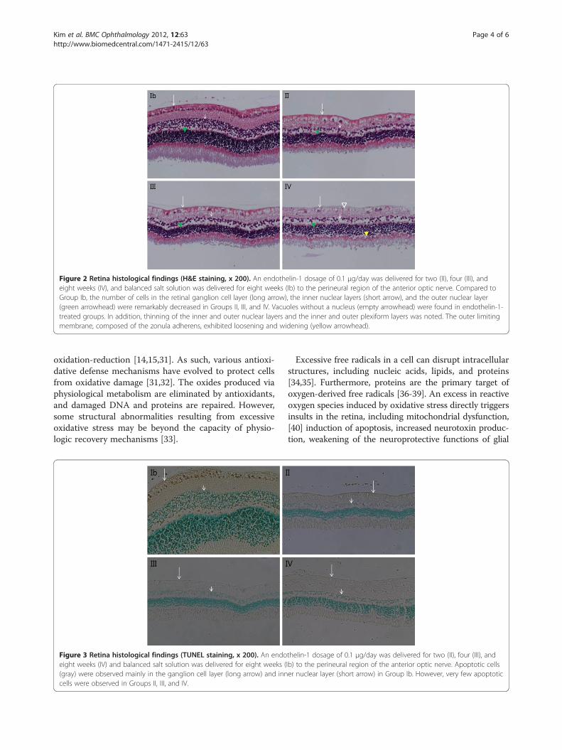
Figure 2 Retina histological findings (H&E staining, x 200). An endothelin-1 dosage of 0.1 μg/day was delivered for two (II), four (III), andeight weeks (IV), and balanced salt solution was delivered for eight weeks (Ib) to the perineural region of the anterior optic nerve. Compared toGroup Ib, the number of cells in the retinal ganglion cell layer (long arrow), the inner nuclear layers (short arrow), and the outer nuclear layer(green arrowhead) were remarkably decreased in Groups II, III, and IV. Vacuoles without a nucleus (empty arrowhead) were found in endothelin-1-treated groups. In addition, thinning of the inner and outer nuclear layers and the inner and outer plexiform layers was noted. The outer limitingmembrane, composed of the zonula adherens, exhibited loosening and widening (yellow arrowhead).
Kim et al. BMC Ophthalmology 2012, 12:63 Page 4 of 6http://www.biomedcentral.com/1471-2415/12/63
oxidation-reduction [14,15,31]. As such, various antioxi-dative defense mechanisms have evolved to protect cellsfrom oxidative damage [31,32]. The oxides produced viaphysiological metabolism are eliminated by antioxidants,and damaged DNA and proteins are repaired. However,some structural abnormalities resulting from excessiveoxidative stress may be beyond the capacity of physio-logic recovery mechanisms [33].
Figure 3 Retina histological findings (TUNEL staining, x 200). An endoeight weeks (IV) and balanced salt solution was delivered for eight weeks ((gray) were observed mainly in the ganglion cell layer (long arrow) and inncells were observed in Groups II, III, and IV.
Excessive free radicals in a cell can disrupt intracellularstructures, including nucleic acids, lipids, and proteins[34,35]. Furthermore, proteins are the primary target ofoxygen-derived free radicals [36-39]. An excess in reactiveoxygen species induced by oxidative stress directly triggersinsults in the retina, including mitochondrial dysfunction,[40] induction of apoptosis, increased neurotoxin produc-tion, weakening of the neuroprotective functions of glial
thelin-1 dosage of 0.1 μg/day was delivered for two (II), four (III), andIb) to the perineural region of the anterior optic nerve. Apoptotic cellser nuclear layer (short arrow) in Group Ib. However, very few apoptotic

Kim et al. BMC Ophthalmology 2012, 12:63 Page 5 of 6http://www.biomedcentral.com/1471-2415/12/63
cells, and activation of immune-mediated neuronal injur-ies. Indeed, oxidative stress in retinal ganglion cells andretinal proteins may be the cause of optic disc deformities[10,11].Quantification of intracellular oxyproteins is necessary
for a comprehensive understanding of oxidative stress.Measurement of DNPH protein bound carbonyl deriva-tives is one of the most commonly used strategies toquantify oxyproteins, and DNPH is quite sensitive withrespect to immunodetection using specific antibodies[41]. In our study, following two weeks of treatmentwith ET-1, the expression of oxyproteins was signifi-cantly increased compared to the control group. How-ever, at four and eight weeks, the level of oxyproteinexpression was equal to or slightly decreased comparedto the control group. When a protein undergoes irre-versible oxidative modifications and cannot be repaired,it is eliminated through degradation. The mechanisms ofprotein elimination are through either proteome-mediatedproteolysis [42] or chaperone-mediated autophagy [43]. Ineither case, vulnerable cells damaged by ischemia and oxi-dative stress may have their damaged proteins eliminatedby these mechanisms, thereby allowing resistant cells topersist. Thus, equivalent levels of oxyprotein expression atfour and eight weeks suggests the presence of a relative in-crease in stressful environments as well as expression ofoxidative proteins in the ET-1- administered group com-pared to the control group.The results of the present study also demonstrate the
presence of oxidative stress in the control group. Specific-ally, apoptosis was more apparent in the control groupthan in the ET-1-treated group, which may be a reflectionof the effects of natural stress such as light, chemicals, orradiation. Furthermore, the basal production of reactiveoxygen species, which are regulated by antioxidants, ispart of normal cellular redox homeostasis. Thus, the bal-ance between native and oxidatively-damaged proteinsdepends on the rates of protein biosynthesis, oxidativemodification, and oxidized protein elimination [44].In our results, there was no expression of oxyproteins
in the retina by oxyblot, yet cell loss was observed in ret-inal layers. In this study, we analyzed separation of theoptic nerve head from the retinal layer. For this analysis,we collected optic nerve head tissue, which protrudesinto the globe. The optic nerve head consists mainly ofthe surface nerve fiber layer and the prelaminar layer.Those layers include mainly axons and some deep ret-inal layers. The structure of the retina containing wholelayer is different from that of the surface retinal layers.Therefore, if oxyprotein is expressed more in the axons,the oxyblot result will be different from that of the thinaxons of the existing retinal layers. Since oxyblot is aquantitative method, a certain amount of oxyprotein isneeded for detection. Therefore, the absence of detection
can be attributed to a low amount of oxyprotein in thetissue. To date, there have been no studies regardingwhether oxyprotein can migrate along the axon. In aprevious animal model study, the injection of ET-1 intothe posterior chamber caused a significant constrictionof retinal vessels and impaired retrograde axonal trans-port by decreasing the number of retinal ganglion cells[45]. The number of retinal ganglion cells decreased by44.2% four weeks after the injection of ET-1 into theposterior chamber. For this reason, apoptosis in retinalganglion cells should be rarely observed in the ET-1-treated group.
ConclusionThe exact mechanisms that lead to damage of the opticnerve head and retinal ganglion cells in glaucoma arestill unknown, especially in glaucoma without highintraocular pressure. However, multiple mechanisms andfactors are undoubtedly involved in glaucoma. Themodel used in the present study simulates glaucomacaused by microcirculatory compromise in terms ofoptic nerve blood flow reduction. Thus, the currentdemonstration of increased expression of oxyprotein inthe optic nerve head in optic nerve ischemia justifies itsuse in studies aimed at both understanding and inhibit-ing the mechanisms associated with retinal ganglion celldamage.
Competing interestsThe authors declare that they have no conflict of interest.
Authors’ contributionsLiterature screening and selection was performed by JMK and DMK. JMK,YJK, and DMK participated in the design of the study, and JMK performedthe statistical analysis. Preparation of the first draft of the manuscript wasdone by JMK, and review and approval of the manuscript was performed byYJK and DMK. All authors read and approved the final manuscript.
Financial disclosureThe authors have no proprietary or commercial interest in any of thematerials mentioned in this article.
AcknowledgementsThis study was supported by a grant of the Korea Health technology R&DProject, Ministry of Health and Welfare, Republic of Korea (A101727).
Author details1Department of Ophthalmology, Kangbuk Samsung Hospital, SungkyunkwanUniversity School of Medicine, Seoul, Korea. 2Seoul National UniversityHospital Clinical Research Institute, Seoul, Korea. 3Department ofOphthalmology, Seoul National University College of Medicine, 28Yongon-Dong, Chongno-Gu, Seoul 110-744, Korea.
Received: 22 August 2012 Accepted: 27 November 2012Published: 5 December 2012
References1. Phelps CD, Corbett JJ: Migraine and low tension glaucoma:
a case–control study. Invest Ophthalmol Vis Sci 1985, 26:1105–1108.2. Drance SM, Douglas GR, Wijsman K, Schulzer M, Britton RJ: Response of
blood flow to warm and cold in normal and low-tension glaucomapatients. Am J Ophthalmol 1988, 105:35–39.

Kim et al. BMC Ophthalmology 2012, 12:63 Page 6 of 6http://www.biomedcentral.com/1471-2415/12/63
3. Becker B: Diabetes mellitus and primary open angle glaucoma.Am J Ophthalmol 1971, 71:1–16.
4. Corbett JJ, Phelps CD, Eslinger P, Montaque PR: The neurologic evaluationof patients with low-tension glaucoma. Invest Ophthalmol Vis Sci 1985,26:1101–1104.
5. Flammer J, Gasser P, Prunte C, Yao K: The probable involvement of factorsother than intraocular pressure in the pathogenesis of glaucoma. InApplied Pharmacology of the Glaucoma. Edited by Drance SM, Van BuskirkEM, Neufeld AH. Baltimore: Williams and Wilkins; 1992:273–283.
6. Mcleod SD, West SK, Quigley HA, Forzard JL: A longitudinal study of therelationship between intraocular and blood pressure. Invest OphthalmolVis Sci 1990, 31:2361–2366.
7. Saccà SC, Izzotti A: Oxidative stress and glaucoma: injury in the anteriorsegment of the eye. Prog Brain Res 2008, 173:385–407.
8. Izzotti A, Saccà SC, Longobardi M, Cartiglia C: Sensitivity of ocular anteriorchamber tissues to oxidative damage and its relevance to thepathogenesis of glaucoma. Invest Ophthalmol Vis Sci 2009, 50:5251–5258.
9. Agar A, Li S, Agarwal N, Coroneo MT, Hill MA: Retinal ganglion cell lineapoptosis induced by hydrostatic pressure. Brain Res 2006, 1086:191–200.
10. Liu Q, Ju WK, Crowston JG, Xie F, Perry G, Smith MA, Lindsey JD, WeinrebRN: Oxidative stress is an early event in hydrostatic pressure-inducedretinal ganglion cell damage. Invest Ophthalmol Vis Sci 2007,48:4580–4589.
11. Garcia-Valenzuela E, Shareef S, Walsh J, Sharma SC: Programmed cell deathof retinal ganglion cells during experimental glaucoma. Exp Eye Res 1995,61:33–44.
12. Pang IH, Yorio T: Ocular actions of endothelins. Proc Soc Exp Biol Med 1997,215:21–34.
13. Yorio T, Krishnamoorthy R, Prasanna G: Endothelin: is it a contributorglaucoma pathophysiology? J Glaucoma 2002, 11:259–270.
14. Cellini M, Possati GL, Profazio V, Sbrocca M, Caramazza N, Caramazza R:Color Doppler imaging and plasma levels of endothelin-1 in low-tensionglaucoma. Acta Ophthalmol Scand 1997, 224(Suppl):11–13.
15. Thanos S, Naskar R: Correlation between retinal ganglion cell death andchronically developing inherited glaucoma in a new rat mutant.Exp Eye Res 2004, 79:119–129.
16. Flammer J, Orgül S, Costa VP, Orzalesi N, Krieglstein GK, Serra LM, Renard JP,Stefánsson E: The impact of ocular blood flow in glaucoma. Prog Retin EyeRes 2002, 21:359–393.
17. Flammer J: Glaucomatous optic neuropathy: a reperfusion injury.Klin Monatsbl Augenheilkd 2001, 218:290–291.
18. Emre M, Orgul S, Haufschild T, Shaw SG, Flammer J: Increased plasmaendothelin-1 levels in patients with progressive open angle glaucoma.Br J Ophthalmol 2005, 89:60–63.
19. Grieshaber MC, Mozaffarieh M, Flammer J: What is the link betweenvascular dysregulation and glaucoma? Surv Ophthalmol 2007,52(Suppl 2):S144–S154.
20. Pache M, Kaiser HJ, Akhalbedashvili N, et al: Extraocular blood flow andendothelin-1 plasma levels in patients with multiple sclerosis. Eur Neurol2003, 49:164–168.
21. Zimmermann M: Endothelin in cerebral vasospasm. Clinical andexperimental results. J Neurosurg Sci 1997, 41:139–151.
22. Meyer P, Flammer J, Lüscher TF: Endothelium-dependent regulation ofthe ophthalmic microcirculation in the perfused porcine eye: role ofnitric oxide and endothelins. Invest Ophthalmol Vis Sci 1993, 34:3614–3621.
23. Arden GB, Sivaprasad S: Hypoxia and oxidative stress in the causation ofdiabetic retinopathy. Curr Diabetes Rev 2011, 7:e291–e304.
24. Cioffi GA, Van Buskirk EM: Microvasculature of the anterior optic nerve.Surv Ophthalmol 1994, 38:S107–S117.
25. Cioffi GA, Orgul S, Onda E, Bacon DR, Van Buskirk EM: An in vivo model ofchronic optic nerve ischemia: the dose-dependent effects of endothelin-1 on the optic nerve microvasculature. Curr Eye Res 1995, 14:1147–1153.
26. Orgul S, Cioffi GA, Wilson DJ, Bacon DR, Van Buskirk EM:An endothelin-1 induced model of optic nerve ischemia in the rabbit.Invest Ophthalmol Vis Sci 1996, 37:1860–1869.
27. Kim TW, Kang KB, Choung HK, Park KH, Kim DM: Elevated glutamate levelsin the vitreous body of an in vivo model of optic nerve ischemia.Arch Ophthalmol 2000, 118:533–536.
28. Aktaş Z, Gürelik G, Akyürek N, Onol M, Hasanreisoğlu B: Neuroprotectiveeffect of topically applied brimonidine tartrate 0.2% in
endothelin-1-induced optic nerve ischemia model. Clin Exp Ophthalmol2007, 35:527–534.
29. Friguet B: Protein repair and degradation during aging. Sci World J 2002,2:248–254.
30. Sies H: Oxidative stress. San Diego: Academic Press; 1985:1–7.31. Halliwell B, Gutteridge JMC: Role of free radicals and catalytic metal ions
in human diseases: an overview. Methods Enzymol 1990, 186:1–85.32. Stadtman ER: Protein oxidation and aging. Science 1992, 257:1250–1258.33. Mozaffarieh M, Grieshaber MC, Flammer J: Oxygen and blood flow: players
in the pathogenesis of glaucoma. Mol Vis 2008, 14:224–233. 31. Review.34. Cance B, Sies H, Boveris A: Hydroperoxide metabolism in mammalian
organs. Physiol Rev 1979, 59:527–605.35. Davies KJA: Protein damage and degradation by oxygen radicals.
J Biol Chem 1987, 262:9902–9907.36. Stadtman ER: Oxidation of free amino acids and amino acid residues in
proteins by radiolysis and by metal-catalyzed reactions. Ann Rev Biochem.1993, 62:797–821.
37. Davies KJ, Delsignore ME, Lin SW: Protein damage and degradation byoxygen radicals II. Modification of amino acids. J Biol Chem 1987,262:9902–9907.
38. Uchida K, Kawakishi S: Site-specific oxidation of angiotensin I bycopper(II) and L-ascorbate: conversion of histidine residues to2-imidazolones. Arch Biochem Biophys 1990, 283:20–26.
39. Heinecke JW, Li W, Daehnke HL 3rd, Goldstein JA: Dityrosine, a specificmarker of oxidation, is synthesized by the myeloperoxidase-hydrogenperoxidase system of human neutrophil and macrophages. J Biol Chem1993, 268:4069–4077.
40. Flammer J, Mozaffarieh M: What is the present pathogenetic concept ofglaucomatous optic neuropathy? Surv Ophthalmol 2007, 52:S162–S173.
41. Robbins CE, Keshavarzian A, Pasco DS, Frommel TO, Winship DH, HolmesEW: Determination of protein carbonyl groups by immunoblotting. AnalBiochem 1999, 266:48–57.
42. Davies KJ: Degradation of oxidized proteins by the 20S proteasome.Biochimie 2001, 83:301–310.
43. Kiffin R, Christian C, Knecht E, Cuervo AM: Activation ofchaperone-mediated autophagy during oxidative stress. Mol Biol Cell2004, 15:1055–1064.
44. Farout L, Friguet B: Proteasome function in aging and oxidative stress:implications in protein maintenance failure. Antioxid Redox Signal 2006,8:205–216.
45. Lau J, Dang M, Hockmann K, Alexander K: Ball Effects of acute delivery ofendothelin-1 on retinal ganglion cell loss in the rat. Exp Eye Res 2006,82:132–145.
doi:10.1186/1471-2415-12-63Cite this article as: Kim et al.: Increased expression of oxyproteins in theoptic nerve head of an in vivo model of optic nerve ischemia. BMCOphthalmology 2012 12:63.
Submit your next manuscript to BioMed Centraland take full advantage of:
• Convenient online submission
• Thorough peer review
• No space constraints or color figure charges
• Immediate publication on acceptance
• Inclusion in PubMed, CAS, Scopus and Google Scholar
• Research which is freely available for redistribution
Submit your manuscript at www.biomedcentral.com/submit
Related Documents