ACCELERATED COMMUNICATION The Novel Antipsychotic Drug Lurasidone Enhances N-Methyl- D-aspartate Receptor-Mediated Synaptic Responses Eunice Y. Yuen, Xiangning Li, Jing Wei, Masakuni Horiguchi, Herbert Y. Meltzer, and Zhen Yan Department of Physiology and Biophysics, State University of New York at Buffalo School of Medicine and Biomedical Sciences, Buffalo, New York (E.Y.Y., X.L., J.W., Z.Y.); Department of Psychiatry, Vanderbilt University School of Medicine, Nashville, Tennessee (M.H., H.Y.M.); and Dainippon Sumitomo Pharma Co., Ltd., Osaka, Japan (M.H.) Received October 3, 2011; accepted November 9, 2011 ABSTRACT N-Methyl-D-aspartate (NMDA) receptor (NMDAR) hypofunction has been postulated to contribute to the cognitive deficit of schizophrenia. In this study, we examined the effect of lurasi- done (Latuda; Dainippon Sumitomo Pharma Co. Ltd., Tokyo, Japan), a newly approved atypical antipsychotic drug (APD), on NMDAR synaptic function in rat frontal cortical pyramidal neu- rons. In vivo administration of lurasidone produced a significant and selective enhancement of NMDAR-mediated synaptic re- sponses and surface expression of NR2A and NR2B subunits. Lurasidone has high affinity for serotonin 5-HT 1A , 5-HT 2A , and 5-HT 7 receptors and dopamine D 2 receptors. In vivo adminis- tration of the 5-HT 7 receptor antagonist (2R)-1-[(3-hydroxyphe- nyl)sulfonyl]-2 -(2-(4-methyl-1-piperidinyl)ethyl)pyrrolidine (SB- 269970) mimicked the enhancing effect of lurasidone on NMDAR responses, whereas the D 2 receptor antagonist halo- peridol failed to do so. Previous studies have found that short- term administration of lurasidone reverses the cognitive impair- ment induced by subchronic administration of phencyclidine (PCP), an NMDAR noncompetitive antagonist. In this study, we found that lurasidone, as well as the prototypical atypical APD clozapine, restored NMDAR-mediated synaptic responses to normal levels in the PCP model of schizophrenia. These results suggest that NMDAR is the potential key molecular target of lurasidone, possibility via antagonizing 5-HT 7 receptors, which is consistent with evidence that 5-HT 7 receptor antagonism contributes to cognitive enhancement by atypical APDs in pa- tients with schizophrenia. Introduction Schizophrenia is characterized by positive symptoms (de- lusions and hallucinations), negative symptoms (e.g., affec- tive flattening, anhedonia, anergia), abnormalities in mood, and deficits in multiple domains of cognition, including work- ing memory, declarative memory, and executive function, often leading to severe functional impairment from the time of diagnosis (Meltzer et al., 1989; Sawa and Snyder, 2002). Abnormalities in prefrontal cortex (PFC) and temporal cortex are considered to be the most likely basis for the cognitive impairment of schizophrenia (Weinberger et al., 1986). Hy- pofunction of glutamatergic pyramidal neurons in cortex and hypodopaminergic activity are believed to underlie the cog- nitive deficit of schizophrenia (Lewis and Lieberman, 2000; Tsai and Coyle, 2002). The evidence for the hypoglutamater- gic theory includes the ability of noncompetitive NMDA re- This work was supported by grants from the National Institutes of Health National Institute on Mental Health [Grants MH84233, MH55441] (to Z.Y.). H.Y.M. is a stockholder of ACADIA, Astra Zeneca, and SureGene and has received grant support in the last 3 years from BioLine Rx, Cephalon, Daini- ppon Sumitomo, Eli Lilly, EnVivo, Janssen, Otsuka, Pfizer, and Sunovion. He is, or has been, a consultant to ACADIA, Alkemes, Astellas, Boehringer Mann- heim, Bristol Myers Squibb, Cypress, Janssen, Lundbeck, Ovation, Merck, Novartis, Pfizer, Teva, and Valeant (BioVail). Article, publication date, and citation information can be found at http://molpharm.aspetjournals.org. http://dx.doi.org/10.1124/mol.111.076141. ABBREVIATIONS: PFC, prefrontal cortex; NMDA, N-methyl-D-aspartate; NMDAR, N-methyl-D-aspartate receptor; PCP, phencyclidine; MK-801, 5H-dibenzo[a,d]cyclohepten-5,10-imine (dizocilpine maleate); APD, antipsychotic drug; NOR, novel object recognition; SB-269970, (2R)-1-[(3- hydroxyphenyl)sulfonyl]-2 -(2-(4-methyl-1-piperidinyl)ethyl)pyrrolidine; ACSF, artificial cerebrospinal fluid; EPSC, excitatory postsynaptic current; mEPSC, miniature excitatory postsynaptic current; AMPA, -amino-3-hydroxy-5-methyl-4-isoxazolepropionic acid; AMPAR, -amino-3-hydroxy- 5-methyl-4-isoxazolepropionic acid receptor. 1521-0111/12/8102-113–119$25.00 MOLECULAR PHARMACOLOGY Vol. 81, No. 2 Copyright © 2012 The American Society for Pharmacology and Experimental Therapeutics 76141/3744653 Mol Pharmacol 81:113–119, 2012 113 at State Univ Of NY At Buffalo on February 3, 2012 molpharm.aspetjournals.org Downloaded from

Welcome message from author
This document is posted to help you gain knowledge. Please leave a comment to let me know what you think about it! Share it to your friends and learn new things together.
Transcript

ACCELERATED COMMUNICATION
The Novel Antipsychotic Drug Lurasidone Enhances N-Methyl-D-aspartate Receptor-Mediated Synaptic Responses
Eunice Y. Yuen, Xiangning Li, Jing Wei, Masakuni Horiguchi, Herbert Y. Meltzer,and Zhen YanDepartment of Physiology and Biophysics, State University of New York at Buffalo School of Medicine and BiomedicalSciences, Buffalo, New York (E.Y.Y., X.L., J.W., Z.Y.); Department of Psychiatry, Vanderbilt University School of Medicine,Nashville, Tennessee (M.H., H.Y.M.); and Dainippon Sumitomo Pharma Co., Ltd., Osaka, Japan (M.H.)
Received October 3, 2011; accepted November 9, 2011
ABSTRACTN-Methyl-D-aspartate (NMDA) receptor (NMDAR) hypofunctionhas been postulated to contribute to the cognitive deficit ofschizophrenia. In this study, we examined the effect of lurasi-done (Latuda; Dainippon Sumitomo Pharma Co. Ltd., Tokyo,Japan), a newly approved atypical antipsychotic drug (APD), onNMDAR synaptic function in rat frontal cortical pyramidal neu-rons. In vivo administration of lurasidone produced a significantand selective enhancement of NMDAR-mediated synaptic re-sponses and surface expression of NR2A and NR2B subunits.Lurasidone has high affinity for serotonin 5-HT1A, 5-HT2A, and5-HT7 receptors and dopamine D2 receptors. In vivo adminis-tration of the 5-HT7 receptor antagonist (2R)-1-[(3-hydroxyphe-nyl)sulfonyl]-2 -(2-(4-methyl-1-piperidinyl)ethyl)pyrrolidine (SB-269970) mimicked the enhancing effect of lurasidone on
NMDAR responses, whereas the D2 receptor antagonist halo-peridol failed to do so. Previous studies have found that short-term administration of lurasidone reverses the cognitive impair-ment induced by subchronic administration of phencyclidine(PCP), an NMDAR noncompetitive antagonist. In this study, wefound that lurasidone, as well as the prototypical atypical APDclozapine, restored NMDAR-mediated synaptic responses tonormal levels in the PCP model of schizophrenia. These resultssuggest that NMDAR is the potential key molecular target oflurasidone, possibility via antagonizing 5-HT7 receptors, whichis consistent with evidence that 5-HT7 receptor antagonismcontributes to cognitive enhancement by atypical APDs in pa-tients with schizophrenia.
IntroductionSchizophrenia is characterized by positive symptoms (de-
lusions and hallucinations), negative symptoms (e.g., affec-
tive flattening, anhedonia, anergia), abnormalities in mood,and deficits in multiple domains of cognition, including work-ing memory, declarative memory, and executive function,often leading to severe functional impairment from the timeof diagnosis (Meltzer et al., 1989; Sawa and Snyder, 2002).Abnormalities in prefrontal cortex (PFC) and temporal cortexare considered to be the most likely basis for the cognitiveimpairment of schizophrenia (Weinberger et al., 1986). Hy-pofunction of glutamatergic pyramidal neurons in cortex andhypodopaminergic activity are believed to underlie the cog-nitive deficit of schizophrenia (Lewis and Lieberman, 2000;Tsai and Coyle, 2002). The evidence for the hypoglutamater-gic theory includes the ability of noncompetitive NMDA re-
This work was supported by grants from the National Institutes of HealthNational Institute on Mental Health [Grants MH84233, MH55441] (to Z.Y.).
H.Y.M. is a stockholder of ACADIA, Astra Zeneca, and SureGene and hasreceived grant support in the last 3 years from BioLine Rx, Cephalon, Daini-ppon Sumitomo, Eli Lilly, EnVivo, Janssen, Otsuka, Pfizer, and Sunovion. Heis, or has been, a consultant to ACADIA, Alkemes, Astellas, Boehringer Mann-heim, Bristol Myers Squibb, Cypress, Janssen, Lundbeck, Ovation, Merck,Novartis, Pfizer, Teva, and Valeant (BioVail).
Article, publication date, and citation information can be found athttp://molpharm.aspetjournals.org.
http://dx.doi.org/10.1124/mol.111.076141.
ABBREVIATIONS: PFC, prefrontal cortex; NMDA, N-methyl-D-aspartate; NMDAR, N-methyl-D-aspartate receptor; PCP, phencyclidine; MK-801,5H-dibenzo[a,d]cyclohepten-5,10-imine (dizocilpine maleate); APD, antipsychotic drug; NOR, novel object recognition; SB-269970, (2R)-1-[(3-hydroxyphenyl)sulfonyl]-2 -(2-(4-methyl-1-piperidinyl)ethyl)pyrrolidine; ACSF, artificial cerebrospinal fluid; EPSC, excitatory postsynaptic current;mEPSC, miniature excitatory postsynaptic current; AMPA, �-amino-3-hydroxy-5-methyl-4-isoxazolepropionic acid; AMPAR, �-amino-3-hydroxy-5-methyl-4-isoxazolepropionic acid receptor.
1521-0111/12/8102-113–119$25.00MOLECULAR PHARMACOLOGY Vol. 81, No. 2Copyright © 2012 The American Society for Pharmacology and Experimental Therapeutics 76141/3744653Mol Pharmacol 81:113–119, 2012
113
at State U
niv Of N
Y A
t Buffalo on F
ebruary 3, 2012m
olpharm.aspetjournals.org
Dow
nloaded from

ceptor (NMDAR) antagonists, such as phencyclidine (PCP),5H-dibenzo[a,d]cyclohepten-5,10-imine (dizocilpine maleate)(MK-801), and ketamine, to produce behavioral symptomsand cognitive dysfunction that have some similarity toschizophrenia in healthy volunteers and to exacerbate posi-tive and negative symptoms in schizophrenia (Javitt andZukin, 1991). Short-term or subchronic administration ofNMDAR antagonists also increases locomotor activity anddisrupts prepulse inhibition in rodents, both of which arethought to model schizophrenia symptoms (Jentsch et al.,1997; Jentsch and Roth, 1999). Furthermore, mice with ge-netic knockdown of the NMDAR subunit NR1, as well asother rodent models in which specific glutamate receptorsubtypes are genetically altered, also have phenotypes sug-gestive of schizophrenia, including increased locomotor activ-ity, stereotypy, and deficits in cognitive and social function(Mohn et al., 1999).
Typical antipsychotic drugs (APDs; e.g., haloperidol andperphenazine) are believed to diminish positive symptoms inpatients with schizophrenia through blockade of limbic do-pamine D2 receptors (Creese et al., 1976; Sawa and Snyder,2002), but blockade of D2 receptors in the dorsal striatumproduce unwanted extrapyramidal side effects (Meltzer,1992). Clozapine, the prototypical atypical APD, as well asmany other atypical APDs, are more potent serotonin 5-HT2A
than dopamine D2 receptor antagonists, which has been sug-gested to be the basis for some of their advantages overtypical APDs, including low extrapyramidal side effects(Meltzer et al., 1989; Meltzer and Huang, 2008). Actions atadrenergic and muscarinic receptors may also contribute tothe efficacy of various atypical APDs (Meltzer et al., 1989).Thus, clozapine and related atypical APDs have been re-ferred to as multireceptor antagonists to reflect the contri-bution of receptors other than 5-HT2A and D2 receptors totheir efficacy and side effects (Meltzer and Huang, 2008).
Lurasidone (Latuda; Dainippon Sumitomo Pharma Co.Ltd., Tokyo, Japan) is a novel atypical APD recently ap-proved for treatment of schizophrenia by the U.S. Food andDrug Administration. Lurasidone has potent binding affini-ties for 5-HT2A, 5-HT7, 5-HT1A, D2, and noradrenaline �2C
receptors (Ishibashi et al., 2010). Clinical trials have shownthat lurasidone is a safe and effective treatment for patientswith schizophrenia and has minimal extrapyramidal, cardio-vascular, and metabolic complications (Nakamura et al.,2009; Citrome, 2011; Meltzer et al., 2011a). Lurasidone hasbeen reported to improve short-term MK-801-induced mem-ory impairment in rats (Ishiyama et al., 2007; Enomoto et al.,2008), as well as subchronic PCP-induced impairment innovel object recognition (NOR) (Horiguchi et al., 2011; Melt-zer et al., 2011b). The molecular mechanism for the atypicalAPDs to improve cognition is not fully known. We sought to testthe hypothesis that lurasidone and clozapine may reverseNMDAR hypofunction via their 5-HT7 receptor antagonism.
Materials and MethodsAnimals. All animal experiments were performed with the approval
of the Institutional Animal Care and Use Committee of the StateUniversity of New York at Buffalo. Sprague-Dawley (3–4-week-old) ratswere used in all experiments. Animals were injected with lurasidone(0.1 mg/kg s.c.), clozapine (5 mg/kg s.c.), (2R)-1-[(3-hydroxyphenyl)sul-fonyl]-2 -(2-(4-methyl-1-piperidinyl)ethyl)pyrrolidine (SB-269970) (1
mg/kg i.p.), or haloperidol (0.1 mg/kg i.p.). One hour later, animals wereanesthetized by halothane inhalation (Sigma-Aldrich, St. Louis, MO)for �30 s and decapitated quickly. In some experiments, PCP (Sigma)was administered (5 mg/kg i.p.) once daily for 7 days (Wang et al., 2006).One day after the last administration of PCP, animals were injectedwith lurasidone (0.1 mg/kg s.c.) or clozapine (5 mg/kg s.c.) and sacrificed1 h later.
Slice Preparation. Brains were removed, iced, and blocked, andcoronal slices (300 �m) containing prelimbic/infralimbic regions wereobtained with a Vibratome (VP1000S; Leica, Wetzlar, Germany) inthe presence of a low-Ca2� HEPES-buffered salt solution (140 mMsodium isethionate, 2 mM KCl, 4 mM MgCl2, 0.1 mM CaCl2, 23 mMglucose, and 15 mM HEPES, pH 7.4; 300–305 mOsM). These sliceswere then incubated for 1 to 5 h at room temperature (20–22°C) in aNaHCO3-buffered saline bubbled with 95% O2/5% CO2.
Electrophysiological Recordings. To record evoked synapticcurrents in prefrontal cortical slices, the whole-cell voltage-clamptechnique was used as described previously (Yuen et al., 2005, 2009).The internal solution contained 130 mM cesium methanesulfonate,10 mM CsCl, 4 mM NaCl, 1 mM MgCl2, 10 mM HEPES, 5 mMEGTA, 2.2 mM QX-314, 12 mM phosphocreatine, 5 mM MgATP, and0.5 mM Na2GTP, pH 7.2–7.3; 265 to 270 mOsM. Slices were perfusedwith ACSF (130 mM NaCl, 26 mM NaHCO3, 3 mM KCl, 5 mMMgCl2, 1.25 NaH2PO4, 1 mM CaCl2, and 10 mM glucose, pH 7.4; 300mOsM) bubbled with 95% O2/5% CO2. 6-Cyano-2,3-dihydroxy-7-nit-roquinoxaline (25 �M) and bicuculline (10 �M) were added whenNMDAR-excitatory postsynaptic currents (EPSCs) were recorded,whereas D-2-amino-5-phosphonovalerate (25 �M) and bicuculline (10�M) were added when AMPAR-EPSCs were measured. Recordingswere conducted at room temperature. Neurons were visualized witha 40� water-immersion lens and illuminated with near-IR light. Allrecordings were performed using a Multiclamp 700A amplifier anddigitized with Digidata 1322A (Molecular Devices, Sunnyvale, CA).Tight seals (2–10 G�) were generated by applying negative pressure,followed by additional suction to disrupt the membrane and obtainthe whole-cell configuration. The access resistance ranged from 8 to15 M�. Evoked currents were generated with a 50-�s pulse from astimulation isolation unit controlled by a pulse generator (S48; As-tro-Med, Inc., West Warwick, RI). A bipolar stimulating electrode(FHC, Inc., Bowdoinham, ME) was positioned �100 �m from theneuron under study. The same stimulation intensity was used inindividual neurons across groups with various drug treatments,similar to what was described previously (Yuen et al., 2009). ForNMDAR-EPSC recording, cells (voltage-clamped at �70 mV) weredepolarized to �60 mV for 3 s before stimulation to fully relieve thevoltage-dependent Mg2� block of NMDAR channels. For AMPAR-EPSC recording, cells were constantly held at �70 mV. When min-iature EPSC (mEPSC) was recorded, tetrodotoxin (1 �M) was addedto ACSF. The capacitance of the recorded neurons ranged from 80 to120 pF. Signals were acquired at a bandwidth of 20 KHz (EPSC) or2 KHz (mEPSC) and filtered with a 2-KHz low-pass Bessel filter.
To minimize experimental variations between cells, the followingcriteria were used: 1) layer V medial prefrontal cortex pyramidal neu-rons with comparable membrane capacitances were selected; 2) thestimulating electrode was positioned at the same location (layer VI,�100 �m horizontally) from the recording neuron, and the electrode tipwas cleaned after every recording to allow precise stimulation capacity;and 3) recordings from animals injected with different drugs wereinterleaved throughout the course of experiments.
Data analyses were performed with Clampfit (Molecular Devices),Mini Analysis (Synaptosoft, Decatur, GA) and Kaleidagraph (Syn-ergy Software, Reading, PA). Rise time was measured from 10 to 90%of peak amplitude. No correlation between EPSC amplitude and risetime was found. Because the distribution of EPSC amplitudes indifferent animals was often found to be non-normal, statistical anal-ysis was performed with Kruskal-Wallis test. For comparisons ofdata (two groups) with normal distributions, Student’s t test wasused.
114 Yuen et al.
at State U
niv Of N
Y A
t Buffalo on F
ebruary 3, 2012m
olpharm.aspetjournals.org
Dow
nloaded from
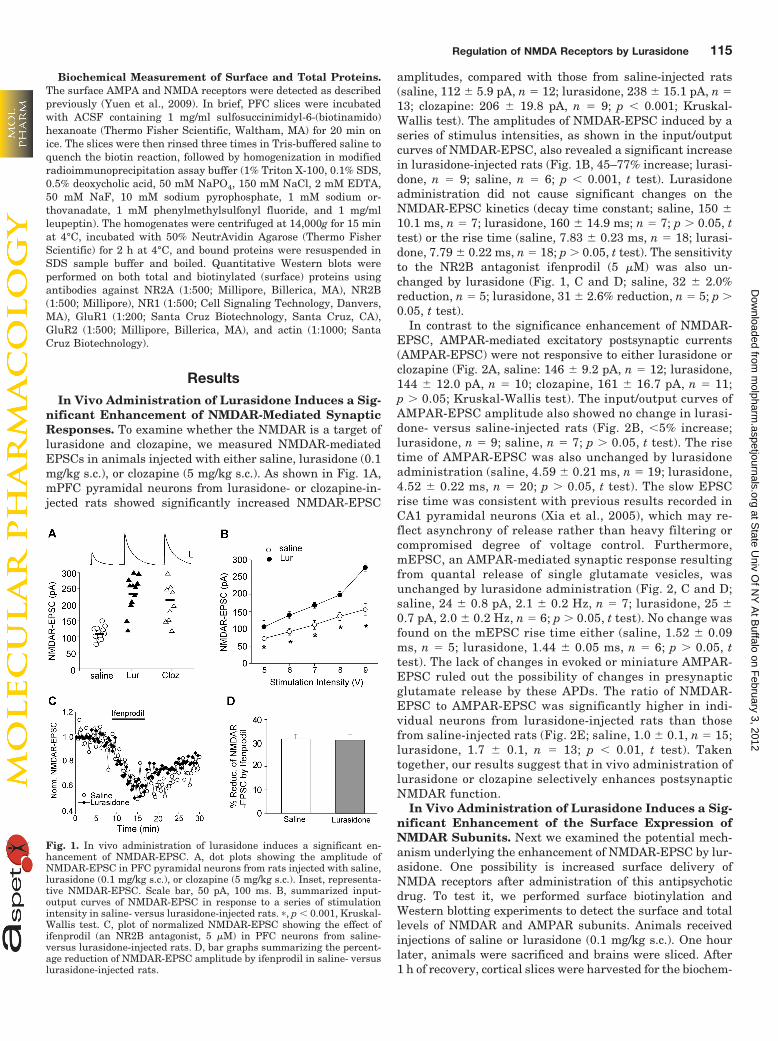
Biochemical Measurement of Surface and Total Proteins.The surface AMPA and NMDA receptors were detected as describedpreviously (Yuen et al., 2009). In brief, PFC slices were incubatedwith ACSF containing 1 mg/ml sulfosuccinimidyl-6-(biotinamido)hexanoate (Thermo Fisher Scientific, Waltham, MA) for 20 min onice. The slices were then rinsed three times in Tris-buffered saline toquench the biotin reaction, followed by homogenization in modifiedradioimmunoprecipitation assay buffer (1% Triton X-100, 0.1% SDS,0.5% deoxycholic acid, 50 mM NaPO4, 150 mM NaCl, 2 mM EDTA,50 mM NaF, 10 mM sodium pyrophosphate, 1 mM sodium or-thovanadate, 1 mM phenylmethylsulfonyl fluoride, and 1 mg/mlleupeptin). The homogenates were centrifuged at 14,000g for 15 minat 4°C, incubated with 50% NeutrAvidin Agarose (Thermo FisherScientific) for 2 h at 4°C, and bound proteins were resuspended inSDS sample buffer and boiled. Quantitative Western blots wereperformed on both total and biotinylated (surface) proteins usingantibodies against NR2A (1:500; Millipore, Billerica, MA), NR2B(1:500; Millipore), NR1 (1:500; Cell Signaling Technology, Danvers,MA), GluR1 (1:200; Santa Cruz Biotechnology, Santa Cruz, CA),GluR2 (1:500; Millipore, Billerica, MA), and actin (1:1000; SantaCruz Biotechnology).
ResultsIn Vivo Administration of Lurasidone Induces a Sig-
nificant Enhancement of NMDAR-Mediated SynapticResponses. To examine whether the NMDAR is a target oflurasidone and clozapine, we measured NMDAR-mediatedEPSCs in animals injected with either saline, lurasidone (0.1mg/kg s.c.), or clozapine (5 mg/kg s.c.). As shown in Fig. 1A,mPFC pyramidal neurons from lurasidone- or clozapine-in-jected rats showed significantly increased NMDAR-EPSC
amplitudes, compared with those from saline-injected rats(saline, 112 � 5.9 pA, n � 12; lurasidone, 238 � 15.1 pA, n �13; clozapine: 206 � 19.8 pA, n � 9; p � 0.001; Kruskal-Wallis test). The amplitudes of NMDAR-EPSC induced by aseries of stimulus intensities, as shown in the input/outputcurves of NMDAR-EPSC, also revealed a significant increasein lurasidone-injected rats (Fig. 1B, 45–77% increase; lurasi-done, n � 9; saline, n � 6; p � 0.001, t test). Lurasidoneadministration did not cause significant changes on theNMDAR-EPSC kinetics (decay time constant; saline, 150 �10.1 ms, n � 7; lurasidone, 160 � 14.9 ms; n � 7; p 0.05, ttest) or the rise time (saline, 7.83 � 0.23 ms, n � 18; lurasi-done, 7.79 � 0.22 ms, n � 18; p 0.05, t test). The sensitivityto the NR2B antagonist ifenprodil (5 �M) was also un-changed by lurasidone (Fig. 1, C and D; saline, 32 � 2.0%reduction, n � 5; lurasidone, 31 � 2.6% reduction, n � 5; p 0.05, t test).
In contrast to the significance enhancement of NMDAR-EPSC, AMPAR-mediated excitatory postsynaptic currents(AMPAR-EPSC) were not responsive to either lurasidone orclozapine (Fig. 2A, saline: 146 � 9.2 pA, n � 12; lurasidone,144 � 12.0 pA, n � 10; clozapine, 161 � 16.7 pA, n � 11;p 0.05; Kruskal-Wallis test). The input/output curves ofAMPAR-EPSC amplitude also showed no change in lurasi-done- versus saline-injected rats (Fig. 2B, �5% increase;lurasidone, n � 9; saline, n � 7; p 0.05, t test). The risetime of AMPAR-EPSC was also unchanged by lurasidoneadministration (saline, 4.59 � 0.21 ms, n � 19; lurasidone,4.52 � 0.22 ms, n � 20; p 0.05, t test). The slow EPSCrise time was consistent with previous results recorded inCA1 pyramidal neurons (Xia et al., 2005), which may re-flect asynchrony of release rather than heavy filtering orcompromised degree of voltage control. Furthermore,mEPSC, an AMPAR-mediated synaptic response resultingfrom quantal release of single glutamate vesicles, wasunchanged by lurasidone administration (Fig. 2, C and D;saline, 24 � 0.8 pA, 2.1 � 0.2 Hz, n � 7; lurasidone, 25 �0.7 pA, 2.0 � 0.2 Hz, n � 6; p 0.05, t test). No change wasfound on the mEPSC rise time either (saline, 1.52 � 0.09ms, n � 5; lurasidone, 1.44 � 0.05 ms, n � 6; p 0.05, ttest). The lack of changes in evoked or miniature AMPAR-EPSC ruled out the possibility of changes in presynapticglutamate release by these APDs. The ratio of NMDAR-EPSC to AMPAR-EPSC was significantly higher in indi-vidual neurons from lurasidone-injected rats than thosefrom saline-injected rats (Fig. 2E; saline, 1.0 � 0.1, n � 15;lurasidone, 1.7 � 0.1, n � 13; p � 0.01, t test). Takentogether, our results suggest that in vivo administration oflurasidone or clozapine selectively enhances postsynapticNMDAR function.
In Vivo Administration of Lurasidone Induces a Sig-nificant Enhancement of the Surface Expression ofNMDAR Subunits. Next we examined the potential mech-anism underlying the enhancement of NMDAR-EPSC by lur-asidone. One possibility is increased surface delivery ofNMDA receptors after administration of this antipsychoticdrug. To test it, we performed surface biotinylation andWestern blotting experiments to detect the surface and totallevels of NMDAR and AMPAR subunits. Animals receivedinjections of saline or lurasidone (0.1 mg/kg s.c.). One hourlater, animals were sacrificed and brains were sliced. After1 h of recovery, cortical slices were harvested for the biochem-
Fig. 1. In vivo administration of lurasidone induces a significant en-hancement of NMDAR-EPSC. A, dot plots showing the amplitude ofNMDAR-EPSC in PFC pyramidal neurons from rats injected with saline,lurasidone (0.1 mg/kg s.c.), or clozapine (5 mg/kg s.c.). Inset, representa-tive NMDAR-EPSC. Scale bar, 50 pA, 100 ms. B, summarized input-output curves of NMDAR-EPSC in response to a series of stimulationintensity in saline- versus lurasidone-injected rats. �, p � 0.001, Kruskal-Wallis test. C, plot of normalized NMDAR-EPSC showing the effect ofifenprodil (an NR2B antagonist, 5 �M) in PFC neurons from saline-versus lurasidone-injected rats. D, bar graphs summarizing the percent-age reduction of NMDAR-EPSC amplitude by ifenprodil in saline- versuslurasidone-injected rats.
Regulation of NMDA Receptors by Lurasidone 115
at State U
niv Of N
Y A
t Buffalo on F
ebruary 3, 2012m
olpharm.aspetjournals.org
Dow
nloaded from
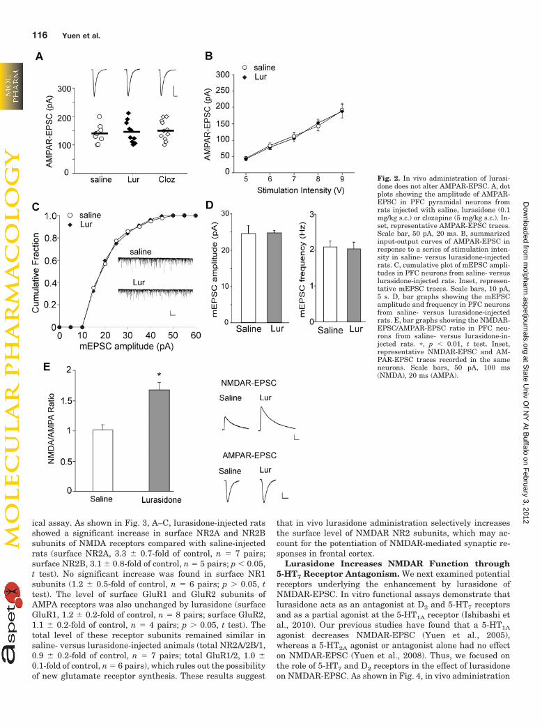
ical assay. As shown in Fig. 3, A–C, lurasidone-injected ratsshowed a significant increase in surface NR2A and NR2Bsubunits of NMDA receptors compared with saline-injectedrats (surface NR2A, 3.3 � 0.7-fold of control, n � 7 pairs;surface NR2B, 3.1 � 0.8-fold of control, n � 5 pairs; p � 0.05,t test). No significant increase was found in surface NR1subunits (1.2 � 0.5-fold of control, n � 6 pairs; p 0.05, ttest). The level of surface GluR1 and GluR2 subunits ofAMPA receptors was also unchanged by lurasidone (surfaceGluR1, 1.2 � 0.2-fold of control, n � 8 pairs; surface GluR2,1.1 � 0.2-fold of control, n � 4 pairs; p 0.05, t test). Thetotal level of these receptor subunits remained similar insaline- versus lurasidone-injected animals (total NR2A/2B/1,0.9 � 0.2-fold of control, n � 7 pairs; total GluR1/2, 1.0 �0.1-fold of control, n � 6 pairs), which rules out the possibilityof new glutamate receptor synthesis. These results suggest
that in vivo lurasidone administration selectively increasesthe surface level of NMDAR NR2 subunits, which may ac-count for the potentiation of NMDAR-mediated synaptic re-sponses in frontal cortex.
Lurasidone Increases NMDAR Function through5-HT7 Receptor Antagonism. We next examined potentialreceptors underlying the enhancement by lurasidone ofNMDAR-EPSC. In vitro functional assays demonstrate thatlurasidone acts as an antagonist at D2 and 5-HT7 receptorsand as a partial agonist at the 5-HT1A receptor (Ishibashi etal., 2010). Our previous studies have found that a 5-HT1A
agonist decreases NMDAR-EPSC (Yuen et al., 2005),whereas a 5-HT2A agonist or antagonist alone had no effecton NMDAR-EPSC (Yuen et al., 2008). Thus, we focused onthe role of 5-HT7 and D2 receptors in the effect of lurasidoneon NMDAR-EPSC. As shown in Fig. 4, in vivo administration
Fig. 2. In vivo administration of lurasi-done does not alter AMPAR-EPSC. A, dotplots showing the amplitude of AMPAR-EPSC in PFC pyramidal neurons fromrats injected with saline, lurasidone (0.1mg/kg s.c.) or clozapine (5 mg/kg s.c.). In-set, representative AMPAR-EPSC traces.Scale bar, 50 pA, 20 ms. B, summarizedinput-output curves of AMPAR-EPSC inresponse to a series of stimulation inten-sity in saline- versus lurasidone-injectedrats. C, cumulative plot of mEPSC ampli-tudes in PFC neurons from saline- versuslurasidone-injected rats. Inset, represen-tative mEPSC traces. Scale bars, 10 pA,5 s. D, bar graphs showing the mEPSCamplitude and frequency in PFC neuronsfrom saline- versus lurasidone-injectedrats. E, bar graphs showing the NMDAR-EPSC/AMPAR-EPSC ratio in PFC neu-rons from saline- versus lurasidone-in-jected rats. �, p � 0.01, t test. Inset,representative NMDAR-EPSC and AM-PAR-EPSC traces recorded in the sameneurons. Scale bars, 50 pA, 100 ms(NMDA), 20 ms (AMPA).
116 Yuen et al.
at State U
niv Of N
Y A
t Buffalo on F
ebruary 3, 2012m
olpharm.aspetjournals.org
Dow
nloaded from

of the selective 5-HT7 antagonist SB-269970 (1 mg/kg i.p.;Gasbarri et al., 2008) produced significantly increasedNMDAR-EPSC (saline, 111 � 6.2 pA, n � 19; SB-269970:274 � 25.5 pA, n � 20; p � 0.001, Kruskal-Wallis test),mimicking the effects of clozapine and lurasidone. Note thata subset of neurons (5 of 20) had no response to the 5-HT7
antagonist, consistent with the previous report that 5-HT7 is
not expressed in every mature PFC pyramidal neuron(Beïque et al., 2004). Moreover, coinjection of lurasidone (0.1mg/kg) plus SB-269970 (1 mg/kg) did not produce an additiveenhancement of NMDAR-EPSC (231 � 32.6 pA, n � 18) atthese doses. On the other hand, in vivo administration of theselective D2 antagonist haloperidol (0.1 mg/kg i.p.) failed tochange NMDAR-EPSC (112 � 7.9 pA, n � 10). These resultssuggest that 5-HT7 antagonism may underlie the lurasidone-induced enhancement of NMDAR function.
Lurasidone Rescues the NMDAR Hypofunction in aPCP Model of Schizophrenia. Because lurasidone re-versed the effect of subchronic PCP administration on novelobject recognition (Horiguchi et al., 2011) and was shownhere to potently increase NMDAR function, we further testedits ability to restore NMDAR hypofunction in PCP-treatedrats. Thus, we administered lurasidone after subchronictreatment of PCP (5 mg/kg i.p., 7 days). One day after PCPwithdrawal, animals were given one injection of lurasidone(0.1 mg/kg s.c.) and tested 1 to 4 h later. As shown in Fig. 5A,PCP-treated animals showed significantly diminishedNMDAR-EPSC (saline, 128 � 12.8pA, n � 12; PCP, 66 � 6.5pA, n � 15; p � 0.001, Kruskal-Wallis test). Administrationof lurasidone increased NMDAR-EPSC in saline-injected an-imals (lurasidone, 207 � 13.3 pA; n � 14), and restored
Fig. 3. In vivo administration of lurasidone significantly increases thesurface expression of NMDA receptor NR2 subunits. A to C, immunoblotsand quantification analysis of the surface and total AMPAR and NMDARsubunits in cortical slices from saline- versus lurasidone-injected rats. �,p � 0.05 t test.
Fig. 4. The enhancing effect of lurasidone on NMDAR-EPSC is mimickedby antagonizing 5-HT7 receptors. A, dot plot showing the amplitude ofNMDAR-EPSC in PFC pyramidal neurons from animals injected withsaline, the 5-HT7 antagonist SB-269970 (1 mg/kg i.p.), lurasidone (0.1mg/kg s.c.), plus SB-269970 or the D2 antagonist haloperidol (0.1 mg/kgi.p.). B, representative NMDAR-EPSC traces in rats injected with differ-ent agents. Scale bar, 50 pA, 100 ms.
Fig. 5. Lurasidone reverses NMDAR hypofunction in the PCP model ofschizophrenia. A and B, dot plots showing the amplitude of NMDAR-EPSC in PFC pyramidal neurons from PCP-treated animals injected withlurasidone (0.1 mg/kg s.c.; A) or clozapine (5 mg/kg s.c.; B). Inset, repre-sentative NMDAR-EPSC traces. Scale bar, 50 pA, 100 ms.
Regulation of NMDA Receptors by Lurasidone 117
at State U
niv Of N
Y A
t Buffalo on F
ebruary 3, 2012m
olpharm.aspetjournals.org
Dow
nloaded from

NMDAR-EPSC in the PCP-treated animals (PCP � lurasi-done, 168 � 20.3 pA, n � 12). A similar rescue was observedwith in vivo administration of clozapine (5 mg/kg s.c., saline�clozapine, 244 � 9.6 pA, n � 14; PCP � clozapine, 227 � 18.2pA, n � 9, Fig. 5B). These results suggest that lurasidone,like clozapine, is capable of reversing the NMDAR hypofunc-tion induced by repeated PCP treatment, which is a widelystudied animal model of schizophrenia.
DiscussionDespite the ability of lurasidone and clozapine to improve
cognition in animal models of schizophrenia (Ishiyama et al.,2007; Enomoto et al., 2008; Nakamura et al., 2009; Snigdhaet al., 2010; Horiguchi et al., 2011), little is known about themolecular and cellular mechanism underlying this action.Repeated exposure to lurasidone increases the mRNA andprotein levels of brain-derived neurotrophic factor (Fum-agalli et al., 2011), an important determinant of synapticplasticity of glutamatergic synapses, consistent with the ideathat antipsychotic treatment may change the expression,trafficking, and interaction of essential components of gluta-matergic synapses (Fumagalli et al., 2008; Iasevoli et al.,2010). This study has provided the first electrophysiologicalevidence showing that in vivo administration of lurasidone orclozapine produces a significant enhancement of NMDAR-mediated EPSC in PFC neurons. Moreover, administration ofa single dose of lurasidone or clozapine restored NMDARresponses in subchronic PCP-treated rats.
Similar to the pharmacological profile of clozapine (Melt-zer, 1994), lurasidone has high binding affinity to variousmonoamine receptors, such as 5-HT2A, 5-HT7, 5-HT1A, D2,and �2C receptors (Meyer et al., 2009; Ishibashi et al., 2010).Drugs that affect several 5-HT receptors (e.g., 5-HT2A antag-onists and 5-HT2C agonists) are effective to prevent the ef-fects of NMDAR noncompetitive blockers on locomotor activ-ity (Marquis et al., 2007) and to restore NOR in thesubchronic PCP model (Meltzer et al., 2011b). Our previousstudies have found that selective agonists or antagonists for5-HT1A or 5-HT2A receptors either reduce NMDAR-EPSC orhave no effect (Yuen et al., 2005, 2008). In this study, weshow that the selective 5-HT7 antagonist SB-269970 mimicsthe enhancing effect of lurasidone on NMDAR-EPSC,whereas the D2 antagonist haloperidol (a typical APD) isineffective. These results suggest that antagonism of 5-HT7
receptors may contribute to the ability of some atypical APDsto potentiate NMDAR function. The electrophysiological re-sults reported here are consistent with the behavioral effectsof these compounds in schizophrenia models. Horiguchi et al.(2011) have found that lurasidone, clozapine, and SB-269970,but not haloperidol, improve the impairment in NOR inducedby subchronic PCP treatment. Moreover, the ability of lur-asidone to reverse the PCP-induced NOR deficit is blocked bythe 5-HT7 agonist AS19 (Horiguchi et al., 2011).
Emerging evidence suggests that NMDAR trafficking,which is regulated by interactions with PDZ proteins andtyrosine phosphorylation, plays a key role in controllingNMDAR function at synapses (Wenthold et al., 2003). Ourbiochemical evidence indicates that the surface levels ofNR2A and NR2B subunits of NMDA receptors are selectivelyand significantly elevated after lurasidone administration.Because the availability of NR2 subunits determines the
number of functional NMDARs at synapses, our results sug-gest that the potential molecular mechanism underlying theenhancing effect of in vivo administration of lurasidone onNMDAR synaptic responses is the increased delivery or de-creased internalization of synaptic NMDA receptors.
Pharmacological data suggest that the effect of lurasidoneon NMDARs is likely to be through a mechanism involving5-HT7 receptor antagonism. 5-HT7 is a Gs-coupled GPCRthat stimulates type 1 and type 8 Ca2�/calmodulin-sensitiveadenylyl cyclases (Baker et al., 1998). The 5-HT7 receptor isenriched in brain regions mediating complex cognitive pro-cesses, such as the limbic system, hippocampus, amygdala,and PFC (Ruat et al., 1993; Beïque et al., 2004). 5-HT7
expression and function also correlate with neuronal depo-larization in the developing rat PFC (Beïque et al., 2004).5-HT7 receptor knockout or blockade of the 5-HT7 receptorenhances learning and memory (Gasbarri et al., 2008). Agrowing body of evidence supports the concept for targeting5-HT7 antagonism as a possible mechanism for the treatmentof cognitive deficits and a potential target for novel anxiolyticand antidepressant drugs (Hedlund et al., 2005; Abbas et al.,2009; Mnie-Filali et al., 2009; Horiguchi et al., 2011). Inconclusion, the results reported here suggest that the procog-nitive effect of 5-HT7 receptor antagonism (Gasbarri et al.,2008; Horiguchi et al., 2011) may result from enhancement ofNMDAR function.
Acknowledgments
We thank Xiaoqing Chen for excellent technical support.
Authorship Contributions
Participated in research design: Yuen, Meltzer, and Yan.Conducted experiments: Yuen, Li, and Wei.Performed data analysis: Yuen, Li, and Wei.Wrote or contributed to the writing of the manuscript: Horiguchi,
Meltzer, and Yan.
ReferencesAbbas AI, Hedlund PB, Huang XP, Tran TB, Meltzer HY, and Roth BL (2009)
Amisulpride is a potent 5-HT7 antagonist: relevance for antidepressant actions invivo. Psychopharmacology (Berl) 205:119–128.
Baker LP, Nielsen MD, Impey S, Metcalf MA, Poser SW, Chan G, Obrietan K,Hamblin MW, and Storm DR (1998) Stimulation of type 1 and type 8 Ca2�/calmodulin-sensitive adenylyl cyclases by the Gs-coupled 5-hydroxytryptaminesubtype 5-HT7A receptor. J Biol Chem 273:17469–17476.
Beïque JC, Campbell B, Perring P, Hamblin MW, Walker P, Mladenovic L, andAndrade R (2004) Serotonergic regulation of membrane potential in developing ratprefrontal cortex: coordinated expression of 5-hydroxytryptamine (5-HT)1A,5-HT2A, and 5-HT7 receptors. J Neurosci 24:4807–4817.
Citrome L (2011) Lurasidone for schizophrenia: a brief review of a new second-generation antipsychotic. Clin Schizophr Relat Psychoses 4:251–257.
Creese I, Burt DR, and Snyder SH (1976) Dopamine receptor binding predictsclinical and pharmacological potencies of antischizophrenic drugs. Science 192:481–483.
Enomoto T, Ishibashi T, Tokuda K, Ishiyama T, Toma S, and Ito A (2008) Lurasidonereverses MK-801-induced impairment of learning and memory in the Morris watermaze and radial-arm maze tests in rats. Behav Brain Res 186:197–207.
Fumagalli F, Calabrese F, Luoni A, Bolis F, Racagni G, and Riva MA (2011)Modulation of BDNF expression by repeated treatment with the novel antipsy-chotic lurasidone under basal condition and in response to acute stress. Int J Neu-ropsychopharmacol 24:1–12.
Fumagalli F, Frasca A, Racagni G, and Riva MA (2008) Dynamic regulation ofglutamatergic postsynaptic activity in rat prefrontal cortex by repeated adminis-tration of antipsychotic drugs. Mol Pharmacol 73:1484–1490.
Gasbarri A, Cifariello A, Pompili A, and Meneses A (2008) Effect of 5-HT(7) antag-onist SB-269970 in the modulation of working and reference memory in the rat.Behav Brain Res 195:164–170.
Hedlund PB, Huitron-Resendiz S, Henriksen SJ, and Sutcliffe JG (2005) 5-HT7receptor inhibition and inactivation induce antidepressantlike behavior and sleeppattern. Biol Psychiatry 58:831–837.
Horiguchi M, Huang M, and Meltzer HY (2011) The role of 5-hydroxytryptamine 7receptors in the phencyclidine-induced novel object recognition deficit in rats.J Pharmacol Exp Ther 338:605–614.
118 Yuen et al.
at State U
niv Of N
Y A
t Buffalo on F
ebruary 3, 2012m
olpharm.aspetjournals.org
Dow
nloaded from

Iasevoli F, Tomasetti C, Marmo F, Bravi D, Arnt J, and de Bartolomeis A (2010)Divergent acute and chronic modulation of glutamatergic postsynaptic densitygenes expression by the antipsychotics haloperidol and sertindole. Psychopharma-cology (Berl) 212:329–344.
Ishibashi T, Horisawa T, Tokuda K, Ishiyama T, Ogasa M, Tagashira R, MatsumotoK, Nishikawa H, Ueda Y, Toma S, et al. (2010) Pharmacological profile of lurasi-done, a novel antipsychotic agent with potent 5-hydroxytryptamine 7 (5-HT7) and5-HT1A receptor activity. J Pharmacol Exp Ther 334:171–181.
Ishiyama T, Tokuda K, Ishibashi T, Ito A, Toma S, and Ohno Y (2007) Lurasidone(SM-13496), a novel atypical antipsychotic drug, reverses MK-801-induced impair-ment of learning and memory in the rat passive-avoidance test. Eur J Pharmacol572:160–170.
Javitt DC and Zukin SR (1991) Recent advances in the phencyclidine model ofschizophrenia. Am J Psychiatry 148:1301–1308.
Jentsch JD and Roth RH (1999) The neuropsychopharmacology of phencyclidine:from NMDA receptor hypofunction to the dopamine hypothesis of schizophrenia.Neuropsychopharmacology 20:201–225.
Jentsch JD, Tran A, Le D, Youngren KD, and Roth RH (1997) Subchronic phency-clidine administration reduces mesoprefrontal dopamine utilization and impairsprefrontal cortical-dependent cognition in the rat. Neuropsychopharmacology 17:92–99.
Lewis DA and Lieberman JA (2000) Catching up on schizophrenia: natural historyand neurobiology. Neuron 28:325–334.
Marquis KL, Sabb AL, Logue SF, Brennan JA, Piesla MJ, Comery TA, Grauer SM,Ashby CR Jr, Nguyen HQ, Dawson LA, et al. (2007) WAY-163909 ((7bR,10aR)-1,2,3,4,8,9,10,10a-octahydro-7bH-cyclopenta-[b][1,4]diazepino[6,7,1hi]indole): anovel 5-hydroxytryptamine 2C receptor-selective agonist with preclinical antipsy-chotic-like activity. J Pharmacol Exp Ther 320:486–496.
Meltzer HY (1992) Dimensions of outcome with clozapine. Br J Psychiatry Suppl(17):46–53.
Meltzer HY (1994) An overview of the mechanism of action of clozapine. J ClinPsychiatry 55 (Suppl B):47–52.
Meltzer HY, Cucchiaro J, Silva R, Ogasa M, Phillips D, Xu J, Kalali AH SchweizerE, Pikalov A, and Loebel A (2011a) Lurasidone in the treatment of schizophrenia:a randomized, double-blind, placebo- and olanzapine-controlled study. Am J Psy-chiatry 168:957–967.
Meltzer HY, Horiguchi M, and Massey BW (2011b) The role of serotonin in theNMDA receptor antagonist models of psychosis and cognitive impairment. Psy-chopharmacology (Berl) 213:289–305.
Meltzer HY and Huang M (2008) In vivo actions of atypical antipsychotic drug onserotonergic and dopaminergic systems. Prog Brain Res 172:177–197.
Meltzer HY, Matsubara S, and Lee JC (1989) Classification of typical and atypicalantipsychotic drugs on the basis of dopamine D-1, D-2 and serotonin2 pKi values.J Pharmacol Exp Ther 251:238–246.
Meyer JM, Loebel AD, and Schweizer E (2009) Lurasidone: a new drug in develop-ment for schizophrenia. Expert Opin Investig Drugs 18:1715–1726.
Mnie-Filali O, Lambas-Senas L Scarna H, and Haddjeri N (2009) Therapeuticpotential of 5-HT7 receptors in mood disorders. Curr Drug Targets 10:1109–1117.
Mohn AR, Gainetdinov RR, Caron MG, and Koller BH (1999) Mice with reducedNMDA receptor expression display behaviors related to schizophrenia. Cell 98:427–436.
Nakamura M, Ogasa M, Guarino J, Phillips D, Severs J, Cucchiaro J, and Loebel A(2009) Lurasidone in the treatment of acute schizophrenia: a double-blind, place-bo-controlled trial. J Clin Psychiatry 70:829–836.
Ruat M, Traiffort E, Leurs R, Tardivel-Lacombe J, Diaz J, Arrang JM, and SchwartzJC (1993) Molecular cloning, characterization, and localization of a high-affinityserotonin receptor (5-HT7) activating cAMP formation. Proc Natl Acad Sci USA90:8547–8551.
Sawa A and Snyder SH (2002) Schizophrenia: diverse approaches to a complexdisease. Science 296:692–695.
Snigdha S, Horiguchi M, Huang M, Li Z, Shahid M, Neill JC, and Meltzer HY (2010)Attenuation of phencyclidine-induced object recognition deficits by the combina-tion of atypical antipsychotic drugs and pimavanserin (ACP 103), a5-hydroxytryptamine(2A) receptor inverse agonist. J Pharmacol Exp Ther 332:622–631.
Tsai G and Coyle JT (2002) Glutamatergic mechanisms in schizophrenia. Annu RevPharmacol Toxicol 42:165–179.
Wang X, Gu Z, Zhong P, Chen G, Feng J, and Yan Z (2006) Aberrant regulation ofNMDA receptors by dopamine D4 signaling in rats after phencyclidine exposure.Mol Cell Neurosci 31:15–25.
Weinberger DR, Berman KF, and Zec RF (1986) Physiologic dysfunction of dorsolat-eral prefrontal cortex in schizophrenia. I. Regional cerebral blood flow evidence.Arch Gen Psychiatry 43:114–124.
Wenthold RJ, Prybylowski K, Standley S, Sans N, and Petralia RS (2003) Traffickingof NMDA receptors. Annu Rev Pharmacol Toxicol 43:335–358.
Xia YF, Kessler M, and Arai AC (2005) Positive alpha-amino-3-hydroxy-5-methyl-4-isoxazolepropionic acid (AMPA) receptor modulators have different impact onsynaptic transmission in the thalamus and hippocampus. J Pharmacol Exp Ther313:277–285.
Yuen EY, Jiang Q, Chen P, Feng J, and Yan Z (2008) Activation of 5-HT2A/C receptorscounteracts 5-HT1A regulation of N-methyl-D-aspartate receptor channels in py-ramidal neurons of prefrontal cortex. J Biol Chem 283:17194–17204.
Yuen EY, Jiang Q, Chen P, Gu Z, Feng J, and Yan Z (2005) Serotonin 5-HT1Areceptors regulate NMDA receptor channels through a microtubule-dependentmechanism. J. Neurosci 25:5488–5501.
Yuen EY, Liu W, Karatsoreos IN, Feng J, McEwen BS, and Yan Z (2009) Acute stressenhances glutamatergic transmission in prefrontal cortex and facilitates workingmemory. Proc Natl Acad Sci USA 106:14075–14079.
Address correspondence to: Zhen Yan, State University of New York atBuffalo, 124 Sherman Hall, Buffalo, NY 14214. E-mail: [email protected]
Regulation of NMDA Receptors by Lurasidone 119
at State U
niv Of N
Y A
t Buffalo on F
ebruary 3, 2012m
olpharm.aspetjournals.org
Dow
nloaded from
Related Documents