The Neural Substrates of Visual Perceptual Learning of Words: Implications for the Visual Word Form Area Hypothesis Gui Xue and Russell A. Poldrack Abstract & It remains under debate whether the fusiform visual word form area (VWFA) is specific to visual word form and whether visual expertise increases its sensitivity (Xue et al., 2006; Cohen et al., 2002). The present study examined three related issues: (1) whether the VWFA is also involved in processing foreign writing that significantly differs from the native one, (2) the effect of visual word form training on VWFA activation after controlling the task difficulty, and (3) the transfer of visual word form learning. Eleven native English speakers were trained, dur- ing five sessions, to judge whether two subsequently flashed (100-msec duration with 200-msec interval) foreign characters (i.e., Korean Hangul) were identical or not. Visual noise was added to the stimuli to manipulate task difficulty. In functional magnetic resonance imaging scans before and after training, subjects performed the task once with the same noise level (i.e., parameter-matched scan) and once with noise level changed to match performance from pretraining to posttraining (i.e., performance-matched scan). Results indicated that training in- creased the accuracy in parameter-matched condition but re- mained constant in performance-matched condition (because of increasing task difficulty). Pretraining scans revealed stronger activation for English words than for Korean characters in the left inferior temporal gyrus and the left inferior frontal cortex, but not in the VWFA. Visual word form training significantly decreased the activation in the bilateral middle and left pos- terior fusiform when either parameters or performance were matched and for both trained and new items. These results con- firm our conjecture that the VWFA is not dedicated to words, and visual expertise acquired with training reduces rather than increases its activity. & INTRODUCTION The role of the visual word form area (VWFA) located in the left midfusiform cortex is a matter of continuing de- bate (Xue, Chen, Jin, & Dong, 2006; Cohen & Dehaene, 2004; Price & Devlin, 2003). Although the VWFA is con- sistently involved in visual word processing and shows cross-culture consistency (Bolger, Perfetti, & Schneider, 2005; Xue et al., 2005; McCandliss, Cohen, & Dehaene, 2003), researchers do not agree on whether the VWFA is specific to visual word form. It has been argued that VWFA is specified for prelexical visual word form pro- cessing (Cohen & Dehaene, 2004; McCandliss et al., 2003; Cohen et al., 2002), whereas others hold that the VWFA is also involved in lexical processing (Hillis et al., 2005; Kronbichler et al., 2004) and in processing other visual objects such as faces, houses, and tools (see Price & Devlin, 2003, for a review) and foreign writing 1 (Xue et al., 2006). The VWFA has also been proposed to play a role in integrating visual, phonological, and semantic information (Devlin, Jamison, Gonnerman, & Matthews, 2006; McCrory, Mechelli, Frith, & Price, 2005; Price & Friston, 2005). Neuropsychological studies could help to resolve this issue, but the results are not consistent (Henry et al., 2005; Hillis et al., 2005; Cohen, Henry, et al., 2004; Leff et al., 2001; Cohen et al., 2000, 2003). One major reason is that lesions in this region due to tumors or strokes are usually not restricted to the VWFA but, rather, extend to a large part of the cortex. More recently, Gaillard et al. (2006) have been able to test a rare patient who under- went surgical resection of a small patch of cortex near the VWFA. The patient developed a specific reading def- icit after surgery, whereas his general language ability (e.g., phonological and semantic processing) and ability to recognize and name other visual category (i.e., hous- es, faces, and tools) remained intact. This result provides support for some degree of regional specificity for visual word processing in the VWFA. Recent advances in this area have led to finer function- al division in the region. Particularly, researchers have proposed several models suggesting a posterior-versus- anterior distinction on VWFA function, including visuo- perceptual versus lexical/semantic (Simons, Koutstaal, Prince, Wagner, & Schacter, 2003), unimodal versus mul- timodal (Cohen, Jobert, Le Bihan, & Dehaene, 2004), and local combination versus larger fragments of words (Dehaene, Cohen, Sigman, & Vinckier, 2005; Cohen & University of California, Los Angeles D 2007 Massachusetts Institute of Technology Journal of Cognitive Neuroscience 19:10, pp. 1643–1655

Welcome message from author
This document is posted to help you gain knowledge. Please leave a comment to let me know what you think about it! Share it to your friends and learn new things together.
Transcript
The Neural Substrates of Visual Perceptual Learningof Words: Implications for the Visual Word Form
Area Hypothesis
Gui Xue and Russell A. Poldrack
Abstract
& It remains under debate whether the fusiform visual wordform area (VWFA) is specific to visual word form and whethervisual expertise increases its sensitivity (Xue et al., 2006; Cohenet al., 2002). The present study examined three related issues:(1) whether the VWFA is also involved in processing foreignwriting that significantly differs from the native one, (2) theeffect of visual word form training on VWFA activation aftercontrolling the task difficulty, and (3) the transfer of visual wordform learning. Eleven native English speakers were trained, dur-ing five sessions, to judge whether two subsequently f lashed(100-msec duration with 200-msec interval) foreign characters(i.e., Korean Hangul) were identical or not. Visual noise wasadded to the stimuli to manipulate task difficulty. In functionalmagnetic resonance imaging scans before and after training,subjects performed the task once with the same noise level (i.e.,
parameter-matched scan) and once with noise level changedto match performance from pretraining to posttraining (i.e.,performance-matched scan). Results indicated that training in-creased the accuracy in parameter-matched condition but re-mained constant in performance-matched condition (becauseof increasing task difficulty). Pretraining scans revealed strongeractivation for English words than for Korean characters in theleft inferior temporal gyrus and the left inferior frontal cortex,but not in the VWFA. Visual word form training significantlydecreased the activation in the bilateral middle and left pos-terior fusiform when either parameters or performance werematched and for both trained and new items. These results con-firm our conjecture that the VWFA is not dedicated to words,and visual expertise acquired with training reduces rather thanincreases its activity. &
INTRODUCTION
The role of the visual word form area (VWFA) located inthe left midfusiform cortex is a matter of continuing de-bate (Xue, Chen, Jin, & Dong, 2006; Cohen & Dehaene,2004; Price & Devlin, 2003). Although the VWFA is con-sistently involved in visual word processing and showscross-culture consistency (Bolger, Perfetti, & Schneider,2005; Xue et al., 2005; McCandliss, Cohen, & Dehaene,2003), researchers do not agree on whether the VWFA isspecific to visual word form. It has been argued thatVWFA is specified for prelexical visual word form pro-cessing (Cohen & Dehaene, 2004; McCandliss et al., 2003;Cohen et al., 2002), whereas others hold that the VWFAis also involved in lexical processing (Hillis et al., 2005;Kronbichler et al., 2004) and in processing other visualobjects such as faces, houses, and tools (see Price &Devlin, 2003, for a review) and foreign writing1 (Xueet al., 2006). The VWFA has also been proposed to play arole in integrating visual, phonological, and semanticinformation (Devlin, Jamison, Gonnerman, & Matthews,2006; McCrory, Mechelli, Frith, & Price, 2005; Price &Friston, 2005).
Neuropsychological studies could help to resolve thisissue, but the results are not consistent (Henry et al.,2005; Hillis et al., 2005; Cohen, Henry, et al., 2004; Leffet al., 2001; Cohen et al., 2000, 2003). One major reasonis that lesions in this region due to tumors or strokes areusually not restricted to the VWFA but, rather, extend toa large part of the cortex. More recently, Gaillard et al.(2006) have been able to test a rare patient who under-went surgical resection of a small patch of cortex nearthe VWFA. The patient developed a specific reading def-icit after surgery, whereas his general language ability(e.g., phonological and semantic processing) and abilityto recognize and name other visual category (i.e., hous-es, faces, and tools) remained intact. This result providessupport for some degree of regional specificity for visualword processing in the VWFA.
Recent advances in this area have led to finer function-al division in the region. Particularly, researchers haveproposed several models suggesting a posterior-versus-anterior distinction on VWFA function, including visuo-perceptual versus lexical/semantic (Simons, Koutstaal,Prince, Wagner, & Schacter, 2003), unimodal versus mul-timodal (Cohen, Jobert, Le Bihan, & Dehaene, 2004),and local combination versus larger fragments of words(Dehaene, Cohen, Sigman, & Vinckier, 2005; Cohen &University of California, Los Angeles
D 2007 Massachusetts Institute of Technology Journal of Cognitive Neuroscience 19:10, pp. 1643–1655
Dehaene, 2004). These results suggest that differentstrategies should be used to examine the specificityhypothesis in the subregions of the VWFA. For example,object–word comparisons are ideal to examine the spec-ificity in the anterior VWFA because both objects andwords are associated with both phonological and seman-tic information (Price & Devlin, 2003). On the other side,because of the great differences in visual features be-tween words and other categories of visual objects, thecomparison between word and visually matched non-words (e.g., consonant strings, false fonts, foreign writ-ings) may be better able to address the specificity in themiddle and posterior VWFA (Xue et al., 2006). However,neuroimaging studies along the latter line have obtainedmixed results (Ben-Shachar, Dougherty, Deutsch, &Wandell, 2007; Xue et al., 2006; James, James, Jobard,Wong, & Gauthier, 2005; Wong, Gauthier, Woroch,DeBuse, & Curran, 2005; Cohen et al., 2002; Polk &Farah, 2002; Tagamets, Novick, Chalmers, & Friedman,2000; Indefrey et al., 1997; Petersen, Fox, Snyder, &Raichle, 1990). For example, several studies have notfound word sensitivity in this region (Gaillard et al., 2006;Xue et al., 2006; Cohen et al., 2003; Tagamets et al., 2000;Indefrey et al., 1997). Some studies have revealed stron-ger fusiform activation for words/letters than for visual-matched nonwords/symbols, but the exact location variedsignificantly across studies (James et al., 2005; Cohenet al., 2002; Polk & Farah, 2002; Petersen et al., 1990).
It has been argued that word sensitivity/specificity inthe VWFA is associated with the visual expertise thatdeveloped through years of reading experience (Cohen& Dehaene, 2004; McCandliss et al., 2003; Cohen et al.,2002). To the contrary, we have argued that differences inactivity in the word-nonword comparison might reflectcombined effects of several aspects of language experi-ence (i.e., visual familiarity, phonology, and semantics)(Xue et al., 2006). We adopted an artificial languagetraining paradigm to disentangle the roles of visual famil-iarity, phonology, and semantics in modulating VWFAactivation. Results indicated that visual form trainingsignificantly decreased the activation in the VWFA, where-as phonology and semantic training increased VWFA ac-tivation, along with the increase in the left inferior frontalcortex (IFC) activation. This study shows that artificiallanguage training paradigm is a useful tool to examinethe developmental mechanisms of the VWFA. Moreover,it emphasizes that during word-nonword comparison,instead of merely focusing on the VWFA, it is also criticalto examine the neural differences in the frontal lobe thatsupport phonological and semantic processing (Poldrack,Wagner, et al., 1999), which could provide us with betterunderstanding of mechanisms of VWFA activation.
The present study was aimed at extending previousstudies to examine the mechanisms of the neural changesassociated with visual word form training without thecontamination of phonology and semantic information.Specifically, we aimed to address three questions. First,
we asked whether the VWFA is also involved in theprocessing of foreign writings that are significantly differ-ent from the native words, such as Korean Hangul versusEnglish. The use of significant different systems mini-mizes the transfer of native language experience to thenew system.
Second, we aimed to separate the effects of neuralplasticity and task difficulty. Research on neural plasticitywith training is often confounded by overall performancelevel and/or task difficulty (Poldrack, 2000). In the previ-ous study, we used a passive viewing task to reduce theeffect of task requirement. To further address this issue,the present study added visual noise to the characters tomanipulate task difficulty. Subjects were scanned undertwo conditions in each scanning session, once before andonce after training. In one condition, the amount of visualnoise used in the pretraining and posttraining scan re-mained the same (i.e., parameter-matched scan). In theother condition, a parameter estimation procedure wasused to determine the task parameters that would equatesubjects’ performance between the pretraining and post-training scans (i.e., performance-matched scan).
Finally, visual word form learning could reflect eitheritem-specific learning (i.e., repetition priming) or somegeneral visual skills that can be transferred to new items(i.e., skill learning) (Poldrack & Gabrieli, 2001; Poldrack,Selco, Field, & Cohen, 1999; Poldrack, Desmond, Glover,& Gabrieli, 1998). To further examine the nature of visualword form learning, the present study included both oldand new items in the training and functional magneticresonance imaging (fMRI) scans. We also examined short-term repetition priming (STRP) by presenting sets of oldor new items and then repeating the same items soonafter (within five trials).
METHODS
Subjects
Eleven normal young adult subjects participated in thisstudy (mean age = 26 years; 5 women and 6 men). Allsubjects had normal or corrected-to-normal vision andjudged as right-handed using the Edinburgh Handed-ness Inventory (Oldfield, 1971). They were all nativeEnglish speakers, and none had experience with anymajor logographic language (e.g., Chinese, Korean, Jap-anese). They provided informed consent according to aprocedure approved by the University of California, LosAngeles Human Subject Committee. Three additionalsubjects were excluded because of substantial motionartifacts in pretraining and/or posttraining session.
Material
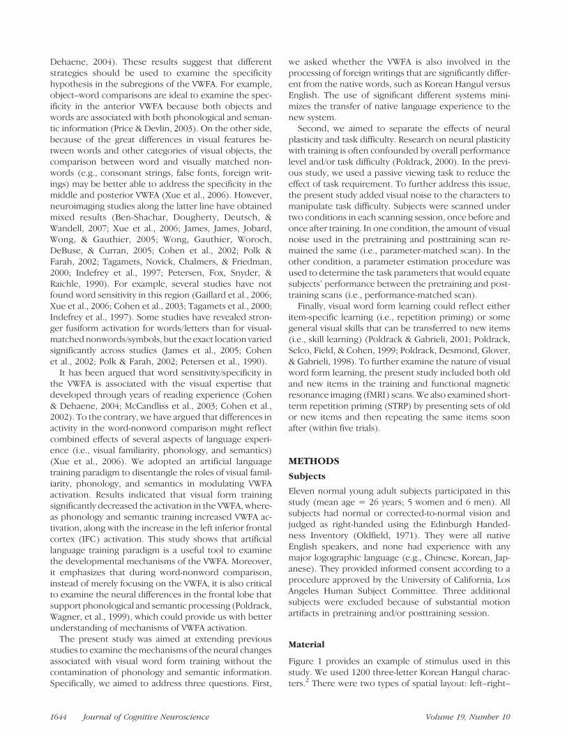
Figure 1 provides an example of stimulus used in thisstudy. We used 1200 three-letter Korean Hangul charac-ters.2 There were two types of spatial layout: left–right–
1644 Journal of Cognitive Neuroscience Volume 19, Number 10
bottom and top–middle–bottom (see Figure 1A, for anexample). To control task difficulty in the visual discrim-ination task to be detailed later, special attention was paidin constructing the word pairs. First, these characterswere organized into 600 pairs in which the two charactersshared the same spatial layout and visual complexity butdiffered from each other by only one letter. Second, those600 pairs were then divided into 20 matched groups thatwere assigned to each training and test condition andcounterbalanced across subjects. Finally, to create equalnumber of ‘‘same’’ trial and ‘‘different’’ trial, we randomlychose one third of the pairs, broke them up to form the‘‘same’’ trials. As a result, each group of stimuli (60 char-acters, 30 pair) produced 40 test pairs (i.e., 20 ‘‘same’’ and20 ‘‘different’’). Another 240 three-letter English wordswere used as control in fMRI scan (Figure 1B). Followinga similar procedure, they were divided into four matchedgroups of 30 pairs, in which the two words only differedby one letter. We used the uppercase letters and a boldfont for the English words. Pilot data indicated that thisresulted in comparable performance on Korean andEnglish.
The experiment was created in Matlab (Mathworks,Sherborn, MA) using the Psychophysics Toolbox exten-sions (Brainard, 1997; Pelli, 1997). Each stimulus wasframed in a 113 � 113-pixel window, drawn in whiteagainst a gray background. Random visual noise wasadded to the picture to manipulate the task difficulty(Figure 1C). The percentage of noise for English wordswas 28% for all the tasks, whereas that for Korean charac-ters varied from 28% to 38% across conditions. To createthe noise, we randomly chose a given percentage of pixelsin the picture and reversed their color (white to gray orvice versa). The pattern of the visual noise was differentfor each presentation of each stimulus.
Behavioral Training and Test
Subjects participated in five training sessions. The basicparadigm throughout the training was a same–different
judgment task. At the beginning of each trial, there was a400-msec fixation point followed by 200-msec blankscreen. Two characters then f lashed subsequently(100-msec duration with 200-msec interval). Subjectswere asked to judge whether the two characters wereidentical or not with a key press. The next trial started1 sec after the subjects making a response. Two groupsof stimuli (120 characters in total) were trained everysession (i.e., old items). One group of new stimuli (i.e.,new items) was added in each training session to allowseparation of repetition priming and skill learning(Poldrack, Selco, et al., 1999).
At the beginning of each training session, subjects tooka parameter setup test to find the desirable task difficultylevel. New items with four different noise levels (40 trialseach) were mixed. Based on subject’s performance, anoise level that corresponded to accuracy closest to 70%was chosen. Subjects then finished 4 blocks of 280 trials(80 old trials repeated 3 times, plus 40 new trials) withthe chosen noise level. There was a 2-min break be-tween each block.
Upon finishing all the training, a recognition task wasadministered to examine subjects’ explicit memory ofthese old items. Sixty old items and 60 new items wererandomly mixed, and subjects were to decide whetherthe character on the screen was new or old by pressingthe button. Each trial began with a fixation of 400 msec,followed by 200-msec blank screen. The character thenappeared and remained on the screen until subjectindicated a key press. If no responses were made in1900 msec, the character also disappeared. In either case,the next trial would start after a 1-sec interval.
fMRI Task
Each subject participated two fMRI scan sessions, onebefore training and one after training, separated by 6 to8 days. Two scanning runs were included in both pre-training and posttraining sessions: one with matchedparameters and one with parameters adjusted to match
Figure 1. Example of the stimuli. (A) Korean characters. The top and bottom show an example character with left–right–bottom, andtop–middle–bottom structure, respectively. The letters of the character are marked with different colors. (B) English words. All English words
consisted of three letters. Capital letters and bold font were chosen to increase the difficulty of recognition. (C) Korean characters presented
in varying amounts of visual noise; labels on each character indicate the percentage of noise.
Xue and Poldrack 1645
performance. The noise level for the parameter-matchedscan was 28% for pretraining and posttraining scans, aswell as for the pretraining performance-matched scan.For the posttraining performance-matched scan, the noiselevel was set to match subjects’ performance in pretrain-ing scan, using a parameter setup procedure similar tothat used in the training sessions. This task was adminis-tered in the scanner during an anatomical scan.
In each scan, there were 40 trials of old characters and40 trials of new characters (they were all new at thepretraining scan). To examine the STRP effect, they wererepeated once within five trials after its first presenta-tion. In addition, there were 40 trials of English words.The same–different task described above was used inthe fMRI scans. To improve the design efficiency, nullevents of random duration were imposed between everytrial. The duration of null time was sampled from anexponential distribution with mean of 1 sec (range =0.2-6 sec). A large number of sequences were generatedwithin these constraints, and the sequences with thehighest efficiency to detect the desired differences wereselected (Dale, 1999). In total, each scan included 200 trialsand lasted 9 min 36 sec.
MRI Data Acquisition
Imaging data collection was performed using a 3-TSiemens (Iselin, NJ) Allegra MRI scanner at the UCLAAhmanson-Lovelace Brain Mapping Center. Blood oxy-genation level-dependent-sensitive functional imageswere collected using a T2*-weighted echo-planar plussequence (repetition time [TR] = 2 sec, echo time [TE] =30 msec, flip angle = 908, matrix = 64 � 64, field ofview = 200, 33 slices, 4-mm slice thickness). Additional-ly, a T2-weighted matched-bandwidth high-resolutionanatomical scan (same slice prescription as echo-planarimaging) and Magnetization Prepared RApid GradientEcho (MP-RAGE) were acquired for each subject to aidregistration. The parameters for MP-RAGE were the fol-lowing: TR = 2.3, TE = 2.1, matrix = 192 � 192, field ofview = 256, sagittal plane, 160 slices, 1-m thickness.
Data Analysis
Image preprocessing and statistical analyses were per-formed with Statistical Parametric Mapping (SPM2; Well-come Department of Cognitive Neurology, London,UK),which is implemented in Matlab. The first two imagesin each time series were excluded from analysis to al-low T1 equilibration. Functional images were realigned,unwarped (Andersson, Hutton, Ashburner, Turner, &Friston, 2001), normalized using a combination of linearand nonlinear basis functions to the Montreal Neurolog-ical Institute 305 (MNI305) template (Friston et al.,1995), and smoothed with an 8-mm full width halfmaximum Gaussian filter. A two-level statistical analysisapproach was used. The general linear model was first
used to estimate condition effects in each individual par-ticipant (Friston et al., 1994) after convolving the refer-ence functions with a canonical hemodynamic response.Statistical parametric maps were computed for each con-trast of interest, and the contrast effect maps from theseanalyses were entered into a second-level model, whichtreats subjects as a random effect. Unless otherwisenoted, clusters of at least 10 contiguous voxels thatexceeded an uncorrected threshold of p < .001 wereconsidered significant for the exploratory analysis. Fur-ther analyses focused on regions of interest (ROIs) inthe fusiform/inferior temporal region and IFC, based onprevious results.
ROI Selection and Quantification
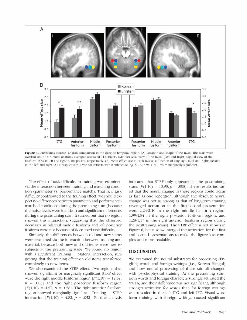
Based on a previous study (Xue et al., 2006), the centerof the fusiform ROI was defined as �39, �60, and �18(x, y, and z, respectively, in MNI coordinates). This isnear the VWFA region defined by Cohen et al. (2002):�42, �57, and �15. Following James et al. (2005), wesplit the fusiform region into three smaller equal sizedregions, namely, the anterior fusiform region (MNI cen-ter: �39, �48, and �18), middle fusiform region (MNIcenter: �39, �60, and �18), and posterior fusiformregion (MNI center: �39, �72, and �18). A lateral regionin the inferior temporal gyrus (ITG) (MNI center: �51,�48, and �18) was also selected based on the result ofthe pretraining Korean versus English comparison. Theright homologue of these regions was also defined.
In the left IFC, analysis using small volume correction(SVC) with a search volume based on the automated ana-tomical labeling atlas (Tzourio-Mazoyer et al., 2002) iden-tified two foci, one in the pars opercularis (MNI center:�45, 15, and 36), and one in pars triangularis (MNIcenter: �45, 33, and 18). All voxels within 6-mm radiusof the above coordinates were defined as ROI usingMarsbar (Brett, Anton, Valabregue, & Poline, 2002). Themean effect size for each subject and each condition wascalculated and entered into SPSS (Chicago, IL) forfurther statistical analysis.
For both the behavioral and ROI results, unless oth-erwise noted, we used within-subject repeated measureanalysis of variance (ANOVA), and within-subject stan-dard error (SE) was reported (Loftus & Masson, 1994).
RESULTS
Behavioral Results
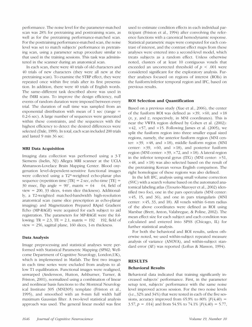
Behavioral data indicated that training significantly in-creased subjects’ performance. First, in the parametersetup test, subjects’ performance with the same noiselevel improved across session. For the two noise levels(i.e., 32% and 34%) that were tested in each of the five ses-sions, accuracy improved from 65.9% to 80% [F(4,40) =3.57, p = .014] and from 54.5% to 74.1% [F(4,40) = 5.77,
1646 Journal of Cognitive Neuroscience Volume 19, Number 10
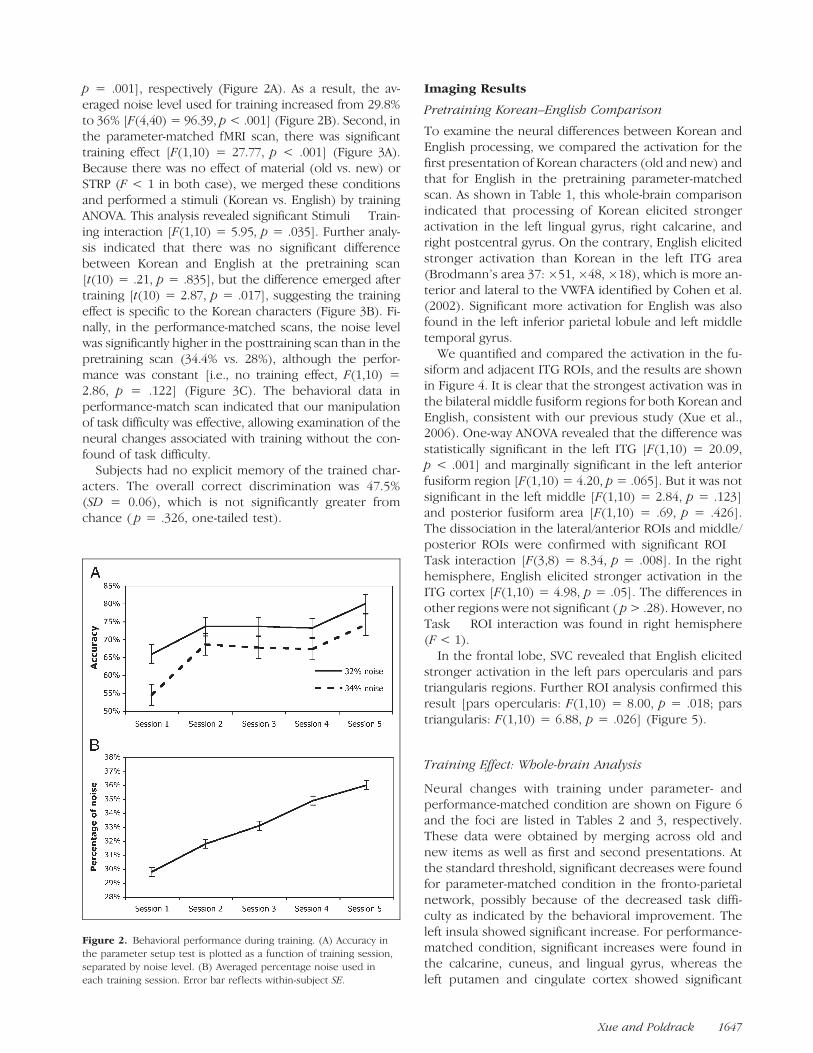
p = .001], respectively (Figure 2A). As a result, the av-eraged noise level used for training increased from 29.8%to 36% [F(4,40) = 96.39, p < .001] (Figure 2B). Second, inthe parameter-matched fMRI scan, there was significanttraining effect [F(1,10) = 27.77, p < .001] (Figure 3A).Because there was no effect of material (old vs. new) orSTRP (F < 1 in both case), we merged these conditionsand performed a stimuli (Korean vs. English) by trainingANOVA. This analysis revealed significant Stimuli � Train-ing interaction [F(1,10) = 5.95, p = .035]. Further analy-sis indicated that there was no significant differencebetween Korean and English at the pretraining scan[t(10) = .21, p = .835], but the difference emerged aftertraining [t(10) = 2.87, p = .017], suggesting the trainingeffect is specific to the Korean characters (Figure 3B). Fi-nally, in the performance-matched scans, the noise levelwas significantly higher in the posttraining scan than in thepretraining scan (34.4% vs. 28%), although the perfor-mance was constant [i.e., no training effect, F(1,10) =2.86, p = .122] (Figure 3C). The behavioral data inperformance-match scan indicated that our manipulationof task difficulty was effective, allowing examination of theneural changes associated with training without the con-found of task difficulty.
Subjects had no explicit memory of the trained char-acters. The overall correct discrimination was 47.5%(SD = 0.06), which is not significantly greater fromchance ( p = .326, one-tailed test).
Imaging Results
Pretraining Korean–English Comparison
To examine the neural differences between Korean andEnglish processing, we compared the activation for thefirst presentation of Korean characters (old and new) andthat for English in the pretraining parameter-matchedscan. As shown in Table 1, this whole-brain comparisonindicated that processing of Korean elicited strongeractivation in the left lingual gyrus, right calcarine, andright postcentral gyrus. On the contrary, English elicitedstronger activation than Korean in the left ITG area(Brodmann’s area 37: �51, �48, �18), which is more an-terior and lateral to the VWFA identified by Cohen et al.(2002). Significant more activation for English was alsofound in the left inferior parietal lobule and left middletemporal gyrus.
We quantified and compared the activation in the fu-siform and adjacent ITG ROIs, and the results are shownin Figure 4. It is clear that the strongest activation was inthe bilateral middle fusiform regions for both Korean andEnglish, consistent with our previous study (Xue et al.,2006). One-way ANOVA revealed that the difference wasstatistically significant in the left ITG [F(1,10) = 20.09,p < .001] and marginally significant in the left anteriorfusiform region [F(1,10) = 4.20, p = .065]. But it was notsignificant in the left middle [F(1,10) = 2.84, p = .123]and posterior fusiform area [F(1,10) = .69, p = .426].The dissociation in the lateral/anterior ROIs and middle/posterior ROIs were confirmed with significant ROI �Task interaction [F(3,8) = 8.34, p = .008]. In the righthemisphere, English elicited stronger activation in theITG cortex [F(1,10) = 4.98, p = .05]. The differences inother regions were not significant ( p > .28). However, noTask � ROI interaction was found in right hemisphere(F < 1).
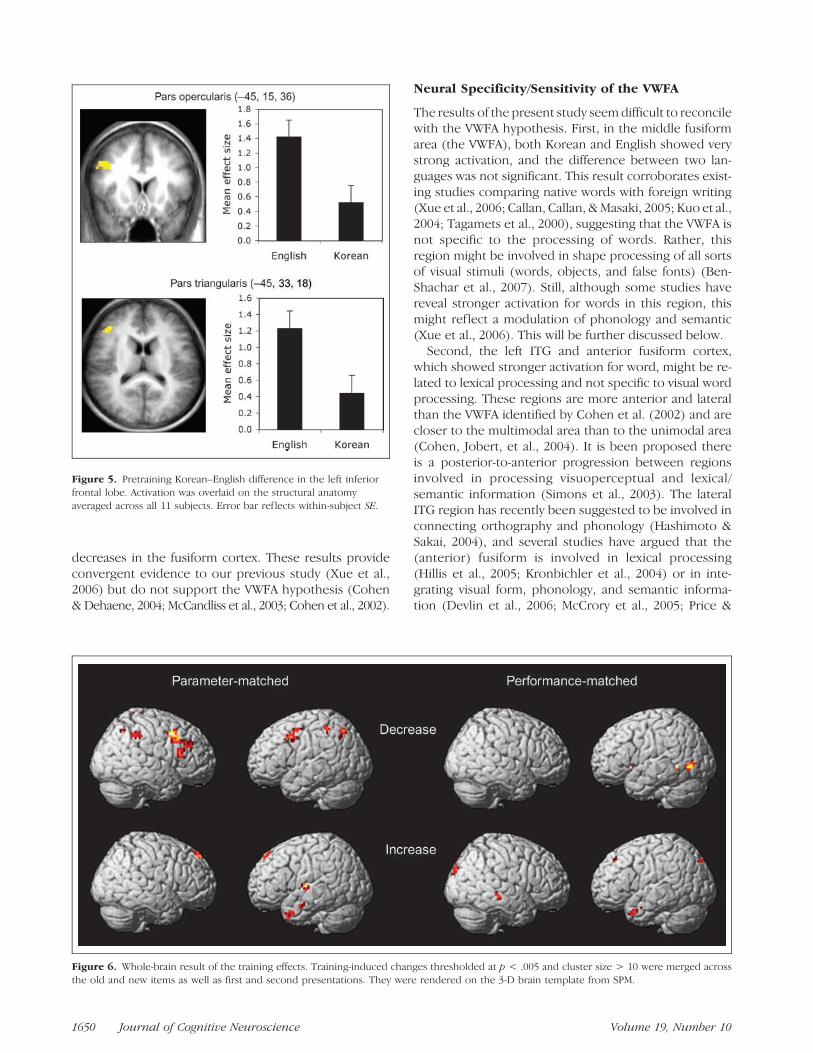
In the frontal lobe, SVC revealed that English elicitedstronger activation in the left pars opercularis and parstriangularis regions. Further ROI analysis confirmed thisresult [pars opercularis: F(1,10) = 8.00, p = .018; parstriangularis: F(1,10) = 6.88, p = .026] (Figure 5).
Training Effect: Whole-brain Analysis
Neural changes with training under parameter- andperformance-matched condition are shown on Figure 6and the foci are listed in Tables 2 and 3, respectively.These data were obtained by merging across old andnew items as well as first and second presentations. Atthe standard threshold, significant decreases were foundfor parameter-matched condition in the fronto-parietalnetwork, possibly because of the decreased task diffi-culty as indicated by the behavioral improvement. Theleft insula showed significant increase. For performance-matched condition, significant increases were found inthe calcarine, cuneus, and lingual gyrus, whereas theleft putamen and cingulate cortex showed significant
Figure 2. Behavioral performance during training. (A) Accuracy in
the parameter setup test is plotted as a function of training session,separated by noise level. (B) Averaged percentage noise used in
each training session. Error bar ref lects within-subject SE.
Xue and Poldrack 1647

decreased activation. At a slightly reduced threshold(i.e., p < .005, cluster size > 10), the superior frontallobe and left anterior inferior temporal lobe extendingto temporal pole showed significant increased for bothconditions, and the left lingual also showed increase inthe parameter-matched condition. Based on our previ-
ous study (Xue et al., 2006), SVC over the anatomicalregion of the occipital and fusiform gyrus were applied.This revealed significant decrease for both conditions inthe bilateral fusiform cortex and left inferior occipitalgyrus. The left frontal lobe showed no increase usingSVC, suggesting the pretraining difference is not becauseof visual expertise but might more likely reflect phonol-ogy and/or semantic processing.
Training Effect: ROI Results
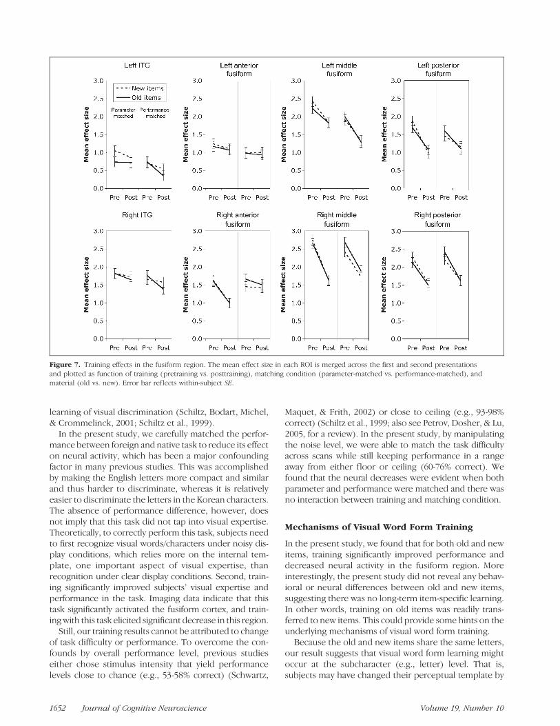
Focusing on each ROI in the fusiform and adjacentITG area, we performed a four-way repeated measuresANOVA, including training (pretraining vs. posttraining),material (old vs. new), matching condition (parametervs. performance), and STRP (first vs. second presenta-tion) as within-subject factors. The result is shown inFigure 7.
We found three regions that showed significant train-ing effects: left middle fusiform region [F(1,10) = 5.18,p = .046], left posterior fusiform region [F(1,10) = 7.30,p = .022], and right middle fusiform region [F(1,10) =7.62, p = .02]. Training effects in the right anteriorfusiform region [F(1,10) = 3.36 p = .097] and rightposterior fusiform region [F(1,10) = 3.20, p = .104]were marginally significant.
Figure 3. Behavioral
performance during fMRI
scans. (A) Accuracy for
Korean characters in theparameter-matched scans.
(B) Mean accuracy for English
and Korean characters
(merged across old and newas well as first and second
presentation). (C) Accuracy
for Korean characters inperformance-matched scans.
Pre = pretraining; Post =
posttraining; First = first
presentation; Second =second presentation. Error
bar ref lects within-subject SE.
Table 1. Foci of Difference between English and Korean atthe Pretraining Scan
Regions x y z Z Value
English > Korean
Left inferior frontal pars triangularisa �45 33 18 2.75
Left inferior frontal pars opercularisa �45 15 36 2.92
Left inferior parietal lobule �45 �48 39 3.59
Left middle temporal gyrus �57 �33 0 3.84
Left ITG �51 �48 �18 3.49
Korean > English
Right postcentral gyrus 45 �12 60 4.23
Right calcarine 6 �84 18 3.83
Left lingual gyrus �18 �69 3 4.90
aSVC over the search volume of left inferior frontal cortex.
1648 Journal of Cognitive Neuroscience Volume 19, Number 10
The effect of task difficulty in training was examinedvia the interaction between training and matching condi-tion (parameter vs. performance match). That is, if taskdifficulty contributed to the training effect, we should ex-pect no differences between parameter- and performance-matched conditions during the pretraining scan (becausethe noise levels were identical) and significant differencesduring the posttraining scan. It turned out that no regionshowed this interaction, suggesting that the observeddecreases in bilateral middle fusiform and left posteriorfusiform were not because of decreased task difficulty.
Similarly, the differences between old and new itemswere examined via the interaction between training andmaterial, because both new and old items were new tosubjects at the pretraining stage. We found no regionwith a significant Training � Material interaction, sug-gesting that the training effect on old items transferredcompletely to new items.
We also examined the STRP effect. Two regions thatshowed significant or marginally significant STRP effectwere the right middle fusiform region [F(1,10) = 12.62,p = .005] and the right posterior fusiform region[F(1,10) = 4.57, p = .058]. The right anterior fusiformregion showed marginally significant Training � STRPinteraction [F(1,10) = 4.82, p = .052]. Further analysis
indicated that STRP only appeared in the posttrainingscans [F(1,10) = 10.90, p = .008]. These results indicat-ed that the neural change in these regions could occuras fast as one repetition, although the absolute neuralchange was not as strong as that of long-term training(averaged activation in the first/second presentationwere 2.24/2.10 in the right middle fusiform region,1.99/1.84 in the right posterior fusiform region, and1.28/1.17 in the right anterior fusiform region duringthe posttraining scans). The STRP effect is not shown inFigure 6, because we merged the activation for the firstand second presentations to make the figure less com-plex and more readable.
DISCUSSION
We examined the neural substrates for processing (En-glish) words and foreign writings (i.e., Korean Hangul)and how neural processing of these stimuli changedwith psychophysical training. At the pretraining scan,both words and foreign characters strongly activated theVWFA, and their difference was not significant, althoughstronger activation for words than for foreign writingswas revealed in the left ITG and left IFC. Visual wordform training with foreign writings caused significant
Figure 4. Pretraining Korean–English comparison in the occipito-temporal region. (A) Location and shape of the ROIs. The ROIs wereoverlaid on the structural anatomy averaged across all 11 subjects. (Middle) Axial view of the ROIs. (Left and Right) sagittal view of the
fusiform ROIs in left and right hemispheres, respectively. (B) Mean effect size in each ROI as a function of language. (Left and right) Results
in the left and right ROIs, respectively. Error bar ref lects within-subject SE. *p < .05, **p < .01; ms = marginally significant.
Xue and Poldrack 1649
decreases in the fusiform cortex. These results provideconvergent evidence to our previous study (Xue et al.,2006) but do not support the VWFA hypothesis (Cohen& Dehaene, 2004; McCandliss et al., 2003; Cohen et al., 2002).
Neural Specificity/Sensitivity of the VWFA
The results of the present study seem difficult to reconcilewith the VWFA hypothesis. First, in the middle fusiformarea (the VWFA), both Korean and English showed verystrong activation, and the difference between two lan-guages was not significant. This result corroborates exist-ing studies comparing native words with foreign writing(Xue et al., 2006; Callan, Callan, & Masaki, 2005; Kuo et al.,2004; Tagamets et al., 2000), suggesting that the VWFA isnot specific to the processing of words. Rather, thisregion might be involved in shape processing of all sortsof visual stimuli (words, objects, and false fonts) (Ben-Shachar et al., 2007). Still, although some studies havereveal stronger activation for words in this region, thismight reflect a modulation of phonology and semantic(Xue et al., 2006). This will be further discussed below.
Second, the left ITG and anterior fusiform cortex,which showed stronger activation for word, might be re-lated to lexical processing and not specific to visual wordprocessing. These regions are more anterior and lateralthan the VWFA identified by Cohen et al. (2002) and arecloser to the multimodal area than to the unimodal area(Cohen, Jobert, et al., 2004). It is been proposed thereis a posterior-to-anterior progression between regionsinvolved in processing visuoperceptual and lexical/semantic information (Simons et al., 2003). The lateralITG region has recently been suggested to be involved inconnecting orthography and phonology (Hashimoto &Sakai, 2004), and several studies have argued that the(anterior) fusiform is involved in lexical processing(Hillis et al., 2005; Kronbichler et al., 2004) or in inte-grating visual form, phonology, and semantic informa-tion (Devlin et al., 2006; McCrory et al., 2005; Price &
Figure 5. Pretraining Korean–English difference in the left inferior
frontal lobe. Activation was overlaid on the structural anatomy
averaged across all 11 subjects. Error bar ref lects within-subject SE.
Figure 6. Whole-brain result of the training effects. Training-induced changes thresholded at p < .005 and cluster size > 10 were merged across
the old and new items as well as first and second presentations. They were rendered on the 3-D brain template from SPM.
1650 Journal of Cognitive Neuroscience Volume 19, Number 10
Friston, 2005). Existing studies show that both objectsnaming/recognition and words processing activate this re-gion (Moore & Price, 1999; Price et al., 2006; also see Price& Devlin, 2003, for a review). This idea is not inconsistentwith the patient study mentioned above, which showedthe posterior fusiform may not be involved in semanticand phonological processing (Gaillard et al., 2006).
Furthermore, along with the difference in the inferiortemporal cortex and anterior fusiform region, there weredifferences in the left IFC. A tight coupling of frontal andfusiform activation has been identified by numerous stud-ies. For example, many studies have revealed strongeractivation in the left inferior frontal lobe and midfusiformarea for pseudowords than for words (see Mechelli,Gorno-Tempini, & Price, 2003, for a review). Repetitionpriming usually elicits significant neural decrease in bothfrontal lobe and fusiform cortex (Buckner, Koutstaal,Schacter, & Rosen, 2000; Wagner, Koutstaal, Maril,Schacter, & Buckner, 2000; Buckner et al., 1998). Neuralsuppression in the fusiform cortex and frontal lobe hasalso been found when the priming stimuli and targetshare no visual similarity, such as priming cross letter case(Dehaene et al., 2001), script (Nakamura, Dehaene,Jobert, Le Bihan, & Kouider, 2005), and language (Chee,Soon, & Lee, 2003). Our previous study found thatphonological training elicited increased activation in bothregions (Xue et al., 2006). These studies are consistent
with the view that semantic and/or phonological process-ing may involve top-down modulation of occipital andtemporal cortices. As a direct evidence of this top-downmodulation, it has been shown that repetition-inducedneural suppression in the fusiform cortex is eliminated bytranscranial magnetic stimulation that disrupted the ac-tivity in the left frontal cortex during repeated classifica-tion of objects (Wig, Grafton, Demos, & Kelley, 2005).
Finally, whereas comparisons between visual word andother object categories argue against the specificity hy-pothesis, results from visual word form training provideevidence against the visual expertise hypothesis. Thepresent study replicates and extends previous results onChinese readers (Xue et al., 2006). We found that trainingsignificantly decreased the activation in the bilateralmiddle fusiform cortex and left posterior fusiform cortex.Similar to the long-term training effect, STRP also causedsignificant decrease in the right middle and posteriorfusiform cortex and in the right anterior fusiform cortexduring the posttraining scan. We have argued that be-cause of the tight connection among visual form, pho-nology and semantic, visual word form training providesthe most direct evidence to test the visual expertisehypothesis (Xue et al., 2006). The two studies using thisparadigm obtained convergent evidence to against thishypothesis. Consistent with our findings, decreased acti-vation in the fusiform has also been found in perceptual
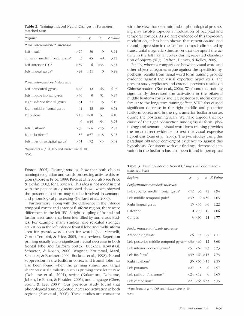
Table 2. Training-induced Neural Changes in Parameter-matched Scan
Regions x y z Z Value
Parameter-matched: increase
Left insula �27 30 9 3.91
Superior medial frontal gyrus* 3 45 48 3.42
Left anterior ITG* �39 6 �33 3.62
Left lingual gyrus* �24 �51 0 3.28
Parameter-matched: decrease
Left precentral gyrus �48 12 45 4.05
Left middle frontal gyrus �30 0 51 3.89
Right inferior frontal gyrus 51 21 15 4.15
Right middle frontal gyrus 42 18 39 3.74
Precuneus �12 �60 51 4.18
0 �45 54 3.75
Left fusiforma �39 �66 �15 2.82
Right fusiforma 36 �57 �18 3.02
Left inferior occipital gyrusa �51 �72 �3 3.34
*Significant at p < .005 and cluster size > 10.aSVC.
Table 3. Training-induced Neural Changes in Performance-matched Scan
Regions x y z Z Value
Performance-matched: increase
Left superior medial frontal gyrus* �12 36 42 2.94
Left middle temporal pole* �39 9 �30 4.03
Right lingual gyrus 15 �36 �6 4.22
Calcarine 0 �75 15 4.86
Cuneus 3 �90 21 4.77
Performance-matched: decrease
Anterior cingulate �6 27 27 4.11
Left posterior middle temporal gyrus* �36 �60 12 3.68
Left inferior occipital gyrusa �51 �69 �3 3.23
Left fusiforma �39 �66 �15 2.73
Right fusiforma 36 �66 �15 2.55
Left putamen �27 15 0 4.57
Left pallidum/thalamus* �24 �12 6 3.65
Left cerebellum* �21 �63 �33 3.35
*Significant at p < .005 and cluster size > 10.aSVC.
Xue and Poldrack 1651
learning of visual discrimination (Schiltz, Bodart, Michel,& Crommelinck, 2001; Schiltz et al., 1999).
In the present study, we carefully matched the perfor-mance between foreign and native task to reduce its effecton neural activity, which has been a major confoundingfactor in many previous studies. This was accomplishedby making the English letters more compact and similarand thus harder to discriminate, whereas it is relativelyeasier to discriminate the letters in the Korean characters.The absence of performance difference, however, doesnot imply that this task did not tap into visual expertise.Theoretically, to correctly perform this task, subjects needto first recognize visual words/characters under noisy dis-play conditions, which relies more on the internal tem-plate, one important aspect of visual expertise, thanrecognition under clear display conditions. Second, train-ing significantly improved subjects’ visual expertise andperformance in the task. Imaging data indicate that thistask significantly activated the fusiform cortex, and train-ing with this task elicited significant decrease in this region.
Still, our training results cannot be attributed to changeof task difficulty or performance. To overcome the con-founds by overall performance level, previous studieseither chose stimulus intensity that yield performancelevels close to chance (e.g., 53-58% correct) (Schwartz,
Maquet, & Frith, 2002) or close to ceiling (e.g., 93-98%correct) (Schiltz et al., 1999; also see Petrov, Dosher, & Lu,2005, for a review). In the present study, by manipulatingthe noise level, we were able to match the task difficultyacross scans while still keeping performance in a rangeaway from either floor or ceiling (60-76% correct). Wefound that the neural decreases were evident when bothparameter and performance were matched and there wasno interaction between training and matching condition.
Mechanisms of Visual Word Form Training
In the present study, we found that for both old and newitems, training significantly improved performance anddecreased neural activity in the fusiform region. Moreinterestingly, the present study did not reveal any behav-ioral or neural differences between old and new items,suggesting there was no long-term item-specific learning.In other words, training on old items was readily trans-ferred to new items. This could provide some hints on theunderlying mechanisms of visual word form training.
Because the old and new items share the same letters,our result suggests that visual word form learning mightoccur at the subcharacter (e.g., letter) level. That is,subjects may have changed their perceptual template by
Figure 7. Training effects in the fusiform region. The mean effect size in each ROI is merged across the first and second presentations
and plotted as function of training (pretraining vs. posttraining), matching condition (parameter-matched vs. performance-matched), and
material (old vs. new). Error bar ref lects within-subject SE.
1652 Journal of Cognitive Neuroscience Volume 19, Number 10
learning specific components of the Korean characters.This pattern might reflect the distinct nature of visualword form learning. As we all know, the countless wordsin one language are usually composed of many fewerbasic writing units (e.g., letters). To learn to recognizethe visual form words, the more efficient way is first tolearn the basic letters. Moreover, because similar words(e.g., only differing by one letter) are abundant in agiven language, subjects have to identify every letter tocorrectly recognize each word. This could prevent thevisual system from forming higher perceptual units. Asa result, there is cumulative evidence indicating thatwhole-word recognition might not be possible undercertain situation. In alphabetic scripts, it has been shownthat even very fluent readers cannot recognize wordsbeyond the level of individual letters (i.e., holistic rec-ognition) (Pelli, Farell, & Moore, 2003). Comparable toletters in alphabetic scripts, the basic functioning unitfor Chinese characters is strokes pattern, which is de-fined as the group of strokes functioning dependently asthe basic orthographic component in different characters(Chen, Allport, & Marshall, 1996). Studies have suggestedthat the basic perceptual unit of Chinese is strokes pat-tern, rather than whole character or single stroke (Chenet al., 1996; Xue, unpublished dissertation).
The absence of item-specific learning may also reflectthe nature of our specific training paradigm. First, wetrained subjects with character pairs that only differed byone letter. In the Korean Hangul language, it is verycommon to generate a number of different charactersby changing just one letter. Moreover, in the presentstudy, the old items only had been trained 12 times ineach session (60 times in total). Third, during training, allstimuli were present very briefly and under very noisyconditions. Finally, to perform the same–different judg-ment task, subjects had to direct their attention to theconsisting letters to point out the subtle differencesbetween two characters. All these factors would make itextremely difficult, if not impossible, to form perceptualtemplate beyond single letters. These differences inexperimental tasks and training methods might accountfor the discrepancies with previous studies, which showthat supraletter orthographic information is acquiredin normal reading development (Cunningham, Perry,Stanovich, & Share, 2002) and plays important role innormal reading (Coltheart, 2004). Further studies need toexamine how the design principles of the particularlanguage and specific training method affect the forma-tion of item-specific learning.
Existing studies suggest different perceptual learningmechanisms under clear and noisy display (Dosher & Lu,2005). For noisy displays, practice improves performanceby learned external noise filtering, that is, the retuning ofthe perceptual template (Lu & Dosher, 2004), whereasfor clear displays, performance can be improved byimproved amplification or enhancement of the stimulusthrough the reduction of internal noise. In high-noise
displays, amplification of stimulus would amplify signaland external noise as well, and the reduction of internalnoise would similarly be of no benefit. This model couldwell explain the results in the present study and ourprevious study. In a previous study (Xue et al., 2006),subjects were trained in clear displays, and performance isimproved by reduction of internal noise. Thus, training ispartially transferred to Chinese task. Under the noisydisplay in the present study, however, internal noise orefficiency cannot be trained, resulting in an absence oftransfer to English task. On the contrary, performance forKorean character is improved by retuning of perceptualtemplate, in our case, the Korean Hangul letters. Consis-tent with our result, a previous study of perceptuallearning of Roman letter identification under noisy con-ditions also found improvement in perceptual templatebut not in internal noise (Chung, Levi, & Tjan, 2005).
Conclusion
We have shown that foreign writing and words equally en-gage the VWFA, and visual word form training reducedVWFA activation. These results replicate our previousfindings and suggest that the VWFA is neither specific towords nor sensitized by visual expertise with specific writ-ing systems. Ample evidence has suggested that reading issupported by a distributed and interconnected neural net-work (Fiez & Petersen, 1998). Given these facts, our re-sults hint that a better understanding of the nature of theVWFA could be achieved both by interpreting the findingsin the whole language network and by breaking downthese connections in artificial language learning situation.
Acknowledgments
This study is supported by a grant from FPR-UCLA Center forCulture, Brain, and Development to G.X. We thank StephenEngel, Zili Liu, and Zhonglin Lu for helpful discussion and twoanonymous reviewers’ valuable comments on an early versionof this manuscript.
Reprint requests should be sent to Gui Xue, Department ofPsychology, University of Southern California, Seely G. Mudd501, Los Angeles, CA 90089-161; or Russell A. Poldrack, Depart-ment of Psychology, University of California, Los Angeles, FranzHall, Box 951563, Los Angeles, CA 90095-1563, or via e-mail:[email protected] or [email protected].
Notes
1. ‘‘Foreign writing’’ in this article is used to refer to an actualwriting system (e.g., Korean Hangul) that subjects are not ableto read. Thus, ‘‘Korean’’ here means a foreign writing but not asecond language. It may be functionally similar to ‘‘false fonts,’’but the latter does not share the same degree of systematicityin their visual features as an actual writing system.2. Hangul script consists of 24 letters (10 vowels and 14 con-sonants), which represent the phonemes of the language.These letters are combined into a compact square-framedsyllable blocks, visually similar to Chinese. The blocks are, thus,called ‘‘characters.’’
Xue and Poldrack 1653
REFERENCES
Andersson, J. L., Hutton, C., Ashburner, J., Turner, R., &Friston, K. (2001). Modeling geometric deformations in EPItime series. Neuroimage, 13, 903–919.
Ben-Shachar, M., Dougherty, R. F., Deutsch, G. K., & Wandell,B. A. (2007). Differential sensitivity to words and shapes inventral occipito-temporal cortex. Cerebral Cortex, 17,1604-1611.
Bolger, D. J., Perfetti, C. A., & Schneider, W. (2005). Cross-cultural effect on the brain revisited: Universal structuresplus writing system variation. Human Brain Mapping, 25,92–104.
Brainard, D. H. (1997). The psychophysics toolbox. SpatialVision, 10, 433–436.
Brett, M., Anton, J.-L., Valabregue, R., & Poline, J.-B. (2002).Region of interest analysis using an SPM toolbox [abstract].Presented at the 8th International Conference on FunctionalMapping of the Human Brain, June 2–6, 2002, Sendai, Japan.Available on CD-ROM in Neuroimage, 16.
Buckner, R. L., Goodman, J., Burock, M., Rotte, M., Koutstaal,W., Schacter, D., et al. (1998). Functional-anatomiccorrelates of object priming in humans revealed by rapidpresentation event-related fMRI. Neuron, 20, 285–296.
Buckner, R. L., Koutstaal, W., Schacter, D. L., & Rosen, B. R.(2000). Functional MRI evidence for a role of frontal andinferior temporal cortex in amodal components of priming.Brain, 123, 620–640.
Callan, A. M., Callan, D. E., & Masaki, S. (2005). Whenmeaningless symbols become letters: Neural activitychange in learning new phonograms. Neuroimage, 28,553–562.
Chee, M. W., Soon, C. S., & Lee, H. L. (2003). Common andsegregated neuronal networks for different languagesrevealed using functional magnetic resonance adaptation.Journal of Cognitive Neuroscience, 15, 85–97.
Chen, Y.-P., Allport, D. A., & Marshall, J. C. (1996). What are thefunctional orthographic units in Chinese word recognition:The stroke or the stroke pattern? Quarterly Journal ofExperimental Psychology: Human ExperimentalPsychology, 49A, 1024–1043.
Chung, S. T., Levi, D. M., & Tjan, B. S. (2005). Learning letteridentification in peripheral vision. Vision Research, 45,1399–1412.
Cohen, L., & Dehaene, S. (2004). Specialization within theventral stream: The case for the visual word form area.Neuroimage, 22, 466–476.
Cohen, L., Dehaene, S., Naccache, L., Lehericy, S., Dehaene-Lambertz, G., Henaff, M. A., et al. (2000). The visual wordform area: Spatial and temporal characterization of an initialstage of reading in normal subjects and posterior split-brainpatients. Brain, 123, 291–307.
Cohen, L., Henry, C., Dehaene, S., Martinaud, O., Lehericy, S.,Lemer, C., et al. (2004). The pathophysiology of letter-by-letter reading. Neuropsychologia, 42, 1768–1780.
Cohen, L., Jobert, A., Le Bihan, D., & Dehaene, S. (2004).Distinct unimodal and multimodal regions for wordprocessing in the left temporal cortex. Neuroimage, 23,1256–1270.
Cohen, L., Lehericy, S., Chochon, F., Lemer, C., Rivaud, S., &Dehaene, S. (2002). Language-specific tuning of visualcortex? Functional properties of the visual word form area.Brain, 125, 1054–1069.
Cohen, L., Martinaud, O., Lemer, C., Lehericy, S., Samson, Y.,Obadia, M., et al. (2003). Visual word recognition in theleft and right hemispheres: Anatomical and functionalcorrelates of peripheral alexias. Cerebral Cortex, 13,1313–1333.
Coltheart, M. (2004). Are there lexicons? Quarterly Journalof Experimental Psychology: A, 57, 1153–1171.
Cunningham, A. E., Perry, K. E., Stanovich, K. E., & Share, D. L.(2002). Orthographic learning during reading: Examiningthe role of self-teaching. Journal of Experimental ChildPsychology, 82, 185–199.
Dale, A. M. (1999). Optimal experimental design for event-related fMRI. Human Brain Mapping, 8, 109–114.
Dehaene, S., Cohen, L., Sigman, M., & Vinckier, F. (2005).The neural code for written words: A proposal. Trends inCognitive Science, 9, 335–341.
Dehaene, S., Naccache, L., Cohen, L., Bihan, D. L., Mangin, J. F.,Poline, J. B., et al. (2001). Cerebral mechanisms of wordmasking and unconscious repetition priming. NatureNeuroscience, 4, 752–758.
Devlin, J. T., Jamison, H. L., Gonnerman, L. M., & Matthews,P. M. (2006). The role of the posterior fusiform gyrusin reading. Journal of Cognitive Neuroscience, 18,911–922.
Dosher, B. A., & Lu, Z. L. (2005). Perceptual learning in cleardisplays optimizes perceptual expertise: Learning thelimiting process. Proceedings of the National Academy ofSciences, U.S.A., 102, 5286–5290.
Fiez, J. A., & Petersen, S. E. (1998). Neuroimaging studies ofword reading. Proceedings of the National Academy ofSciences, U.S.A., 95, 914–921.
Friston, K. J., Ashburner, J., Frith, C. D., Poline, J. B., Heather,J. D., & Frackowiak, R. S. J. (1995). Spatial registrationand normalization of images. Human Brain Mapping, 3,165–189.
Friston, K. J., Holmes, A. P., Worsley, K. J., Poline, J. P., Frith,C. D., & Frackowiak, R. S. J. (1994). Statistical parametricmaps in functional imaging: A general linear approach.Human Brain Mapping, 2, 189–210.
Gaillard, R., Naccache, L., Pinel, P., Clemenceau, S., Volle, E.,Hasboun, D., et al. (2006). Direct intracranial, FMRI, andlesion evidence for the causal role of left inferotemporalcortex in reading. Neuron, 50, 191–204.
Hashimoto, R., & Sakai, K. L. (2004). Learning letters inadulthood: Direct visualization of cortical plasticity forforming a new link between orthography and phonology.Neuron, 42, 311–322.
Henry, C., Gaillard, R., Volle, E., Chiras, J., Ferrieux, S.,Dehaene, S., et al. (2005). Brain activations during letter-by-letter reading: A follow-up study. Neuropsychologia, 43,1983–1989.
Hillis, A. E., Newhart, M., Heidler, J., Barker, P., Herskovits, E.,& Degaonkar, M. (2005). The roles of the ‘‘visual wordform area’’ in reading. Neuroimage, 24, 548–559.
Indefrey, P., Kleinschmidt, A., Merboldt, K. D., Kruger, G.,Brown, C., Hagoort, P., et al. (1997). Equivalent responsesto lexical and nonlexical visual stimuli in occipital cortex:A functional magnetic resonance imaging study.Neuroimage, 5, 78–81.
James, K. H., James, T. W., Jobard, G., Wong, A. C., &Gauthier, I. (2005). Letter processing in the visualsystem: Different activation patterns for single lettersand strings. Cognitive, Affective & Behavioral Neuroscience,5, 452–466.
Kronbichler, M., Hutzler, F., Wimmer, H., Mair, A., Staffen, W.,& Ladurner, G. (2004). The visual word form area andthe frequency with which words are encountered:Evidence from a parametric fMRI study. Neuroimage,21, 946–953.
Kuo, W. J., Yeh, T. C., Lee, J. R., Chen, L. F., Lee, P. L.,Chen, S. S., et al. (2004). Orthographic and phonologicalprocessing of Chinese characters: An fMRI study.Neuroimage, 21, 1721–1731.
1654 Journal of Cognitive Neuroscience Volume 19, Number 10
Leff, A. P., Crewes, H., Plant, G. T., Scott, S. K., Kennard, C.,& Wise, R. J. (2001). The functional anatomy of single-wordreading in patients with hemianopic and pure alexia.Brain, 124, 510–521.
Loftus, G., & Masson, M. (1994). Using confidence intervalsin within-subject designs. Psychonomic Bulletin & Review,1, 476–490.
Lu, Z. L., & Dosher, B. A. (2004). Perceptual learningretunes the perceptual template in foveal orientationidentification. Journal of Vision, 4, 44–56.
McCandliss, B. D., Cohen, L., & Dehaene, S. (2003). Thevisual word form area: Expertise for reading in the fusiformgyrus. Trends in Cognitive Science, 7, 293–299.
McCrory, E. J., Mechelli, A., Frith, U., & Price, C. J. (2005).More than words: A common neural basis for readingand naming deficits in developmental dyslexia? Brain,128, 261–267.
Mechelli, A., Gorno-Tempini, M. L., & Price, C. J. (2003).Neuroimaging studies of word and pseudoword reading:Consistencies, inconsistencies, and limitations. Journal ofCognitive Neuroscience, 15, 260–271.
Moore, C. J., & Price, C. J. (1999). Three distinct ventraloccipitotemporal regions for reading and object naming.Neuroimage, 10, 181–192.
Nakamura, K., Dehaene, S., Jobert, A., Le Bihan, D., &Kouider, S. (2005). Subliminal convergence of Kanji andKana words: Further evidence for functional parcellation ofthe posterior temporal cortex in visual word perception.Journal of Cognitive Neuroscience, 17, 954–968.
Oldfield, R. C. (1971). The assessment and analysis ofhandedness: The Edinburgh Inventory. Neuropsychologia,9, 97–113.
Pelli, D. G. (1997). The VideoToolbox software for visualpsychophysics: Transforming numbers into movies. SpatialVision, 10, 437–442.
Pelli, D. G., Farell, B., & Moore, D. C. (2003). The remarkableinefficiency of word recognition. Nature, 423, 752–756.
Petersen, S. E., Fox, P. T., Snyder, A. Z., & Raichle, M. E.(1990). Activation of extrastriate and frontal cortical areasby visual words and word-like stimuli. Science, 249,1041–1044.
Petrov, A. A., Dosher, B. A., & Lu, Z.-L. (2005). The dynamics ofperceptual learning: An incremental reweighting model.Psychological Review, 112, 715–743.
Poldrack, R. A. (2000). Imaging brain plasticity: Conceptual andmethodological issues—A theoretical review. Neuroimage,12, 1–13.
Poldrack, R. A., Desmond, J. E., Glover, G. H., & Gabrieli, J. D.(1998). The neural basis of visual skill learning: An fMRIstudy of mirror reading. Cerebral Cortex, 8, 1–10.
Poldrack, R. A., & Gabrieli, J. D. (2001). Characterizing theneural mechanisms of skill learning and repetition priming:Evidence from mirror reading. Brain, 124, 67–82.
Poldrack, R. A., Selco, S. L., Field, J. E., & Cohen, N. J. (1999).The relationship between skill learning and repetitionpriming: Experimental and computational analyses. Journalof Experimental Psychology: Learning, Memory, andCognition, 25, 208–235.
Poldrack, R. A., Wagner, A. D., Prull, M. W., Desmond, J. E.,Glover, G. H., & Gabrieli, J. D. (1999). Functionalspecialization for semantic and phonological processing inthe left inferior prefrontal cortex. Neuroimage, 10, 15–35.
Polk, T. A., & Farah, M. J. (2002). Functional MRI evidence foran abstract, not perceptual, word-form area. Journal ofExperimental Psychology: General, 131, 65–72.
Price, C., & Friston, K. (2005). Functional ontologies forcognition: The systematic definition of structure andfunction. Cognitive Neuropsychology, 22, 262–275.
Price, C. J., & Devlin, J. T. (2003). The myth of the visual wordform area. Neuroimage, 19, 473–481.
Price, C. J., McCrory, E., Noppeney, U., Mechelli, A., Moore,C. J., Biggio, N., et al. (2006). How reading differs fromobject naming at the neuronal level. Neuroimage, 29,643–648.
Schiltz, C., Bodart, J. M., Dubois, S., Dejardin, S., Michel, C.,Roucoux, A., et al. (1999). Neuronal mechanisms ofperceptual learning: Changes in human brain activity withtraining in orientation discrimination. Neuroimage, 9,46–62.
Schiltz, C., Bodart, J. M., Michel, C., & Crommelinck, M.(2001). A pet study of human skill learning: Changes inbrain activity related to learning an orientationdiscrimination task. Cortex, 37, 243–265.
Schwartz, S., Maquet, P., & Frith, C. (2002). Neural correlatesof perceptual learning: A functional MRI study of visualtexture discrimination. Proceedings of the NationalAcademy of Sciences, U.S.A., 99, 17137–17142.
Simons, J. S., Koutstaal, W., Prince, S., Wagner, A. D., &Schacter, D. L. (2003). Neural mechanisms of visualobject priming: Evidence for perceptual and semanticdistinctions in fusiform cortex. Neuroimage, 19,613–626.
Tagamets, M. A., Novick, J. M., Chalmers, M. L., & Friedman,R. B. (2000). A parametric approach to orthographicprocessing in the brain: An fMRI study. Journal ofCognitive Neuroscience, 12, 281–297.
Tzourio-Mazoyer, N., Landeau, B., Papathanassiou, D., Crivello,F., Etard, O., Delcroix, N., et al. (2002). Automatedanatomical labeling of activations in SPM using amacroscopic anatomical parcellation of the MNI MRIsingle-subject brain. Neuroimage, 15, 273–289.
Wagner, A. D., Koutstaal, W., Maril, A., Schacter, D. L., &Buckner, R. L. (2000). Task-specific repetition priming in leftinferior prefrontal cortex. Cerebral Cortex, 10, 1176–1184.
Wig, G. S., Grafton, S. T., Demos, K. E., & Kelley, W. M.(2005). Reductions in neural activity underlie behavioralcomponents of repetition priming. Nature Neuroscience,8, 1228–1233.
Wong, A. C., Gauthier, I., Woroch, B., DeBuse, C., & Curran, T.(2005). An early electrophysiological response associatedwith expertise in letter perception. Cognitive, Affective &Behavioral Neuroscience, 5, 306–318.
Xue, G. (2004). Cognitive and neural mechanisms of visualword recognition and the role of language experiences: Anartificial language training study. Dissertation. BeijingNormal University, Beijing China.
Xue, G., Chen, C., Jin, Z., & Dong, Q. (2006). Languageexperience shapes fusiform activation when processing alogographic artificial language: An fMRI training study.Neuroimage, 31, 1315–1326.
Xue, G., Dong, Q., Chen, K., Jin, Z., Chen, C., Zeng, Y., et al.(2005). Cerebral asymmetry in children when readingChinese characters. Cognitive Brain Research, 24,206–214.
Xue and Poldrack 1655
Related Documents