The Muscular System Specialized tissue that enable the body and its parts to move.

The muscular-system-powerpoint-1227697713114530-8
Aug 15, 2015
Welcome message from author
This document is posted to help you gain knowledge. Please leave a comment to let me know what you think about it! Share it to your friends and learn new things together.
Transcript
The Muscular System
Specialized tissue that enable the body and its parts to move.
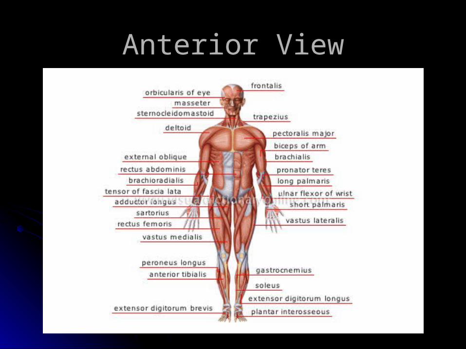
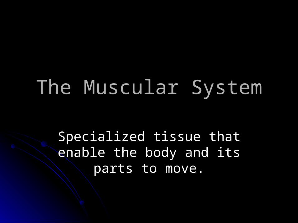
Anterior View
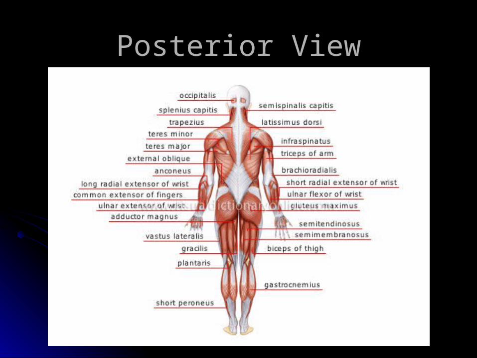
Posterior View
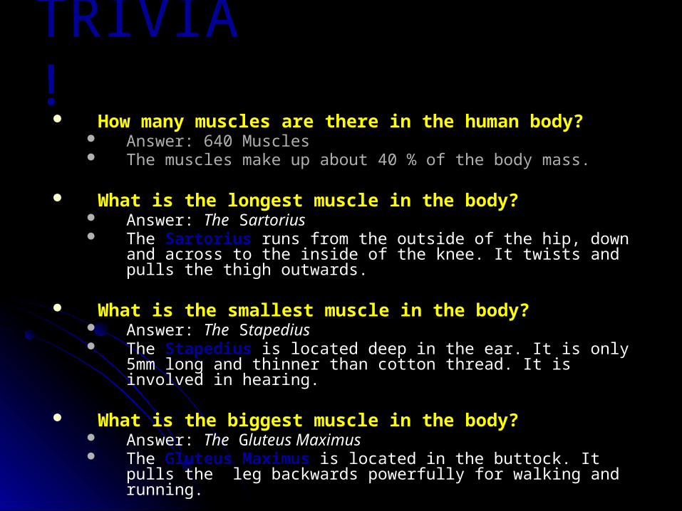
TRIVIA! How many muscles are there in the human body?
Answer: 640 Muscles The muscles make up about 40 % of the body mass.
What is the longest muscle in the body? Answer: The Sartorius The Sartorius runs from the outside of the hip, down and across to the
inside of the knee. It twists and pulls the thigh outwards.
What is the smallest muscle in the body? Answer: The Stapedius The Stapedius is located deep in the ear. It is only 5mm long and thinner
than cotton thread. It is involved in hearing.
What is the biggest muscle in the body? Answer: The Gluteus Maximus The Gluteus Maximus is located in the buttock. It pulls the leg
backwards powerfully for walking and running.
Functions of the Muscles
MovementMaintenance of posture and muscle
toneHeat productionProtects the bones and internal
organs.
Muscle Classification
FunctionallyVoluntarily – can be moved at willInvoluntarily – can’t be moved
intentionally
StructurallyStriated – have stripes across the fiberSmooth – no striations
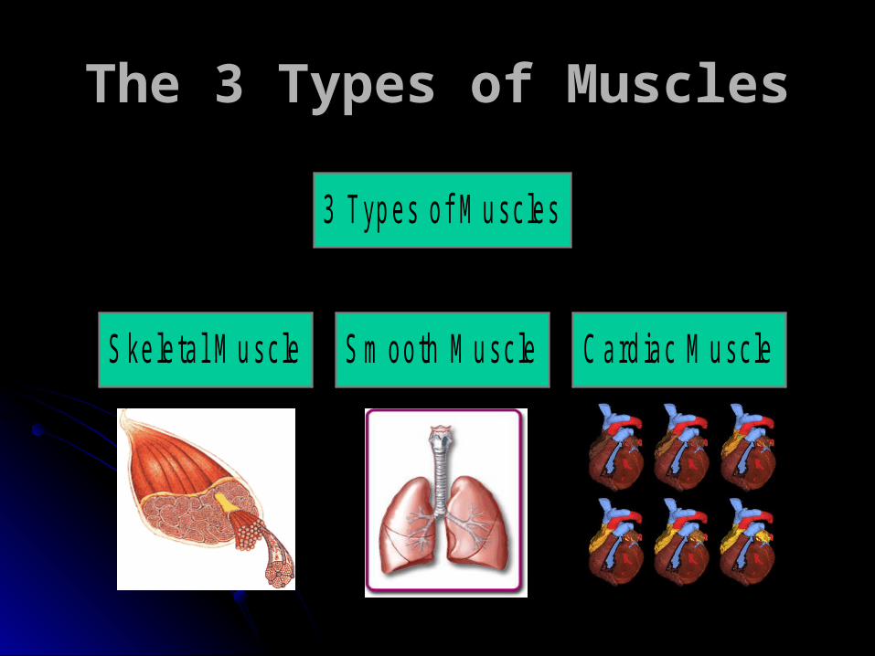
The 3 Types of Muscles
S ke le ta l M u sc le S m ooth M u sc le C ard iac M u sc le
3 Typ es o f M u sc les
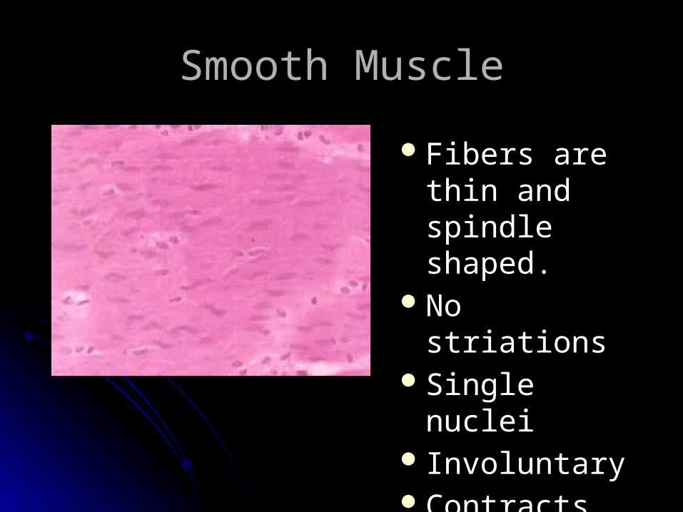
Smooth Muscle
Fibers are thin and spindle shaped.
No striationsSingle nuclei InvoluntaryContracts slowly

Smooth Muscle They fatigue… but very slowly Found in the circulatory system
Lining of the blood vessels Helps in the circulation of the blood
Found in the digestive system Esophagus, stomach, intestine Controls digestion
Found in the respiratory system Controls breathing
Found in the urinary system Urinary bladder Controls urination
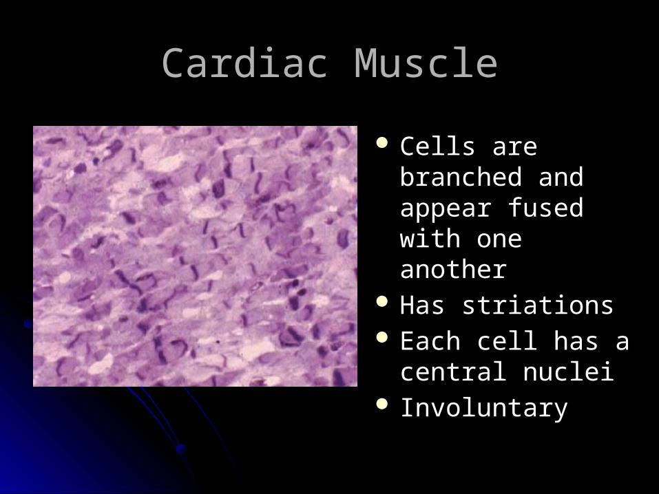
Cardiac Muscle
Cells are branched and appear fused with one another
Has striations Each cell has a
central nuclei Involuntary
Cardiac Muscle
Found ONLY in the heartContractions of the heart muscles
pump blood throughout the body and account for the heartbeat.
Healthy cardiac muscle NEVER fatigues or else…
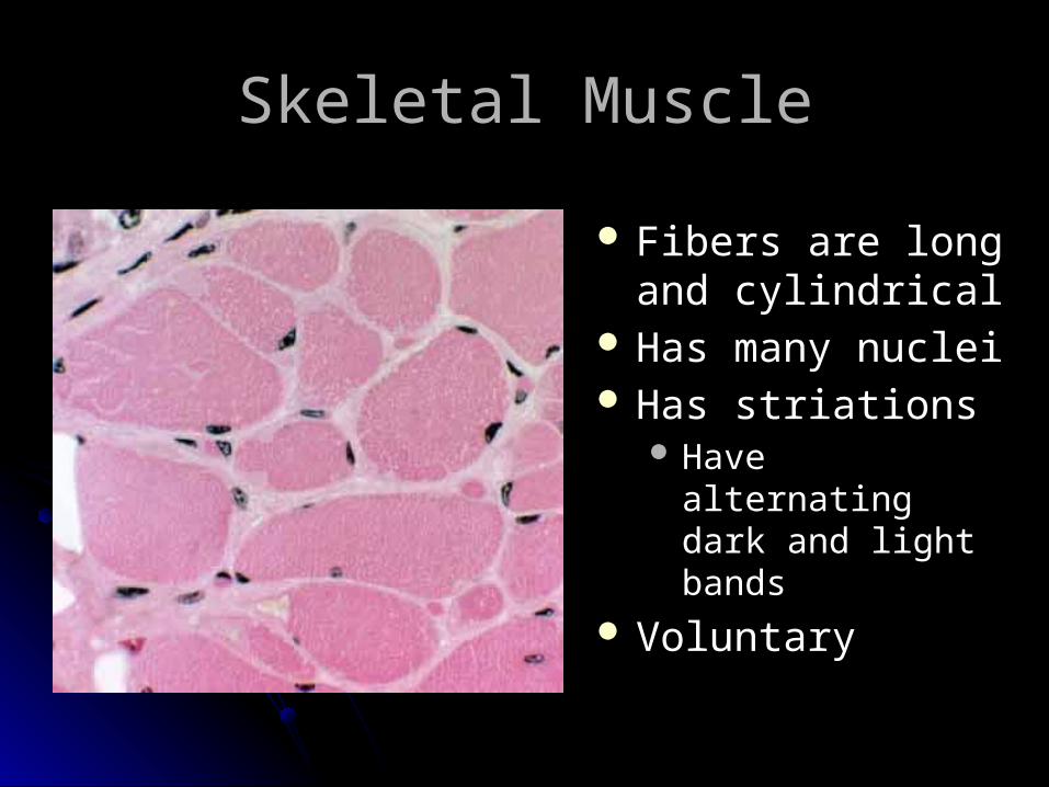
Skeletal Muscle
Fibers are long and cylindrical
Has many nuclei Has striations
Have alternating dark and light bands
Voluntary

Skeletal Muscle
Attached to skeleton by tendonsCauses movement of bones at the
joints.And yes… they do fatigue Muscle fatigue activity what
substance forms causing muscle fatigue???
Functions of Skeletal Muscle
Movement – muscle move bones by pulling not pushing. Synergists – any movement is generally
accomplished by more than one muscle. All of the muscles responsible for the movement are synergists.
The one that is most responsible for the movement is the Prime Mover (agonist).
Functions of Skeletal Muscle
Movement Antagonists – muscles and muscle groups
usually work in pairs
– example the biceps flex your arm and its partner the triceps extend your arm. The two muscles are antagonists, i.e. cause opposite actions.
– when one contracts the other relaxes. Levators – muscle that raise a body part.

Functions of Skeletal Muscle
Maintenance of posture or muscle tone We are able to maintain our body position because
of tonic contractions in our skeletal muscles. These contractions don’t produce movement yet hold our muscles in position.
Heat production – contraction of muscles produces most of the heat required to maintain body temperature.

Structure of Skeletal Muscle
Composed of striated muscle cells (=muscle fibers) and connective tissue. Most muscles attach to 2 bones that have a
moveable joint between them. The attachment to the bone that does not move is the
origin. The attachment to the bone that moves is the insertion.
Tendons anchor muscle firmly to bones. Tendons are made of dense fibrous connective tissue.
Ligaments connect bone to bone at a joint.
Structure of Skeletal Muscle
Bursae – small fluid filled sacs that lie between some tendons and the bones beneath them. They are made of connective tissue and are lined with synovial membrane that secretes synovial fluid.

Structure of Skeletal Muscle
Contribution of the nervous system Electrochemical impulses travel from the
frontal lobes of the cerebrum via motor nerves to the muscle fibers and cause them to contract.
Sensation is a function of the brain – impulses are integrated in the parietal lobes of the cerebrum (conscious muscle sense) and in the cerebellum (unconscious). These activities promote coordination.

Structure of Skeletal Muscle
Microscopic anatomy Muscle cells (fibers) are grouped in a highly
organized way in the muscle. The membrane that surrounds the muscle cell is called the sarcolemma.
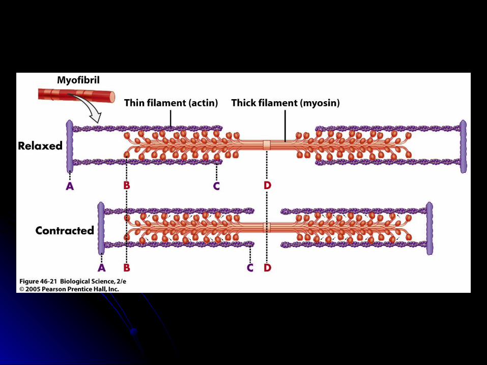
Muscle cells are filled with 2 types of fine threadlike proteins called myofilaments: myosin (thick) and actin (thin). These structures slide past each other causing the muscle cell to contract or shorten.
The myofilaments are arranged in the cells in small units called sarcomeres.

Structure of Skeletal Muscle
Neuromuscular junction Spot where the axon of a motor nerve nears
the muscle fiber. The axon terminal does not touch the muscle
but comes close. The space between the axon and the muscle cell is called the synapse.
Within the terminal end of the axon are small sacs filled with a neurotransmitter called acetylcholine.

Muscle Contraction
Sequence Electrical impulse travels down a motor
neuron. When it reaches the end, acetylcholine (chemical) is released into the synapse.
Acetylcholine bind to special receptors on the muscle cell and causes an electrical impulse to spread over the cell.
The sarcomeres shorten and the muscle cell contracts.

MUSCLE
MUSCLE FIBER
MYOFIBRIL
SARCOMERE

Sarcomere
Z ZZ
Z ZZ
I
AA

Movement of Muscles
Origin: the attachment of the muscle to the bone that remains stationary
Insertion: the attachment of the muscle to the bone that moves
Belly: the fleshy part of the muscle between the tendons of origin and/or insertion
origin
insertion
belly

Movement of skeletal muscle
These muscles move when the brain sends messages to the muscle
Always work in pairs2 movements of skeletal muscle
Contraction (shorten)Extension (lengthen)

Categories of skeletal muscle actions
Categories Actions
Extensor Increases the angle at a joint Flexor Decreases the angle at a joint Abductor Moves limb away from midline of
body Adductor Moves limb toward midline of body Levator Moves insertion upward Depressor Moves insertion downward Rotator Rotates a bone along its axis Sphincter Constricts an opening


Practice these Movements
1. Bend arm- biceps contract- triceps extend
2. Straighten arm- biceps extend - triceps contract
3. Bend knee- quadriceps extend- hamstrings contract

4. Straighten knee - quadriceps contract - hamstrings extend
5. Crunches- abdomen contract- back muscles
extend6. Point toes
- calf muscle contract
- shin muscle extend
More Movements

Naming Skeletal Muscles
Location of the muscleShape of the muscleRelative Size of the muscleDirection/Orientation of the muscle
fibers/cellsNumber of OriginsLocation of the AttachmentsAction of the muscle

Muscles Named by Location
Epicranius
(around cranium)
Tibialis anterior (front of tibia)
tibialis anterior
Naming Skeletal Muscles
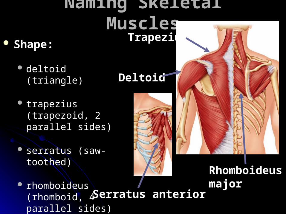
Shape:
deltoid (triangle)
trapezius (trapezoid, 2 parallel sides)
serratus (saw-toothed)
rhomboideus (rhomboid, 4 parallel sides)
orbicularis and sphincters (circular)
Rhomboideusmajor
Trapezius
Deltoid
Serratus anterior

maximus (largest)minimis (smallest) longus (longest)brevis (short)major (large)minor (small)
Psoas major
Psoas minor
Muscles Named by Size
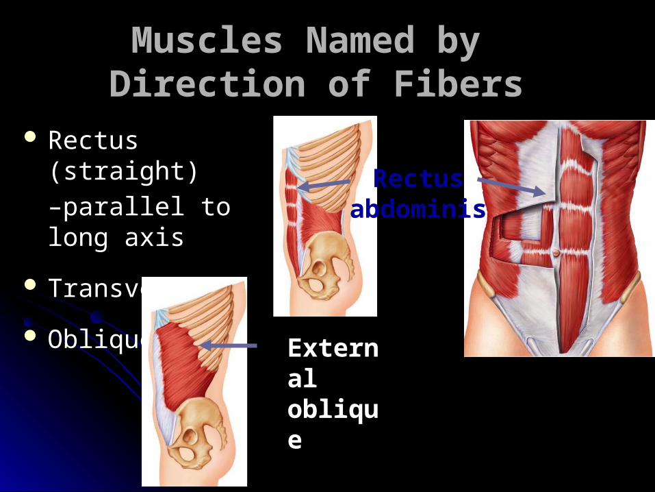
Muscles Named by Direction of Fibers
Rectus (straight)
–parallel to long axis
Transverse
Oblique
Rectusabdominis
External oblique

Biceps (2)
Triceps (3)
Quadriceps (4)
Muscles Named for Number of Origins
Biceps brachii
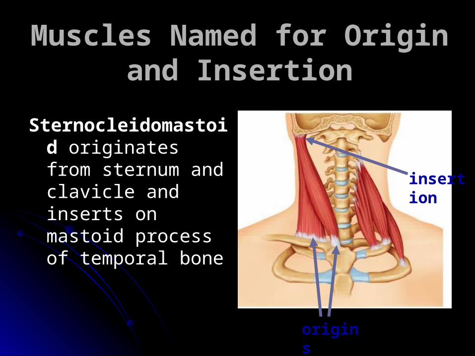
Muscles Named for Origin and Insertion
Sternocleidomastoid originates from sternum and clavicle and inserts on mastoid process of temporal bone
origins
insertion

Muscles Named for Action
Flexor carpi radialis (extensor carpi radialis) – flexes wrist
Abductor pollicis brevis (adductor pollicis)– flexes thumb
Abductor magnus – abducts thigh
Extensor digitorum – extends fingers
Adductormagnus

Arrangement of Fascicles
Parallel strap-like ex: sartorius
Fusiform spindle shaped ex: biceps femoris

Arrangement of Fascicles
Pennate "feather shaped”
Unipennate ex: extensor
digitorum longus
Bipennate ex: rectus femoris
Multipennate ex: deltoid
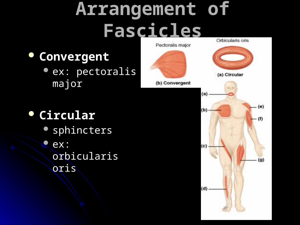
Arrangement of Fascicles
Convergent ex: pectoralis
major
Circular sphincters ex: orbicularis oris

There are about 60 muscles in the face.
Smiling is easier than frowning.
It takes 20 muscles to smile and over 40 to frown.
Smile and make someone happy.
Related Documents