(CANCER RESEARCH 58. 3401-3408, August I, I998| The Murine F hit Locus: Isolation, Characterization, and Expression in Normal and Tumor Cells1 Yuri Pekarsky,2 Teresa Druck,2 Maria Grazia Cotticelli, Masataka Ohta,3 Jiang Shou, Jeannine Menarola, Jeffry C. Montgomery, Arthur M. Buchberg, Linda D. Siracusa, Giacomo Manenti, Louise Y. Y. Fong, Tommaso A. Dragani, Carlo M. Croce, and Kay Huebner4 Kimmel Cancer Institute. Jefferson Medical College. Philadelphia. Pennsylvania 19107 [Y. P.. T. D.. M. G. C, M. O.. J. S.. J. M.. J. C. M.. A. M. B.. L D. S.. L Y. Y. F.. C. M. C.. K. H.¡;and Istituti! Na-ionale Tumori. Milan. Italy 20133 ¡C.M.. T. A. D.¡ ABSTRACT The murine Fhit locus maps near the centromere v proximal Ptprg locus on mouse chromosome 14. The cDNA sequence and structure are similar to those of the human gene, with exons 5-9 encoding the protein. The predominant mRNA in the tissues and cell lines tested was an alternatively spliced form missing exon 3. Most murine cell lines tested, including lines established from normal mouse embryos and tumors, expressed very low or undetectable levels of Fhit mRNA. Most normal mouse tissues ex pressed wild-type l-'liit mRNA, whereas —¿40% of murine lung carcinomas expressed wild-type and aberrant Fhit RT-PCR products that lacked various exons. Several tumorigenic mouse cell lines exhibited homozygous deletions of Fhit exons. We conclude that the murine I lui gene, like its human counterpart, is a target of alterations involved in murine carcino- genesis. INTRODUCTION The human FHIT gene at chromosome region 3pl4.2, encompass ing the most active constitutive chromosomal fragile site, is a candi date tumor suppressor gene for the most common forms of human cancer (1). Intragenic rearrangements within the FHIT locus, often resulting in independent deletions within both alÃ-eles(2, 3), have been observed in numerous cancer-derived cell lines and tumors derived from head and neck (4), esophagus (5), lung (6), stomach, colon (1), uterine cervix (7), and other organs. The FHIT locus is > 1 Mb in size, whereas the mRNA is 1.1 kb, encoded by 10 small exons, with exons 5-9 encoding a protein of 147 amino acids (M, 16,800). Although the gene is specifically involved in loss of heterozygosity in many, if not most human cancers, point mutations in the gene are rare (2, 8). Like the pl(>/CDKN2 locus, alterations occur more frequently through rearrangement within both alÃ-eles;unlike the p\6/CDKN2 locus, which is included in a single cosmid, the FHIT gene is very large. Even when both alÃ-elessustain a deletion, the deletions need not overlap, making such deletions very difficult to detect in primary tumors. We and others have, thus, used RT5 of tumor RNA followed by PCR amplification of nearly full- length products to assess FHIT expression in cancer cells. A large fraction of several tumor types exhibited aberrant-sized RT-PCR products that correlated with DNA and/or Fhit protein alterations (2). Some investigators have reported amplification of aberrant-sized products even from normal cell RNA (9, 10). Additionally, in many Received 3/19/98: accepted 5/26/98. The costs of publication of this article were defrayed in part by the payment of page charges. This article must therefore be hereby marked advertisement in accordance with 18 U.S.C. Section 1734 solely to indicate this fact. 1 Supported by United States Public Health Service Grants CA21124. CA39860. and CA56336 from the National Cancer Institute and a gift from R. R. M. Carpenter III and M. K. Carpenter. 2 The first two authors contributed equally to this work. ' Present address: Banyu Tsukuba Research Inslitute. Okubo 3. Tsukuba. 300-33 Japan. 4 To whom requests for reprints should be addressed, at Kimmel Cancer Institute. Thomas Jefferson University. Room 1008 BLSB, 233 South 10th Street. Philadelphia. PA 19107. Phone: (215)503-4656; Fax: (215)923-4498; E-mail: [email protected]. 5 The abbreviations used are: RT. reverse transcription; RACE, rapid amplification of cDNA ends; YAC. yeast artificial chromosome; MCA, methylcholanthrene. cancer cell lines and primary tumors, both normal and aberrant am plification products have been observed. Because the alterations to the FHIT locus in many cancer cells appeared not to fit the classic tumor suppressor paradigm, a number of investigators have suggested that the FHIT gene may not be a tumor suppressor gene. Alternative explanations for the very high frequency of alterations within the FHIT locus are: (a) the position of the FRA3B fragile region within FHIT leads to frequent deletion within the region, which sometimes accidentally includes FHIT exons; or (b) there is another suppressor gene(s) within the region that is the real target of the frequent loss of heterozygosity and deletion. Replacement and reexpression of Fhit in cancer cell lines lacking endogenous Fhit eliminated tumorigenicity of the lung, stomach, and kidney cancer cell lines tested, a result con sistent with a role for Fhit as a tumor suppressor (11). Otterson et al. (12) have replaced Fhit in HeLa cells and have observed growth of a transfected clone as a tumor; however, immunohistochemistry of the HeLa tumor appears to show loss of Fhit expression in most of the tumor section. Most recently, immunohistochemical analysis of hu man cervical, lung, and kidney tumors using anti-Fhit antisera has shown that most of the tumors either do not express Fhit or express reduced levels of Fhit, whereas the normal epithelial counterpart cells strongly stain for Fhit (7, 13, 14). The fragile region, FRA3B, is highly susceptible to carcinogen damage (15, 16), which may be the case for other common fragile regions as well. A long-standing hypothesis (17) suggests that genes altered by fragile site rearrangement may be involved in cancer development and progression. The FHIT gene within FRA3B is such an altered gene. It is, thus, important, as suggested in a commentary summarizing recent studies of the human FHIT locus (18), to continue examination of its bonafides as a tumor suppressor and its function in normal and tumor cells. We have begun a study of the murine Fhit gene and its organization and expression in normal and tumor cells, with a view toward use of the murine model system to study the interactions among carcinogens, Fhit alterations, and tumor development. MATERIALS AND METHODS cDNA and Genomic Library Screening and Sequencing. One million plaques each of the mouse kidney cDNA library (Clontech, Palo Alto, CA) and mouse genomic library (bacteriophage library from strain SVJ129; Stratagene, La Jolla, CA) were screened with a human FHIT cDNA probe ( 1). Clones were purified, and DNA was isolated using standard procedures (19). Sequencing was performed using Perkin-Elmer thermal cyclers and an Applied Biosystems model 377 automated DNA sequencer. Murine Fhit sequences compiled dur ing this study have been deposited in the GenBank database (accession nos. AF047699-AF047702). Interspecific Backcross Mapping. Progeny from a (AEJ/Gn-a bp"/a bp" X Mus spretus)F, X AEJ/Gn-a bp"/a bp" backcross (20) were typed by Southern blot analysis of genomic DNA with a mouse Fhit cDNA probe representing exons 4-10. This probe detected common Oral fragments of 3.0. 1.9, and 0.9 kb in DNA from AEJ and M. spretus mice and unique fragments of 8.6, 7.0, and 2.8 kb in AEJ DNA and of 6.1 and 3.3 kb in M. spretus DNA. The cosegregating 6.1- and 3.3-kb M. spretus fragments were followed in N2 offspring of the backcross. Mapping of the Ptprg. Plau, and Psp-rxl (formerly 3401 on July 9, 2015. © 1998 American Association for Cancer Research. cancerres.aacrjournals.org Downloaded from

Welcome message from author
This document is posted to help you gain knowledge. Please leave a comment to let me know what you think about it! Share it to your friends and learn new things together.
Transcript

(CANCER RESEARCH 58. 3401-3408, August I, I998|
The Murine F hit Locus: Isolation, Characterization, and Expression in Normal andTumor Cells1
Yuri Pekarsky,2 Teresa Druck,2 Maria Grazia Cotticelli, Masataka Ohta,3 Jiang Shou, Jeannine Menarola,
Jeffry C. Montgomery, Arthur M. Buchberg, Linda D. Siracusa, Giacomo Manenti, Louise Y. Y. Fong,Tommaso A. Dragani, Carlo M. Croce, and Kay Huebner4
Kimmel Cancer Institute. Jefferson Medical College. Philadelphia. Pennsylvania 19107 [Y. P.. T. D.. M. G. C, M. O.. J. S.. J. M.. J. C. M.. A. M. B.. L D. S.. L Y. Y. F.. C. M. C..K. H.¡;and Istituti! Na-ionale Tumori. Milan. Italy 20133 ¡C.M.. T. A. D.¡
ABSTRACT
The murine Fhit locus maps near the centromere v proximal Ptprg locuson mouse chromosome 14. The cDNA sequence and structure are similarto those of the human gene, with exons 5-9 encoding the protein. The
predominant mRNA in the tissues and cell lines tested was an alternativelyspliced form missing exon 3. Most murine cell lines tested, including linesestablished from normal mouse embryos and tumors, expressed very lowor undetectable levels of Fhit mRNA. Most normal mouse tissues expressed wild-type l-'liit mRNA, whereas —¿�40%of murine lung carcinomas
expressed wild-type and aberrant Fhit RT-PCR products that lacked
various exons. Several tumorigenic mouse cell lines exhibited homozygousdeletions of Fhit exons. We conclude that the murine I lui gene, like itshuman counterpart, is a target of alterations involved in murine carcino-
genesis.
INTRODUCTION
The human FHIT gene at chromosome region 3pl4.2, encompassing the most active constitutive chromosomal fragile site, is a candidate tumor suppressor gene for the most common forms of humancancer (1). Intragenic rearrangements within the FHIT locus, oftenresulting in independent deletions within both alÃeles(2, 3), have beenobserved in numerous cancer-derived cell lines and tumors derived
from head and neck (4), esophagus (5), lung (6), stomach, colon (1),uterine cervix (7), and other organs.
The FHIT locus is > 1 Mb in size, whereas the mRNA is 1.1 kb,encoded by 10 small exons, with exons 5-9 encoding a protein of 147
amino acids (M, 16,800). Although the gene is specifically involved inloss of heterozygosity in many, if not most human cancers, pointmutations in the gene are rare (2, 8). Like the pl(>/CDKN2 locus,alterations occur more frequently through rearrangement within bothalÃeles;unlike the p\6/CDKN2 locus, which is included in a singlecosmid, the FHIT gene is very large. Even when both alÃelessustaina deletion, the deletions need not overlap, making such deletions verydifficult to detect in primary tumors. We and others have, thus, usedRT5 of tumor RNA followed by PCR amplification of nearly full-
length products to assess FHIT expression in cancer cells. A largefraction of several tumor types exhibited aberrant-sized RT-PCR
products that correlated with DNA and/or Fhit protein alterations (2).Some investigators have reported amplification of aberrant-sized
products even from normal cell RNA (9, 10). Additionally, in many
Received 3/19/98: accepted 5/26/98.The costs of publication of this article were defrayed in part by the payment of page
charges. This article must therefore be hereby marked advertisement in accordance with18 U.S.C. Section 1734 solely to indicate this fact.
1Supported by United States Public Health Service Grants CA21124. CA39860. and
CA56336 from the National Cancer Institute and a gift from R. R. M. Carpenter III andM. K. Carpenter.
2 The first two authors contributed equally to this work.' Present address: Banyu Tsukuba Research Inslitute. Okubo 3. Tsukuba. 300-33
Japan.4 To whom requests for reprints should be addressed, at Kimmel Cancer Institute.
Thomas Jefferson University. Room 1008 BLSB, 233 South 10th Street. Philadelphia. PA19107. Phone: (215)503-4656; Fax: (215)923-4498; E-mail: [email protected].
5 The abbreviations used are: RT. reverse transcription; RACE, rapid amplification of
cDNA ends; YAC. yeast artificial chromosome; MCA, methylcholanthrene.
cancer cell lines and primary tumors, both normal and aberrant amplification products have been observed. Because the alterations to theFHIT locus in many cancer cells appeared not to fit the classic tumorsuppressor paradigm, a number of investigators have suggested thatthe FHIT gene may not be a tumor suppressor gene. Alternativeexplanations for the very high frequency of alterations within theFHIT locus are: (a) the position of the FRA3B fragile region withinFHIT leads to frequent deletion within the region, which sometimesaccidentally includes FHIT exons; or (b) there is another suppressorgene(s) within the region that is the real target of the frequent loss ofheterozygosity and deletion. Replacement and reexpression of Fhit incancer cell lines lacking endogenous Fhit eliminated tumorigenicity ofthe lung, stomach, and kidney cancer cell lines tested, a result consistent with a role for Fhit as a tumor suppressor (11). Otterson et al.(12) have replaced Fhit in HeLa cells and have observed growth of atransfected clone as a tumor; however, immunohistochemistry of theHeLa tumor appears to show loss of Fhit expression in most of thetumor section. Most recently, immunohistochemical analysis of human cervical, lung, and kidney tumors using anti-Fhit antisera has
shown that most of the tumors either do not express Fhit or expressreduced levels of Fhit, whereas the normal epithelial counterpart cellsstrongly stain for Fhit (7, 13, 14). The fragile region, FRA3B, is highlysusceptible to carcinogen damage (15, 16), which may be the case forother common fragile regions as well. A long-standing hypothesis
(17) suggests that genes altered by fragile site rearrangement may beinvolved in cancer development and progression. The FHIT genewithin FRA3B is such an altered gene. It is, thus, important, assuggested in a commentary summarizing recent studies of the humanFHIT locus (18), to continue examination of its bonafides as a tumorsuppressor and its function in normal and tumor cells. We have beguna study of the murine Fhit gene and its organization and expression innormal and tumor cells, with a view toward use of the murine modelsystem to study the interactions among carcinogens, Fhit alterations,and tumor development.
MATERIALS AND METHODS
cDNA and Genomic Library Screening and Sequencing. One millionplaques each of the mouse kidney cDNA library (Clontech, Palo Alto, CA) andmouse genomic library (bacteriophage library from strain SVJ129; Stratagene,
La Jolla, CA) were screened with a human FHIT cDNA probe ( 1). Clones werepurified, and DNA was isolated using standard procedures (19). Sequencingwas performed using Perkin-Elmer thermal cyclers and an Applied Biosystems
model 377 automated DNA sequencer. Murine Fhit sequences compiled during this study have been deposited in the GenBank database (accession nos.AF047699-AF047702).
Interspecific Backcross Mapping. Progeny from a (AEJ/Gn-a bp"/abp" X Mus spretus)F, X AEJ/Gn-a bp"/a bp" backcross (20) were typed by
Southern blot analysis of genomic DNA with a mouse Fhit cDNA proberepresenting exons 4-10. This probe detected common Oral fragments of 3.0.
1.9, and 0.9 kb in DNA from AEJ and M. spretus mice and unique fragmentsof 8.6, 7.0, and 2.8 kb in AEJ DNA and of 6.1 and 3.3 kb in M. spretus DNA.The cosegregating 6.1- and 3.3-kb M. spretus fragments were followed in N2offspring of the backcross. Mapping of the Ptprg. Plau, and Psp-rxl (formerly
3401
on July 9, 2015. © 1998 American Association for Cancer Research. cancerres.aacrjournals.org Downloaded from

THE MURINE FHIT LOCUS
Psp-2) loci in this cross, using data generated from a subset of the mice used
in this work, has been described previously (21). One hundred fifteen micewere typed at all four loci and were used for haplotype analysis; distancesbetween adjacent markers were calculated from recombination frequenciesdetermined from a larger number of mice typed at both loci. Linkage data weregenerated and analyzed with the computer program Spretus Madness: PartDeux (developed by Karl Smalley. Jim Averbach. Linda D. Siracusa, andArthur M. Buchberg, Jefferson Medical College. Philadelphia. PA).
DNA and RNA Analysis by Southern and Northern Blot Hybridization.A mouse multiple tissue Northern blot (Clontech) was hybridized with a mouseFhit cDNA probe using the supplier's protocol and washed at 60°Cin 0.1%
SDS and 0.1X SSC. For cell lines, total RNA was isolated from 1-5 X IO8
cells using TRIZOL reagent (Life Technologies, Inc., Gaithersburg, MD).Poly(A)+ RNA was isolated from 0.2-1.0 mg of total RNA using the Mini-Oligo (dT) Cellulose Spin Column Kit (5 Prime-3 Prime). Three /ng of
poly(A)+ RNA from each cell line were electrophoresed in 0.8% agarose gelin a borate buffer containing formaldehyde and transferred to HybondN +(Amersham, Arlington Heights, IL). For RT-PCR analysis of mouse lungtissue and tumors, total RNAs were extracted using Ultraspec-II RNA Kit
(Biotecx. Houston, TX) according to previously described methods (22, 23).Restriction enzyme digestions, transfers to membrane, and hybridizations wereperformed as described previously (21).
PCR and RT-PCR Amplification. PCR amplification of Fhit exons from
cellular DNA templates was performed essentially as described (2) usingmurine Fhit specific primers. Nested RT-PCR amplification of Fhit from
murine total RNAs was performed as described for the human gene (2) or bythe following modified method. Two hundred ng of poly(A)+ RNA or 3 /ngof total RNA were treated with DNasel (amplification grade; Life Technologies, Inc.) following the manufacturer's protocol. DNase-treated RNA was
used in RT reactions as follows: 10 nM each dNTP. 100 pmol of randomhexamers [oligo(dT) priming was used in some cases], DNase I-treated RNA,
and 200 units of MuLV reverse transcriptase (Life Technologies, Inc.), in atotal volume of 20 n\, were incubated at 42°Cfor 1 h; then 10 ¿tgof RNaseA were added, followed by incubation at 37°Cfor 30 min. One jil of the
reaction or 100 ng of genomic DNA were used for each PCR. PCR primers andtheir locations are shown in Table 1. PCRs were carried out under standardconditions using 10 pmol of each gene-specific primer and 25-35 cycles of95°Cfor 30 s, 55-60°C for 30 s, and 72°Cfor 1 min. In some cases, nested
PCR was performed using 0.1 /¿Iof the original PCR. Products were separatedon 1.5% agarose gels and sometimes isolated and sequenced or cloned andsequenced.
F9 and ES RT-PCR products were ligated into the pCR2.1 vector (Invitro-
gen, San Diego, CA) using the procedure recommended in the TA Cloning Kit,and l /J.1 of the ligation reaction transformed into DH5a-competent cells.
Several colonies were selected, and plasmid DNA was isolated (Qiagen) forsequencing.
Cell Lines. Murine cell lines were purchased from American Type CultureCollection (WEHI164, CRL1412, LMTK~, L929, and A9) or obtained from
other investigators at the Kimmel Cancer Institute (ES, NP3, RENCA, CUD,MEL, IT22, BW5147, and F9). The murine melanoma cell lines K1735cllO.K1735cl4, and K1735M2X21 were obtained from Dr. Sen Pathak (M. D.Anderson Cancer Center; 24) and lines MEI and ME2 were established in ourlaboratory from mouse embryos, by standard tissue culture procedures, and
were passaged for 20 subcultures during these studies. Cell lines were maintained in MEM or RPMI supplemented with 10% fetal bovine serum. CellularDNA was prepared from proteinase K-treated cell lysates by standard phenol/
chloroform extraction and ethanol precipitation.RACE. Oligo(dT)-primed double-stranded cDNA was synthesized using
mouse kidney mRNA (Clontech) and procedures and reagents from the Marathon RACE cDNA amplification kit (Clontech); the cDNA was ligated toMarathon adapters (Clontech). 3' and 5' RACE products were generated by
long-range PCR using gene-specific primers and the API primer (Clontech).
To increase the specificity of the procedure, the second PCR was carried outusing nested gene-specific primers and the AP2 primer (Clontech). PCRs were
performed according to the Marathon protocol using the Expand long templatePCR system (Boehringer Mannheim) and 30 cycles of 94°Cfor 30 s, 60°Cfor30 s, and 68°Cfor 4 min. RACE products were electrophoresed, identified by
hybridization, and sequenced.YAC Clone Isolation and Characterization. DNA pools from the WI/
MIT 820 strain C57BU6J YAC library were purchased from Research Genetics (Huntsville, AL). Pools were screened by PCR with the exon 1 primersaccording to the supplier's protocol. YAC DNAs were isolated in solution, and
agarose plugs were prepared as described (25). Pulsed-field gel electrophoresiswas carried out using the CHEF MAPPER pulsed-field electrophoresis system(Bio-Rad, Richmond, CA) with a pulse time of 60 s for 15 h, followed by 90 s
for 9 h. DNA from gels was transferred onto a nylon membrane and hybridizedwith mouse Fhit cDNA under standard conditions.
Immunohistochemical Methods. Routine deparaffinization from xylenethrough a graded alcohol series and rehydration, prior to microwave antigenrecovery, were carried out on a Leica Autostainer (Leica, Inc., Deerfield, IL).The deparaffinization process included a 30-min hydrogen peroxide block for
endogenous peroxidase activity. The antigen recovery step was carried out inan 800-W microwave oven in 200 ml of ChemMate H.I.E.R. Buffer, pH5.5-5.7 (Ventana Medical Systems, Tucson, AZ).
The slides were cooled and washed in distilled H20 and placed in ChemMate Buffer 1 (PBS containing carrier protein and sodium azide). The immu-
nostaining was performed on a Techmate 1000 (Ventana Medical Systems,Tucson, AZ) using capillary gap technology. Tissue sections were exposed for10 min to 10% normal goat serum, followed by overnight primary antibodyincubation ( 1:600). The primary antibody was a commercially prepared poly-
clonal chicken immunoglobulin raised against purified human Fhit protein.The next day, slides were washed, incubated for 30 min with a biotinylatedgoat antichicken IgY secondary antibody ( 1:200), and exposed to avidin-biotincomplex for 45 min. Following a wash, slides were subjected to a 10-minapplication of 3,3'-diaminobenzidine. Slides were also lightly counterstained
with hematoxylin and coverslipped using Permount (Fisher) mounting mediumprior to evaluation.
To assess specificity of staining, —¿�1.5/ig/ml purified gstFhit protein was
added during the overnight incubation of the tissue with antiserum.Western Blot Analysis. Mouse cell lines and tissues were lysed, and total
protein was prepared as described previously (2). Approximately 75 fig of totalprotein were electrophoresed on a 14% SDS-polyacrylamide gel. The mem
brane was prepared and blocked as described previously (2) before incubationfor —¿�17h at 4°Cwith chicken anti-Fhit (1:2000). After three 10-min washes,
in TEST buffer, the blot was incubated for 1 h with rabbit antichicken IgGheavy and light chain horseradish peroxidase-labeled antibody (Pierce, Rock-
Table 1 Oligonucleotide primers for PCR ampliation
Numbers in primer names refer to the cDNA nucleotide sequence number for the first nucleotide of the primer; primers in capital letters are within exons. and those in lowercaseletters are in introns. Primer pairs spanning several exons were used in amplification from reverse transcripts; primer pairs within or flanking individual exons were used in detectionof homozygous deletion of exons in cellular DNAs.
PrimerpairMF105F/MF826RMF127F/MF806RMF68F/MF843RMF106F/MF792RMFIF/MF77RMF170F/MF261Rmfiex5F/Rmfiex6F/Rmfiex7F/MF623Rmfiex8FMF696RSequence(5' ->3')TTTGAAGCCCAGCAAAGAAGGGA/GCCTTGGGAATCGTTTGAGTTACAATCCACTGTGAAGAGTCAGGAA/TACTCTCAGGCCTGAAAGTAGACCTTGCCTTTCATTCCAGC
/GGGGTCCATTTTCTTATTGAAGCCCAGCAAAGAAG/AAAGTAGACCCGCAGAGCGTCAGTTCCCAGGTCCAC
/ATCTTGGTGGCTGGAATGTACTTTGGAATACCTCCTCACAG/CTTCAGATTAGTCCAACCTATCAGGAGGGACAATTAAAAGGGAC/ATTCCTTGCTTACCTTTTGGCCCTCAAAGTAAACCAACAA/AAATGCAGACCACAAAAACCGGCCTGCTGGATAATTCATA/ACAGTCTGCCCAGCTTCACACTGTCAAGTC
AAAATATAG/ CTCATCATAGATGTTGTCATTLocationExon
2/10Exon2/10Exon1/10Exon2/9Exon1Exon3Intron
4/intron5Intron5/intron6Intron6/exon7Intron7/exon 8Product
size(bp)7226807766877792361456260135
3402
on July 9, 2015. © 1998 American Association for Cancer Research. cancerres.aacrjournals.org Downloaded from

THE MURINE FHIT LOCUS
ford, IL) diluted 1:40,000. Signal was detected using the Super Signal chemi-
luminescent substrate (Pierce).Mouse Lung Tumors. Male and female A/J, C3H/He, and BALB/cJ mice
were purchased from The Jackson Laboratory (Bar Harbor, ME) and thencrossed in our laboratory (26). Lung tumors were induced by a single i.p.injection of 300 mg of urethane dissolved in water per kg body weight (27).The tumors were excised from female mice and ranged from 4 to 15 mm indiameter. Normal lungs were excised from untreated adult (A/J x C3H/He)F,female mice.
RESULTS
Fhit cDNA Sequence and Genomic Structure. The mouse kidney cDNA library was screened using the human FHIT cDNA; aftertertiary screening, the few positive cDNA clones were amplified,
sequenced, and compared to human FHIT cDNA sequence and to thepublic databases. The first cDNA clone was found to represent exons4-10. The 5' sequence was determined by performing 5' RACE on a
reverse transcript of mouse kidney RNA. Primer pairs from differentregions of the murine Fhit cDNA were used to amplify differentoverlapping fragments of the cDNA reverse-transcribed from RNAderived from a number of tissues and cell lines. PCR-amplified
fragments were also sequenced so that each portion of Fhit cDNA wassequenced numerous times to assemble the full sequence shown inFig. Iß.Oligonucleotide primer pairs used in amplification of Fhitfragments are listed in Table 1.
Primer pairs within exon 1 were used to screen the mouse YAClibrary, and several YAC clones were identified. YAC clone 158E2appeared to be part of the WC14.2 contig in the Whitehead/MIT
158E2(-1350kb)
•¿�138F12(-1125kb)
•¿�384C6(~1070kb)
1 GTCAGTTCCCAGGTCCACCCCAGTGGGGACACGATCAGGCGGCACTCCTGTCCTGTGCTC 601V2«
61 CCGCCCTCTTGCCTTTCATTCCAGCCACCAAGATCTTGGCAGCTTTGAAGCCCAGCAAAG 1202\/3
121 AAGGGAAATCCACTGAGAAGAGTCAGGAAAGGCACATGAATGCTCAGCCTACTTTGGAAT 180*** M L S L L W N
181 ACCTCCCCACAGTGCAACCAAATGCAAGACGTTGGTGCCTTCAAGATAGTATTCTACCTG 240TSPQCNQMQDVGAFKIVFYL
3\/4241 ATAGGTTGGACTAATCTGAAGGTGCCCTGGGACAACCTGCTCGGTCACACATCTGCAAGA 300
IGWTNLKVPWDNLLGHTSAR4\/5
301 ACTCTCCATCTTGCACTGAGGACCCCAAGGAGCTTTTACTGTGAGACCATGTCATTTAGA 360TLHLALRTPRSFYCET M S F R 4
361 TTTGGCCAACATCTCATCAAGCCCTCTGTGGTTTTTCTCAAAACTGAACTGTCCTTCGCC 420FGOHLI KPSVVFLKTELSFA 24
5\/6421 CTGGTGAATAGGAAACCCGTTGTACCTGGCCATGTCCTCGTGTGCCCGCTGAGGCCAGTA 480
LVNRKPVVPGHVLVCPLRPV 44
481 GAGCGCTTCCGTGACCTACATCCTGATGAAGTGGCCGATTTGTTTCAAGTGACCCAGAGA 540ERFRDLHPDEVADLFOVTOR 64
H R T 6\/7541 GTTGGGACAGTGGTGGAGAAGCATTTCCAGGGGACCTCCATCACCTTCTCCATGCAAGAT 600
VGTVVEKHFOGTSITFSMOD 84H
7\/8601 GGTCCTGAAGCTGGGCAGACTGTGAAGCATGTACATGTCCACGTTCTTCCCAGGAAGGCA 660
GPEAGOTVKHVHVHVLPRKA 1048\/9
661 GGGGACTTCCCCCGGAATGACAACATCTATGATGAGCTCCAGAAACATGACAGAGAAGAA 720GDFPRNDNIYDELOKHDREE 124
H S E K721 GAGGACTCGCCAGCCTTTTGGAGATCTGAGAAGGAGATGGCTGCAGAGGCGGAGGCTCTG 780
EDSPAFWRSEKEMAAEAEAL 144F S 9V10S A
781 CGGGTCTACTTTCAGGCCTGAGAGTAACTCAAACGATTCCCAAGGCATAAGAAAATGGAC 840R V Y F O A *** 150
841 CCCATTCTTCTGGAGTCTTCGACATTGGAAATGAAGCTAACCACTCTTTATTGTACCCCA 900901 CGACCCCACAGCCTTGTACAAAGAACTTTATTGCATGTGTGGAAGCCACTAGTATTATGC 960961 TTCCATCACGATGACAAATAGGCTAGCTTCCCAAACAGTGGCATTGACAGCCTGCCACCG 1020
1021 TTCCTCAGTTCTGTACTTAGAATANTAACATTTCCAAACTGTCCCTGAACCTAGATGAAA 10801081 ATAAAGTTGGTTGCT
Fig. l. Genomic structure and cDNA sequence of the mouse Fhit gene. A, YAC contig covering the gene. The mouse Fhit gene is 1.2-2 Mb in size. YAC names and sizes areindicated. »,provisional exon positions. B, mouse Fhit cDNA. Exon boundaries are shown above the sequence. $, provisional exon boundaries based only on homology belween humanand mouse cDNAs. Underlined letters, amino acid sequence encoded by the major transcript (that lacks exon 3). Amino acids that differ in human Fhit protein sequence are shownbelow the murine amino acid sequence. Boldface letters, amino acids that are absent in human protein.
3403
on July 9, 2015. © 1998 American Association for Cancer Research. cancerres.aacrjournals.org Downloaded from

THE MURINE FHIT LOCUS
Fhit,Ptprg M D D •¿�D •¿�
Plan M D •¿�D D •¿�Psp-rsl •¿�D •¿�D •¿�D
48 54 0 157
14
Flau
Psp-rs1
10q21.1-q21.2. 3p24.3-p24.2, XY. _ _ _ _3pJ4.2_J_3pl4.2_ _ _ _.0.8 I0q24, 13q14-q22, 10q22.1-q23, 21
8p23.1-p22 -- Edili
10.83p21.3-p21.2, 3p21-p14
10cen-q24
3p21.2-p14.110q22-q23
10q11.214q22-q23, 10q22-q23, 10q22-q23
10q11.210q11.2, 10q23.3, 3
13,
Anx7. Rarb. Il3ra. Tal
Pou4f 1. Vçl,Vdac2
-DnahcS
-Cacnald. Dnahd, WntSa
-Adk. Tncc'.HIM
.SfÎËâßmpr^ Q,jf-¡Q^ pibps
Bmp4. Sftel, Sftp4, Sftpd15_ywfc
•¿�Chat.Oud, Pkcd
~Apex.Psp-rs1~Qa3, Qb2, NQ, NrÃ,Jera. Tçrd14q11.2-q12
, 14q11.2, 2Q14q11.2, 14q11.2 '
Fig. 2. Chromosomal location of mouse Fhit. A. haplotype analysis of 115 N2 offspring from the interspecific backcross. Left, mapped loci. Each column represents the chromosomeinherited from the (AEJ/Gn X M. spretus}Â¥{parent. •¿�AEJ/Gn alÃeles;D, M. spreius alÃeles.Numbers, number of progeny carrying that particular haplotype. B, genetic linkage mapshowing the position of Fhil in the mouse genome. Left, chromosome showing the loci typed in the interspecific backcross with intergenic distances given in cM. Right, chromosomethat is a partial representation of the chromosome 14 committee report consensus map (accessible on-line via the Mouse Genome Database at www.informatics.jax.org). Distances fromthe centromere are given in cM. , locus used to align the maps. Underlined loci, loci that have been mapped in the human genome; their gene locations on human chromosomesare shown between the maps.
database. YACs 138F12 and 384C6 from this contig appeared tocover the entire Fhit locus (Fig. \A), as determined by PCR amplification and Southern hybridization. Sizes of the YACs were determined by pulsed-field gel electrophoresis of DNA from each of the
YACs; the assembled contig (Fig. IA) suggests that the mouse Fhitlocus is, like the human, 1-2 Mb in size. DNA from the YACs was
also digested with enzymes that do not cleave within Fhit, andSouthern blot hybridizations were performed to determine the presence and size of individual Fhit exons within the YACs.
The murine Fhit protein is 91% identical to human and 93%similar; the amino acids that differ are indicated in Fig. Iß.This figurealso shows that upstream of the exon 5 Met codon, which is theputative start codon for the Fhit protein (Fig. Iß,underlined letters),there is a conceptual open reading frame extending into exon 3. Thisstart codon would rarely be used, however, because the main murineFhit transcript is an alternative transcript with exon 2 spliced to exon4, eliminating exon 3 and its Met codon (see below). The ~ll-kbexon 5 genomic fragment is 38% GC, the ~15-kb exon 6 genomicclone is 38.7% GC, and the ~2-kb exon 8 genomic fragment is 38.1%
GC, suggesting that the mouse Fhit locus, like the human (3), mayhave an overall high AT content.
The Fhit cDNA probe was used in linkage analysis to determine the
chromosomal location of the murine gene. Interspecific backcrossanalysis of 115 N2 mice showed that the Fhit locus cosegregated withseveral previously mapped loci from the centromere proximal end ofchromosome 14. The distribution of haplotypes shown in Fig. 2indicates the following gene order: centromere-Pfprg-F/¡/r-(0.8 ±0.8cM)-P/iZM-(10.8 ±2.8 cM)-/V-rj/-telomere. No recombination be
tween the Fhit and Ptprg loci in 187 N2 animals suggests tightlinkage, with a maximum distance between the two genes of 1.6 cM(95% confidence).
The cDNA probe was also used in selection of Fhit genomic clonesfrom the 129/SvJ library; clones for exons 5, 6, and 8 were obtained,and intron-exon boundaries were sequenced, as described previously(2, 28). A ~8-kb PCR product containing intron 6 was amplified from
mouse YACs 138F12 and 384C6, purified, and sequenced. Other exonboundaries were determined by sequencing of alternative transcripts(see below). Primer pairs flanking exons 5, 6, 7, and 8 were thendesigned (see Table 1) to test DNAs from murine cell lines forpresence of these exons. Primer pairs within exons 1 and 3 (Table 1)were designed and used in similar experiments. Results are summarized in Table 2, and examples of the homozygous deletions detectedare shown in Fig. 3, which illustrates the absence of exon 5 in theBALB/c derived renal carcinoma cell line RENCA (see Fig. 3A, Lane
3404
on July 9, 2015. © 1998 American Association for Cancer Research. cancerres.aacrjournals.org Downloaded from
THE MURINE FHIT LOCUS
Table 2 The Fhit locus in murine cell lines
CelllineES
F9MEIME2IT22NP3
MELBW5I47WEHI164CRL1412RENCA
K1735cllOK1735M2X21Origin
FhitexonsEmbryonicstem +°
Teratocarcinoma +Mouse embryo +Mouse embryo +3T3 TK~ clone+Myeloma
+FeLv erythroleukemia - exons 6, 7, Õ
Thymoma +MCA-treated adult+connective
tissueMCA-induced+fibrosarcomaC3H101I/2
MCA-treated+Kidneycarcinoma - exon 5
Melanoma+Metastaticclone +Fhit
RT-PCRWild
type*
Wild typeWild typeWildtype+
exons5-10——_-Northern
blot+c-t-
short''——_-
" Presence of exons I. 3, 5, 6, 7, and 8 was detected in genomic DNA from cell lines
by PCR amplifications using intron primers flanking exons 5, 6, 7. and 8 or primers withinexons 1 and 3. +, ail six exons were present.
Wild-type RT-PCR product consisted of three species in mouse tissues and cell lines,
as described in Fig. 3 legend.1 MEI and ME2 showed a very faint signal of expected size on Northerns.
LMTK , a clone of L cells, was previously shown to be missing exons 3-5 of thePtpy gene, as were all L-derived lines tested {21 ).
' LMTK~ Fhit mRNA was abundant but shorter than the expected size (see Fig. 2) and
had an aberrant 5' untranslated region.
J), and the absence of exon 6 in the Feline leukemia virus-induced
murine erythroleukemia cell line, MEL (Fig. 3A, Lane 12). The MELcells also lack exons 7 and 8 (data not shown). Other cell line DNAsexhibited presence of each of the exons tested, as summarized inTable 2. Exons 2, 4, and 9 are too small to detect using primer pairswithin them. Some cellular DNAs were also tested for integrity of theFhit locus by Southern blot analysis, and alterations in additional cellswere not detected. Restriction fragment length polymorphisms wereobserved for exon 3 (Xbal, 3.7 or 3.3 kb) and exon 8 (BamHl. 18 or6.7 kb; Xbal, 5.8 or 2.6 kb). Fig. 3ßillustrates the Xbal polymorphismof exon 8, as well as the absence of exons 6 and 7 in MEL cells (Fig.3fi, Lane /;, note that absence of exon 8 is obscured by overlap of theexon 4 fragment with the position of exon 8 alÃeleai). Homozygousdeletion of exon 5 in RENCA cells is also confirmed by the Southernblot results (Fig. 3B, Lane 2).
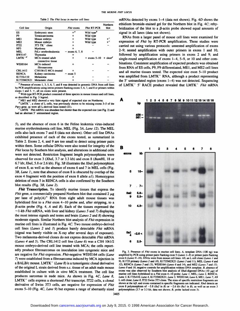
Fhit Transcription. To identify murine tissues that express theFhit gene, a commercially prepared Northern blot that contained 2 ^.gper lane of poly(A)+ RNA from eight adult mouse tissues was
hybridized first to a Fhit exon 4-10 probe and, after stripping, to aß-actinprobe (Fig. 4, A and B). Each of the tissues expressed the~l-kb Fhit mRNA, with liver and kidney (Lanes 5 and 7) displaying
the most intense signals and testes and brain (Lanes 2 and 8) showingmoderate signals. Similar Northern blot analysis of Fhit expression inmurine cell lines is illustrated in Fig. 4C. Two mouse embryo-derived
cell lines (Lanes 2 and 3) produce barely detectable Fhit mRNA(signal was barely visible on X-ray after several days of exposure).Two melanoma-derived clones do not express detectable Fhit mRNA
(Lanes 4 and 5). The CRL1412 cell line (Lane 6) was a C3H 10tl/2mouse embryo-derived cell line treated with MCA; the cells report
edly produce fibrosarcomas on inoculation into syngeneic mice andare negative for Fhit expression. FWÃ-negativeWEHI164 cells (Lane
7) were established from a fibrosarcoma induced by MCA injection ina BALB/c mouse. LMTK" (Lane 8) is a tumorigenic clonal derivative
of the original L strain derived from s.c. tissue of an adult male mouseestablished in culture with in vitro MCA treatment. The cell lineproduces sarcomas in nude mice. As shown in Fig. 4C, Lane 8,LMTK" cells express a shortened Fhit transcript. IT22 cells, a clonal
derivative of Swiss 3T3 cells, are negative for expression of Fhitexons 5-10 (Fig. 4C, Lane 9) but express a range of aberrantly sized
mRNAs detected by exons 1-4 (data not shown). Fig. 4D shows theethidium bromide-stained gel for the Northern blot in Fig. 4C; rehy-bridization of the blot to a ß-actinprobe showed equal amounts of
signal in all lanes (data not shown).RNAs from a larger panel of mouse cell lines were examined for
expression of Fhit by RT-PCR amplification. These studies were
carried out using various protocols: unnested amplification of exons2-9; nested amplification with outer primers in exons 1 and 10,
followed by amplification using primers in exons 2 and 9; andsingle-round amplification of exons 1-4, 5-9, or 10 and other com
binations. Consistent amplification of expected products was obtainedfrom RNA of ES cells, F9, F9 differentiated, MEI, and ME2 cell linesand all murine tissues tested. The expected size exon 5-10 productwas amplified from LMTK" RNA, although a product representing
the 5' untranslated region (exons 1-4) was not detected. Sequencingof LMTK" 5' RACE product revealed that LMTK" Fhit mRNA
1 2 3 4 5 6 7 8 M 9 1011 1213141516
Baxons
8a29
10
kb
15-
. 7-
5.8-5.3-
4.3-
8a14
-2.6 <
2.3-
~ m <•
»*•¿�
-9-3..IFig. 3. Presence of Fhit exons in murine cell lines. A. template DNA (100 ng) was
amplified by PCR using primer pairs Hanking exon 5 (Lanes 1-8} or primer pairs flankingexon 6 (Lanes 9-16). DNAs were from mouse cell lines: A9. an L cell clone (Lanes 1 and
9), KI735 primary (Lanes 2 and IO), K1735M2X21 (Lanes 3 and 11), MEL (Lanes 4 and12). RENCA (Lanes 5 and 13). WEHI164 (Limes 6 and 14), and ME2 (Lanes 7 and IS).Lanes 8 and 16. negative controls for amplification without DNA template, a. absence ofexons was also observed by Southern blot analysis of Mai-digested DNAs (10 /ig) ofmurine cell lines hybridized to a Fhit exon 4-10 probe: Lane I. MEL; Lane 2. RENCA:Lane 3. KI735cllO: Lane 4. K1735M2X2I; Lane 5. WEHI164; Lane 6. MEt; Lane 7, A9L cell clone; Lane 8, IT22 Swiss 3T3 clone. The sizes of specific restriction fragments areshown at the left, and exons contained in specific fragments arc indicated. Xha\ detects anexon 8 polymorphism of —¿�5.8(8a2 in B) or —¿�2.6kb (8al in ß),as well as an exon 3
polymorphism (3.7 or 3.3 kb, data not shown; B. probe lacked exon 3).
3405
on July 9, 2015. © 1998 American Association for Cancer Research. cancerres.aacrjournals.org Downloaded from
THE MURINE FHIT LOCUS
Fig. 4. Expression of Fhit mRNA in tissues andcell lines. Two jj.g of mRNA from adult murinetissues or from murine cell lines were electrophore-sed, transferred to membrane, and hybridized toradiolabeled cDNA probes. A, mRNA from heart(Lane /), brain (Lane 2), spleen (Lane 3), lung(Lane 4), liver (Lane 5), muscle (Lane 6), kidney(Lane 7), and testes (Lane 8) hybridized to radio-labeled Fhir exon 5-10 probe. B, the membraneshown in A was stripped and hybridized to radio-labeled j3-actin cDNA. C, mRNA from mouse
brain (Lane I), cell lines MEI (Lane 2), ME2 (Lane3), K1735M2X21 (Lane 4), KI735cl 10 (Lane 5),CRL1412 (Lane 6), WEHI164 (Lane 7), LMTKT(Lane 8), and IT22 (Lane 9) was hybridized toradiolabeled Fhit exon 5-10 probe. D, ethidiumbromide-stained gel photographed before transfer
of mRNAs from the gel, illustrating quality andquantity of mRNA; the RNAs shown in C weretransferred from this gel.
kb* 122.4-
1.35-
345678 C1 23456789
1.35-
B
2.4-
1.35-
lacks exons 1-4 and exhibits an unknown 100-bp sequence 5' of exon
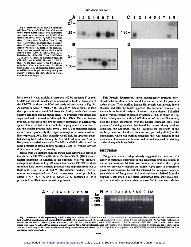
5 (data not shown). Results are summarized in Table 2. Examples ofthe RT-PCR products amplified and analyzed are shown in Fig. 5A.
As shown in Lanes 4 (MEI), 8 (ME2), and 9 (mouse brain), at leastthree products were amplified from the freshly established mouseembryo cell lines and the mouse brain. The products were cloned andsequenced and compared to full-length Fhit cDNA. The most intenseproduct, at just above the 500-bp marker, represents an alternatively
spliced transcript that lacks exon 3; the larger product retains exon 3,and the smaller product lacks exons 3 and 4. The transcript lackingexon 3 was reproducibly the major transcript in all tissues and celllines expressing Fhit. This transcript would lack the upstream potential starting Met codon, leaving the first Met codon in exon 5 as thelikely major translation start site. The MEI and ME2 cells showed thesame products at tissue culture passages 2 and 20 without obviousdifferences in quality or quantity.
RNAs from 30 urethane-induced mouse lung tumors also served astemplates for RT-PCR amplification. Twelve of the 30 (40%) showedshorter fragments, in addition to the expected wild-type products;examples are shown in Fig. 5ß.Lanes 1-9 contain RT-PCR products
from nine lung tumors; aberrant products were amplified from four ofthese nine (Lanes 5, 7, 8, and 9). The aberrant products from alltumors were sequenced and found to represent transcripts lackingexons 4-7, 4-8, 4-6, or 5-8. Lanes 10-12 contained RT-PCR
products from RNA from normal lung tissues.
Fhit Protein Expression. Three independently prepared poly-clonal rabbit anti-Fhit sera did not detect murine or rat Fhit protein in
rodent tissues. Thus, purified human Fhit protein was injected into achicken, and after the fourth injection, the antiserum was used inimmunohistochemical analysis of several mouse tissues. Epithelialcells of various tissues expressed cytoplasmic Fhit, as shown in Fig.6A for kidney reacted with a 1:600 dilution of the anti-Fhit serum;
note the brown chromagen over the tubular epithelial cells. Thispattern of staining matches that found for human kidney sectionsusing anti-Fhit antiserum. Fig. 6ßillustrates the specificity of the
antibody detection; for this kidney section, purified gstFhit (not theimmunogen, which was purified untagged Fhit) was included in theincubation of antiserum with tissue and has outcompeted the stainingof the kidney tubule epithelia.
DISCUSSION
Cytogenetic studies had previously suggested the presence of atumor or metastasis suppressor in the centromere proximal region ofmurine chromosome 14 (24). We became interested in this regionwhen we previously mapped the murine Ptprg gene to centromereproximal chromosome 14 and characterized the intragenic homozy-gous deletion of Ptprg exons 3-5 in all cell clones derived from the
original L cell strain, a cell strain established from adult male connective and adipose tissue after in vitro MCA treatment. Murine
45678 9101112
Fig. 5. Assessment of Fhit expression by RT-PCR analysis. A, murine cell or tissue RNA was reverse-transcribed, and single-stranded cDNA products served as templates forfirst-round PCR amplification with primers MF68F and MF843R to amplify exons 1-10; second-round amplification of exons 2-9 was with primers MF106F/MF792R. PCR productsfrom mouse cell lines IT22 (Lane /), CRL1412 (Lane 2), K1735M2X21 (Lane 3), ME2 (Lane 4), WEHI164 (Lane 5), LMTK~ (Lane 6), KI735cllO (Lane 7), MEI (Lane 8). adult
mouse brain (Lane 9), and no cDNA template as negative control (Lane 10) were electrophoresed and visualized after ethidium bromide staining. B, murine lung tumor RNAs (Lanes1-9) and normal lung RNAs (Lanes 10-12) were similarly reverse-transcribed followed by PCR amplification using primer pairs MF105F/MF826R and MF127F/MF806R for first-and second-round amplification.
3406
on July 9, 2015. © 1998 American Association for Cancer Research. cancerres.aacrjournals.org Downloaded from

THE MURINE FHIT LIXTS
A % •¿�'^ $* *
-9
« HI•¿�
t*
\r w
'•*f
I
Fig. 6. Immunohistochemical analysis of Fhil expression in adult mouse tissues. Expression of endogenous Fhit in murine tissues was detected by immunohisiochemical analysisusing polyclonal chicken antiserum raised against purified human Fhit protein. Expression of Fhit was detected in adult mouse liver, esophagus, lung, and kidney paraffin sections asshown in A for kidney. The Fhit expression is detected as a brown stain in the cytoplasm confined to the tubular epithelium. B. a similar section from the same kidney, but purifiedgstFhit protein was added during the incubation with antiserum. Note that the gstFhit protein competed out the antibody staining. Sections were lightly counterstained with hematoxylin.
chromosome 14 is involved in t(14;14) translocations in the K-1735
parental melanoma, a murine lymphoma, a colon cancer cell line, anda mammary tumor (24).
The human PTPRG locus, encoding the PTPy receptor tyrosinephosphatase gene, maps to human chromosome region 3pl4.2 cen-
tromeric to a kidney cancer associated translocation break and themost inducible common human fragile site, FRA3B (28). The humanFHIT gene crosses the same translocation break with its 5' end and
three untranslated exons on the centromeric side of the break, facingthe 5' end of the PTPy gene. The FRA3B site is actually a broad
region of fragility extending over the entire FHIT locus, with mostfragile gaps induced by aphidicolin mapping from FHIT mid-intron 4to mid-intron 5, flanking the first protein coding exon 5 (29). The
human chromosome region 3pl4.2 is involved in frequent allelicdeletions, specifically within the FHIT gene, in most types of humancancer, perhaps because of breakage and repair of fragile breaks. Wehave cloned the murine homologue of the human FHIT/FRA3B regionwith several goals, the most important of which were to obtain clonesfor preparation of Fhil knockout mice, to determine whether themurine Fhil gene might also be altered in cancer cells, and to investigate the possible fragility of this locus in mice. In a parallel study.Glover et al. (30) have demonstrated that the mouse Fhit gene does,indeed, encompass a common fragile site.
The murine Fhit locus resembles the human homologue in its largesize and position near the Pipy gene. The introns are large, and theportions of the locus sequenced are very low in GC content, like thehuman locus. The murine Fhit cDNA is highly homologous to thehuman cDNA, with an important difference. The murine cDNA has anopen reading frame extending from exon 3 to exon 9, whereas thehuman open reading frame extends from exon 5 to exon 9; that is, inthe human cDNA there is an in-frame stop codon upstream of the
exon 5 starting Met codon, whereas in mouse Fhit, there is not anin-frame stop codon upstream of the analogous exon 5 Met within
exon 5. Nevertheless, murine tissues most likely encode a proteinstarting with the exon 5 Met because the major transcript detected inall murine tissues and cell lines was a transcript lacking exon 3. Aminor transcript, representing fewer than 10% of the cloned RT-PCR
products, retains exons 3 and 4, but whether this transcript actuallyencodes a larger protein is not yet known.
In distribution and quantity, the expression pattern of Fhit mRNAin mouse adult tissues was very similar to that of human, as wasprotein expression in the tissues examined by immunohistochemistryor Western blot.
Expression of Fhit in murine cell lines, on the other hand, wasunexpectedly low. It was difficult to detect Fhil expression in celllines, even by RT-PCR and some cell lines, such as MEI and ME2,
3407
on July 9, 2015. © 1998 American Association for Cancer Research. cancerres.aacrjournals.org Downloaded from

THE MURINE FHIT LOCUS
expressed an extremely low level of Fhit mRNA on Northernanalysis, although the RT-PCR amplification showed expression ofthe expected products. Only one murine cell line, LMTK~, ex
pressed detectable levels of Fhit mRNA, and this mRNA was notof the normal size. LMTK" Fhit mRNA is aberrant, lacking exons
1-4 and instead containing a short sequence (—100bp) of unknown origin at its 5' end. It is possible that this transcript results
from a rearrangement in intron 4, such as an insertion or translocation. Interestingly, this cell line carries a homozygous deletionwithin the nearby 5' end of the Ptpy gene (21). IT22TK" cells,
derived from Swiss 3T3 cells after mutagenization and selection in5-bromodeoxyuridine, also expressed an aberrant Fhit transcript,detected by hybridization to Fhit exons 1-4 but not by exons 5-10.MEL cells and RENCA cells, two cancer cell lines, do not expressFhit mRNA and exhibit homozygous deletions of exons 6-8 andexon 5, respectively. Thus, 4 of 11 transformed or malignantmurine cell lines (not including ES, MEI, and ME2 cell lines)showed clear abnormalities of the Fhit locus. The abnormality inthe IT22 cell line, a 3T3-derived clone, was unexpected, as was thevery low level of expression of Fhit in F9 and ME cells, in whichwe have not detected Fhit protein by Western blot. It may be thatembryonic and fetal cells express very low levels of Fhit, so thatabsence or low level Fhit expression in embryo-derived cell linesmay be the expected result. This possibility is under investigation.Expression of Fhit mRNA and protein in tissues is very similar toexpression in human tissues. Expression of Fhit RNA is lung tissuewas easily detected by single-round RT-PCR, suggesting relativelyabundant expression of Fhit in adult murine lung and possiblyaltered expression in a large fraction of lung tumors. In futureexperiments, reduction of expression of Fhit will be assessed byimmunohistochemical analysis in murine lung tumors; absence ofexpression of Fhit in a large fraction of lung cancers would beconsistent with involvement of Fhit alterations in murine lungcarcinogenesis, as was previously suggested for human lung cancers (6, 13).
Our investigation of the murine Fhit locus, together with thedemonstration of fragility of the mouse Fhit gene (30), suggeststhat the mouse will be an excellent model system to study therelationship between carcinogen treatment and alterations withinthe Fhit gene. Ultimately, the mouse model could also allowdelineation of the relationship between Fhit alteration and tumorprogression.
ACKNOWLEDGMENTS
We thank A. Mathis for preparation of the manuscript, Becky Cusick andJean Letofsky for technical assistance, and Dr. Larry Barnes for helpfuldiscussion and for purified human Fhit protein.
REFERENCES
1. Ohta. M., Inoue, H., Cotticeli!, M. G., Kastury, K., Baffa, R., Palazzo, J.. Siprashvili,Z., Mori. M.. McCue, P.. Druck. T., Croce, C. M., and Huebner, K. The FHIT gene,spanning the chromosome 3pl4.2 fragile site and renal carcinoma-associated t(3;8)breakpoint, is abnormal in digestive tract cancers. Cell, 84: 587-597, 1996.
2. Druck, T., Hadaczek, P., Fu, T-B.. Ohta, M., Siprashvili, Z., Baffa, R., Negrini, M.,
Kastury, K., Veronese, M. L., Rosen, D„Rothstein, J., McCue, P., Cotticelli, M. G.,Inoue, H., Croce, C. M., and Huebner, K. Structure and expression of the humanFHIT gene in normal and tumor cells. Cancer Res., 57: 504-512. 1997.
3. Inoue, H., Ishii, H., Alder, H., Snyder, E., Druck, T., Huebner, K., and Croce, C. M.Sequence of the FRA3B common fragile region: implications for the mechanism ofFHIT deletion. Proc. Nati. Acad. Sci. USA, 94: 14584-14589, 1997.
4. Virgilio, L., Schuster, M., Gollin, S. M., Veronese. M. L., Ohta, M., Huebner, K., andCroce, C. M. FHIT gene alterations in head and neck squamous cell carcinomas. Proc.Nati. Acad. Sci. USA, 93: 9770-9775, 1996.
5. Michael, D., Beer, D. G., Wilke, C. W.. Miller, D. E.. and Glover, T. W. Frequentdeletions of FHIT and FRA3B in Barrett's metaplasia and esophageal adenocarcino-
mas. Oncogene, 15: 1553-1559, 1997.
10.
12.
13.
14.
15.
16.
17.
18.
19.
20.
21.
22.
23.
24.
25.
26.
27.
28.
29.
30.
Sozzi, G., Veronese, M. L., Negrini, M., Baffa. R., Cotticelli, M. G., Inoue. H.,Tomielli, S., Pilotti, S., DeGregorio, L., Pastorino, V., Pierotti, M. A., Ohta, M.,Huebner, K., and Croce, C. M. The FHIT ¿eneat 3pl4.2 is abnormal in lung cancer.Cell, 85: 17-26, 1996.
Greenspan, D. L., Connolly, D. C., Wu, R., Lei, R. Y., Vogelstein, J. T. C., Kim,Y-T., Mok, J. E., Muñoz,N., Bosch, X., Shah, K., and Cho, K. R. Loss of FHIT
expression in cervical carcinoma cell lines and primary tumors. Cancer Res., 57:4692-4698, 1997.
Gemma, A., Hagiwara, K., Ke, Y., Burke, L. M., Khan, M. A., Nagashima. M.,Bennett, W. P., and Harris, C. C. FHIT mutations in human primary gastric cancer.Cancer Res., 57: 1435-1437, 1997.
Panagopoulos, L, Thelin, S.. Mertens, F., Mitelman, F., and Aman, P. Variable FHITtranscripts in non-neoplastic tissues. Genes Chromosomes Cancer, 19: 215-219.
1997.van den Berg, A., Draaijers, T. G., Kok, K., Timmer, T., Van der Veen. A. Y.,Veldhuis, P. M. J. F., de Leij, L., Gerhartz, C. D., Naylor, S. L., Smith, D. !.. andBuys, C. H. C. M. Normal FHIT transcripts in renal cell cancer- and lung cancer-
derived cell lines, including a cell line with a homozygous deletion in the FRA3Bregion. Genes Chromosomes Cancer, 19: 220-227, 1997.
Siprashvili, Z., Sozzi, G.. Barnes. L. D., McCue. P., Robinson, A. K., Eryomin. V..Sard, L., Tagliabue, E., Greco, A., Fusetti, L., Schwartz, G., Pierotti, M. A., Croce,C. M., and Huebner, K. Replacement of Fhit in cancer cells suppresses tumorigenic-ity. Proc. Nati. Acad. Sci. USA, 94: 13771-13776. 1997.Otterson, G. A., Xiao, G-H., Geradts. J., Jin, F.. Chen, W., Niklinska, W., Kaye, F. J..
and Yeung, R. S. Protein expression and functional analysis of the FHIT gene inhuman tumor cells. J. Nati. Cancer Inst. (Bethesda), 90: 426-432, 1998.
Sozzi, G., Tomielli, S., Tagliabue, E., Sard, L., Pezzella, F., Pastorino, U.. Minoletti.F., Pilotti, S., Ratcliffe, C., Veronese, M. L., Goldstraw, P., Huebner, K., Croce,C. M., and Pierotti, M. A. Absence of Fhit protein in primary lung tumors and celllines with FHIT gene abnormalities. Cancer Res.. 57: 5207-5212, 1997.Xiao, G-H., Jin, F., Klein-Szanto, A. J. P., Goodrow, T. L., Linehan, M. W., and
Yeung, R. S. The FHIT gene product is highly expressed in the cytoplasm of renaltubular epithelium and is down-regulated in kidney cancers. Am. J. Pathol.. 151:1541-1547, 1997.
Mao, L., Lee, J. S., Kurie, J. M., Fan, Y. H., Lippman, S. M., Lee, J. J., Ro. J. Y.,Broxson, A., Yu, R., Morice, R. C., Kemp, B. L., Khuri, F. R., Walsh, G. L.,Hittelman, W. N., and Hong, W. K. Clonal genetic alterations in the lungs of currentand former smokers. J. Nati. Cancer Inst. (Bethesda), 89: 857-862, 1997.
Wistuba, 1.1., Lam, S., Behrens, C., Virmani, A. K., Fong, K. M., LeRiche, J., Samet,J. M., Srivastava, S., Minna, J. D.. and Gazdar, A. F. Molecular damage in thebronchial epithelium of current and former smokers. J. Nati. Cancer Inst. (Bethesda).89: 1366-1373, 1997.
Yunis. J. J., and Soreng, A. L. Constitutive fragile sites and cancers. Science(Washington DC). 226: 1199-1204, 1984.
Mao, L. Tumor suppressor genes: does FHIT fit? J. Nati. Cancer Inst. (Bethesda), 90:412-414, 1998.
Sambrook, J., Fritsch, E. F., and Maniatis, T. Molecular Cloning: A LaboratoryManual, Ed. 2. Cold Spring Harbor, NY: Cold Spring Harbor Laboratory, 1989.Marini, J. C., Nelson, K. K., Battey. J., and Siracusa, L. D. The pituitary hormonesarginine vasopressin-neurophysin II and oxytocin-neurophysin I show close linkagewith interleukin-1 on mouse chromosome 2. Genomics, 15: 200-202, 1993.
Wary. K., Lou. Z.. Buchberg, A., Siracusa, L., Druck, T., LaForgia, S., and Huebner.K. A homozygous deletion in the carbonic anhydrase-like domain of the Pipg gene inmurine L cells. Cancer Res., 53: 1498-1502, 1993.
Canzian, F., Gariboldi, M., Manenti, G.. De Gregorio, L., Osada. S., Ohno, S.,Dragani. T. A., and Pierotti, M. A. Expression in lung tumors and genetic mapping ofthe murine protein kinase Cn (nPKCn). Mol. Carcinog., 9: 111-113. 1994.
Dragani, T. A., Falvella, F. S., Manenti, G., Pierotti, M. A., and Gambetta. R. A.Downexpression of aldehyde dehydrogenase 1 in murine lung tumors. Mol. Carcinog., 16: 123-125, 1996.
Pathak, S., Dave, B. J., and Gadhia, P. K. Mouse chromosome 14 is altered indifferent metastatic murine neoplasias. Cancer Genet. Cytogenet., 83: 172-173,
1995.Williams, R. F., Pekarsky, Y., Cheng, S., and Gardiner, K. YAC clones targetinggene-rich regions of human chromosome 3. Mamm. Genome, 5: 380-383, 1994.
Dragani, T. A., Manenti, G., and Della Porta, G. Quantitative analysis of geneticsusceptibility to liver and lung carcinogenesis in mice. Cancer Res., 57: 6299-6303,
1991.Gariboldi, M., Manenti, G.. Canzian, F., Falvella, F. S., Radice, M. T., Pierotti, M. A..Della Porta, G., Binelli, G., and Dragani, T. A. A major susceptibility locus to murinelung carcinogenesis maps on chromosome 6. Nat. Genet. 3: 132-136, 1993.
Kastury. K., Ohta, M.. Lasota, J., Moir, D., Dormán,T., LaForgia, S., Druck, T., andHuebner, K. Structure of the human receptor tyrosine phosphatase gamma gene(PTPRG) and relation to the familial RCC t(3;8) chromosome translocation. Genomics 32: 225-235. 1996.
Zimonjic, D. B., Druck, T., Ohta, M., Kastury, K., Popescu. N. C., and Huebner. K.Positions of chromosome 3pl4.2 fragile sites (FRA3B) within the FHlTgene. CancerRes. 57: 1166-1170, 1997.Glover, T. W., Hoge, A., Miller, D. E., Askara-Wilke, J., Wilke, C. M., Adam, A.,
Dagenais, S. L., Dierick, H. A., and Beer, D. G. The murine FHIT gene is highlysimilar to its human orthologue and maps to a common fragile site region. CancerRes., 58: 3409-3414, 1998.
3408
on July 9, 2015. © 1998 American Association for Cancer Research. cancerres.aacrjournals.org Downloaded from

1998;58:3401-3408. Cancer Res Yuri Pekarsky, Teresa Druck, Maria Grazia Cotticelli, et al. Expression in Normal and Tumor Cells
Locus: Isolation, Characterization, andFhitThe Murine
Updated version
http://cancerres.aacrjournals.org/content/58/15/3401
Access the most recent version of this article at:
E-mail alerts related to this article or journal.Sign up to receive free email-alerts
Subscriptions
Reprints and
To order reprints of this article or to subscribe to the journal, contact the AACR Publications
Permissions
To request permission to re-use all or part of this article, contact the AACR Publications
on July 9, 2015. © 1998 American Association for Cancer Research. cancerres.aacrjournals.org Downloaded from
Related Documents