Review The monolayer technique: a potent tool for studying the interfacial properties of antimicrobial and membrane-lytic peptides and their interactions with lipid membranes Re ¤gine Maget-Dana * Centre de Biophysique Mole ¤culaire, rue Charles Sadron, 45071 Orle ¤ans cedex 2, France Accepted 5 October 1999 Abstract Erudites of the antiquity already knew the calming effect of oil films on the sea waves. But one had to wait until 1774 to read the first scientific report on oil films from B. Franklin and again 1878 to learn the thermodynamic analysis on adsorption developed by J. Gibbs. Then, in 1891, Agnes Pockels described a technique to manipulate oil films by using barriers. Finally, in 1917, I. Langmuir introduced the experimental and theoretical modern concepts on insoluble monolayers. Since that time, and because it has been found to provide invaluable information at the molecular scale, the monolayer technique has been more and more extensively used, and, during the past decade, an explosive increase in the number of publications has occurred. Over the same period, considerable and ever-increasing interest in the antimicrobial peptides of various plants, bacteria, insects, amphibians and mammals has grown. Because many of these antimicrobial peptides act at the cell membrane level, the monolayer technique is entirely suitable for studying their physicochemical and biological properties. This review describes monolayer experiments performed with some of these antimicrobial peptides, especially gramicidin A, melittin, cardiotoxins and defensin A. After giving a few basic notions of surface chemistry, the surface-active properties of these peptides and their behavior when they are arranged in monomolecular films are reported and discussed in relation to their tridimensional structure and their amphipathic character. The penetration of these antimicrobial peptides into phospholipid monolayer model membranes, as well as their interactions with lipids in mixed films, are also emphasized. ß 1999 Elsevier Science B.V. All rights reserved. Keywords : Antimicrobial peptide ; Membrane-lytic peptide ; Surface-active property ; Lipid monolayer ; Lipid^peptide interaction ; Mono- layer technique 0005-2736 / 99 / $ ^ see front matter ß 1999 Elsevier Science B.V. All rights reserved. PII:S0005-2736(99)00203-5 Abbreviations : BLM, bilayer lipid membrane ; CTX, cardiotoxin ; CTX RM, cardiotoxin reduced and methylated ; DLPS, dilauroyl- phosphatidylserine ; DMPC, dimyristoylphosphatidylcholine ; DMPA, dimyristoylphosphatidic acid ; DMPG, dimyristoylphosphatidyl- glycerol ; DOPC, dioleoylphosphatidylcholine ; DOPE, dioleoylphosphatidylethanolamine ; DOPG, dioleoylphosphatidylglycerol ; DPPA, dipalmitoylphosphatidic acid ; DPPC, dipalmitoylphosphatidylcholine ; DPPE, dipalmitoylphosphatidylethanolamine ; DPPG, dipalmi- toylphosphatidylglycerol ; egg-PC, phosphatidylcholine extracted from egg yolk ; FTIR, Fourier-transformed infra red spectroscopy ; GA, gramicidin A ; HNP, human neutrophil peptide ; IRRAS, infrared external re£ection-absorption spectroscopy ; L-C, liquid-condensed state ; L-E, liquid-expanded state ; LUV, large unilamellar vesicles ; PS, phosphatidylserine ; QCM, quartz-crystal microbalance ; SUV, single unilamellar vesicles * Fax : +33-2-3863-1517 ; E-mail : [email protected] Biochimica et Biophysica Acta 1462 (1999) 109^140 www.elsevier.com/locate/bba

Welcome message from author
This document is posted to help you gain knowledge. Please leave a comment to let me know what you think about it! Share it to your friends and learn new things together.
Transcript
Review
The monolayer technique: a potent tool for studying the interfacialproperties of antimicrobial and membrane-lytic peptides and their
interactions with lipid membranes
Regine Maget-Dana *Centre de Biophysique Moleculaire, rue Charles Sadron, 45071 Orleans cedex 2, France
Accepted 5 October 1999
Abstract
Erudites of the antiquity already knew the calming effect of oil films on the sea waves. But one had to wait until 1774 toread the first scientific report on oil films from B. Franklin and again 1878 to learn the thermodynamic analysis onadsorption developed by J. Gibbs. Then, in 1891, Agnes Pockels described a technique to manipulate oil films by usingbarriers. Finally, in 1917, I. Langmuir introduced the experimental and theoretical modern concepts on insolublemonolayers. Since that time, and because it has been found to provide invaluable information at the molecular scale, themonolayer technique has been more and more extensively used, and, during the past decade, an explosive increase in thenumber of publications has occurred. Over the same period, considerable and ever-increasing interest in the antimicrobialpeptides of various plants, bacteria, insects, amphibians and mammals has grown. Because many of these antimicrobialpeptides act at the cell membrane level, the monolayer technique is entirely suitable for studying their physicochemical andbiological properties. This review describes monolayer experiments performed with some of these antimicrobial peptides,especially gramicidin A, melittin, cardiotoxins and defensin A. After giving a few basic notions of surface chemistry, thesurface-active properties of these peptides and their behavior when they are arranged in monomolecular films are reportedand discussed in relation to their tridimensional structure and their amphipathic character. The penetration of theseantimicrobial peptides into phospholipid monolayer model membranes, as well as their interactions with lipids in mixedfilms, are also emphasized. ß 1999 Elsevier Science B.V. All rights reserved.
Keywords: Antimicrobial peptide; Membrane-lytic peptide; Surface-active property; Lipid monolayer; Lipid^peptide interaction; Mono-layer technique
0005-2736 / 99 / $ ^ see front matter ß 1999 Elsevier Science B.V. All rights reserved.PII: S 0 0 0 5 - 2 7 3 6 ( 9 9 ) 0 0 2 0 3 - 5
Abbreviations: BLM, bilayer lipid membrane; CTX, cardiotoxin; CTX RM, cardiotoxin reduced and methylated; DLPS, dilauroyl-phosphatidylserine; DMPC, dimyristoylphosphatidylcholine; DMPA, dimyristoylphosphatidic acid; DMPG, dimyristoylphosphatidyl-glycerol ; DOPC, dioleoylphosphatidylcholine; DOPE, dioleoylphosphatidylethanolamine; DOPG, dioleoylphosphatidylglycerol; DPPA,dipalmitoylphosphatidic acid; DPPC, dipalmitoylphosphatidylcholine; DPPE, dipalmitoylphosphatidylethanolamine; DPPG, dipalmi-toylphosphatidylglycerol ; egg-PC, phosphatidylcholine extracted from egg yolk; FTIR, Fourier-transformed infra red spectroscopy;GA, gramicidin A; HNP, human neutrophil peptide; IRRAS, infrared external re£ection-absorption spectroscopy; L-C, liquid-condensedstate; L-E, liquid-expanded state; LUV, large unilamellar vesicles ; PS, phosphatidylserine; QCM, quartz-crystal microbalance; SUV,single unilamellar vesicles
* Fax: +33-2-3863-1517; E-mail : [email protected]
BBAMEM 77747 30-11-99 Cyaan Magenta Geel Zwart
Biochimica et Biophysica Acta 1462 (1999) 109^140www.elsevier.com/locate/bba
Contents
1. Introduction . . . . . . . . . . . . . . . . . . . . . . . . . . . . . . . . . . . . . . . . . . . . . . . . . . . . . . . . . . 110
2. Elementary notions of surface chemistry . . . . . . . . . . . . . . . . . . . . . . . . . . . . . . . . . . . . . 1122.1. Interfaces . . . . . . . . . . . . . . . . . . . . . . . . . . . . . . . . . . . . . . . . . . . . . . . . . . . . . . . . . 1122.2. Surface tension . . . . . . . . . . . . . . . . . . . . . . . . . . . . . . . . . . . . . . . . . . . . . . . . . . . . . 1122.3. Adsorption . . . . . . . . . . . . . . . . . . . . . . . . . . . . . . . . . . . . . . . . . . . . . . . . . . . . . . . . 1132.4. Surface pressure measurements . . . . . . . . . . . . . . . . . . . . . . . . . . . . . . . . . . . . . . . . . 1142.5. Surface potential . . . . . . . . . . . . . . . . . . . . . . . . . . . . . . . . . . . . . . . . . . . . . . . . . . . . 1142.6. Surface potential measurements . . . . . . . . . . . . . . . . . . . . . . . . . . . . . . . . . . . . . . . . . 115
3. Adsorption of antimicrobial peptides at the air/water interface . . . . . . . . . . . . . . . . . . . . . 1153.1. In£uence of the primary sequence . . . . . . . . . . . . . . . . . . . . . . . . . . . . . . . . . . . . . . . 1163.2. In£uence of the tridimensional structure . . . . . . . . . . . . . . . . . . . . . . . . . . . . . . . . . . 1173.3. Unfolding of antimicrobial peptides upon adsorption at the air/water interface . . . . . . 1173.4. In£uence of the subphase bulk composition . . . . . . . . . . . . . . . . . . . . . . . . . . . . . . . . 119
4. Spread monolayers of antimicrobial peptides at the air/water interface . . . . . . . . . . . . . . . 1194.1. General properties of spread monolayers [53,54] . . . . . . . . . . . . . . . . . . . . . . . . . . . . . 1194.2. Spread monolayers of gramicidin A and its analogs . . . . . . . . . . . . . . . . . . . . . . . . . . 1224.3. Spread monolayers of melittin, bombolitin III and N-lysin . . . . . . . . . . . . . . . . . . . . . 1244.4. Spread monolayers of defensin A [66] . . . . . . . . . . . . . . . . . . . . . . . . . . . . . . . . . . . . 125
5. Penetration of antimicrobial peptides into lipid monolayers . . . . . . . . . . . . . . . . . . . . . . . 1265.1. Lipid monolayers as membrane models . . . . . . . . . . . . . . . . . . . . . . . . . . . . . . . . . . . 1265.2. General considerations on the penetration of soluble compounds into spread insoluble
monolayers [111] . . . . . . . . . . . . . . . . . . . . . . . . . . . . . . . . . . . . . . . . . . . . . . . . . . . . 1275.3. Penetration of N-lysin and melittin into phospholipid monolayers . . . . . . . . . . . . . . . . 1285.4. Penetration of cardiotoxins into phospholipid monolayers . . . . . . . . . . . . . . . . . . . . . 1295.5. Penetration of nisin Z and other lantibiotic derivatives into phospholipid monolayers . 1305.6. Penetration of androctonin and defensin A [129] into phospholipid monolayers . . . . . 131
6. Mixed antimicrobial peptide/phospholipid monolayers . . . . . . . . . . . . . . . . . . . . . . . . . . . 1326.1. General considerations upon mixed (two-component) monolayers [54,132,133] . . . . . . 1326.2. Mixed gramicidin/lipid monolayers . . . . . . . . . . . . . . . . . . . . . . . . . . . . . . . . . . . . . . 1346.3. Mixed melittin/lipid monolayers . . . . . . . . . . . . . . . . . . . . . . . . . . . . . . . . . . . . . . . . . 1346.4. Mixed bombolitin III/phospholipid monolayers . . . . . . . . . . . . . . . . . . . . . . . . . . . . . 1356.5. Mixed defensin A/phospholipid monolayers [129] . . . . . . . . . . . . . . . . . . . . . . . . . . . . 136
7. Conclusion . . . . . . . . . . . . . . . . . . . . . . . . . . . . . . . . . . . . . . . . . . . . . . . . . . . . . . . . . . . 137
Acknowledgements . . . . . . . . . . . . . . . . . . . . . . . . . . . . . . . . . . . . . . . . . . . . . . . . . . . . . . . . . 137
References . . . . . . . . . . . . . . . . . . . . . . . . . . . . . . . . . . . . . . . . . . . . . . . . . . . . . . . . . . . . . . . 138
1. Introduction
In the last decade, several reviews have been de-voted to antimicrobial peptides [1^18] isolated from awide variety of bacteria [9,19,20], plants [14], insects[3,5], amphibians [1] and mammals including humans[11], that is indicative of a growing interest in thesecompounds.
Table 1 lists some representative antimicrobialpeptides of various origins. The upper part of Table1 is devoted to host defense (also called immunogen-ic) peptides either induced in the insect hemolymphafter septic injury, such as defensin A, or producedconstitutively, such as frog skin magainins, androc-tonin or mammalian defensins. The second part ofTable 1 concerns peptides generally known as toxins
BBAMEM 77747 30-11-99 Cyaan Magenta Geel Zwart
R. Maget-Dana / Biochimica et Biophysica Acta 1462 (1999) 109^140110
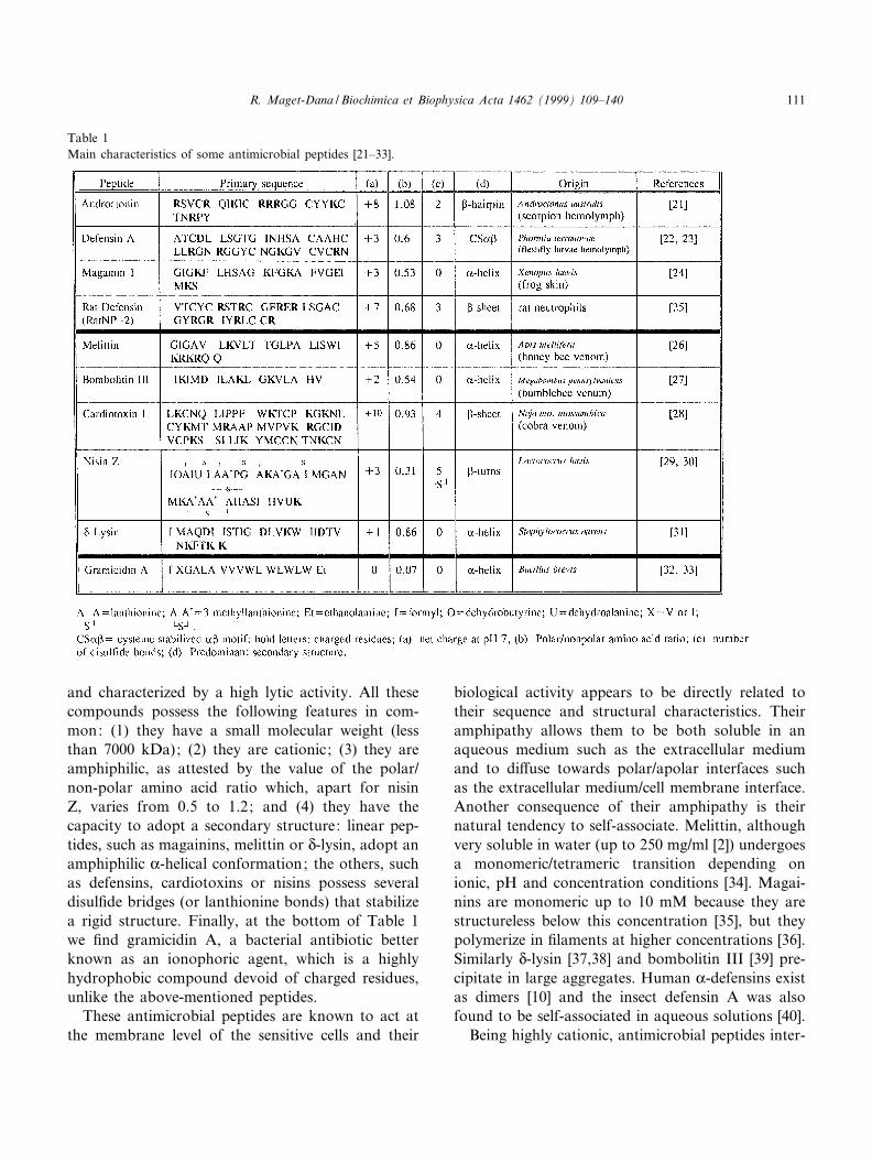
and characterized by a high lytic activity. All thesecompounds possess the following features in com-mon: (1) they have a small molecular weight (lessthan 7000 kDa); (2) they are cationic; (3) they areamphiphilic, as attested by the value of the polar/non-polar amino acid ratio which, apart for nisinZ, varies from 0.5 to 1.2; and (4) they have thecapacity to adopt a secondary structure: linear pep-tides, such as magainins, melittin or N-lysin, adopt anamphiphilic K-helical conformation; the others, suchas defensins, cardiotoxins or nisins possess severaldisul¢de bridges (or lanthionine bonds) that stabilizea rigid structure. Finally, at the bottom of Table 1we ¢nd gramicidin A, a bacterial antibiotic betterknown as an ionophoric agent, which is a highlyhydrophobic compound devoid of charged residues,unlike the above-mentioned peptides.
These antimicrobial peptides are known to act atthe membrane level of the sensitive cells and their
biological activity appears to be directly related totheir sequence and structural characteristics. Theiramphipathy allows them to be both soluble in anaqueous medium such as the extracellular mediumand to di¡use towards polar/apolar interfaces suchas the extracellular medium/cell membrane interface.Another consequence of their amphipathy is theirnatural tendency to self-associate. Melittin, althoughvery soluble in water (up to 250 mg/ml [2]) undergoesa monomeric/tetrameric transition depending onionic, pH and concentration conditions [34]. Magai-nins are monomeric up to 10 mM because they arestructureless below this concentration [35], but theypolymerize in ¢laments at higher concentrations [36].Similarly N-lysin [37,38] and bombolitin III [39] pre-cipitate in large aggregates. Human K-defensins existas dimers [10] and the insect defensin A was alsofound to be self-associated in aqueous solutions [40].
Being highly cationic, antimicrobial peptides inter-
Table 1Main characteristics of some antimicrobial peptides [21^33].
BBAMEM 77747 30-11-99 Cyaan Magenta Geel Zwart
R. Maget-Dana / Biochimica et Biophysica Acta 1462 (1999) 109^140 111
act preferentially with acidic lipids which are partic-ularly abundant in bacterial membranes [41]. It hastherefore been suggested that di¡erences in theamount of acidic membrane phospholipids play amajor role in sensitive cell speci¢city [42]. In partic-ular, antimicrobial peptides bind to lipopolysaccha-rides on the outer membrane of Gram-negative bac-teria, leading to the disruption of this ¢rst barrier[43]. However, the killing event for both Gram-neg-ative and Gram-positive bacteria is the permeabiliza-tion of the cytoplasmic membrane induced by theformation of voltage-dependent channels, as demon-strated with numerous peptides such as mammaliandefensins [44], insect defensins [45], magainins [46],melittin [47] and nisins [48]. In the case of linearpeptides, channels within the cytoplasmic membraneare thought to be made of bundles of K-helices [13]whereas, for L-sheet peptides, dimer associations maybe the basis of the channel structures [49]. The hy-drophobic gramicidin A forms helical dimer channelsspeci¢c for the transport of monovalent cationsacross membranes [50^52].
The amphipathic character of antimicrobial pep-tides makes them surface-active products and as theirbiological activity occurs at lipid membrane interfa-ces, the monolayer technique is entirely suitable tostudy their physicochemical and biological proper-ties.
In this review, we will describe some aspects of thistechnology by focusing on the procedures needed toinvestigate a particular property or phenomenon,such as adsorption of amphipathic peptides at theair/water interface, their behavior when arranged ina monomolecular ¢lm, how they penetrate into aphospholipid monolayer taken as a membrane modeland ¢nally their interactions with lipid molecules in amixed peptide^lipid monolayer. Each procedure willbe illustrated with examples of investigations per-formed on some antimicrobial peptides.
2. Elementary notions of surface chemistry
In the following, all the basic notions concerningsurface chemistry are based on the book by Adam-son [53] and by that of Gaines [54], still considered asthe bible for research workers in the ¢eld of mono-molecular ¢lms.
2.1. Interfaces
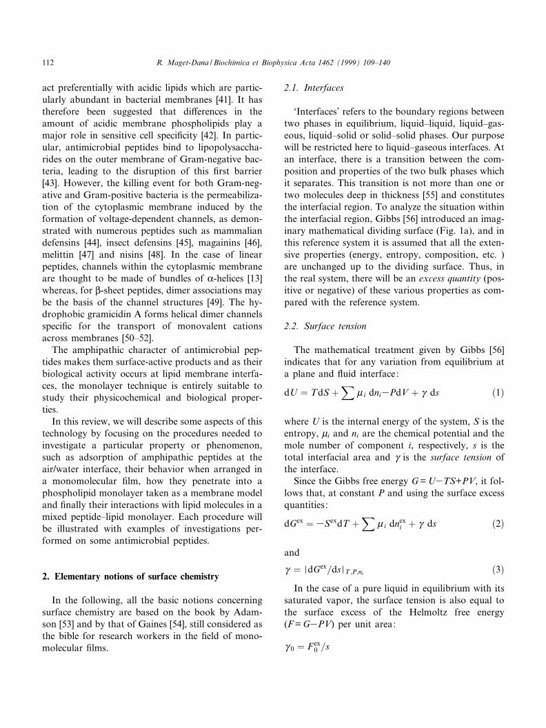
`Interfaces' refers to the boundary regions betweentwo phases in equilibrium, liquid^liquid, liquid^gas-eous, liquid^solid or solid^solid phases. Our purposewill be restricted here to liquid^gaseous interfaces. Atan interface, there is a transition between the com-position and properties of the two bulk phases whichit separates. This transition is not more than one ortwo molecules deep in thickness [55] and constitutesthe interfacial region. To analyze the situation withinthe interfacial region, Gibbs [56] introduced an imag-inary mathematical dividing surface (Fig. 1a), and inthis reference system it is assumed that all the exten-sive properties (energy, entropy, composition, etc. )are unchanged up to the dividing surface. Thus, inthe real system, there will be an excess quantity (pos-itive or negative) of these various properties as com-pared with the reference system.
2.2. Surface tension
The mathematical treatment given by Gibbs [56]indicates that for any variation from equilibrium ata plane and £uid interface:
dU � TdS �X
W i dni3PdV � Q ds �1�
where U is the internal energy of the system, S is theentropy, Wi and ni are the chemical potential and themole number of component i, respectively, s is thetotal interfacial area and Q is the surface tension ofthe interface.
Since the Gibbs free energy G = U3TS+PV, it fol-lows that, at constant P and using the surface excessquantities:
dGex � 3SexdT �X
W i dnexi � Q ds �2�
and
Q � MdGex=dsMT ;P;ni �3�In the case of a pure liquid in equilibrium with its
saturated vapor, the surface tension is also equal tothe surface excess of the Helmoltz free energy(F = G3PV) per unit area:
Q 0 � F ex0 =s
BBAMEM 77747 30-11-99 Cyaan Magenta Geel Zwart
R. Maget-Dana / Biochimica et Biophysica Acta 1462 (1999) 109^140112
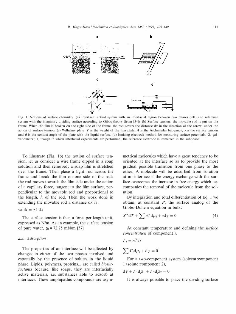
To illustrate (Fig. 1b) the notion of surface ten-sion, let us consider a wire frame dipped in a soapsolution and then removed: a soap ¢lm is stretchedover the frame. Then place a light rod across theframe and break the ¢lm on one side of the rod:the rod moves towards the ¢lm side under the actionof a capillary force, tangent to the ¢lm surface, per-pendicular to the movable rod and proportional tothe length, l, of the rod. Then the work done inextending the movable rod a distance dx is :
work � Q l dx
The surface tension is then a force per length unit,expressed as N/m. As an example, the surface tensionof pure water, Q0 = 72.75 mN/m [57].
2.3. Adsorption
The properties of an interface will be a¡ected bychanges in either of the two phases involved andespecially by the presence of solutes in the liquidphase. Lipids, polymers, proteins... are called biosur-factants because, like soaps, they are interfaciallyactive materials, i.e. substances able to adsorb atinterfaces. These amphipathic compounds are asym-
metrical molecules which have a great tendency to beoriented at the interface so as to provide the mostgradual possible transition from one phase to theother. A molecule will be adsorbed from solutionat an interface if the energy exchange with the sur-face overcomes the increase in free energy which ac-companies the removal of the molecule from the sol-ution.
By integration and total di¡erentiation of Eq. 1 weobtain, at constant P, the surface analog of theGibbs^Duhem equation in bulk:
SexdT �X
nexi dW i � sdQ � 0 �4�
At constant temperature and de¢ning the surfaceconcentration of component i,
y i � nexi =sX
y idW i � dQ � 0
For a two-component system (solvent:component1+solute:component 2),
dQ � y 1dW 1 � y 2dW 2 � 0
It is always possible to place the dividing surface
Fig. 1. Notions of surface chemistry. (a) Interface: actual system with an interfacial region between two phases (left) and referencesystem with the imaginary dividing surface according to Gibbs theory (from [54]). (b) Surface tension: the movable rod is put on theframe. When the ¢lm is broken on the right side of the frame, the rod covers the distance dx in the direction of the arrow, under theaction of surface tension. (c) Wilhelmy plate: P is the weight of the thin plate, A is the Archimedes buoyancy, Q is the surface tensionand a is the contact angle of the plate with the liquid surface. (d) Ionizing electrode method for measuring surface potentials. G, gal-vanometer; T, trough in which interfacial experiments are performed; the reference electrode is immersed in the subphase.
BBAMEM 77747 30-11-99 Cyaan Magenta Geel Zwart
R. Maget-Dana / Biochimica et Biophysica Acta 1462 (1999) 109^140 113
in such a position that the surface excess of solventvanishes (y1 = 0), then,
dQ � 3y 2dW 2
with W2 =W02+RT ln a2 (a2 being the activity of the
solute and W02 the standard chemical potential of the
solute)
y 2 � 31=RT �dQ =dlna2�If the solution is dilute enough so that the behav-
ior is ideal, then the volume concentration, C, of thesolute is proportional to the activity, and:
y � 31=RT �dQ =dlnC� �5�This is the Gibbs adsorption isotherm widely used
in evaluating the extent of adsorption of surface-ac-tive compounds in dilute solutions, from surface ten-sion measurements. This relation shows clearly thatif there is adsorption (that is to say a positive excess)of the solute at the interface, the surface tension ofthe solution decreases. The di¡erence,
Z � Q solvent3Q solution
is called surface pressure of the ¢lm formed by theadsorbed molecules.
2.4. Surface pressure measurements
A £uid interface, such as air/water interface, hasthe advantage of being a plane interface, the changein interfacial free energy of which can be obtained bya simple measurement of surface pressure.
Herein, we will describe only the Wilhelmy methodwhich is the most commonly used to measure surfacepressure of plane and £uid interfaces. A thin plate,usually made of glass, mica or platinum, is partiallyimmersed in the liquid phase and is connected to anelectromicrobalance. The forces acting on the plateare its weight P and surface tension e¡ects down-ward, and Archimedes buoyancy A upward (Fig.1c). The net downward force is:
F � P� 2Q �w� t�cosa3A
where w and t (tIw) are the width and the thicknessof the plate, respectively, and a is the contact angleof the liquid with the solid plate. If the plate is com-pletely wetted (platinum o¡ers the advantage that£aming not only removes contaminants but also ren-
ders the plate wettable) the contact angle a= 0 andcosa= 1, so that,
F � P� 2Q w3A
When the composition of the interface varies, Pand A (provided the plate is maintained in a ¢xedposition) stay constant, then,
vF � 2w�Q solution3Q water� � 32wZ
and
Z � 3vF=2w
2.5. Surface potential
As seen above, there is a tendency for moleculesnear the liquid surface to have a speci¢c orientation.The orientation of water molecules, which behave asdipoles, produces an asymmetrical ¢eld near the sur-face. Spreading a monolayer on a clean water sur-face, will produce a change in the orientation at theinterface and then a change in the nature of theelectrical ¢eld. The di¡erence, vV, in surface poten-tials between that for the aqueous phase and that forthe ¢lm covered phase, is attributed to the ¢lm. vV iscalled surface potential or `Volta potential' and isalso de¢ned as the work required to bring a unitcharge from in¢nity just up to the phase. A qualita-tive interpretation of vV is generally given in termsof analogy with a condenser: a polarized moleculewith an e¡ective dipole moment1 M= qd is similar totwo conducting plates separated by a distance d andenclosing a charge density q. Then, the potential dif-ference vV is given by
vV � 4Zqd=O
where O is the dielectric constant.If there is an array of n dipoles by area unit,
vV � 4ZnMP=O
where MP is the normal component of the dipolemoment to the surface.
There are several contributions to vV : (1) the
1 The dipole moment is expressed as Debye (D). As an exam-ple, the dipole moment of the peptide unit in an K-helix is 3.6 D[58].
BBAMEM 77747 30-11-99 Cyaan Magenta Geel Zwart
R. Maget-Dana / Biochimica et Biophysica Acta 1462 (1999) 109^140114
change induced by the reorientation of the water di-poles in the presence of the ¢lm forming molecules;(2) the dipoles of the ¢lm forming molecules, namelythat of the polar group and that of the alkyl part,which can be located in di¡erent dielectric constantmedia [59]; then,
vV � 4ZX�nMP=O �
(3) we have seen that most of the antimicrobial pep-tides are positively charged molecules. Thereforethere is an additional contribution to vV due tothe Gouy^Chapman electrical double layer, arisingfrom the charged monolayer and the counter-ionsin the subphase solution.
The surface potential at a liquid interface is thengiven by:
vV � 4ZX�nMP=O � �8 0 �6�
where the ¢rst term is the conventional surface dipolemoment expression for uncharged monolayers and80 represents the potential di¡erence (due to thedouble layer) between the surface and the bulk ofthe subphase solution. The subscript 0 means that80 is the potential at the distance x = 0 from theinterface.
The relevant principles of membrane electrochem-istry are well described in [60].
2.6. Surface potential measurements
Surface potential measurements can be accom-plished either by the ionizing electrode method orby the vibrating plate method. The ¢rst method in-volves ionization of the air above the ¢lm, so that itbecomes conducting. The potential di¡erence be-tween two electrodes, one in the aqueous subphaseand the other in the air above the surface, can thenbe measured directly. The ionization in the air abovethe ¢lm is produced by coating the air electrode withan K-emitter, such as polonium or americium. Thereference electrode placed in the bulk subphase canbe an Ag^AgCl electrode. The gap between the ra-dioactive electrode and the liquid surface (V5 mm)is generally su¤ciently conducting that vV can bemeasured by means of a high impedance (1016 6)voltameter. The principle of this method is shownin Fig. 1d.
In the vibrating plate method, the radioactive elec-trode is replaced by a vibrating electrode which con-stitutes one of the plates of a condenser, the secondbeing the surface of the aqueous subphase. The alter-nating signal, generated across a resistance when theplate is vibrated rapidly, is detected using a high-gainampli¢er.
3. Adsorption of antimicrobial peptides at theair/water interface
The adsorption of peptides at the air/water inter-face is therefore currently monitored by following the
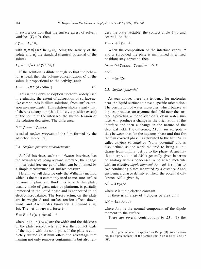
Fig. 2. Adsorption of peptides at the air/water interface.(a) Time dependence of defensin A adsorption (vZAW versus t)plot. The defensin A concentration is 4U1036 M(res) in 30mM Tris-Cl, pH 7.4; T = 20³C. Defensin A was added at thearrow. (b) Corresponding kinetics plot according to Eq. 8 [66].
BBAMEM 77747 30-11-99 Cyaan Magenta Geel Zwart
R. Maget-Dana / Biochimica et Biophysica Acta 1462 (1999) 109^140 115
increase in surface pressure as a function of time, asexempli¢ed in Fig. 2a in the case of defensin A. Therate of adsorption is regulated by the rate of di¡u-sion of the peptide molecules from the bulk solution,at a peptide concentration C, up to the subsurfaceclose to the interface at zero peptide concentration.This ¢rst step is promoted by the concentration gra-dient of peptide molecules in the early stage of theprocess [61,62]. The di¡usion rate of peptide mole-cules towards the interface can be described by [63]:
y � 2C�Dt=3:14�1=2 �7�where D is the di¡usion coe¤cient and y the surfaceconcentration of the peptide at time t. To give anorder of magnitude, the D values are 9.5U1035
and 3.5U1035 cm2/min. for the K-helical (LKKL)4
and the L-sheet (LK)8 model peptides, respectively[64]. As stated above, there is initially no energybarrier to adsorption and the rate of arrival of mol-ecules at the air/water interface is di¡usion-con-trolled, therefore Z and y change at the same rateand the measurement of Z as a function of timeallows the adsorption kinetics to be determined byapplying the following ¢rst-order equation [65]:
ln��Zr3Z t�=�Zr3Z 0�� � 3Kt �8�where Zr, Zt and Z0 are the surface pressures at theplateau, at time t and at zero time and K is the rateconstant. (Zr3Z0) called vZAW
r , is the maximum in-crease in surface pressure induced by the adsorptionof a peptide (at a given concentration) at a clean air/water interface. As seen in Fig. 2b, in the case ofdefensin A the plot presents a slope change de¢ningtwo rate constants K1 = 6U1033 min31 andK2 = 4U1032 min31 [66] values in the range of thosefound for various other proteins [65,67].
The surface activity of antimicrobial peptides isin£uenced by conformational factors, such as amphi-pathic structure, molecular size, molecular £exibilityand net charge.
3.1. In£uence of the primary sequence
The amphipathic character of a given peptide is aconsequence of its amino acid sequence. As seen inTable 1, melittin and N-lysin possess a highly asym-metrical polar/non-polar amino acid distributionwith a cluster of six polar (including four cationic)
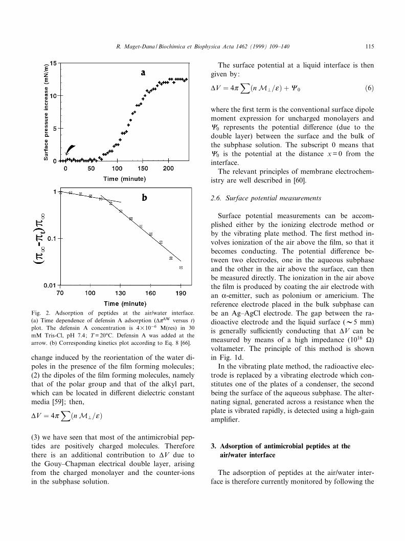
amino acids at the C-terminus in the case of melittin.On the contrary, non-polar amino acids are distrib-uted all along the sequence of defensin A, bombolitinor cardiotoxin. As for androctonin, which possessesa high content in polar, including eight cationic, res-idues, it is an only slightly amphiphilic peptide, and,as shown in Fig. 3, it exhibits a very low surfaceactivity. The in£uence of the primary sequence ofantimicrobial peptides upon adsorption is well dem-onstrated in the case of the four cardiotoxins isolatedfrom Naja mossambica mossambica, the sequence ofwhich does not di¡er to a great extent [28]. Bougis etal. [68] have shown that, for the same concentration,6U1035 M(res)2 in the subphase, the surface pres-sure increase varies from 6 mN/m for CTX II andCTX III to 9 mN/m for CTX IV and to 11 mN/m forCTX III RM. In the same way, Ksenzhek et al. [69]indicated that the N-acetylated toxin is more surface
Fig. 3. Adsorption of some antimicrobial peptides at the air/water interface. Maximum increase in surface pressure (vZAW
r )versus peptide concentration in the subphase. w, melittin in
0.145 M NaCl pH 5.6 (data from [73]); 8, defensin A in 30
mM Tris buffer pH 7.6, 0.1 M KCl, T = 20³C (data from [66]) ;b, androctonin in 10 mM Tris bu¡er, pH 7.4, 0.1 M NaCl,T = 20³C (unpublished results) ; F, cardiotoxin isolated fromNaja naja oxiana in 0.1 M KCl (data from [69]) ; +, N-lysin(composition of the subphase unspeci¢ed) pH 6.8, T = 22³C(data from [82]).
2 In order to compare the properties of the various antimicro-bial peptides, all the concentrations will be expressed as mole ofamino acid residue, i. e. M(res).
BBAMEM 77747 30-11-99 Cyaan Magenta Geel Zwart
R. Maget-Dana / Biochimica et Biophysica Acta 1462 (1999) 109^140116
active than the native cardiotoxin from Naja najaoxiana. Antimicrobial peptides are polyelectrolytesand the presence of cationic residues might have agreat in£uence on their adsorption behavior. In spiteof this, Schro«der et al. [70] have found an unchangedsurface activity upon removing the hydrophilic andhighly cationic (21^26) hexapeptide to the native me-littin. However, it is likely that, although the adsorp-tion extent remains the same, the adsorption kineticschanges in the absence of repulsive forces arisingfrom the charged residues. Nevertheless, theSchro«der ¢nding indicates that, as a ¢rst approxima-tion, the energy of adsorption would be determinedprimarily by the non-polar amino acid residues.
3.2. In£uence of the tridimensional structure
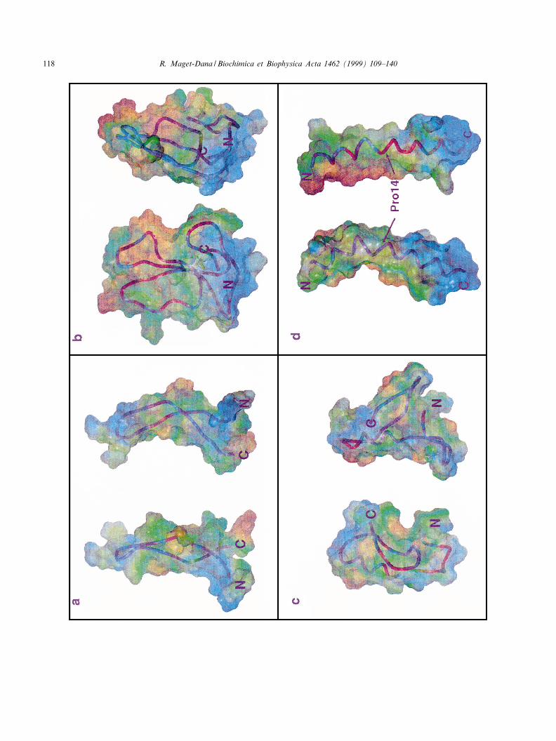
The molecular size of peptides depends not onlyupon their sequence length, but mainly upon theirtridimensional structure in solution (see in Fig. 4the tridimensional structure of some antimicrobialpeptides). In aqueous solution, the melittin monomeradopts a predominantly random-coil structure,whereas the tetramer is rich in K-helix [71]. In thetetrameric form, melittin has the conformation of abent K-helical rod which exhibits a distinctive orien-tational segregation of hydrophilic and hydrophobicside chains, the latter being oriented mainly towardsthe inside of the helix bend [72]. The strong surfaceactivity of melittin [73] can be therefore relatedmainly to its tridimensional structure as an assemblyof amphipathic K-helices. Cardiotoxins, mammaliandefensins or insect defensins are compact moleculesrigidi¢ed by disul¢de bridges which generally fold insuch a manner that hydrophobic patches are createdat the surface of the molecule. Cardiotoxins are ableto adopt a three-¢ngered L-strand structure whichpresents an important hydrophobic region locatedat the tip of loop 1, in the middle of loop 2 andalong a portion of loop 3 [74,75]. The structure ofmammalian defensins, such as HNP-3, is dominatedby a three-stranded antiparallel L-sheet self-associ-ated as an amphipathic dimer [49]. Defensin A con-sists of three distinct domains: a N-terminal £exibleloop, a helical fragment connected via two S^Sbridges (16^36, 20^38) to an antiparallel two-stranded L-structure, the overall fold being stabilizedby a third (3^30) S^S bridge. At the surface of the
structured defensin A molecule, three hydrophobicpatches are visible [23]. Conversely, the twisted L-hairpin structure of androctonin presents, mainly atthe C-terminus, hydrophobic zones of only reducedstretch [76] and, as a result, it exhibits a low surfaceactivity.
3.3. Unfolding of antimicrobial peptides uponadsorption at the air/water interface
Molecular £exibility is also an important factor forprotein or peptide adsorption since, unlike other sur-factants, these molecules tend to unfold at the inter-face, mainly at low surface pressures, in such a man-ner that their conformation is consistent with themaximum lowering of the surface free energy. Sucha conformation will allow the maximum number ofhydrophilic side groups to be oriented into the sub-phase and most of the hydrophobic ones to be pro-jected away from the aqueous phase. It is generallyadmitted that secondary structures (K-helices and L-sheets) are preserved in the course of protein adsorp-tion at the air/water interface [77]. This assumptionhas been demonstrated by spectroscopic analysis oftransferred K-helical and L-sheet polypeptide mono-layers by the technique of Langmuir^Blodgett [78].Therefore, it is likely that melittin amphiphilic K-heli-ces are adsorbed at the air/water interface withoutnoticeable unfolding. Conversely, the CD spectrumrecorded for defensin A transferred monolayers in-dicates a loss in the K-helix content (7%) as com-pared to that (20%) found in solution [79]. As amatter of fact, although the defensin A structure isrigidi¢ed by three disul¢de bridges, it possesses a£exible N-terminal loop (residues 4^14) able to un-fold at the air/water interface and containing one ofthe hydrophobic regions [23]. According to the pro-tein adsorption model of MacRichtie [62] and assuggested by the sigmoidal aspect of the (vZAW^t)plot (Fig. 2a), the defensin A adsorption may be acooperative process, the adsorption of one hydro-phobic patch being the ¢rst step leading other hydro-phobic parts of the peptide up to the interface.
The complete denaturation of N-lysin in concen-trated urea leads to a very high adsorption [80] prob-ably because, in the absence of structural constraints,most of the amino acid residues are located at theinterface.
BBAMEM 77747 30-11-99 Cyaan Magenta Geel Zwart
R. Maget-Dana / Biochimica et Biophysica Acta 1462 (1999) 109^140 117
BBAMEM 77747 30-11-99 Cyaan Magenta Geel Zwart
R. Maget-Dana / Biochimica et Biophysica Acta 1462 (1999) 109^140118
3.4. In£uence of the subphase bulk composition
The adsorption of proteins or peptides from solu-tion to interface is in£uenced by variables such asconcentration, ionic strength, pH and temperature[81]. The in£uence of the peptide concentration inbulk upon adsorption is shown in Fig. 3 for someantimicrobial peptides at comparable ionic strength.The plateau in the (vZAW
r ^C) plot corresponds to thesaturation of the interface by the adsorbed peptides.The higher the pressure value at the plateau, thegreater the surface-active properties of the peptide.We can see that the amphipathic helices, melittin andN-lysin, are the most surface active and, conversely,the very hydrophilic androctonin is the least active inthis concentration range. Increasing the bulk peptideconcentration also favors self association. In the caseof N-lysin [82], the adsorption enhancement up to1035 M(res) (Fig. 3) may correspond to the forma-tion of amphipathic aggregates. Colacicco et al. [80]reported a very low adsorption of N-lysin when dis-solved in distilled water (vZAW
r 6 1 mN/m atCV8U1036 M(res)) as compared to the valuereached in a bu¡er subphase. Likewise, increasingthe ionic strength of the subphase results in an en-hancement of both the adsorption rate and the extentof the defensin A adsorption [66]. The adsorption ofcharged peptides is maximum when the subphase pHis near their isoelectric point, thus avoiding electro-static repulsion between already adsorbed peptidesand peptides in solution.
4. Spread monolayers of antimicrobial peptides at theair/water interface
Another way to obtain a peptide ¢lm is to spread apeptide solution at the air/water interface. Con-versely to monolayers resulting from adsorptionand which are in equilibrium with molecules in thebulk phase, spread monolayers are in a metastable
state. Peptides are generally spread by carefully add-ing a small amount of a concentrated aqueous solu-tion at the air/water interface, with a microsyringe.Our personal method is to dilute the aqueous peptidesolution with a given amount of a volatile solvent(hexa£uoroisopropanol) miscible with water. Thesurface area to volume ratio of the trough must pref-erentially be higher than 1 cm31.
Owing to the high hydrosolubility of most anti-microbial peptides, the hypothesis of a total absenceof desorption of the spread peptide into the sub-phase must be veri¢ed. Schwartz and Taylor [83]have elaborated a pertinent thermodynamic analysisto evaluate the partition of a spread peptide betweenthe monolayer and the subphase. In each experi-ment the surface pressure, Z, is measured with agiven amount, ns, of spread peptide for a gradual-ly decreasing trough area, s. A series of Z versus sisotherms are recorded using a number of suf-¢ciently di¡erent values of ns. Then pairs of ns
and s taken from all the available isotherms at thesame surface pressure are collected. Because of massconservation, these quantities must obey the rela-tion:
ns � y s� VC �9�where y is the surface concentration of the peptide, Vis the volume of the subphase and C is the peptideconcentration in the subphase.
Thus, plotting ns as a function of s gives a straightline with a slope equal to y, and C can be determinedfrom the intercept with the ordinate. From this anal-ysis it is possible to show clearly any peptide desorp-tion from the spread monolayer.
4.1. General properties of spread monolayers [53,54]
The main parameters which characterize the ¢lmstate of a given substance spread on an aqueous sub-phase are the temperature T, the surface pressure Z,the surface area and the number of molecules, these
Fig. 4. Tridimensional structure of some antimicrobial peptides. Hydrophobic potential surfaces MOLCAD (Sybyl-Tripos, Saint-Louis,MO, USA): from blue (hydrophilic) to brown (hydrophobic). Orthographic views of: (a) androctonin, Protein Data Bank ID code 1CZ6 [76]; (b) cardiotoxin from Taiwan Cobra (Naja Naja Atra), Protein Data Bank ID code 2 CDX [75]. Note the three loops in thetube representation; (c) defensin A, Protein Data Bank ID code 1 ICA [23] ; (d) melittin, Protein Data Bank ID code 2 MLT (M.Gribskov, L. Wesson, D. Eisenberg, 1990). Note the bend of the K-helix and the high hydrophilicity of the C-terminal part.6
BBAMEM 77747 30-11-99 Cyaan Magenta Geel Zwart
R. Maget-Dana / Biochimica et Biophysica Acta 1462 (1999) 109^140 119
two last parameters being expressed as area per mol-ecule, A.
Therefore, at constant T, an equation of state ofthe ¢lm has the general form:
Z � Z �A�TThe surface pressure^area (Z^A) isotherm of a mono-layer constitutes the essential characterization of theproperties of the ¢lm although surface potentialmeasurements, which give information regardingthe orientation of the ¢lm constituents, are also usu-ally performed.
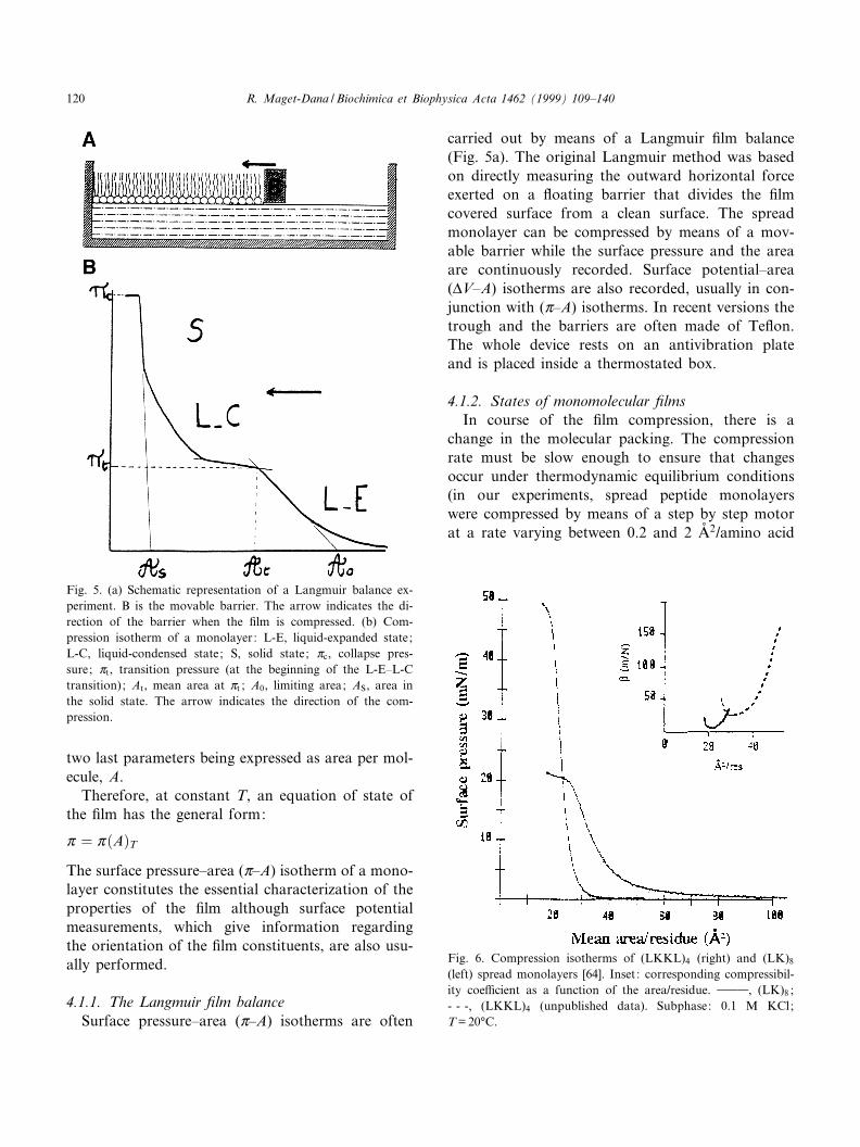
4.1.1. The Langmuir ¢lm balanceSurface pressure^area (Z^A) isotherms are often
carried out by means of a Langmuir ¢lm balance(Fig. 5a). The original Langmuir method was basedon directly measuring the outward horizontal forceexerted on a £oating barrier that divides the ¢lmcovered surface from a clean surface. The spreadmonolayer can be compressed by means of a mov-able barrier while the surface pressure and the areaare continuously recorded. Surface potential^area(vV^A) isotherms are also recorded, usually in con-junction with (Z^A) isotherms. In recent versions thetrough and the barriers are often made of Te£on.The whole device rests on an antivibration plateand is placed inside a thermostated box.
4.1.2. States of monomolecular ¢lmsIn course of the ¢lm compression, there is a
change in the molecular packing. The compressionrate must be slow enough to ensure that changesoccur under thermodynamic equilibrium conditions(in our experiments, spread peptide monolayerswere compressed by means of a step by step motorat a rate varying between 0.2 and 2 Aî 2/amino acid
Fig. 6. Compression isotherms of (LKKL)4 (right) and (LK)8
(left) spread monolayers [64]. Inset: corresponding compressibil-ity coe¤cient as a function of the area/residue. 999, (LK)8 ;- - -, (LKKL)4 (unpublished data). Subphase: 0.1 M KCl;T = 20³C.
Fig. 5. (a) Schematic representation of a Langmuir balance ex-periment. B is the movable barrier. The arrow indicates the di-rection of the barrier when the ¢lm is compressed. (b) Com-pression isotherm of a monolayer: L-E, liquid-expanded state;L-C, liquid-condensed state; S, solid state; Zc, collapse pres-sure; Zt, transition pressure (at the beginning of the L-E^L-Ctransition); At, mean area at Zt ; A0, limiting area; AS, area inthe solid state. The arrow indicates the direction of the com-pression.
BBAMEM 77747 30-11-99 Cyaan Magenta Geel Zwart
R. Maget-Dana / Biochimica et Biophysica Acta 1462 (1999) 109^140120
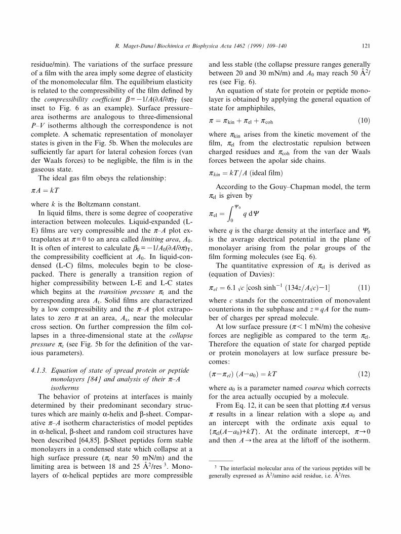
residue/min). The variations of the surface pressureof a ¢lm with the area imply some degree of elasticityof the monomolecular ¢lm. The equilibrium elasticityis related to the compressibility of the ¢lm de¢ned bythe compressibility coe¤cient L=31/A(DA/DZ)T (seeinset to Fig. 6 as an example). Surface pressure^area isotherms are analogous to three-dimensionalP^V isotherms although the correspondence is notcomplete. A schematic representation of monolayerstates is given in the Fig. 5b. When the molecules aresu¤ciently far apart for lateral cohesion forces (vander Waals forces) to be negligible, the ¢lm is in thegaseous state.
The ideal gas ¢lm obeys the relationship:
ZA � kT
where k is the Boltzmann constant.In liquid ¢lms, there is some degree of cooperative
interaction between molecules. Liquid-expanded (L-E) ¢lms are very compressible and the Z^A plot ex-trapolates at Z= 0 to an area called limiting area, A0.It is often of interest to calculate L0 =31/A0(DA/DZ)T,the compressibility coe¤cient at A0. In liquid-con-densed (L-C) ¢lms, molecules begin to be close-packed. There is generally a transition region ofhigher compressibility between L-E and L-C stateswhich begins at the transition pressure Zt and thecorresponding area At. Solid ¢lms are characterizedby a low compressibility and the Z^A plot extrapo-lates to zero Z at an area, As, near the molecularcross section. On further compression the ¢lm col-lapses in a three-dimensional state at the collapsepressure Zc (see Fig. 5b for the de¢nition of the var-ious parameters).
4.1.3. Equation of state of spread protein or peptidemonolayers [84] and analysis of their Z^Aisotherms
The behavior of proteins at interfaces is mainlydetermined by their predominant secondary struc-tures which are mainly K-helix and L-sheet. Compar-ative Z^A isotherm characteristics of model peptidesin K-helical, L-sheet and random coil structures havebeen described [64,85]. L-Sheet peptides form stablemonolayers in a condensed state which collapse at ahigh surface pressure (Zc near 50 mN/m) and thelimiting area is between 18 and 25 Aî 2/res 3. Mono-layers of K-helical peptides are more compressible
and less stable (the collapse pressure ranges generallybetween 20 and 30 mN/m) and A0 may reach 50 Aî 2/res (see Fig. 6).
An equation of state for protein or peptide mono-layer is obtained by applying the general equation ofstate for amphiphiles,
Z � Z kin � Z el � Z coh �10�where Zkin arises from the kinetic movement of the¢lm, Zel from the electrostatic repulsion betweencharged residues and Zcoh from the van der Waalsforces between the apolar side chains.
Z kin � kT=A �ideal film�According to the Gouy^Chapman model, the term
Zel is given by
Z el �Z 8 0
0q d8
where q is the charge density at the interface and 80
is the average electrical potential in the plane ofmonolayer arising from the polar groups of the¢lm forming molecules (see Eq. 6).
The quantitative expression of Zel is derived as(equation of Davies):
Z el � 6:1 kc �cosh sinh31 �134z=Akc�31� �11�where c stands for the concentration of monovalentcounterions in the subphase and z = qA for the num-ber of charges per spread molecule.
At low surface pressure (Z6 1 mN/m) the cohesiveforces are negligible as compared to the term Zel.Therefore the equation of state for charged peptideor protein monolayers at low surface pressure be-comes:
�Z3Z el� �A3a0� � kT �12�where a0 is a parameter named coarea which correctsfor the area actually occupied by a molecule.
From Eq. 12, it can be seen that plotting ZA versusZ results in a linear relation with a slope a0 andan intercept with the ordinate axis equal to{Zel(A3a0)+kT}. At the ordinate intercept, ZC0and then ACthe area at the lifto¡ of the isotherm.
3 The interfacial molecular area of the various peptides will begenerally expressed as Aî 2/amino acid residue, i.e. Aî 2/res.
BBAMEM 77747 30-11-99 Cyaan Magenta Geel Zwart
R. Maget-Dana / Biochimica et Biophysica Acta 1462 (1999) 109^140 121
When the experimental value found at the ordinateintercept di¡ers from the theoretical (calculated) val-ue, this discrepancy is generally attributed to a mo-lecular aggregation in the ¢lm [54] with an aggrega-tion number nag = (calculated value)/(value at theintercept). Applying this method to the (LKKL)4
and (LK)8 isopeptides, we concluded that the L-sheetpeptide has a higher degree of self association at theair/water interface than its K-helical isomer [64]. This¢nding agrees with the situation encountered in sol-ution where L-sheet peptides have a greater tendencythan K-helix peptides to form aggregates because thehydrogen bonds in L-sheet are arranged between onestrand and another with a thermodynamic preferencefor the antiparallel structure [86].
According to Birdi [87], it is possible to estimatethe degree of unfolding of proteins at the air/waterinterface from their Z^A isotherms. He suggestedthat the wider the di¡erence between the work ofcompression and the work of expansion, the moreimportant the degree of unfolding of the protein.This statement is valid provided that no loss of pro-tein into the subphase occurs in the course of thecompression. It is therefore better to stop the com-pression well before the collapse of the monolayerwhen recording a compression^expansion cycle.
The work of compression (or of expansion) isgiven by:
W �Z final
initialZdA
4.2. Spread monolayers of gramicidin A and itsanalogs
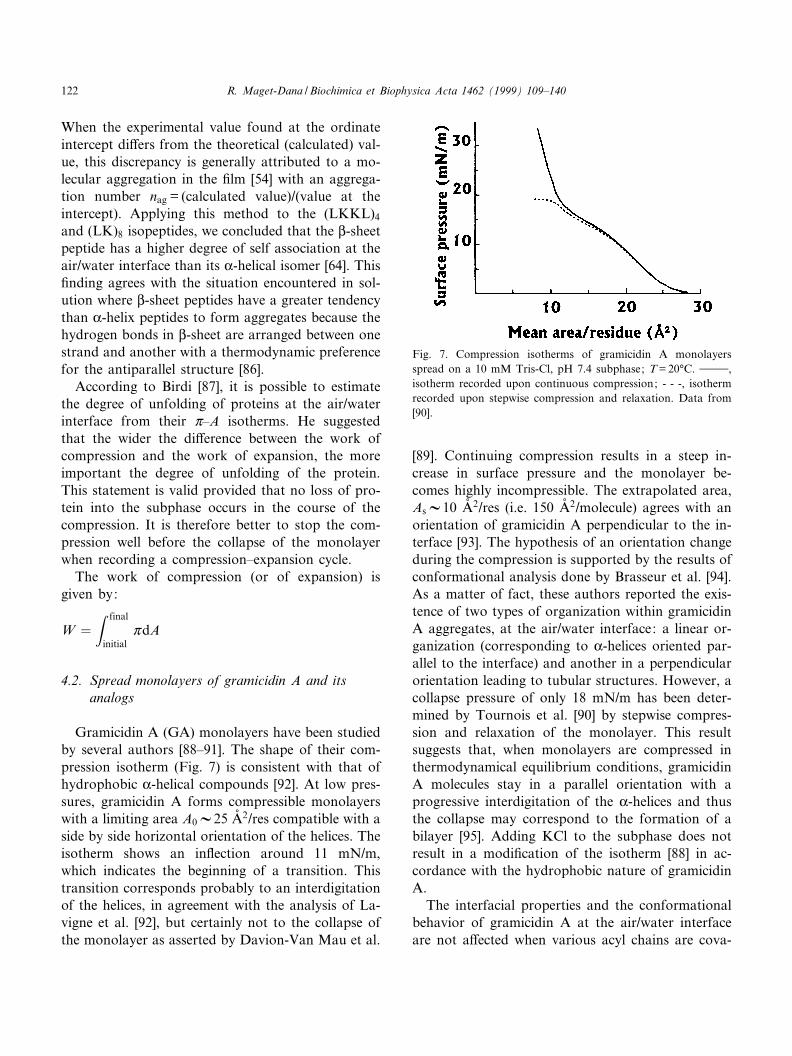
Gramicidin A (GA) monolayers have been studiedby several authors [88^91]. The shape of their com-pression isotherm (Fig. 7) is consistent with that ofhydrophobic K-helical compounds [92]. At low pres-sures, gramicidin A forms compressible monolayerswith a limiting area A0V25 Aî 2/res compatible with aside by side horizontal orientation of the helices. Theisotherm shows an in£ection around 11 mN/m,which indicates the beginning of a transition. Thistransition corresponds probably to an interdigitationof the helices, in agreement with the analysis of La-vigne et al. [92], but certainly not to the collapse ofthe monolayer as asserted by Davion-Van Mau et al.
[89]. Continuing compression results in a steep in-crease in surface pressure and the monolayer be-comes highly incompressible. The extrapolated area,AsV10 Aî 2/res (i.e. 150 Aî 2/molecule) agrees with anorientation of gramicidin A perpendicular to the in-terface [93]. The hypothesis of an orientation changeduring the compression is supported by the results ofconformational analysis done by Brasseur et al. [94].As a matter of fact, these authors reported the exis-tence of two types of organization within gramicidinA aggregates, at the air/water interface: a linear or-ganization (corresponding to K-helices oriented par-allel to the interface) and another in a perpendicularorientation leading to tubular structures. However, acollapse pressure of only 18 mN/m has been deter-mined by Tournois et al. [90] by stepwise compres-sion and relaxation of the monolayer. This resultsuggests that, when monolayers are compressed inthermodynamical equilibrium conditions, gramicidinA molecules stay in a parallel orientation with aprogressive interdigitation of the K-helices and thusthe collapse may correspond to the formation of abilayer [95]. Adding KCl to the subphase does notresult in a modi¢cation of the isotherm [88] in ac-cordance with the hydrophobic nature of gramicidinA.
The interfacial properties and the conformationalbehavior of gramicidin A at the air/water interfaceare not a¡ected when various acyl chains are cova-
Fig. 7. Compression isotherms of gramicidin A monolayersspread on a 10 mM Tris-Cl, pH 7.4 subphase; T = 20³C. 999,isotherm recorded upon continuous compression; - - -, isothermrecorded upon stepwise compression and relaxation. Data from[90].
BBAMEM 77747 30-11-99 Cyaan Magenta Geel Zwart
R. Maget-Dana / Biochimica et Biophysica Acta 1462 (1999) 109^140122
lently coupled to the C-terminal ethanolamine group[96]. When highly compressed, the acyl-gramicidinmolecule would be oriented with its C-terminus to-wards the subphase and the coupled acyl chain lo-cated parallel to the helix axis in between the pro-truding tryptophans. The nature of the aromaticresidue appears to be essential for the channel func-tion of gramicidin A [97]. For this reason, Davion-Van Mau et al. [89] synthetized analogs of GA whereall Trp residues were replaced by Tyr giving GT, Phegiving GM� or Tyr-(OBzl) giving GTP. GT andGM� monolayers are less compressible than GA orGTP monolayers because their side chains are short-er, and the transition occurs at higher pressures (21^22 mN/m). Also, changing only one Trp residue al-ters the interfacial properties of GA. For example,the [Phe-9] analog monolayer is more condensed(A0V20 Aî 2/res instead of 25 Aî 2/res) than the GAmonolayer, and its isotherm lacks a transition region[89]. Conversely, analogs in which the central regionis extended by two hydrophobic residues (Leu-Ala)have comparable isotherm characteristics to those of
GA monolayers [90]. The active structure of grami-cidin A being a head to head dimer [50], Davion-vanMau et al. [89] synthetized a covalent retro GA-DAla-GA dimer. Although the transition occurs at thesame pressure (V11 mN/m), the monolayers of GA-D Ala-GA are more condensed (A0V18 Aî 2/res) thanGA monolayers suggesting that the dimer adopts adi¡erent conformational state than gramicidin A[89].
From surface potential measurements it appearsthat gramicidin analogs can be classi¢ed into twogroups: for the ¢rst group (GA and GT), vV isaround 200 mV at the plateau of the (vV^A) iso-therms (when the monolayer is highly compressed),whereas for the second group (GM� and GTP) vVreaches more than 300 mV. It was suggested that acorrelation may exist between surface potential val-ues and single channel behavior. As a matter of fact,the channel conductance units of GA and GT arehigh and almost independent of the applied voltagewhereas those of GM� and GTP are lower andstrongly voltage dependent [89].
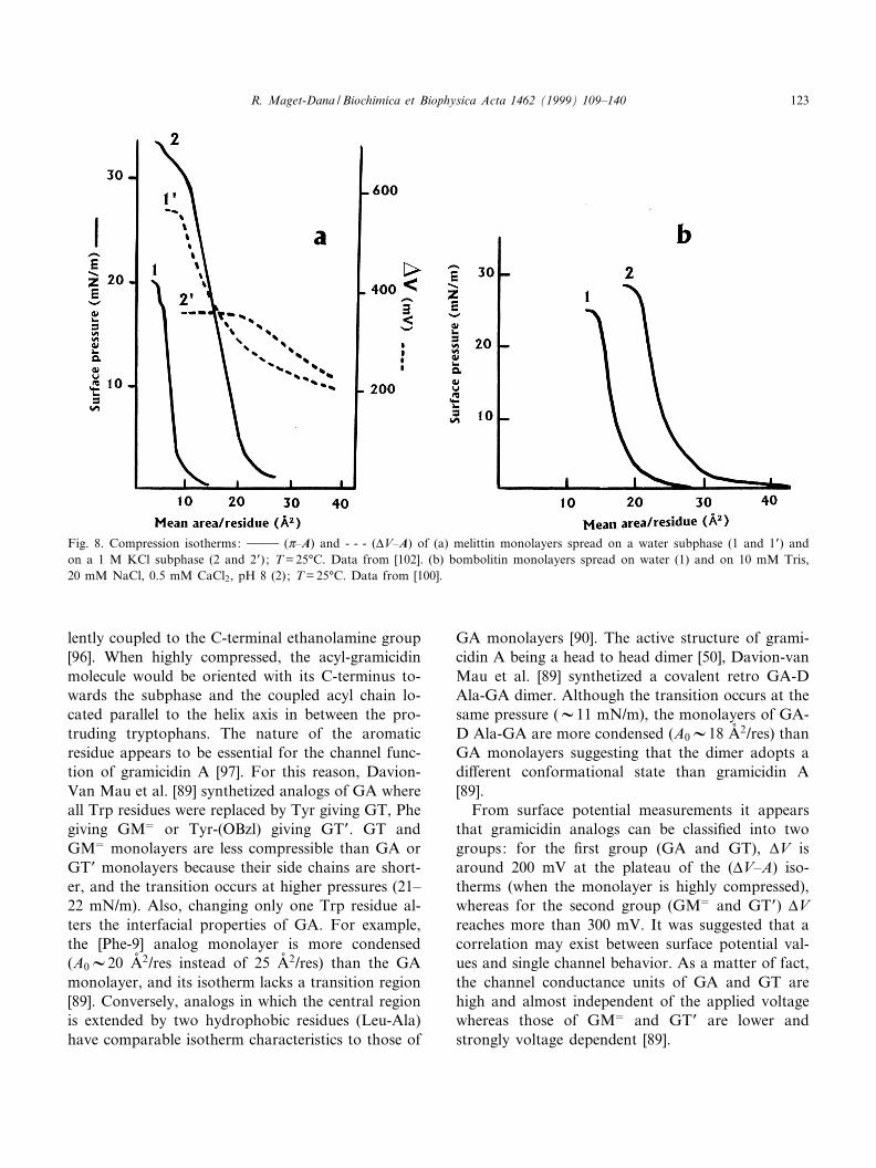
Fig. 8. Compression isotherms: 999 (Z^A) and - - - (vV^A) of (a) melittin monolayers spread on a water subphase (1 and 1P) andon a 1 M KCl subphase (2 and 2P) ; T = 25³C. Data from [102]. (b) bombolitin monolayers spread on water (1) and on 10 mM Tris,20 mM NaCl, 0.5 mM CaCl2, pH 8 (2); T = 25³C. Data from [100].
BBAMEM 77747 30-11-99 Cyaan Magenta Geel Zwart
R. Maget-Dana / Biochimica et Biophysica Acta 1462 (1999) 109^140 123
4.3. Spread monolayers of melittin, bombolitin III andN-lysin
Structured as amphipathic K-helices, melittin[59,73,98,99] and bombolitin III [100] as well as N-lysin [80] form stable monolayers at the air/waterinterface. From the partition analysis of spread me-littin molecules between the monolayer and the sub-phase, Wackerbauer et al. [101] concluded, by apply-ing Eq. 9, that melittin molecules manifest a strongpreference for an accumulation at the air/water inter-face. However, at high surface pressure (about 30mN/m) an appreciable degree of melittin desorptionoccurs, attributed to the excessive mutual repulsionof melittin molecules which limits their surface con-centration. On a pure water subphase, melittinmonolayers (Fig. 8a) are in a very condensed stateand the A0 value (V8.5 Aî 2/res) is far from corre-sponding to that of an K-helix lying £at at the inter-face [102]. The reason may be the presence of a bendat Pro-14: the 15^26 hydrophilic C-terminal peptidemay be fully hydrated while the more hydrophobicN-terminal part points away from the aqueousphase. In such a situation the 15^26 C-terminal pep-tide adsorbed at the interface of the water subphaseis in a medium of dielectric constant O= 78 while theN-terminal part is in a medium of dielectric constantO= 1 and the cationic Lys-7 is not able to fully inter-act with the aqueous phase. This could explain whythe (Z^A) isotherms of melittin monolayers spread ona water subphase cannot be analyzed by using theGouy^Chapman model which does not take su¤-ciently into account the e¡ect of the medium dielec-tric constant [59]. As seen in the Fig. 8b and in con-trast to melittin, the value of A0 (V20 Aî 2/res) ofbombolitin III on a water subphase [100] is compat-ible with a horizontal orientation of the helix part ofthe peptide (the K-helix content of bombolitin IIIbeing 60^70%). The collapse pressure (V25 mN/m)of bombolittin III monolayers is within the samerange as that (20 mN/m) of melittin monolayers,but lower than that (40 mN/m) of N-lysin monolayersunder the same experimental conditions [80].Thehigher stability of N-lysin monolayers may resultfrom the less net charge of the peptide. The additionof KCl in the subphase enhances the stability of bothbombolitin III and melittin monolayers which col-lapse pressure and limiting area become higher
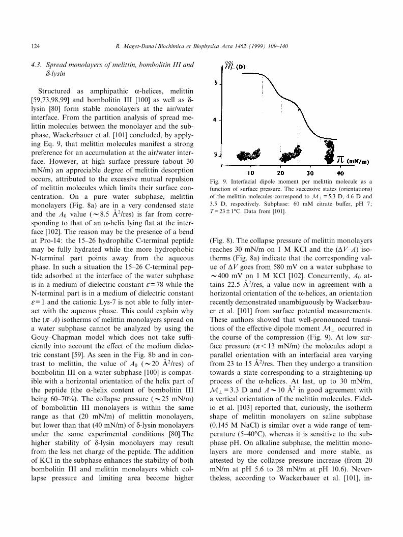
(Fig. 8). The collapse pressure of melittin monolayersreaches 30 mN/m on 1 M KCl and the (vV^A) iso-therms (Fig. 8a) indicate that the corresponding val-ue of vV goes from 580 mV on a water subphase toV400 mV on 1 M KCl [102]. Concurrently, A0 at-tains 22.5 Aî 2/res, a value now in agreement with ahorizontal orientation of the K-helices, an orientationrecently demonstrated unambiguously by Wackerbau-er et al. [101] from surface potential measurements.These authors showed that well-pronounced transi-tions of the e¡ective dipole moment MP occurred inthe course of the compression (Fig. 9). At low sur-face pressure (Z6 13 mN/m) the molecules adopt aparallel orientation with an interfacial area varyingfrom 23 to 15 Aî 2/res. Then they undergo a transitiontowards a state corresponding to a straightening-upprocess of the K-helices. At last, up to 30 mN/m,MP = 3.3 D and AV10 Aî 2 in good agreement witha vertical orientation of the melittin molecules. Fidel-io et al. [103] reported that, curiously, the isothermshape of melittin monolayers on saline subphase(0.145 M NaCl) is similar over a wide range of tem-perature (5^40³C), whereas it is sensitive to the sub-phase pH. On alkaline subphase, the melittin mono-layers are more condensed and more stable, asattested by the collapse pressure increase (from 20mN/m at pH 5.6 to 28 mN/m at pH 10.6). Never-theless, according to Wackerbauer et al. [101], in-
Fig. 9. Interfacial dipole moment per melittin molecule as afunction of surface pressure. The successive states (orientations)of the melittin molecules correspond to MP = 5.3 D, 4.6 D and3.5 D, respectively. Subphase: 60 mM citrate bu¡er, pH 7;T = 23 þ 1³C. Data from [101].
BBAMEM 77747 30-11-99 Cyaan Magenta Geel Zwart
R. Maget-Dana / Biochimica et Biophysica Acta 1462 (1999) 109^140124
creasing the pH apparently favors the melittin parti-tion into the subphase.
From the analysis of the ZA versus Z plot, accord-ing to Eq. 12 and giving (ZA)Z!0 = kT/4, Gevod andBirdi [104] concluded that melittin is present as atetramer at the interface. Given the low value ofA0, the melittin tetramer may be oriented verticallywith the polar end (20^26 residues) situated in theaqueous phase. However, this is a very controversialresult since, on the one hand, De Grado et al. [98]reported that melittin is in a monomer state at theair/water interface and, on the other hand, Terwil-liger et al. [72] suggested, on the basis of crystallo-graphic studies, that a melittin monolayer is consti-tuted by a dimer layer.
4.4. Spread monolayers of defensin A [66]
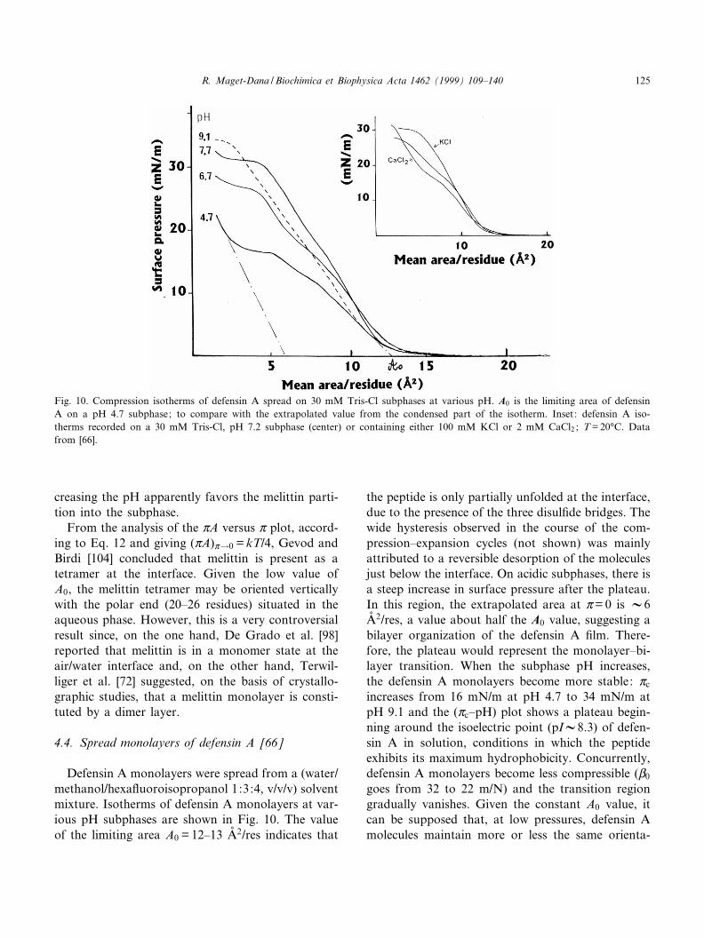
Defensin A monolayers were spread from a (water/methanol/hexa£uoroisopropanol 1:3:4, v/v/v) solventmixture. Isotherms of defensin A monolayers at var-ious pH subphases are shown in Fig. 10. The valueof the limiting area A0 = 12^13 Aî 2/res indicates that
the peptide is only partially unfolded at the interface,due to the presence of the three disul¢de bridges. Thewide hysteresis observed in the course of the com-pression^expansion cycles (not shown) was mainlyattributed to a reversible desorption of the moleculesjust below the interface. On acidic subphases, there isa steep increase in surface pressure after the plateau.In this region, the extrapolated area at Z= 0 is V6Aî 2/res, a value about half the A0 value, suggesting abilayer organization of the defensin A ¢lm. There-fore, the plateau would represent the monolayer^bi-layer transition. When the subphase pH increases,the defensin A monolayers become more stable: Zc
increases from 16 mN/m at pH 4.7 to 34 mN/m atpH 9.1 and the (Zc^pH) plot shows a plateau begin-ning around the isoelectric point (pIV8.3) of defen-sin A in solution, conditions in which the peptideexhibits its maximum hydrophobicity. Concurrently,defensin A monolayers become less compressible (L0
goes from 32 to 22 m/N) and the transition regiongradually vanishes. Given the constant A0 value, itcan be supposed that, at low pressures, defensin Amolecules maintain more or less the same orienta-
Fig. 10. Compression isotherms of defensin A spread on 30 mM Tris-Cl subphases at various pH. A0 is the limiting area of defensinA on a pH 4.7 subphase; to compare with the extrapolated value from the condensed part of the isotherm. Inset: defensin A iso-therms recorded on a 30 mM Tris-Cl, pH 7.2 subphase (center) or containing either 100 mM KCl or 2 mM CaCl2 ; T = 20³C. Datafrom [66].
BBAMEM 77747 30-11-99 Cyaan Magenta Geel Zwart
R. Maget-Dana / Biochimica et Biophysica Acta 1462 (1999) 109^140 125
tion. The addition of 0.1 M KCl in the subphaseleads to similar features to basic pH, probably asthe result of a screening e¡ect. The addition ofonly 2 mM CaCl2 induces signi¢cant changes in theshape of defensin A monolayers. The transition re-gion is now more pronounced and the liquid^con-densed region of the isotherm is less compressible(see Fig. 10 inset). These features were attributed tothe binding of Ca2� ions to defensin A molecules atthe level of Asp-4 and/or of the terminal COO3
group, with possible Ca2� bridges between adjacentdefensin A molecules promoting the formation ofdimers.
5. Penetration of antimicrobial peptides into lipidmonolayers
5.1. Lipid monolayers as membrane models
With the aim of studying interactions between agiven e¡ector (antimicrobial peptide, for instance)and cell membranes, several approaches are possible.We can work with living cells and observe what hap-pens in situ or we can isolate cellular membranes.However, natural membranes are very complex enti-ties with a great variety of lipids and proteins, thus,in this case, we can only investigate global phenom-ena. Therefore, if we are interested in speci¢c aspectsof a given biological phenomenon occurring at mem-brane level, the best choice is to use membrane mod-els. Among them, bilayer lipid membranes (BLM)are the most suitable for the study of ion transportacross membranes [105]. Lipid vesicles (SUV, LUVor multilamellar), a widely used model [106], possessan internal aqueous compartment and are also suit-able for permeability studies. However, although lip-id vesicle experiments are easy to carry out and allowvarious spectroscopic measurements, the lipid vesiclesystem presents some disadvantages. Firstly, it is notalways possible to prepare vesicles: with pure phos-phatidylethanolamine, for example, either with 100%charged lipid or containing more than 50% choles-terol or at a temperature less than the gel^liquidcrystal transition temperature of the phospholipids.Furthermore, it is di¤cult to prepare a homogeneous(in size and in layer number) vesicle suspension andto avoid spontaneous fusion. Another disadvantage
is the small curvature radius of vesicles that imposesstrong constraints at the polar head level.
On the contrary, with the monolayer system, anumber of parameters including the nature and thepacking of the lipid molecules, the composition ofthe subphase and the temperature, can be chosenwithout any limitation. The interest of phospholipidmonolayers as membrane models [107] consists alsoin their homogeneity, their stability and their planargeometry where the lipid molecules have a speci¢corientation. Besides, as seen before, this well-de¢nedbidimensional system provides rigorous thermody-namic analysis. The lipid monolayer is also a verysuitable model to study, in conditions near the actual
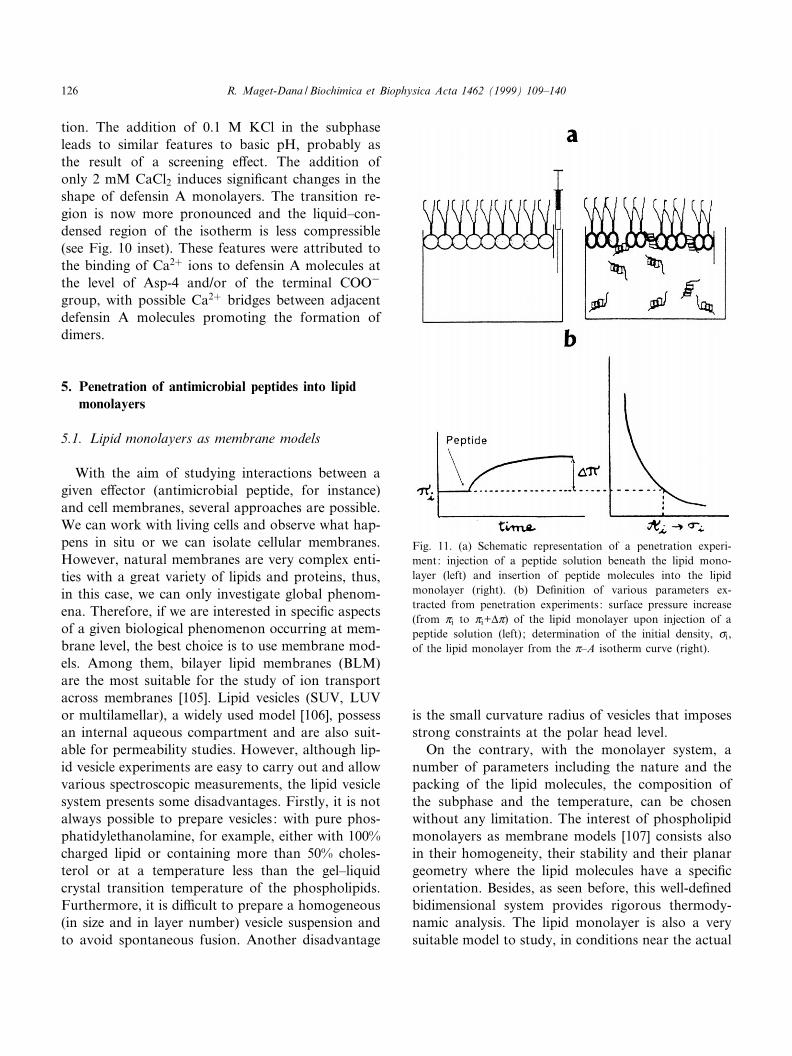
Fig. 11. (a) Schematic representation of a penetration experi-ment: injection of a peptide solution beneath the lipid mono-layer (left) and insertion of peptide molecules into the lipidmonolayer (right). (b) De¢nition of various parameters ex-tracted from penetration experiments: surface pressure increase(from Zi to Zi+vZ) of the lipid monolayer upon injection of apeptide solution (left) ; determination of the initial density, ci,of the lipid monolayer from the Z^A isotherm curve (right).
BBAMEM 77747 30-11-99 Cyaan Magenta Geel Zwart
R. Maget-Dana / Biochimica et Biophysica Acta 1462 (1999) 109^140126
biological ones, what happens when a hydrosolublepeptide, present in the extracellular medium, arrivesat the membrane surface of the target cell. Finally, ina recent article, Brockman [108] argued convincinglyin favor of lipid monolayers over bilayers as a modelto characterize protein^membrane interactions. Forfurther details about bilayer and monolayer corre-spondence, see [109] and [110] as reviews.
5.2. General considerations on the penetration ofsoluble compounds into spread insolublemonolayers [111]
Generally speaking, the term `monolayer penetra-tion' is used to describe the interaction of an insolu-ble monolayer spread at the air/water interface witha soluble active compound present in the aqueous
Fig. 12. Melittin and N-lysin penetration into phospholipid monolayers. (a) Maximum increase in surface pressure (vZ) as a functionof the initial pressure, Zi of the phospholipid monolayer, upon injection of 8.84U1036 M(res) of melittin in the subphase. Data from[82]. (b) Maximum increase in surface pressure as a function of Zi upon injection of 8.84U1036 M(res) of N-lysin in the subphase.Data from [82]. (c) The same as in b, but vZ is plotted as a function of the initial density ci of phospholipids. Data calculated from[82] and [115]. a, DPPC; R, DPPE; F, DPPA; b, DPPG. Pure water subphase, pH 6.8; T = 22³C. The dotted lines represent themaximum value of surface pressure, vZAW
r , induced by the peptide adsorption at a clean air/water interface in similar experimentalconditions (see Fig. 3).
BBAMEM 77747 30-11-99 Cyaan Magenta Geel Zwart
R. Maget-Dana / Biochimica et Biophysica Acta 1462 (1999) 109^140 127

phase. The interaction can be measured in two ways.The ¢lm pressure can be kept constant and the re-action followed by observing the increase in area ofthe ¢lm. The second method, by far the most widelyused, is to maintain the ¢lm area constant and tomeasure the surface pressure changes on adding thepenetrating compound to the subphase. Herein, theinsoluble monolayer is a lipid monolayer and thepenetrating compound an antimicrobial peptide.The experimental device generally consists in a smalldish equipped with a vertical tube through which thepeptide solution is injected (Fig. 11a). Lipid mono-layers are formed at the required pressure by inter-mittent spreading of the lipid solution in the appro-priate solvent mixture (containing generallychloroform and methanol). The parameters thatmust be taken into account are: (1) the initial pres-sure of the lipid ¢lm, Zi, which re£ects the packing ofthe lipids in the monolayer. However, in order tocompare the peptide penetration in various lipidmonolayers, it is best to choose the lipid density,ci, as the lipid packing parameter (Fig. 11b); (2)the concentration, C, of the peptide in the subphase;(3) the maximum change, vZLip
r (called vZ to simpli-fy), in surface pressure of the lipid ¢lm upon inter-action with the peptide dissolved in the subphase. Asfar as possible, C must be low enough for the surfacepressure, vZAW
r , induced by adsorption of the peptideat a clean air/water interface (see Section 3) in thesame experimental conditions, to be negligible. Oth-erwise, the result under consideration must be theinteraction pressure vZLip
r 3vZAWr . The lipid ¢lm pres-
sure at which the peptide no longer penetrates iscalled the exclusion pressure, Zex and, in the case ofa (vZ3ci) representation, an exclusion density, cex isde¢ned.
5.3. Penetration of N-lysin and melittin intophospholipid monolayers
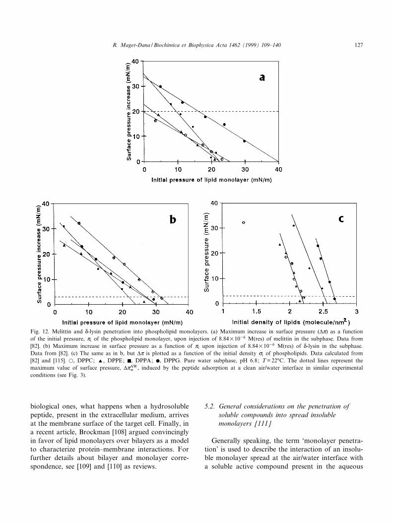
Bhakoo et al. [82] have studied the penetration ofN-lysin and melittin into various phospholipid mono-layers. At the subphase concentration used,C = 8.84U1036 M(res), the adsorption of N-lysin ata clean interface induces an increase in surface pres-sure of only 3 mN/m, but for melittin, vZAW
r reachesV20 mN/m (Fig. 3). Thus, at this concentration,although melittin penetrates into all type of phos-
pholipid monolayers, it interacts speci¢cally onlywith acidic phospholipids (DPPG and DPPA) (Fig.12a) in agreement with its highly cationic nature. Inthe same way, studying the area increase of DPPCand DPPG monolayers upon melittin injection, Hen-drickson et al. [112] found also a greater peptidepenetration into DPPG monolayers. Recently, by insitu infrared re£ection (IRRAS) measurements at theair/water interface, Flach et al. [113] con¢rmed this¢nding and outlined that melittin interacts di¡erentlywith zwitterionic compared with negatively chargedmonolayer surfaces. However, Ohki et al. [114] re-ported an opposite result with natural phospholipidmonolayers (egg-PC and bovine PS), which, owing tothe presence of unsaturated chains, are always in anexpanded state even at high surface pressures. Melit-tin was also found to induce a greater area increaseof DMPC with respect to DMPG monolayers [112].These controversial results could be related to thelength and the nature of the aliphatic chains. As amatter of fact dimyristoyl phospholipid monolayersdo not exhibit L-E^L-C transition and are more ex-panded than the corresponding dipalmitoyl phospho-lipid monolayers [115]. Besides, a dramatic increasein melittin insertion at a Zi corresponding to the L-E^L-C transition of DPPC and DPPG was also re-ported [112]. Therefore, it can legitimately bethought that, in addition to the polar head nature(zwitterionic or acidic) of the phospholipids, thephysical state of the lipid monolayer is involved inthe penetration process. For instance, the bent K-hel-ical structure of melittin could hinder a completepenetration into the rigid chains of DPPC orDPPE ¢lms, but not into the short expanded chainsof DMPC. In addition, the charge of the lysine-7residue prevents an integral insertion of the N-termi-nal hydrophobic segment of melittin [116]. But wemust keep in mind that a weaker penetration intoacidic phospholipid monolayers is not synonymouswith a weaker binding. As a matter of fact, it hasbeen clearly shown that melittin binds £uid nega-tively charged phospholipids intensively [117]. How-ever, owing to their high positive net charge, the ¢rstbound melittin molecules exert a repulsive e¡ect onthe following ones. The ¢rst step of the penetrationprocess is the peptide adsorption at the surface of themonolayer, which is known to induce only negligiblechanges in surface pressure. This happens in all
BBAMEM 77747 30-11-99 Cyaan Magenta Geel Zwart
R. Maget-Dana / Biochimica et Biophysica Acta 1462 (1999) 109^140128
cases, and even with neutral phospholipid mono-layers, at surface pressures higher than Zex. For in-stance, Ebara and Okahata [118] have shown, byQCM experiments, that melittin adsorbs on DPPEmonolayers at Zi = 40 mN/m, a pressure far abovethe exclusion pressure as seen in Fig. 12a. At thesubphase concentration (1036 to 2U1035 M(res))and in pH and ionic strength conditions generallyused in monolayer experiments, melittin is in amonomeric state [34] and mainly in a random coilconformation [119]. Melittin undergoes conforma-tional changes upon binding to phospholipid mono-layers. From CD spectra analysis of transferredDMPA monolayers, Sui et al. [120] showed that in-serted melittin (at Zi = 15 mN/m) contains more K-helix structure than adsorbed melittin (at Zi s Zex).When adsorbed, the K-helix of melittin would liewith its axis parallel to the membrane surface [121]and its hydrophobic face directed toward the mem-brane interior [122]. However, from IRRAS meas-urements, Flach et al. [113] recently suggested thatmelittin adopts a conformation di¡erent from K-helixwhen bound to phospholipid monolayers.
Conversely to melittin, N-lysin interacts speci¢callywith all the phospholipids tested, as seen Fig. 12b.The exclusion pressure in DPPC monolayers reachesV32 mN/m for N-lysin whereas it is only 25 mN/mfor melittin. The same observation can be made forDPPE, giving a further indication of the greater af-¢nity of N-lysin for zwitterionic phospholipids, ascompared to melittin. The penetration power of N-lysin into DPPG monolayers is lower than that ofmelittin since Zex = 31 mN/m instead of 40 mN/m,probably because the net charge of N-lysin is less.However, the exclusion pressure in DPPA mono-layers is the same for both peptides. On the basisof the vZ^Zi plots, Bhakoo et al. [82] concludedthat di¡erences in the phospholipid head grouphave little e¡ect on the degree of interaction betweenN-lysin and the phospholipid ¢lm. However, plottingvZ as a function of the initial molecular density, ci,obtained from the Z^A isotherms of phospholipids[115], leads to the opposite conclusion. As a matterof fact, the vZ^ci plot (Fig. 12c) indicates that thepenetration extent of N-lysin into the various phos-pholipid ¢lms varies in the order: DPPC =DPPE6DPPG. The penetration pro¢les of N-lysinin DPPC and DPPE are identical except at low
DPPC density (corresponding to the expanded partof the Z^A isotherm of DPPC before the L-E^L-Ctransition).
5.4. Penetration of cardiotoxins into phospholipidmonolayers
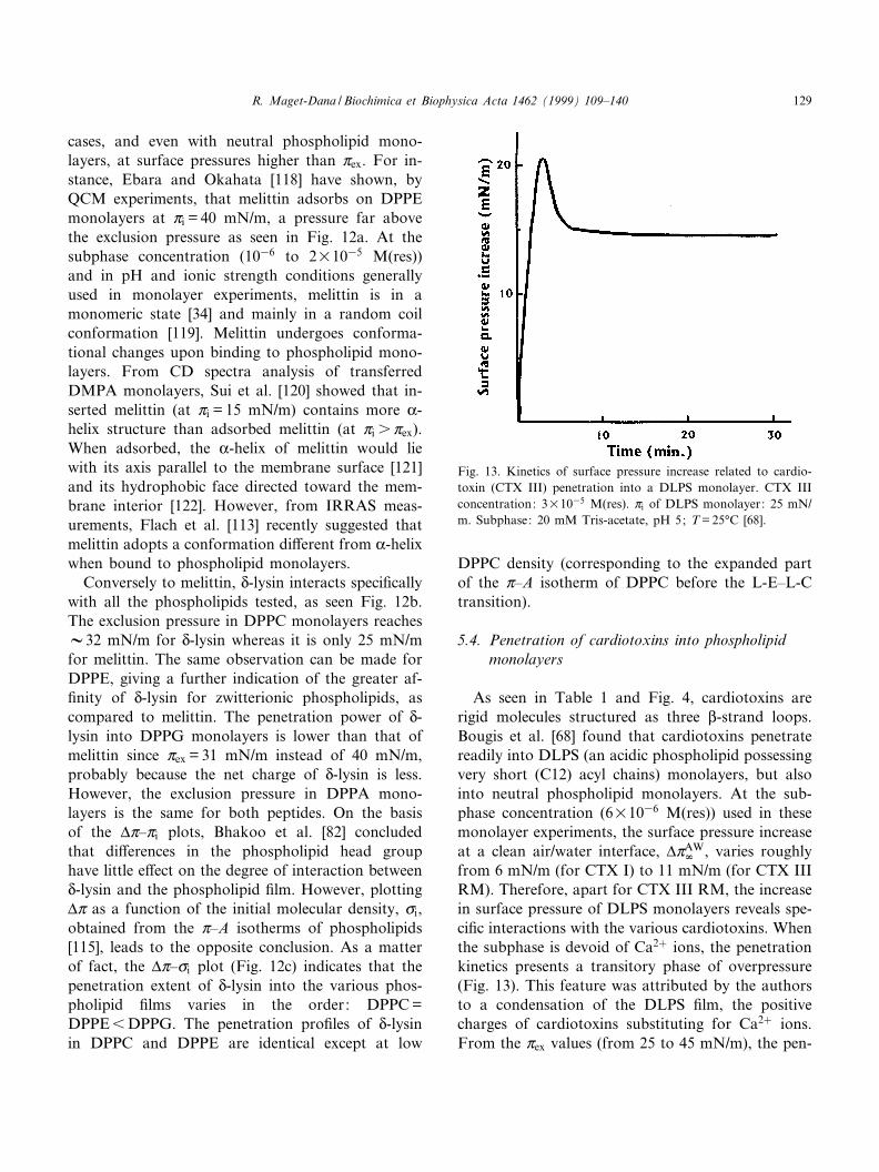
As seen in Table 1 and Fig. 4, cardiotoxins arerigid molecules structured as three L-strand loops.Bougis et al. [68] found that cardiotoxins penetratereadily into DLPS (an acidic phospholipid possessingvery short (C12) acyl chains) monolayers, but alsointo neutral phospholipid monolayers. At the sub-phase concentration (6U1036 M(res)) used in thesemonolayer experiments, the surface pressure increaseat a clean air/water interface, vZAW
r , varies roughlyfrom 6 mN/m (for CTX I) to 11 mN/m (for CTX IIIRM). Therefore, apart for CTX III RM, the increasein surface pressure of DLPS monolayers reveals spe-ci¢c interactions with the various cardiotoxins. Whenthe subphase is devoid of Ca2� ions, the penetrationkinetics presents a transitory phase of overpressure(Fig. 13). This feature was attributed by the authorsto a condensation of the DLPS ¢lm, the positivecharges of cardiotoxins substituting for Ca2� ions.From the Zex values (from 25 to 45 mN/m), the pen-
Fig. 13. Kinetics of surface pressure increase related to cardio-toxin (CTX III) penetration into a DLPS monolayer. CTX IIIconcentration: 3U1035 M(res). Zi of DLPS monolayer: 25 mN/m. Subphase: 20 mM Tris-acetate, pH 5; T = 25³C [68].
BBAMEM 77747 30-11-99 Cyaan Magenta Geel Zwart
R. Maget-Dana / Biochimica et Biophysica Acta 1462 (1999) 109^140 129
etration powers of the various cardiotoxins in DLPSmonolayers are found in the order: CTX IIIRMICTX I = CTX II = CTX IIIICTX IV. Ap-plying the Gibbs relation (Eq. 5) to the penetrationof CTX III into a DLPS ¢lm, the apparent interfa-cial molecular area, A = 1/y, can be determined. Atlow pressures (6 20 mN/m), a value (AV23 Aî 2/res)in good agreement with the interfacial area of L-sheetpeptide models, is found [64]. At higher pressures,the interfacial area of CTX III falls to 5 Aî 2/res, alow value as compared to the value found for the L-sheet peptide models (V15 Aî 2/res). The transitionbetween the two area values (5 and 23 Aî 2/res) occursin a very narrow range of surface pressures (between20 and 30 mN/m). Considering that spectroscopicdata have shown that the rigid structure of cardio-toxins does not change upon interaction with phos-pholipids [123], this behavior has been attributed tothe capacity of cardiotoxins to be oriented `£at' or`edgewise' in the lipid ¢lm. According to Bougis et al.[124], the interaction between acidic phospholipidsand cardiotoxins takes place in several steps: (1) anelectrostatic interaction with the negatively chargedpolar heads of phospholipids; followed by (2) thepenetration of at least the one hydrophobic loop ofthe cardiotoxin molecule; (3) the disorganization ofthe membrane; and, ¢nally (4) a quick change of thebound cardiotoxin orientation. I would add that theorientation change of cardiotoxin from `£at' to`edgewise' may be the cause of the abrupt decreasein pressure observed in the course of the vZ^t experi-ments (Fig. 13).
5.5. Penetration of nisin Z and other lantibioticderivatives into phospholipid monolayers
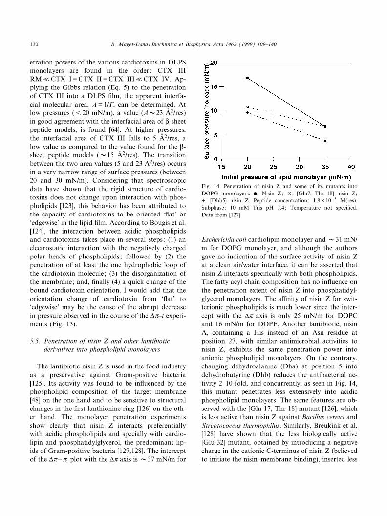
The lantibiotic nisin Z is used in the food industryas a preservative against Gram-positive bacteria[125]. Its activity was found to be in£uenced by thephospholipid composition of the target membrane[48] on the one hand and to be sensitive to structuralchanges in the ¢rst lanthionine ring [126] on the oth-er hand. The monolayer penetration experimentsshow clearly that nisin Z interacts preferentiallywith acidic phospholipids and specially with cardio-lipin and phosphatidylglycerol, the predominant lip-ids of Gram-positive bacteria [127,128]. The interceptof the vZ3Zi plot with the vZ axis is V37 mN/m for
Escherichia coli cardiolipin monolayer and V31 mN/m for DOPG monolayer, and although the authorsgave no indication of the surface activity of nisin Zat a clean air/water interface, it can be asserted thatnisin Z interacts speci¢cally with both phospholipids.The fatty acyl chain composition has no in£uence onthe penetration extent of nisin Z into phosphatidyl-glycerol monolayers. The a¤nity of nisin Z for zwit-terionic phospholipids is much lower since the inter-cept with the vZ axis is only 25 mN/m for DOPCand 16 mN/m for DOPE. Another lantibiotic, nisinA, containing a His instead of an Asn residue atposition 27, with similar antimicrobial activities tonisin Z, exhibits the same penetration power intoanionic phospholipid monolayers. On the contrary,changing dehydroalanine (Dha) at position 5 intodehydrobutyrine (Dhb) reduces the antibacterial ac-tivity 2^10-fold, and concurrently, as seen in Fig. 14,this mutant penetrates less extensively into acidicphospholipid monolayers. The same features are ob-served with the [Gln-17, Thr-18] mutant [126], whichis less active than nisin Z against Bacillus cereus andStreptococcus thermophilus. Similarly, Breukink et al.[128] have shown that the less biologically active[Glu-32] mutant, obtained by introducing a negativecharge in the cationic C-terminus of nisin Z (believedto initiate the nisin^membrane binding), inserted less
Fig. 14. Penetration of nisin Z and some of its mutants intoDOPG monolayers. b, Nisin Z; w, [Gln7, Thr 18] nisin Z;+, [Dhb5] nisin Z. Peptide concentration: 1.8U1035 M(res).Subphase: 10 mM Tris pH 7.4; Temperature not speci¢ed.Data from [127].
BBAMEM 77747 30-11-99 Cyaan Magenta Geel Zwart
R. Maget-Dana / Biochimica et Biophysica Acta 1462 (1999) 109^140130
e¤ciently than nisin Z into DOPG monolayers.Therefore, the lipid monolayer experiments appearto be a useful tool to establish a direct correlationbetween the antibacterial activity of the various ni-sins and their a¤nity for acidic phospholipids.
5.6. Penetration of androctonin and defensin A [129]into phospholipid monolayers
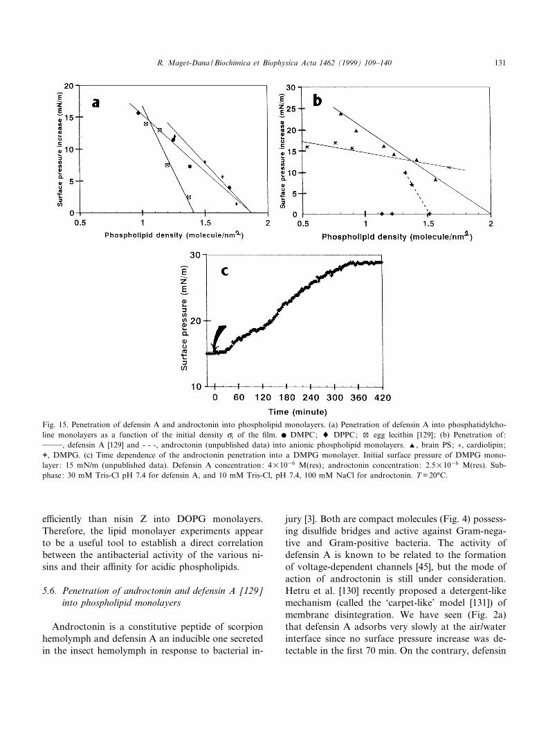
Androctonin is a constitutive peptide of scorpionhemolymph and defensin A an inducible one secretedin the insect hemolymph in response to bacterial in-
jury [3]. Both are compact molecules (Fig. 4) possess-ing disul¢de bridges and active against Gram-nega-tive and Gram-positive bacteria. The activity ofdefensin A is known to be related to the formationof voltage-dependent channels [45], but the mode ofaction of androctonin is still under consideration.Hetru et al. [130] recently proposed a detergent-likemechanism (called the `carpet-like' model [131]) ofmembrane disintegration. We have seen (Fig. 2a)that defensin A adsorbs very slowly at the air/waterinterface since no surface pressure increase was de-tectable in the ¢rst 70 min. On the contrary, defensin
Fig. 15. Penetration of defensin A and androctonin into phospholipid monolayers. (a) Penetration of defensin A into phosphatidylcho-line monolayers as a function of the initial density ci of the ¢lm. b DMPC; 8 DPPC; w egg lecithin [129]; (b) Penetration of:999, defensin A [129] and - - -, androctonin (unpublished data) into anionic phospholipid monolayers. R, brain PS; �, cardiolipin;+, DMPG. (c) Time dependence of the androctonin penetration into a DMPG monolayer. Initial surface pressure of DMPG mono-layer: 15 mN/m (unpublished data). Defensin A concentration: 4U1036 M(res) ; androctonin concentration: 2.5U1036 M(res). Sub-phase: 30 mM Tris-Cl pH 7.4 for defensin A, and 10 mM Tris-Cl, pH 7.4, 100 mM NaCl for androctonin. T = 20³C.
BBAMEM 77747 30-11-99 Cyaan Magenta Geel Zwart
R. Maget-Dana / Biochimica et Biophysica Acta 1462 (1999) 109^140 131
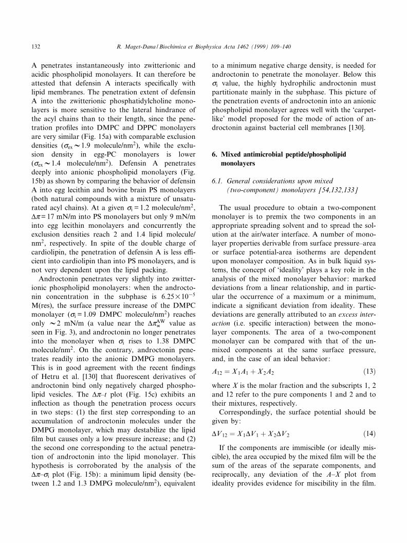
A penetrates instantaneously into zwitterionic andacidic phospholipid monolayers. It can therefore beattested that defensin A interacts speci¢cally withlipid membranes. The penetration extent of defensinA into the zwitterionic phosphatidylcholine mono-layers is more sensitive to the lateral hindrance ofthe acyl chains than to their length, since the pene-tration pro¢les into DMPC and DPPC monolayersare very similar (Fig. 15a) with comparable exclusiondensities (cexV1.9 molecule/nm2), while the exclu-sion density in egg-PC monolayers is lower(cexV1.4 molecule/nm2). Defensin A penetratesdeeply into anionic phospholipid monolayers (Fig.15b) as shown by comparing the behavior of defensinA into egg lecithin and bovine brain PS monolayers(both natural compounds with a mixture of unsatu-rated acyl chains). At a given ci = 1.2 molecule/nm2,vZ= 17 mN/m into PS monolayers but only 9 mN/minto egg lecithin monolayers and concurrently theexclusion densities reach 2 and 1.4 lipid molecule/nm2, respectively. In spite of the double charge ofcardiolipin, the penetration of defensin A is less e¤-cient into cardiolipin than into PS monolayers, and isnot very dependent upon the lipid packing.
Androctonin penetrates very slightly into zwitter-ionic phospholipid monolayers: when the androcto-nin concentration in the subphase is 6.25U1035
M(res), the surface pressure increase of the DMPCmonolayer (ci = 1.09 DMPC molecule/nm2) reachesonly V2 mN/m (a value near the vZAW
r value asseen in Fig. 3), and androctonin no longer penetratesinto the monolayer when ci rises to 1.38 DMPCmolecule/nm2. On the contrary, androctonin pene-trates readily into the anionic DMPG monolayers.This is in good agreement with the recent ¢ndingsof Hetru et al. [130] that £uorescent derivatives ofandroctonin bind only negatively charged phospho-lipid vesicles. The vZ^t plot (Fig. 15c) exhibits anin£ection as though the penetration process occursin two steps: (1) the ¢rst step corresponding to anaccumulation of androctonin molecules under theDMPG monolayer, which may destabilize the lipid¢lm but causes only a low pressure increase; and (2)the second one corresponding to the actual penetra-tion of androctonin into the lipid monolayer. Thishypothesis is corroborated by the analysis of thevZ^ci plot (Fig. 15b): a minimum lipid density (be-tween 1.2 and 1.3 DMPG molecule/nm2), equivalent
to a minimum negative charge density, is needed forandroctonin to penetrate the monolayer. Below thisci value, the highly hydrophilic androctonin mustpartitionate mainly in the subphase. This picture ofthe penetration events of androctonin into an anionicphospholipid monolayer agrees well with the `carpet-like' model proposed for the mode of action of an-droctonin against bacterial cell membranes [130].
6. Mixed antimicrobial peptide/phospholipidmonolayers
6.1. General considerations upon mixed(two-component) monolayers [54,132,133]
The usual procedure to obtain a two-componentmonolayer is to premix the two components in anappropriate spreading solvent and to spread the sol-ution at the air/water interface. A number of mono-layer properties derivable from surface pressure^areaor surface potential-area isotherms are dependentupon monolayer composition. As in bulk liquid sys-tems, the concept of `ideality' plays a key role in theanalysis of the mixed monolayer behavior: markeddeviations from a linear relationship, and in partic-ular the occurrence of a maximum or a minimum,indicate a signi¢cant deviation from ideality. Thesedeviations are generally attributed to an excess inter-action (i.e. speci¢c interaction) between the mono-layer components. The area of a two-componentmonolayer can be compared with that of the un-mixed components at the same surface pressure,and, in the case of an ideal behavior:
A12 � X 1A1 � X 2A2 �13�where X is the molar fraction and the subscripts 1, 2and 12 refer to the pure components 1 and 2 and totheir mixtures, respectively.
Correspondingly, the surface potential should begiven by:
vV12 � X 1vV1 � X 2vV2 �14�If the components are immiscible (or ideally mis-
cible), the area occupied by the mixed ¢lm will be thesum of the areas of the separate components, andreciprocally, any deviation of the A^X plot fromideality provides evidence for miscibility in the ¢lm.
BBAMEM 77747 30-11-99 Cyaan Magenta Geel Zwart
R. Maget-Dana / Biochimica et Biophysica Acta 1462 (1999) 109^140132
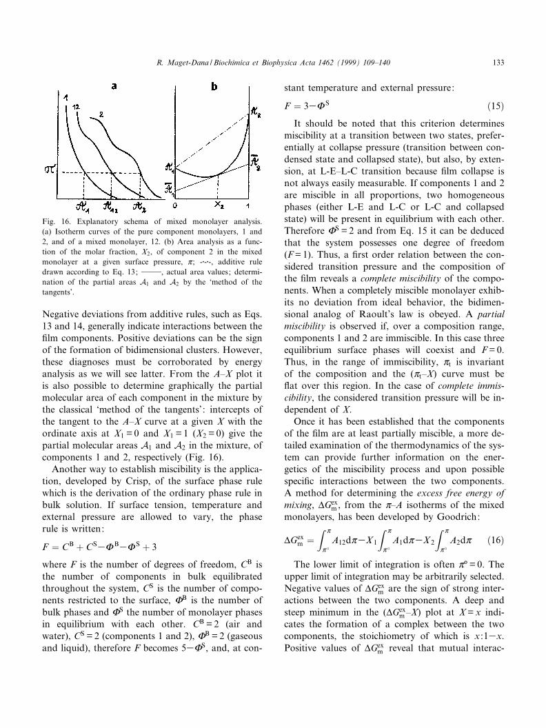
Negative deviations from additive rules, such as Eqs.13 and 14, generally indicate interactions between the¢lm components. Positive deviations can be the signof the formation of bidimensional clusters. However,these diagnoses must be corroborated by energyanalysis as we will see latter. From the A^X plot itis also possible to determine graphically the partialmolecular area of each component in the mixture bythe classical `method of the tangents' : intercepts ofthe tangent to the A^X curve at a given X with theordinate axis at X1 = 0 and X1 = 1 (X2 = 0) give thepartial molecular areas A1 and A2 in the mixture, ofcomponents 1 and 2, respectively (Fig. 16).
Another way to establish miscibility is the applica-tion, developed by Crisp, of the surface phase rulewhich is the derivation of the ordinary phase rule inbulk solution. If surface tension, temperature andexternal pressure are allowed to vary, the phaserule is written:
F � CB � CS3x B3x S � 3
where F is the number of degrees of freedom, CB isthe number of components in bulk equilibratedthroughout the system, CS is the number of compo-nents restricted to the surface, xB is the number ofbulk phases and xS the number of monolayer phasesin equilibrium with each other. CB = 2 (air andwater), CS = 2 (components 1 and 2), xB = 2 (gaseousand liquid), therefore F becomes 53xS, and, at con-
stant temperature and external pressure:
F � 33x S �15�It should be noted that this criterion determines
miscibility at a transition between two states, prefer-entially at collapse pressure (transition between con-densed state and collapsed state), but also, by exten-sion, at L-E^L-C transition because ¢lm collapse isnot always easily measurable. If components 1 and 2are miscible in all proportions, two homogeneousphases (either L-E and L-C or L-C and collapsedstate) will be present in equilibrium with each other.Therefore xS = 2 and from Eq. 15 it can be deducedthat the system possesses one degree of freedom(F = 1). Thus, a ¢rst order relation between the con-sidered transition pressure and the composition ofthe ¢lm reveals a complete miscibility of the compo-nents. When a completely miscible monolayer exhib-its no deviation from ideal behavior, the bidimen-sional analog of Raoult's law is obeyed. A partialmiscibility is observed if, over a composition range,components 1 and 2 are immiscible. In this case threeequilibrium surface phases will coexist and F = 0.Thus, in the range of immiscibility, Zt is invariantof the composition and the (Zt^X) curve must be£at over this region. In the case of complete immis-cibility, the considered transition pressure will be in-dependent of X.
Once it has been established that the componentsof the ¢lm are at least partially miscible, a more de-tailed examination of the thermodynamics of the sys-tem can provide further information on the ener-getics of the miscibility process and upon possiblespeci¢c interactions between the two components.A method for determining the excess free energy ofmixing, vGex
m , from the Z^A isotherms of the mixedmonolayers, has been developed by Goodrich:
vGexm �
Z Z
Z �A12dZ3X 1
Z Z
Z �A1dZ3X 2
Z Z
Z �A2dZ �16�
The lower limit of integration is often Z³ = 0. Theupper limit of integration may be arbitrarily selected.Negative values of vGex
m are the sign of strong inter-actions between the two components. A deep andsteep minimum in the (vGex
m ^X) plot at X = x indi-cates the formation of a complex between the twocomponents, the stoichiometry of which is x :13x.Positive values of vGex
m reveal that mutual interac-
Fig. 16. Explanatory schema of mixed monolayer analysis.(a) Isotherm curves of the pure component monolayers, 1 and2, and of a mixed monolayer, 12. (b) Area analysis as a func-tion of the molar fraction, X2, of component 2 in the mixedmonolayer at a given surface pressure, Z ; -W-W-, additive ruledrawn according to Eq. 13; 999, actual area values; determi-nation of the partial areas A1 and A2 by the `method of thetangents'.
BBAMEM 77747 30-11-99 Cyaan Magenta Geel Zwart
R. Maget-Dana / Biochimica et Biophysica Acta 1462 (1999) 109^140 133
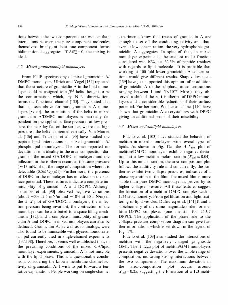
tions between the two components are weaker thaninteractions between the pure component moleculesthemselves: brie£y, at least one component formsbidimensional aggregates. If vGex
m = 0, the mixing isideal.
6.2. Mixed gramicidin/lipid monolayers
From FTIR spectroscopy of mixed gramicidin A/DMPC monolayers, Ulrich and Vogel [134] reportedthat the structure of gramicidin A in the lipid mono-layer could be assigned to a L6:3 helix thought to bethe conformation which, by N^N dimerization,forms the functional channel [135]. They stated alsothat, as seen above for pure gramicidin A mono-layers [89,90], the orientation of the helix in mixedgramicidin A/DMPC monolayers is markedly de-pendent on the applied surface pressure: at low pres-sure, the helix lay £at on the surface, whereas at highpressures, the helix is oriented vertically. Van Mau etal. [136] and Tournois et al. [90] have studied thepeptide^lipid interactions in mixed gramicidin A/phospholipid monolayers. The former reported nodeviations from ideality in the area^composition dia-gram of the mixed GA/DOPC monolayers and thein£ection in the isotherm occurs at the same pressure(V13 mN/m) on the range of composition where it isdetectable (0.59XGA91). Furthermore, the presenceof DOPC in the monolayer has no e¡ect on the sur-face potential. These features indicate a complete im-miscibility of gramicidin A and DOPC. AlthoughTournois et al. [90] observed negative variations(about 35% at 5 mN/m and 310% at 30 mN/m) inthe A^X plot of GA/DOPC monolayers, the in£ec-tion pressure being invariant, the contraction of themonolayer can be attributed to a space-¢lling mech-anism [132], and a complete immiscibility of grami-cidin A and DOPC in mixed monolayers can also bededuced. Gramicidin A, as well as its analogs, werealso found to be immiscible with glyceromonooleate,a lipid currently used in single-channel experiments[137,138]. Therefore, it seems well established that, inthe prevailing conditions of the mixed GA/lipidmonolayer experiments, gramicidin A is not misciblewith the lipid phase. This is a questionable conclu-sion, considering the known membrane channel ac-tivity of gramicidin A. I wish to put forward a ten-tative explanation. People working on single-channel
experiments know that traces of gramicidin A areenough to set o¡ the conducting activity and that,even at low concentration, the very hydrophobic gra-micidin A aggregates. In spite of that, in mixedmonolayer experiments, the smallest molar fractionconsidered was 10%, i.e. 62.5% of peptide residueswith regards to lipid molecules. It is probable thatworking at 100-fold lower gramicidin A concentra-tions would give di¡erent results. Shapovalov et al.[139] have just supported this opinion: after additionof gramicidin A to the subphase, at concentrationsranging between 1 and 5U1036 M(res), they ob-served a shift of the Z^A isotherms of DPPC mono-layers and a considerable reduction of their surfacepotential. Furthermore, Wallace and Janes [140] haveshown that gramicidin A co-crystallizes with DPPCgiving an additional proof of their miscibility.
6.3. Mixed melittin/lipid monolayers
Fidelio et al. [103] have studied the behavior ofmelittin in mixed monolayers with several types oflipids. As shown in Fig. 17a, the A^Xmel plot ofmelittin/DMPC monolayers exhibits negative devia-tions at a low melittin molar fraction (Xmel 9 0.04).Up to this molar fraction, the area^composition plotfollows the additivity rule and concurrently, the iso-therms exhibit two collapse pressures, indicative of aphase separation in the ¢lm. The mixed ¢lm is morestable than pure DMPC monolayer as proved by itshigher collapse pressure. All these features suggestthe formation of a melittin^DMPC complex with a1:24 stoichiometry. From gel ¢ltration and light scat-tering of lipid vesicles, Dufourcq et al. [141] found astoichiometry of the same magnitude order for me-littin^DPPC complexes (one melittin for 25 þ 7DPPC). The application of the phase rule to thecollapse pressure^composition diagram can give fur-ther information, which is set down in the legend ofFig. 17b.
Fidelio et al. [103] also studied the interactions ofmelittin with the negatively charged gangliosideGM1. The A^Xmel plot of melittin/GM1 monolayerspresents negative deviations over the whole range ofcomposition, indicating strong interactions betweenthe two components. The maximum deviation inthe area^composition plot occurs aroundXmel = 0.25, suggesting the formation of a 1:3 melit-
BBAMEM 77747 30-11-99 Cyaan Magenta Geel Zwart
R. Maget-Dana / Biochimica et Biophysica Acta 1462 (1999) 109^140134
tin^GM1 complex. The collapse pressure^composi-tion diagram indicates that a phase separation occursup to Xmel = 0.5. This result can be correlated withthe ¢nding that melittin induces a ratio-dependentphase change from bilayer structure to hexagonalHII phase in melittin^cardiolipin systems [142].
6.4. Mixed bombolitin III/phospholipid monolayers
Another way to obtain mixed monolayers is tospread the lipid ¢lm onto a peptide solution. Thiswas the choice of Signor et al. [100] when studyingthe interaction of bombolitin III with phospholipidmonolayers. It is clear that this method does notallow a precise control of the peptide amount presentat the interface. However, as argued by the authors,the system reaches an equilibrium corresponding tothe peptide amount adsorbed by the monolayer as afunction of the peptide concentration in the sub-phase. Besides, this procedure is more representativeof the natural association process of a soluble pep-tide with a biological membrane. From these experi-ments it appears that there is a rapid adsorption ofbombolitin III into phospholipid monolayers in con-
ditions (low ionic strength and acidic pH) where thepeptide does not adsorb at a clean air/water inter-face. This adsorption is clearly demonstrated by theexpansion of the phospholipid monolayer over time.Upon compression, the mixed bombolitin III/DPPCmonolayers display a two-phase behavior, whereasthe bombolitin III/DMPC monolayers display aone-phase behavior. Introducing traces of a £uores-cent lipid probe into the phospholipid phase allowsthe observation of the mixed monolayer by means ofan epi£uorescence microscope. In this way, the for-mation of phase-separated domains of bombolitin IIIin the DPPC monolayer can be visualized.
C
Fig. 17. Mixed melittin/DMPC monolayers. (a) Area^composi-tion diagram. The area taken into consideration is As (see Fig.5b); - - -, additive rule drawn according to Eq. 13. (b) Collapsepressure^composition diagram. Analysis of the Zc^Xmel diagramaccording to Eq. 15: the line ghi corresponds to the collapsepressure of the 1:24 melittin^DMPC complex; three phases arein equilibrium along the line gh (since F = 0): excess DMPCand the melittin^DMPC complex miscible in a liquid state, ex-cess DMPC in the collapsed state and the melittin^DMPC com-plex in the collapsed state; in the same way, three phases are inequilibrium along the line hi : the melittin^DMPC complex in aliquid state, excess melittin in the collapsed state and the melit-tin^DMPC complex in the collapsed state; two phases are inequilibrium along the line jk (since F = 1): excess melittin andthe melittin^DMPC complex miscible in a liquid state and me-littin in the collapsed state; then, the Zc^Xmel diagram can bedivided into several regions: in region A, the melittin^DMPCcomplex and the excess DMPC are both in a liquid state andmiscible. In region B, the complex and the excess melittin aremiscible in a liquid state. In region C, melittin is in a collapsedstate immiscible with the complex in a liquid state. In regionD1, excess DMPC and the complex are both in the collapsedstate and immiscible and in region D2, excess melittin and thecomplex are both in a collapsed state and immiscible. Datafrom [103].
BBAMEM 77747 30-11-99 Cyaan Magenta Geel Zwart
R. Maget-Dana / Biochimica et Biophysica Acta 1462 (1999) 109^140 135
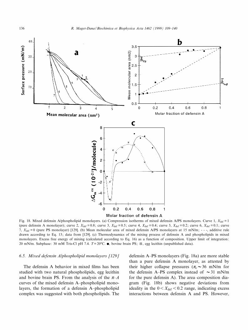
6.5. Mixed defensin A/phospholipid monolayers [129]
The defensin A behavior in mixed ¢lms has beenstudied with two natural phospholipids, egg lecithinand bovine brain PS. From the analysis of the Z^Acurves of the mixed defensin A^phospholipid mono-layers, the formation of a defensin A^phospholipidcomplex was suggested with both phospholipids. The
defensin A^PS monolayers (Fig. 18a) are more stablethan a pure defensin A monolayer, as attested bytheir higher collapse pressures (ZcV36 mN/m forthe defensin A^PS complex instead of V31 mN/mfor the pure defensin A). The area^composition dia-gram (Fig. 18b) shows negative deviations fromideality in the 06Xdef 6 0.2 range, indicating excessinteractions between defensin A and PS. However,
Fig. 18. Mixed defensin A/phospholipid monolayers. (a) Compression isotherms of mixed defensin A/PS monolayers. Curve 1, Xdef = 1(pure defensin A monolayer); curve 2, Xdef = 0.8; curve 3, Xdef = 0.5; curve 4, Xdef = 0.4; curve 5, Xdef = 0.2; curve 6, Xdef = 0.1; curve7, Xdef = 0 (pure PS monolayer) [129]. (b) Mean molecular area of mixed defensin A/PS monolayers at 15 mN/m; - - -, additive ruledrawn according to Eq. 13; data from [129]. (c) Thermodynamics of the mixing process of defensin A and phospholipids in mixedmonolayers. Excess free energy of mixing (calculated according to Eq. 16) as a function of composition. Upper limit of integration:20 mN/m. Subphase: 30 mM Tris-Cl pH 7.4. T = 20³C. b, bovine brain PS; w, egg lecithin (unpublished data).
BBAMEM 77747 30-11-99 Cyaan Magenta Geel Zwart
R. Maget-Dana / Biochimica et Biophysica Acta 1462 (1999) 109^140136
large positive deviations in the defensin-rich region(Xdefv0.5) suggest that the defensin A^PS complexesform clusters in the defensin A monolayers. In thisregion, the partial molecular area of PS is roughlythe same as the area of defensin A, indicating that inthe complex, the PS molecules are intimately boundto defensin A and occupy the same surface. In orderto determine precisely the stoichiometry of the defen-sin A^phospholipid complexes, we calculated the ex-cess energy of mixing vGex
m according to Eq. 16. Theresults are shown in Fig. 18c. The vGex
m ^compositionplot presents a deep and narrow minimum at thesame XdefV0.1 in both cases (egg lecithin and PS),indicating the formation of 1:9 defensin A^phospho-lipid complexes. However, the defensin A^PS com-plex is energetically more stable than the complexwith lecithin since vGex
mV35U10321 J/molecule forthe defensin A^PS complex instead of 32U10321
J/molecule in the case of the defensin A^lecithincomplex. In the 0.29Xdef 6 1 range, the excessenergy of mixing presents positive values, indicatingthe formation of bidimensional aggregates either ofthe defensin A^phospholipid complex or of defensinA itself. In the case of defensin A/PS monolayers, the(vGex
m ^Xdef ) plot presents a plateau at 2U10321
J/molecule before reaching high energy values, sug-gesting that there may be two steps in the aggrega-tion process.
It should be noted that: (1) the peptide rich-regionof the composition diagrams is generally devoid ofinterest since the aim of the study was the determi-nation of the peptide behavior in lipid membranes. Itis obvious in this study that it would have been pref-erable to obtain more values in the 06Xdef 6 0.2range. As a matter of fact, the small number of val-ues in the lipid-rich region leads to an uncertainty onthe complex stoichiometry which actually varies be-tween 1:9 and 1:4; (2) on the basis of area^compo-sition and transition pressure^composition diagramsonly, we previously concluded that the defensin A^phospholipid complexes have a 1:4 stoichiometry.The energy analysis presented here suggests that theactual value is nearer 1:9 than 1:4. This last resultagrees well with the ¢ndings of D. Sy (personal com-munication) deduced from investigation of the elec-trostatic potential surfaces of defensin A. As a mat-ter of fact, one of the molecular faces is markedlymore electropositive than the others with its side
chains directed outwards. This positive face containsnot only the L-sheet Arg-26, Arg-39 and Lys-33 cat-ionic residues as well as the hydrophilic residues Asn-31 and Asn-40, but also the His-19 and Arg-23 res-idues belonging to the K-helix. Therefore, sevenanionic lipids at least could interact strongly withthis particular face of defensin A.
7. Conclusion
The monolayer technique has proved to be an in-valuable approach for the study of the physicochem-ical properties of peptides at hydrophilic/hydropho-bic interfaces. Moreover, the lipid monolayerprovides a unique model membrane of great interestand reliability in understanding the insertion mecha-nism of antimicrobial peptides into cell membranes.We have restricted this review to the basic Langmuir¢lm balance experiments, but it should be pointedout that numerous complementary techniques havebeen developed with the aim of improving and ex-tending the monolayer technique possibilities. Thus,various spectroscopic methods allow direct observa-tion of the phase behavior of the peptide or lipidmonolayer during compression: among them, epi-£uorescence microscopy [100,143] and Brewster anglemicroscopy [143] are the most currently employed.Similarly, in situ infrared spectroscopy allows thedetermination of the structure and orientation ofthe peptide at the interface [134,144]. The Lang-muir^Blodgett technique [145] has long been usedto transfer monolayers onto solid supports. Trans-ferred monolayers can be studied by spectroscopicmethods such as CD [146^148], IR [120,147,148],X-ray di¡raction [149] or atomic force microscopy[150]. This list is not exhaustive, but is given onlyto point out the wide range of technologies devel-oped around the monolayer technique and anotherreview could be devoted to such a subject.
Acknowledgements
I wish to thank Denise Sy for her valuable help inthe PDB structure search and representation. Ourfruitful discussions on androctonin and defensin Abehavior were always greatly appreciated.
BBAMEM 77747 30-11-99 Cyaan Magenta Geel Zwart
R. Maget-Dana / Biochimica et Biophysica Acta 1462 (1999) 109^140 137
References
[1] C.L. Bevins, M. Zaslo¡, Annu. Rev. Biochem. 59 (1990)395^414.
[2] C.E. Dempsey, Biochim. Biophys. Acta 1041 (1990) 143^161.[3] J.A. Ho¡mann, Ch. Hetru, Immunol. Today 13 (1992) 411^
415.[4] I. Cornut, E. Thiaudie©re, J. Dufourcq, in: R.M. Epand
(Ed.), The Amphipathic Helix, CRC Press, 1993, pp. 173^219.
[5] S. Cociancich, P. Bulet, Ch. Hetru, J.A. Ho¡mann, Para-sitol. Today 10 (1994) 132^139.
[6] A.G. Rao, Mol. Plant-Microbe Interact. 8 (1995) 6^13.[7] H.G. Boman, Annu. Rev. Immunol. 13 (1995) 61^92.[8] W.L. Maloy, U.P. Kari, Biopolymers 37 (1995) 105^122.[9] H.-G. Sahl, R.W. Jack, G. Bierbaum, Eur. J. Biochem. 230
(1995) 827^853.[10] S.H. White, W.C. Winley, M.E. Selsted, Curr. Opin. Struct.
Biol. 5 (1995) 521^527.[11] R.I. Lehrer, T. Ganz, Ann. New York Acad. Sci. 797 (1996)
228^239.[12] R.E.W. Hancock, Lancet 349 (1997) 418^422.[13] B. Bechinger, J. Membr. Biol. 156 (1997) 197^211.[14] W.F. Broekaert, B.P. Cammue, M.F. De Bolle, K. Thevis-
sen, G.W. De Samblanx, R.W. Osborn, Crit. Rev. Plant Sci.16 (1997) 297^323.
[15] P.M. Hwang, H.J. Vogel, Biochem. Cell Biol. 76 (1998) 235^246.
[16] R.E.W. Hancock, R. Lehrer, Trends Biotechnol. 16 (1998)82^87.
[17] K. Matsuzaki, Biochim. Biophys. Acta 1376 (1998) 391^400.[18] J.M. Schro«der, Biochem. Pharmacol. 57 (1999) 121^134.[19] A.W. Bernheimer, Biochim. Biophys. Acta 344 (1974) 27^50.[20] E. Katz, A.L. Demain, Bacteriol. Rev. 41 (1977) 449^474.[21] L. Ehret-Sabatier, D. Loew, M. Goy¡on, P. Fehlbaum, J.A.
Ho¡mann, A. van Dorsselaer, Ph. Bulet, J. Biol. Chem. 271(1996) 29537^29544.
[22] J. Lambert, E. Keppi, J.L. Dimarcq, C. Wicker, J.M. Reich-hart, B. Dunbar, P. Lepage, A. van Dorsselaer, J. Ho¡mann,J. Fothergill, D. Ho¡mann, Proc. Natl. Acad. Sci. USA 86(1989) 262^266.
[23] B. Cornet, J.-M. Bonmatin, Ch. Hetru, J.A. Ho¡mann, M.Ptak, F. Vovelle, Structure 3 (1995) 435^448.
[24] M. Zaslo¡, B. Martin, H.C. Chen, Proc. Natl. Acad. Sci.USA 85 (1988) 910^913.
[25] P. Eisenhauer, S. Harwig, R. Lehrer, Infect. Immunol. 60(1992) 3556^3565.
[26] E. Habermann, J. Jentsch, Hoppe-Seyler's Z Physiol. Chem.348 (1967) 37^50.
[27] A. Argolias, J. Pisano, J. Biol. Chem. 260 (1985) 1437^1444.[28] A.I. Louw, Biochim. Biophys. Acta 336 (1974) 470^480.[29] E. Gross, J.L. Morrell, J. Am. Chem. Soc. 93 (1970) 4634^
4635.[30] G. Buchman, S. Banerjee, J. Hansen, J. Biol. Chem. 263
(1988) 16260^16266.[31] J. Fitton, A. Dell, W. Shaw, FEBS Lett. 115 (1980) 209^212.
[32] R. Sarges, B. Witkop, J. Am. Chem. Soc. 87 (1965) 2011^2020.
[33] E. Katz, A.L. Demain, Bacteriol. Rev. 41 (1977) 449^474.[34] J.F. Faucon, J. Dufourcq, C. Lussan, FEBS Lett. 102 (1979)
187^190.[35] M. Jackson, H. Mantsch, J. Spencer, Biochemistry 31 (1992)
7289^7293.[36] R. Urrutia, R.A. Cruciani, J.L. Barker, B. Kachar, FEBS
Lett. 247 (1988) 17^21.[37] J.E. Fitton, FEBS Lett. 102 (1981) 257^260.[38] E. Thiaudie©re, O. Si¡ert, J.-C. Talbot, J. Bolard, J.E. Alouf,
J. Dufourcq, Eur. J. Biochem. 195 (1991) 203^213.[39] E. Bairaktari, D.F. Mierke, S. Mammi, E. Peggion, Bio-
chemistry 29 (1990) 10097^10102.[40] R. Maget-Dana, J.-M. Bonmatin, Ch. Hetru, M. Ptak, J.-C.
Maurizot, Biochimie 77 (1995) 240^244.[41] H. Nikaido, in: Membrane Transport and Information Stor-
age, Alan R. Liss, 1990, pp. 165^190.[42] K. Matsuzaki, K.I. Sugishita, N. Fujii, K. Miyajima, Bio-
chemistry 34 (1995) 3423^3429.[43] K.L. Piers, M.H. Brown, R.E.W. Hancock, Antimicrob.
Agents Chemother. 38 (1994) 2311.[44] B.L. Kagan, M.E. Selsted, T. Ganz, R.I. Lehrer, Proc. Natl.
Acad. Sci. USA 87 (1990) 210^214.[45] S. Cociancich, A. Ghazi, Ch. Hetru, J.A. Ho¡mann, L. Le-
tellier, J. Biol. Chem. 268 (1993) 19239^19245.[46] H. Duclohier, G. Molle, G. Spach, Biophys. J. 56 (1989)
1017^1021.[47] M.T. Tosteson, D.C. Tosteson, Biophys. J. 36 (1981) 109^
116.[48] M.J. Garcia Garcera, M.G. Elferink, A.J. Driessen, W.N.
Konings, Eur. J. Biochem. 212 (1993) 417^428.[49] C.P. Hill, J. Yee, M.E. Selsted, D. Eisenberg, Science 251
(1991) 1481^1485.[50] D.W. Urry, Proc. Natl. Acad. Sci. USA 68 (1972) 672^676.[51] G. Spach, H. Duclohier, G. Molle, J.-M. Valleton, Biochi-
mie 71 (1989) 11^21.[52] W.L. Duax, J.F. Gri¤n, D.A. Langs, G.D. Smith, P. Gro-
chulski, V. Pletnev, V. Ivanov, Biopolymers 40 (1996) 141^155.
[53] A.W. Adamson, Physical Chemistry of Surfaces, 3rd edn.,J. Wiley and Sons, New York, 1976.
[54] G.L. Gaines, Insoluble Monolayers at Liquid^Gas Interfa-ces, Prigogine (Ed.), J. Wiley and Sons, New York, 1966.
[55] J.W. McBain, R.C. Bacon, H.D. Bruce, J. Chem. Phys. 7(1939) 818^821.
[56] J.W. Gibbs, Collected Works, Vol. 1, Longmans, New York,1931.
[57] R. Bresow, T. Guo, Proc. Natl. Acad. Sci. USA 87 (1990)167^169.
[58] R. Pethig, Dielectric and Electronic Properties of BiologicalMaterials, Wiley, New York, 1979.
[59] K. Birdi, V. Gevod, O. Ksenzhek, E. Stenby, K. Rasmussen,Colloid Polymer Sci. 261 (1983) 767^775.
[60] N. Lakhsminarayanaiah, Equations of Membrane Bio-physics, Academic Press, London, 1984.
BBAMEM 77747 30-11-99 Cyaan Magenta Geel Zwart
R. Maget-Dana / Biochimica et Biophysica Acta 1462 (1999) 109^140138
[61] R. Miller, G. Kretzschmar, Adv. Colloid Interface Sci. 37(1991) 97^121.
[62] F. MacRichtie, Colloids Surfaces A. Physicochem. Eng. As-pects 76 (1993) 159^166.
[63] A.F. Ward, L. Tordai, J. Chem. Phys. 14 (1946) 453^461.[64] R. Maget-Dana, D. Lelie©vre, A. Brack, Biopolymers 49
(1999) 415^423.[65] D.E. Graham, M.C. Phillips, J. Colloid Interface Sci. 70
(1979) 403^414.[66] R. Maget-Dana, M. Ptak, Colloids Surfaces B: Biointerfaces
7 (1996) 135^143.[67] P. Suttiprasit, V. Krisdhasima, J. McGuire, J. Colloid Inter-
face Sci. 154 (1992) 316^326.[68] P. Bougis, H. Rochat, G. Pieroni, R. Verger, Biochemistry
20 (1981) 4915^4920.[69] O.S. Ksenzhek, V.S. Gevod, A.M. Omel'chenko, S.N. Seme-
nov, A.I. Sotnichenko, Mol. Biol. (Mosk.) 12 (1978) 1057^1065.
[70] E. Schro«der, K. Lu«bke, M. Lehmann, I. Beetz, Experientia27 (1971) 764^765.
[71] A.S. Tatham, R.C. Hider, A.F. Drake, Biochem. J. 211(1983) 683^686.
[72] T.C. Terwilliger, L. Weissman, D. Eisenberg, Biochim. Bio-phys. Acta 37 (1982) 353^361.
[73] G.D. Fidelio, B. Maggio, F.A. Cumar, Biochem. J. 203(1982) 717^725.
[74] M.J. Dufton, R.C. Hider, Pharmacol. Ther. 36 (1988) 1^40.[75] W. Jahnke, D.F. Mierke, L. Beress, H. Kessler, J. Mol. Biol.
240 (1994) 445^458.[76] N. Mandard, D. Sy, C. Maufrais, J.-M. Bonmatin, Ph. Bu-
let, Ch. Hetru, F. Vovelle, J. Biomol. Struct. Dynam. 17(1999) 367^380.
[77] F. Uraizee, G. Narsimhan, J. Colloid Interface Sci. 146(1991) 169^178.
[78] R. Maget-Dana, A. Brack, D. Lelie©vre, Supramol. Sci. 4(1997) 365^368.
[79] R. Maget-Dana, Ch. Hetru, M. Ptak, Thin Solid Films 284/285 (1996) 841^844.
[80] G. Colacicco, M.K. Basu, A.R. Buchelew, A.W. Bernheimer,Biochim. Biophys. Acta 465 (1977) 378^390.
[81] C.W. Cumper, A.E. Alexander, Trans. Faraday Soc. 46(1950) 235^238.
[82] M. Bhakoo, T.H. Birbeck, J.H. Freer, Biochemistry 21(1982) 6879^6883.
[83] G. Schwarz, S. Taylor, Langmuir 11 (1995) 4341^4346.[84] K.S. Birdi, Lipid and Biopolymer Monolayers at Liquid In-
terfaces, Plenum Press, New York, 1989.[85] J.W. Taylor, Biochemistry 29 (1990) 5364^5373.[86] K. Kobayashi, J.R. Granja, M.R. Ghadiri, Angew. Chem.
Int. Ed. Engl. 34 (1995) 95^98.[87] K. Birdi, J. Colloid Interface Sci. 43 (1973) 545^547.[88] G. Kemp, K.A. Jacobson, C.E. Wenner, Biochim. Biophys.
Acta 255 (1972) 493^501.[89] N. Davion-Van Mau, P. Daumas, D. Lelie©vre, Y. Trudelle,
F. Heitz, Biophys. J. 51 (1987) 843^845.
[90] H. Tournois, P. Gieles, R. Demel, J. de Gier, B. de Kruij¡,Biophys. J. 55 (1989) 557^569.
[91] H.E. Ries, H. Swift, Colloids Surfaces 40 (1989) 145^165.[92] P. Lavigne, P. Tancre©de, F. Lamarche, J.-J. Max, Langmuir
8 (1992) 1988^1993.[93] W. Veatch, E. Fossel, E. Blout, Biochemistry 13 (1974)
5249^5256.[94] R. Brasseur, J.A. Killian, B. de Kruij¡, J.M. Ruysschaert,
Biochim. Biophys. Acta 903 (1987) 11^17.[95] B.R. Malcolm, Prog. Surf. Membr. Sci. 7 (1973) 183^229.[96] T. Vogt, J. Killian, R. Demel, B. De Kruij¡, Biochim.
Biophys. Acta 1069 (1991) 157^164.[97] D. Jones, E. Hayon, D. Busath, Biochim. Biophys. Acta
861 (1986) 62^67.[98] W.F. DeGrado, F.J. Kezdy, E.T. Kaiser, J. Am. Chem.
Soc. 103 (1981) 679^681.[99] G. Sessa, J.H. Freer, G. Colacicco, G. Weissmann, J. Biol.
Chem. 244 (1969) 3575^3582.[100] G. Signor, S. Mammi, E. Peggion, H. Ringsdorf, A. Wa-
genknecht, Biochemistry 33 (1994) 6659^6670.[101] G. Wackerbauer, I. Weis, G. Schwarz, Biophys. J. 71 (1996)
1422^1427.[102] V. Gevod, K. Birdi, Biophys. J. 45 (1984) 1079^1083.[103] G.D. Fidelio, B. Maggio, F.A. Cumar, Biochim. Biophys.
Acta 862 (1986) 49^56.[104] V. Gevod, K. Birdi, Colloid Polymer Sci. 265 (1987) 257^
261.[105] H.T. Tien, Bilayer Lipid Membranes (BLM): Theory and
Practice, M. Dekker, 1974.[106] G.D. Eytan, Biochim. Biophys. Acta 694 (1982) 185^202.[107] R.A. Demel, Subcell. Biochem. 23 (1994) 83^120.[108] H. Brockman, Curr. Opin. Struct. Biol. 9 (1999) 438^443.[109] D. Marsh, Biochim. Biophys. Acta 1286 (1996) 183^223.[110] S. Feng, Langmuir 15 (1999) 998^1010.[111] B. Pethica, Trans Faraday Soc. 51 (1955) 1402^1411.[112] H.S. Hendrickson, P. Fan, D. Kaufman, D. Kleiner, Arch.
Biochem. Biophys. 227 (1983) 242^247.[113] C.R. Flach, F.G. Prendergast, R. Mendelsohn, Biophys. J.
70 (1996) 539^546.[114] S. Ohki, E. Marcus, D.K. Sukumaran, K. Arnold, Biochim.
Biophys. Acta 1194 (1994) 223^232.[115] A.F. Mingotaud, C. Mingotaud, L.K. Patterson, in: Hand-
book of Monolayers, Academic Press, New York, 1993, pp.778^986.
[116] B. Stanislawski, H. Ru«terjans, Eur. Biophys. J. 15 (1987) 1^12.
[117] G. Schwarz, G. Beschiaschvili, Biochim. Biophys. Acta 979(1989) 82^90.
[118] Y. Ebara, Y. Okahata, Langmuir 9 (1993) 574^576.[119] J. Bello, H.R. Bello, E. Granados, Biochemistry 21 (1982)
461^465.[120] S.-F. Sui, H. Wu, Y. Guo, K.-S. Chen, J. Biochem. 116
(1994) 482^487.[121] C. Altenbach, W. Froncisz, J. Hyde, W. Hubbel, Biophys.
J. 56 (1989) 1183^1191.
BBAMEM 77747 30-11-99 Cyaan Magenta Geel Zwart
R. Maget-Dana / Biochimica et Biophysica Acta 1462 (1999) 109^140 139
[122] C. Dempley, G. Butler, Biochemistry 31 (1992) 11973^11977.
[123] M. Pezolet, L. Duchesneau, P. Bougis, J.F. Faucon, J. Du-fourcq, Biochim. Biophys. Acta 704 (1982) 515^523.
[124] P. Bougis, M. Tessier, J. Van Rietschoten, H. Rochat, J.F.Faucon, J. Dufourq, Mol. Cell. Biochem. 55 (1983) 49^64.
[125] J. Delves-Broughton, J. Soc. Dairy Technol. 43 (1990) 73^76.
[126] O. Kuipers, H. Rollema, W. Yap, H. Boot, R. Siezen, W.De Vos, J. Biol. Chem. 267 (1992) 24340^24346.
[127] A.R. Demel, T. Peelen, R. Siezen, B. De Kruij¡, O.Kuipers, Eur. J. Biochem. 235 (1996) 267^274.
[128] E. Breukink, C. van Kraaij, R. Demel, R. Siezen, O.Kuipers, B. de Kruij¡, Biochemistry 36 (1997) 6968^6976.
[129] R. Maget-Dana, M. Ptak, Biophys. J. 73 (1997) 2527^2533.[130] Ch. Hetru, L. Letellier, Z. Oren, J.A. Ho¡mann, Y. Shai,
Biochem. J., in press.[131] E. Gazit, A. Boman, H. Boman, Y. Shai, Biochemistry 34
(1995) 11470^11488.[132] I. Costin, G. Barnes, J. Colloid Interface Sci. 51 (1975)
106^121.[133] K. Bacon, G. Barnes, J. Colloid Interface Sci. 67 (1978) 70^
77.[134] W.P. Ulrich, H. Vogel, Biophys. J. 76 (1999) 1639^1647.[135] B.A. Wallace, Biophys. J. 49 (1986) 295^306.[136] N. Van Mau, Y. Trudelle, P. Daumas, F. Heitz, Biophys. J.
54 (1988) 563^567.
[137] A. Benayad, D. Benamar, N. Van Mau, G. Page, F. Heitz,Eur. Biophys. J. 20 (1991) 209^213.
[138] N. Van Mau, B. Bonnet, A. Benayad, F. Heitz, Eur. Bio-phys. J. 22 (1994) 447^452.
[139] V.L. Shapovalov, E.A. Kotova, T.I. Rokitskaya, Y.N. An-tonenko, Biophys. J. 77 (1999) 299^305.
[140] B.A. Wallace, R.W. Janes, J. Mol. Biol. 217 (1991) 625^627.
[141] J. Dufourcq, J.F. Faucon, G. Fourche, J.J. Dasseux, M. LeMaire, T. Gulik-Krzywicki, Biochim. Biophys. Acta 859(1986) 33^46.
[142] A.M. Batenburg, J.C. Hibbeln, A.J. Verkleij, B. de Kruij¡,Biochim. Biophys. Acta 903 (1987) 142^154.
[143] M. Lipp, K.Y.C. Lee, A. Waring, J.A. Zsadzinski, Biophys.J. 72 (1997) 2783^2804.
[144] S. Castano, B. Desbat, M. Laguerre, J. Dufourq, Biochim.Biophys. Acta 1416 (1999) 176^194.
[145] G.G. Roberts, Langmuir^Blodgett Films, Plenum, New-York, 1990.
[146] D.G. Cornell, J. Colloid Interface Sci. 70 (1979) 167^180.[147] R. Maget-Dana, G. Bolbach, Y. Trudelle, Biopolymers 35
(1994) 629^637.[148] M. Boncheva, H. Vogel, Biophys. J. 73 (1997) 1056^1072.[149] S. Ludtke, K. He, H. Huang, Biochemistry 34 (1995)
16764^16769.[150] N. Van Mau, V. Vie, L. Chaloin, E. Lesniewska, F. Heitz,
C. Le Grimellec, J. Membr. Biol. 167 (1999) 241^249.
BBAMEM 77747 30-11-99 Cyaan Magenta Geel Zwart
R. Maget-Dana / Biochimica et Biophysica Acta 1462 (1999) 109^140140
Related Documents