J. Clin. Med. 2022, 11, 2513. https://doi.org/10.3390/jcm11092513 www.mdpi.com/journal/jcm Review The Leading Role of the Immune Microenvironment in Multiple Myeloma: A New Target with a Great Prognostic and Clinical Value Vanessa Desantis 1, *, Francesco Domenico Savino 1,† , Antonietta Scaringella 1,† , Maria Assunta Potenza 1 , Carmela Nacci 1 , Maria Antonia Frassanito 2 , Angelo Vacca 3 and Monica Montagnani 1 1 Department of Biomedical Sciences and Human Oncology, Pharmacology Section, University of Bari Aldo Moro Medical School, 70124 Bari, Italy; [email protected] (F.D.S.); [email protected] (A.S.); [email protected] (M.A.P.); [email protected] (C.N.); [email protected] (M.M.) 2 Unit of General Pathology, Department of Biomedical Sciences and Human Oncology, University of Bari Aldo Moro Medical School, 70124 Bari, Italy; [email protected] 3 Unit of Internal Medicine and Clinical Oncology, Department of Biomedical Sciences and Human Oncology, University of Bari Aldo Moro Medical School, 70124 Bari, Italy; [email protected] * Correspondence: [email protected]; Tel.: +39‐080‐5478452 † These authors contributed equally to this work. Abstract: Multiple myeloma (MM) is a plasma cell (PC) malignancy whose development flourishes in the bone marrow microenvironment (BMME). The BMME components’ immunoediting may foster MM progression by favoring initial immunotolerance and subsequent tumor cell escape from immune surveillance. In this dynamic process, immune effector cells are silenced and become progressively anergic, thus contributing to explaining the mechanisms of drug resistance in unresponsive and relapsed MM patients. Besides traditional treatments, several new strategies seek to re‐establish the immunological balance in the BMME, especially in already‐treated MM patients, by targeting key components of the immunoediting process. Immune checkpoints, such as CXCR4, T cell immunoreceptor with immunoglobulin and ITIM domains (TIGIT), PD‐1, and CTLA‐4, have been identified as common immunotolerance steps for immunotherapy. B‐cell maturation antigen (BCMA), expressed on MMPCs, is a target for CAR‐T cell therapy, antibody‐(Ab) drug conjugates (ADCs), and bispecific mAbs. Approved anti‐CD38 (daratumumab, isatuximab), anti‐VLA4 (natalizumab), and anti‐SLAMF7 (elotuzumab) mAbs interfere with immunoediting pathways. New experimental drugs currently being evaluated (CD137 blockers, MSC‐derived microvesicle blockers, CSF‐1/CSF‐1R system blockers, and Th17/IL‐17/IL‐17R blockers) or already approved (denosumab and bisphosphonates) may help slow down immune escape and disease progression. Thus, the identification of deregulated mechanisms may identify novel immunotherapeutic approaches to improve MM patients’ outcomes. Keywords: multiple myeloma; bone marrow niche; immune escape; immune exhaustion; immune checkpoint inhibitors; immune microenvironment; immunotherapy 1. Introduction Multiple myeloma (MM) is a neoplastic plasma cell (PC) disorder characterized by clonal proliferation of malignant PCs in the bone marrow microenvironment (BMME). The abnormal and uncontrolled proliferation of PCs translates into the accumulation of monoclonal proteins in the blood, urine, and tissues with associated organ dysfunction [1]. The clinical onset of MM is often preceded by an asymptomatic premalignant condition called monoclonal gammopathy of undetermined significance (MGUS). MGUS, in turn, can evolve into smoldering MM (SMM), an intermediate phase in which PC Citation: Desantis, V.; Savino, F.D.; Scaringella, A.; Potenza, M.A.; Nacci, C.; Frassanito, M.A.; Vacca, A.; Montagnani, M. The Leading Role of the Immune Microenvironment in Multiple Myeloma: A New Target with a Great Prognostic and Clinical Value. J. Clin. Med. 2022, 11, 2513. https://doi.org/10.3390/jcm11092513 Academic Editors: Antonio G. Solimando and Tadeusz Robak Received: 4 April 2022 Accepted: 28 April 2022 Published: 29 April 2022 Publisher’s Note: MDPI stays neutral with regard to jurisdictional claims in published maps and institutional affiliations. Copyright: © 2022 by the authors. Licensee MDPI, Basel, Switzerland. This article is an open access article distributed under the terms and conditions of the Creative Commons Attribution (CC BY) license (https://creativecommons.org/license s/by/4.0/).

Welcome message from author
This document is posted to help you gain knowledge. Please leave a comment to let me know what you think about it! Share it to your friends and learn new things together.
Transcript

J. Clin. Med. 2022, 11, 2513. https://doi.org/10.3390/jcm11092513 www.mdpi.com/journal/jcm
Review
The Leading Role of the Immune Microenvironment
in Multiple Myeloma: A New Target with a Great Prognostic
and Clinical Value
Vanessa Desantis 1,*, Francesco Domenico Savino 1,†, Antonietta Scaringella 1,†, Maria Assunta Potenza 1,
Carmela Nacci 1, Maria Antonia Frassanito 2, Angelo Vacca 3 and Monica Montagnani 1
1 Department of Biomedical Sciences and Human Oncology, Pharmacology Section, University of Bari Aldo
Moro Medical School, 70124 Bari, Italy; [email protected] (F.D.S.); [email protected] (A.S.);
[email protected] (M.A.P.); [email protected] (C.N.);
[email protected] (M.M.) 2 Unit of General Pathology, Department of Biomedical Sciences and Human Oncology, University of Bari
Aldo Moro Medical School, 70124 Bari, Italy; [email protected] 3 Unit of Internal Medicine and Clinical Oncology, Department of Biomedical Sciences and Human Oncology,
University of Bari Aldo Moro Medical School, 70124 Bari, Italy; [email protected]
* Correspondence: [email protected]; Tel.: +39‐080‐5478452
† These authors contributed equally to this work.
Abstract: Multiple myeloma (MM) is a plasma cell (PC) malignancy whose development flourishes
in the bone marrow microenvironment (BMME). The BMME components’ immunoediting may
foster MM progression by favoring initial immunotolerance and subsequent tumor cell escape from
immune surveillance. In this dynamic process, immune effector cells are silenced and become
progressively anergic, thus contributing to explaining the mechanisms of drug resistance in
unresponsive and relapsed MM patients. Besides traditional treatments, several new strategies seek
to re‐establish the immunological balance in the BMME, especially in already‐treated MM patients,
by targeting key components of the immunoediting process. Immune checkpoints, such as CXCR4,
T cell immunoreceptor with immunoglobulin and ITIM domains (TIGIT), PD‐1, and CTLA‐4, have
been identified as common immunotolerance steps for immunotherapy. B‐cell maturation antigen
(BCMA), expressed on MMPCs, is a target for CAR‐T cell therapy, antibody‐(Ab) drug conjugates
(ADCs), and bispecific mAbs. Approved anti‐CD38 (daratumumab, isatuximab), anti‐VLA4
(natalizumab), and anti‐SLAMF7 (elotuzumab) mAbs interfere with immunoediting pathways.
New experimental drugs currently being evaluated (CD137 blockers, MSC‐derived microvesicle
blockers, CSF‐1/CSF‐1R system blockers, and Th17/IL‐17/IL‐17R blockers) or already approved
(denosumab and bisphosphonates) may help slow down immune escape and disease progression.
Thus, the identification of deregulated mechanisms may identify novel immunotherapeutic
approaches to improve MM patients’ outcomes.
Keywords: multiple myeloma; bone marrow niche; immune escape; immune exhaustion; immune
checkpoint inhibitors; immune microenvironment; immunotherapy
1. Introduction
Multiple myeloma (MM) is a neoplastic plasma cell (PC) disorder characterized by
clonal proliferation of malignant PCs in the bone marrow microenvironment (BMME).
The abnormal and uncontrolled proliferation of PCs translates into the accumulation of
monoclonal proteins in the blood, urine, and tissues with associated organ dysfunction
[1]. The clinical onset of MM is often preceded by an asymptomatic premalignant
condition called monoclonal gammopathy of undetermined significance (MGUS). MGUS,
in turn, can evolve into smoldering MM (SMM), an intermediate phase in which PC
Citation: Desantis, V.; Savino, F.D.;
Scaringella, A.; Potenza, M.A.;
Nacci, C.; Frassanito, M.A.; Vacca,
A.; Montagnani, M. The Leading
Role of the Immune
Microenvironment in Multiple
Myeloma: A New Target
with a Great Prognostic and Clinical
Value. J. Clin. Med. 2022, 11, 2513.
https://doi.org/10.3390/jcm11092513
Academic Editors: Antonio G.
Solimando and Tadeusz Robak
Received: 4 April 2022
Accepted: 28 April 2022
Published: 29 April 2022
Publisher’s Note: MDPI stays
neutral with regard to jurisdictional
claims in published maps and
institutional affiliations.
Copyright: © 2022 by the authors.
Licensee MDPI, Basel, Switzerland.
This article is an open access article
distributed under the terms and
conditions of the Creative Commons
Attribution (CC BY) license
(https://creativecommons.org/license
s/by/4.0/).
J. Clin. Med. 2022, 11, 2513 2 of 20
expansion and gene mutations increase the risk of evolution to active MM [2–4]. MM
exhibits broad heterogeneity in clinical presentation, molecular features, and treatment
effectiveness [5]. Current MM therapies are based on a combination of conventional
chemotherapy, corticosteroids, and one or more of the newer agents—such as proteasome
inhibitors (i.e., bortezomib, carfilzomib, and ixazomib), checkpoint inhibitors,
immunomodulating compounds (i.e., lenalidomide, thalidomide, and pomalidomide)—
or biological therapies, including monoclonal antibodies (mAbs) (i.e., daratumumab and
elotuzumab) and chimeric antigen receptor (CAR)‐T cell therapy. The leading role of the
BMME components in MM progression and heterogeneity suggests that further
characterization of its specific activities may help identify key therapeutic targets and
foster the development of new approaches that aim to reinforce the immune system.
The BMME includes a non‐cellular compartment formed by extracellular matrix
(ECM) proteins (laminin, fibronectin, and collagen) and soluble factors (cytokines, growth
factors, and chemokines) and a rich cellular compartment constituting hematopoietic cells
(myeloid cells, T lymphocytes, B lymphocytes, and natural killer (NK) cells) and non‐
hematopoietic cells (fibroblasts (FBs), osteoblasts, osteoclasts, endothelial cells (ECs),
endothelial progenitor cells (EPCs), dendritic cells (DCs), pericytes, mesenchymal stem
cells (MSCs), and mesenchymal stromal cells) [6]. In this specialized BM niche, all cells are
protected from apoptotic stimuli and may, therefore, actively promote disease
progression. Since the BM niche is the primary residence of long‐lived PCs, the complex
interaction among its cellular components, ECM proteins, and soluble factors may play a
major role in the survival of malignant PCs [7].
The immune system acts as a critical rheostat that fine‐tunes the balance between
dormancy and disease progression in MM. Even if malignant PCs are not completely
eliminated, the immune system is critical for maintaining functional dormancy at early stages;
however, malignant PCs eventually evade immune control and foster progression toward
active MM, in which dysfunctional effector lymphocytes, tumor‐educated
immunosuppressive cells, and soluble mediators act in coordination as a barrier against anti‐
MM immune response. An in‐depth understanding of this dynamic process, known as
“cancer immunoediting”, will provide important insights into the immunopathology of PC
dyscrasias and, hopefully, help organize the most effective anti‐MM immunotherapy [8].
Interestingly, a growing body of evidence suggests that a complex interaction
between non‐hematopoietic stromal cells and the BM immune system may display unique
functions to support pro‐ and anti‐tumor events in the BM niche, thus highlighting the
relevant roles of immune components in the impaired anti‐MM immune responses and
disease progression [9].
Among BM immune cells, MSCs have long been recognized as key players in
immune response, actively promoting the homing of PCs in the BM by secreting C‐X‐C
Motif Chemokine Ligand 12 (CXCL12) (CXCR4 ligand), hence providing contact‐
dependent support for PCs by integrins and enhancing the secretion of pro‐survival
factors, such as interleukin‐6 (IL‐6) and vascular endothelial growth factor (VEGF). The
reciprocal interactions between PCs and MSCs induce MM progression [10]. According to
previous studies, MM‐educated MSCs acquire the ability to produce high numbers of pro‐
inflammatory cytokines and growth factors that favor the accumulation and
chemoresistance of malignant PCs [11,12]. These observations suggest that MM evolution
might progressively define unique and complex immune phenotypes in the BM
components, including bystander immune cells.
Notably, immune changes represented by an increased number of terminal effector
T cells and group 1 innate lymphoid cells can be observed from the MGUS stage to active
MM [13]. Moreover, although stem‐like tissue‐resident T cells can still be detected in
MGUS patients, subjects with advanced MM are characterized by the progressive loss of
this T cell subset and the accumulation of senescent and exhausted T cells, suggesting that
the T cell phenotype changes dynamically during disease progression [14]. Similarly,
altered polarization of T cells, particularly T helper (Th) 17‐skewed cells, has been
J. Clin. Med. 2022, 11, 2513 3 of 20
reported in patients with active MM and associated with the increased release of IL‐1, IL‐
6, and transforming growth factor‐β (TGF‐β) in the pro‐inflammatory BM niche [15].
Despite some evidence pointing toward an accumulation of hypoproliferative, senescent
CD571 and CD81 T cells in MM, the presence of exhausted T cells in newly diagnosed patients
is controversial. Indeed, CD81 T cells from these patients rarely express high levels of multiple
immune checkpoint receptors (i.e., programmed death‐1 (PD‐1), cytotoxic T‐lymphocyte
associated antigen‐4 (CTLA‐4), TIM‐3, and LAG‐3), which represents a cardinal feature of T
cell exhaustion [16]. In addition, increased levels of PD‐1 on T CD4+, which has been observed
more in relapsed MM patients compared with MM and MGUS ones and is able to interact
with PD‐ligand 1 (PD‐L1) on PCs and DCs, are correlated with MM progression [17].
Moreover, NK cells from MM patients display reduced expression of activating
receptors and parallel upregulation of PD‐1 receptors, the latter facilitating the inhibition
of NK cytotoxicity by MM cells expressing higher levels of PD‐L1 [18].
As professional antigen‐presenting cells (APCs), DCs act as a link between innate and
adaptive immunity. DCs from MM patients are also dysfunctional, are involved in PC
survival, and may be included among the key determinants for the progression from
MGUS to active MM [19].
The specific significance of different immune players in the BMME of MM patients
is still an open field of investigation since the uncertain clinical responses to immune
checkpoints inhibitors make it difficult to identify reliable predictive biomarkers. Next,
we summarize the current knowledge on the importance of immunoediting in MM
progression, focusing on the most common MM immune checkpoints identified so far and
the relevant clinical/prognostic value of specific drug inhibitors.
2. Immunoediting in MM Progression
In MM, the “cancer immunoediting” is attributable to multiple factors, including the
suppressive activity of tumor‐associated macrophages (TAMs) and myeloid‐derived
suppressor cells (MDSCs), the immunotolerance toward malignant cell antigens, progressive
T cell exhaustion, and the alteration in cytokine production [9,20,21]. The complex and
dynamic rearrangement of all these elements occurs throughout three phases (elimination,
equilibrium, and escape), whose combination fosters disease development [22].
The immunosuppression mechanism, reinforced by both tumor cells and the BMME
components, involves lymphocyte effectors, immunosuppressive cells, and immunity‐
dampening molecules. With disease progression, somatic mutations in PCs can be
immunogenic and induce neoantigen‐specific NK and T cell activation. Concomitantly,
the effector cells become silenced and gradually less effective, as confirmed by the
increased numbers of neo‐antigens found in relapsed MM patients compared with newly
diagnosed subjects [23,24]. Compared with the MGUS stage, exhaustion and senescent
signs of T cells appear more evidently in the advanced MM stage [13,14,25].
The elimination phase, which limits PC growth, matches with strong immune
activity against PCs and subsists in a dynamic balance (the intermediate phase) with the
escape stage. The equilibrium phase can last for a long time before switching to the escape
phase; it is characterized by malignant PC dormancy, which may correspond to MGUS
and/or SMM clinical disease appearance. Importantly, in patients undergoing medical
therapies and autologous stem cell transplantation, the switch to the last stage is
potentially a reversible process [9,26,27]. In this context, NK and CD8+ T cells are key
actors in immunoediting evolution, and among the main mediators, perforin and
interferon‐γ (IFN‐γ) as well as the adhesion receptor CD226 (DNAM‐1) are responsible
for the elimination process [28]. When triggered by its stress‐induced ligands
(Nectin2/CD112 and PVR/CD155), which are frequently over‐expressed on malignant PC
surfaces, the DNAM‐1 receptor controls T and NK cell activation [29] and confers
resistance to bortezomib and cyclophosphamide, contributing to slowed paraproteinemia
and enhanced survival in mice models [28]. DNAM‐1 also has a functional role in the NK‐
dependent killing of malignant PCs, strictly depending on the presence of Nectin‐1 and
J. Clin. Med. 2022, 11, 2513 4 of 20
PVR on their surfaces [30]. Compared with patients in remission and healthy controls, a
reduced amount of DNAM‐1 on CD56dim NK cells from patients with active disease was
discovered, explaining the role of NK cells in MM pathogenesis [30]. Like CD226, the NK
group 2D (NKG2D) receptor binds two stress‐induced ligands (major histocompatibility
complex class I‐related chains A and B, MICA/B) and contributes to NK and T cell activity
during the elimination phase, acting in combination with UL16‐binding proteins (ULBPs)
[31]. Overall, the data reveal that alterations in the NKG2D pathway are associated with
the progression from MGUS to active MM [32], indicate that early changes in both innate
and adaptive immunity are in place in MGUS‐stage PC tumors (including an increasing
number of T CD8+ and group 1 innate lymphoid cells), and indicate that a decrease in
stem‐like cells may underlie the loss of immune surveillance in MM progression [13].
The transition from MGUS to active MM also entails changes in inflammatory
molecules. Indeed, the tumor secretion of phlogistic mediators, such as TGF‐β, IL‐10, IL‐
6, and prostaglandin E2 (PGE2), contributes to immunological imbalance and, combined
with non‐tumoral PC dysregulation, exposes MM patients to infections [33]. The
numerical alteration and progressive impairment of the NK cell population toward the
MM stage are related to TGF‐β secretion, which leads to the defective release of INF‐γ and
suppresses antibody‐dependent cellular cytotoxicity (ADCC) [34].
Further support for the role played by the BMME in MM progression comes from the
contribution to the escape mechanism through the osteoclasts’ production of Gal‐9 and
proliferation‐inducing ligand (APRIL), two signaling molecules promoting T cell
apoptosis, and through PD‐L1 expression in MM cells [35]. As previously mentioned, PD‐
L1 expression on malignant PCs has been associated with drug resistance, and serum
levels of PD‐L1 are predictive of worse progression‐free survival (PFS) [36,37].
T and B regulatory cells (Tregs and Bregs), MDSCs, and TAMs all play relevant roles
in MM immune escape by inhibiting the cytotoxic functions of T cells and NK cells and
stimulating angiogenesis and proliferation, thereby promoting disease progression [38].
In the BMME, crosstalk between toll‐like receptor (TLR)‐2 and damage‐associated
molecular patterns (DAMPs) [39] is among the hypothesized mechanisms through which
TAM precursors support MM progression [40]. In this context, S100A9, a fundamental
DAMP that stimulates IL‐18 and promotes MM progression by interacting with TLR‐4
and RAGE [41], fosters a pro‐inflammatory environment in the BM niche [41]. Moreover,
TAM survival and differentiation in the BMME are favored by colony‐stimulating factor‐
1 (CSF‐1), whose increased levels are related to disease progression [42,43].
During the active phase of MM disease, the involvement of BMME in “cancer
immunoediting” becomes more evident: specifically, MSCs upregulate IL‐6 production and
exhibit high levels of CD40/CD40L and adhesion molecules, such as VCAM‐1, ICAM‐1,
LFA‐3, junctional adhesion molecule‐A (JAM‐A), and human leukocyte antigen (HLA)
system molecules (HLA‐DR and HLA‐ABC) [38]. Combined with MSC‐derived
microvesicles, all these elements contribute to immunoescape, drug resistance, and
proliferation mechanisms. This concept is supported by recent findings showing that, by
slowing down PCs’ uptake of microvesicles, integrin inhibitors (targeting α4β1 integrin and
CD29 together) undermine malignant progression [44]. Similarly, a reduced inhibition of T
cells proliferation with a shift in the Th17/Tregs balance when T cells were co‐cultured with
MM BM‐MSCs [45] further supported the presence of immune dysfunction in both
upstream mechanisms related to the dysregulated interactions of immune cells and the
altered expression of downstream signaling by adhesion molecules and cytokines.
Lastly, the dormancy phase, starting from the early stage of disease, is embedded in
MM progression and is a process involving genetic aberrations [46]. The dormancy status
and its evolution in pre‐cancerous lesions are tightly interconnected with the immune
system and its balance, the BMME cells and their molecular components, and the genetic
modifications of cancerous cells. Moreover, because of their role in homing PCs in BM
niches, MSCs seem to be crucial to the survival of malignant PCs, drug resistance, and
disease progression. MSCs and FBs ease the pathogenetic interaction between PCs and

J. Clin. Med. 2022, 11, 2513 5 of 20
BMME via the excretion of CXCL12 (CXCR4 ligand, expressed on ECs and malignant
PCs), which is relevant in the “cancer immunoediting” process [10,47]. The triggering of
the CXCR4/CXCL12 signaling pathway enhances trans‐endothelial migration and the
homing and adhesion of cancer cells in the BM niche; it also increases the expression and
excretion of other disease‐progressing molecules (i.e., IL‐6, integrins, and growth factors)
[48,49]. Moreover, PCs expressing CD28 interact with plasmacytoid dendritic cells (pDCs)
and the CD11c+ conventional dendritic cells (cDCs) CD80/CD86, triggering the former to
excrete IL‐6 and the latter to survive. CD28 pathway is regulated by a cDC, Treg, and PC
cross‐talking mechanism, and its signaling pathway is suppressed by CTLA‐4 [50,51].
Furthermore, pDCs promote drug resistance, foster chemotaxis, and excrete high levels of
IL‐6 and IFN‐γ [52]. They also contribute to the upregulation of kynurenine‐3‐mono‐
oxygenase. In the context of the BMME, this enzyme shifts the balance between
tryptophan and kynurenine metabolites, abolishing the immune anti‐tumoral response in
both newly diagnosed MM patients and MM patients under treatment [53].
Interestingly, osteoblasts inside the BM niche allow malignant PCs to retain a
quiescence state and stem‐like characteristics. RANKL‐mediated osteoclast activation
(associated with disease progression and malignancy) importantly contributes to
interrupting the cancer cells’ dormancy [54].
3. T Cell Dysfunction Occurs in MM Progression
Despite the role of other immune cells, CD4+ Treg and T cytotoxic CD8+ cells have
emerged as the dominant effectors of host control for MM PCs (Figure 1). The progression
from MGUS to active MM is associated with alterations in Tregs and terminal effector CD8+ T
cells (TTEs) and is correlated with reduced survival in patients with recent MM diagnosis [1].
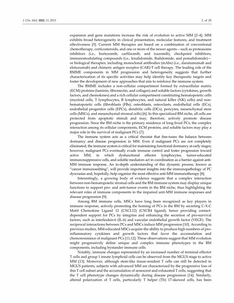
Figure 1. The bone marrow microenvironment (BMME) in multiple myeloma (MM). Complex
interactions between non‐hematopoietic stromal cells and BM immune system may support pro‐
and anti‐tumor events in the BMME, highlighting the roles of immune components in the impaired
anti‐MM immune responses and disease progression. Innate and adaptive immune cells can
recognize malignant plasma cells (PCs) and generate an anti‐tumor immune response against
tumors. A predominant role is attributed to immune cells, such as CD4+ Tregs and T cytotoxic CD8+
cells, which are considered effectors of host control for the MM PCs.
The clinical finding of an association between improved clinical outcome and reduced
Tregs/Th17 cells ratios or Treg frequency and oligoclonal expansion of TTEs suggests that
Tregs and the expansion of TTEs are key players in immune surveillance in MM [55]. This
J. Clin. Med. 2022, 11, 2513 6 of 20
further entails that T cells may recognize MM PC antigens and differentiate into TTEs, which
are able to exert cytotoxicity against PCs through perforin and granzyme expression.
However, whether BM residency is necessary to confer tumor control is still unclear.
Nevertheless, the same BM residency of immune cells can be considered the “Green Card”
that allows permanent MM surveillance at the site of disease initiation and progression [56].
Tregs are a subset of CD4+ T lymphocytes characterized on the surface by the CD25+
CD127low phenotype and the expression of the transcription factor forkhead box P3 (FoxP3)
[57]. If the homeostatic balance between Treg‐mediated suppression and T effector cell
activation is unbalanced in favor of effector activation, autoimmune disease emerges. In the
case of malignancy, excessive Treg activity leads to the suppression or exhaustion of effector
cells and a lack of tumor immune surveillance. Compared with MGUS, MM displays a
skewing of the Treg and pro‐inflammatory Th17 cell balance in favor of Tregs [58].
It has been postulated that Tregs are implicated in MM progression on the basis of
their contribution to the complex immunosuppressive environment through the secretion
of IL‐10 and TGF‐β by APRIL/TACI‐dependent mechanisms and through the CD39/CD73
adenosine pathway and direct inhibition of effector T cell responses [59]. In particular, the
secretion of IL‐6, TGF‐β, and IL‐1β in the BM niche promotes Th17 production, inducing
IL‐17 release, which correlates with MM cell growth [60].
Treg variation between the elimination/equilibrium (MGUS) and the escape stage
(MM) seems particularly intriguing, and, rather than a skewing of the balance between
Tregs and pro‐inflammatory Th17 in favor of Tregs, it might represent active Treg
differentiation involving the regulation of ectonucleotidase CD39 expression and
activation at BM residency [56]. Understanding changes in the Treg compartment holds
the potential to improve our comprehension of the clinical stability in MGUS and MM
progression, with relevant implications for the clinical diagnosis, prognosis, and
successful therapeutic approaches.
4. Dendritic Cells as Important Players in MM Immune Response
Since DCs represent a bridge between the innate and adaptative immune responses, they
are master APCs in all tissues, able to capture, process, and present tumor‐(neo)antigens (Ags)
to naïve T cells via major histocompatibility complex (MHC) molecules (Figure 2). Because of
their capacity for cross‐presenting Ags and inducing specific T cellular and B humoral
immune responses, DCs are a promising tool for immunotherapy in MM [61].
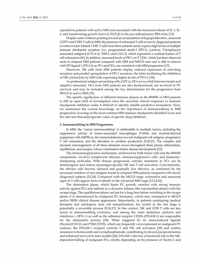
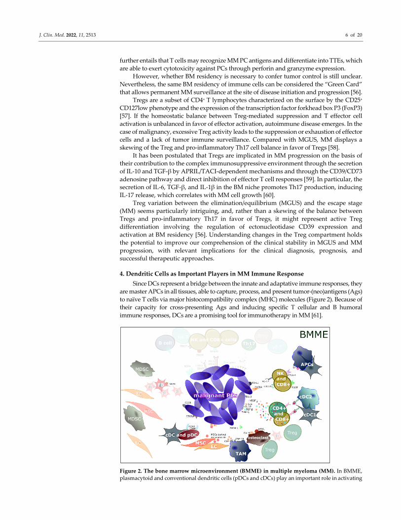
Figure 2. The bone marrow microenvironment (BMME) in multiple myeloma (MM). In BMME,
plasmacytoid and conventional dendritic cells (pDCs and cDCs) play an important role in activating
J. Clin. Med. 2022, 11, 2513 7 of 20
tumor‐specific T cells with natural killer (NK), NKT phenotype, and gamma delta (γδ) T cells,
inducing INF‐γ production. Concomitantly, CD8+ and CD4+ cells realize an immunosuppressive
milieu, producing transforming growth factor (TGF)‐β, vascular‐endothelial growth factor (VEGF),
interleukin (IL)‐10, IL‐6, IL‐17, and IL‐2; interacting with antigen‐presenting cells (APCs); and
inducing T regulatory (Treg) cell differentiation and proliferation. The same interaction stimulates
the CD28‐CD80/CD86 contact and decreases the processing and presentation of tumor antigens,
thus reducing malignant PC recognition by cytotoxic T CD8+ cells. Stromal cells, such as endothelial
cells (ECs), fibroblasts (FBs), mesenchymal stem cells (MSCs), DCs, myeloid‐derived suppressor
cells (MDSCs), osteoclasts, and tumor‐associated macrophages (TAMs), alongside immune cells,
regulate different mechanisms (i.e., cell‐to‐cell adhesion; release of soluble factors, cytokines,
chemokines, and growth factors) and activate several signaling pathways leading to MM
progression. Among them, note that MSC‐derived microvesicles are introjected by malignant PCs;
B‐cell maturation antigen (BCMA), CD38, and SLAMF7 are hyper‐expressed on malignant PCs;
osteoclasts highly activate and release RANK‐L, Gal‐9, and APRIL; VLA4 is expressed on FBs;
CD137, CD226 (interacting with CD112 and CD155 on malignant PCs), and NKG2D (interacting
with MICA\B on malignant PCs) are expressed on NK and CD8+ cells.
As sentinel cells of the innate immune system, DCs are able to recognize endogenous
danger molecules called DAMPs, which are released by damaged or dying cells via
pattern recognition receptors (PRRs) on the cell surface. Subsequently, DCs secrete
necessary cytokines allowing the activation of innate immune cells [62]. Simultaneously,
DCs process and present these Ags on the cell surface, allowing immature DCs (imDCs)
to switch to mature DCs (mDCs). The latter increase the expression of co‐stimulatory
molecules and immunostimulatory cytokines (CD80, CD86, and CD83, IL‐12, IL‐10, and
tumor necrosis factor (TNF)) (Figure 2) [63]. In addition, imDCs conserve the MHC‐II
molecules in the late endosomal and lysosomal compartments, whereas in mDCs, the
molecules are located on the cell surface. Upon Ag presentation via MHC molecules,
mDCs migrate to draining lymph nodes in a chemokine‐dependent manner. CCR7 and its
cognate ligands (C‐C motif ligand (CCL)‐19 and CCL‐21) allow the homing of DCs
through the lymphatic vessels to the T lymphocyte‐enriched zone in the secondary
lymphoid organs. In the draining lymph nodes, mDCs trigger naïve T cells to differentiate
into disparate T effector cells (i.e., Th1, Th2, Th17, T follicular helper (TFH) cells, Tregs,
and CD8+ CTLs), resulting in specific T cell responses [64,65]. Depending on the Ag nature,
by engaging the MHC‐I presenting endogenous Ags and MHC‐II presenting exogenous
Ags, DCs elicit respectively adaptive CD8+ or CD4+ T cell immune responses. Here, a
process called “cross‐presentation” between exogenous Ags on MHC‐I molecules results
in CD8+ CTL activation. Interestingly, each DCs subset contributes differently to the
immune response; for instance, cDC1s excel in cross‐presenting exogenous Ags via MHC‐
I to CD8+ CTLs and secrete IL‐12, thereby promoting Th1 responses [66].
Extensive literature data are available on the number, phenotypic profile, and
functional status of DCs in MM progression. Specifically, the number of circulating DCs
in healthy subjects includes 0.1–2.0% of the mononuclear cells [67], while a significant
alteration is observed in MM patients, with approximately a 50% reduction in myeloid
DCs (BDCA1+) and pDCs (BDCA2+) that is independent of the patient’s disease stage.
Leone et al., found that during disease progression from MGUS to active/symptomatic
MM, the myeloid DCs (CD11c+) and pDCs (CD11c– CD123+) accumulate in the BM niche.
This is paralleled by an increase in tumor burden, as both mDCs and pDCs exert
immunosuppressive and tumor‐promoting properties [68].
BMME immunological inhibitory cytokines induce phenotypic alterations and
functional deficiencies, which include impaired DC differentiation, maturation, and
activation. The most involved cytokines are TGF‐β1, VEGF, IL‐6, and IL‐10. These factors
can induce hyperactivation of STAT3 and extracellular signal‐regulated kinase (ERK)
pathways, which may be responsible for defective DC differentiation. TGF‐β1 and IL‐10
are both secreted by MM cells and play a significant role in deficient CD80/86
upregulation during DC maturation. In addition, the excessive production of TGF‐β1 by
MM cells suppressed allogeneic T cell responses and favored the differentiation and
J. Clin. Med. 2022, 11, 2513 8 of 20
expansion of Tregs, resulting in tumor‐associated immune tolerance [69]. While tumor‐
derived VEGF is engaged in the impaired DC function due to inhibitory effects on DC
maturation and differentiation, it is also responsible for T cell exhaustion. Previous
researchers confirmed the importance of MM cell adhesion to bone marrow stromal cells
(BMSCs), which in turn secrete IL‐6 in an NF‐κB‐dependent manner, supporting MM cell
growth and survival. IL‐6, which can also be secreted by MM cells, affects CD4+ T cell
differentiation, inhibiting Th1 polarization and promoting Th2 differentiation.
Furthermore, IL‐6 stimulates CD34+ precursor cell differentiation into monocytes instead
of DC progenitors through the upregulation of CD14 and the downregulation of CD1a,
HLA‐DR, CD40, and CD80 [70].
These large numbers of observations and experimental findings support the concept
that MM cells are intelligent evaders of immunosurveillance and may employ a variety of
mechanisms to disturb B cell immunity, promote Treg expansion, and suppress CTL
activity while concomitantly directing their inhibitory activity on DC differentiation,
maturation, and functions [70].
5. Immune Checkpoints and MM Progression
The high heterogeneity among MM patients has increased the need to identify immune
checkpoints in the BMME that regulate MM physiopathology. These molecules represent the
modulators of the signaling pathways responsible for immunological tolerance, a concept that
prevents the immune system from destroying its own cells. In MM pathogenesis, recognition
of biomarkers for the identification of patient populations that are likely to respond to therapy
and/or have fewer side effects from therapy is needed. Accordingly, several factors that
provide prognostic information and/or predict responses to checkpoint inhibitors have been
identified. Immune tolerance is partly mediated by CTLA‐4 and PD‐1, two
immunomodulatory receptors expressed on T cells that trigger inhibitory pathways
dampening T cell activity. CTLA‐4 and PD‐1 immune checkpoints constitute the major
immune escape mechanism in MM (Figure 2). CTLA‐4 regulates T cell proliferation early in
the immune response, primarily in the lymph nodes, and is more prominently expressed in
patients with active MM compared with MGUS patients [71]. Instead, PD‐1 suppresses T cells
in the immune response, primarily in the peripheral tissues, and its expression in NK and T
cells differs between relapsed/refractory MM patients and patients with MGUS or newly
diagnosed MM [72]. The clinical profiles of immuno‐oncology agents targeting these
checkpoints may vary according to their mechanistic differences.
5.1. CTLA‐4
CTLA‐4 is a member of the immunoglobulin superfamily and a negative regulator of
T cell activation. The T cell receptor complex initially recognizes Ags; then, the binding of
CD28 to CD80/CD86 on T cells and APCs, respectively, generates a primary positive co‐
stimulatory signal (Figure 2). After activation, CTLA‐4 is expressed on T cells and exerts
its negative regulatory effects by competing with CD28 for CD80/CD86 and blocking
downstream pathway activation.
In MM T cells, CTLA‐4 is upregulated and, via competitive bidding for the co‐
stimulatory molecules CD80/CD86, negatively modulates the activated T cells [73].
5.2. PD‐1/PD‐L1
PD‐1 is a member of the B7/CD28 family of co‐stimulatory receptors. It regulates T cell
activation through the binding to PD‐L1 and PD‐L2 ligands (Figure 2). Similar to CTLA‐4
signaling, the PD‐1 binding inhibits T cell proliferation, production of IFN‐γ, TNF‐α, and
IL‐2, and reduces T cell survival. PD‐1 expression is a hallmark of “exhausted” T cells that
have experienced high levels of stimulation or reduced CD4+ T cell help [25]. This state of
exhaustion, which occurs during chronic infections and cancer, is characterized by T cell
dysfunction, resulting in suboptimal control of infections and tumors.

J. Clin. Med. 2022, 11, 2513 9 of 20
In MM pathophysiology, PD‐L1, which was first identified as B7 homolog‐1 (B7‐H1),
is widely expressed on PCs and inhibits antitumor T cell responses associated with poor
prognosis. PD‐L1 expression levels are higher in MM PCs compared with those in MGUS
patients and healthy PCs, and its expression is often upregulated upon relapse or in the
refractory phase [74].
Interestingly, levels of soluble PD‐L1 are elevated in the peripheral blood of newly
diagnosed MM patients, and they are associated with a low response to treatment and
shorter PFS [37]. PD‐1 is overexpressed on T cells and NK cells in MM patients, and PD‐1+
T cells are highly enriched in MM‐specific effector cells [75]. Unfortunately, PD‐1/PD‐L1
interactions seem to undermine an effective anti‐MM immune response and contribute to
severe immune suppression and MM drug resistance. Accordingly, patients with an
increased frequency of PD‐1‐expression on T cells after autologous stem cell transplant may
present a higher risk of relapse [76]. Blockade of PD‐1/PD‐L1 enhances T cell and NK cell‐
mediated anti‐MM responses in vitro and in vivo, and the administration of anti‐PD‐L1 or
anti‐PD‐1 antibodies significantly decreases disease progression in MM mouse models [77].
6. Immunotherapy in MM
Conventional treatment of MM is age‐ and disease‐stage‐related. While SMM
patients require only periodic observation, the active disease needs to be instantly treated.
The conventional first‐line therapy consists of the administration of
thalidomide/lenalidomide, bortezomib, and dexamethasone.
Autologous stem cell transplantation can be performed in addition to pharmacological
treatment or singularly, depending on comorbidity and age‐related factors [78].
A certain number of MM patients are treated with allogenic bone marrow
transplantation (BMT) in association with maintenance drug administration. Because of
the excretion of IL‐17 by donor cells, IFN‐γ is included in long‐term therapy to avoid post‐
BMT relapse; lenalidomide is also part of the treatment because of its ability to ensure
residual MM cell dormancy [79].
Results from immunotherapy studies suggest that, when administered separately,
allogeneic BMT, immune checkpoint inhibitors, and DC‐based vaccines have limited
effects in a small number of patients (Figure 3) [38].
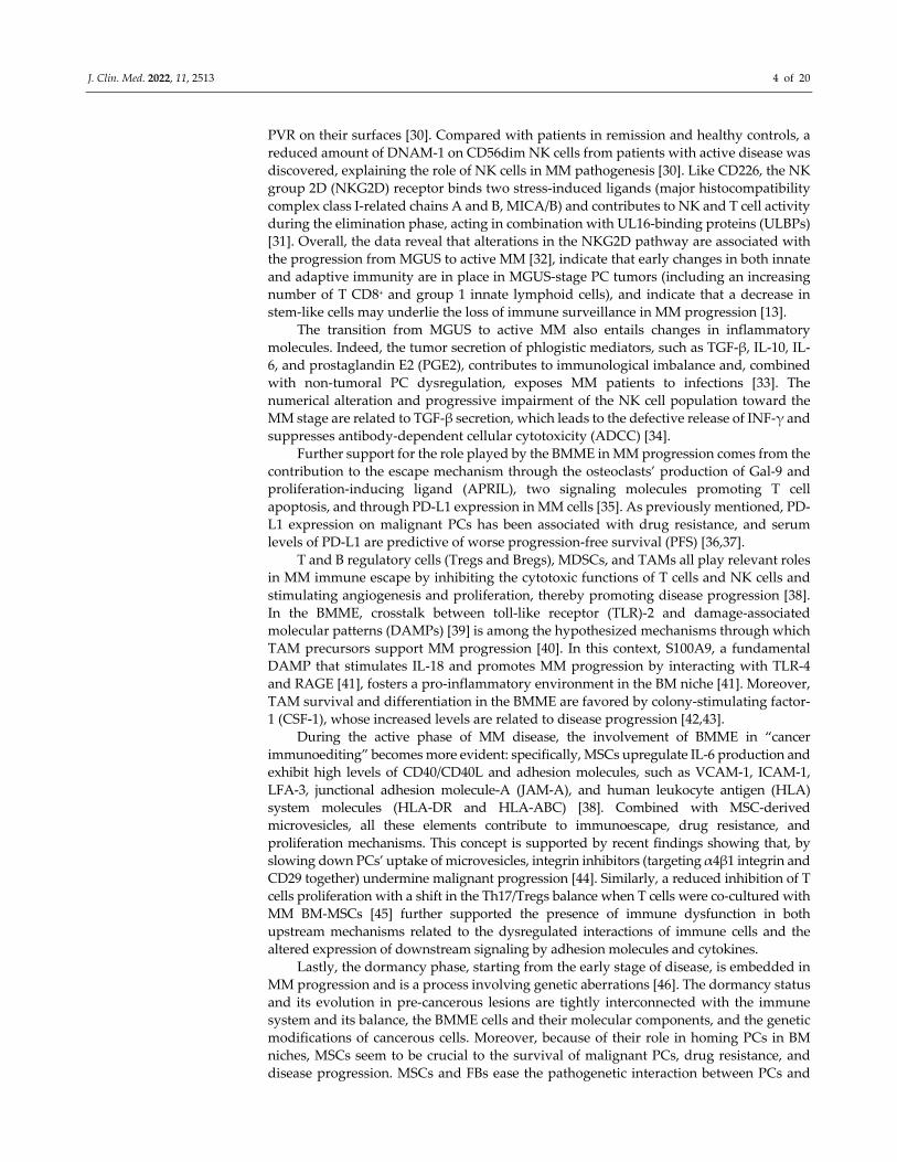
Figure 3. Immunomodulatory drugs in MM. Tumors have been shown to evade the immune
system. This has led to the development of new agents to be used in combination with both well‐
established and innovative therapeutical schemes. Some of these immunological drugs include anti‐
CTLA‐4 (ipilimumab), anti‐CXCR4/CSCL12 system (ulocuplumab and olaptesed pegol), anti‐PD‐

J. Clin. Med. 2022, 11, 2513 10 of 20
1/PDL‐1 (nivolumab and pembrolizumab), and TIGIT, which counteract the blockade caused by
immune checkpoints, enhance the immune response, and induce selective control on tumor growth,
sometimes in the long term. As a result, the immune system is more active in recognizing the tumor
as a foreign entity. CAR‐T therapy is directed against the B‐cell maturation antigen (BCMA) found
on the surface of cancer cells; recognition and binding of BCMA lead to the proliferation of CAR‐T
cells, which can thus attack and kill the cancer cells expressing this antigen. Recent agents with the
same target are Ab‐drug conjugates (ADCs) and bispecific monoclonal Abs (such as BiTEs). A long
list of mAbs capable of interfering with immunoediting pathways is currently approved, especially
in combination regimes, and includes anti‐CD38 (daratumumab and isatuximab), anti‐VLA4
(natalizumab), and anti‐SLAMF7 (elotuzumab) drugs; in addition, several new experimental
compounds are currently under evaluation, such as CD137 blockers, MSC‐derived microvescicle
blockers, CSF‐1/CSF‐1R system blockers, and Th17/IL‐17/IL‐17R system blockers. Lastly,
denosumab and bisphosphonates have been shown to be effective in slowing clinical disease
progression and the immune escape process.
6.1. ImiDs and mAbs
Immunomodulatory drugs (ImiDs, including thalidomide and its analogs) promote
cancer cells apoptosis, foster the proliferation and activity of NK and T cells (through
cereblon‐dependent degradation of the transcription factors IKZF1 and IKZF3) [80,81],
improve the production of INF‐γ and IL‐2 by Th1 cells, enhance ADCC [38], and contain
CD4+ and CD8+ IL‐10 release, which enhances NK cell activation [82]. For treatment of
refractory MM patient, these agents can be combined with mAb directed to specific targets:
daratumumab, as well as isatuximab, by targeting CD38 on the MM cells surface, either in
monotherapy or in combination with bortezomib and/or dexamethasone, is capable of
promoting ADCC, apoptosis, complement‐mediated cytotoxicity, antibody‐dependent
cellular phagocytosis (ADCP) and T cells response, targeting CD38 on the MM cells surface.
In addition, this reduces MDSC, Treg, and Breg cell activity, leading to enhanced PFS [38].
It has also been observed that daratumumab depletes the CD38+ cell pool, resulting in an
increased number of cytotoxic cells (Figure 3) [83]. The Dara‐VTD regimen (daratumumab
plus bortezomib, thalidomide, and dexamethasone) was approved in early 2020 by the Food
and Drug Administration (FDA) and the European Medicines Agency (EMA) as a new
induction therapy capable of improving the overall survival (OS) and response rate (RR),
thus ensuring longer follow‐ups [84]. Several other regimens with anti‐CD38 Abs, in
combination with carfilzomib and traditional chemotherapeutics, are currently being
evaluated for first‐ and second‐line and relapsed patients treatments [85,86]. Whether anti‐
CD38 mAb administration is compatible with CAR‐T therapy is still an open question
because of the presence of the antigen on activated T cells [87].
Elotuzumab targets SLAMF7 (CD319), improving NK cell function and ADCC,
whether associated with lenalidomide and dexamethasone [88]. SLAMF7 is over‐
exhibited in MM patients with the chromosomal translocation t(4;14)(p16;q32) and is
associated with very poor prognosis [89]. Several elotuzumab‐enriched schemes of
therapy have shown promising results in relapsed patients [88,90], and analogous
outcomes are expected from an ongoing trial on a quadruple‐drug induction and
consolidation regime for newly diagnosed MM patients eligible for transplantation (HD6
trial, Elo‐VRD; DSMMXVII trial, Elo plus carfilzomib, lenalidomide, and dexamethasone).
An ongoing phase 3 trial on relapsed MM patients is investigating the combination of
elotuzumab with anti‐PD‐1/anti‐PD‐L1 mAbs (NCT02726581).
Anti‐IL17A mAb, in conjunction with PDR001 (an anti‐PD‐1 mAb), is currently being
evaluated for the treatment of MM‐relapsed patients. Preclinical studies have evaluated
ulocuplumab, an anti‐CXCR4 mAb, as well as olaptesed pegol (PEGylated mirror‐image
l‐oligonucleotide capable of inhibiting CXCL12 signaling activity), as two possible
strategies to limit the spread of PCs and MM progression [91,92].
Further studies have investigated the efficacy of natalizumab, an anti‐VLA4 mAb
used for the treatment of multiple sclerosis that binds α4 integrins, in order to prevent the
interaction between ECM components, BM stromal cells, and malignant PCs; it has

J. Clin. Med. 2022, 11, 2513 11 of 20
emerged that the drug slows tumor cell proliferation, VEGF secretion, and angiogenesis
and strengthens the effects of bortezomib and dexamethasone [93].
6.2. Immune Checkpoints Inhibitors
PD‐L1 plays a fundamental prognostic and progression role in MM pathogenesis
[94]. It is over‐exhibited on malignant PCs because of INF‐γ and IL‐6 activation of
intracellular pathways (i.e., MEK/ERK) and induces both drug resistance and anti‐
apoptotic mechanisms (higher expression of Ki67 and BCL‐2) [95]. Nivolumab and
pembrolizumab (anti‐PD‐1 mAbs) show better performance in stable MM disease patients
rather than in refractory ones when combined with pomalidomide and dexamethasone or
radiotherapy regimes [96,97] (Figure 3). The association between ImiDs and anti‐PD‐
1/PD‐L1 mAbs has recently been discontinued by FDA since this combination could cause
fatally excessive immune responses, such as autoimmune cardiomyopathy [38].
Conversely, several preclinical trials show promising results when anti‐PD‐1 mAbs are
administered in monotherapy after transplantation and at an early disease stage
[28,79,98,99]. A phase II study (NCT02681302, ClinicalTrials.gov) analyzing the
combination of nivolumab and ipilimumab (noted for its Treg suppression activity in
vivo) [100,101] (Figure 3) reported positive preliminary results in high‐risk transplanted
patients (both newly diagnosed and recurrent ones), although effectiveness is limited by
concomitant severe immune‐related adverse effects (65%).
Recently, new potential target antigens have emerged. A preclinical trial investigated
the effectiveness of elotuzumab plus an anti‐CD137 agonist (4‐1BB) mAb on the
promotion of T cell proliferation and cytotoxicity in an early disease stage [102].
Unfortunately, a preclinical study on a group of recently transplanted patients observed
that the anti‐CD137 mAb treatment upregulated PD‐1 and TIM‐3 on CD8+ cells [103], thus
suggesting that an anti‐PD‐1 mAb should be able to counterbalance the anti‐CD137
upregulating effects [104].
T cell immunoreceptor with immunoglobulin and ITIM domains (TIGIT) works by
competitively contrasting CD226 action [16] (Figure 3). It inhibits NK and CD8+ activation
against cancer cells and interferes with TIGIT1 Treg activity and the T–DC interface [105].
Consequently, TIGIT blockade interrupts DC‐derived IL‐10 excretion, hindering the
immune‐escape process [98]. TIGIT represents an interesting target given its recurrent
presence on BM CD8+ cells surface. Its blockade can reverse the T cell exhaustion process
and improve disease control in MM patients and post‐BMT patients [98]. However, given
the unfavorable benefit–risk profile and higher toxicity revealed, immune checkpoint
inhibitor trials, such as NCT02579863, have been put on hold by the FDA.
6.3. CAR‐T Cells
CAR‐T cell technology is used for the treatment of a few hematological neoplasias
and has recently been approved in a certain clinical subset of refractory MM patients.
Among all possible targets, the main target of the engineered T cells is B‐cell maturation
antigen (BCMA); other targets are CD19 [106], CD138 [107], isoform variant 6 of CD44
(CD44v6) [108], CD70, SLAMF7 [109], integrin β7, Igκ [110], and TGF‐β [111], which are
currently being investigated (Figure 3).
BCMA is frequently expressed in MM PCs and constitutes a promising target in
refractory patients [112]. A phase I trial showed that, in heavily pre‐treated patients with
refractory and relapsed MM, the BCMA/CAR‐T cell treatment idecabtagene vicleucel (ide‐
cel, also called bb2121) produced an OS rate of 85% (with a complete response in 45% of
patients) [113,114]. A “real‐life” study on belantamab mafodotin, a highly selective MM
targeted therapy, enrolled a cohort of patients that received a median of eight prior lines
of therapy and revealed an overall response rate (ORR) of 33%, very similar to the ORR
reported in the DREAMM‐2 trial [115]. Other phase I–II studies have confirmed the strong
effectiveness of BCMA/CAR‐T cell therapy, thus proposing it as the future first‐line
therapy in relapsed or refractory patients [114,116–118]. BCMA/CAR‐T therapy has
J. Clin. Med. 2022, 11, 2513 12 of 20
demonstrated a good safety profile, assuring a low incidence of neurotoxicity and
cytokines release syndrome events compared with other CAR‐T protocols used to treat B‐
cell lymphoma and leukemia [114,119].
Unfortunately, the majority of patients relapse in any case [117,118], suggesting the
existence of tumor resistance and circumventing mechanisms. Among them, the possible
BCMA downregulation or complete loss by PCs, the accelerated CAR‐T cell life, and the
limited strength conditions of T cells [114,120], especially in highly treated patients, are
currently being investigated.
To date, countermeasures pursued to address the resistance mechanisms include the use
of γ‐secretase inhibitors, which increase BCMA cellular expression on PCs to the detriment of
the soluble BCMA fragment capable of inhibiting CAR‐T cell function [121]; the redefining of
CAR‐T cell manufacturing protocols to improve suitability [122]; and new CAR‐T‐cell
composition and humanized targeting domains to lower the immune reaction against CAR‐T
cells and enhance engraftment and in vivo expansion [121,123,124].
6.4. Ab‐Drug Conjugates (ADCs)
For MM patients who are refractory or suffering from high‐impact adverse effects
from CAR‐T, ImiDs, and mAbs therapies, BCMA‐specific ADCs could represent a suitable
alternative treatment. Belantamab mafodotin is an ADC that binds specifically to BCMA,
eliciting an antibody‐dependent cytotoxic response and releasing the cytotoxic agent
auristatin F. It has shown an acceptable safety profile, except for the occurrence of
keratopathy (31%) and hematologic dyscrasias [125], and an OS rate of approximately 30%
in phase II trial patients refractory to daratumumab, ImiDs, and proteasome inhibitors
[126]. Considering that its toxicity profile appeared manageable in relapsed and refractory
patients, belantamab mafodotin has been recently approved by EMA for adult MM
patients who have received at least four prior therapies and were refractory to at least one
proteasome inhibitor, one IMiD, and an anti‐CD38 mAB, and for patients who have
shown disease progression on the last therapy regimen [125].
6.5. Bispecific mAbs
Bispecific T cell engager mAbs (BiTEs) are able to target two different antigen‐
binding sites, CD3 (or other T cell receptors) and BCMA (or other tumor cell receptors)
and have been proposed as new agents to promote immune response (Figure 3). They
seem able to strongly enforce T cell engagement and activation independently of the T cell
receptor recognition mechanism [127] and have demonstrated a proper response in
heavily treated patients [128] in combination with ImiDs [129].
7. Final Remarks and Future Perspectives
Here, we reviewed the most important components involved in the tight interaction
between MMPCs and the BMME during MM progression. Since malignant PCs depend
on the BMME for their survival, an in‐depth understanding of the BM niche structure
might also provide clues for more effective immune‐mediated control.
Increasing knowledge has undoubtedly clarified that MM cells use several
concomitant strategies to escape immune surveillance. While is undeniable that successful
treatment approaches should prevent immune cells from becoming MM’s best friends,
several gaps in the comprehension of the crosstalk between MM and BM immune cells
are still present, and a number of key questions remain unanswered.
In the search for the best combination treatment, one of the most compelling needs is
to overcome drug resistance while allowing a sustainable therapy approach. The evolving
process of cancer immunoediting shows that a treatment strategy that considers the BMME
and clonal evolution is as important as treating the MM cells themselves. Unfortunately,
because of the cumulative toxic and side effects of multi‐drug treatments, the combined
management with single molecule‐driven drugs is still difficult to achieve, as highlighted
J. Clin. Med. 2022, 11, 2513 13 of 20
by some recent clinical studies. In this respect, BCMA‐based therapy may represent a
promising strategy, supporting research on targets similar to BCMA in the future.
At present, while several specific inhibitors for the BMME have been evaluated in
preclinical studies, most of them are not available in clinical practice. Incorporating the
molecular approaches related to diagnosis and risk stratification into the routine
diagnostic workup of patients remains a necessary approach for the use of personalized,
biologically based treatments for MM.
Overall, the therapeutic strategies described in this review underline the importance of a
novel approach to fighting MM heterogeneity and further support the notion that the
individual patient profile may contribute to developing specific immune microenvironment‐
based prognostic and predictive scores. From this point of view, the identification of
deregulated mechanisms may also translate into immune biomarkers able to distinguish
patients at higher risk of progression of aggressive disease and become the starting point for
planning novel immunotherapeutic approaches to improve MM patients’ outcomes.
Author Contributions: Conceptualization, V.D. and M.M.; investigation, V.D., F.D.S. and A.S.;
writing‐original draft preparation, V.D., F.D.S. and A.S.; writing‐review and editing, V.D., M.A.P.,
C.N., and M.M.; supervision, M.A.F., A.V. and M.M. All authors have read and agreed to the
published version of the manuscript.
Funding: This work was supported by “Programma Regionale”—Research for Innovation REFIN—
POR Puglia FESR‐FSE 2014/2020 to V.D.; INNOLABS‐POR Puglia FESR‐FSE 2014‐2020 (CITEL‐
Telemedicine Reasearch Center) and “Progetto Regione Puglia—Fondo europeo di sviluppo
regionale e Fondo sociale europeo (FESR e FSE)” to A.V. The sponsors of this study are public or
non‐profit organizations that support science in general.
Institutional Review Board Statement: Not Applicable.
Informed Consent Statement: Not Applicable.
Data Availability Statement: All data generated or analyzed during this study are included in this
published article.
Conflicts of Interest: The authors declare no conflicts of interest.
Abbreviations
Ab Antibody
ADCC Antibody‐dependent cellular cytotoxicity
ADCP Antibody‐dependent cellular phagocytosis
ADCs Antibody–drug conjugates
Ag Antigen
APCs Antigen‐presenting cells
APRIL A proliferation‐inducing ligand
B7‐H1 B7‐homologue 1
BCL‐2 B‐cell lymphoma‐2
BCMA B‐cell maturation antigen
BiTEs Bispecific T cell engager mAbs
BM Bone marrow
BMME Bone marrow microenvironment
BMSCs Bone marrow stromal cells
BMT Bone marrow transplantation
Bregs Regulatory B cells
CAR‐T Chimeric antigen receptor–T cell therapy
cDCs Conventional dendritic cells
CCL C‐C motif ligand
CSF‐1 Colony‐stimulating factor 1
CTLA‐4 Cytotoxic T‐lymphocyte antigen 4
CTLs Cytotoxic T lymphocytes
CXCL C‐X‐C motif Chemokine ligand

J. Clin. Med. 2022, 11, 2513 14 of 20
CXCR C‐X‐C motif Chemokine receptor
DAMPs Damage‐associated molecular patterns
Dara‐VTD Daratumumab plus bortezomib, thalidomide, and dexamethasone
DCs Dendritic cells
ECM Extracellular matrix
ECs Endothelial cells
EMA European Medicines Agency
EPCs Endothelial progenitor cells
ERK Extracellular signal‐regulated kinase
FDA Food and Drug Administration
FBs Fibroblasts
Foxp3 Forkhead box P3
HLA Human leukocyte antigen
IL Interleukin
imDCs Immature DCs
ImiDs Immunomodulatory drugs
INF‐γ Interferon‐γ
JAM‐A Junctional adhesion molecule A
mAb Monoclonal antibodies
mDCs Mature DCs
MDSCs Myeloid‐derived suppressor cells
MGUS Monoclonal gammopathy of undetermined significance
MHC Major histocompatibility complex
MM Multiple myeloma
MSCs Mesenchymal stromal cells
NK Natural killer
OS Overall survival
PCs Plasma cells
PD‐1 Programmed death‐1
pDCs Plasmacytoid dendritic cells
PD‐L1 PD ligand 1
PFS Progression‐free survival
PGE2 Prostaglandin E2
PRRs Pattern recognition receptors
RANKL Receptor activator of nuclear factor kappa‐B ligand
RR Response rate
SMM Smoldering multiple myeloma
TACI Transmembrane activator CAML interactor
TAMs Tumor‐associated macrophages
TFH T follicular helper
TGF‐β Transforming growth factor‐β
Th T helper
TIGIT T cell immunoreceptor with immunoglobulin and ITIM domains
TLR Toll‐like receptor
TNF Tumor necrosis factor
Tregs Regulatory T cells
TTEs Terminal effector CD8+ T cells
ULBPs UL‐binding proteins
VEGF Vascular endothelial growth factor

J. Clin. Med. 2022, 11, 2513 15 of 20
References
1. Alrasheed, N.; Lee, L.; Ghorani, E.; Henry, J.Y.; Conde, L.; Chin, M.; Galas‐Filipowicz, D.; Furness, A.J.S.; Chavda, S.J.; Richards,
H.; et al. Marrow‐Infiltrating Regulatory T Cells Correlate with the Presence of Dysfunctional CD4+ PD‐1+ Cells and Inferior
Survival in Patients with Newly Diagnosed Multiple Myeloma. Clin. Cancer Res. 2020, 26, 3443–3454. https://doi.org/10.1158/1078‐
0432.CCR‐19‐1714.
2. Landgren, O.; Kyle, R.A.; Pfeiffer, R.M.; Katzmann, J.A.; Caporaso, N.E.; Hayes, R.B.; Dispenzieri, A.; Kumar, S.; Clark, R.J.; Baris,
D.; et al. Monoclonal gammopathy of undetermined significance (MGUS) consistently precedes multiple myeloma: A prospective
study. Blood 2009, 113, 5412–5417. https://doi.org/10.1182/blood‐2008‐12‐194241.
3. Oben, B.; Froyen, G.; Maclachlan, K.H.; Leongamornlert, D.; Abascal, F.; Zheng‐Lin, B.; Yellapantula, V.; Derkach, A.; Geerdens,
E.; Diamond, B.T.; et al. Whole‐genome sequencing reveals progressive versus stable myeloma precursor conditions as two distinct
entities. Nat. Commun. 2021, 12, 1861. https://doi.org/10.1038/s41467‐021‐22140‐0.
4. Boyle, E.M.; Deshpande, S.; Tytarenko, R.; Ashby, C.; Wang, Y.; Bauer, M.A.; Johnson, S.K.; Wardell, C.P.; Thanendrarajan, S.;
Zangari, M.; et al. The molecular make up of smoldering myeloma highlights the evolutionary pathways leading to multiple
myeloma. Nat. Commun. 2021, 12, 293. https://doi.org/10.1038/s41467‐020‐20524‐2.
5. Laganà, A.; Perumal, D.; Melnekoff, D.; Readhead, B.; Kidd, B.A.; Leshchenko, V.; Kuo, P.‐Y.; Keats, J.; DeRome, M.; Yesil, J.; et al.
Integrative network analysis identifies novel drivers of pathogenesis and progression in newly diagnosed multiple myeloma.
Leukemia 2018, 32, 120–130. https://doi.org/10.1038/leu.2017.197.
6. Di Marzo, L.; Desantis, V.; Solimando, A.G.; Ruggieri, S.; Annese, T.; Nico, B.; Fumarulo, R.; Vacca, A.; Frassanito, M.A.
Microenvironment drug resistance in multiple myeloma: Emerging new players. Oncotarget 2016, 7, 60698–60711.
https://doi.org/10.18632/oncotarget.10849.
7. Manier, S.; Sacco, A.; Leleu, X.; Ghobrial, I.M.; Roccaro, A.M. Bone marrow microenvironment in multiple myeloma progression.
J. Biomed. Biotechnol. 2012, 2012, 157496. https://doi.org/10.1155/2012/157496.
8. Lopes, R.; Caetano, J.; Ferreira, B.; Barahona, F.; Carneiro, E.A.; João, C. The Immune Microenvironment in Multiple Myeloma:
Friend or Foe? Cancers 2021, 13, 625. https://doi.org/10.3390/cancers13040625.
9. Schreiber, R.D.; Old, L.J.; Smyth, M.J. Cancer immunoediting: Integrating immunity’s roles in cancer suppression and promotion.
Science 2011, 331, 1565–1570. https://doi.org/10.1126/science.1203486.
10. Corre, J.; Mahtouk, K.; Attal, M.; Gadelorge, M.; Huynh, A.; Fleury‐Cappellesso, S.; Danho, C.; Laharrague, P.; Klein, B.; Rème, T.;
et al. Bone marrow mesenchymal stem cells are abnormal in multiple myeloma. Leukemia 2007, 21, 1079–1088.
https://doi.org/10.1038/sj.leu.2404621.
11. Botta, C.; Mendicino, F.; Martino, E.A.; Vigna, E.; Ronchetti, D.; Correale, P.; Morabito, F.; Neri, A.; Gentile, M. Mechanisms of
Immune Evasion in Multiple Myeloma: Open Questions and Therapeutic Opportunities. Cancers 2021, 13, 3213.
https://doi.org/10.3390/cancers13133213.
12. Nakamura, K.; Smyth, M.J.; Martinet, L. Cancer immunoediting and immune dysregulation in multiple myeloma. Blood 2020, 136,
2731–2740. https://doi.org/10.1182/blood.2020006540.
13. Bailur, J.K.; McCachren, S.S.; Doxie, D.B.; Shrestha, M.; Pendleton, K.; Nooka, A.K.; Neparidze, N.; Parker, T.L.; Bar, N.; Kaufman,
J.L.; et al. Early alterations in stem‐like/resident T cells, innate and myeloid cells in the bone marrow in preneoplastic gammopathy.
JCI Insight 2019, 5, 127807. https://doi.org/10.1172/jci.insight.127807.
14. Suen, H.; Brown, R.; Yang, S.; Weatherburn, C.; Ho, P.J.; Woodland, N.; Nassif, N.; Barbaro, P.; Bryant, C.; Hart, D.; et al. Multiple
myeloma causes clonal T‐cell immunosenescence: Identification of potential novel targets for promoting tumour immunity and
implications for checkpoint blockade. Leukemia 2016, 30, 1716–1724. https://doi.org/10.1038/leu.2016.84.
15. Prabhala, R.H.; Pelluru, D.; Fulciniti, M.; Prabhala, H.K.; Nanjappa, P.; Song, W.; Pai, C.; Amin, S.; Tai, Y.‐T.; Richardson, P.G.; et
al. Elevated IL‐17 produced by TH17 cells promotes myeloma cell growth and inhibits immune function in multiple myeloma.
Blood 2010, 115, 5385–5392. https://doi.org/10.1182/blood‐2009‐10‐246660.
16. Guillerey, C.; Harjunpää, H.; Carrié, N.; Kassem, S.; Teo, T.; Miles, K.; Krumeich, S.; Weulersse, M.; Cuisinier, M.; Stannard, K.; et
al. TIGIT immune checkpoint blockade restores CD8+ T‐cell immunity against multiple myeloma. Blood 2018, 132, 1689–1694.
https://doi.org/10.1182/blood‐2018‐01‐825265.
17. Dhodapkar, M.V.; Sexton, R.; Das, R.; Dhodapkar, K.M.; Zhang, L.; Sundaram, R.; Soni, S.; Crowley, J.J.; Orlowski, R.Z.; Barlogie,
B. Prospective analysis of antigen‐specific immunity, stem‐cell antigens, and immune checkpoints in monoclonal gammopathy.
Blood 2015, 126, 2475–2478. https://doi.org/10.1182/blood‐2015‐03‐632919.
18. Benson, D.M.; Bakan, C.E.; Mishra, A.; Hofmeister, C.C.; Efebera, Y.; Becknell, B.; Baiocchi, R.A.; Zhang, J.; Yu, J.; Smith, M.K.; et
al. The PD‐1/PD‐L1 axis modulates the natural killer cell versus multiple myeloma effect: A therapeutic target for CT‐011, a novel
monoclonal anti‐PD‐1 antibody. Blood 2010, 116, 2286–2294. https://doi.org/10.1182/blood‐2010‐02‐271874.
19. International Myeloma Working Group Criteria for the classification of monoclonal gammopathies, multiple myeloma and related
disorders: A report of the International Myeloma Working Group. Br. J. Haematol. 2003, 121, 749–757.
20. Mittal, D.; Gubin, M.M.; Schreiber, R.D.; Smyth, M.J. New insights into cancer immunoediting and its three component phases‐‐
elimination, equilibrium and escape. Curr. Opin. Immunol. 2014, 27, 16–25. https://doi.org/10.1016/j.coi.2014.01.004.
21. Guillerey, C.; Nakamura, K.; Vuckovic, S.; Hill, G.R.; Smyth, M.J. Immune responses in multiple myeloma: Role of the natural
immune surveillance and potential of immunotherapies. Cell. Mol. Life Sci. 2016, 73, 1569–1589. https://doi.org/10.1007/s00018‐016‐
2135‐z.

J. Clin. Med. 2022, 11, 2513 16 of 20
22. Minnie, S.A.; Hill, G.R. Immunotherapy of multiple myeloma. J. Clin. Investig. 2020, 130, 1565–1575.
https://doi.org/10.1172/JCI129205.
23. Perumal, D.; Imai, N.; Laganà, A.; Finnigan, J.; Melnekoff, D.; Leshchenko, V.V.; Solovyov, A.; Madduri, D.; Chari, A.; Cho, H.J.; et
al. Mutation‐derived Neoantigen‐specific T‐cell Responses in Multiple Myeloma. Clin. Cancer Res. 2020, 26, 450–464.
https://doi.org/10.1158/1078‐0432.CCR‐19‐2309.
24. Miller, A.; Asmann, Y.; Cattaneo, L.; Braggio, E.; Keats, J.; Auclair, D.; Lonial, S.; MMRF CoMMpass Network; Russell, S.J.; Stewart,
A.K. High somatic mutation and neoantigen burden are correlated with decreased progression‐free survival in multiple myeloma.
Blood Cancer J. 2017, 7, e612. https://doi.org/10.1038/bcj.2017.94.
25. Zelle‐Rieser, C.; Thangavadivel, S.; Biedermann, R.; Brunner, A.; Stoitzner, P.; Willenbacher, E.; Greil, R.; Jöhrer, K. T cells in
multiple myeloma display features of exhaustion and senescence at the tumor site. J. Hematol. Oncol. 2016, 9, 116.
https://doi.org/10.1186/s13045‐016‐0345‐3.
26. Koebel, C.M.; Vermi, W.; Swann, J.B.; Zerafa, N.; Rodig, S.J.; Old, L.J.; Smyth, M.J.; Schreiber, R.D. Adaptive immunity maintains
occult cancer in an equilibrium state. Nature 2007, 450, 903–907. https://doi.org/10.1038/nature06309.
27. Park, S.L.; Buzzai, A.; Rautela, J.; Hor, J.L.; Hochheiser, K.; Effern, M.; McBain, N.; Wagner, T.; Edwards, J.; McConville, R.; et al.
Tissue‐resident memory CD8+ T cells promote melanoma‐immune equilibrium in skin. Nature 2019, 565, 366–371.
https://doi.org/10.1038/s41586‐018‐0812‐9.
28. Guillerey, C.; Ferrari de Andrade, L.; Vuckovic, S.; Miles, K.; Ngiow, S.F.; Yong, M.C.R.; Teng, M.W.L.; Colonna, M.; Ritchie, D.S.;
Chesi, M.; et al. Immunosurveillance and therapy of multiple myeloma are CD226 dependent. J. Clin. Investig. 2015, 125, 2077–2089.
https://doi.org/10.1172/JCI77181.
29. Carbone, E.; Neri, P.; Mesuraca, M.; Fulciniti, M.T.; Otsuki, T.; Pende, D.; Groh, V.; Spies, T.; Pollio, G.; Cosman, D.; et al. HLA class
I, NKG2D, and natural cytotoxicity receptors regulate multiple myeloma cell recognition by natural killer cells. Blood 2005, 105,
251–258. https://doi.org/10.1182/blood‐2004‐04‐1422.
30. El‐Sherbiny, Y.M.; Meade, J.L.; Holmes, T.D.; McGonagle, D.; Mackie, S.L.; Morgan, A.W.; Cook, G.; Feyler, S.; Richards, S.J.;
Davies, F.E.; et al. The requirement for DNAM‐1, NKG2D, and NKp46 in the natural killer cell‐mediated killing of myeloma cells.
Cancer Res. 2007, 67, 8444–8449. https://doi.org/10.1158/0008‐5472.CAN‐06‐4230.
31. Groh, V.; Wu, J.; Yee, C.; Spies, T. Tumour‐derived soluble MIC ligands impair expression of NKG2D and T‐cell activation. Nature
2002, 419, 734–738. https://doi.org/10.1038/nature01112.
32. Jinushi, M.; Vanneman, M.; Munshi, N.C.; Tai, Y.‐T.; Prabhala, R.H.; Ritz, J.; Neuberg, D.; Anderson, K.C.; Carrasco, D.R.; Dranoff,
G. MHC class I chain‐related protein A antibodies and shedding are associated with the progression of multiple myeloma. Proc.
Natl. Acad. Sci. USA 2008, 105, 1285–1290. https://doi.org/10.1073/pnas.0711293105.
33. Pratt, G.; Goodyear, O.; Moss, P. Immunodeficiency and immunotherapy in multiple myeloma. Br. J. Haematol. 2007, 138, 563–579.
https://doi.org/10.1111/j.1365‐2141.2007.06705.x.
34. Trotta, R.; Dal Col, J.; Yu, J.; Ciarlariello, D.; Thomas, B.; Zhang, X.; Allard, J.; Wei, M.; Mao, H.; Byrd, J.C.; et al. Immunodeficiency
and immunotherapy in multiple myeloma. J. Immunol. 2008, 181, 3784–3792. https://doi.org/10.4049/jimmunol.181.6.3784.
35. An, G.; Acharya, C.; Feng, X.; Wen, K.; Zhong, M.; Zhang, L.; Munshi, N.C.; Qiu, L.; Tai, Y.‐T.; Anderson, K.C. Osteoclasts promote
immune suppressive microenvironment in multiple myeloma: Therapeutic implication. Blood 2016, 128, 1590–1603.
https://doi.org/10.1182/blood‐2016‐03‐707547.
36. Yousef, S.; Marvin, J.; Steinbach, M.; Langemo, A.; Kovacsovics, T.; Binder, M.; Kröger, N.; Luetkens, T.; Atanackovic, D.
Immunomodulatory molecule PD‐L1 is expressed on malignant plasma cells and myeloma‐propagating pre‐plasma cells in the
bone marrow of multiple myeloma patients. Blood Cancer J. 2015, 5, e285. https://doi.org/10.1038/bcj.2015.7.
37. Wang, L.; Wang, H.; Chen, H.; Wang, W.; Chen, X.‐Q.; Geng, Q.‐R.; Xia, Z.‐J.; Lu, Y. Serum levels of soluble programmed death
ligand 1 predict treatment response and progression free survival in multiple myeloma. Oncotarget 2015, 6, 41228–41236.
https://doi.org/10.18632/oncotarget.5682.
38. Tamura, H. Immunopathogenesis and immunotherapy of multiple myeloma. Int. J. Hematol. 2018, 107, 278–285.
https://doi.org/10.1007/s12185‐018‐2405‐7.
39. Hope, C.; Foulcer, S.; Jagodinsky, J.; Chen, S.X.; Jensen, J.L.; Patel, S.; Leith, C.; Maroulakou, I.; Callander, N.; Miyamoto, S.; et al.
Immunoregulatory roles of versican proteolysis in the myeloma microenvironment. Blood 2016, 128, 680–685.
https://doi.org/10.1182/blood‐2016‐03‐705780.
40. Zavidij, O.; Haradhvala, N.J.; Mouhieddine, T.H.; Sklavenitis‐Pistofidis, R.; Cai, S.; Reidy, M.; Rahmat, M.; Flaifel, A.; Ferland, B.;
Su, N.K.; et al. Single‐cell RNA sequencing reveals compromised immune microenvironment in precursor stages of multiple
myeloma. Nat. Cancer 2020, 1, 493–506. https://doi.org/10.1038/s43018‐020‐0053‐3.
41. Nakamura, K.; Kassem, S.; Cleynen, A.; Chrétien, M.‐L.; Guillerey, C.; Putz, E.M.; Bald, T.; Förster, I.; Vuckovic, S.; Hill, G.R.; et al.
Dysregulated IL‐18 Is a Key Driver of Immunosuppression and a Possible Therapeutic Target in the Multiple Myeloma
Microenvironment. Cancer Cell 2018, 33, 634–648.e5. https://doi.org/10.1016/j.ccell.2018.02.007.
42. Ao, J.‐Y.; Zhu, X.‐D.; Chai, Z.‐T.; Cai, H.; Zhang, Y.‐Y.; Zhang, K.‐Z.; Kong, L.‐Q.; Zhang, N.; Ye, B.‐G.; Ma, D.‐N.; et al. Colony‐
Stimulating Factor 1 Receptor Blockade Inhibits Tumor Growth by Altering the Polarization of Tumor‐Associated Macrophages in
Hepatocellular Carcinoma. Mol. Cancer Ther. 2017, 16, 1544–1554. https://doi.org/10.1158/1535‐7163.MCT‐16‐0866.
43. Ries, C.H.; Cannarile, M.A.; Hoves, S.; Benz, J.; Wartha, K.; Runza, V.; Rey‐Giraud, F.; Pradel, L.P.; Feuerhake, F.; Klaman, I.; et al.
Targeting tumor‐associated macrophages with anti‐CSF‐1R antibody reveals a strategy for cancer therapy. Cancer Cell 2014, 25,
846–859. https://doi.org/10.1016/j.ccr.2014.05.016.

J. Clin. Med. 2022, 11, 2513 17 of 20
44. Dabbah, M.; Jarchowsky‐Dolberg, O.; Attar‐Schneider, O.; Tartakover Matalon, S.; Pasmanik‐Chor, M.; Drucker, L.; Lishner, M.
Multiple myeloma BM‐MSCs increase the tumorigenicity of MM cells via transfer of VLA4‐enriched microvesicles. Carcinogenesis
2020, 41, 100–110. https://doi.org/10.1093/carcin/bgz169.
45. André, T.; Najar, M.; Stamatopoulos, B.; Pieters, K.; Pradier, O.; Bron, D.; Meuleman, N.; Lagneaux, L. Immune impairments in
multiple myeloma bone marrow mesenchymal stromal cells. Cancer Immunol. Immunother. 2015, 64, 213–224.
https://doi.org/10.1007/s00262‐014‐1623‐y.
46. Dutta, A.K.; Fink, J.L.; Grady, J.P.; Morgan, G.J.; Mullighan, C.G.; To, L.B.; Hewett, D.R.; Zannettino, A.C.W. Subclonal evolution
in disease progression from MGUS/SMM to multiple myeloma is characterised by clonal stability. Leukemia 2019, 33, 457–468.
https://doi.org/10.1038/s41375‐018‐0206‐x.
47. Xu, S.; De Veirman, K.; De Becker, A.; Vanderkerken, K.; Van Riet, I. Mesenchymal stem cells in multiple myeloma: A therapeutical
tool or target? Leukemia 2018, 32, 1500–1514. https://doi.org/10.1038/s41375‐018‐0061‐9.
48. Menu, E.; Asosingh, K.; Indraccolo, S.; De Raeve, H.; Van Riet, I.; Van Valckenborgh, E.; Vande Broek, I.; Van de Broek, I.; Fujii, N.;
Tamamura, H.; et al. The involvement of stromal derived factor 1alpha in homing and progression of multiple myeloma in the
5TMM model. Haematologica 2006, 91, 605–612.
49. Alsayed, Y.; Ngo, H.; Runnels, J.; Leleu, X.; Singha, U.K.; Pitsillides, C.M.; Spencer, J.A.; Kimlinger, T.; Ghobrial, J.M.; Jia, X.; et al.
Mechanisms of regulation of CXCR4/SDF‐1 (CXCL12)‐dependent migration and homing in multiple myeloma. Blood 2007, 109,
2708–2717. https://doi.org/10.1182/blood‐2006‐07‐035857.
50. Murray, M.E.; Gavile, C.M.; Nair, J.R.; Koorella, C.; Carlson, L.M.; Buac, D.; Utley, A.; Chesi, M.; Bergsagel, P.L.; Boise, L.H.; et al.
CD28‐mediated pro‐survival signaling induces chemotherapeutic resistance in multiple myeloma. Blood 2014, 123, 3770–3779.
https://doi.org/10.1182/blood‐2013‐10‐530964.
51. Glatman Zaretsky, A.; Konradt, C.; Dépis, F.; Wing, J.B.; Goenka, R.; Atria, D.G.; Silver, J.S.; Cho, S.; Wolf, A.I.; Quinn, W.J.; et al. T
Regulatory Cells Support Plasma Cell Populations in the Bone Marrow. Cell Rep. 2017, 18, 1906–1916.
https://doi.org/10.1016/j.celrep.2017.01.067.
52. Jego, G.; Palucka, A.K.; Blanck, J.‐P.; Chalouni, C.; Pascual, V.; Banchereau, J. Plasmacytoid dendritic cells induce plasma cell
differentiation through type I interferon and interleukin 6. Immunity 2003, 19, 225–234. https://doi.org/10.1016/s1074‐7613(03)00208‐5.
53. Ray, A.; Song, Y.; Du, T.; Tai, Y.‐T.; Chauhan, D.; Anderson, K.C. Targeting tryptophan catabolic kynurenine pathway enhances
antitumor immunity and cytotoxicity in multiple myeloma. Leukemia 2020, 34, 567–577. https://doi.org/10.1038/s41375‐019‐0558‐x.
54. Lawson, M.A.; McDonald, M.M.; Kovacic, N.; Hua Khoo, W.; Terry, R.L.; Down, J.; Kaplan, W.; Paton‐Hough, J.; Fellows, C.; Pettitt,
J.A.; et al. Osteoclasts control reactivation of dormant myeloma cells by remodelling the endosteal niche. Nat. Commun. 2015, 6,
8983. https://doi.org/10.1038/ncomms9983.
55. Brown, R.D.; Spencer, A.; Joy Ho, P.; Kennedy, N.; Kabani, K.; Yang, S.; Sze, D.M.; Aklilu, E.; Gibson, J.; Joshua, D.E. Prognostically
significant cytotoxic T cell clones are stimulated after thalidomide therapy in patients with multiple myeloma. Leuk. Lymphoma
2009, 50, 1860–1864. https://doi.org/10.3109/10428190903216804.
56. Joshua, D.E.; Vuckovic, S.; Favaloro, J.; Lau, K.H.A.; Yang, S.; Bryant, C.E.; Gibson, J.; Ho, P.J. Treg and Oligoclonal Expansion of
Terminal Effector CD8+ T Cell as Key Players in Multiple Myeloma. Front. Immunol. 2021, 12, 620596.
https://doi.org/10.3389/fimmu.2021.620596.
57. Miyara, M.; Yoshioka, Y.; Kitoh, A.; Shima, T.; Wing, K.; Niwa, A.; Parizot, C.; Taflin, C.; Heike, T.; Valeyre, D.; et al. Functional
delineation and differentiation dynamics of human CD4+ T cells expressing the FoxP3 transcription factor. Immunity 2009, 30, 899–
911. https://doi.org/10.1016/j.immuni.2009.03.019.
58. Favaloro, J.; Brown, R.; Aklilu, E.; Yang, S.; Suen, H.; Hart, D.; Fromm, P.; Gibson, J.; Khoo, L.; Ho, P.J.; et al. Myeloma skews
regulatory T and pro‐inflammatory T helper 17 cell balance in favor of a suppressive state. Leuk. Lymphoma 2014, 55, 1090–1098.
https://doi.org/10.3109/10428194.2013.825905.
59. Brown, R.; Suen, H.; Favaloro, J.; Yang, S.; Ho, P.J.; Gibson, J.; Joshua, D. Trogocytosis generates acquired regulatory T cells adding
further complexity to the dysfunctional immune response in multiple myeloma. Oncoimmunology 2012, 1, 1658–1660.
https://doi.org/10.4161/onci.22032.
60. Shen, C.‐J.; Yuan, Z.‐H.; Liu, Y.‐X.; Hu, G.‐Y. Increased numbers of T helper 17 cells and the correlation with clinicopathological
characteristics in multiple myeloma. J. Int. Med. Res. 2012, 40, 556–564. https://doi.org/10.1177/147323001204000217.
61. Wang, Y.; Xiang, Y.; Xin, V.W.; Wang, X.‐W.; Peng, X.‐C.; Liu, X.‐Q.; Wang, D.; Li, N.; Cheng, J.‐T.; Lyv, Y.‐N.; et al. Dendritic cell
biology and its role in tumor immunotherapy. J. Hematol. Oncol. 2020, 13, 107. https://doi.org/10.1186/s13045‐020‐00939‐6.
62. Garg, A.D.; Nowis, D.; Golab, J.; Vandenabeele, P.; Krysko, D.V.; Agostinis, P. Immunogenic cell death, DAMPs and anticancer
therapeutics: An emerging amalgamation. Biochim. Biophys. Acta 2010, 1805, 53–71. https://doi.org/10.1016/j.bbcan.2009.08.003.
63. Dudek, A.M.; Martin, S.; Garg, A.D.; Agostinis, P. Immature, Semi‐Mature, and Fully Mature Dendritic Cells: Toward a DC‐Cancer
Cells Interface That Augments Anticancer Immunity. Front. Immunol. 2013, 4, 438. https://doi.org/10.3389/fimmu.2013.00438.
64. Hilligan, K.L.; Ronchese, F. Antigen presentation by dendritic cells and their instruction of CD4+ T helper cell responses. Cell. Mol.
Immunol. 2020, 17, 587–599. https://doi.org/10.1038/s41423‐020‐0465‐0.
65. Guermonprez, P.; Valladeau, J.; Zitvogel, L.; Théry, C.; Amigorena, S. Antigen presentation and T cell stimulation by dendritic
cells. Annu. Rev. Immunol. 2002, 20, 621–667. https://doi.org/10.1146/annurev.immunol.20.100301.064828.
66. Tussiwand, R.; Everts, B.; Grajales‐Reyes, G.E.; Kretzer, N.M.; Iwata, A.; Bagaitkar, J.; Wu, X.; Wong, R.; Anderson, D.A.; Murphy,
T.L.; et al. Klf4 expression in conventional dendritic cells is required for T helper 2 cell responses. Immunity 2015, 42, 916–928.
https://doi.org/10.1016/j.immuni.2015.04.017.

J. Clin. Med. 2022, 11, 2513 18 of 20
67. Do, T.H.; Johnsen, H.E.; Kjaersgaard, E.; Taaning, E.; Svane, I.M. Impaired circulating myeloid DCs from myeloma patients.
Cytotherapy 2004, 6, 196–203. https://doi.org/10.1080/14653240410006004.
68. Leone, P.; Berardi, S.; Frassanito, M.A.; Ria, R.; De Re, V.; Cicco, S.; Battaglia, S.; Ditonno, P.; Dammacco, F.; Vacca, A.; et al. Dendritic
cells accumulate in the bone marrow of myeloma patients where they protect tumor plasma cells from CD8+ T‐cell killing. Blood
2015, 126, 1443–1451. https://doi.org/10.1182/blood‐2015‐01‐623975.
69. Zelante, T.; Fric, J.; Wong, A.Y.W.; Ricciardi‐Castagnoli, P. Interleukin‐2 production by dendritic cells and its immuno‐regulatory
functions. Front. Immunol. 2012, 3, 161. https://doi.org/10.3389/fimmu.2012.00161.
70. Rutella, S.; Locatelli, F. Targeting multiple‐myeloma‐induced immune dysfunction to improve immunotherapy outcomes. Clin.
Dev. Immunol. 2012, 2012, 196063. https://doi.org/10.1155/2012/196063.
71. Braga, W.M.T.; da Silva, B.R.; de Carvalho, A.C.; Maekawa, Y.H.; Bortoluzzo, A.B.; Rizzatti, E.G.; Atanackovic, D.; Colleoni, G.W.B.
FOXP3 and CTLA4 overexpression in multiple myeloma bone marrow as a sign of accumulation of CD4(+) T regulatory cells.
Cancer Immunol. Immunother. 2014, 63, 1189–1197. https://doi.org/10.1007/s00262‐014‐1589‐9.
72. Paiva, B.; Azpilikueta, A.; Puig, N.; Ocio, E.M.; Sharma, R.; Oyajobi, B.O.; Labiano, S.; San‐Segundo, L.; Rodriguez, A.; Aires‐Mejia,
I.; et al. PD‐L1/PD‐1 presence in the tumor microenvironment and activity of PD‐1 blockade in multiple myeloma. Leukemia 2015,
29, 2110–2113. https://doi.org/10.1038/leu.2015.79.
73. Pianko, M.J.; Liu, Y.; Bagchi, S.; Lesokhin, A.M. Immune checkpoint blockade for hematologic malignancies: A review. Stem Cell
Investig. 2017, 4, 32. https://doi.org/10.21037/sci.2017.03.04.
74. Leone, P.; Solimando, A.G.; Malerba, E.; Fasano, R.; Buonavoglia, A.; Pappagallo, F.; De Re, V.; Argentiero, A.; Silvestris, N.; Vacca,
A.; et al. Actors on the Scene: Immune Cells in the Myeloma Niche. Front. Oncol. 2020, 10, 599098.
https://doi.org/10.3389/fonc.2020.599098.
75. Fernandez‐Poma, S.M.; Salas‐Benito, D.; Lozano, T.; Casares, N.; Riezu‐Boj, J.‐I.; Mancheño, U.; Elizalde, E.; Alignani, D.; Zubeldia,
N.; Otano, I.; et al. Expansion of Tumor‐Infiltrating CD8+ T cells Expressing PD‐1 Improves the Efficacy of Adoptive T‐cell Therapy.
Cancer Res. 2017, 77, 3672–3684. https://doi.org/10.1158/0008‐5472.CAN‐17‐0236.
76. Chung, D.J.; Pronschinske, K.B.; Shyer, J.A.; Sharma, S.; Leung, S.; Curran, S.A.; Lesokhin, A.M.; Devlin, S.M.; Giralt, S.A.; Young,
J.W. T‐cell Exhaustion in Multiple Myeloma Relapse after Autotransplant: Optimal Timing of Immunotherapy. Cancer Immunol.
Res. 2016, 4, 61–71. https://doi.org/10.1158/2326‐6066.CIR‐15‐0055.
77. Atanackovic, D.; Luetkens, T. Biomarkers for checkpoint inhibition in hematologic malignancies. Semin. Cancer Biol. 2018, 52, 198–
206. https://doi.org/10.1016/j.semcancer.2018.05.005.
78. Palumbo, A.; Avet‐Loiseau, H.; Oliva, S.; Lokhorst, H.M.; Goldschmidt, H.; Rosinol, L.; Richardson, P.; Caltagirone, S.; Lahuerta,
J.J.; Facon, T.; et al. Revised International Staging System for Multiple Myeloma: A Report from International Myeloma Working
Group. J. Clin. Oncol. 2015, 33, 2863–2869. https://doi.org/10.1200/JCO.2015.61.2267.
79. Itchaki, G.; Brown, J.R. Lenalidomide in the treatment of chronic lymphocytic leukemia. Expert Opin Investig Drugs. 2017, 26,
633–650. https://doi.org/10.1080/13543784.2017.1313230.
80. Krönke, J.; Udeshi, N.D.; Narla, A.; Grauman, P.; Hurst, S.N.; McConkey, M.; Svinkina, T.; Heckl, D.; Comer, E.; Li, X.; et al.
Lenalidomide causes selective degradation of IKZF1 and IKZF3 in multiple myeloma cells. Science 2014, 343, 301–305.
https://doi.org/10.1126/science.1244851.
81. Lu, G.; Middleton, R.E.; Sun, H.; Naniong, M.; Ott, C.J.; Mitsiades, C.S.; Wong, K.‐K.; Bradner, J.E.; Kaelin, W.G. The myeloma drug
lenalidomide promotes the cereblon‐dependent destruction of Ikaros proteins. Science 2014, 343, 305–309.
https://doi.org/10.1126/science.1244917.
82. Luptakova, K.; Rosenblatt, J.; Glotzbecker, B.; Mills, H.; Stroopinsky, D.; Kufe, T.; Vasir, B.; Arnason, J.; Tzachanis, D.; Zwicker, J.I.;
et al. Lenalidomide enhances anti‐myeloma cellular immunity. Cancer Immunol. Immunother. 2013, 62, 39–49.
https://doi.org/10.1007/s00262‐012‐1308‐3.
83. Krejcik, J.; Casneuf, T.; Nijhof, I.S.; Verbist, B.; Bald, J.; Plesner, T.; Syed, K.; Liu, K.; van de Donk, N.W.C.J.; Weiss, B.M.; et al.
Daratumumab depletes CD38+ immune regulatory cells, promotes T‐cell expansion, and skews T‐cell repertoire in multiple
myeloma. Blood 2016, 128, 384–394. https://doi.org/10.1182/blood‐2015‐12‐687749.
84. Moreau, P.; Attal, M.; Hulin, C.; Arnulf, B.; Belhadj, K.; Benboubker, L.; Béné, M.C.; Broijl, A.; Caillon, H.; Caillot, D.; et al.
Bortezomib, thalidomide, and dexamethasone with or without daratumumab before and after autologous stem‐cell transplantation
for newly diagnosed multiple myeloma (CASSIOPEIA): A randomised, open‐label, phase 3 study. Lancet 2019, 394, 29–38.
https://doi.org/10.1016/S0140‐6736(19)31240‐1.
85. Zhou, X.; Flüchter, P.; Nickel, K.; Meckel, K.; Messerschmidt, J.; Böckle, D.; Knorz, S.; Steinhardt, M.J.; Krummenast, F.; Danhof, S.;
et al. Carfilzomib Based Treatment Strategies in the Management of Relapsed/Refractory Multiple Myeloma with Extramedullary
Disease. Cancers 2020, 12, 1035. https://doi.org/10.3390/cancers12041035.
86. Chari, A.; Martinez‐Lopez, J.; Mateos, M.‐V.; Bladé, J.; Benboubker, L.; Oriol, A.; Arnulf, B.; Rodriguez‐Otero, P.; Pineiro, L.;
Jakubowiak, A.; et al. Daratumumab plus carfilzomib and dexamethasone in patients with relapsed or refractory multiple
myeloma. Blood 2019, 134, 421–431. https://doi.org/10.1182/blood.2019000722.
87. Rasche, L.; Hudecek, M.; Einsele, H. What is the future of immunotherapy in multiple myeloma? Blood 2020, 136, 2491–2497.
https://doi.org/10.1182/blood.2019004176.
88. Lonial, S.; Dimopoulos, M.; Palumbo, A.; White, D.; Grosicki, S.; Spicka, I.; Walter‐Croneck, A.; Moreau, P.; Mateos, M.‐V.; Magen,
H.; et al. Elotuzumab Therapy for Relapsed or Refractory Multiple Myeloma. N. Engl. J. Med. 2015, 373, 621–631.
https://doi.org/10.1056/NEJMoa1505654.

J. Clin. Med. 2022, 11, 2513 19 of 20
89. Xie, Z.; Gunaratne, J.; Cheong, L.L.; Liu, S.C.; Koh, T.L.; Huang, G.; Blackstock, W.P.; Chng, W.J. Plasma membrane proteomics
identifies biomarkers associated with MMSET overexpression in T(4;14) multiple myeloma. Oncotarget 2013, 4, 1008–1018.
https://doi.org/10.18632/oncotarget.1049.
90. Dimopoulos, M.A.; Dytfeld, D.; Grosicki, S.; Moreau, P.; Takezako, N.; Hori, M.; Leleu, X.; LeBlanc, R.; Suzuki, K.; Raab, M.S.; et al.
Elotuzumab plus Pomalidomide and Dexamethasone for Multiple Myeloma. N. Engl. J. Med. 2018, 379, 1811–1822.
https://doi.org/10.1056/NEJMoa1805762.
91. Roccaro, A.M.; Mishima, Y.; Sacco, A.; Moschetta, M.; Tai, Y.‐T.; Shi, J.; Zhang, Y.; Reagan, M.R.; Huynh, D.; Kawano, Y.; et al.
CXCR4 Regulates Extra‐Medullary Myeloma through Epithelial‐Mesenchymal‐Transition‐like Transcriptional Activation. Cell Rep.
2015, 12, 622–635. https://doi.org/10.1016/j.celrep.2015.06.059.
92. Roccaro, A.M.; Sacco, A.; Purschke, W.G.; Moschetta, M.; Buchner, K.; Maasch, C.; Zboralski, D.; Zöllner, S.; Vonhoff, S.; Mishima,
Y.; et al. SDF‐1 inhibition targets the bone marrow niche for cancer therapy. Cell Rep. 2014, 9, 118–128.
https://doi.org/10.1016/j.celrep.2014.08.042.
93. Podar, K.; Zimmerhackl, A.; Fulciniti, M.; Tonon, G.; Hainz, U.; Tai, Y.‐T.; Vallet, S.; Halama, N.; Jäger, D.; Olson, D.L.; et al. The
selective adhesion molecule inhibitor Natalizumab decreases multiple myeloma cell growth in the bone marrow
microenvironment: Therapeutic implications. Br. J. Haematol. 2011, 155, 438–448. https://doi.org/10.1111/j.1365‐2141.2011.08864.x.
94. Dong, H.; Strome, S.E.; Salomao, D.R.; Tamura, H.; Hirano, F.; Flies, D.B.; Roche, P.C.; Lu, J.; Zhu, G.; Tamada, K.; et al. Tumor‐
associated B7‐H1 promotes T‐cell apoptosis: A potential mechanism of immune evasion. Nat. Med. 2002, 8, 793–800.
https://doi.org/10.1038/nm730.
95. Liu, J.; Hamrouni, A.; Wolowiec, D.; Coiteux, V.; Kuliczkowski, K.; Hetuin, D.; Saudemont, A.; Quesnel, B. Plasma cells from
multiple myeloma patients express B7‐H1 (PD‐L1) and increase expression after stimulation with IFN‐{gamma} and TLR ligands
via a MyD88‐, TRAF6‐, and MEK‐dependent pathway. Blood 2007, 110, 296–304. https://doi.org/10.1182/blood‐2006‐10‐051482.
96. Lesokhin, A.M.; Ansell, S.M.; Armand, P.; Scott, E.C.; Halwani, A.; Gutierrez, M.; Millenson, M.M.; Cohen, A.D.; Schuster, S.J.;
Lebovic, D.; et al. Nivolumab in Patients with Relapsed or Refractory Hematologic Malignancy: Preliminary Results of a Phase Ib
Study. J. Clin. Oncol. 2016, 34, 2698–2704. https://doi.org/10.1200/JCO.2015.65.9789.
97. Levy, A.; Massard, C.; Soria, J.‐C.; Deutsch, E. Concurrent irradiation with the anti‐programmed cell death ligand‐1 immune
checkpoint blocker durvalumab: Single centre subset analysis from a phase 1/2 trial. Eur. J. Cancer 2016, 68, 156–162.
https://doi.org/10.1016/j.ejca.2016.09.013.
98. Minnie SA, Kuns RD, Gartlan KH; et al. Myeloma escape after stem cell transplantation is a consequence of T‐cell exhaustion and
is prevented by TIGIT blockade. Blood. 2018;132(16):1675‐1688. Blood 2019, 134, 1878. https://doi.org/10.1182/blood.2019003727.
99. Jing, W.; Gershan, J.A.; Weber, J.; Tlomak, D.; McOlash, L.; Sabatos‐Peyton, C.; Johnson, B.D. Combined immune checkpoint
protein blockade and low dose whole body irradiation as immunotherapy for myeloma. J. Immunother. Cancer 2015, 3, 2.
https://doi.org/10.1186/s40425‐014‐0043‐z.
100. Du, X.; Tang, F.; Liu, M.; Su, J.; Zhang, Y.; Wu, W.; Devenport, M.; Lazarski, C.A.; Zhang, P.; Wang, X.; et al. A reappraisal of CTLA‐
4 checkpoint blockade in cancer immunotherapy. Cell Res. 2018, 28, 416–432. https://doi.org/10.1038/s41422‐018‐0011‐0.
101. Arce Vargas, F.; Furness, A.J.S.; Litchfield, K.; Joshi, K.; Rosenthal, R.; Ghorani, E.; Solomon, I.; Lesko, M.H.; Ruef, N.; Roddie, C.;
et al. Fc Effector Function Contributes to the Activity of Human Anti‐CTLA‐4 Antibodies. Cancer Cell 2018, 33, 649–663.e4.
https://doi.org/10.1016/j.ccell.2018.02.010.
102. Yonezawa, A.; Dutt, S.; Chester, C.; Kim, J.; Kohrt, H.E. Boosting Cancer Immunotherapy with Anti‐CD137 Antibody Therapy.
Clin. Cancer Res. 2015, 21, 3113–3120. https://doi.org/10.1158/1078‐0432.CCR‐15‐0263.
103. Vuckovic, S.; Minnie, S.A.; Smith, D.; Gartlan, K.H.; Watkins, T.S.; Markey, K.A.; Mukhopadhyay, P.; Guillerey, C.; Kuns, R.D.;
Locke, K.R.; et al. Bone marrow transplantation generates T cell‐dependent control of myeloma in mice. J. Clin. Investig. 2019, 129,
106–121. https://doi.org/10.1172/JCI98888.
104. McKee, S.J.; Doff, B.L.; Soon, M.S.F.; Mattarollo, S.R. Therapeutic Efficacy of 4‐1BB Costimulation Is Abrogated by PD‐1 Blockade
in a Model of Spontaneous B‐cell Lymphoma. Cancer Immunol. Res. 2017, 5, 191–197. https://doi.org/10.1158/2326‐6066.CIR‐16‐0249.
105. Manieri, N.A.; Chiang, E.Y.; Grogan, J.L. TIGIT: A Key Inhibitor of the Cancer Immunity Cycle. Trends Immunol. 2017, 38, 20–28.
https://doi.org/10.1016/j.it.2016.10.002.
106. Garfall, A.L.; Maus, M.V.; Hwang, W.‐T.; Lacey, S.F.; Mahnke, Y.D.; Melenhorst, J.J.; Zheng, Z.; Vogl, D.T.; Cohen, A.D.; Weiss,
B.M.; et al. Chimeric Antigen Receptor T Cells against CD19 for Multiple Myeloma. N. Engl. J. Med. 2015, 373, 1040–1047.
https://doi.org/10.1056/NEJMoa1504542.
107. Guo, B.; Chen, M.; Han, Q.; Hui, F.; Dai, H.; Zhang, W.; Zhang, Y.; Wang, Y.; Zhu, H.; Han, W. CD138‐directed adoptive
immunotherapy of chimeric antigen receptor (CAR)‐modified T cells for multiple myeloma. J. Cell. Immunother. 2016, 2, 28–35.
https://doi.org/10.1016/j.jocit.2014.11.001.
108. Casucci, M.; Nicolis di Robilant, B.; Falcone, L.; Camisa, B.; Norelli, M.; Genovese, P.; Gentner, B.; Gullotta, F.; Ponzoni, M.;
Bernardi, M.; et al. CD44v6‐targeted T cells mediate potent antitumor effects against acute myeloid leukemia and multiple
myeloma. Blood 2013, 122, 3461–3472. https://doi.org/10.1182/blood‐2013‐04‐493361.
109. Chu, J.; He, S.; Deng, Y.; Zhang, J.; Peng, Y.; Hughes, T.; Yi, L.; Kwon, C.‐H.; Wang, Q.‐E.; Devine, S.M.; et al. Genetic modification
of T cells redirected toward CS1 enhances eradication of myeloma cells. Clin. Cancer Res. 2014, 20, 3989–4000.
https://doi.org/10.1158/1078‐0432.CCR‐13‐2510.

J. Clin. Med. 2022, 11, 2513 20 of 20
110. Ramos, C.A.; Savoldo, B.; Torrano, V.; Ballard, B.; Zhang, H.; Dakhova, O.; Liu, E.; Carrum, G.; Kamble, R.T.; Gee, A.P.; et al. Clinical
responses with T lymphocytes targeting malignancy‐associated κ light chains. J. Clin. Investig. 2016, 126, 2588–2596.
https://doi.org/10.1172/JCI86000.
111. Tang, N.; Cheng, C.; Zhang, X.; Qiao, M.; Li, N.; Mu, W.; Wei, X.‐F.; Han, W.; Wang, H. TGF‐β inhibition via CRISPR promotes the
long‐term efficacy of CAR T cells against solid tumors. JCI Insight 2020, 5, 133977. https://doi.org/10.1172/jci.insight.133977.
112. Ali, S.A.; Shi, V.; Maric, I.; Wang, M.; Stroncek, D.F.; Rose, J.J.; Brudno, J.N.; Stetler‐Stevenson, M.; Feldman, S.A.; Hansen, B.G.; et
al. T cells expressing an anti‐B‐cell maturation antigen chimeric antigen receptor cause remissions of multiple myeloma. Blood 2016,
128, 1688–1700. https://doi.org/10.1182/blood‐2016‐04‐711903.
113. Munshi, N.C.; Anderson, L.D.; Shah, N.; Madduri, D.; Berdeja, J.; Lonial, S.; Raje, N.; Lin, Y.; Siegel, D.; Oriol, A.; et al. Idecabtagene
Vicleucel in Relapsed and Refractory Multiple Myeloma. N. Engl. J. Med. 2021, 384, 705–716.
https://doi.org/10.1056/NEJMoa2024850.
114. Raje, N.; Berdeja, J.; Lin, Y.; Siegel, D.; Jagannath, S.; Madduri, D.; Liedtke, M.; Rosenblatt, J.; Maus, M.V.; Turka, A.; et al. Anti‐
BCMA CAR T‐Cell Therapy bb2121 in Relapsed or Refractory Multiple Myeloma. N. Engl. J. Med. 2019, 380, 1726–1737.
https://doi.org/10.1056/NEJMoa1817226.
115. Vaxman, I.; Abeykoon, J.; Dispenzieri, A.; Kumar, S.K.; Buadi, F.; Lacy, M.Q.; Dingli, D.; Hwa, Y.; Fonder, A.; Hobbs, M.; et al.
“Real‐life” data of the efficacy and safety of belantamab mafodotin in relapsed multiple myeloma‐the Mayo Clinic experience.
Blood Cancer J. 2021, 11, 196. https://doi.org/10.1038/s41408‐021‐00592‐3.
116. Zhao, W.‐H.; Liu, J.; Wang, B.‐Y.; Chen, Y.‐X.; Cao, X.‐M.; Yang, Y.; Zhang, Y.‐L.; Wang, F.‐X.; Zhang, P.‐Y.; Lei, B.; et al. A phase 1,
open‐label study of LCAR‐B38M, a chimeric antigen receptor T cell therapy directed against B cell maturation antigen, in patients
with relapsed or refractory multiple myeloma. J. Hematol. Oncol. 2018, 11, 141. https://doi.org/10.1186/s13045‐018‐0681‐6.
117. Cohen, A.D.; Garfall, A.L.; Stadtmauer, E.A.; Melenhorst, J.J.; Lacey, S.F.; Lancaster, E.; Vogl, D.T.; Weiss, B.M.; Dengel, K.; Nelson,
A.; et al. B cell maturation antigen‐specific CAR T cells are clinically active in multiple myeloma. J. Clin. Investig. 2019, 129, 2210–
2221. https://doi.org/10.1172/JCI126397.
118. Brudno, J.N.; Maric, I.; Hartman, S.D.; Rose, J.J.; Wang, M.; Lam, N.; Stetler‐Stevenson, M.; Salem, D.; Yuan, C.; Pavletic, S.; et al. T
Cells Genetically Modified to Express an Anti‐B‐Cell Maturation Antigen Chimeric Antigen Receptor Cause Remissions of Poor‐
Prognosis Relapsed Multiple Myeloma. J. Clin. Oncol. 2018, 36, 2267–2280. https://doi.org/10.1200/JCO.2018.77.8084.
119. Moreau, P.; Sonneveld, P.; Boccadoro, M.; Cook, G.; Mateos, M.V.; Nahi, H.; Goldschmidt, H.; Dimopoulos, M.A.; Lucio, P.; Bladé,
J.; et al. Chimeric antigen receptor T‐cell therapy for multiple myeloma: A consensus statement from The European Myeloma
Network. Haematologica 2019, 104, 2358–2360. https://doi.org/10.3324/haematol.2019.224204.
120. Yu, B.; Jiang, T.; Liu, D. BCMA‐targeted immunotherapy for multiple myeloma. J. Hematol. Oncol. 2020, 13, 125.
https://doi.org/10.1186/s13045‐020‐00962‐7.
121. Pont, M.J.; Hill, T.; Cole, G.O.; Abbott, J.J.; Kelliher, J.; Salter, A.I.; Hudecek, M.; Comstock, M.L.; Rajan, A.; Patel, B.K.R.; et al. γ‐
Secretase inhibition increases efficacy of BCMA‐specific chimeric antigen receptor T cells in multiple myeloma. Blood 2019, 134,
1585–1597. https://doi.org/10.1182/blood.2019000050.
122. D’Agostino, M.; Raje, N. Anti‐BCMA CAR T‐cell therapy in multiple myeloma: Can we do better? Leukemia 2020, 34, 21–34.
https://doi.org/10.1038/s41375‐019‐0669‐4.
123. Maude, S.L.; Frey, N.; Shaw, P.A.; Aplenc, R.; Barrett, D.M.; Bunin, N.J.; Chew, A.; Gonzalez, V.E.; Zheng, Z.; Lacey, S.F.; et al.
Chimeric Antigen Receptor T Cells for Sustained Remissions in Leukemia. N. Engl. J. Med. 2014, 371, 1507–1517.
https://doi.org/10.1056/NEJMoa1407222.
124. Sommermeyer, D.; Hudecek, M.; Kosasih, P.L.; Gogishvili, T.; Maloney, D.G.; Turtle, C.J.; Riddell, S.R. Chimeric antigen receptor‐
modified T cells derived from defined CD8+ and CD4+ subsets confer superior antitumor reactivity in vivo. Leukemia 2016, 30, 492–
500. https://doi.org/10.1038/leu.2015.247.
125. Tzogani, K.; Penttilä, K.; Lähteenvuo, J.; Lapveteläinen, T.; Lopez Anglada, L.; Prieto, C.; Garcia‐Ochoa, B.; Enzmann, H.;
Gisselbrecht, C.; Delgado, J.; et al. EMA Review of Belantamab Mafodotin (Blenrep) for the Treatment of Adult Patients with
Relapsed/Refractory Multiple Myeloma. Oncologist 2021, 26, 70–76. https://doi.org/10.1002/onco.13592.
126. Lonial, S.; Lee, H.C.; Badros, A.; Trudel, S.; Nooka, A.K.; Chari, A.; Abdallah, A.‐O.; Callander, N.; Lendvai, N.; Sborov, D.; et al.
Belantamab mafodotin for relapsed or refractory multiple myeloma (DREAMM‐2): A two‐arm, randomised, open‐label, phase 2
study. Lancet Oncol. 2020, 21, 207–221. https://doi.org/10.1016/S1470‐2045(19)30788‐0.
127. Demichelis‐Gómez, R.; Pérez‐Sámano, D.; Bourlon, C. Bispecific Antibodies in Hematologic Malignancies: When, to Whom, and
How Should Be Best Used? Curr. Oncol. Rep. 2019, 21, 17. https://doi.org/10.1007/s11912‐019‐0759‐5.
128. Einsele, H.; Rasche, L.; Topp, M.S.; Martin Kortüm, K.; Duell, J. The use of bispecific antibodies to optimize the outcome of patients
with acute leukemia, lymphoma and multiple myeloma after SCT. Bone Marrow Transplant. 2019, 54, 721–726.
https://doi.org/10.1038/s41409‐019‐0596‐z.
129. Cho, S.‐F.; Xing, L.; Anderson, K.C.; Tai, Y.‐T. Promising Antigens for the New Frontier of Targeted Immunotherapy in Multiple
Myeloma. Cancers 2021, 13, 6136. https://doi.org/10.3390/cancers13236136.
Related Documents