1 CD40-mediated maintenance of immune homeostasis in the adipose tissue microenvironment Zuoan Yi 1 , Laura L. Stunz 1 , Gail A. Bishop 1,2,3,4 1 Departments of Microbiology and 2 Internal Medicine, 3 Graduate Immunology Program, University of Iowa, 4 VA Medical Center, Iowa City, IA 52242 Running title: Immune regulation of metabolism by CD40 Corresponding author: Gail Bishop 2193B MERF, The University of Iowa Iowa City, IA 52242 Phone #: 319-335-7945 Fax #: 319-335-9006 email: [email protected] Word count: 4,085 Number of figures: 6; 5 online supplementary figures Page 1 of 38 For Peer Review Only Diabetes Diabetes Publish Ahead of Print, published online March 19, 2014

Welcome message from author
This document is posted to help you gain knowledge. Please leave a comment to let me know what you think about it! Share it to your friends and learn new things together.
Transcript

1
CD40-mediated maintenance of immune homeostasis in the adipose tissue
microenvironment
Zuoan Yi1, Laura L. Stunz
1, Gail A. Bishop
1,2,3,4
1Departments of Microbiology and
2Internal Medicine,
3Graduate Immunology Program,
University of Iowa, 4
VA Medical Center, Iowa City, IA 52242
Running title: Immune regulation of metabolism by CD40
Corresponding author: Gail Bishop
2193B MERF, The University of Iowa
Iowa City, IA 52242
Phone #: 319-335-7945
Fax #: 319-335-9006
email: [email protected]
Word count: 4,085
Number of figures: 6; 5 online supplementary figures
Page 1 of 38
For Peer Review Only
Diabetes
Diabetes Publish Ahead of Print, published online March 19, 2014

2
ABSTRACT
Chronic inflammation in visceral adipose tissue is considered a key element for induction of
insulin resistance in obesity. CD40 is required for efficient systemic adaptive immune responses,
and is implicated in various inflammatory conditions. However, its role in modulating immunity
in the microanatomical niches of adipose tissue remains largely undefined. Here we show that, in
contrast to its well-documented co-stimulatory effects, CD40 regulates development of insulin
resistance in a diet-induced obesity (DIO) mouse model, by ameliorating local inflammation in
adipose tissues. CD40 deficiency (CD40KO) resulted in greater body weight gain, more severe
inflammation in epididymal adipose tissue (EAT) and aggravated insulin resistance, in response
to DIO. Interestingly, we found that CD40KO CD8+ T lymphocytes were major contributors to
exacerbated insulin resistance. Specifically, CD8+ T cells in EAT of DIO CD40KO mice
produced elevated chemokines and proinflammatory cytokines and were critical for macrophage
recruitment. These results indicate that CD40 plays distinct roles in different tissues, and
unexpectedly plays an important role in maintaining immune homeostasis in EAT. Further study
of how CD40 promotes maintenance of healthy metabolism could contribute to better
understanding of, and ability to therapeutically manipulate the increasing health problem of
obesity and insulin resistance.
Page 2 of 38
For Peer Review Only
Diabetes

3
The worldwide increase of obesity and type 2 diabetes causes a substantial burden in human
health problems and their associated financial costs. Although type 2 diabetes is a heterogenous
disease, obesity and insulin resistance are recognized as commonly preceding the development
of most cases. Insulin resistance is the driving force of type 2 diabetes, but how resistance
develops is still incompletely understood. Recent studies support the concept that low-grade
inflammation mediated by both innate and adaptive immunity in visceral adipose tissue is a
critical step in this process (1,2). It has been reported that immune cells, including macrophages
(Mφ), T cells, B cells, mast cells and others accumulate in visceral adipose tissue (VAT) and
promote or regulate insulin resistance (3-9). Cytokines and/or chemokines produced by these
cells, such as TNF-α, MCP-1 and IL-6, are essential inflammation mediators (10-13). VAT is a
special microenvironment for immune responses compared to lymphoid tissues or even to
subcutaneous adipose tissue (SAT). The specificity of this immune environment is characterized
by a sharp distinction in the composition, phenotype and function of different immune cells,
compared to those in lymphoid and other non-lymphoid tissues (6,9). In addition, specific
cognate antigen engagement in this discrete microenvironment may contribute to its unique
immune profile, as biased T cell receptor repertoires in CD4+ T cells were found in VAT in
obese mice (7). The immune homeostasis in VAT must be delicately regulated, because any
changes in the composition of immune cells and the interactions between different cell types
might disturb the homeostatic balance and induce severe inflammation, thus aggravating insulin
resistance.
CD40, a member of the TNF receptor superfamily, supplies essential co-stimulatory signals
during interactions between antigen presenting cells and T cells. Both humans with deficiencies
in expression of the ligand for CD40 (X-linked Hyper-IgM syndrome) and CD40 deficient
Page 3 of 38
For Peer Review Only
Diabetes

4
(CD40KO) mice have lower basal serum levels of IgG and IgA, and cannot mount efficient
humoral and cellular adaptive immune responses (14-16). In addition to its constitutive
expression on antigen presenting cells, CD40 can also be expressed on some activated T cells.
The expression of CD40 on a subset of CD4+ T cells plays an important role in pathogenesis in
animal models of autoimmune insulin-dependent type 1 diabetes and collagen-induced arthritis
(17,18). CD40 expressed on CD8+ T cells is involved in pathogen-associated immune responses
(19-21). Recent studies showed that preadipocytes and adipocytes also express CD40, and
demonstrated that adipose tissue inflammation requires the ligand for CD40, CD154 (22-24).
However, the roles of CD154 and CD40 might be distinct, as CD154 has other potential
receptors in addition to CD40 (25). Therefore, it remains unclear whether CD40 plays a
causative role in regulating immune homeostasis in the inflamed obese adipose tissue, and
whether it is involved in the initiation and development of obesity-related insulin resistance and
type 2 diabetes. In this study, we tested the role of CD40 in obesity and insulin resistance in a
high fat diet (HFD)-induced obese mouse model (diet-induced obesity, DIO). Our results reveal
a physiological role for adipose tissue CD40 in preventing obesity and insulin resistance.
Page 4 of 38
For Peer Review Only
Diabetes

5
Research Design and Methods
Mice and diets
CD40 deficient (CD40KO) mice on a C57BL/6Ncr background and control C57BL/6J (WT)
mice were obtained from The Jackson Laboratory (Bar Harbor, ME). Rag1-deficient (Rag1-/-
)
mice were originally provided by Dr. F. Sutterwala (University of Iowa, Iowa City, IA). Age
matched male mice were used for diet-induced obesity induction with a high fat diet. All mice
were maintained in facilities under specific pathogen-free conditions at The University of Iowa
and were used in accordance with National Institutes of Health guidelines under an animal
protocol approved by the Animal Care and Use Committee of the University of Iowa. Mice were
fed with either a standard chow diet containing 6% fat (Oriental Yeast Company, Vista, CA) or a
HFD containing 60% of calories from fat (D12492, Research Diets, New Brunswick, NJ). To
induce obesity, unless otherwise indicated, male mice were fed with the HFD for 18-20 weeks
starting at the age of 6 weeks.
Antibodies and reagents
Anti-CD8 depleting Ab (YTS169.4) and relevant isotype control Abs were purchased from
BioXell (West Lebanon, NH). Fluorescence-conjugated anti-F4/80 (BM8), anti-CD3 (145-
2C11), anti-CD28 (37.51), anti-CD4 (GK1.5), anti-CD8 (53-6.7), anti-Foxp3 (FJK-16s), anti-
CD16/32 (93), anti-B220 (RA3-6B2), anti-CD11c (N418), anti-CD40 (HM40.3), anti-CD11b
(M1/70) and anti-perilipin (D1D8) Abs were purchased from eBioscience (San Diego, CA), BD
Bioscience (San Jose, CA), R&D Systems (Minneapolis, MN), or Cell Signaling Techology
(Danvers, MA).
Metabolic parameter measurements
Page 5 of 38
For Peer Review Only
Diabetes
6
Food consumption and body weight were monitored weekly. After 18 weeks on a HFD as
indicated, fasting blood glucose was measured. Serum was collected before mice were
euthanized. Triglycerides, total cholesterol, LDL/vLDL, HDL and insulin quantification kits
were purchased from Abcam (Cambridge, MA) and ELISA detection was performed according
to the manufacturer’s instructions. For glucose tolerance tests (GTT), mice were fasted for 14 hrs
and 1g glucose/kg body weight was injected intraperitoneally; for insulin tolerance tests (ITT),
mice were fasted for 6 hrs and 0.75U/kg body weight recombinant human insulin (InVitrogen,
Grand Island, NY) was injected intraperitoneally. Blood glucose was measured at 0, 15, 30, 60
and 120 min after glucose or insulin injection.
Stromal vascular fraction (SVF) isolation and analysis
SVF isolation was performed as described (3,5,7). Some freshly isolated SVF was directly used
for mRNA extraction and real-time PCR analysis as indicated in figure legend. In some
experiments, 2.5x10⁵ total SVF cells were cultured for 48 hrs. Cytokines were tested with ELISA
kits (eBioscience). In others, SVF were stained for flow cytometry analysis or for sorting CD8+
T cells or CD4+ T cells using flow cytometry. 2x10
5 sorted CD3
+CD8
+ T cells or CD3
+CD4
+ T
cells were cultured with/without 1ug/ml anti-CD3 Ab and 5ug/ml anti-CD28 Ab ± anti-CD40 Ab
for 48 hrs. Culture supernatant was collected for cytokine and chemokine detection by ELISA
(kits from eBioscience) or Luminex assays with the MILLIPLEX MAP Kit (Billerica, MA).
Flow cytometric analysis and cell sorting were performed using a FACS LSRII or Aria (BD
Bioscience) at The University of Iowa Flow Cytometry Facility. Flow data were analyzed with
FlowJo software (Tree Star).
Histology and immunohistochemical (IHC) staining
Page 6 of 38
For Peer Review Only
Diabetes

7
IHC staining was performed as described (5). Fluorochrome-labeled anti-F4/80 and anti-perilipin
Ab was used for staining. Confocal microscopy was performed with a Zeiss LSM 710 in the
Central Microscopy Research Facility at The University of Iowa. A crown-like structure (CLS)
was defined as F4/80+ Mφ aggregates comprising at least 50% of the circumference circularly
surrounding adipocytes. For Oil-Red-O staining, liver sections were snap frozen in Tissue-Tek
OCT, sectioned and fixed in 10% neutral buffered formalin (Harleco, Billerica, MA) and
incubated in fresh prepared Oil-Red-O staining solution for 25 min at 60°C. Nuclei were stained
with alum hematoxylin. Staining and microscopy analysis were done in the Pathology Core
Facility at The University of Iowa.
Immune cell adoptive transfer
CD4+ or CD8
+ T cells and B cells were purified from the spleens of 8-week-old CD40KO or WT
standard chow diet fed mice using negative selection kits (Miltenyi Biotech, San Diego, CA).
Rag1-/-
mice fed with HFD for 12 weeks were given 5x10⁶ CD8
+ T cells, CD4
+ T cells, or 10x10
6
B cells intravenously. Mice were fed with HFD for 4 more weeks. Metabolic parameters and
SVF were examined at the end of this time period.
Bone marrow chimeric mice
WT and CD40KO mice were irradiated with 950 rads γ-irradiation. 10x106 bone marrow (BM)
cells isolated from WT or CD40KO mice were transferred into these mice by i.v. injection. BM
recipients were fed with HFD for 12 weeks starting at 4 weeks after irradiation. In another set of
experiments, irradiated WT mice were reconstituted with mixed BM (Rag1-/-
BM with WT BM
or CD40KO BM at a 10:1 ratio). 4 weeks later, HFD was given for 12 weeks. Metabolic
parameters were monitored as indicated in the Figure legends.
Page 7 of 38
For Peer Review Only
Diabetes
8
Antibody depletion in vivo
For CD8+ T cell depletion, WT and CD40KO mice fed with a HFD for 12 weeks were injected
intraperitoneally with 100ug anti-CD8 or isotype control Abs in 100ul PBS, once per week for
six weeks. HFD was given continuously. Metabolic parameters were monitored as indicated in
Figure legends.
Real Time PCR
Adipose tissue (<100mg) was minced into small pieces. RNA was extracted with RNeasy lipid
extraction kit (QIAGEN, Gaithersburg, MD) and cDNA synthesized using SuperScript III
(Invitrogen). RT-PCR was performed on an ABI PRISM 7900 Sequence Detection System using
SYBR Green (Applied Biosystems, Grand Island, NY). cDNA levels were determined with a
standard curve and normalized to β-actin. Primers for IFN-γ (forward: 5’-
CTTTGCAGCTCTTCCTCATGGCTGTTTCTG-3’, reverse: 5’-
TGACGCTTATGTTGTTGCTGATGGCCTG-3’), TNF-α (forward: 5’-
CACAGAAAGCATGATCCGCGACGT-3’, reverse: 5’-
CGGCAGAGAGGAGGTTGACTTTCT-3’), IL-1β (forward: 5’-
CCAGCTTCAAATCTCACAGCAG-3’, reverse: 5’- CTTCTTTGGGTATTGCTTGGGATC-
3’), IL-6 (forward: 5’- ACAACGATGATGCACTTGCAGA-3’, reverse: 5’-
GATGAATTGGATGGTCTTGGTC-3’), IL-4 (forward: 5’-TGTACCAGGAGCCATATCCAC-
3’, reverse: 5’-GTTCTTCGTTGCTGTGAGGAC-3’), IL-10 (forward: 5’-
TGGCCCAGAAATCAAGGAGC-3’, reverse: 5’-CAGCAGACTCAATACACACT-3’),
RANTES (forward: 5’-TTGCCTACCTCTCCCTAGAGC-3’, reverse:5’-
GGTTCCTTCGAGTGACAAACA-3’), IP-10, (forward: 5’-
Page 8 of 38
For Peer Review Only
Diabetes

9
CCATCAGCACCATGAACCCAAGTCCTGCCG-3’, reverse: 5’-
GGACGTCCTCCTCATCGTCGACTACACTGG-3’), MCP-1 (forward: 5’-
TTGACCCGTAAATCTGAAGC-3’, reverse: 5’-CGAGTCACACTAGTTCACTG-3’), MIP-1α
( forward: 5’-TGTTTGCTGCCAAGTAGCCACATC-3’, reverse: 5’-
AACAGTGTGAACAACTGGGAGGGA-3’) and β-actin (forward: 5’-
TGTGATGGTGGGAATGGGTCAG-3’, reverse: 5’-TTTGATGTCACGCACGATTTCC-3’)
were purchased from IDT (Coralville, IA).
Statistical analysis
Results are presented as mean values ± SEM. Statistical differences between two means were
evaluated using unpaired Student’s t-test. For comparisons of multiple groups, two-way ANOVA
was used. Statistical significance was set at a P value of <0.05.
Page 9 of 38
For Peer Review Only
Diabetes

10
Results
Exacerbation of HFD-induced metabolic complications by CD40 deficiency
The broad expression of CD40 on immune cells and adipocytes, and the proinflammatory effects
mediated via CD40 signaling during immune responses encouraged us to explore whether CD40
signaling plays a role in the development of obesity and insulin resistance. For this purpose, age-
matched WT and CD40KO mice were fed with a HFD (60 kcal%) for 18 weeks. Interestingly,
CD40KO mice gained significantly more body weight than WT mice (Fig. 1A), and this could be
attributed to visceral fat deposition, as they also showed remarkably increased weight of
epididymal adipose tissue (EAT), but only slightly increased weight of femoral SAT (Fig. 1B).
DIO CD40KO mice also exhibited aggravated glucose intolerance and insulin resistance
compared to DIO WT mice (Fig. 1C and D). Notably, 4 of 12 CD40KO mice, but none of the
WT mice, occasionally showed hyperglycemia (>250mg/dL) after 12 weeks on a HFD, although
the blood glucose levels after 14 hours of fasting remained unchanged (Supplementary Fig. 1).
The fluctuation of blood glucose further supports the impaired glucose and insulin tolerance in
CD40KO mice. Consistent with the increased fat pad weight and insulin resistance, CD40KO
mice also showed higher levels of triglycerides, total and (v)LDL cholesterol, but comparable
HDL cholesterol in serum compared to WT mice (Fig. 1E-H). We also found slightly lower
levels of serum insulin in CD40KO versus WT mice (Fig. 1I). In addition, liver histology
showed enhanced Oil Red-O-specific lipid deposition in CD40KO mice (Supplementary Fig. 2),
indicating more severe hepatic steatosis. Taken together, our data demonstrate that CD40 plays
an important role in inhibiting the development of insulin resistance and dysfunctional
metabolism in response to DIO.
Page 10 of 38
For Peer Review Only
Diabetes

11
Requirement of CD40 expression on immune cells for preventing aggravated obesity and
insulin resistance
CD40 is widely expressed on immune cells, adipocytes, and endothelial cells (26). Although
there are many published reports on CD40 functions in immune cells, the roles of CD40 in
various types of nonimmune cells are less understood. To explore whether prevention of obesity
and insulin resistance in the DIO model requires CD40 expression by immune cells, WT and
CD40KO mice were lethally irradiated and reconstituted with WT bone marrow cells (BM) or
CD40KO BM, and received a HFD starting 4 weeks later. In this model, immune cells lacking or
expressing CD40 are reconstituted in recipient mice in which non-immune cells are CD40-
sufficient or deficient. After 12 weeks on the HFD, recipients of CD40KO BM gained more
body weight and EAT weight than recipients of WT BM (Fig. 2A and B). Consistently, these
mice also displayed decreased glucose tolerance and elevated insulin resistance (Fig. 2C and D).
These data indicate that CD40 must be expressed on immune cells to exert its effects in
inhibiting obesity and insulin resistance.
Impact of CD40 upon local immune responses in adipose tissues
Chronic, low-level inflammation in adipose tissue is crucial for the development of insulin
resistance. CD40 plays multiple roles in different types of immune cells, so we next explored
whether CD40 deficiency alters the local immune environment in adipose tissue and induces
more immune cell infiltration and inflammation. For this purpose, stromal vascular fractions
(SVF) in obese EAT were extracted and analyzed. Flow cytometric analysis showed that there
were consistently more CD8+ T cells and Mφ accumulated in EAT of DIO CD40KO mice than
in DIO WT mice, but little if any change in numbers of other immune cells (Fig. 3A). A similar
Page 11 of 38
For Peer Review Only
Diabetes

12
trend was also seen in mice fed a standard chow diet (Fig. 3B). Interestingly, CD40KO mice
consuming a standard chow diet also showed lowered glucose tolerance and insulin tolerance
(Fig. 3C and D), indicating that CD40 plays an important role in preventing insulin resistance
even without the extra risk factor of HFD consumption.
Obesity is usually accompanied by necrosis of adipocytes, which can induce Mφ infiltration
and the formation of crown-like structures (CLS) (27). Consistent with our flow cytometry data,
immunohistochemical staining (IHC) also revealed higher numbers of CLS in CD40KO EAT
following HFD consumption (Fig 3E). When analyzing different subsets of Mφ, we found that
the numbers of both M1-like Mφ (F4/80+ and CD11c
+) and M2-like Mφ (F4/80
+ and CD11c
-)
were equally increased in CD40KO EAT (Fig 3F). To evaluate whether CD40 deficiency
predisposes to a more proinflammatory EAT milieu, total RNA from EAT and SAT was
extracted and real time PCR was conducted. Results showed that the mRNA expression levels of
IFN-γ, TNF-α, IL-1β and IL-6 were remarkably increased in EAT, but not in SAT in CD40KO
mice (Fig. 3G). Notably, we also found slightly less IL-4 in EAT in CD40KO mice (Fig. 3G). To
further assess the function of immune cells that infiltrated into EAT, total SVF isolated from
EAT were cultured for 48 hrs. TNF-α and IFN-γ, which affect insulin resistance (2), were
measured in culture supernatants. Results showed that significantly more TNF-α and IFN-γ were
produced in the absence of CD40 (Fig. 3H). Collectively, CD40 deficiency allows more severe
inflammation to occur in obese EAT.
Role of lymphoid cells in the susceptibility of HFD-induced obesity and insulin resistance in
CD40KO mice
Page 12 of 38
For Peer Review Only
Diabetes

13
Both myeloid and lymphoid immune cells are important for the induction of insulin resistance in
obese mouse models. To assess the relative importance of each cell type in contributing to the
dysfunctional metabolism in CD40KO mice, BM from Rag1 deficient (Rag1-/-
) mice was mixed
with WT BM or CD40KO BM at a 10:1 ratio and transferred into lethally irradiated WT mice.
HFD was started 4 weeks later. In this model, most of the myeloid cells are derived from Rag1-/-
BM and are thus CD40 sufficient, while the lymphoid cells are either CD40 sufficient or
deficient. Interestingly, Rag1/CD40KO chimeric mice gained more body weight than Rag1/WT
chimeric mice during HFD consumption for 12 weeks (Fig. 4A). Rag1/CD40KO chimeric mice
also consistently exhibited exacerbated glucose intolerance and insulin resistance (Fig. 4B and
C). These results indicate that lymphoid cells are the major immune cells contributing to the
susceptibility to enhanced HFD-induced obesity and insulin resistance in CD40KO mice.
Essential role of CD8+ T cells in exacerbating HFD-induced insulin resistance in CD40KO
mice
It has been reported that different lymphocyte subsets play distinct roles in insulin resistance
induction in obese mice. To further investigate which lymphoid cell types are most important in
the CD40 role in protecting against insulin resistance, Rag1-/-
mice fed with a HFD for 12 weeks
were reconstituted with B cells, CD4+ T cells or CD8
+ T cells isolated from spleens of either WT
or CD40KO mice. HFD was given for 4 more weeks and glucose and insulin tolerance tests were
conducted. Consistent with previously reported findings (7), WT CD4+ T cell transfer improved
glucose tolerance and insulin tolerance (Supplementary Fig. 3A and B), but no impact was
observed with B cell transfer (Supplementary Fig. 3C and D). Additionally, CD4+ T cell and B
cell transfer produced the same results regardless of whether these cells came from WT or
CD40KO mice, indicating that CD40 expressed on CD4+ T cells and B cells does not account for
Page 13 of 38
For Peer Review Only
Diabetes

14
the impact of CD40 in preventing insulin resistance in the DIO model. In contrast, mice
transferred with CD40 deficient CD8+ T cells exhibited worsened glucose tolerance and insulin
sensitivity compared to mice receiving WT CD8+ T cells (Fig. 5A and B). In addition, we found
that more Mφ and CD8+ T cells accumulated in EAT in recipient mice transferred with CD40
deficient CD8+ T cells (Fig. 5C), suggesting that the presence of CD8
+ T cells from CD40-
deficient mice facilitates Mφ recruitment or maintenance in visceral adipose tissue. To further
establish the role of CD40-deficient CD8+ T cells in promoting insulin resistance, CD8
+ T cells
were depleted by injection of anti-CD8 depleting Ab for 6 weeks in CD40KO mice which were
fed with a HFD for 12 weeks. We found that CD8+ T cell depletion greatly improved glucose
tolerance and insulin tolerance in both WT and CD40KO mice, and the difference between these
two groups disappeared (Fig. 5D and E). This is accompanied by significant decrease of Mφ in
EAT (Fig. 5F), suggesting that recruitment or maintenance of Mφ in adipose tissue requires the
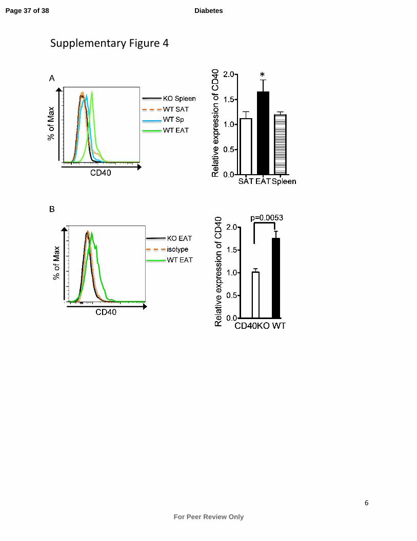
presence of CD8+ T cells. Consistent with these findings, we found that CD40 expression is
greatly upregulated on CD8+ T cells in EAT in DIO mice, compared to minimal or no expression
by CD8+ T cells harvested from SAT and spleen (Supplementary Fig. 4a). In addition, CD8
+ T
cells in EAT in older WT mice with normal chow diet also upregulated CD40 expression
(Supplementary Fig. 4b). Collectively, these results indicate that CD8+ T cells are essential in
inducing inflammation in adipose tissue and insulin resistance following HFD consumption, and
that CD40 plays a critical role in restraining this pathologic function.
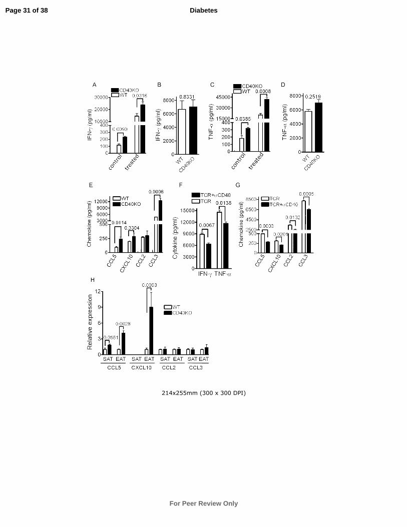
CD40 deficiency promotes production of chemokines and proinflammatory cytokines by
CD8+ T cells in obese EAT
To begin to determine how CD40-deficient CD8+ T cells affect insulin sensitivity, CD8
+ T cells
were sorted from SAT and EAT in DIO WT and CD40KO mice and cultured in vitro for 48 hrs
Page 14 of 38
For Peer Review Only
Diabetes

15
with/without anti-CD3 and CD28 Ab stimulation. Cytokine measurements in culture
supernatants showed that CD8+ T cells from CD40KO EAT produced significantly more IFN-γ
and TNF-α (Fig. 6A and C) both before and after stimulation. In contrast, there was no
discernible difference attributable to CD40 when CD8+ T cells were isolated from SAT (Fig. 6B
and D), which indicates that the microenvironment in EAT is key in promoting CD40-deficient
CD8+ T cells to produce proinflammatory cytokines. Consistent with the expression of CD40 on
CD8+ T cells in EAT, CD40 stimulation suppressed cytokine production from CD8
+ T cells
sorted from WT EAT (Fig. 6F). In contrast to CD8+ T cells, cytokine production by sorted CD4
+
T cells from EAT in CD40KO vs. WT mice showed no detectable difference (Supplementary
Fig. 5A and B).
CD8+
T cell infiltration in adipose tissue precedes that of Mφ, and CD8+ T cells can interact
with adipocytes and enhance Mφ migration by secreting chemokines (5). We found that sorted
CD40 deficient CD8+ T cells from EAT secreted more CCL5 and CCL3 (Fig. 6E) upon anti-CD3
and CD28 Ab stimulation than WT CD8+ T cells. Additionally, CD40 stimulation suppressed
CCL5, CXCL10, CCL2 and CCL3 production from CD8+ T cells sorted from WT EAT (Fig.
6G). Furthermore, real time PCR with whole SVF isolated from EAT revealed higher levels of
CCL5 and CXCL10, but not CCL2 and CCL3 mRNAs in CD40-deficient EAT than in WT, but
no difference was observed in SAT (Fig. 6H). Therefore, these results suggest that CD40
deficiency facilitates forming an environment in EAT with high concentrations of chemokines,
which may contribute to immune cell migration and residence in this adipose tissue.
Page 15 of 38
For Peer Review Only
Diabetes

16
Discussion
Inflammation in obese adipose tissue can be modulated by alterations of the composition and/or
interactions of local immune cells. Our finding that CD40KO mice in the DIO model exhibited
exacerbated local inflammation in EAT, insulin resistance and metabolic abnormalities was
initially surprising, as CD40 itself is a co-stimulatory receptor and its deficiency impairs general
immune responses. Of particular interest, we found that CD40-deficient CD8+ T cells in EAT
produced more proinflammatory cytokines and CCL5 and CCL3 and this was critical for
immune cell infiltration and insulin resistance induction in DIO CD40KO mice. We propose that
CD40 may play a special role in the microenvironment of obese EAT through regulating CD8+ T
cell function.
In contrast to our observations with CD40KO mice, a previous study showed that
CD154/CD40L is a requirement to maintain adipose tissue inflammation in mice (24), and
CD154 deficiency attenuates diet-induced steatosis and improves insulin resistance (28).
However, CD154 can function through distinct receptors additional to CD40, e.g. Mac-1/CD11b
(25). Therefore, the varied phenotypes observed may result from overlapping but different
physiologic alterations in mice lacking CD154 vs. CD40. For example, CD154 promotes
atherogenesis, whereas CD40 does not have an effect in a mouse model (29). Thus, the roles
played by CD154 and CD40 may be context-dependent. This concept is further supported by a
very recent report showing that CD40-deficient mice exhibit more severe hepatic steatosis,
adipose tissue inflammation and insulin resistance, although the immunological mechanisms
were not explored (30).
Page 16 of 38
For Peer Review Only
Diabetes

17
Antigen presenting cells, as well as T lymphocytes in certain situations can express CD40,
and both are crucial for inducing or regulating insulin resistance in DIO mice. Although there are
more Mφ and CD8+ T cells residing in the DIO CD40KO EAT, we found that the CD8
+ T cell is
the most critical element for inducing severe immune cell infiltration in the EAT and insulin
resistance in DIO CD40KO mice. Our results are consistent with a previous report that CD8+ T
cells can mediate Mφ recruitment and activation in adipose tissue (5), as transfer or depletion of
CD8+ T cells increased or decreased Mφ infiltration in DIO CD40KO mice, respectively. CD40
can be expressed on activated CD8+ T cells, but its role in CD8
+ T cell activation and
differentiation is not established (31-33). Recent studies indicate that CD40 signaling in CD8+ T
cells impacts pathogen-associated immune responses (19-21). CD40 signaling also promotes
optimal CD8+ effector T cell responses and blocks the differentiation of antigen specific
inducible T regulatory cells in a skin transplant model (34). In contrast to the proinflammatory
effect of CD40 in CD8+ T cells in these infectious disease models, our findings with the DIO
mouse model showed that CD40 plays an important anti-inflammatory role in obese EAT.
Interactions between adipocytes and CD8+ T cells are crucial for CD8
+ T cells to mediate Mφ
migration and activation (5), highlighting the unique immune environment of VAT. Our findings
that high numbers of Mφ and CD8+ T cells in EAT in DIO CD40KO mice are not accompanied
by increased numbers of these cells in lymphoid organs, further supports the specificity of this
unique tissue environment in inducing immune responses. Consistent with this point, although
there are less Foxp3+ regulatory T cells (Treg) in lymphoid tissues in CD40KO mice than in WT
mice (35), Treg number in obese EAT was not significantly different in WT vs CD40KO mice.
This result also indicates that Treg do not make a major contribution to the enhanced insulin
resistance in CD40KO mice. Our observation that CD40-deficient CD8+ T cells specifically in
Page 17 of 38
For Peer Review Only
Diabetes

18
EAT exhibit a more severe proinflammatory phenotype, suggests that EAT supplies a special
milieu for the interaction between CD8+ T cells and their environment. In this special
environment, CD40 provides a negative signal for activation of CD8+ T cells. The detailed
underlying molecular mechanisms await further investigation. Nevertheless, our current results,
together with the previous studies discussed above, indicate that the impact of CD40 upon CD8+
T cell functions is context-dependent. Consistent with our findings, a very recent report showed
enhanced adipose inflammation and insulin resistance due to reduced Treg cell development and
proliferation in CD80 and CD86-deficient mice (36). Although the mechanisms are distinct from
CD40KO mice, this further supports our concept that co-stimulatory signals may play different
roles in the microenvironment of obese adipose tissue compared to other disease conditions.
We here present the unexpected finding that CD40 potently restrains adipose tissue
inflammation and promotes metabolic homeostasis in mice. As CD40 polymorphisms are
associated with human diseases (37-39), further investigation into how CD40 in human
populations affects obesity and other metabolic diseases can continue to reveal important roles of
CD40 and new opportunities for disease management and intervention.
Page 18 of 38
For Peer Review Only
Diabetes

19
Acknowledgments:
We thank Dr. Lyse Norian (U. of Iowa, Iowa City, IA) for advice on the use of the HFD in the
DIO mouse model and for critical review of the manuscript. The work of the authors was
supported by a Research Career Scientist Award from the Dept. of Veterans Affairs (GAB) and a
Biological Sciences Funding Program pilot award from The University of Iowa (GAB). This
material is based upon work supported in part by the Department of Veterans Affairs, Veterans
Health Administration, Office of Research and Development.
The authors declare no financial or commercial conflict of interest.
Z.Y. designed and performed experiments and wrote the manuscript. L.S. performed
experiments, contributed to discussion and edited the manuscript. G.B. contributed to
experimental design and discussion, wrote and edited the manuscript.
Dr. Bishop is the guarantor of this work, had full access to all the data, and takes full
responsibility for the integrity of data and the accuracy of data analysis
Page 19 of 38
For Peer Review Only
Diabetes

20
References
1. Hotamisligil GS. 2006. Inflammation and metabolic disorders. Nature. 444:860–867.
2. Mathis D. 2013. Immunological goings-on inVAT. Cell Metab. 17:851-859.
3. Feuerer M, Herrero L, Cipolletta D, Naaz A, Wong J, Nayer A, Lee J, Goldfine AB,
Benoist C, Shoelson S, Mathis D. 2009. Lean, but not obese, fat is enriched for a unique
population of regulatory T cells that affect metabolic parameters. Nat Med. 15:930-939.
4. Liu J, Divoux A, Sun J, Zhang J, Clément K, Glickman JN, Sukhova GK, Wolters PJ, Du
J, Gorgun CZ, Doria A, Libby P, Blumberg RS, Kahn BB, Hotamisligil GS, Shi GP.
2009. Genetic deficiency and pharmacological stabilization of mast cells reduce diet-
induced obesity and diabetes in mice. Nat Med. 15:940-945.
5. Nishimura S, Manabe I, Nagasaki M, Eto K, Yamashita H, Ohsugi M, Otsu M, Hara K,
Ueki K, Sugiura S, Yoshimura K, Kadowaki T, Nagai R. 2009. CD8+ effector T cells
contribute to macrophage recruitment and adipose tissue inflammation in obesity. Nat
Med. 15: 914-920.
6. Weisberg SP, McCann D, Desai M, Rosenbaum M, Leibel RL, Ferrante AW Jr. 2003.
Obesity is associated with macrophage accumulation in adipose tissue. J Clin Invest.
112:1796–1808.
7. Winer S, Chan Y, Paltser G, Truong D, Tsui H, Bahrami J, Dorfman R, Wang Y,
Zielenski J, Mastronardi F, Maezawa Y, Drucker DJ, Engleman E, Winer D, Dosch HM.
2009. Normalization of obesity-associated insulin resistance through immunotherapy. Nat
Med. 15:921–929.
8. Winer DA, Winer S, Shen L, Wadia PP, Yantha J, Paltser G, Tsui H, Wu P, Davidson
MG, Alonso MN, Leong HX, Glassford A, Caimol M, Kenkel JA, Tedder TF,
McLaughlin T, Miklos DB, Dosch HM, Engleman EG. 2011. B cells promote insulin
resistance through modulation of T cells and production of pathogenic IgG antibodies.
Nat Med. 17:610-617.
9. Xu H, Barnes GT, Yang Q, Tan G, Yang D, Chou CJ, Sole J, Nichols A, Ross JS,
Tartaglia LA, Chen H. 2003. Chronic inflammation in fat plays a crucial role in the
development of obesity-related insulin resistance. J Clin Invest. 112:1821–1830.
10. Hotamisligil GS, Shargill NS, Spiegelman BM. 1993. Adipose expression ofTNF-α:
direct role in obesity-linked insulin resistance. Science. 259:87–91.
11. Kanda H, Tateya S, Tamori Y, Kotani K, Hiasa K, Kitazawa R, Kitazawa S, Miyachi H,
Maeda S, Egashira K, Kasuga M. 2006. MCP-1 contributes to macrophage infiltration
into adipose tissue, insulin resistance, and hepatic steatosis in obesity. J Clin Invest.
116:1494-1505.
12. Ouchi N, Parker JL, Lugus JJ, Walsh K. 2011. Adipokines in inflammation and metabolic
disease. Nat Rev Immunol. 11:85-97.
13. Pradhan AD, Manson JE, Rifai N, Buring JE, Ridker PM. 2001. C-reactive protein,
interleukin 6, and risk of developing type 2 diabetes mellitus. JAMA. 286:327–334.
14. Callard E, Armitage RJ, Fanslow WC, Spriggs MK. 1993. CD40L and its role in X-
linked hyper-IgM syndrome. Immunol Today. 14: 559–564.
15. DiSanto JP, Bonnefoy JY, Gauchat JF, Fischer A, de Saint Basile G. 1993. CD40L
mutations in X-linked immunodeficiency with hyper-IgM. Nature. 361:541–543.
16. Kawabe T, Naka T, Yoshida K, Tanaka T, Fujiwara H, Suematsu S, Yoshida N,
Kishimoto T, Kikutani H. 1994. The immune responses in CD40-deficient mice:
Page 20 of 38
For Peer Review Only
Diabetes

21
impaired immunoglobulin class switching and germinal center formation. Immunity.
1:167-178.
17. Wagner DH Jr, Vaitaitis G, Sanderson R, Poulin M, Dobbs C, Haskins K. 2002.
Expression of CD40 identifies a unique pathogenic T cell population in type 1 diabetes.
Proc Natl Acad Sci U S A. 99:3782-3787.
18. Munroe ME, Bishop GA. 2007. A costimulatory function for T cell CD40. J Immunol.
178:671-682.
19. Bhadra R, Gigley JP, Khan IA. 2011. Cutting edge: CD40-CD40L pathway plays a
critical CD8-intrinsic and -extrinsic role during rescue of exhausted CD8 T cells. J.
Immunol. 187:4421–4425.
20. Johnson S, Zhan Y, Sutherland RM, Mount AM, Bedoui S, Brady JL, Carrington EM,
Brown LE, Belz GT, Heath WR, Lew AM. 2009. Selected Toll-like receptor ligands and
viruses promote helper-independent cytotoxic T cell priming by upregulating CD40L on
dendritic cells. Immunity. 30:218–227.
21. Martin S, Pahari S, Sudan R, Saha B. 2010. CD40 signaling in CD8+CD40
+ T cells turns
on contra-T regulatory cell functions. J. Immunol. 184:5510–5518.
22. Missiou A, Wolf D, Platzer I, Ernst S, Walter C, Rudolf P, Zirlik K, Köstlin N, Willecke
FK, Münkel C, Schönbeck U, Libby P, Bode C, Varo N, Zirlik A. 2010. CD40L induces
inflammation and adipogenesis in adipose cells--a potential link between metabolic and
cardiovascular disease. Thromb Haemost. 103:788-796.
23. Poggi M, Jager J, Paulmyer-Lacroix O, Peiretti F, Gremeaux T, Verdier M, Grino M,
Stepanian A, Msika S, Burcelin R, de Prost D, Tanti JF, Alessi MC. 2009. The
inflammatory receptor CD40 is expressed on human adipocytes: contribution to crosstalk
between lymphocytes and adipocytes. Diabetologia. 52:1152-1163.
24. Wolf D, Jehle F, Ortiz Rodriguez A, Dufner B, Hoppe N, Colberg C, Lozhkin A, Bassler
N, Rupprecht B, Wiedemann A, Hilgendorf I, Stachon P, Willecke F, Febbraio M, von
zur Muhlen C, Binder CJ, Bode C, Zirlik A, Peter K. 2012. CD40L deficiency attenuates
diet-induced adipose tissue inflammation by impairing immune cell accumulation and
production of pathogenic IgG-antibodies. PLoS One. 7:e33026.
25. Zirlik A, Maier C, Gerdes N, MacFarlane L, Soosairajah J, Bavendiek U, Ahrens I, Ernst
S, Bassler N, Missiou A, Patko Z, Aikawa M, Schönbeck U, Bode C, Libby P, Peter K.
2007. CD40L mediates inflammation independently of CD40 by interaction with Mac-1.
Circulation. 115:1571–1580.
26. Schönbeck U, Libby P. 2001. CD40 signaling and plaque instability. Circ Res. 89:1092-
1103.
27. Cinti S, Mitchell G, Barbatelli G, Murano I, Ceresi E, Faloia E, Wang S, Fortier M,
Greenberg AS, Obin MS. 2005. Adipocyte death defines macrophage localization and
function in adipose tissue of obese mice and humans. J. Lipid Res. 46: 2347–2355.
28. Poggi M, Engel D, Christ A, Beckers L, Wijnands E, Boon L, Driessen A, Cleutjens J,
Weber C, Gerdes N, Lutgens E. 2011. CD40L deficiency ameliorates adipose tissue
inflammation and metabolic manifestations of obesity in mice. Arterioscler Thromb Vasc
Biol. 31:2251–2260.
29. Gerdes, N, and Zirlik, A 2011. Co-stimulatory molecules in and beyond co-stimulation-
tipping the balance in atherosclerosis? Thromb Haemost. 106:804-813.
30. Guo CA, Kogan S, Amano SU, Wang M, Dagdeviren S, Friedline RH, Aouadi M, Kim
JK, Czech MP. 2013. CD40 deficiency in mice exacerbates obesity-induced adipose
Page 21 of 38
For Peer Review Only
Diabetes

22
tissue inflammation, hepatic steatosis and insulin resistance. Am J Physiol Endocrinol
Metab. 304(9):E951-963.
31. Bourgeois C, Rocha B, Tanchot C. 2002. A role for CD40 expression on CD8+ T cells in
the generation of CD8+ T cell memory. Science. 297:2060-2063.
32. Lee BO, Hartson L, Randall TD. 2003. CD40-deficient, influenza-specific CD8 memory
T cells develop and function normally in a CD40-sufficient environment. J Exp Med
198:1759–1764.
33. Sun JC, Bevan MJ. 2004. Cutting edge: long-lived CD8 memory and protective
immunity in the absence of CD40 expression on CD8 T cells. J Immunol. 172:3385–
3389.
34. Liu D, Ferrer IR, Konomos M, Ford ML. 2013. Inhibition of CD8+ T cell-derived CD40
signals is necessary but not sufficient for Foxp3+ induced regulatory T cell generation in
vivo. J Immunol. 15;191(4):1957-64
35. Spence PJ, Green EA. 2008. Foxp3+ regulatory T cells promiscuously accept thymic
signals critical for their development. Proc Natl Acad Sci U S A. 105:973-8.
36. Zhong J, Rao X, Braunstein Z, Taylor A, Narula V, Hazey J, Mikami D, Needleman B,
Rutsky J, Sun Q, Deiuliis JA, Satoskar AR, Rajagopalan S. T cell costimulation protects
obesity-induced adipose inflammation and insulin resistance. Diabetes. 2013 Nov 12.
[Epub ahead of print]
37. Bishop G.A. 2009. The many faces of CD40: multiple roles in normal immunity and
disease. Semin Immunol. 21:255-256.
38. Jacobson EM, Concepcion E, Oashi T and Tomer Y. 2005. A Graves’ disease-associated
Kozak sequence single-nucleotide polymorphism enhances the efficiency of CD40 gene
translation: A case for translational pathophysiology. Thyroid. 146:2684-2691.
39. Park JH, Chang HS, Park CS, Jang AS, Park BL, Rhim TY, Uh ST, Kim YH, Chung IY,
Shin HD. 2007. Association analysis of CD40 polymorphisms with asthma and the level
of serum total IgE. Am J Respir Crit Care Med. 175:775-782.
Page 22 of 38
For Peer Review Only
Diabetes

23
FIGURE LEGENDS:
Figure 1. CD40 deficiency aggravates HFD-induced metabolic complications. WT and
CD40KO mice consumed a HFD for 18 weeks. Relative increase of body weight (% increase of
starting body weight) (A) and weight of EAT and SAT after 18 weeks of diet (B) were shown.
(C) GTT and (D) ITT were performed after intraperitoneal injection of glucose or insulin.
Triglyceride (E), total cholesterol (F), LDL/vLDL (G), HDL (H) and insulin (I) levels in mouse
sera were examined (SCD=standard chow diet). Data are presented as mean ± SEM of at least 12
animals for A–D and of 8 animals for E–J per group. *p<0.05.
Figure 2. CD40 expression on immune cells is required for preventing aggravated obesity
and insulin resistance. 8-week-old WT and CD40KO mice were lethally irradiated and
reconstituted with BM from WT or CD40KO mice. 4 weeks later, mice were fed with HFD for
12 weeks. Relative increase in body weight during HFD feeding (A) and weight of EAT (B) was
quantified at the end of the study. (C) GTT and (D) ITT were performed after intraperitoneal
injection of glucose or insulin. Data are presented as mean ± SEM of >8 animals per group.
*p<0.05 (WT+KOBM or KO+KOBM vs. WT+WTBM or KO+WTBM).
Figure 3. CD40 deficiency results in severe local inflammation in EAT. WT and CD40KO
mice consumed a HFD or standard chow diet for 18 weeks. (A) Immune cells in EAT in DIO
mice were identified by flow cytometry (left) and quantified (right). Cell number is expressed
per gram of fat pads. (B) Immune cells in EAT in standard chow diet-fed mice were quantified
(Cell number expressed per gram of fat pads), and GTT (C) and ITT (D) were performed. (E)
Epididymal fat pad sections from DIO mice were stained for F4/80 (green) and perilipin (red)
(left) (scale bar=50µm). CLS were counted (right) (4 mice for each group, n=20 low-power
Page 23 of 38
For Peer Review Only
Diabetes
24
fields in each group). (F) Mφ (F4/80+) in epididymal fat pads in DIO mice were gated, and M1-
like Mφ (CD11c+) and M2-like Mφ (CD11c
-) were further analyzed and quantified. Cell number
is expressed per gram of fat pads. (G) Cytokines in freshly isolated SVF from EAT and SAT in
DIO mice were measured by real time PCR and fold changes of mRNA expression level
(CD40KO vs. WT) are shown (4 mice for each group). (H) Isolated SVF was cultured for 48 hrs,
and TNF-α and IFN-γ were quantified by ELISA (one of three individual experiments is shown).
Data are presented as mean ± SEM of 8 to 12 mice for all the panels except E and G.
Figure 4. Role of lymphoid cells in the susceptibility of HFD-induced obesity and insulin
resistance in CD40KO mice. WT mice were lethally irradiated and immune systems were
reconstituted with mixed BM (Rag1-/-
BM vs. WT BM or CD40KO BM with a 10:1 ratio). 4
weeks later, HFD was given for 12 weeks. (A) Relative increase of body weight during HFD
feeding was shown. (B) GTT and (C) ITT were conducted. Data are presented as mean ± SEM of
8 animals per group. *p<0.05.
Figure 5. CD8+ T cells are essential in exacerbating HFD-induced insulin resistance in
CD40KO mice. For (A) to (C), Rag1-/-
mice were fed with HFD for 12 weeks, then 5x10⁶ CD8
+
T cells isolated from spleen of WT or CD40KO mice were transferred intravenously. HFD was
given for 4 more weeks. GTT (A) and ITT (B) were conducted. (C) CD8+ T cell and Mφ in EAT
were quantified (Cell number is expressed per gram of fat pads). For (D) to (F), WT and
CD40KO mice were fed with HFD for 12 weeks, anti-CD8 depleting or isotype control Ab were
injected intraperitoneally once per week for 6 weeks. HFD was given simultaneously. GTT (D)
and ITT (E) were conducted (The list of legends shows the order of graph from high to low). (F)
Immune cells in EAT were quantified (Cell number is expressed per gram of fat pads). Data are
presented as mean ± SEM of 6-8 animals per group. *p<0.05.
Page 24 of 38
For Peer Review Only
Diabetes
25
Figure 6. CD40 deficiency promotes chemokine and proinflammatory cytokine production
by CD8+ T cells in EAT. WT and CD40KO mice consumed a HFD for 18 weeks. In A-E, CD8
+
T cells in EAT and SAT were sorted from WT and CD40KO mice and stimulated with anti-CD3
and CD28 Ab for 48 hours. IFN-γ and TNF-α produced by control and treated CD8+ T cell in
EAT (A and C) and by treated CD8+ T cells in SAT (B and D) were measured by ELISA.
Chemokines produced by treated CD8+ T cells in EAT were measured by Luminex beads (E)
(one of three individual experiments are shown). In F and G, CD8+ T cells in EAT were sorted
from obese WT mice and stimulated with anti-CD3 and CD28 Ab and in the presence or absence
of anti-CD40 Ab for 48 hours. IFN-γ and TNF-α (F) and chemokines (G) were quantified with
luminex beads (Data represent 4 mixed mice). (H) Chemokine mRNA in whole SVF isolated
from EAT and SAT were measured by real time PCR and fold changes of mRNA expression
level (CD40KO vs. WT) are shown (4 mice for each group). Data are presented as mean ± SEM.
Page 25 of 38
For Peer Review Only
Diabetes

106x62mm (300 x 300 DPI)
Page 26 of 38
For Peer Review Only
Diabetes

62x21mm (300 x 300 DPI)
Page 27 of 38
For Peer Review Only
Diabetes

181x182mm (300 x 300 DPI)
Page 28 of 38
For Peer Review Only
Diabetes

60x24mm (300 x 300 DPI)
Page 29 of 38
For Peer Review Only
Diabetes

140x109mm (300 x 300 DPI)
Page 30 of 38
For Peer Review Only
Diabetes
214x255mm (300 x 300 DPI)
Page 31 of 38
For Peer Review Only
Diabetes

1
Online Supplementary Data:
Supplementary Figure 1. Fasting blood glucose levels in DIO CD40KO mice vs. WT mice.
WT and CD40KO mice consumed a HFD for 18 weeks. They were fasted for 14 hrs prior to
measurement of blood glucose levels. Data are presented as mean ± SEM of 12 animals per
group.
Supplementary Figure 2. Hepatic lipid deposition in DIO mice. WT and CD40KO mice
consumed a HFD for 18 weeks. Frozen liver sections were stained with Oil-Red-O. Data are
representative of one of 5 mice from each group.
Supplementary Figure 3. CD40 deficiency in CD4+ T cells and B cells does not contribute to
the aggravated insulin tolerance in DIO CD40KO mice. Rag1-/-
mice were fed with HFD for
12 weeks, after which 5x10⁶ CD4
+ T cells or 10x10
6 B cells isolated from spleen of WT or
CD40KO mice were transferred. HFD was given for 4 more weeks. GTT and ITT with CD4+ T
cell transferred mice (A and B) and B cell transferred mice (C and D) were conducted. Data are
presented as mean ± SEM of 6-8 animals per group. *p<0.05.
Supplementary Figure 4. CD40 expression in CD8+ T cells is upregulated in EAT. (A) WT
and CD40 KO mice consumed a HFD for 18 weeks. CD8+ T cells in EAT, SAT and spleen were
stained for CD40 (left). The relative expression levels of CD40 are shown (right, WT/CD40KO
mean fluorescence intensity). Data are presented as mean ± SEM of 6 animals per group.
*p<0.05 (EAT vs. SAT or spleen). (B) WT and CD40KO mice consumed normal chow diet for
30 weeks. CD8+ T cells in EAT were stained for CD40 (left). The relative expression levels of
CD40 are shown (right, WT/CD40KO mean fluorescence intensity). Data are presented as mean
± SEM of 4 animals per group.
Page 32 of 38
For Peer Review Only
Diabetes

2
Supplementary Figure 5. CD40 deficiency in EAT CD4+ T cells does not affect cytokine
production. WT and CD40 KO mice consumed a HFD for 18 weeks. CD4+ T cells in EAT were
sorted and stimulated with anti-CD3 and anti-CD28 Abs for 48 hours. IFN-γ (A) and TNF-α (B)
were measured by ELISA. Data are representative of two independent experiments.
Page 33 of 38
For Peer Review Only
Diabetes

3
WT
CD40KO
0
50
100
150
200 0.9644
Blood Glucose (mg/dL)
Supplementary Figure 1
Page 34 of 38
For Peer Review Only
Diabetes

4
WT CD40KO
Supplementary Figure 2
Page 35 of 38
For Peer Review Only
Diabetes

5
A B
C D
0 1530 60 1200
20
40
60
80
100
120no transfer
WT CD4
KO CD4
minBlood Glucose (% of start)
*
*
*
0 15 30 60 1200
200
400
600no transfer
WT CD4
KO CD4
min
Blood Glucose (mg/dl)
* *
*
*
0 15 30 60 1200
200
400
600no transfer
WT B
KO B
min
Blood Glucose (mg/dl)
0 1530 60 1200
20
40
60
80
100
120WT B
KO B
no transfer
min
Blood Glucose (% of start)
Supplementary Figure 3
Page 36 of 38
For Peer Review Only
Diabetes
6
Supplementary Figure 4
Page 37 of 38
For Peer Review Only
Diabetes

7
Supplementary Figure 5
WT
CD40KO
0
3000
6000
9000
12000
15000 0.9145
TNF- α (pg/ml)
A B
WT
CD40KO
0
3000
6000
9000
0.5467
IFN- γ (pg/ml)
Page 38 of 38
For Peer Review Only
Diabetes
Related Documents