The Influence of the Maxillary Frenum on the Development and Pattern of Dental Caries on Anterior Teeth in Breastfeeding Infants: Prevention, Diagnosis, and Treatment Lawrence A. Kotlow, DDS [AQ: 1] Keywords: breastfeeding; dental caries; infant development Accepted for publication December 14, 2009. Disclosure: No company, individuals, or other sources supported this article. Lawrence A. Kotlow, DDS, has had a private dental practice located in Albany, New York, since 1974. He specializes in pediatric dentistry. He is a graduate of SUNY Buffalo School of Dentistry and the Cincinnati Children’s Hospital pediatric dental postgraduate program. He is board certified in pediatric dentistry by the American Board of Pediatric Dentistry. He received his advanced proficiency certification from the Academy of Laser Dentistry in the Erbium laser and has standard proficiency using the Nd:YAG and diode laser. He is a recognized standard proficiency course provider for the Academy of Laser Dentistry and has achieved mastership in the Academy of Laser Dentistry. He has published many articles on using hard and soft tissue lasers and topics in pediatric dentistry. He contributed a chapter to the October 2004 Dental Clinics of North America: Lasers in Clinical Dentistry titled “Lasers in Pediatric Dentistry.” He has lectured throughout the United States, Canada, Australia, and Taiwan about pediat- ric dentistry and lasers. Address correspondence to Lawrence A. Kotlow, DDS, 340 Fuller Road, Albany, NY 12203. J Hum Lact XX(X), XXXX DOI: 10.1177/0890334410362520 © Copyright 2010 International Lactation Consultant Association 1 The concept that the first dental examination for chil- dren should occur no later than the first birthday or 6 months after the first teeth erupt has been a long sought after goal of the Academy of Pediatric Dentistry. 1 Implementation of this concept has been slow and often difficult because parents may have difficulty locating a dentist who treats infants this young. An infant’s first year represents many “firsts” in developmental milestones: nursing, teething, facial recognition, rolling over, crawling, walking, and even some word development. 2 Additional firsts should include an inspection of the oral cavity to observe any soft tissue abnormalities. An excellent time for this inspection is just after the first feeding. Such anoma- lies that could affect nursing are neonatal teeth, 3 gingival lesions, 4-6 ankyloglossia, and position of the maxillary labial frenum attachment. It is also important that new parents become properly educated as to their child’s oral development and be made aware that children as young as 6 months of age can begin to develop early infant dental caries. Because it is a recent concept to universally have a routine examination of the oral structures of newborns and infants, clinicians should be sure to check for any and all deviations from normal, especially those that could affect the formation of dental caries. Abnormal frena attachments fall into this category. Despite the lack of documenting studies to compare and reference the risks of leaving abnormal frenum attachments intact, revising such a frenum may very well preclude the development of dental caries in the breastfeeding infant. The purpose of this article is to alert health care professionals treating infants and nursing mothers that although nursing alone may not cause dental caries, nursing in conjunction with an abnormal maxillary frenum attachment may be a contributing cause of facial caries development in a nursing infant. In 1977, it was first reported that some infants who were exclusively nursing presented with a pattern of anterior decay different than the decay observed in chil- dren who were allowed to sleep or rest with a baby bottle containing liquids other than water. 7,8 Differential diagnostic discussions with nursing mothers indicated that these infants were not receiving nutrition other than by the breast and that in all cases were sleeping through- out the night with the mother and engaging in at-will nursing. Two to 3 times during the night, the infant would nurse, a feeding pattern that continued after the eruption of the upper and lower central and lateral inci- sors and in some cases beyond the age of 2 or 3 years. The pattern of decay seen in baby bottle caries pres- ents as lingual caries on the upper central and lateral incisors, followed by decay of the upper first molars. In most cases, parents usually attempt to wean infants from the bottle by 12 to 14 months. The primary canines and second primary molars usually do not erupt until after 14 to 16 months of age and thus are spared the effects of sleeping or resting with a baby bottle. JHL362520 for WEB.indd 1 04/02/2010 8:27:56 PM

Welcome message from author
This document is posted to help you gain knowledge. Please leave a comment to let me know what you think about it! Share it to your friends and learn new things together.
Transcript

The Influence of the Maxillary Frenum on the Development and Pattern of Dental Caries on Anterior Teeth in Breastfeeding Infants: Prevention, Diagnosis, and TreatmentLawrence A. Kotlow, DDS
[AQ: 1]
Keywords: breastfeeding; dental caries; infant development
Accepted for publication December 14, 2009.
Disclosure: No company, individuals, or other sources supported this article.
Lawrence A. Kotlow, DDS, has had a private dental practice located in Albany, New York, since 1974. He specializes in pediatric dentistry. He is a graduate of SUNY Buffalo School of Dentistry and the Cincinnati Children’s Hospital pediatric dental postgraduate program. He is board certified in pediatric dentistry by the American Board of Pediatric Dentistry. He received his advanced proficiency certification from the Academy of Laser Dentistry in the Erbium laser and has standard proficiency using the Nd:YAG and diode laser. He is a recognized standard proficiency course provider for the Academy of Laser Dentistry and has achieved mastership in the Academy of Laser Dentistry. He has published many articles on using hard and soft tissue lasers and topics in pediatric dentistry. He contributed a chapter to the October 2004 Dental Clinics of North America: Lasers in Clinical Dentistry titled “Lasers in Pediatric Dentistry.” He has lectured throughout the United States, Canada, Australia, and Taiwan about pediat-ric dentistry and lasers.
Address correspondence to Lawrence A. Kotlow, DDS, 340 Fuller Road, Albany, NY 12203.
J Hum Lact XX(X), XXXXDOI: 10.1177/0890334410362520© Copyright 2010 International Lactation Consultant Association
1
The concept that the first dental examination for chil-dren should occur no later than the first birthday or 6 months after the first teeth erupt has been a long sought after goal of the Academy of Pediatric Dentistry.1 Implementation of this concept has been slow and often difficult because parents may have difficulty locating a dentist who treats infants this young.
An infant’s first year represents many “firsts” in developmental milestones: nursing, teething, facial recognition, rolling over, crawling, walking, and even some word development.2 Additional firsts should include an inspection of the oral cavity to observe any soft tissue abnormalities. An excellent time for this inspection is just after the first feeding. Such anoma-lies that could affect nursing are neonatal teeth,3 gingival lesions,4-6 ankyloglossia, and position of the maxillary labial frenum attachment. It is also important that new parents become properly educated as to their child’s oral development and be made aware that children as
young as 6 months of age can begin to develop early infant dental caries.
Because it is a recent concept to universally have a routine examination of the oral structures of newborns and infants, clinicians should be sure to check for any and all deviations from normal, especially those that could affect the formation of dental caries. Abnormal frena attachments fall into this category. Despite the lack of documenting studies to compare and reference the risks of leaving abnormal frenum attachments intact, revising such a frenum may very well preclude the development of dental caries in the breastfeeding infant. The purpose of this article is to alert health care professionals treating infants and nursing mothers that although nursing alone may not cause dental caries, nursing in conjunction with an abnormal maxillary frenum attachment may be a contributing cause of facial caries development in a nursing infant.
In 1977, it was first reported that some infants who were exclusively nursing presented with a pattern of anterior decay different than the decay observed in chil-dren who were allowed to sleep or rest with a baby bottle containing liquids other than water.7,8 Differential diagnostic discussions with nursing mothers indicated that these infants were not receiving nutrition other than by the breast and that in all cases were sleeping through-out the night with the mother and engaging in at-will nursing. Two to 3 times during the night, the infant would nurse, a feeding pattern that continued after the eruption of the upper and lower central and lateral inci-sors and in some cases beyond the age of 2 or 3 years.
The pattern of decay seen in baby bottle caries pres-ents as lingual caries on the upper central and lateral incisors, followed by decay of the upper first molars. In most cases, parents usually attempt to wean infants from the bottle by 12 to 14 months. The primary canines and second primary molars usually do not erupt until after 14 to 16 months of age and thus are spared the effects of sleeping or resting with a baby bottle.
JHL362520 for WEB.indd 1 04/02/2010 8:27:56 PM
Larry
Larry
Larry
Larry

2 Kotlow J Hum Lact XX(X), XXXX
Figure 1. Various patterns of facial dental caries in children nurs-ing at will every night.
A normal functioning tongue will cover the man-dibular incisors, protecting them from caries forma-tion. Infants who breastfeed and are born with an abnormal maxillary frenum attachment often present with a different pattern of dental caries: facial caries on the upper incisors (Figures 1 A-C and Figure 2) that often include incisal notching (Figures 3 and 4A).
Figure 2. Nursing infant 2.5 years of age exhibiting an abnormal frenum attachment.
Figure 3. Notching decay pattern in nursing infants exhibiting abnormal frenum attachments.
Breastfeeding-induced dental caries appear to be uncommon but not rare. Breastfed infants, having a tight maxillary frenum, examined immediately after completion of nursing may show retained milk on the facial surfaces of the upper anterior teeth (Figure 1C). Children who do not nurse yet present with facial car-ies and no history of sleeping or resting with a bottle or sippy cup also appear to have this low or tight max-illary frenum.
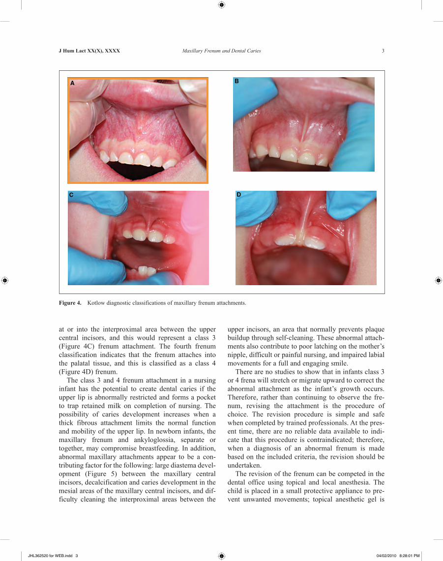
The author has developed a clinical set of criteria based on examination of more than 350 infants in the age group of newborns to 3 years that are based on the position of the maxillary frenum. A class 1 (Figure 4A) frenum indicates little or no tissue attachment of the lip to the gingival tissue. A Class 2 (Figure 4B) frenum indicates the frenum is attached above or at the border between free and attached gingival tissue. Abnormal attachments begin to occur when the frenum attaches
JHL362520 for WEB.indd 2 04/02/2010 8:27:59 PM
J Hum Lact XX(X), XXXX Maxillary Frenum and Dental Caries 3
upper incisors, an area that normally prevents plaque buildup through self-cleaning. These abnormal attach-ments also contribute to poor latching on the mother’s nipple, difficult or painful nursing, and impaired labial movements for a full and engaging smile.
There are no studies to show that in infants class 3 or 4 frena will stretch or migrate upward to correct the abnormal attachment as the infant’s growth occurs. Therefore, rather than continuing to observe the fre-num, revising the attachment is the procedure of choice. The revision procedure is simple and safe when completed by trained professionals. At the pres-ent time, there are no reliable data available to indi-cate that this procedure is contraindicated; therefore, when a diagnosis of an abnormal frenum is made based on the included criteria, the revision should be undertaken.
The revision of the frenum can be competed in the dental office using topical and local anesthesia. The child is placed in a small protective appliance to pre-vent unwanted movements; topical anesthetic gel is
Figure 4. Kotlow diagnostic classifications of maxillary frenum attachments.
at or into the interproximal area between the upper central incisors, and this would represent a class 3 (Figure 4C) frenum attachment. The fourth frenum classification indicates that the frenum attaches into the palatal tissue, and this is classified as a class 4 (Figure 4D) frenum.
The class 3 and 4 frenum attachment in a nursing infant has the potential to create dental caries if the upper lip is abnormally restricted and forms a pocket to trap retained milk on completion of nursing. The possibility of caries development increases when a thick fibrous attachment limits the normal function and mobility of the upper lip. In newborn infants, the maxillary frenum and ankyloglossia, separate or together, may compromise breastfeeding. In addition, abnormal maxillary attachments appear to be a con-tributing factor for the following: large diastema devel-opment (Figure 5) between the maxillary central incisors, decalcification and caries development in the mesial areas of the maxillary central incisors, and dif-ficulty cleaning the interproximal areas between the
JHL362520 for WEB.indd 3 04/02/2010 8:28:01 PM

4 Kotlow J Hum Lact XX(X), XXXX
Figure 5. Diastema formation.
Figure 6. Laser safety goggles.
then placed over the maxillary frenum area followed by a small amount of local anesthesia. The local anes-thetic should be placed directly into the frenum slowly to prevent distortion of the frenum. Appropriate laser safety glasses are placed on all individuals in the immediate surgical operating area. Laser safety glasses are placed on the infant; goggles are used on newborns (Figure 6).
The revision is completed using an Erbium:YAG laser at 2970 nm, laser energy of 20 Hz and 65 mJ, 600 microseconds pulse duration, and no water (PowerLase AT, Lares Research, Chico, California). An alternative is the 980-nm or similar wavelength diode at 1 W (Figures 7 A-C). The procedure takes less than 60 sec-onds. Postoperative instructions require the parent to
Figure 7. A, laser revision using the Erbium:YAG laser. B, immediately after treatment. C, healing of frenum 6 days after surgery.
separate the lip from the gingival tissue by pulling up the lip twice a day, once in the morning and once at bedtime, followed by placing a small amount of vita-min E on the area. The area heals uneventfully, and the child is rarely uncomfortable. Patients are seen for a postsurgical follow-up in 6 days.
Conclusion
In breastfeeding infants, class 3 and class 4 maxillary frena have the potential to become a significant factor contributing to facial caries formation on the maxillary anterior teeth. This is in part due to the inability of an
JHL362520 for WEB.indd 4 04/02/2010 8:28:04 PM

J Hum Lact XX(X), XXXX Maxillary Frenum and Dental Caries 5
infant to remove residual milk from the area between the lip and facial surfaces of the maxillary central and lateral incisors at the completion of nursing. A nursing mother should be cautious and practice good oral hygiene in the infant after the upper front teeth erupt, making sure no residual milk remains in contact with the facial surfaces of the infant’s teeth on completion of nursing. If a parent sees any signs of enamel decalcification, white spots, or discoloration developing, a visit to a dentist should occur followed by any needed corrective care. Early diagnosis and treatment of an abnormal frenum attachment with the simple and quick revision technique using the Erbium:YAG or Diode laser can prevent decay from occurring and do not present any risk to the infant.
References1. Reference manual 07/08PG 82: clinical guidelines. Pediatr Dent. 29.
[AQ: 2]2. March of Dimes. Developmental milestones for babies. Available at: www
.marchofdimes.com/pnhec/298_10203.asp. Accessed January 27, 2010.3. Leung AKC, Robson W. Natal teeth: a review. J Natl Med Assoc.
2006;98:226-228.4. Kotlow L. Ankyloglossia (tongue-tie): a diagnostic and treatment quan-
dary. Quintessence Int. 1999;30:259-262.5. Kotlow L. Oral diagnosis of abnormal frenum attachments in neonates
and infants. J Pediatr Dent Care. 2004;10:[AQ: 3]6. Kotlow L. Using the Erbium:YAG laser to correct abnormal lingual fre-
num attachments in newborns. J Acad Laser Dent. 2004;12:[AQ: 4]7. Kotlow LA. Breast feeding: a cause of dental caries in children. ASDC
J Dent Child. 1977;44:192-193.8. Gardner DE, Norwood JR, Eisenson JE. At-will breast feeding and
dental caries: four case reports. ASDC J Dent Child. 1977;44:186-191.9. Weissinger D, Miller M. Breastfeeding difficulties as the result of tight
lingual and labial frena. J Hum Lact. 1995;11:313-316. [AQ: 5]
JHL362520 for WEB.indd 5 04/02/2010 8:28:04 PM
Larry
Larry
Larry
Larry
Larry
Larry
Related Documents