The Impact of cHS4 Insulators on DNA Transposon Vector Mobilization and Silencing in Retinal Pigment Epithelium Cells Nynne Sharma, Anne Kruse Hollensen, Rasmus O. Bak, Nicklas Heine Staunstrup, Lisbeth Dahl Schrøder, Jacob Giehm Mikkelsen* Department of Biomedicine, Aarhus University, Aarhus, Denmark Abstract DNA transposons have become important vectors for efficient non-viral integration of transgenes into genomic DNA. The Sleeping Beauty (SB), piggyBac (PB), and Tol2 transposable elements have distinct biological properties and currently represent the most promising transposon systems for animal transgenesis and gene therapy. A potential obstacle, however, for persistent function of integrating vectors is transcriptional repression of the element and its genetic cargo. In this study we analyze the insulating effect of the 1.2-kb 59-HS4 chicken b-globin (cHS4) insulator element in the context of SB, PB, and Tol2 transposon vectors. By examining transgene expression from genomically inserted transposon vectors encoding a marker gene driven by a silencing-prone promoter, we detect variable levels of transcriptional silencing for the three transposon systems in retinal pigment epithelium cells. Notably, the PB system seems less vulnerable to silencing. Incorporation of cHS4 insulator sequences into the transposon vectors results in 2.2-fold and 1.5-fold increased transgene expression levels for insulated SB and PB vectors, respectively, but an improved persistency of expression was not obtained for insulated transgenes. Colony formation assays and quantitative excision assays unveil enhanced SB transposition efficiencies by the inclusion of the cHS4 element, resulting in a significant increase in the stable transfection rate for insulated SB transposon vectors in human cell lines. Our findings reveal a positive impact of cHS4 insulator inclusion for SB and PB vectors in terms of increased transgene expression levels and improved SB stable transfection rates, but also the lack of a long-term protective effect of the cHS4 insulator against progressive transgene silencing in retinal pigment epithelium cells. Citation: Sharma N, Hollensen AK, Bak RO, Staunstrup NH, Schrøder LD, et al. (2012) The Impact of cHS4 Insulators on DNA Transposon Vector Mobilization and Silencing in Retinal Pigment Epithelium Cells. PLoS ONE 7(10): e48421. doi:10.1371/journal.pone.0048421 Editor: Ferenc Mueller, University of Birmingham, United Kingdom Received June 7, 2012; Accepted September 25, 2012; Published October 26, 2012 Copyright: ß 2012 Sharma et al. This is an open-access article distributed under the terms of the Creative Commons Attribution License, which permits unrestricted use, distribution, and reproduction in any medium, provided the original author and source are credited. Funding: This work was supported by The Danish Council for Independent Research, Medical Sciences (http://en.fi.dk/councils-commissions/the-danish-council- for-independent-research/scientific-research-councils/medical-sciences); The Lundbeck Foundation, (http://www.lundbeckfoundation.com); The Novo Nordisk Foundation (http://www.novonordiskfonden.dk/en/); The Danish National Advanced Technology Foundation (http://hoejteknologifonden.dk/en); The Foundation of 17-12-1981 (http://hoejteknologifonden.dk); Kgl. Hofbuntmager Aage Bangs Foundation (http://www.danderm-pdv.is.kkh.dk/h11u-5.htm); Helga and Peter Kornings Foundation (http://www.korningfonden.dk); Novo Scholarship Programme in Biotechnology and Pharmaceutical Sciences (http://www.novozymes. com); and the EU (EU-FP6-STREP, contract number 018961). The funders had no role in study design, data collection and analysis, decision to publish, or preparation of the manuscript. Competing Interests: The authors received funding from The Lundbeck Foundation and the Novo Scholarship Programme in Biotechnology and Pharmaceutical Sciences. This does not alter the authors’ adherence to all the PLOS ONE policies on sharing data and materials. * E-mail: [email protected] Introduction DNA transposons are mobile DNA elements with a natural ability to integrate genetic material into genomic DNA. Consisting of only two parts, a transposon element defined by inverted terminal repeat sequences and a transposase enzyme mediating excision and reintegration of the transposon element, DNA transposons can easily be transformed into plasmid-based gene vector systems. Transposons have long been used for gene transfer applications in invertebrate model organisms, such as Drosophila and Caenorhabditis elegans [1,2], but elements with efficient transposition in mammalian cells have been in high demand for biomedical and therapeutic applications. Reconstruction of Sleeping Beauty (SB), a Tc1/mariner element assembled from inactive salmonid fish transposon sequences, revealed the first DNA transposon vector reported to have high activity in vertebrate cells [3]. Since its resurrection, the SB system has proven to be active in a wide range of vertebrate species, which has made it a widely used non-viral tool for transgenesis and insertional mutagenesis studies [4,5]. In addition, observations of long-term gene expression after SB-mediated delivery in human primary cell types (including CD34 + [6,7,8], primary T [9,10,11,12], and embryonic stem cells [13,14]), have made the SB transposon a highly studied vector system for gene therapy applications [15,16]. In consequence, the first clinical trial utilizing SB-directed gene insertion has recently been initiated for adoptive immunotherapy treatment of patients with B-cell malignancies [17]. Since the re-activation of the SB transposon other transposable elements, capable of high-efficient transposition in mammalian cells, have been discovered. Amongst these are two naturally active elements, the piggyBac (PB) transposon, originally isolated from the cabbage looper moth Trichoplusia ni [18,19], and the Tol2 transposon, isolated from the genome of the Japanese medaka fish Oryzias latipes [20]. PB, the founding member of the piggyBac PLOS ONE | www.plosone.org 1 October 2012 | Volume 7 | Issue 10 | e48421

Welcome message from author
This document is posted to help you gain knowledge. Please leave a comment to let me know what you think about it! Share it to your friends and learn new things together.
Transcript
The Impact of cHS4 Insulators on DNA Transposon VectorMobilization and Silencing in Retinal Pigment EpitheliumCellsNynne Sharma, Anne Kruse Hollensen, Rasmus O. Bak, Nicklas Heine Staunstrup, Lisbeth Dahl Schrøder,
Jacob Giehm Mikkelsen*
Department of Biomedicine, Aarhus University, Aarhus, Denmark
Abstract
DNA transposons have become important vectors for efficient non-viral integration of transgenes into genomic DNA. TheSleeping Beauty (SB), piggyBac (PB), and Tol2 transposable elements have distinct biological properties and currentlyrepresent the most promising transposon systems for animal transgenesis and gene therapy. A potential obstacle, however,for persistent function of integrating vectors is transcriptional repression of the element and its genetic cargo. In this studywe analyze the insulating effect of the 1.2-kb 59-HS4 chicken b-globin (cHS4) insulator element in the context of SB, PB, andTol2 transposon vectors. By examining transgene expression from genomically inserted transposon vectors encoding amarker gene driven by a silencing-prone promoter, we detect variable levels of transcriptional silencing for the threetransposon systems in retinal pigment epithelium cells. Notably, the PB system seems less vulnerable to silencing.Incorporation of cHS4 insulator sequences into the transposon vectors results in 2.2-fold and 1.5-fold increased transgeneexpression levels for insulated SB and PB vectors, respectively, but an improved persistency of expression was not obtainedfor insulated transgenes. Colony formation assays and quantitative excision assays unveil enhanced SB transpositionefficiencies by the inclusion of the cHS4 element, resulting in a significant increase in the stable transfection rate forinsulated SB transposon vectors in human cell lines. Our findings reveal a positive impact of cHS4 insulator inclusion for SBand PB vectors in terms of increased transgene expression levels and improved SB stable transfection rates, but also the lackof a long-term protective effect of the cHS4 insulator against progressive transgene silencing in retinal pigment epitheliumcells.
Citation: Sharma N, Hollensen AK, Bak RO, Staunstrup NH, Schrøder LD, et al. (2012) The Impact of cHS4 Insulators on DNA Transposon Vector Mobilization andSilencing in Retinal Pigment Epithelium Cells. PLoS ONE 7(10): e48421. doi:10.1371/journal.pone.0048421
Editor: Ferenc Mueller, University of Birmingham, United Kingdom
Received June 7, 2012; Accepted September 25, 2012; Published October 26, 2012
Copyright: � 2012 Sharma et al. This is an open-access article distributed under the terms of the Creative Commons Attribution License, which permitsunrestricted use, distribution, and reproduction in any medium, provided the original author and source are credited.
Funding: This work was supported by The Danish Council for Independent Research, Medical Sciences (http://en.fi.dk/councils-commissions/the-danish-council-for-independent-research/scientific-research-councils/medical-sciences); The Lundbeck Foundation, (http://www.lundbeckfoundation.com); The Novo NordiskFoundation (http://www.novonordiskfonden.dk/en/); The Danish National Advanced Technology Foundation (http://hoejteknologifonden.dk/en); The Foundationof 17-12-1981 (http://hoejteknologifonden.dk); Kgl. Hofbuntmager Aage Bangs Foundation (http://www.danderm-pdv.is.kkh.dk/h11u-5.htm); Helga and PeterKornings Foundation (http://www.korningfonden.dk); Novo Scholarship Programme in Biotechnology and Pharmaceutical Sciences (http://www.novozymes.com); and the EU (EU-FP6-STREP, contract number 018961). The funders had no role in study design, data collection and analysis, decision to publish, orpreparation of the manuscript.
Competing Interests: The authors received funding from The Lundbeck Foundation and the Novo Scholarship Programme in Biotechnology andPharmaceutical Sciences. This does not alter the authors’ adherence to all the PLOS ONE policies on sharing data and materials.
* E-mail: [email protected]
Introduction
DNA transposons are mobile DNA elements with a natural
ability to integrate genetic material into genomic DNA. Consisting
of only two parts, a transposon element defined by inverted
terminal repeat sequences and a transposase enzyme mediating
excision and reintegration of the transposon element, DNA
transposons can easily be transformed into plasmid-based gene
vector systems. Transposons have long been used for gene transfer
applications in invertebrate model organisms, such as Drosophila
and Caenorhabditis elegans [1,2], but elements with efficient
transposition in mammalian cells have been in high demand for
biomedical and therapeutic applications. Reconstruction of Sleeping
Beauty (SB), a Tc1/mariner element assembled from inactive
salmonid fish transposon sequences, revealed the first DNA
transposon vector reported to have high activity in vertebrate
cells [3]. Since its resurrection, the SB system has proven to be
active in a wide range of vertebrate species, which has made it a
widely used non-viral tool for transgenesis and insertional
mutagenesis studies [4,5]. In addition, observations of long-term
gene expression after SB-mediated delivery in human primary cell
types (including CD34+ [6,7,8], primary T [9,10,11,12], and
embryonic stem cells [13,14]), have made the SB transposon a
highly studied vector system for gene therapy applications [15,16].
In consequence, the first clinical trial utilizing SB-directed gene
insertion has recently been initiated for adoptive immunotherapy
treatment of patients with B-cell malignancies [17].
Since the re-activation of the SB transposon other transposable
elements, capable of high-efficient transposition in mammalian
cells, have been discovered. Amongst these are two naturally active
elements, the piggyBac (PB) transposon, originally isolated from
the cabbage looper moth Trichoplusia ni [18,19], and the Tol2
transposon, isolated from the genome of the Japanese medaka fish
Oryzias latipes [20]. PB, the founding member of the piggyBac
PLOS ONE | www.plosone.org 1 October 2012 | Volume 7 | Issue 10 | e48421
transposon family, has high transposition activity in numerous
invertebrate organisms [21], and is highly active in mouse and
human cells [22,23,24], including therapeutically relevant cells
such as human embryonic stem cells [25] and human primary T
cells [26,27,28,29]. The PB transposon has also been shown to be
a suitable vector system for reprogramming of human and mouse
fibroblasts to induced pluripotent stem cells [30]. The Tol2
element, a member of the hAT transposon family, is the favored
transposon system for transgenesis and insertional mutagenesis
studies in zebrafish ([31]). In addition, the Tol2 transposon system
has been shown to be active, although with lower efficiency than
SB and PB, in all vertebrate cells tested so far [32].
The SB, PB and Tol2 transposons originate from different
phylogenetic backgrounds and possess different biological proper-
ties that may represent strengths or weaknesses in a vector context.
Understanding functional differences contributes to an improved
basis for choosing the most suited transposon vector system for a
particular experimental or therapeutic application. Such differ-
ences include, first of all, the way of transposition. Excision of the
SB transposon results in 2 or 3 bp 39-overhangs at the transposon
ends, and repair of the 39-overhang creates, together with the TA
target site duplication, a characteristic transposition footprint [33].
Excision of PB, in contrast, results in hairpin formations at the
excised transposon ends, and the 59-TTAA overhangs created in
the flanking DNA after excision anneal in the absence of DNA
synthesis, leaving an intact excision site without any transposition
footprint [34]. Tol2 also forms hairpin structures during transpo-
sition, but the hairpins are formed at the ends of the flanking
donor DNA instead of at the ends of the excised element, and
footprints of variable lengths are created at the excision site [35].
Secondly, SB, PB and Tol2 differ in their cargo-capacity. Whereas
SB vectors have been observed to have a reduced transposition
activity with increased transposon size [36,37], PB and Tol2 seem
less affected by cargo size and may carry up to at least 10-kb
sequences without a significant reduction in transposition activity
[24,38]. Notably, the PB transposase was recently reported to
mobilize a 100-kb transposon in mouse embryonic stem cells [39].
Thirdly, the integration site preferences of the three transposon
systems vary. At the primary DNA sequence level, the SB and PB
transposons are very strict in their choice of target site (TA
dinucleotide or TTAA tetranucleotide, respectively), whereas the
Tol2 transposon is less discriminating by targeting typically an 8-bp
sequence that can vary in nucleotide composition. At the genomic
level, SB has a fairly random integration profile with no preference
for or against genes [40,41,42,43]. PB, in contrast, exhibits a
nonrandom integration pattern with about 50% integration events
within intragenic regions (dependent on the cell type) and a bias
towards transcriptional start sites [23,28,42,43,44] and actively
transcribed genes [28]. In case of Tol2, the integration pattern
seems to vary depending on cell type with reports of less than 40%
to almost 50% of the insertion events within transcriptional units
and with a preference for integration near transcriptional start sites
[43,44,45,46].
Transcriptional repression of transgene cassettes constitutes a
potential problem for transgenesis and therapeutic gene transfer
applications. Recombinant retroviruses are highly susceptible to
transcriptional silencing and position effects conducted by
chromosomal sequences at the integration sites [47,48,49]. We
have previously detected transcriptional silencing of a SB
transposon-delivered transgene cassette in HeLa and F9 murine
teratocarcinoma cells [50,51]. By analyzing gene expression from
F9 clones harboring SB transposons encoding a reporter gene
driven by an RSV promoter, we observed that approximately 50%
of the clones were silenced after 7 weeks of passage under non-
selective conditions. Thus, SB does not seem to contain natural
insulator elements that protect its content from pathways of
transcriptional silencing. Such insulators are cis-acting elements
that can block enhancer-activation of a promoter and/or protect
transcribed regions from chromosomal position effects, offering
protection against spreading of condensed inactive chromatin. The
59-HS4 chicken b-globin (cHS4) insulator, a 1.2-kb sequence
located at the 59 end of the chicken b-globin domain, possesses
both enhancer- and barrier-blocking effects, the latter which are
coupled to hyperacetylation of histones H3 and H4 as well as a
reduction in promoter CpG methylation [52]. Incorporation of
cHS4 sequences into retroviral vectors improves expression of
integrated transgene cassettes [53,54] and provides increased and
consistent expression of therapeutic transgenes delivered by
lentiviral vectors [55]. The incorporation of cHS4 sequences into
SB DNA transposon vectors also has positive influence on the
stability of transgene expression in embryonic cells [51].
Cells of the retina pigment epithelium (RPE) are target cells of
ongoing gene therapy trials [56,57]. A spontaneously arising
human RPE cell line, ARPE-19, has functional properties of RPE
cells in vivo [58] and has been used frequently as an in vitro platform
for molecular and genetic studies of retinal pigment epithelium.
Recently, genetically engineered ARPE-19 cells were explored as a
cellular source of nerve growth factor (NGF). ARPE-19 cells were
encapsulated in a biodelivery device that was implanted in the
basal forebrain of minipigs, facilitating in vivo delivery of NGF [59].
The therapeutic potential of this approach was recently further
supported by the production of an improved ARPE-19-derived
cell line in which NGF was expressed from a total of four
integrated SB DNA transposon vectors encoding NGF [60]. In the
present study, we further investigated long-term expression of
DNA transposon-directed transgene expression in ARPE-19 cells
with the goal of investigating transcriptional repression in these
cells and compare cHS4-mediated insulation of expression
cassettes contained within SB, PB, and Tol2 transposon vectors.
By analyzing eGFP expression driven by the silencing-prone RSV
promoter in stably transfected ARPE-19 cell clones, we observed
progressive transgene silencing as expected but notably detected
variable levels of transgene silencing for the three transposon
systems. Under these conditions, the PB system was least affected
by transcriptional repression. Although cHS4 insulator sequences
flanking the transgene cassette were found to protect against early
position effects, resulting in increased initial transgene expression,
our data also demonstrate that cHS4 elements do not offer long-
term protection against repression in ARPE-19 cells, and shut-
down of expression was seen even in clones with multiple
transposon insertions. Interestingly, our findings demonstrate that
incorporation of cHS4 sequences is beneficial for mobilizing SB
transposons from plasmid DNA supporting an improved stable
transfection rate.
Results
Efficient SB, PB and Tol2 transposition in ARPE-19 cellsTo study mobilization and transgene expression levels of DNA
transposon-derived vectors in ARPE-19 cells, we generated vectors
in the context of SB, PB, and Tol2, containing an RSV-driven
bicistronic gene cassette consisting of the eGFP reporter gene, an
internal ribosomal entry site (IRES), and the puromycin resistance
gene (puro) [51]. The vectors were designated pSBT/RGIP,
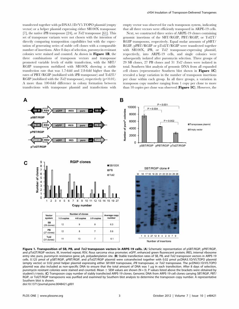
pPBT/RGIP, and pTol2T/RGIP, respectively (Figure 1A).
Transposition efficiencies of the three vectors in ARPE-19 cells
were measured by standard colony formation assays. Equal molar
amounts of pSBT/RGIP, pPBT/RGIP, and pTol2T/RGIP were
cHS4 Insulation of Transposon-Delivered Transgenes
PLOS ONE | www.plosone.org 2 October 2012 | Volume 7 | Issue 10 | e48421
transfected together with pcDNA3.1D/V5.TOPO plasmid (empty
vector) or a helper plasmid expressing either SB100X transposase
[7], the native iPB transposase [24], or Tol2 transposase [61]. This
set of transposase variants were not chosen with the intention of
directly comparing transposition capabilities but with the expec-
tation of generating series of stable cell clones with a comparable
number of insertions. After 8 days of selection, puromycin-resistant
colonies were stained and counted. As shown in Figure 1B, the
three combinations of transposon vectors and transposase
promoted variable levels of stable transfection, with the SBT/
RGIP transposon mobilized with SB100X showing a stable
transfection rate that was 1.7-fold and 2.8-fold higher than the
rates of PBT/RGIP (mobilized with iPB transposase) and Tol2T/
RGIP (mobilized with the Tol2 transposase), respectively (p,0.01).
A more than 100-fold difference in colony formation between
transfections with transposase plasmid and transfections with
empty vector was observed for each transposon system, indicating
that all three vectors were efficiently transposed in ARPE-19 cells.
Next, we constructed three series of ARPE-19 clones containing
genomic insertions of the SBT/RGIP, PBT/RGIP, or Tol2T/
RGIP transposons, respectively. Equal molar amounts of pSBT/
RGIP, pPBT/RGIP or pTol2T/RGIP were transfected together
with SB100X, iPB, or Tol2 transposase-expressing plasmid,
respectively, into ARPE-19 cells, and single colonies were
subsequently isolated after puromycin selection. Three groups of
29 SB clones, 27 PB clones and 31 Tol2 clones were isolated in
total. Southern blot analysis of genomic DNA from all expanded
cell clones (representative Southern blot shown in Figure 1C)
revealed a large variation in the number of transposon insertions
per clone within each group. In all three groups, a variation in
transposon copy number ranging from 1 copy per clone to more
than 10 copies per clone was observed (Figure 1C). However, the
Figure 1. Transposition of SB, PB, and Tol2 transposon vectors in ARPE-19 cells. (A) Schematic representation of pSBT/RGIP, pPBT/RGIP,and pTol2T/RGIP vectors. IR, inverted repeat; RSV, Rous sarcoma virus promoter; eGFP, enhanced green fluorescent protein; IRES, internal ribosomeentry site; puro, puromycin resistance gene; pA, polyadenylation site. (B) Stable transfection rates of SB, PB, and Tol2 transposon vectors in ARPE-19cells. 0.125 pmol of pSBT/RGIP, pPBT/RGIP, and pTol2T/RGIP plasmid were cotransfected together with 0.02 pmol pcDNA3.1D/V5.TOPO plasmid(empty vector) or 0.02 pmol helper plasmid expressing either SB100X transposase, iPB transposase, or Tol2 transposase. The pcDNA3.1D/V5.TOPOplasmid was also included as non-specific DNA to ensure that the total amount of DNA was 1 mg in each transfection. After 8 days of selection,puromycin resistant colonies were stained and counted. Mean 6 SEM values are shown (N = 3). P values listed above the brackets were obtained bystudent’s t-tests. (C) Transposon copy number of stably transfected ARPE-19 clones. Genomic DNA from ARPE-19 cell clones carrying SBT/RGIP, PBT/RGIP, or Tol2T/RGIP transposons was purified and examined by Southern blot analysis to determine the transposon copy number. A representativeSouthern blot is shown.doi:10.1371/journal.pone.0048421.g001
cHS4 Insulation of Transposon-Delivered Transgenes
PLOS ONE | www.plosone.org 3 October 2012 | Volume 7 | Issue 10 | e48421
average copy number was comparable for all three groups, with
6.3 copies per clone for SB clones, 5.9 copies per clone for PB
clones and 6.1 copies per clone for Tol2 clones. Hence, the small
difference in transposition activity obtained with the three
transposase variants did not translate into a substantial difference
in the transposon copy number.
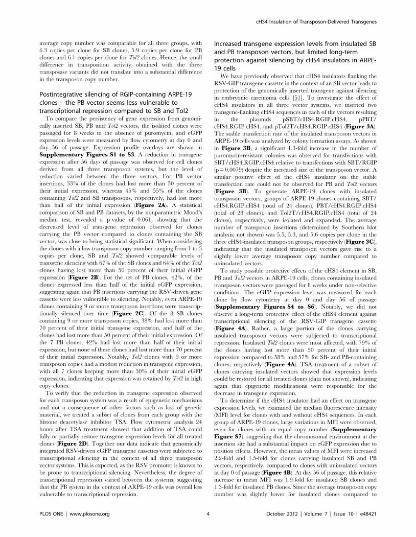
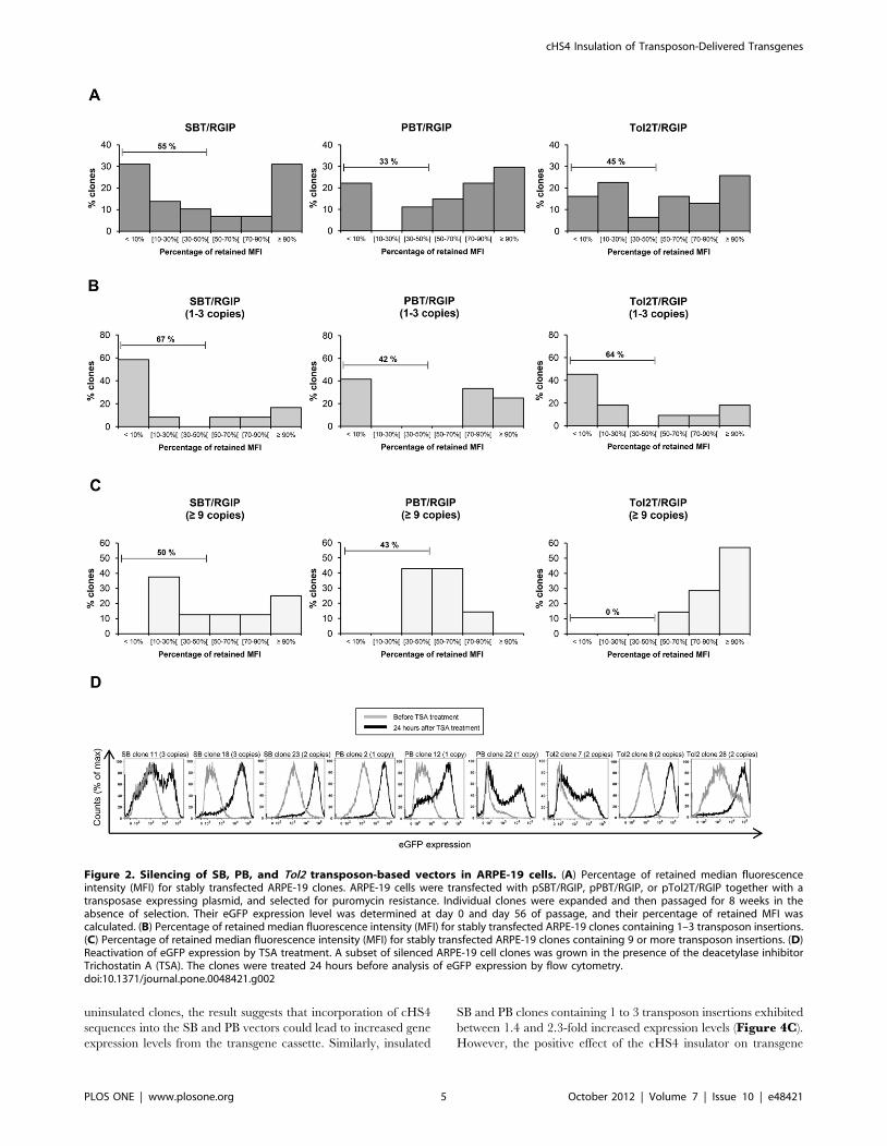
Postintegrative silencing of RGIP-containing ARPE-19clones – the PB vector seems less vulnerable totranscriptional repression compared to SB and Tol2
To compare the persistency of gene expression from genomi-
cally inserted SB, PB and Tol2 vectors, the isolated clones were
passaged for 8 weeks in the absence of puromycin, and eGFP
expression levels were measured by flow cytometry at day 0 and
day 56 of passage. Expression profile overlays are shown in
Supplementary Figures S1 to S3. A reduction in transgene
expression after 56 days of passage was observed for cell clones
derived from all three transposon systems, but the level of
reduction varied between the three vectors. For PB vector
insertions, 33% of the clones had lost more than 50 percent of
their initial expression, whereas 45% and 55% of the clones
containing Tol2 and SB transposons, respectively, had lost more
than half of the initial expression (Figure 2A). A statistical
comparison of SB and PB datasets, by the nonparametric Mood’s
median test, revealed a p-value of 0.061, showing that the
decreased level of transgene repression observed for clones
carrying the PB vector compared to clones containing the SB
vector, was close to being statistical significant. When considering
the clones with a low transposon copy number ranging from 1 to 3
copies per clone, SB and Tol2 showed comparable levels of
transgene silencing with 67% of the SB clones and 64% of the Tol2
clones having lost more than 50 percent of their initial eGFP
expression (Figure 2B). For the set of PB clones, 42%, of the
clones expressed less than half of the initial eGFP expression,
suggesting again that PB insertions carrying the RSV-driven gene
cassette were less vulnerable to silencing. Notably, even ARPE-19
clones containing 9 or more transposon insertions were transcrip-
tionally silenced over time (Figure 2C). Of the 8 SB clones
containing 9 or more transposon copies, 38% had lost more than
70 percent of their initial transgene expression, and half of the
clones had lost more than 50 percent of their initial expression. Of
the 7 PB clones, 42% had lost more than half of their initial
expression, but none of these clones had lost more than 70 percent
of their initial expression. Notably, Tol2 clones with 9 or more
transposon copies had a modest reduction in transgene expression,
with all 7 clones keeping more than 50% of their initial eGFP
expression, indicating that expression was retained by Tol2 in high
copy clones.
To verify that the reduction in transgene expression observed
for each transposon system was a result of epigenetic mechanisms
and not a consequence of other factors such as loss of genetic
material, we treated a subset of clones from each group with the
histone deacetylase inhibitor TSA. Flow cytometric analysis 24
hours after TSA treatment showed that addition of TSA could
fully or partially restore transgene expression levels for all treated
clones (Figure 2D). Together our data indicate that genomically
integrated RSV-driven eGFP transgene cassettes were subjected to
transcriptional silencing in the context of all three transposon
vector systems. This is expected, as the RSV promoter is known to
be prone to transcriptional silencing. Nevertheless, the degree of
transcriptional repression varied between the systems, suggesting
that the PB system in the context of ARPE-19 cells was overall less
vulnerable to transcriptional repression.
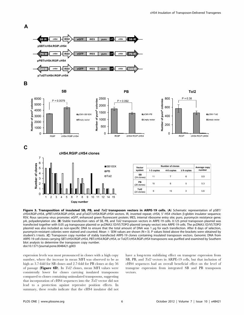
Increased transgene expression levels from insulated SBand PB transposon vectors, but limited long-termprotection against silencing by cHS4 insulators in ARPE-19 cells
We have previously observed that cHS4 insulators flanking the
RSV-GIP transgene cassette in the context of an SB vector leads to
protection of the genomically inserted transgene against silencing
in embryonic carcinoma cells [51]. To investigate the effect of
cHS4 insulators in all three vector systems, we inserted two
transgene-flanking cHS4 sequences in each of the vectors resulting
in the plasmids pSBT/cHS4.RGIP.cHS4, pPBT/
cHS4.RGIP.cHS4, and pTol2T/cHS4.RGIP.cHS4 (Figure 3A).
The stable transfection rate of the insulated transposon vectors in
ARPE-19 cells was analyzed by colony formation assays. As shown
in Figure 3B, a significant 1.3-fold increase in the number of
puromycin-resistant colonies was observed for transfections with
SBT/cHS4.RGIP.cHS4 relative to transfections with SBT/RGIP
(p = 0.0079) despite the increased size of the transposon vector. A
similar positive effect of the cHS4 insulator on the stable
transfection rate could not be observed for PB and Tol2 vectors
(Figure 3B). To generate ARPE-19 clones with insulated
transposon vectors, groups of ARPE-19 clones containing SBT/
cHS4.RGIP.cHS4 (total of 24 clones), PBT/cHS4.RGIP.cHS4
(total of 28 clones), and Tol2T/cHS4.RGIP.cHS4 (total of 24
clones), respectively, were isolated and expanded. The average
number of transposon insertions (determined by Southern blot
analysis; not shown) was 5.5, 5.3, and 5.6 copies per clone in the
three cHS4-insulated transposon groups, respectively (Figure 3C),
indicating that the insulated transposon vectors gave rise to a
slightly lower average transposon copy number compared to
uninsulated vectors.
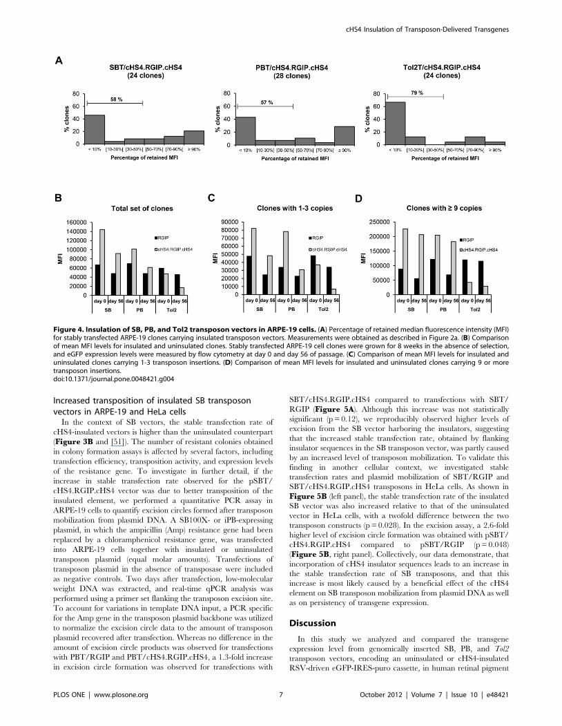
To study possible protective effects of the cHS4 element in SB,
PB and Tol2 vectors in ARPE-19 cells, clones containing insulated
transposon vectors were passaged for 8 weeks under non-selective
conditions. The eGFP expression level was measured for each
clone by flow cytometry at day 0 and day 56 of passage
(Supplementary Figures S4 to S6). Notably, we did not
observe a long-term protective effect of the cHS4 element against
transcriptional silencing of the RSV-GIP transgene cassette
(Figure 4A). Rather, a large portion of the clones carrying
insulated transposon vectors were subjected to transcriptional
repression. Insulated Tol2 clones were most affected, with 79% of
the clones having lost more than 50 percent of their initial
expression compared to 58% and 57% for SB- and PB-containing
clones, respectively (Figure 4A). TSA treatment of a subset of
clones carrying insulated vectors showed that expression levels
could be restored for all treated clones (data not shown), indicating
again that epigenetic modifications were responsible for the
decrease in transgene expression.
To determine if the cHS4 insulator had an effect on transgene
expression levels, we examined the median fluorescence intensity
(MFI) level for clones with and without cHS4 sequences. In each
group of ARPE-19 clones, large variations in MFI were observed,
even for clones with an equal copy number (SupplementaryFigure S7), suggesting that the chromosomal environment at the
insertion site had a substantial impact on eGFP expression due to
position effects. However, the mean values of MFI were increased
2.2-fold and 1.5-fold for clones carrying insulated SB and PB
vectors, respectively, compared to clones with uninsulated vectors
at day 0 of passage (Figure 4B). At day 56 of passage, this relative
increase in mean MFI was 1.9-fold for insulated SB clones and
1.3-fold for insulated PB clones. Since the average transposon copy
number was slightly lower for insulated clones compared to
cHS4 Insulation of Transposon-Delivered Transgenes
PLOS ONE | www.plosone.org 4 October 2012 | Volume 7 | Issue 10 | e48421
uninsulated clones, the result suggests that incorporation of cHS4
sequences into the SB and PB vectors could lead to increased gene
expression levels from the transgene cassette. Similarly, insulated
SB and PB clones containing 1 to 3 transposon insertions exhibited
between 1.4 and 2.3-fold increased expression levels (Figure 4C).
However, the positive effect of the cHS4 insulator on transgene
Figure 2. Silencing of SB, PB, and Tol2 transposon-based vectors in ARPE-19 cells. (A) Percentage of retained median fluorescenceintensity (MFI) for stably transfected ARPE-19 clones. ARPE-19 cells were transfected with pSBT/RGIP, pPBT/RGIP, or pTol2T/RGIP together with atransposase expressing plasmid, and selected for puromycin resistance. Individual clones were expanded and then passaged for 8 weeks in theabsence of selection. Their eGFP expression level was determined at day 0 and day 56 of passage, and their percentage of retained MFI wascalculated. (B) Percentage of retained median fluorescence intensity (MFI) for stably transfected ARPE-19 clones containing 1–3 transposon insertions.(C) Percentage of retained median fluorescence intensity (MFI) for stably transfected ARPE-19 clones containing 9 or more transposon insertions. (D)Reactivation of eGFP expression by TSA treatment. A subset of silenced ARPE-19 cell clones was grown in the presence of the deacetylase inhibitorTrichostatin A (TSA). The clones were treated 24 hours before analysis of eGFP expression by flow cytometry.doi:10.1371/journal.pone.0048421.g002
cHS4 Insulation of Transposon-Delivered Transgenes
PLOS ONE | www.plosone.org 5 October 2012 | Volume 7 | Issue 10 | e48421
expression levels was most pronounced in clones with a high copy
number, where the increase in mean MFI was observed to be as
high as 3.7-fold for SB clones and 2.7-fold for PB clones at day 56
of passage (Figure 4D). In Tol2 clones, mean MFI values were
consistently lower for clones carrying insulated transposons
compared to clones containing uninsulated transposons, suggesting
that incorporation of cHS4 sequences into the Tol2 vector did not
lead to a protection against repressive position effects. In
summary, these results indicate that the cHS4 insulator did not
have a long-term stabilizing effect on transgene expression from
SB, PB, and Tol2 vectors in ARPE-19 cells, but that inclusion of
cHS4 sequences had an overall beneficial effect on the level of
transgene expression from integrated SB and PB transposon
vectors.
Figure 3. Transposition of insulated SB, PB, and Tol2 transposon vectors in ARPE-19 cells. (A) Schematic representation of pSBT/cHS4.RGIP.cHS4, pPBT/cHS4.RGIP.cHS4, and pTol2T/cHS4.RGIP.cHS4 vectors. IR, inverted repeat; cHS4, 59-HS4 chicken b-globin insulator sequence;RSV, Rous sarcoma virus promoter; eGFP, enhanced green fluorescent protein; IRES, internal ribosome entry site; puro, puromycin resistance gene;pA, polyadenylation site. (B) Stable transfection rates of SB, PB, and Tol2 transposon vectors in ARPE-19 cells. 0.125 pmol transposon plasmid wastransfected together with 0.05 mg transposase plasmid or pcDNA3.1D/V5.TOPO plasmid (empty vector) into ARPE-19 cells. The pcDNA3.1D/V5.TOPOplasmid was also included as non-specific DNA to ensure that the total amount of DNA was 1 mg for each transfection. After 8 days of selection,puromycin-resistant colonies were stained and counted. Mean 6 SEM values are shown (N = 3). P values listed above the brackets were obtained bystudent’s t-tests. (C) Transposon copy number of stably transfected ARPE-19 clones containing insulated transposon vectors. Genomic DNA fromARPE-19 cell clones carrying SBT/cHS4.RGIP.cHS4, PBT/cHS4.RGIP.cHS4, or Tol2T/cHS4.RGIP.cHS4 transposons was purified and examined by Southernblot analysis to determine the transposon copy number.doi:10.1371/journal.pone.0048421.g003
cHS4 Insulation of Transposon-Delivered Transgenes
PLOS ONE | www.plosone.org 6 October 2012 | Volume 7 | Issue 10 | e48421
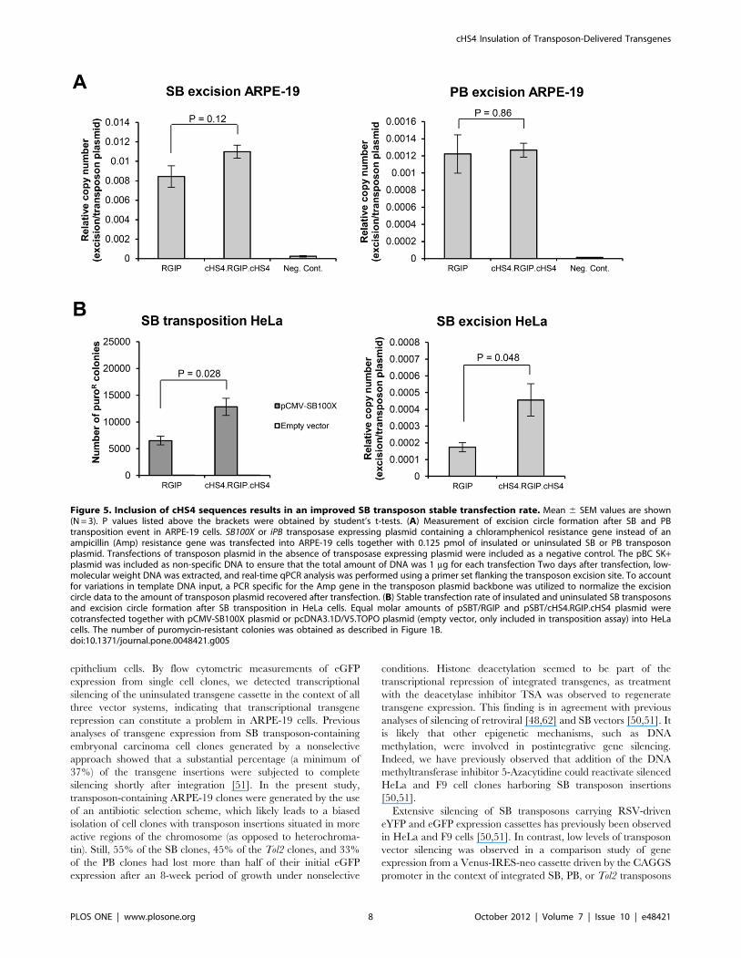
Increased transposition of insulated SB transposonvectors in ARPE-19 and HeLa cells
In the context of SB vectors, the stable transfection rate of
cHS4-insulated vectors is higher than the uninsulated counterpart
(Figure 3B and [51]). The number of resistant colonies obtained
in colony formation assays is affected by several factors, including
transfection efficiency, transposition activity, and expression levels
of the resistance gene. To investigate in further detail, if the
increase in stable transfection rate observed for the pSBT/
cHS4.RGIP.cHS4 vector was due to better transposition of the
insulated element, we performed a quantitative PCR assay in
ARPE-19 cells to quantify excision circles formed after transposon
mobilization from plasmid DNA. A SB100X- or iPB-expressing
plasmid, in which the ampicillin (Amp) resistance gene had been
replaced by a chloramphenicol resistance gene, was transfected
into ARPE-19 cells together with insulated or uninsulated
transposon plasmid (equal molar amounts). Transfections of
transposon plasmid in the absence of transposase were included
as negative controls. Two days after transfection, low-molecular
weight DNA was extracted, and real-time qPCR analysis was
performed using a primer set flanking the transposon excision site.
To account for variations in template DNA input, a PCR specific
for the Amp gene in the transposon plasmid backbone was utilized
to normalize the excision circle data to the amount of transposon
plasmid recovered after transfection. Whereas no difference in the
amount of excision circle products was observed for transfections
with PBT/RGIP and PBT/cHS4.RGIP.cHS4, a 1.3-fold increase
in excision circle formation was observed for transfections with
SBT/cHS4.RGIP.cHS4 compared to transfections with SBT/
RGIP (Figure 5A). Although this increase was not statistically
significant (p = 0.12), we reproducibly observed higher levels of
excision from the SB vector harboring the insulators, suggesting
that the increased stable transfection rate, obtained by flanking
insulator sequences in the SB transposon vector, was partly caused
by an increased level of transposon mobilization. To validate this
finding in another cellular context, we investigated stable
transfection rates and plasmid mobilization of SBT/RGIP and
SBT/cHS4.RGIP.cHS4 transposons in HeLa cells. As shown in
Figure 5B (left panel), the stable transfection rate of the insulated
SB vector was also increased relative to that of the uninsulated
vector in HeLa cells, with a twofold difference between the two
transposon constructs (p = 0.028). In the excision assay, a 2.6-fold
higher level of excision circle formation was obtained with pSBT/
cHS4.RGIP.cHS4 compared to pSBT/RGIP (p = 0.048)
(Figure 5B, right panel). Collectively, our data demonstrate, that
incorporation of cHS4 insulator sequences leads to an increase in
the stable transfection rate of SB transposons, and that this
increase is most likely caused by a beneficial effect of the cHS4
element on SB transposon mobilization from plasmid DNA as well
as on persistency of transgene expression.
Discussion
In this study we analyzed and compared the transgene
expression level from genomically inserted SB, PB, and Tol2
transposon vectors, encoding an uninsulated or cHS4-insulated
RSV-driven eGFP-IRES-puro cassette, in human retinal pigment
Figure 4. Insulation of SB, PB, and Tol2 transposon vectors in ARPE-19 cells. (A) Percentage of retained median fluorescence intensity (MFI)for stably transfected ARPE-19 clones carrying insulated transposon vectors. Measurements were obtained as described in Figure 2a. (B) Comparisonof mean MFI levels for insulated and uninsulated clones. Stably transfected ARPE-19 cell clones were grown for 8 weeks in the absence of selection,and eGFP expression levels were measured by flow cytometry at day 0 and day 56 of passage. (C) Comparison of mean MFI levels for insulated anduninsulated clones carrying 1-3 transposon insertions. (D) Comparison of mean MFI levels for insulated and uninsulated clones carrying 9 or moretransposon insertions.doi:10.1371/journal.pone.0048421.g004
cHS4 Insulation of Transposon-Delivered Transgenes
PLOS ONE | www.plosone.org 7 October 2012 | Volume 7 | Issue 10 | e48421
epithelium cells. By flow cytometric measurements of eGFP
expression from single cell clones, we detected transcriptional
silencing of the uninsulated transgene cassette in the context of all
three vector systems, indicating that transcriptional transgene
repression can constitute a problem in ARPE-19 cells. Previous
analyses of transgene expression from SB transposon-containing
embryonal carcinoma cell clones generated by a nonselective
approach showed that a substantial percentage (a minimum of
37%) of the transgene insertions were subjected to complete
silencing shortly after integration [51]. In the present study,
transposon-containing ARPE-19 clones were generated by the use
of an antibiotic selection scheme, which likely leads to a biased
isolation of cell clones with transposon insertions situated in more
active regions of the chromosome (as opposed to heterochroma-
tin). Still, 55% of the SB clones, 45% of the Tol2 clones, and 33%
of the PB clones had lost more than half of their initial eGFP
expression after an 8-week period of growth under nonselective
conditions. Histone deacetylation seemed to be part of the
transcriptional repression of integrated transgenes, as treatment
with the deacetylase inhibitor TSA was observed to regenerate
transgene expression. This finding is in agreement with previous
analyses of silencing of retroviral [48,62] and SB vectors [50,51]. It
is likely that other epigenetic mechanisms, such as DNA
methylation, were involved in postintegrative gene silencing.
Indeed, we have previously observed that addition of the DNA
methyltransferase inhibitor 5-Azacytidine could reactivate silenced
HeLa and F9 cell clones harboring SB transposon insertions
[50,51].
Extensive silencing of SB transposons carrying RSV-driven
eYFP and eGFP expression cassettes has previously been observed
in HeLa and F9 cells [50,51]. In contrast, low levels of transposon
vector silencing was observed in a comparison study of gene
expression from a Venus-IRES-neo cassette driven by the CAGGS
promoter in the context of integrated SB, PB, or Tol2 transposons
Figure 5. Inclusion of cHS4 sequences results in an improved SB transposon stable transfection rate. Mean 6 SEM values are shown(N = 3). P values listed above the brackets were obtained by student’s t-tests. (A) Measurement of excision circle formation after SB and PBtransposition event in ARPE-19 cells. SB100X or iPB transposase expressing plasmid containing a chloramphenicol resistance gene instead of anampicillin (Amp) resistance gene was transfected into ARPE-19 cells together with 0.125 pmol of insulated or uninsulated SB or PB transposonplasmid. Transfections of transposon plasmid in the absence of transposase expressing plasmid were included as a negative control. The pBC SK+plasmid was included as non-specific DNA to ensure that the total amount of DNA was 1 mg for each transfection Two days after transfection, low-molecular weight DNA was extracted, and real-time qPCR analysis was performed using a primer set flanking the transposon excision site. To accountfor variations in template DNA input, a PCR specific for the Amp gene in the transposon plasmid backbone was utilized to normalize the excisioncircle data to the amount of transposon plasmid recovered after transfection. (B) Stable transfection rate of insulated and uninsulated SB transposonsand excision circle formation after SB transposition in HeLa cells. Equal molar amounts of pSBT/RGIP and pSBT/cHS4.RGIP.cHS4 plasmid werecotransfected together with pCMV-SB100X plasmid or pcDNA3.1D/V5.TOPO plasmid (empty vector, only included in transposition assay) into HeLacells. The number of puromycin-resistant colonies was obtained as described in Figure 1B.doi:10.1371/journal.pone.0048421.g005
cHS4 Insulation of Transposon-Delivered Transgenes
PLOS ONE | www.plosone.org 8 October 2012 | Volume 7 | Issue 10 | e48421
in HeLa cells [45]. As transcriptional repression of integrated
vectors has been demonstrated to occur in a promoter-dependent
manner [50,63], such discrepancies most likely reflect that the
experiments were conducted with transposons containing different
cargo sequences, suggesting that silencing of transposon-based
vectors can be diminished by careful transgene design. However,
substantial differences between cell types may affect the silencing
profile. In a recent report examining transgene expression from SB
vectors stably transfected into K562 erythroid cells, progressive
transgene silencing of the CAGGS promoter was observed and
could be significantly reduced by flanking the CAGGS-DsRed or
IHK-b-globin gene cassettes with cHS4-insulators [64], indicating
that incorporation of protective insulating elements can be
beneficial, even when promoter sequences are carefully chosen.
In the present study, all three transposon systems contained the
same transgenic cassette, which means that potential intrinsic
silencing triggers resident in the RSV promoter or the eGFP gene
were present in all vectors. Nevertheless, PB vectors were less
affected by transgene silencing compared to vectors based on SB
and Tol2. Notably, PB has a preference for integrating into
transcriptional units, whereas SB has a fairly random integration
profile with no preference for or against genes [23,28,40,41,42,43].
The integration profile of Tol2 vectors seems to vary dependent on
cell type with a high preference for transcriptional start sites in
some cells (HeLa and primary human T cells) and a more random
integration pattern in others (zebrafish and HEK293 cells)
[43,44,45,46]. Although the integration profile of PB, SB, and
Tol2 vectors has not been examined in ARPE-19 cells, the
increased integration frequency of the PB transposon in transcrip-
tionally ‘‘open’’ regions compared to SB and Tol2 transposons is
an obvious explanation for the decreased level of transgene
silencing observed for PB. Alternatively, the PB vector may
contain protective cis-elements in its terminal regions. Analysis of
the 59 and 39-terminal repeats of the PB transposon has revealed
the existence of promoter activity in the 59-terminal repeat and
enhancer activity in the 39-terminal repeat [65,66], but the
existence of protective motifs has not been reported.
In the attempt to create silencing-protected transposon vectors,
we tested the effect of incorporating 1.2-kb cHS4 insulator
sequences into the three vector systems, which to our knowledge is
the first time the barrier function of the cHS4 element has been
tested in PB and Tol2 transposon vectors. Flanking the transgene
cassette with the cHS4 insulator resulted in a 2.2-fold and 1.5-fold
increase in the initial eGFP expression level of SB and PB vectors,
respectively, despite the fact that the average copy numbers were
reduced with the cHS4-containing vectors. Similar results have
been observed in studies of transgene expression from retroviral
vectors in which cHS4 inclusion led to increased transgene
expression levels in transduced murine fibroblast NIH3T3 cell
clones [67,68], human fibrosarcoma HT1080 cell clones [53], and
murine primary bone marrow progenitor clones [53]. By
comparing the MFI level of stably expressing ARPE-19 clones
carrying SB-, PB-, and Tol2-derived vectors, we detected large
variations of transgene expression within clones containing equal
transposon copy numbers, suggesting that eGFP expression levels
were not simply determined by transgene copy number, but that
chromosomal position effects had a large influence on eGFP
expression. Since the cHS4 element does not exhibit conventional
enhancer activity, the increase in transgene expression caused by
the cHS4 elements likely reflects the ability of the cHS4 insulator
to reduce the impact of repressive chromosomal position effects on
vector transgene expression. It remains to be elucidated, however,
why inclusion of cHS4 sequences into the Tol2 vector did not seem
to benefit the eGFP expression level.
By measuring eGFP expression levels of stably expressing
ARPE-19 clones grown for 8 weeks in the absence of selection, we
discovered that the cHS4 insulator did not protect against
progressive transgene silencing of SB, PB, and Tol2 transposon
vectors in ARPE-19 cells. Our previous eGFP expression studies of
F9 cell clones harboring the SBT/RGIP or SBT/
cHS4.RGIP.cHS4 transposon vector showed that inclusion of
cHS4 sequences into the SB transposon had a profound effect on
the stability of transgene expression in F9 cell clones after
prolonged passaging in culture. Since identical SB transposon
constructs were used in this study, differences between cell types
most likely accounted for the variable benefit of including cHS4
insulators. In support of this notion, Rivella et al. observed that
inclusion of the cHS4 insulator into a recombinant retroviral
vector could decrease vector methylation and transgene silencing
in murine erythroleukemia cells, but not in murine embryonic
stem cells [54], indicating that the barrier function of the cHS4
insulator is not uniformly active in all kinds of cell types. To
improve the protection against progressive silencing of DNA
transposon-embedded transgenes in ARPE-19 cells, the ubiqui-
tously-acting chromatin opening element (UCOE) derived from
the human HNRPA2B1-CBX3 locus [69] may represent an
attractive alternative to the cHS4 insulator. The UCOE sequence
contains a methylation-free CpG island which has been found to
shield flanking heterologous promoters from transcriptional
silencing, allowing sustained transgene expression from lentiviral
vectors [70,71,72]. However, this element remains to be investi-
gated in conjunction with a series of promoters and in the context
of DNA transposon-based vectors.
Previous studies have determined an inverse relationship
between transposon length and transposition frequency for SB
transposon vectors [36,37]. In the present study, we did not
observe a reduction in transposition activity when two 1.2-kb
cHS4 insulator sequences were incorporated into the pSBT/RGIP
vector. Instead, significantly increased stable transfection rates
were observed for the insulated SB vector in both ARPE-19 and
HeLa cells. By quantitative measurements of SB excision circle
formation, we detected a positive effect of the cHS4 sequences on
SB transposon mobilization from plasmid DNA. The mechanisms
responsible for insulator function are still poorly understood, but
an ability of insulator binding proteins to form closed looped
chromatin domains has been proposed as one model for insulator
enhancer blocking activity [73]. The DNA-bending protein
HMGBI is a cofactor of SB transposition in mammalian cells
and is believed to stimulate transposition by assisting the SB
transposase during synaptic complex formation either by bringing
the transposon binding sites and/or the terminal repeats physically
closer to each other [74]. Hypothetically, cHS4 insulator binding
proteins may also stimulate DNA bending and bring the
transposon binding sites closer to each other by loop formation
of DNA sequences between the cHS4 element sequences, resulting
in increased stabilization of the synaptic complex and increased
transposition activities. This hypothesis, however, remains to be
tested.
Genotoxicity caused by activation of proto-oncogenes near the
vector integration site constitutes a serious problem to gene
therapy. As none of the SB, PB, or Tol2 transposon vectors
integrate in a site-specific manner, a risk of insertional mutagenesis
upon vector integration exists, a risk especially pronounced for PB
and Tol2 transposons due to their increased preference for
integrating into transcriptional units. The cHS4 element contains
enhancer blocking activity which enables it to block molecular
communication between enhancers and genes. Inclusion of the
cHS4 insulator in retroviral vectors has been reported to reduce
cHS4 Insulation of Transposon-Delivered Transgenes
PLOS ONE | www.plosone.org 9 October 2012 | Volume 7 | Issue 10 | e48421
genotoxicity by 6-fold in a murine tumor transplantation model
[75]. The enhancer blocking activity of the cHS4 insulator was not
addressed in this study, but previous analysis of promoter
activation by an SV40-neo transgene unit within an SB transposon
vector showed that the cHS4 element could effectively block
transactivation of a nearby TATA-box minimal promoter in HeLa
and primary human T cells [76]. Incorporation of cHS4 sequences
could therefore potentially lead to an increased safety profile.
By following the expression of a silencing-prone promoter, we
demonstrate in this study that the extent of silencing may depend on
the type of carrier, most likely due to overall differences in the
integration profile of the different DNA transposon carriers. We
show that incorporation of cHS4 insulator sequences can lead to an
increase in transgene expression levels for genomically integrated
SB- and PB-based vectors in ARPE19 cells. In addition, improved
stable transfection rates are obtained for cHS4-insulated SB vectors,
possibly due to the increased mobilization of cHS4-containing
transposons from plasmid DNA. Finally, we find that inclusion of
cHS4 elements in SB-, PB- and Tol2-derived vectors does not lead
to long-term protection against progressive transgene silencing in
ARPE19 cells, supporting the notion that the barrier activity of the
cHS4 insulator is not uniformly active in all cell types.
Materials and Methods
Plasmid constructionThe plasmids pSBT/RGIP, pSBT/cHS4.RGIP.cHS4, and
pCMV-SB100X have been described previously [51,77]. The
pPBT/RGIP and pTol2T/RGIP plasmids were constructed by
ligation of a RSV.eGFP.IRES.puro PCR fragment, amplified
from pSBT/RGIP, into ClaI/NotI-digested pXL-BacII [78] and
NheI/ClaI-digested pT2AL200R150 [20], respectively. To gen-
erate pPBT/cHS4.RGIP.cHS4 and pTol2T/cHS4.RGIP.cHS4,
the 1200-bp cHS4 insulator element was amplified from pSBT/
cHS4.RGIP.cHS4 by PCR and inserted in front of and after the
RSV.eGFP.IRES.puro cassette in pPBT/RGIP (using a ClaI and
NotI site) and in pTol2T/RGIP (using a NheI and ClaI site),
respectively. The pCMV-PB and pCMV-Tol2 plasmids have been
previously described in [79]. The pCMV-SB100X.chloramp and
pCMV-PB.chloramp plasmids were generated by ligation of a
chloramphenicol PCR fragment amplified from pBC SK+(Stratagene, Santa Clara, CA) into PvuI-digested pCMV-
SB100X and pCMV-PB, respectively. To generate pPBT4tp,
the transposon sequence of pPBT/RGIP was cut out by NsiI/PstI-
digestion, and the digested plasmid backbone was then exposed to
T4 DNA polymerase treatment and blunt-end ligation. All
produced DNA constructs were verified by restriction digestion
and DNA sequencing.
Cell culture and transposition assaysHeLa (human cervical cancer) cells were maintained in
Dulbecco’s modified Eagle’s medium (DMEM) (Lonza, Basel,
Switzerland) supplemented with 10% fetal bovine serum (Lonza,
Basel, Switzerland), 0.26 mg/ml glutamine, 54 ng/ml penicillin
and 36 ug/ml streptomycin. ARPE-19 human retinal pigment
epithelium cells [58] were maintained in culture medium
containing 50% Ham’s F-12 Nutrient Mixture (Invitrogen,
Carlsbad, CA) and 50% DMEM with serum, glutamine, penicillin,
and streptomycin, as described above. To measure rates of stable
transfection, cells were plated at 1.5 x 10⁵ cells/well in 6-well
dishes 1 day before cotransfection with 0.125 pmol transposon
plasmid and 0.016 pmol transposase plasmid or pcDNA3.1D/
V5.TOPO plasmid (Invitrogen, Carlsbad, CA) as a negative
control. The pcDNA3.1D/V5.TOPO plasmid was used as stuffer
DNA to obtain equal DNA amounts in each transfection.
Transfections were carried out using FuGene-6 (Roche, Basel,
Switzerland) according to manufacturer’s instructions using 3 ml of
reagent per 1 mg of DNA. One day after transfection, cells were
split in varying densities and plated in 10-cm dishes. Two days
after transfection, selection medium containing 1 mg/ml puromy-
cin (Invitrogen, Carlsbad, CA) was added to the cells. After 8 days
of selection, colonies of cells were stained with 0.6% methylene
blue, air-dried and counted.
Generation of stable expressing cell clones and long-term expression analysis
ARPE-19 cells were seeded in 6-well dishes (1.56105 cells/well)
and transfected with 0.125 pmol transposon plasmid together with
0.05 mg transposase plasmid using FuGene-6 transfection reagent
according to manufacturer’s instructions. One day after transfec-
tion cells were split in varying densities and plated in 10-cm dishes.
Selection medium, containing 1 mg/ml puromycin, was added to
the cells two days after transfection. After 10 days of selection,
single clones were isolated and expanded for genomic DNA
extraction and long-term eGFP expression analysis. The isolated,
stably expressing cell clones were passaged for 8 weeks in standard
culture medium, and analyzed by flow cytometry on day 0 and day
56 on a BD FACSAria III cell sorter (BD Biosciences, San Jose,
CA). In the flow cytometric analysis, non-transfected cells were
included as a negative control, and propidium iodide (Sigma
Aldrich, St Louis, MO) was used to exclude non-viable cells.
Transposon excision assayHeLa or ARPE-19 cells were seeded in 6-well dishes (1.56105
cells/well) and transfected with 0.25 pmol (HeLa) or 0.125 pmol
(ARPE-19) transposon plasmid together with 0.25 pmol (HeLa) or
0.05 mg (ARPE-19) transposase plasmid or pBC SK+ plasmid as
negative control. The pBC SK+ plasmid was also used as stuffer
DNA. Transfections were carried out using FuGene-6 according
to manufacturer’s instructions using 3 ml of reagent per 1 mg of
DNA. Low-molecular weight DNA was extracted 2 days after
transfection using the QIAprep miniprep kit (CA 91355; Qiagen,
Valencia, Spain) according to the manufacturer’s instructions,
except for a 1-h incubation period at 55uC (0.6% SDS and 0.08
mg/ml proteinase K together with buffer P1) instead of the lysis
step with buffer P2 after resuspension of the cell pellet. 50 ng of
extracted DNA from each transfection was used in a quantitative
PCR analysis using a primer set recognizing transposon excision
circle products and a primer set amplifying an amplicon within the
Amp resistance gene. The plasmids pUC19 and pPBT4tp were
used as templates for standard curve formation. The analysis was
performed on a Lightcycler 480 (Roche, Basel, Switzerland) using
the Lightcycler DNA Master SYBR Green I kit (Roche, Basel,
Switzerland). Amplification was performed under the following
conditions: Thirty cycles (95uC 30s, 65uC 30s, and 72uC 5s).
Primer sequences are as follows: SB excision (S): 59-CGAT-
TAAGTTGGGTAACGCCAGGG-39, SB excision (AS): 59-
CAGCTGGCACGACAGGTTTCCCG-39, PB excision (S): 59-
CCGTGGAGGACGGGCAGACTCGCG-39, PB excision (AS):
59-GGCGTGCATGGCCACACCTTCCCG-39, Amp (S): 59-
CAAGAGCAACTCGGTCGC-39, Amp (AS): 59-
TCGTTGTCAGAAGTAAGTTGGC-39.
Southern Blot analysisGenomic DNA was prepared from cell pellets following NaCl
extraction and ethanol precipitation. 15 mg genomic DNA was
digested with PstI+KpnI (pSBT/RGIP stable clones), KpnI+SalI
cHS4 Insulation of Transposon-Delivered Transgenes
PLOS ONE | www.plosone.org 10 October 2012 | Volume 7 | Issue 10 | e48421
(pSBT/cHS4.RGIP.cHS4 stable clones), KpnI+PvuII (pPBT/
RGIP stable clones) or KpnI (pTol2T/RGIP, pPBT/
cHS4.RGIP.cHS4, and pTol2T/cHS4.RGIP.cHS4 stable clones).
Genomic DNA of non-transfected cells was used as a negative
control. Genomic DNA of non-transfected cells spiked with
plasmid DNA corresponding to 1 copy/cell or 3 copies/cell was
used as a positive control. The digested DNA was electrophoresed
in a 0.8% agarose gel and transferred to a Hybond membrane (GE
Healthcare, Buckinghamshire, UK). The membrane was hybrid-
ized overnight using a puro-specific [a-32P] dCTP-labelled probe.
TSA treatmentSilenced ARPE-19 cell clones were grown in the presence of
1200 nmol/l TSA (Sigma Aldrich, St Louis, MO). The clones
were treated 24 hours before analysis of eGFP expression by flow
cytometry on a BD FACSAria III cell sorter (BD Biosciences, San
Jose, CA). Non-transfected cells were included as a negative
control, and propidium iodide (Sigma Aldrich, St Louis, MO) was
used to exclude non-viable cells.
Supporting Information
Figure S1 eGFP expression profiles of pSBT/RGIPclones measured by flow cytometry at day 0 and day56 of growth in non-selection medium.(PDF)
Figure S2 eGFP expression profiles of pPBT/RGIPclones measured by flow cytometry at day 0 and day56 of growth in non-selection medium.(PDF)
Figure S3 eGFP expression profiles of pTol2/RGIPclones measured by flow cytometry at day 0 and day56 of growth in non-selection medium.(PDF)
Figure S4 eGFP expression profiles of pSBT/cHS4.RGIP.cHS4 clones measured by flow cytometryat day 0 and day 56 of growth in non-selection medium.
(PDF)
Figure S5 eGFP expression profiles of pPBT/cHS4.RGIP.cHS4 clones measured by flow cytometryat day 0 and day 56 of growth in non-selection medium.
(PDF)
Figure S6 eGFP expression profiles of pTol2T/cHS4.RGIP.cHS4 clones measured by flow cytometryat day 0 and day 56 of growth in non-selection medium.
(PDF)
Figure S7 Median fluorescence intensity (MFI) values ofRPE transposon clones measured by flow cytometry atday 0 of passage.
(PDF)
Acknowledgments
We thank the staff at the FACS core facility, The Faculty of Health
Sciences, Aarhus University, for their technical assistance. We also thank
Jakob Grove for statistical assistance. The authors would like to kindly
acknowledge Gary Felsenfeld for providing the cHS4 insulator, Malcolm
Fraser for providing piggyBac constructs, Koichi Kawakami for providing
Tol2 constructs, and Lajos Mates, Zoltan Ivics, and Zsuzanna Izsvak for
providing the plasmid encoding the SB100X transposase.
Author Contributions
Conceived and designed the experiments: NS JGM. Performed the
experiments: NS AKH ROB NHS LDS. Analyzed the data: NS JGM.
Contributed reagents/materials/analysis tools: JGM. Wrote the paper: NS
JGM.
References
1. Bachmann A, Knust E (2008) The use of P-element transposons to generate
transgenic flies. Methods Mol Biol 420: 61–77.
2. Moerman DG, Barstead RJ (2008) Towards a mutation in every gene in
Caenorhabditis elegans. Brief Funct Genomic Proteomic 7: 195–204.
3. Ivics Z, Hackett PB, Plasterk RH, Izsvak Z (1997) Molecular reconstruction of
Sleeping Beauty, a Tc1-like transposon from fish, and its transposition in human
cells. Cell 91: 501–510.
4. Grabundzija I, Izsvak Z, Ivics Z (2011) Insertional engineering of chromosomes
with Sleeping Beauty transposition: an overview. Methods Mol Biol 738: 69–85.
5. Ivics Z, Izsvak Z (2010) The expanding universe of transposon technologies for
gene and cell engineering. Mob DNA 1: 25.
6. Xue X, Huang X, Nodland SE, Mates L, Ma L, et al. (2009) Stable gene transfer
and expression in cord blood-derived CD34+ hematopoietic stem and
progenitor cells by a hyperactive Sleeping Beauty transposon system. Blood
114: 1319–1330.
7. Mates L, Chuah MK, Belay E, Jerchow B, Manoj N, et al. (2009) Molecular
evolution of a novel hyperactive Sleeping Beauty transposase enables robust
stable gene transfer in vertebrates. Nat Genet 41: 753–761.
8. Sumiyoshi T, Holt NG, Hollis RP, Ge S, Cannon PM, et al. (2009) Stable
transgene expression in primitive human CD34+ hematopoietic stem/progen-
itor cells, using the Sleeping Beauty transposon system. Hum Gene Ther 20:
1607–1626.
9. Jin Z, Maiti S, Huls H, Singh H, Olivares S, et al. (2011) The hyperactive
Sleeping Beauty transposase SB100X improves the genetic modification of T
cells to express a chimeric antigen receptor. Gene Ther.
10. Huang X, Wilber A, McIvor RS, Zhou X (2009) DNA transposons for
modification of human primary T lymphocytes. Methods Mol Biol 506: 115–
126.
11. Singh H, Manuri PR, Olivares S, Dara N, Dawson MJ, et al. (2008) Redirecting
specificity of T-cell populations for CD19 using the Sleeping Beauty system.
Cancer Res 68: 2961–2971.
12. Huang X, Guo H, Kang J, Choi S, Zhou TC, et al. (2008) Sleeping Beauty
transposon-mediated engineering of human primary T cells for therapy of
CD19+ lymphoid malignancies. Mol Ther 16: 580–589.
13. Wilber A, Linehan JL, Tian X, Woll PS, Morris JK, et al. (2007) Efficient and
stable transgene expression in human embryonic stem cells using transposon-
mediated gene transfer. Stem Cells 25: 2919–2927.
14. Orban TI, Apati A, Nemeth A, Varga N, Krizsik V, et al. (2009) Applying a
‘‘double-feature’’ promoter to identify cardiomyocytes differentiated from
human embryonic stem cells following transposon-based gene delivery. Stem
Cells 27: 1077–1087.
15. Aronovich EL, McIvor RS, Hackett PB (2011) The Sleeping Beauty transposon
system: a non-viral vector for gene therapy. Hum Mol Genet 20: R14–20.
16. Izsvak Z, Hackett PB, Cooper LJ, Ivics Z (2010) Translating Sleeping Beauty
transposition into cellular therapies: victories and challenges. Bioessays 32: 756–
767.
17. Williams DA (2008) Sleeping beauty vector system moves toward human trials in
the United States. Mol Ther 16: 1515–1516.
18. Fraser MJ, Cary L, Boonvisudhi K, Wang HG (1995) Assay for movement of
Lepidopteran transposon IFP2 in insect cells using a baculovirus genome as a
target DNA. Virology 211: 397–407.
19. Cary LC, Goebel M, Corsaro BG, Wang HG, Rosen E, et al. (1989) Transposon
mutagenesis of baculoviruses: analysis of Trichoplusia ni transposon IFP2
insertions within the FP-locus of nuclear polyhedrosis viruses. Virology 172:
156–169.
20. Kawakami K, Shima A, Kawakami N (2000) Identification of a functional
transposase of the Tol2 element, an Ac-like element from the Japanese medaka
fish, and its transposition in the zebrafish germ lineage. Proc Natl Acad Sci U S A
97: 11403–11408.
21. Handler AM (2002) Use of the piggyBac transposon for germ-line transforma-
tion of insects. Insect Biochem Mol Biol 32: 1211–1220.
22. Wang W, Lin C, Lu D, Ning Z, Cox T, et al. (2008) Chromosomal transposition
of PiggyBac in mouse embryonic stem cells. Proc Natl Acad Sci U S A 105:
9290–9295.
23. Wilson MH, Coates CJ, George AL Jr (2007) PiggyBac transposon-mediated
gene transfer in human cells. Mol Ther 15: 139–145.
24. Ding S, Wu X, Li G, Han M, Zhuang Y, et al. (2005) Efficient transposition of
the piggyBac (PB) transposon in mammalian cells and mice. Cell 122: 473–483.
cHS4 Insulation of Transposon-Delivered Transgenes
PLOS ONE | www.plosone.org 11 October 2012 | Volume 7 | Issue 10 | e48421
25. Lacoste A, Berenshteyn F, Brivanlou AH (2009) An efficient and reversible
transposable system for gene delivery and lineage-specific differentiation inhuman embryonic stem cells. Cell Stem Cell 5: 332–342.
26. Nakazawa Y, Huye LE, Dotti G, Foster AE, Vera JF, et al. (2009) Optimization
of the PiggyBac transposon system for the sustained genetic modification ofhuman T lymphocytes. J Immunother 32: 826–836.
27. Manuri PV, Wilson MH, Maiti SN, Mi T, Singh H, et al. (2010) piggyBac
transposon/transposase system to generate CD19-specific T cells for thetreatment of B-lineage malignancies. Hum Gene Ther 21: 427–437.
28. Galvan DL, Nakazawa Y, Kaja A, Kettlun C, Cooper LJ, et al. (2009) Genome-
wide mapping of PiggyBac transposon integrations in primary human T cells.J Immunother 32: 837–844.
29. Nakazawa Y, Huye LE, Salsman VS, Leen AM, Ahmed N, et al. (2011)
PiggyBac-mediated Cancer Immunotherapy Using EBV-specific Cytotoxic T-cells Expressing HER2-specific Chimeric Antigen Receptor. Mol Ther.
30. Woltjen K, Michael IP, Mohseni P, Desai R, Mileikovsky M, et al. (2009)
piggyBac transposition reprograms fibroblasts to induced pluripotent stem cells.Nature 458: 766–770.
31. Kawakami K (2005) Transposon tools and methods in zebrafish. Dev Dyn 234:
244–254.
32. Kawakami K (2007) Tol2: a versatile gene transfer vector in vertebrates.
Genome Biol 8 Suppl 1: S7.
33. Liu G, Aronovich EL, Cui Z, Whitley CB, Hackett PB (2004) Excision ofSleeping Beauty transposons: parameters and applications to gene therapy.
J Gene Med 6: 574–583.
34. Mitra R, Fain-Thornton J, Craig NL (2008) piggyBac can bypass DNA synthesisduring cut and paste transposition. EMBO J 27: 1097–1109.
35. Zhou L, Mitra R, Atkinson PW, Hickman AB, Dyda F, et al. (2004)
Transposition of hAT elements links transposable elements and V(D)Jrecombination. Nature 432: 995–1001.
36. Geurts AM, Yang Y, Clark KJ, Liu G, Cui Z, et al. (2003) Gene transfer into
genomes of human cells by the sleeping beauty transposon system. Mol Ther 8:108–117.
37. Izsvak Z, Ivics Z, Plasterk RH (2000) Sleeping Beauty, a wide host-range
transposon vector for genetic transformation in vertebrates. J Mol Biol 302: 93–102.
38. Balciunas D, Wangensteen KJ, Wilber A, Bell J, Geurts A, et al. (2006)Harnessing a high cargo-capacity transposon for genetic applications in
vertebrates. PLoS Genet 2: e169.
39. Li MA, Turner DJ, Ning Z, Yusa K, Liang Q, et al. (2011) Mobilization of giantpiggyBac transposons in the mouse genome. Nucleic Acids Res.
40. Vigdal TJ, Kaufman CD, Izsvak Z, Voytas DF, Ivics Z (2002) Common physical
properties of DNA affecting target site selection of sleeping beauty and otherTc1/mariner transposable elements. J Mol Biol 323: 441–452.
41. Yant SR, Wu X, Huang Y, Garrison B, Burgess SM, et al. (2005) High-
resolution genome-wide mapping of transposon integration in mammals. MolCell Biol 25: 2085–2094.
42. Liang Q, Kong J, Stalker J, Bradley A (2009) Chromosomal mobilization and
reintegration of Sleeping Beauty and PiggyBac transposons. Genesis 47: 404–408.
43. Huang X, Guo H, Tammana S, Jung YC, Mellgren E, et al. (2010) Gene
transfer efficiency and genome-wide integration profiling of Sleeping Beauty,Tol2, and piggyBac transposons in human primary T cells. Mol Ther 18: 1803–
1813.
44. Meir YJ, Weirauch MT, Yang HS, Chung PC, Yu RK, et al. (2011) Genome-wide target profiling of piggyBac and Tol2 in HEK 293: pros and cons for gene
discovery and gene therapy. BMC Biotechnol 11: 28.
45. Grabundzija I, Irgang M, Mates L, Belay E, Matrai J, et al. (2010) Comparativeanalysis of transposable element vector systems in human cells. Mol Ther 18:
1200–1209.
46. Kondrychyn I, Garcia-Lecea M, Emelyanov A, Parinov S, Korzh V (2009)Genome-wide analysis of Tol2 transposon reintegration in zebrafish. BMC
Genomics 10: 418.
47. Pannell D, Osborne CS, Yao S, Sukonnik T, Pasceri P, et al. (2000) Retrovirusvector silencing is de novo methylase independent and marked by a repressive
histone code. EMBO J 19: 5884–5894.
48. Yao S, Sukonnik T, Kean T, Bharadwaj RR, Pasceri P, et al. (2004) Retrovirus
silencing, variegation, extinction, and memory are controlled by a dynamic
interplay of multiple epigenetic modifications. Mol Ther 10: 27–36.
49. Persons DA, Hargrove PW, Allay ER, Hanawa H, Nienhuis AW (2003) The
degree of phenotypic correction of murine beta -thalassemia intermedia
following lentiviral-mediated transfer of a human gamma-globin gene isinfluenced by chromosomal position effects and vector copy number. Blood
101: 2175–2183.
50. Garrison BS, Yant SR, Mikkelsen JG, Kay MA (2007) Postintegrative genesilencing within the Sleeping Beauty transposition system. Mol Cell Biol 27:
8824–8833.
51. Dalsgaard T, Moldt B, Sharma N, Wolf G, Schmitz A, et al. (2009) Shielding of
sleeping beauty DNA transposon-delivered transgene cassettes by heterologous
insulators in early embryonal cells. Mol Ther 17: 121–130.
52. Mutskov VJ, Farrell CM, Wade PA, Wolffe AP, Felsenfeld G (2002) The barrier
function of an insulator couples high histone acetylation levels with specific
protection of promoter DNA from methylation. Genes Dev 16: 1540–1554.
53. Li CL, Emery DW (2008) The cHS4 chromatin insulator reduces gammare-
troviral vector silencing by epigenetic modifications of integrated provirus. GeneTher 15: 49–53.
54. Rivella S, Callegari JA, May C, Tan CW, Sadelain M (2000) The cHS4
insulator increases the probability of retroviral expression at randomchromosomal integration sites. J Virol 74: 4679–4687.
55. Arumugam PI, Scholes J, Perelman N, Xia P, Yee JK, et al. (2007) Improvedhuman beta-globin expression from self-inactivating lentiviral vectors carrying
the chicken hypersensitive site-4 (cHS4) insulator element. Mol Ther 15: 1863–
1871.56. Hauswirth WW, Aleman TS, Kaushal S, Cideciyan AV, Schwartz SB, et al.
(2008) Treatment of leber congenital amaurosis due to RPE65 mutations byocular subretinal injection of adeno-associated virus gene vector: short-term
results of a phase I trial. Hum Gene Ther 19: 979–990.57. Jacobson SG, Cideciyan AV, Ratnakaram R, Heon E, Schwartz SB, et al. (2011)
Gene Therapy for Leber Congenital Amaurosis Caused by RPE65 Mutations:
Safety and Efficacy in 15 Children and Adults Followed Up to 3 Years. ArchOphthalmol.
58. Dunn KC, Aotaki-Keen AE, Putkey FR, Hjelmeland LM (1996) ARPE-19, ahuman retinal pigment epithelial cell line with differentiated properties. Exp Eye
Res 62: 155–169.
59. Fjord-Larsen L, Kusk P, Tornoe J, Juliusson B, Torp M, et al. (2010) Long-termdelivery of nerve growth factor by encapsulated cell biodelivery in the Gottingen
minipig basal forebrain. Mol Ther 18: 2164–2172.60. Fjord-Larsen L, Kusk P, Emerich DF, Thanos C, Torp M, et al. (2011)
Increased encapsulated cell biodelivery of nerve growth factor in the brain bytransposon-mediated gene transfer. Gene Ther.
61. Kawakami K, Noda T (2004) Transposition of the Tol2 element, an Ac-like
element from the Japanese medaka fish Oryzias latipes, in mouse embryonicstem cells. Genetics 166: 895–899.
62. He J, Yang Q, Chang LJ (2005) Dynamic DNA methylation and histonemodifications contribute to lentiviral transgene silencing in murine embryonic
carcinoma cells. J Virol 79: 13497–13508.
63. Xia X, Zhang Y, Zieth CR, Zhang SC (2007) Transgenes delivered by lentiviralvector are suppressed in human embryonic stem cells in a promoter-dependent
manner. Stem Cells Dev 16: 167–176.64. Sjeklocha LM, Park CW, Wong PY, Roney MJ, Belcher JD, et al. (2011)
Erythroid-Specific Expression of beta-globin from Sleeping Beauty-TransducedHuman Hematopoietic Progenitor Cells. PLoS One 6: e29110.
65. Cadinanos J, Bradley A (2007) Generation of an inducible and optimized
piggyBac transposon system. Nucleic Acids Res 35: e87.66. Shi X, Harrison RL, Hollister JR, Mohammed A, Fraser MJ Jr, et al. (2007)
Construction and characterization of new piggyBac vectors for constitutive orinducible expression of heterologous gene pairs and the identification of a
previously unrecognized activator sequence in piggyBac. BMC Biotechnol 7: 5.
67. Emery DW, Yannaki E, Tubb J, Stamatoyannopoulos G (2000) A chromatininsulator protects retrovirus vectors from chromosomal position effects. Proc
Natl Acad Sci U S A 97: 9150–9155.68. Aker M, Tubb J, Groth AC, Bukovsky AA, Bell AC, et al. (2007) Extended core
sequences from the cHS4 insulator are necessary for protecting retroviral vectorsfrom silencing position effects. Hum Gene Ther 18: 333–343.
69. Antoniou M, Harland L, Mustoe T, Williams S, Holdstock J, et al. (2003)
Transgenes encompassing dual-promoter CpG islands from the human TBPand HNRPA2B1 loci are resistant to heterochromatin-mediated silencing.
Genomics 82: 269–279.70. Brendel C, Muller-Kuller U, Schultze-Strasser S, Stein S, Chen-Wichmann L, et
al. (2011) Physiological regulation of transgene expression by a lentiviral vector
containing the A2UCOE linked to a myeloid promoter. Gene Ther.71. Zhang F, Frost AR, Blundell MP, Bales O, Antoniou MN, et al. (2010) A
ubiquitous chromatin opening element (UCOE) confers resistance to DNAmethylation-mediated silencing of lentiviral vectors. Mol Ther 18: 1640–1649.
72. Zhang F, Thornhill SI, Howe SJ, Ulaganathan M, Schambach A, et al. (2007)
Lentiviral vectors containing an enhancer-less ubiquitously acting chromatinopening element (UCOE) provide highly reproducible and stable transgene
expression in hematopoietic cells. Blood 110: 1448–1457.73. Gaszner M, Felsenfeld G (2006) Insulators: exploiting transcriptional and
epigenetic mechanisms. Nat Rev Genet 7: 703–713.74. Zayed H, Izsvak Z, Khare D, Heinemann U, Ivics Z (2003) The DNA-bending
protein HMGB1 is a cellular cofactor of Sleeping Beauty transposition. Nucleic
Acids Res 31: 2313–2322.75. Li CL, Xiong D, Stamatoyannopoulos G, Emery DW (2009) Genomic and
functional assays demonstrate reduced gammaretroviral vector genotoxicityassociated with use of the cHS4 chromatin insulator. Mol Ther 17: 716–724.
76. Walisko O, Schorn A, Rolfs F, Devaraj A, Miskey C, et al. (2008)
Transcriptional activities of the Sleeping Beauty transposon and shielding itsgenetic cargo with insulators. Mol Ther 16: 359–369.
77. Staunstrup NH, Sharma N, Bak RO, Svensson L, Petersen TK, et al. (2011) ASleeping Beauty DNA transposon-based genetic sensor for functional screening
of vitamin D3 analogues. BMC Biotechnol 11: 33.78. Li X, Harrell RA, Handler AM, Beam T, Hennessy K, et al. (2005) piggyBac
internal sequences are necessary for efficient transformation of target genomes.
Insect Mol Biol 14: 17–30.79. Sharma N, Moldt B, Dalsgaard T, Jensen TG, Mikkelsen JG (2008) Regulated
gene insertion by steroid-induced PhiC31 integrase. Nucleic Acids Res 36: e67.
cHS4 Insulation of Transposon-Delivered Transgenes
PLOS ONE | www.plosone.org 12 October 2012 | Volume 7 | Issue 10 | e48421
Related Documents