Mini-review The igneous oceanic crust e Earth's largest fungal habitat? M. Ivarsson a, * , S. Bengtson a , A. Neubeck b a Department of Palaeobiology and Nordic Center for Earth Evolution (NordCEE), Swedish Museum of Natural History, Box 50007, Svante Arrhenius v€ ag 9, SE-104 05, Stockholm, Sweden b Deptartment of Geological Sciences, Stockholm University, Svante Arrhenius v€ ag 8, SE-106 91, Stockholm, Sweden article info Article history: Received 26 November 2015 Received in revised form 13 January 2016 Accepted 27 January 2016 Available online 18 February 2016 Corresponding Editor: Felix B€ arlocher Keywords: Deep subseafloor biosphere Endoliths Basalt Fungi Extreme environments abstract In recent years the igneous oceanic crust has been recognized as a substantial microbial habitat and a scientific frontier within Geology, Biology, and Oceanography. A few successful metagenomic in- vestigations have indicated the presence of Archaea and Bacteria, but also fungi in the subseafloor igneous crust. A comprehensive fossil record supports the presence of fungi in these deep environments and provides means of investigating the fungal presence that complements metagenomic methods. Considering the vast volume of the oceanic crust and that it is the largest aquifer on Earth, we put forward that it is the largest fungal habitat on the planet. This review aims to introduce a yet unexplored fungal habitat in an environment considered extreme from a biological perspective. We present the current knowledge of fungal abundance and diversity and discuss the ecological role of fungi in the igneous oceanic crust. © 2016 The Authors. Published by Elsevier Ltd. This is an open access article under the CC BY-NC-ND license (http://creativecommons.org/licenses/by-nc-nd/4.0/). 1. Introduction Coordinated scientific drilling and exploration have during the last three decades recognized a deep biosphere in the deep-sea sediments and a seafloor biota that was previously unknown (Schrenk et al., 2009). So far, the emphasis has been on prokaryotes but, although neglected at the beginning, the presence of eukary- otes including fungi in these environments is now being investi- gated (Orsi et al., 2013a,b). It is evident that fungi occur in abundance and high diversity in such varied environments as the deep-sea sediments (Nagano et al., 2010; Orsi et al., 2013a,b), at hydrothermal vents (Gadanho and Sampajo, 2005; L opez-García et al., 2007; Connell et al., 2009), and at methane cold-seeps (Nagano et al., 2010; Nagahama et al., 2011). The underlying igneous oceanic crust, on the other hand, is still more or less unexplored in terms of biology. Considering that deep sea sediments are estimated to contain the largest proportion of Earth's microorganisms (Whitman et al., 1998; Schrenk et al., 2009), it would be reasonable to assume that a significant biosphere is hosted in deep crustal environments as well. However, owing to sampling issues, next to nothing is known about microbial life in the igneous crust. Apart from a few successful molecular studies that have shown the presence of Archaea and Bacteria in subseafloor basalts (Mason et al., 2010; Orcutt et al., 2010; Lever et al., 2013) the concept of a deep biosphere in subseafloor crust is based on a fossil record (Staudigel et al., 2008; Ivarsson et al., 2012, 2013a; Bengtson et al., 2014). Ichnofossils in volcanic glass represent the most studied fossil type (Staudigel et al., 2008) but fossilized microorganisms in open pore spaces have, during the last 10 yr, become more acknowledged and investigated (Ivarsson et al., 2008a,b; Peckmann et al., 2008). Unexpectedly, a majority of the fossilized microorganisms are the remains of fungal communities. The fungi seem to play an important ecological role in the igneous oceanic crust as they exist in symbiosis with chemoautotrophic prokaryotes, decompose organic matter from overlying sediments, dissolve and form minerals, and are involved in cycling of elements (Ivarsson et al., 2012, 2013a, 2015a,b,c; Bengtson et al., 2014). It is obvious that the igneous oceanic crust is a previously unrecognized and unexplored fungal niche that might be of great importance considering the distribution of fungi both spatially and in time. This review aims at presenting the occurrence, diversity, and ecological role of fungi in one of Earth's most extreme environments e the igneous oceanic crust. 2. The igneous oceanic crust as a microbial habitat The igneous oceanic crust consists of three main parts: an upper 500e1000 m section of permeable basalts, a middle layer down to * Corresponding author. E-mail address: [email protected] (M. Ivarsson). Contents lists available at ScienceDirect Fungal Ecology journal homepage: www.elsevier.com/locate/funeco http://dx.doi.org/10.1016/j.funeco.2016.01.009 1754-5048/© 2016 The Authors. Published by Elsevier Ltd. This is an open access article under the CC BY-NC-ND license (http://creativecommons.org/licenses/by-nc-nd/4.0/). Fungal Ecology 20 (2016) 249e255

Welcome message from author
This document is posted to help you gain knowledge. Please leave a comment to let me know what you think about it! Share it to your friends and learn new things together.
Transcript

lable at ScienceDirect
Fungal Ecology 20 (2016) 249e255
Contents lists avai
Fungal Ecology
journal homepage: www.elsevier .com/locate/ funeco
Mini-review
The igneous oceanic crust e Earth's largest fungal habitat?
M. Ivarsson a, *, S. Bengtson a, A. Neubeck b
a Department of Palaeobiology and Nordic Center for Earth Evolution (NordCEE), Swedish Museum of Natural History, Box 50007, Svante Arrhenius v€ag 9,SE-104 05, Stockholm, Swedenb Deptartment of Geological Sciences, Stockholm University, Svante Arrhenius v€ag 8, SE-106 91, Stockholm, Sweden
a r t i c l e i n f o
Article history:Received 26 November 2015Received in revised form13 January 2016Accepted 27 January 2016Available online 18 February 2016
Corresponding Editor: Felix B€arlocher
Keywords:Deep subseafloor biosphereEndolithsBasaltFungiExtreme environments
* Corresponding author.E-mail address: [email protected] (M. Ivar
http://dx.doi.org/10.1016/j.funeco.2016.01.0091754-5048/© 2016 The Authors. Published by Elsevier
a b s t r a c t
In recent years the igneous oceanic crust has been recognized as a substantial microbial habitat and ascientific frontier within Geology, Biology, and Oceanography. A few successful metagenomic in-vestigations have indicated the presence of Archaea and Bacteria, but also fungi in the subseafloorigneous crust. A comprehensive fossil record supports the presence of fungi in these deep environmentsand provides means of investigating the fungal presence that complements metagenomic methods.Considering the vast volume of the oceanic crust and that it is the largest aquifer on Earth, we putforward that it is the largest fungal habitat on the planet. This review aims to introduce a yet unexploredfungal habitat in an environment considered extreme from a biological perspective. We present thecurrent knowledge of fungal abundance and diversity and discuss the ecological role of fungi in theigneous oceanic crust.© 2016 The Authors. Published by Elsevier Ltd. This is an open access article under the CC BY-NC-ND
license (http://creativecommons.org/licenses/by-nc-nd/4.0/).
1. Introduction
Coordinated scientific drilling and exploration have during thelast three decades recognized a deep biosphere in the deep-seasediments and a seafloor biota that was previously unknown(Schrenk et al., 2009). So far, the emphasis has been on prokaryotesbut, although neglected at the beginning, the presence of eukary-otes including fungi in these environments is now being investi-gated (Orsi et al., 2013a,b). It is evident that fungi occur inabundance and high diversity in such varied environments as thedeep-sea sediments (Nagano et al., 2010; Orsi et al., 2013a,b), athydrothermal vents (Gadanho and Sampajo, 2005; L�opez-Garcíaet al., 2007; Connell et al., 2009), and at methane cold-seeps(Nagano et al., 2010; Nagahama et al., 2011).
The underlying igneous oceanic crust, on the other hand, is stillmore or less unexplored in terms of biology. Considering that deepsea sediments are estimated to contain the largest proportion ofEarth's microorganisms (Whitman et al., 1998; Schrenk et al.,2009), it would be reasonable to assume that a significantbiosphere is hosted in deep crustal environments as well. However,owing to sampling issues, next to nothing is known about microbiallife in the igneous crust. Apart from a few successful molecular
sson).
Ltd. This is an open access article u
studies that have shown the presence of Archaea and Bacteria insubseafloor basalts (Mason et al., 2010; Orcutt et al., 2010; Leveret al., 2013) the concept of a deep biosphere in subseafloor crustis based on a fossil record (Staudigel et al., 2008; Ivarsson et al.,2012, 2013a; Bengtson et al., 2014). Ichnofossils in volcanic glassrepresent the most studied fossil type (Staudigel et al., 2008) butfossilized microorganisms in open pore spaces have, during the last10 yr, become more acknowledged and investigated (Ivarsson et al.,2008a,b; Peckmann et al., 2008). Unexpectedly, a majority of thefossilized microorganisms are the remains of fungal communities.The fungi seem to play an important ecological role in the igneousoceanic crust as they exist in symbiosis with chemoautotrophicprokaryotes, decompose organic matter from overlying sediments,dissolve and formminerals, and are involved in cycling of elements(Ivarsson et al., 2012, 2013a, 2015a,b,c; Bengtson et al., 2014). It isobvious that the igneous oceanic crust is a previously unrecognizedand unexplored fungal niche that might be of great importanceconsidering the distribution of fungi both spatially and in time. Thisreview aims at presenting the occurrence, diversity, and ecologicalrole of fungi in one of Earth's most extreme environments e theigneous oceanic crust.
2. The igneous oceanic crust as a microbial habitat
The igneous oceanic crust consists of three main parts: an upper500e1000 m section of permeable basalts, a middle layer down to
nder the CC BY-NC-ND license (http://creativecommons.org/licenses/by-nc-nd/4.0/).

M. Ivarsson et al. / Fungal Ecology 20 (2016) 249e255250
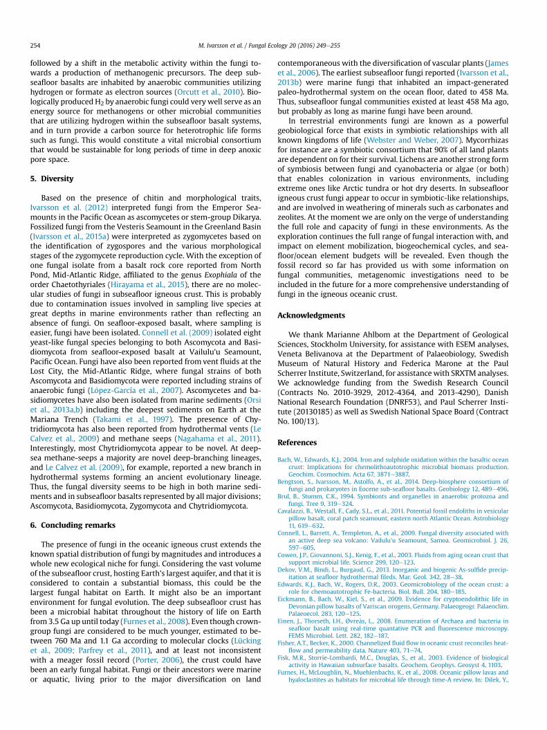
~1.5 km of sheeted dikes, and a deeper layer to about 4 km depth ofgabbroic rock (Fig. 1). The total rock volume is 2.3 � 1018 m3, whichis 6e10 times the total volume of the marine sediments. The upperlayer is characterized by extensive fracturing, about 10% porosity,and with permeabilities of about 10�12 to 10�15 m2 (Bach andEdwards, 2004; Orcutt et al., 2011). Fractures created by tensionrelease or quick cooling occur with varying size and frequency, asdo vesicles as a result of pressure release during magma extrusion.Thus, subseafloor basalts contain coherent systems of micro-fractures and vesicles interconnected with each other in whichseawater and hydrothermal fluids circulate. Roughly 60% of theoceanic crust is hydrologically active and the total fluid volume thatis held within the oceanic crust corresponds to 2% of the total ocean(Orcutt et al., 2011). The entirewater volume of the ocean circulatesthrough the oceanic igneous crust every 105e107 years, whichmeans that the oceanic igneous crust is the largest aquifer systemon Earth (Fisher and Becker, 2000; Orcutt et al., 2011). Indirectlythis means that the oceanic igneous crust is the largest potentialmicrobial habitat on Earth. Microorganisms are passively trans-ported or activelymigrate through this systemwherever pore spaceand fluid flow permit. The host rock and secondary mineralizationsof the fracture walls are used for colonization and anchoring ofmicrobial communities (Figs. 1 and 2A,B), and the minerals of thehost rock can be used as energy sources for the microorganisms.Little is known of metabolic reactions in the subseafloor crustbecause of restricted accessibility of live microbial communities,and discussions on possible metabolic pathways tend to be rather
Fig. 1. Diagram showing the oceani
speculative. However, in the absence of sunlight, a majority of thedeep subseafloor biosphere is thought to consist of chemo-autotrophs; organisms that obtain energy from inorganic sourcesand synthesize all necessary organic compounds from inorganiccarbon sources, in contrast to photoautotrophs that utilize solarenergy.
Basalts exposed at the seafloor, and thus more accessiblecompared to deeper basalts, are commonly coated with biofilmsmainly dominated by Bacteria according to quantative molecularstudies like PCR or FISH (Edwards et al., 2003; Sudek et al., 2010;Templeton et al., 2009). Taxonomically, the microbial commu-nities are dominated by Actinobacteria, Bacteroidetes, Chloroflexi,Firmicutes, Planctomycetes, and Proteobacteria phyla (Mason et al.,2007, 2008; Einen et al., 2008; Santelli et al., 2008; Santelli et al.,2009). The Archaea are much less known but tend to be domi-nated by members of Crenarchaeota and Thaumarchaeota(Thorseth et al., 2001; Fisk et al., 2003; Lysnes et al., 2004; Masonet al., 2007). Eukaryotes, including fungi, have been reportedfrom dredged basalts (Connell et al., 2009) and hydrothermal ventsites (L�opez-García et al., 2007).
Subseafloor basalts are more difficult to access and sample thanseafloor-exposed basalts, and molecular studies from such envi-ronments are therefore sparse. Alpha-, Beta-, and Gammaproteo-bacteria lineages in gabbroic layers (Mason et al., 2010) have beenreported as well as anaerobic Archaea such as Archaeoglobus andMethanosarcina (Cowen et al., 2003; Orcutt et al., 2010). Lever et al.(2013) reported Methanosarcinales, anaerobic methane-oxidizing
c crust and endolithic habitats.

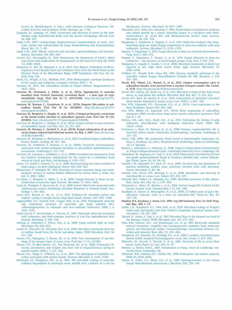
Fig. 2. (A) Optical microphotograph showing a vug (small cavity inside rock) in basalt with a few calcite crystals and a fungal mycelium attached to the wall of the pore space. Thevug is covered by a biofilm fromwhich the mycelium protrudes and grows. (B) ESEM image showing close-up of the fungal mycelium in A. At the bottom is the biofilm from whichthe hyphae grew. The biofilm and the hyphae are mineralized as montmorillonite. In the upper right corner are the remains of microstromatolitic bacterial colonies, called Frutexites,seen as cauliflower-like structures. The bacteria were probably iron oxidizers living in symbiosis with the fungi. (C) Tomographic reconstruction showing a myceliumwith symbioticorganisms; Frutexites and prokaryotic cells suspended between the hyphae in a cobweb-like fashion. Abbreviations: ba, basalt; ca, calcite; bf, biofilm; my, mycelium; hy, hyphae; fx,Frutexites; cw, “cobweb”; ze, zeolite; md, mineral dissolution. Fig. 2A is modified from Ivarsson et al. (2013a), and Fig. 2B is modified from Bengtson et al. (2014).
M. Ivarsson et al. / Fungal Ecology 20 (2016) 249e255 251
Archaea (ANME-1), and a cluster of microbes that falls within un-cultured sulfate reducers. Furthermore, studies of crustal fluidscollected on ocean ridge flanks have indicated the presence ofFirmicutes (Cowen et al., 2003; Nakagawa et al., 2006), Bacteria thatdo not group closely with cultivated microorganisms and whosemetabolism is unclear, although they have been connected withnitrogen or sulfur cycling. Only one fungal isolate from a basalt drillcore has been reported so far (Hirayama et al., 2015). The drill corewas collected at North Pond on the western flank of the Mid-Atlantic ridge. The isolated fungal strain was affiliated to thegenus Exophiala of the order Chaetothyriales.
Apart from this rather fragmentary and circumstantial evidence,our understanding of the biosphere of the subseafloor crust isbased on a fossil record. Granular and tubular ichnofossils (tracefossils) in volcanic glass are the most abundant type of fossil(Staudigel et al., 2008). Numerous reports have discussed the
abundance, diversity and origin of these ichnofossils, but still todaytheir biological origin is questioned (Lepot et al., 2009), especiallythe ancient ones found in ophiolites as old as 3.5 Ga (Grosch andMcLoughlin, 2014). During the last decade fossilized microorgan-isms have been observed in drilled cores and dredged samples fromthe ocean floor and in ophiolites (Schumann et al., 2004; Ivarssonet al., 2008a, b; 2012; Peckmann et al., 2008; Eickmann et al.,2009; Cavalazzi et al., 2011; Sakakibara et al., 2014). The microor-ganisms have been found fossilized in carbonate- and zeolite-filledveins and vesicles in the basalts or open pore spaces, and representmicrobial communities that once lived in these fractures. Surpris-ingly, a majority of these findings represents fungi (Ivarsson et al.,2012, 2013a; Bengtson et al., 2014).

M. Ivarsson et al. / Fungal Ecology 20 (2016) 249e255252
3. Endolithic fungi in the igneous oceanic crust
Schumann et al. (2004) first described fossilized fungal hyphaein carbonate-filled vesicles in North Pacific Eocene basalts.Morphological traits including branching, septa, and a central porewere used to argue for a fungal interpretation of the fossilized fil-aments. These were interpreted as endolithic fungi that wereintroduced to the system after the carbonate formation. Thus, theywere interpreted as active borers of the carbonates. Reitner et al.(2006) reported fungal hyphae in dredged vesicular basalt fromthe Kolbeinsey Ridge, north of Iceland, which has later been revisedas the Vesteris Seamount in the Greenland basin (Ivarsson et al.,2015a). Stephen et al. (2003) reported fungal hyphae incarbonate-filled veins from basalts collected during the OceanDrilling Program (ODP) 200 (Site 1224) in the Pacific Ocean outsideHawaii. The fungal interpretation of both Reitner et al. (2006) andStephen et al. (2003) is based on appearance only and does not relyon any characteristic fungal morphologies or fungal biomarkers.Peckmann et al. (2008) and Eickmann et al. (2009) furtherdescribed similar filamentous fossilized microorganisms in ophio-lites without interpreting them as remains of fungi, even thoughthat was discussed as one possibility. However, lack of morpho-logical characteristics made a fungal interpretation frail. Ivarssonet al. (2008a, b) also reported long, curvilinear filamentous fossil-ized microorganisms in subseafloor basalts from the EmperorSeamounts in the Pacific Ocean, without interpreting the findingsas fungi. All these reports referred to similar filamentous microor-ganisms in carbonate-filled veins in basalts of varying age; the fil-aments were preserved and mineralized by a clay phase likechamosite or illite (Peckmann et al., 2008), or by a composition ofthe end member bertierine-chamosite and illite-glauconite(Eickmann et al., 2009). Ivarsson et al. (2008a, b) described thecomposition of the filamentous microorganisms as a poorly crys-talline clay-like phase of Si, Al, Mg, Fe and C, and/or iron oxides.Many of the filamentous structures contained high amounts ofcarbon (10e50 wt%), but also phosphates, hydrocarbons and lipids,which were used as evidence for their biogenic origin (Ivarssonet al., 2008b). Propidium iodide (PI), a dye that binds to cells withdamaged cell membranes and traces of DNA, were used to stainfossils successfully (Ivarsson et al., 2008a). The high content oforganic remains was explained as a result of instant entombment ofthe microorganisms by the carbonate phase, and lack of diageneticprocesses and metamorphosis after fossilization.
The first study that was able to combine fungal morphologieswith chemical data characteristic for fungi was Ivarsson et al.(2012). They described mycelium-like networks of hyphae in ve-sicular basalts from the Emperor Seamounts in the Pacific Oceanwith the aid of synchrotron-radiation X-ray tomographic micro-scopy (SRXTM). This technique enabled a much more detailedstudy of the fossil morphologies and spatial occurrence. The hyphaewere characterized by repetitive septa, anastomoses betweenbranches and a central strand. They were also able to detect chitinin the cell walls by staining with WGA-FITC under fluorescencemicroscopy. Chitin is absent in prokaryotes but common amongseveral lineages of fungi and thus a strong indication of a fungalaffinity. Based on the presence of chitin and morphological traitsIvarsson et al. (2012) interpreted the fungi as ascomycetes or stem-group Dikarya. This study involved a re-interpretation as hyphalfungi of the filamentous microfossils previously reported byIvarsson et al. (2008a,b) from the same samples. Ivarsson (2012)further showed the presence of fruit bodies, spores, thallic con-idiogenesis, and hyphal tips with preserved hyphal vesicles.Abundant reproductive structures indicated that the fungi do notsimply represent specimens randomly introduced by downwardtransport of seawater but that they existed in vital, sustainable
colonies.Ivarsson et al. (2013a) further reported mycelium-like networks
in open veins and vesicles in subseafloor basalts (Fig. 2). The maindifference compared to the previous studies was the mode ofpreservation in open vesicles compared to the entombment bycarbonates and subsequent mineralization. This type of preserva-tion enabled in-detail studies of an entire fungal community andnot only slices of communities visible in petrographic thin sections.The fungal communities in the open vesicles were also mineralizedand preserved by a clay-phase, normally montmorillonite, butlacked the high carbon content that characterized the fossilsembedded in carbonates. This is probably due to fossilization in anopen system where microbial activity has decomposed, reminer-alized and thus removed all remains of carbon compounds. Besideshyphae, yeast-like growth states were also described. The yeastoccurred as spherical bodies with a diameter of ~20 mm and couldform assemblages of hundreds of cells. Ivarsson et al. (2013a)further described structures associated with the mycelium thatthey interpreted as resting structures, similar to sclerotia.
Using SRXTM Bengtson et al. (2014) revealed new informationon the fungal communities described by Ivarsson et al. (2013a)(Figs. 2c and 3a). They showed that the fungal colonization isinitiated by the formation of a biofilm that is laid down directly onthe vesicle walls. The entire walls are covered by this biofilm fromwhich hyphae as well as sporophores protrude. The biofilm andassociated mycelium are partially overgrown by secondary calcite,and Bengtson et al. (2014) were able to show that the fungi survivedthis overgrowth and bored in the calcite after its formation, pro-ducing long tubular cavities in the calcite (Fig. 3a). The hyphalboring started both from the biofilm overgrown by calcite and fromnon-overgrown parts of the mycelium. The dissolution of thecalcite was most likely mediated by the production of organic acids,which is a common trait among fungi (Gadd, 2007, 2010). A uniquefeature of these borings is that the responsible organisms, withoutexception, remain in the bored structure.
Bengtson et al. (2014) further showed that the fungi existed in aclose symbiotic-like relationship with two types of prokaryotes,and that the prokaryotes used the structural framework of themycelia for their growth. Minute cells suspended between thehyphae formed a “cobweb” with an ultrastructure similar to that ofPyrodictium or Euryarchaeon SM1, common sulfate-reducingarchaea found in hydrothermally active areas. Based on elementanalyses these were interpreted as remains of chemoautotrophicprokaryotes involved in iron oxidation. The other type of prokary-ote was represented by microstromatolites called Frutexites. Thesestructures also grew on the hyphal network and showed a distinctgrowth direction in cross section. The Frutexiteswere interpreted asremains of bacterial communities involved in iron oxidation aswell. The fungi obviously gained from the close relationship withchemoautotrophic prokaryotes, which probably made the fungalcolonization in the nutrient-poor environment possible. In thisinterpretation the chemoautotrophs fixed dissolved carbon fromfluids and built biomass, which the heterotrophic fungi couldscavenge for their metabolism. There might also have been afeedback mechanism when the fungi decomposed the carbohy-drates to CO2 accessible for the chemoautotrophs, although theextent of this feedback is unknown.
4. Metabolic pathways and cycling of elements in the oceanicigneous crust
The presence of fungi in subseafloor igneous crust raises ques-tions regarding metabolic pathways, access to bioavailable ele-ments, metals, and carbon sources (Schumann et al., 2004; Ivarssonet al., 2012). Fungi are heterotrophs and need a constant supply of

Fig. 3. (A) Tomographic reconstruction of a fungal mycelium with symbiotic prokaryotic cells suspended in between the hyphae in a cobweb-like fashion. The mycelium is drapedaround the calcite surface (blue) and the hyphae penetrate the calcite creating tunnels within the mineral. (B) ESEM image of a zeolite surface on which hyphal boring is seen. Notethe dissolution features on the mineral surface around the hyphae. Fig. 3B is modified from Ivarsson et al. (2015c). Abbreviations as for Fig. 2.
M. Ivarsson et al. / Fungal Ecology 20 (2016) 249e255 253
carbohydrates for their metabolism. In a nutrient-poor environ-ment such as the oceanic crust, occurrence of accessible biomassand organic compounds is poorly understood and investigated.Possible sources could be the trickling down from overlying sedi-ments and seawater, or chemoautotrophic communities, butwhether such carbon is evenly or patchily distributed throughoutthe oceanic crust is not known. The symbiotic-like relationshipwith chemoautotrophs reported by Bengtson et al. (2014) describesone possible way fungi can get access to biomass. Ivarsson et al.(2015c) describe a similar community structure from DetroitSeamount, where fungi basically overgrow and graze on the pro-karyotic portion of the community. Thus, chemoautotrophic com-munities seem to be a carbon source in deep basalts. In dredgedsamples from the Vesteris Seamount in the Greenland basin, rep-resenting depths of no more than 1 m, Ivarsson et al. (2015a)showed how fungi feed on marine organisms such as algae,which have been introduced to the basalts by ingress of seawater.The fungi produce haustoria with which they attack and penetratethe algae; even the silica frustules of diatoms are attacked anddegraded by the haustoria. Thus, in shallow basalts, where marinesediments rich in organic remains are easily introduced, the maincarbon source for fungal metabolism seems to be organic matter inthe sediments. At greater depths, where sediments do not reach bydownwardmigration, the fungi need to find other accessible carbonsources of which symbiotic chemoautotrophic communities areone candidate (Bengtson et al., 2014; Ivarsson et al., 2015c).
In addition to cycling carbon, fungi are also involved in thecycling of metals and other elements through bioweathering ofminerals and biomineralization. The dissolution and subsequentboring of tunnels in carbonates (Bengtson et al., 2014) and zeolites(Ivarsson et al., 2015c) (Fig. 3) mobilizemetals and elements such asCa, Si, Al, Na, and K. The reason for the mineral boring could bemigration, acquisition of habitable space, response to environ-mental stress, or trophic strategies (Bengtson et al., 2014; Ivarssonet al., 2015c). Ca, Na, and K are essential metals for fungal growthand metabolism (Gadd, 2007, 2010), and they could thus be minedfor metabolic reasons.
Ivarsson et al. (2015b) showed fungal communities associatedwith biogenic Mn oxides. The biogenicity of these Mn oxides wassupported by electron paramagnetic resonance (EPR) analysis, butwhether the fungi or an associated prokaryotic community wasresponsible for the direct oxidation of Mn(II) to Mn(IV) oxidemineral could not be fully determined. Fungal involvement in Mnoxidation has been shown (Tebo et al., 1997), also from exposedseafloor-basalts (Connell et al., 2009). In the Vesteris Seamountsamples fungal spores mineralize by Mn(II) oxide minerals
indicating a primary process in which fungi are involved in Mn(II)oxidation and formation of subsequent Mn(IV) oxide minerals(Ivarsson et al., 2015a).
Dekov et al. (2013) reported fungal hyphae in hydrothermalsulfide-sulfate samples from seafloor and subseafloor rocksmineralized as orpiments (As-sulfides). They interpreted the fungias endolithic organisms killed by As-rich pulses of fluids, withfungal organic matter serving as a geochemical trap for hydro-thermal As. The As reacted with S from the basement or theseawater and mineralized the hyphae as As2S3.
Considering the plethora of elements fungi are known tointeract with in terrestrial environments (Gadd, 2007, 2010), it isprobable that the geobiological impact of fungi in the oceanicigneous crust is far from resolved and understood. One unresolvedissue is the concentration of O2 in the oceanic crust, which is poorlyinvestigated, and would greatly impact on the fungal abundanceand diversity since most known fungi are aerobic. Shallow igneouscrustal environments appear to be oxygenated since they introduceoxygen-rich pore waters into the anoxic bottom sediments (Orcuttet al., 2013), but further down in the igneous portion of the crustoxygen levels are far from being understood. Local areas ofoxygenated seawater can probably be introduced, but there shouldalso be provinces poor in oxygen. In a heterogeneous environment,such as the igneous crust, geochemical conditions vary on a localscale and anaerobic niches are probably common. Most fungi areaerobic but anaerobic fungi are known from freshwater lakes,landfill sites (McDonald et al., 2012), and deep sea sediments(Nagano and Nagahama, 2012) but are best known from the rumenof ruminating herbivores (Khejornsart and Wanapat, 2010;Liggenstoffer et al., 2010), an anaerobic environment in whichthey play an important role in energy metabolism including theformation of H2. Anaerobic fungal species have no mitochondriaand are unable to produce energy by either aerobic or anaerobicrespiration (Yarlett et al., 1986; O'Fallon et al., 1991). Instead,anaerobic fungi have hydrogenosomes, organelles capable ofcoupling the metabolism of glucose to cellular energy production.Hydrogenosomes contain hydrogenase and produce H2, CO2, ace-tate, formate, lactate, and ethanol as metabolic waste products(Yarlett et al., 1986; Brul and Stumm, 1994; Khejornsart andWanapat, 2010). Anaerobic fungi consort with methanogenicarchaea in the rumen. Methanogens are microorganisms thatproduce methane using carbon dioxide, acetate or one-carbonsubstrates as carbon sources and electron acceptors whilehydrogen functions as an electron donor (Mountfort et al., 1982;Hook et al., 2010). The methanogens increase the enzymatic ac-tivity in the fungi through the removal of hydrogen, which is
M. Ivarsson et al. / Fungal Ecology 20 (2016) 249e255254
followed by a shift in the metabolic activity within the fungi to-wards a production of methanogenic precursors. The deep sub-seafloor basalts are inhabited by anaerobic communities utilizinghydrogen or formate as electron sources (Orcutt et al., 2010). Bio-logically produced H2 by anaerobic fungi could very well serve as anenergy source for methanogens or other microbial communitiesthat are utilizing hydrogen within the subseafloor basalt systems,and in turn provide a carbon source for heterotrophic life formssuch as fungi. This would constitute a vital microbial consortiumthat would be sustainable for long periods of time in deep anoxicpore space.
5. Diversity
Based on the presence of chitin and morphological traits,Ivarsson et al. (2012) interpreted fungi from the Emperor Sea-mounts in the Pacific Ocean as ascomycetes or stem-group Dikarya.Fossilized fungi from the Vesteris Seamount in the Greenland Basin(Ivarsson et al., 2015a) were interpreted as zygomycetes based onthe identification of zygospores and the various morphologicalstages of the zygomycete reproduction cycle. With the exception ofone fungal isolate from a basalt rock core reported from NorthPond, Mid-Atlantic Ridge, affiliated to the genus Exophiala of theorder Chaetothyriales (Hirayama et al., 2015), there are no molec-ular studies of fungi in subseafloor igneous crust. This is probablydue to contamination issues involved in sampling live species atgreat depths in marine environments rather than reflecting anabsence of fungi. On seafloor-exposed basalt, where sampling iseasier, fungi have been isolated. Connell et al. (2009) isolated eightyeast-like fungal species belonging to both Ascomycota and Basi-diomycota from seafloor-exposed basalt at Vailulu'u Seamount,Pacific Ocean. Fungi have also been reported from vent fluids at theLost City, the Mid-Atlantic Ridge, where fungal strains of bothAscomycota and Basidiomycota were reported including strains ofanaerobic fungi (L�opez-García et al., 2007). Ascomycetes and ba-sidiomycetes have also been isolated from marine sediments (Orsiet al., 2013a,b) including the deepest sediments on Earth at theMariana Trench (Takami et al., 1997). The presence of Chy-tridiomycota has also been reported from hydrothermal vents (LeCalvez et al., 2009) and methane seeps (Nagahama et al., 2011).Interestingly, most Chytridiomycota appear to be novel. At deep-sea methane-seeps a majority are novel deep-branching lineages,and Le Calvez et al. (2009), for example, reported a new branch inhydrothermal systems forming an ancient evolutionary lineage.Thus, the fungal diversity seems to be high in both marine sedi-ments and in subseafloor basalts represented by all major divisions;Ascomycota, Basidiomycota, Zygomycota and Chytridiomycota.
6. Concluding remarks
The presence of fungi in the oceanic igneous crust extends theknown spatial distribution of fungi bymagnitudes and introduces awhole new ecological niche for fungi. Considering the vast volumeof the subseafloor crust, hosting Earth's largest aquifer, and that it isconsidered to contain a substantial biomass, this could be thelargest fungal habitat on Earth. It might also be an importantenvironment for fungal evolution. The deep subseafloor crust hasbeen a microbial habitat throughout the history of life on Earthfrom3.5 Ga up until today (Furnes et al., 2008). Even though crown-group fungi are considered to be much younger, estimated to be-tween 760 Ma and 1.1 Ga according to molecular clocks (Lückinget al., 2009; Parfrey et al., 2011), and at least not inconsistentwith a meager fossil record (Porter, 2006), the crust could havebeen an early fungal habitat. Fungi or their ancestors were marineor aquatic, living prior to the major diversification on land
contemporaneous with the diversification of vascular plants (Jameset al., 2006). The earliest subseafloor fungi reported (Ivarsson et al.,2013b) were marine fungi that inhabited an impact-generatedpaleo-hydrothermal system on the ocean floor, dated to 458 Ma.Thus, subseafloor fungal communities existed at least 458 Ma ago,but probably as long as marine fungi have been around.
In terrestrial environments fungi are known as a powerfulgeobiological force that exists in symbiotic relationships with allknown kingdoms of life (Webster and Weber, 2007). Mycorrhizasfor instance are a symbiotic consortium that 90% of all land plantsare dependent on for their survival. Lichens are another strong formof symbiosis between fungi and cyanobacteria or algae (or both)that enables colonization in various environments, includingextreme ones like Arctic tundra or hot dry deserts. In subseafloorigneous crust fungi appear to occur in symbiotic-like relationships,and are involved in weathering of minerals such as carbonates andzeolites. At the moment we are only on the verge of understandingthe full role and capacity of fungi in these environments. As theexploration continues the full range of fungal interaction with, andimpact on element mobilization, biogeochemical cycles, and sea-floor/ocean element budgets will be revealed. Even though thefossil record so far has provided us with some information onfungal communities, metagenomic investigations need to beincluded in the future for a more comprehensive understanding offungi in the igneous oceanic crust.
Acknowledgments
We thank Marianne Ahlbom at the Department of GeologicalSciences, Stockholm University, for assistance with ESEM analyses,Veneta Belivanova at the Department of Palaeobiology, SwedishMuseum of Natural History and Federica Marone at the PaulScherrer Institute, Switzerland, for assistancewith SRXTM analyses.We acknowledge funding from the Swedish Research Council(Contracts No. 2010-3929, 2012-4364, and 2013-4290), DanishNational Research Foundation (DNRF53), and Paul Scherrer Insti-tute (20130185) as well as Swedish National Space Board (ContractNo. 100/13).
References
Bach, W., Edwards, K.J., 2004. Iron and sulphide oxidation within the basaltic oceancrust: Implications for chemolithoautotrophic microbial biomass production.Geochim. Cosmochim. Acta 67, 3871e3887.
Bengtson, S., Ivarsson, M., Astolfo, A., et al., 2014. Deep-biosphere consortium offungi and prokaryotes in Eocene sub-seafloor basalts. Geobiology 12, 489e496.
Brul, B., Stumm, C.K., 1994. Symbionts and organelles in anaerobic protozoa andfungi. Tree 9, 319e324.
Cavalazzi, B., Westall, F., Cady, S.L., et al., 2011. Potential fossil endoliths in vesicularpillow basalt, coral patch seamount, eastern north Atlantic Ocean. Astrobiology11, 619e632.
Connell, L., Barrett, A., Templeton, A., et al., 2009. Fungal diversity associated withan active deep sea volcano: Vailulu'u Seamount, Samoa. Geomicrobiol. J. 26,597e605.
Cowen, J.P., Giovannoni, S.J., Kenig, F., et al., 2003. Fluids from aging ocean crust thatsupport microbial life. Science 299, 120e123.
Dekov, V.M., Bindi, L., Burgaud, G., 2013. Inorganic and biogenic As-sulfide precip-itation at seafloor hydrothermal fileds. Mar. Geol. 342, 28e38.
Edwards, K.J., Bach, W., Rogers, D.R., 2003. Geomicrobiology of the ocean crust: arole for chemoautotrophic Fe-bacteria. Biol. Bull. 204, 180e185.
Eickmann, B., Bach, W., Kiel, S., et al., 2009. Evidence for cryptoendolithic life inDevonian pillow basalts of Variscan orogens, Germany. Palaeogeogr. Palaeoclim.Palaeoecol. 283, 120e125.
Einen, J., Thorseth, I.H., Øvreås, L., 2008. Enumeration of Archaea and bacteria inseafloor basalt using real-time quantative PCR and fluorescence microscopy.FEMS Microbiol. Lett. 282, 182e187.
Fisher, A.T., Becker, K., 2000. Channelized fluid flow in oceanic crust reconciles heat-flow and permeability data. Nature 403, 71e74.
Fisk, M.R., Storrie-Lombardi, M.C., Douglas, S., et al., 2003. Evidence of biologicalactivity in Hawaiian subsurface basalts. Geochem. Geophys. Geosyst 4, 1103.
Furnes, H., McLoughlin, N., Muehlenbachs, K., et al., 2008. Oceanic pillow lavas andhyaloclastites as habitats for microbial life through time-A review. In: Dilek, Y.,
M. Ivarsson et al. / Fungal Ecology 20 (2016) 249e255 255
Furnes, H., Muehlenbachs, K. (Eds.), Links between Geological Processes, Mi-crobial Activities and Evolution of Life. Springer, pp. 1e68.
Gadanho, M., Sampajo, J.P., 2005. Occurrence and diversity of yeasts in the mid-Atlantic ridge hydrothermal fields near the Azores Archipelago. Microb. Ecol.50, 408e417.
Gadd, G.M., 2007. Geomycology: biogeochemical transformations of rocks, min-erals, metals and radionuclides by fungi, bioweathering and bioremediation.Mycol. Res. 111, 3e49.
Gadd, G.M., 2010. Metals, minerals and microbes: geomicrobiology and bioreme-diation. Microbiol. 156, 609e643.
Grosch, E.G., McLoughlin, N., 2014. Reassessing the biogenicity of Earth's oldesttrace fossil with implications for biosignatures in the search for early life. PNAS111, 8380e8385.
Hirayama, H., Abe, M., Miyazaki, J., et al., 2015. Data Report: Cultivation of Micro-organisms from Basaltic Rock and Sediment Cores from the North Pond on theWestern Flank of the Mid-Atlantic Ridge, IODP Expedition 336. Proc. Int. Oc.Drill. Prog. 336.
Hook, S.E., Wright, A.-D.G., McBride, B.W., 2010. Methanogens: methane producersof the rumen and mitigation strategies. Archaea 2010, 1e11.
Ivarsson, M., 2012. The subseafloor basalts as fungal habitats. Biogeosciences 9,3625e3635.
Ivarsson, M., Peckmann, J., Tehler, A., et al., 2015a. Zygomycetes in vesicularbasanites from Vesteris Seamount, Greenland Basin e a new type of cryp-toendolithic fungi. PLoS One 10 (7), e01333368. http://dx.doi.org/10.1371/journal.pone.01333368.
Ivarsson, M., Broman, C., Gustafsson, H., et al., 2015b. Biogenic Mn-oxides in sub-seafloor basalts. PLoS One 10 (6), e0128863. http://dx.doi.org/10.1371/journal.pone.0128863.
Ivarsson, M., Bengtson, S., Skogby, H., et al., 2015c. A fungal-prokaryotic consortiumat the basalt-zeolite interface in subseafloor igneous crust. PLoS One 10 (10),e014016. http://dx.doi.org/10.1371/journal.pone.0140106.
Ivarsson, M., Bengtson, S., Skogby, H., et al., 2013a. Fungal colonies in open fracturesof subseafloor basalt. Geo-Mar. Lett. 33, 233e243.
Ivarsson, M., Broman, C., Sturkell, E., et al., 2013b. Fungal colonization of an ordo-vician impact-induced hydrothermal system. Sci. Rep. 3, 3487. http://dx.doi.org/10.1038/srep03487.
Ivarsson, M., Bengtson, S., Belivanova, V., et al., 2012. Fossilized fungi in subseafloorEocene basalts. Geology 40, 163e166.
Ivarsson, M., Lindblom, S., Broman, C., et al., 2008a. Fossilized microorganismsassociated with zeolite-carbonate interfaces in sub-seafloor hydrothermal en-vironments. Geobiology 6, 155e170.
Ivarsson, M., Lausmaa, J., Lindblom, S., et al., 2008b. Fossilized microorganisms fromthe Emperor Seamounts: Implications for the search for a subsurface fossilrecord on Earth and Mars. Astrobiology 8, 1139e1157.
James, T.Y., Kauff, F., Schoch, C.L., et al., 2006. Reconstructing the early evolution offungi using a six-gene phylogeny. Nature 443, 818e822.
Khejornsart, P., Wanapat, M., 2010. Diversity of rumen anaerobic fungi and meth-anogenic archaea in swamp buffalo influenced by various diets. J. Anim. Vet.Adv. 9, 3062e3069.
Le Calvez, T., Burgaud, G., Mahe, S., et al., 2009. Fungal diversity in deep sea hy-drothermal ecosystems. Appl. Environ. Microbiol. 75, 6415e6421.
Lepot, K., Philippot, P., Benzerara, K., et al., 2009. Garnet-filled trails associated withcarbonaceous matter mimicking microbial filaments in Archean basalt. Geo-biology 7, 393e402.
Lever, M.A., Rouxel, A., Alt, J.C., et al., 2013. Evidence for microbial carbon andsulphur cycling in deeply buried ridge flank basalt. Science 339, 1305e1308.
Liggenstoffer, A.S., Youssef, N.H., Couger, M.B., et al., 2010. Phylogenetic diversityand community structure of anaerobic gut fungi (phylum Neo-callimastigomycota) in ruminant and non-ruminant herbivores. ISME J. 4,1225e1235.
L�opez-García, P., Vereshchaka, A., Moreira, D., 2007. Eukaryotic diversity associatedwith carbonates and fluid-seawater interface in Lost-City hydrothermal field.Environ. Microbiol. 9, 546e554.
Lücking, R., Huhndorf, S., Pfister, D.H., et al., 2009. Fungi evolved right on track.Mycologia 101, 810e822.
Lysnes, K., Thorseth, I.H., Steinsbau, B.O., et al., 2004. Microbial community diversityin seafloor basalt from the Arctic spreading ridges. FEMS Microbiol. Ecol. 50,213e230.
Mason, O.U., Nakagawa, T., Rosner, M., et al., 2010. First investigation of microbi-ology of the deepest layer of ocean crust. PLoS One 5 (11), e15399.
Mason, O.U., Di Meo-Savoie, C.A., Van Nostrand, J.D., et al., 2008. Prokaryotic di-versity, distribution, and insights into their role in biogeochemical cycling inmarine basalts. ISME J. 3, 231e242.
Mason, O.U., Stingl, U., Wilhelm, L.J., et al., 2007. The phylogeny of endolithic mi-crobes associated with marine basalts. Environ. Microbiol. 9, 2539e2550.
McDonald, J.E., Houghton, J.N.I., et al., 2012. The microbial ecology of anaerobiccellulose degradation in municipal waste landfill sites: evidence of a role for
fibrobacters. Environ. Microbiol. 14, 1077e1087.Mountfort, D.O., Asher, R.A., Bauchop, T., 1982. Fermentation of cellulose to methane
and carbon dioxide by a rumen anaerobic fungus in a triculture with Meth-anobrevibacter sp. strain RA1 and Methanosarcina barkeri. Appl. Environ.Microbiol. 44, 128e134.
Nagahama, T., Takahashi, E., Nagano, Y., et al., 2011. Molecular evidence that deep-branching fungi are major fungal components in deep-sea methane cold-seepsediments. Environ. Microbiol. 13, 2359e2370.
Nagano, Y., Nagahama, T., 2012. Fungal diversity in deep-sea extreme environments.Fung. Ecol. 5, 463e471.
Nagano, Y., Nagahama, T., Hatada, Y., et al., 2010. Fungal diversity in deep-seasediments e the presence of novel fungal groups. Fung. Ecol. 3, 316e325.
Nakagawa, S., Inagaki, F., Suzuki, Y., et al., 2006. Microbial community in black rustexposed to hot ridge flank crustal fluids. Appl. Environ. Microbiol. 72,6789e6799.
O'Fallon, J.V., Wright, R.W., Calza, R.E., 1991. Glucose metabolic pathways in theanaerobic rumen fungus Neocallimatrix frontalis EB 188. Biochem. J. 274,595e599.
Orcutt, B.N., Wheat, C.G., Rouxel, O., et al., 2013. Oxygen consumption rates insubseafloor basaltic crust derived from a reaction transport model. Nat. Comm.4, 2539. http://dx.doi.org/10.1038/ncomms3539.
Orcutt, B.N., Sylvan, J.B., Knab, N.J., et al., 2011. Microbial ecology of the dark oceanabove, at, and below the seafloor. Microbiol. Mol. Biol. Rev. 75, 361e422.
Orcutt, B.N., Bach, W., Becker, K., et al., 2010. Colonization of subsurface microbialobservatories deployed in young ocean crust. ISME J. 5, 692e703.
Orsi, W.D., Edgcomb, V.D., Christman, G.D., et al., 2013a. Gene expression in thedeep biosphere. Nature 499, 205e208.
Orsi, W.D., Biddle, J.F., Edgcomb, V.D., 2013b. Deep sequencing of subseaflooreukaryotic rRNA reveals active fungi across marine subsurface provinces. PLoSOne 8, 1e10.
Parfrey, L.W., Lahr, D.J.G., Knoll, A.H., et al., 2011. Estimating the timing of earlyeukaryotic diversification with multigene molecular clocks. PNAS 108,13624e13629.
Peckmann, J., Bach, W., Behrens, K., et al., 2008. Putative cryptoendolithic life inDevonian pillow basalt, rheinisches Schiefergebirge, Germany. Geobiology 6,125e135.
Porter, S.M., 2006. The proterozoic fossil record of heterotrophic eukaryotes. In:Xiao, S., Kaufman, A.J. (Eds.), Neoproterozoic Geobiology, Topics in Geobiology,vol. 27. Springer.
Reitner, J., Schumann, G., Pedersen, K., 2006. Fungi in Subterranean Environments.in: Fungi in Biogeochemical Cycles. Cambridge University Press, Cambridge, UK.
Sakakibara, M., Sugawara, H., Tsuji, T., et al., 2014. Filamentous microbial fossil fromlow-grade metamorphosed basalt in northern chichibu belt, central Shikoku.Jpn. Planet. Space Sci. 95, 84e93.
Santelli, C.M., Edgcomb, V.P., Bach, W., et al., 2009. The diversity and abundance ofbacteria inhabiting seafloor lavas positively correlate with rock alteration.Eviron. Microbiol. 11, 86e98.
Santelli, C.M., Orcutt, B.N., Banning, E., et al., 2008. Abundance and diversity ofmicrobial life in ocean crust. Nature 453, 653e657.
Schrenk, M.O., Huber, J.A., Edwards, K.J., 2009. Microbial provinces in the subsea-floor. Annu. Rev. Mar. Sci. 2, 279e304.
Schumann, G., Manz, W., Reitner, J., et al., 2004. Ancient fungal life in North PacificEocene oceanic crust. Geomicrobiol. J. 21, 241e246.
Staudigel, H., Furnes, H., McLoughlin, N., et al., 2008. 3.5 billion years of glass bio-alteration: Volcanic rocks as a basis for microbial life? Earth-Sci. Rev. 89,156e176.
Stephen, R.A., Kasahara, J., Acton, G.D., 2003. Leg 200 Summary. Proc. Oc. Drill. Prog.,Init. Rep.. 200, 1e73.
Sudek, L.A., Templeton, A.S., Tebo, B.M., et al., 2010. Microbial ecology of Fe(hydr)oxide mats and basalts rock from Vailulu'u Seamount, American Samoa. Geo-microbiol. J. 26, 581e596.
Takami, H., Inoue, A., Fuji, F., et al., 1997. Microbial flora in the deepest sea mud ofthe Mariana Trench. FEMS Microbiol. Lett. 152, 279e285.
Tebo, B.M., Ghiorse, W.C., van Waasbergen, L.G., et al., 1997. Bacterially mediatedmineral formation: insighths into managanese(II) oxidation from moleculargenetic and biochemical studies. Geomicrobiology: interactions between mi-crobes and minerals. Revs. Min. 35, 225e266.
Templeton, A.S., Knowles, E.J., Eldridge, K.L., et al., 2009. A seafloor microbial biomehosted within incipient ferromanganese crusts. Nat. Geosci. 2, 872e876.
Thorseth, I.H., Torsvik, T., Torsvik, V., et al., 2001. Diversity of life in ocean floorbasalt. Earth Planet Sci. Lett. 194, 31e37.
Webster, J., Weber, R.W.S., 2007. Introduction to Fungi, third ed. Cambridge Uni-versity Press, Cambridge, UK.
Whitman, W.B., Coleman, D.C., Wiebe, W.J., 1998. Prokaryotes: the unseen majority.PNAS 95, 6578e6583.
Yarlett, N., Orpin, C.G., Munn, E.A., et al., 1986. Hydrogenosomes in the rumenfungus Neocallimastix patriciarum. J. Biochem. 236, 729e739.
Related Documents