DOI: 10.1126/science.287.5461.2185 , 2185 (2000); 287 Science et al. Mark D. Adams Drosophila melanogaster The Genome Sequence of This copy is for your personal, non-commercial use only. clicking here. colleagues, clients, or customers by , you can order high-quality copies for your If you wish to distribute this article to others here. following the guidelines can be obtained by Permission to republish or repurpose articles or portions of articles ): February 26, 2014 www.sciencemag.org (this information is current as of The following resources related to this article are available online at http://www.sciencemag.org/content/287/5461/2185.full.html version of this article at: including high-resolution figures, can be found in the online Updated information and services, http://www.sciencemag.org/content/287/5461/2185.full.html#related found at: can be related to this article A list of selected additional articles on the Science Web sites http://www.sciencemag.org/content/287/5461/2185.full.html#ref-list-1 , 25 of which can be accessed free: cites 75 articles This article 2900 article(s) on the ISI Web of Science cited by This article has been http://www.sciencemag.org/content/287/5461/2185.full.html#related-urls 100 articles hosted by HighWire Press; see: cited by This article has been http://www.sciencemag.org/cgi/collection/genetics Genetics subject collections: This article appears in the following registered trademark of AAAS. is a Science 2000 by the American Association for the Advancement of Science; all rights reserved. The title Copyright American Association for the Advancement of Science, 1200 New York Avenue NW, Washington, DC 20005. (print ISSN 0036-8075; online ISSN 1095-9203) is published weekly, except the last week in December, by the Science on February 26, 2014 www.sciencemag.org Downloaded from on February 26, 2014 www.sciencemag.org Downloaded from on February 26, 2014 www.sciencemag.org Downloaded from on February 26, 2014 www.sciencemag.org Downloaded from on February 26, 2014 www.sciencemag.org Downloaded from on February 26, 2014 www.sciencemag.org Downloaded from on February 26, 2014 www.sciencemag.org Downloaded from on February 26, 2014 www.sciencemag.org Downloaded from on February 26, 2014 www.sciencemag.org Downloaded from on February 26, 2014 www.sciencemag.org Downloaded from on February 26, 2014 www.sciencemag.org Downloaded from on February 26, 2014 www.sciencemag.org Downloaded from

Welcome message from author
This document is posted to help you gain knowledge. Please leave a comment to let me know what you think about it! Share it to your friends and learn new things together.
Transcript
DOI: 10.1126/science.287.5461.2185, 2185 (2000);287 Science
et al.Mark D. AdamsDrosophila melanogasterThe Genome Sequence of
This copy is for your personal, non-commercial use only.
clicking here.colleagues, clients, or customers by , you can order high-quality copies for yourIf you wish to distribute this article to others
here.following the guidelines
can be obtained byPermission to republish or repurpose articles or portions of articles
): February 26, 2014 www.sciencemag.org (this information is current as of
The following resources related to this article are available online at
http://www.sciencemag.org/content/287/5461/2185.full.htmlversion of this article at:
including high-resolution figures, can be found in the onlineUpdated information and services,
http://www.sciencemag.org/content/287/5461/2185.full.html#relatedfound at:
can berelated to this article A list of selected additional articles on the Science Web sites
http://www.sciencemag.org/content/287/5461/2185.full.html#ref-list-1, 25 of which can be accessed free:cites 75 articlesThis article
2900 article(s) on the ISI Web of Sciencecited by This article has been
http://www.sciencemag.org/content/287/5461/2185.full.html#related-urls100 articles hosted by HighWire Press; see:cited by This article has been
http://www.sciencemag.org/cgi/collection/geneticsGenetics
subject collections:This article appears in the following
registered trademark of AAAS. is aScience2000 by the American Association for the Advancement of Science; all rights reserved. The title
CopyrightAmerican Association for the Advancement of Science, 1200 New York Avenue NW, Washington, DC 20005. (print ISSN 0036-8075; online ISSN 1095-9203) is published weekly, except the last week in December, by theScience
on
Feb
ruar
y 26
, 201
4w
ww
.sci
ence
mag
.org
Dow
nloa
ded
from
o
n F
ebru
ary
26, 2
014
ww
w.s
cien
cem
ag.o
rgD
ownl
oade
d fr
om
on
Feb
ruar
y 26
, 201
4w
ww
.sci
ence
mag
.org
Dow
nloa
ded
from
o
n F
ebru
ary
26, 2
014
ww
w.s
cien
cem
ag.o
rgD
ownl
oade
d fr
om
on
Feb
ruar
y 26
, 201
4w
ww
.sci
ence
mag
.org
Dow
nloa
ded
from
o
n F
ebru
ary
26, 2
014
ww
w.s
cien
cem
ag.o
rgD
ownl
oade
d fr
om
on
Feb
ruar
y 26
, 201
4w
ww
.sci
ence
mag
.org
Dow
nloa
ded
from
o
n F
ebru
ary
26, 2
014
ww
w.s
cien
cem
ag.o
rgD
ownl
oade
d fr
om
on
Feb
ruar
y 26
, 201
4w
ww
.sci
ence
mag
.org
Dow
nloa
ded
from
o
n F
ebru
ary
26, 2
014
ww
w.s
cien
cem
ag.o
rgD
ownl
oade
d fr
om
on
Feb
ruar
y 26
, 201
4w
ww
.sci
ence
mag
.org
Dow
nloa
ded
from
o
n F
ebru
ary
26, 2
014
ww
w.s
cien
cem
ag.o
rgD
ownl
oade
d fr
om
The Genome Sequence of Drosophila melanogasterMark D. Adams,1* Susan E. Celniker,2 Robert A. Holt,1 Cheryl A. Evans,1 Jeannine D. Gocayne,1
Peter G. Amanatides,1 Steven E. Scherer,3 Peter W. Li,1 Roger A. Hoskins,2 Richard F. Galle,2 Reed A. George,2
Suzanna E. Lewis,4 Stephen Richards,2 Michael Ashburner,5 Scott N. Henderson,1 Granger G. Sutton,1
Jennifer R. Wortman,1 Mark D. Yandell,1 Qing Zhang,1 Lin X. Chen,1 Rhonda C. Brandon,1 Yu-Hui C. Rogers,1
Robert G. Blazej,2 Mark Champe,2 Barret D. Pfeiffer,2 Kenneth H. Wan,2 Clare Doyle,2 Evan G. Baxter,2
Gregg Helt,6 Catherine R. Nelson,4 George L. Gabor Miklos,7 Josep F. Abril,8 Anna Agbayani,2 Hui-Jin An,1
Cynthia Andrews-Pfannkoch,1 Danita Baldwin,1 Richard M. Ballew,1 Anand Basu,1 James Baxendale,1
Leyla Bayraktaroglu,9 Ellen M. Beasley,1 Karen Y. Beeson,1 P. V. Benos,10 Benjamin P. Berman,2 Deepali Bhandari,1
Slava Bolshakov,11 Dana Borkova,12 Michael R. Botchan,13 John Bouck,3 Peter Brokstein,4 Phillipe Brottier,14
Kenneth C. Burtis,15 Dana A. Busam,1 Heather Butler,16 Edouard Cadieu,17 Angela Center,1 Ishwar Chandra,1
J. Michael Cherry,18 Simon Cawley,19 Carl Dahlke,1 Lionel B. Davenport,1 Peter Davies,1 Beatriz de Pablos,20
Arthur Delcher,1 Zuoming Deng,1 Anne Deslattes Mays,1 Ian Dew,1 Suzanne M. Dietz,1 Kristina Dodson,1
Lisa E. Doup,1 Michael Downes,21 Shannon Dugan-Rocha,3 Boris C. Dunkov,22 Patrick Dunn,1 Kenneth J. Durbin,3
Carlos C. Evangelista,1 Concepcion Ferraz,23 Steven Ferriera,1 Wolfgang Fleischmann,5 Carl Fosler,1
Andrei E. Gabrielian,1 Neha S. Garg,1 William M. Gelbart,9 Ken Glasser,1 Anna Glodek,1 Fangcheng Gong,1
J. Harley Gorrell,3 Zhiping Gu,1 Ping Guan,1 Michael Harris,1 Nomi L. Harris,2 Damon Harvey,4 Thomas J. Heiman,1
Judith R. Hernandez,3 Jarrett Houck,1 Damon Hostin,1 Kathryn A. Houston,2 Timothy J. Howland,1 Ming-Hui Wei,1
Chinyere Ibegwam,1 Mena Jalali,1 Francis Kalush,1 Gary H. Karpen,21 Zhaoxi Ke,1 James A. Kennison,24
Karen A. Ketchum,1 Bruce E. Kimmel,2 Chinnappa D. Kodira,1 Cheryl Kraft,1 Saul Kravitz,1 David Kulp,6
Zhongwu Lai,1 Paul Lasko,25 Yiding Lei,1 Alexander A. Levitsky,1 Jiayin Li,1 Zhenya Li,1 Yong Liang,1 Xiaoying Lin,26
Xiangjun Liu,1 Bettina Mattei,1 Tina C. McIntosh,1 Michael P. McLeod,3 Duncan McPherson,1 Gennady Merkulov,1
Natalia V. Milshina,1 Clark Mobarry,1 Joe Morris,6 Ali Moshrefi,2 Stephen M. Mount,27 Mee Moy,1 Brian Murphy,1
Lee Murphy,28 Donna M. Muzny,3 David L. Nelson,3 David R. Nelson,29 Keith A. Nelson,1 Katherine Nixon,2
Deborah R. Nusskern,1 Joanne M. Pacleb,2 Michael Palazzolo,2 Gjange S. Pittman,1 Sue Pan,1 John Pollard,1
Vinita Puri,1 Martin G. Reese,4 Knut Reinert,1 Karin Remington,1 Robert D. C. Saunders,30 Frederick Scheeler,1
Hua Shen,3 Bixiang Christopher Shue,1 Inga Siden-Kiamos,11 Michael Simpson,1 Marian P. Skupski,1 Tom Smith,1
Eugene Spier,1 Allan C. Spradling,31 Mark Stapleton,2 Renee Strong,1 Eric Sun,1 Robert Svirskas,32 Cyndee Tector,1
Russell Turner,1 Eli Venter,1 Aihui H. Wang,1 Xin Wang,1 Zhen-Yuan Wang,1 David A. Wassarman,33
George M. Weinstock,3 Jean Weissenbach,14 Sherita M. Williams,1 Trevor Woodage,1 Kim C. Worley,3 David Wu,1
Song Yang,2 Q. Alison Yao,1 Jane Ye,1 Ru-Fang Yeh,19 Jayshree S. Zaveri,1 Ming Zhan,1 Guangren Zhang,1 Qi Zhao,1
Liansheng Zheng,1 Xiangqun H. Zheng,1 Fei N. Zhong,1 Wenyan Zhong,1 Xiaojun Zhou,3 Shiaoping Zhu,1
Xiaohong Zhu,1 Hamilton O. Smith,1 Richard A. Gibbs,3 Eugene W. Myers,1 Gerald M. Rubin,34 J. Craig Venter1
The fly Drosophila melanogaster is one of the most intensively studiedorganisms in biology and serves as a model system for the investigation ofmany developmental and cellular processes common to higher eukaryotes,including humans. We have determined the nucleotide sequence of nearlyall of the ;120-megabase euchromatic portion of the Drosophila genomeusing a whole-genome shotgun sequencing strategy supported by exten-sive clone-based sequence and a high-quality bacterial artificial chromo-some physical map. Efforts are under way to close the remaining gaps;however, the sequence is of sufficient accuracy and contiguity to bedeclared substantially complete and to support an initial analysis ofgenome structure and preliminary gene annotation and interpretation. Thegenome encodes ;13,600 genes, somewhat fewer than the smaller Cae-norhabditis elegans genome, but with comparable functional diversity.
The annotated genome sequence of Drosoph-ila melanogaster, together with its associatedbiology, will provide the foundation for anew era of sophisticated functional studies(1–3). Because of its historical importance,large research community, and powerful re-search tools, as well as its modest genomesize, Drosophila was chosen as a test systemto explore the applicability of whole-genomeshotgun (WGS) sequencing for large andcomplex eukaryotic genomes (4). Thegroundwork for this project was laid overmany years by the fly research community,
which has molecularly characterized ;2500genes; this work in turn has been supportedby nearly a century of genetics (5). SinceDrosophila was chosen in 1990 as one of themodel organisms to be studied under theauspices of the federally funded Human Ge-nome Project, genome projects in the UnitedStates, Europe, and Canada have produced abattery of genome-wide resources (Table 1).The Berkeley and European Drosophila Ge-nome Projects (BDGP and EDGP) initiatedgenomic sequencing (Tables 1 to 3) and fin-ished 29 Mb. The bacterial artificial chromo-
some (BAC) map and other genomic resourc-es available for Drosophila serve both as anindependent confirmation of the assemblyof data from the shotgun strategy and as aset of resources for further biological anal-ysis of the genome.
The Drosophila genome is ;180 Mb insize, a third of which is centric heterochro-matin (Fig. 1). The 120 Mb of euchromatin ison two large autosomes and the X chromo-some; the small fourth chromosome containsonly ;1 Mb of euchromatin. The heterochro-matin consists mainly of short, simple se-quence elements repeated for many mega-bases, occasionally interrupted by insertedtransposable elements, and tandem arrays ofribosomal RNA genes. It is known thatthere are small islands of unique sequenceembedded within heterochromatin—for ex-ample, the mitogen-activated protein kinasegene rolled on chromosome 2, which isflanked on each side by at least 3 Mb ofheterochromatin. Unlike the C. elegans ge-nome, which can be completely cloned inyeast artificial chromosomes (YACs), thesimple sequence repeats are not stable inYACs (6 ) or other large-insert cloning sys-
www.sciencemag.org SCIENCE VOL 287 24 MARCH 2000 2185
T H E D R O S O P H I L A G E N O M E
R E V I E W
tems. This has led to a functional definitionof the euchromatic genome as that portionof the genome that can be cloned stably inBACs. The euchromatic portion of the ge-nome is the subject of both the federallyfunded Drosophila sequencing project andthe work presented here. We began WGS
sequencing of Drosophila less than 1 yearago, with two major goals: (i) to test thestrategy on a large and complex eukaryoticgenome as a prelude to sequencing thehuman genome, and (ii) to provide a com-plete, high-quality genomic sequence to theDrosophila research community so as toadvance research in this important modelorganism.
WGS sequencing is an effective and effi-cient way to sequence the genomes of pro-karyotes, which are generally between 0.5and 6 Mb in size (7). In this strategy, all theDNA of an organism is sheared into segmentsa few thousand base pairs (bp) in length andcloned directly into a plasmid vector suitablefor DNA sequencing. Sufficient DNA se-quencing is performed so that each base pairis covered numerous times, in fragments of;500 bp. After sequencing, the fragments areassembled in overlapping segments to recon-struct the complete genome sequence.
In addition to their much larger size,eukaryotic genomes often contain substan-tial amounts of repetitive sequence thathave the potential to interfere with correctsequence assembly. Weber and Myers (8)presented a theoretical analysis of WGSsequencing in which they examined theimpact of repetitive sequences, discussedexperimental strategies to mitigate their ef-fect on sequence assembly, and suggestedthat the WGS method could be appliedeffectively to large eukaryotic genomes. Akey component of the strategy is obtainingsequence data from each end of the clonedDNA inserts; the juxtaposition of theseend-sequences (“mate pairs”) is a criticalelement in producing a correct assembly.
Genomic StructureWGS libraries were prepared with three differ-ent insert sizes of cloned DNA: 2 kb, 10 kb, and130 kb. The 10-kb clones are large enough tospan the most common repetitive sequence el-ements in Drosophila, the retrotransposons.End-sequence from the BACs provided long-range linking information that was used to con-firm the overall structure of the assembly (9).More than 3 million sequence reads were ob-
tained from whole-genome libraries (Fig. 2 andTable 2). Only ;2% of the sequence readscontained heterochromatic simple sequence re-peats, indicating that the heterochromatic DNAis not stably cloned in the small-insert vectorsused for the WGS libraries. A BAC-basedphysical map spanning .95% of the euchro-matic portion of the genome was constructed byscreening a BAC library with sequence-taggedsite (STS) markers (10). More than 29 Mb ofhigh-quality finished sequence has been com-pleted from BAC, P1, and cosmid clones, anddraft sequence data (;1.53 average coverage)were obtained from an additional 825 BAC andP1 clones spanning in total .90% of the ge-nome (Table 3). The clone-based draft se-quence served two purposes: It improved thelikelihood of accurate assembly, and it allowedthe identification of templates and primers forfilling gaps that remain after assembly. An ini-tial assembly was performed using the WGSdata and BAC end-sequence [WGS-only as-sembly (4)]; subsequent assemblies includedthe clone-based draft sequence data ( joint as-sembly). Figure 3 and Table 3 illustrate thestatus of the euchromatic sequence resultingfrom each of these assemblies and the currentstatus following the directed gap closure com-pleted to date. The sequence assembly processis described in detail in an accompanying paper(11).
Assembly resulted in a set of “scaffolds.”Each scaffold is a set of contiguous sequences(contigs), ordered and oriented with respect toone another by mate-pairs such that the gapsbetween adjacent contigs are of known size andare spanned by clones with end-sequencesflanking the gap. Gaps within scaffolds arecalled sequence gaps; gaps between scaffoldsare called “physical gaps” because there are noclones identified spanning the gap. Two meth-ods were used to map the scaffolds to chromo-somes: (i) cross-referencing between STSmarkers present in the assembled sequence andthe BAC-based STS content map, and (ii)cross-referencing between assembled sequenceand shotgun sequence data obtained from indi-vidual tiling-path clones selected from the BACphysical map. The mapped scaffolds from thejoint assembly, totaling 116.2 Mb after initial
1Celera Genomics, 45 West Gude Drive, Rockville, MD20850, USA. 2Berkeley Drosophila Genome Project(BDGP), Lawrence Berkeley National Laboratory,Berkeley, CA 94720, USA. 3Human Genome Sequenc-ing Center, Department of Molecular and HumanGenetics, Baylor College of Medicine, Houston, TX77030, USA. 4BDGP, Department of Molecular andCell Biology, University of California, Berkeley, CA94720, USA. 5European Molecular Biology Laboratory(EMBL)–European Bioinformatics Institute, WellcomeTrust Genome Campus, Hinxton, Cambridge CB101SD, UK. 6Neomorphic Inc., 2612 Eighth Street, Berke-ley, CA 94710, USA. 7GenetixXpress Pty. Ltd., 78Pacific Road, Palm Beach, Sydney, NSW 2108, Aus-tralia. 8Department of Medical Informatics, IMIM–UPFC/Dr. Aiguader 80, 08003 Barcelona, Spain. 9Depart-ment of Molecular and Cellular Biology, Harvard Uni-versity, 16 Divinity Avenue, Cambridge, MA 02138,USA. 10Department of Genetics, Box 8232, Washing-ton University Medical School, 4566 Scott Avenue, St.Louis, MO 63110, USA. 11Institute of Molecular Biol-ogy and Biotechnology, Forth, Heraklion, Greece.12European Drosophila Genome Project (EDGP),EMBL, Heidelberg, Germany. 13Department of Molec-ular and Cell Biology, University of California, Berke-ley, CA 94710, USA. 14Genoscope, 2 rue Gaston Cre-mieux, 91000 Evry, France. 15Section of Molecular andCellular Biology, University of California, Davis, CA95618, USA. 16Department of Genetics, University ofCambridge, Cambridge CB2 3EH, UK. 17EDGP, RennesUniversity Medical School, UPR 41 CNRS Recombinai-sons Genetiques, Faculte de Medicine, 2 av. du Pr.Leon Bernard, 35043 Rennes Cedex, France. 18Depart-ment of Genetics, Stanford University, Palo Alto, CA94305, USA. 19Department of Statistics, University ofCalifornia, Berkeley, CA 94720, USA. 20EDGP, Centrode Biologıa Molecular Severo Ochoa, CSIC, Univer-sidad Autonoma de Madrid, 28049 Madrid, Spain.21MBVL, Salk Institute, 10010 North Torrey PinesRoad, La Jolla, CA 92037, USA. 22Department of Bio-chemistry and Center for Insect Science, University ofArizona, Tucson, AZ 85721, USA. 23EDGP, MontpellierUniversity Medical School, Institut de Genetique Hu-maine, CNRS (CRBM), 114 rue de la Cardonille, 34396Montpellier Cedex 5, France. 24Laboratory of Molec-ular Genetics, National Institute of Child Health andHuman Development, National Institutes of Health(NIH), Bethesda, MD 20892, USA. 25Department ofBiology, McGill University, 1205 Avenue Docteur Pen-field, Montreal, Quebec, Canada. 26The Institute forGenomic Research, Rockville, MD 20850, USA. 27De-partment of Cell Biology and Molecular Genetics,University of Maryland, College Park, MD 20742, USA.28EDGP, Sanger Centre, Wellcome Trust GenomeCampus, Hinxton, Cambridge CB10 1SA, UK. 29De-partment of Biochemistry, University of Tennessee,Memphis, TN 38163, USA. 30EDGP, Department ofAnatomy and Physiology, University of Dundee,Dundee DD1 4HN, UK, and Department of BiologicalSciences, Open University, Milton Keynes MK7 6AA,UK. 31HHMI/Embryology, Carnegie Institution ofWashington, Baltimore, MD 21210, USA. 32MotorolaBioChip Systems, Tempe, AZ 85284, USA. 33Cell Biol-ogy and Metabolism Branch, National Institute ofChild Health and Human Development, NIH, Be-thesda, MD 20892, USA. 34Howard Hughes MedicalInstitute, BDGP, University of California, Berkeley, CA94720, USA.
*To whom correspondence should be addressed.
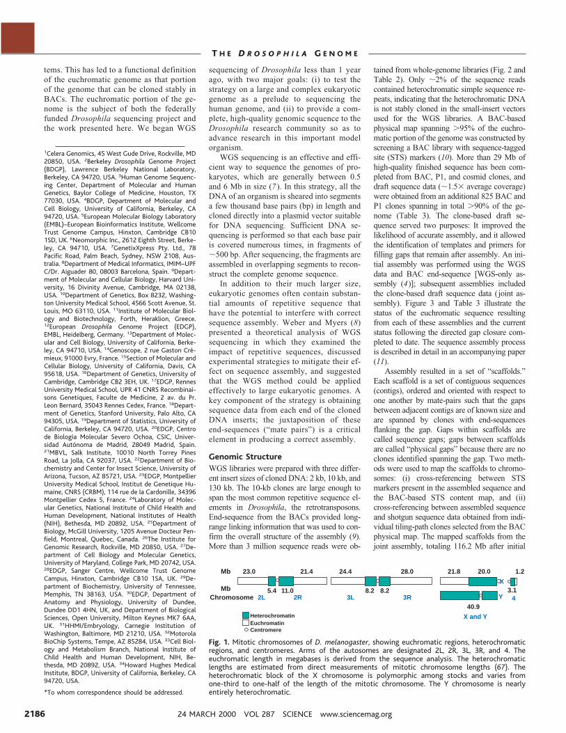
Mb 23.0 21.4 24.4 28.0 21.8 20.0 1.2
3.1
40.9
8.28.211.02R2L 3L 3R
X
Y 4
X and Y
5.4MbChromosome
HeterochromatinEuchromatinCentromere
Fig. 1. Mitotic chromosomes of D. melanogaster, showing euchromatic regions, heterochromaticregions, and centromeres. Arms of the autosomes are designated 2L, 2R, 3L, 3R, and 4. Theeuchromatic length in megabases is derived from the sequence analysis. The heterochromaticlengths are estimated from direct measurements of mitotic chromosome lengths (67). Theheterochromatic block of the X chromosome is polymorphic among stocks and varies fromone-third to one-half of the length of the mitotic chromosome. The Y chromosome is nearlyentirely heterochromatic.
24 MARCH 2000 VOL 287 SCIENCE www.sciencemag.org2186
T H E D R O S O P H I L A G E N O M E
gap closure, were deposited in GenBank (ac-cession numbers AE002566–AE003403) andform the basis for the analysis described in thisarticle.
The WGS-only assembly resulted in 50scaffolds spanning 114.8 Mb that could beplaced unambiguously onto chromosomessolely on the basis of their STS content (la-beled “D” in Fig. 3). The joint assemblyincluded clone-based sequence, but no spe-cific advantage was taken of the location infor-mation of each clone-based read by the whole-genome assembly algorithm. Nonetheless, theclone-based sequence from BACs in the phys-ical map allowed placement of an additional 84small scaffolds (1.4 Mb) on chromosome armsin the joint assembly (labeled “C” in Fig. 3). Asshown in Fig. 3, a few large scaffolds in eachassembly span a large portion of each chromo-some arm, with a number of additional smallerscaffolds located at the centromeric end, excepton the right arm of chromosome 3. Nearly all ofthe scaffolds added to chromosomes in the jointassembly, relative to the WGS-only assembly,are adjacent to the centric heterochromatin,which demonstrates the utility of the physicalmap in these regions. The density of transpos-able elements (labeled “A” in Fig. 3) increasesmarkedly in the transition zone betweeneuchromatin and heterochromatin, as dis-cussed below. An additional 704 scaffoldsin the joint assembly, equivalent to 3.8 Mb,could not be placed with accuracy on thegenome. Most of these do not match clone-based sequence from the physical map, andtherefore they most likely represent smallislands of unique sequence embedded with-in regions of heterochromatin. Because of theinstability of the surrounding genomic regions,these sequences would not have been obtainedthrough a sequencing approach that was depen-
dent on cloning in large-insert vectors.Among the 134 mapped scaffolds, there
were 1636 contigs after assembly (hence 1630gaps, considering that there are six linear chro-mosome arm segments to be assembled). Onthe major autosomes, there are five physicalgaps in the BAC map, three of which are neara centromere or telomere (10). Because theWGS approach did not span these gaps, theylikely contain unclonable regions. Most gaps onthe autosomes—including gaps between scaf-folds—were therefore cloned in either WGSclones or BAC subclones used for clone-baseddraft sequencing and are considered sequencegaps. Directed gap closure was done throughuse of several resources, including whole BACclones, plasmid subclones, and M13 subclones
from the Lawrence Berkeley National Labora-tory (LBNL) and Baylor College of Medicinecenters’ draft sequence of BAC and P1 clones;10-kb subclones from the whole-genome librar-ies; and polymerase chain reaction (PCR) fromgenomic DNA (12). The average size of thegaps filled to date is 771 bp (their predicted sizewas 757 bp); the predicted size of the remaininggaps is 2120 bp. Table 3 provides details of thestatus of each chromosome arm as of 3 March2000.
The accuracy of the assembly was measuredin several ways, as described (11). In summary,the scaffold sequences agree very well with theBAC-based STS content map and with high-quality finished sequence. In the 7 Mb of thegenome where very high-quality sequence was
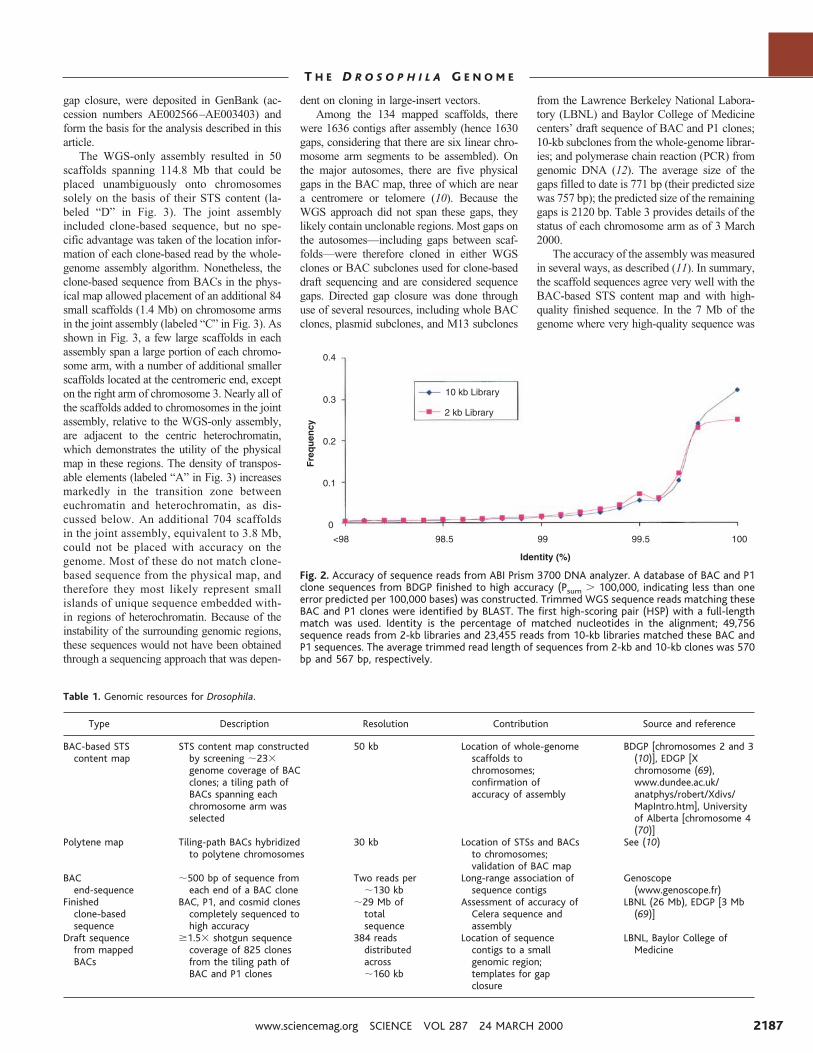
Fig. 2. Accuracy of sequence reads from ABI Prism 3700 DNA analyzer. A database of BAC and P1clone sequences from BDGP finished to high accuracy (Psum . 100,000, indicating less than oneerror predicted per 100,000 bases) was constructed. Trimmed WGS sequence reads matching theseBAC and P1 clones were identified by BLAST. The first high-scoring pair (HSP) with a full-lengthmatch was used. Identity is the percentage of matched nucleotides in the alignment; 49,756sequence reads from 2-kb libraries and 23,455 reads from 10-kb libraries matched these BAC andP1 sequences. The average trimmed read length of sequences from 2-kb and 10-kb clones was 570bp and 567 bp, respectively.
Table 1. Genomic resources for Drosophila.
Type Description Resolution Contribution Source and reference
BAC-based STScontent map
STS content map constructedby screening ;233genome coverage of BACclones; a tiling path ofBACs spanning eachchromosome arm wasselected
50 kb Location of whole-genomescaffolds tochromosomes;confirmation ofaccuracy of assembly
BDGP [chromosomes 2 and 3(10)], EDGP [Xchromosome (69),www.dundee.ac.uk/anatphys/robert/Xdivs/MapIntro.htm], Universityof Alberta [chromosome 4(70)]
Polytene map Tiling-path BACs hybridizedto polytene chromosomes
30 kb Location of STSs and BACsto chromosomes;validation of BAC map
See (10)
BACend-sequence
;500 bp of sequence fromeach end of a BAC clone
Two reads per;130 kb
Long-range association ofsequence contigs
Genoscope(www.genoscope.fr)
Finishedclone-basedsequence
BAC, P1, and cosmid clonescompletely sequenced tohigh accuracy
;29 Mb oftotalsequence
Assessment of accuracy ofCelera sequence andassembly
LBNL (26 Mb), EDGP [3 Mb(69)]
Draft sequencefrom mappedBACs
$1.53 shotgun sequencecoverage of 825 clonesfrom the tiling path ofBAC and P1 clones
384 readsdistributedacross;160 kb
Location of sequencecontigs to a smallgenomic region;templates for gapclosure
LBNL, Baylor College ofMedicine
www.sciencemag.org SCIENCE VOL 287 24 MARCH 2000 2187
T H E D R O S O P H I L A G E N O M E
available for comparison, the accuracy of theassembled sequence was 99.99% in nonrepeti-tive regions. In the ;2.5% of the region com-prising the most highly repetitive sequences, theaccuracy was 99.5%.
Heterochromatin-euchromatin transi-tion zone. The genomes of eukaryotes gen-erally contain heterochromatic regions sur-rounding the centromeres that are intractableto all current sequencing methods. In Dro-sophila, ;60 Mb of the 180-Mb genomeconsists of centric heterochromatin, which iscomposed primarily of simple sequence sat-ellites, transposons, and two large blocks ofribosomal RNA genes (13). We examined thesequence organization at boundaries betweeneuchromatin and centric heterochromatin intwo regions, one in division 20 on the Xchromosome and the other in division 40 on theleft arm of chromosome 2. On the X chromo-some, gene density in division 20 drops abrupt-ly—to two genes in 400 kb around folded gas-trulation—and then rises to 11 genes in 130 kb.Next, at least 10 Mb of largely satellite DNAsequences and the ribosomal RNA gene clusterare located just distal to the centromere itself.On the left arm of chromosome 2, a similarsituation exists: There is a normal gene densityin division 39, followed by only two genes in350 kb near teashirt in division 40, then by a
200-kb region containing 10 genes. These tran-sition zones between euchromatin and hetero-chromatin contain many previously unknowngenes, including counterparts to human cyclinK and mouse Krox-4. None of the 11 genesproximal to teashirt and only one of the 10genes proximal to folded gastrulation wasknown previously.
What is the nature of the sequence in thegene-poor regions? The most common se-quences by far were transposons, consistentwith previous small-scale analyses (14).These include several new elements similarto transposons in other species, as well as the;50 transposon classes previously character-ized in Drosophila. Some short runs of satel-lite sequences are present, but it has not beendetermined whether they might have beentruncated during cloning. In addition, at least110 other simple repeat classes were identi-fied, some of which are distributed widelyoutside of heterochromatin.
Criteria for describing the completionstatus of a eukaryotic genome. Because ofthe unclonable repetitive DNA surrounding thecentromeres, it is highly unlikely that thegenomic sequence of chromosomes from eu-karyotes such as Drosophila or human will everbe “complete.” It is therefore necessary to pro-vide an assessment of the contiguity and accu-
racy of the sequence. Table 4 lists several ob-jective parameters by which the status can bejudged and by which improvements in futurereleases can be measured. We have termed theversion of the sequence associated with thispublication “Release 1” and intend to makeregular future releases as gaps are filled andoverall sequence accuracy is increased.
One measure of the completeness of theassembled sequence is the extent to whichpreviously described genes can be found. Ananalysis of the 2783 Drosophila genes withsome sequence information that have beencompiled by FlyBase (15) resulted in identi-fication of 2778 on the scaffold sequence. Allof the remainder are found in unscaffoldedsequence. The remaining six were all clonedby degenerate PCR, and it is possible thatsome or all of these genes are incorrectlyascribed to Drosophila (16). Of the base pairsrepresented in the 2778 genes, 97.5% arepresent in the assembled sequence.
AnnotationThe initial annotation of the assembled genomeconcentrated on two tasks: prediction of tran-script and protein sequence, and prediction offunction for each predicted protein. Computa-tional approaches can aid each task, but biolo-gists with expertise in particular fields are re-quired for the results to have the most consis-tency, reliability, and utility. Because thebreadth of expertise necessary to annotate acomplete genome does not exist in any singleindividual or organization, we hosted an “An-notation Jamboree” involving more than 40scientists from around the world, primarilyfrom the Drosophila research community. Eachwas responsible for organizing and interpretingthe gene set for a given protein family or bio-logical process. Over a 2-week period, jambo
Table 2. Source of data for assembly: Whole-genome shotgun sequencing. See (65) for more informationabout library construction and sequencing.
VectorInsert size
(kbp)Paired
sequencesTotal
sequencesClone
coverageSequencecoverage
High-copy plasmid 2 732,380 1,903,468 11.23 7.33Low-copy plasmid 10 548,974 1,278,386 42.23 5.43BAC 130 9,869 19,738 11.43 0.073Total 1,290,823 3,201,592 64.83 12.83
Table 3. BAC and P1 clone-based sequencing. EDGP, European Drosophila Genome Project; BCM, Baylor College of Medicine; LBNL, Lawrence Berkeley NationalLaboratory (BCM and LBNL are the genomic sequencing centers of the BDGP).
Clone-based genomic sequencing Gap closure: current status
Chromosomalregion
Group SizeFinished
sequence(Mb)
Draft sequence in jointassembly [BACs, (P1s)]†
Totalsequenced
BACs(P1s) in
jointassembly
Additionalsequenced
BACs intiling path
Percentage of DNA sequencein contigs greater than
ClonesAveragecoverage
30 kb 100 kb 1 Mb
X (1–3) EDGP 3 2.5 0 0 0 79.4 32.7 0X (4–11) BCM 8.8 0.1* 0 1 72X (12–20) LBNL 10 0 71 2.33 71 102L LBNL 23 14.0 103 (8) 1.63 (5.33) 119 (202) 2 97.8 91.4 16.92R LBNL 21.4 8.8 159 (32) 1.33 (4.73) 157 (186) 0 96.4 90.6 32.83L BCM
24.40.1 166 1.33 170 50 95.1 77.7 0
3L LBNL 2.1 22 (7) 1.73 (2.53) 20 (32) 03R LBNL 28 2.1 259 (9) 1.23 (23) 264 (27) 0 98.5 92.6 3.64 LBNL 1.2 0 16 1.43 15 1 85.6 43.5 0Total 120 29.7 796 (56) 817 (447) 135 93.7 77.5 9.9
*Sequenced at LBNL. †A tiling path of clones spanning 97% of the euchromatic portion of the genome was selected from the genome physical maps (10) for clone-basedsequencing. The data include sequence that has been generated since the beginning of the publicly funded (BDGP and EDGP) genome sequencing projects. Tiling path clone identitieswere verified by screening the shotgun sequence for expected STS and BAC end-sequences, sequenced genes with known map locations from genes (and regions flanking P insertions),and sequences of neighboring tiling path clones. The average size of BAC clones in the tiling path is 163 kb. Sequencing methods are described in (66).
24 MARCH 2000 VOL 287 SCIENCE www.sciencemag.org2188
T H E D R O S O P H I L A G E N O M E
Mb0 5 10 15 20
0 5 10 15 20Mb
Mb0 5 10 15 20 25
Mb
A
B
C
D
E
F
Mb0 5 10 15 20
A
B
C
D
E
F
A
B
C
D
E
F
A
B
C
D
E
F
A
B
C
D
E
F
0 5 10 15 20
X
2L
2R
3L
3R
81 82 83 84 85 86 87 88 89 90 91 92 93 94 95 96 97 98 99 100
61 62 63 64 65 66 67 68 69 70 71 72 73 74 75 76 77 78 79 80
41 42 43 44 45 46 47 48 49 50 51 52 53 54 55 56 57 58 59 60
21 22 23 24 25 26 27 28 29 30 31 32 33 34 35 36 37 38 39 40
1 2 3 4 5 6 7 8 9 10 11 12 13 14 15 16 17 18 19 20
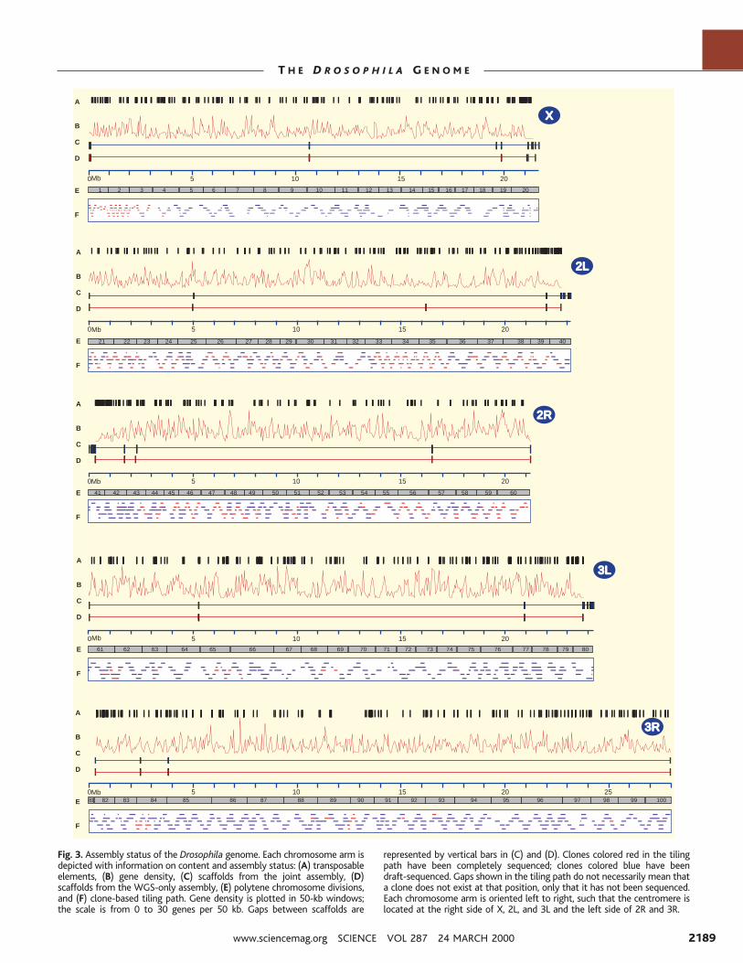
Fig. 3. Assembly status of the Drosophila genome. Each chromosome arm isdepicted with information on content and assembly status: (A) transposableelements, (B) gene density, (C) scaffolds from the joint assembly, (D)scaffolds from the WGS-only assembly, (E) polytene chromosome divisions,and (F) clone-based tiling path. Gene density is plotted in 50-kb windows;the scale is from 0 to 30 genes per 50 kb. Gaps between scaffolds are
represented by vertical bars in (C) and (D). Clones colored red in the tilingpath have been completely sequenced; clones colored blue have beendraft-sequenced. Gaps shown in the tiling path do not necessarily mean thata clone does not exist at that position, only that it has not been sequenced.Each chromosome arm is oriented left to right, such that the centromere islocated at the right side of X, 2L, and 3L and the left side of 2R and 3R.
www.sciencemag.org SCIENCE VOL 287 24 MARCH 2000 2189
T H E D R O S O P H I L A G E N O M E
ree participants worked to define genes, toclassify them according to predicted function,and to begin synthesizing information from agenome-wide perspective.
For definition of gene structure, we relied onthe use of different gene-finding approaches:the gene-finding programs Genscan (17) and aversion of Genie that uses expressed sequencetag (EST) data (18), plus the results of comple-mentary DNA (cDNA) and protein databasesearches, followed by review by human anno-tators (19). Genscan predicted 17,464 genes,and Genie predicted 13,189. We believe thatthe lower estimate is more accurate, because ina test that used the extensively studied andannotated 2.9-Mb Adh region (3), the Geniepredictions were closer to the number of exper-imentally determined genes; Genscan predictedfar too many (20). This is likely because Geniewas optimized for Drosophila, whereas Gen-scan parameters suitable for Drosophila gene-finding are not available.
Results of the computational analyses werepresented to annotators by means of a customvisualization tool that allowed annotators to de-fine transcripts on the basis of EST (21) andprotein sequence similarity information, Geniepredictions, and Genscan predictions, in de-creasing order of confidence. The present anno-tation of the Drosophila genome predicts 13,601genes, encoding 14,113 transcripts through al-ternative splicing in some genes. The number ofalternative splice forms that can be annotated islimited by the available cDNA data and is asubstantial underestimate of the total number ofalternatively spliced genes. More than 10,000genes with database matches were reviewedmanually. The remaining ;3000 genes werepredicted by Genie but have no database match-es that can be used to refine intron-exon bound-aries. Genes predicted by Genscan that did notoverlap Genie predictions or database matcheswere not included in the set of predicted pro-teins. Table 5 summarizes the evidence for thesegenes: 38% of the Genie predictions are sup-ported by evidence from both EST and proteinmatches, 27% by ESTs alone, and 12% byprotein matches alone. Altogether there are ESTmatches for 65% of the genes, but nearly half ofthe total ESTs match only 5% of the genes; 23%of the predicted proteins do not match sequences
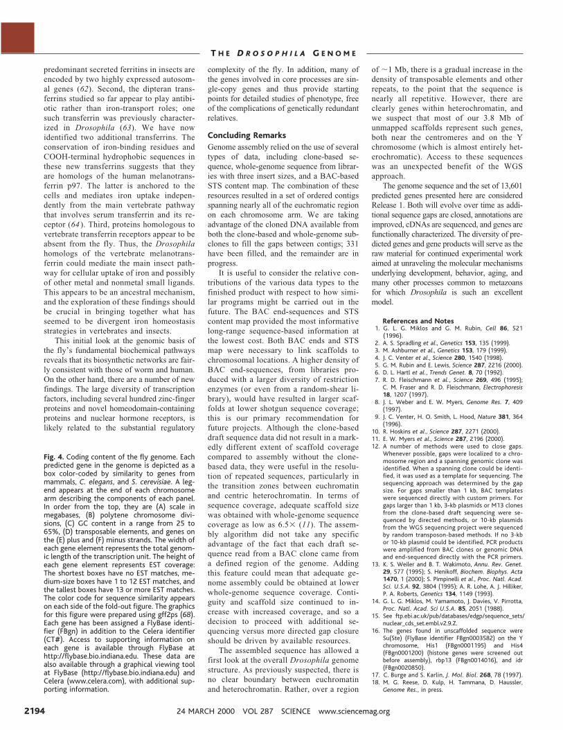
from other organisms or Drosophila ESTs. Thisset of annotations is considered provisional andwill improve as additional full-length cDNAsequence and functional information becomesavailable for each gene. Figure 4 provides agraphical overview of the gene content of thefly.
Genes were classified according to a func-tional classification scheme called Gene Ontol-ogy (GO). The GO project (22) is a collabora-tion among FlyBase, the Saccharomyces Ge-nome Database (23), and Mouse Genome In-formatics (24). It consists of a set of controlledvocabularies providing a consistent descriptionof gene products in terms of their molecularfunction, biological role, and cellular location.At the time of our annotation, proteins encodedby 1539 Drosophila genes had already beenannotated by FlyBase using ;1200 differentGO classifications. In addition, a set of 718proteins from S. cerevisiae and 1724 proteinsfrom mouse had been annotated and placed intoGO categories. Predicted Drosophila genes andgene products were used as queries against adatabase made up of the sequences of thesethree sets of proteins (by BLASTX orBLASTP) (25) and grouped on the basis of theGO classification of the proteins matched.About 7400 transcripts have been assigned to39 major functional categories, and about 4500have been assigned to 47 major process cate-gories (Table 6).
The largest predicted protein is Kakapo, acytoskeletal linker protein required for adhesionbetween and within cell layers, with 5201 aminoacids; the smallest is the 21–amino acid ribo-somal protein L38. There are 56,673 predictedexons, an average of four per gene, occupying24.1 Mb of the 120-Mb euchromatic sequencetotal. The size of the average predicted transcriptis 3058 bp. There was a systematic underpredic-tion of 59 and 39 untranslated sequence as aresult of less than complete EST coverage andthe inability of gene-prediction programs to pre-dict the noncoding regions of transcripts, so thenumber of exons and introns and the averagetranscription unit size are certain to be underes-timates. There are at least 41,000 introns, occu-pying 20 Mb of sequence. Intron sizes in Dro-sophila are heterogeneous, ranging from 40 bpto more than 70 kb, with a clear peak between
59 and 63 bp (26). The average number ofexons is four, although this is an underestimatebecause of a systematic underprediction of 59and 39 untranslated exons. We identified 292transfer RNA genes and 26 genes for spliceoso-mal small nuclear RNAs (snRNAs). We did notattempt to predict other noncoding RNAs.
The total number of protein-coding genes,13,601, is less than that predicted for the wormC. elegans (27) (18,425; WormPep 18, 11 Oc-tober 1999) and far less than the ;27,000 esti-mated for the plant Arabidopsis thaliana (28).The average gene density in Drosophila is onegene per 9 kb. There is substantial variation ingene density, ranging from 0 to nearly 30 genesper 50 kb, but the gene-rich regions are notclustered as they are in C. elegans. Regions ofhigh gene density correlate with G1C-rich se-quences. In the ;1 Mb adjacent to the centricheterochromatin, both G1C content and genedensity decrease, although there is not a markeddecrease in EST coverage as has been seen in A.thaliana (28).
Genomic ContentThe genomic sequence has shed light on someof the processes common to all cells, such asreplication, chromosome segregation, and ironmetabolism. There are also new findings aboutimportant classes of chromosomal proteins thatallow insights into gene regulation and the cellcycle. Overall, the correspondence of Drosoph-ila proteins involved in gene expression andmetabolism to their human counterparts reaf-firms that the fly represents a suitable experi-mental platform for the examination of humandisease networks involved in replication, repair,translation, and the metabolism of drugs andtoxins. In an accompanying manuscript (29),the protein complement of Drosophila is com-pared to those of the two eukaryotes with com-plete genome sequences, C. elegans and S.cerevisiae, and other developmental and cellbiological processes are discussed.
Replication. Genes encoding the basicDNA replication machinery are conservedamong eukaryotes (30); in particular, all of theproteins known to be involved in start siterecognition are encoded by single-copy genesin the fly. These include members of the six-subunit heteromeric origin recognition complex(ORC) (31), the MCM helicase complex (32),and the regulatory factors CDC6 and CDC45,which are thought to determine processing ofpre-initiation complexes. The fly ORC3 andORC6 proteins, for example, share close se-quence similarity with vertebrate proteins, butnot only are they highly divergent relative toyeast ORCs, they have no obvious counterpartsin the worm. It is striking that the ORC genesexist as single copies, given the orthologousfunctions for some of the subunits in otherprocesses (33). It had been considered possiblethat a large family of ORCs, each with a dif-ferent binding specificity, might account for
Table 4. Measures of completion. Analyses supporting many of these values are found in (11).
Number of scaffolds mapped to chromosome arms 134Number of scaffolds not mapped to chromosomes 704Number of base pairs in scaffolds mapped to chromosome arms 116.2 MbNumber of base pairs in scaffolds not mapped to chromosome arms 3.8 MbLargest unmapped scaffold 64 kbPercentage of total base pairs in mapped scaffolds .100 kb 98.2%Percentage of total base pairs in mapped scaffolds .1 Mb 95.5%Percentage of total base pairs in mapped scaffolds .10 Mb 68.0%Number of gaps remaining among mapped scaffolds 1299Base pair accuracy against LBNL BACs (nonrepetitive sequence) 99.99%Known genes accounted for in scaffold set 99.7%
24 MARCH 2000 VOL 287 SCIENCE www.sciencemag.org2190
T H E D R O S O P H I L A G E N O M E
different origin usage in development. Clearly,given the single-copy ORC genes, other as-yet-undiscovered cis-acting elements and trans-act-ing factors participate in developmentally reg-ulated processes such as switches in origin us-age, gene amplification, and specialized repli-cation of euchromatin in certain endocycles. Incontrast, the fly has two distinct homologs ofthe proliferating cell nuclear antigen (PCNA),the processivity factor for the DNA poly-merases (d and ε) involved in chain elongation.Human PCNA is blocked from interaction withthe replication enzymes by the checkpoint reg-ulator p21 in response to DNA damage (34);perhaps one of the fly PCNA proteins is im-mune to such regulation and is thus left activefor repair or replication.
Chromosomal proteins. Analysis of pro-tein families involved in chromosome inheri-tance reveals both expected findings and somesurprises. As expected, the fly has all fourmembers of the conserved SMC family in-volved in sister chromatid cohesion, condensa-tion, DNA repair, and dosage compensation(35). The fly also contains at least one orthologof each of the MAD/Bub metaphase-anaphasecheckpoint proteins that are conserved fromyeast to mammals. However, Drosophila doesnot appear to have orthologs to most of theproteins identified previously in mammals oryeast that are associated with centromericDNA, such as the CENP-C/MIF-2 family andthe yeast CBF3 complex (36). One exception isthe presence of a histone H3-like protein thatshares sequence similarity with mammalianCENP-A, a centromere-specific H3-like pro-tein. There are at least nine histone acetyltrans-ferases (HATs) and five histone deacetylases(HDACs), which are involved in regulatingchromatin structure (37); only three of eachhave been reported previously. There are also17 members of the SNF2 adenosine triphos-phatase (ATPase) family, which represent 9of the 10 known subfamilies. Many of theseATPases are involved in chromatin remodeling(38). The fly also contains at least 14 proteinswith chromodomains (39), six of which arenew, including two HP1-related proteins. Al-though many of these chromodomain-contain-ing proteins have orthologs in vertebrates, onlyone (CHD1) appears in yeast, flies, and verte-brates. There are also at least 13 bromodomain-containing proteins, seven of which are new;the bromodomain may interact with the acety-lated NH2-terminus of histones and is involvedin chromatin remodeling and gene silencing(40). Only three of these appear to have coun-terparts in yeast. Furthermore, Drosophila telo-meres lack the simple repeats that are charac-teristic of most eukaryotic telomeres (41), andthe known telomerase components of verte-brates, for example, are absent from flies. Thefly does, however, contain five proteins that areclose relatives of the yeast and human SIR2telomere silencing proteins.
DNA repair. The importance of DNA re-pair in maintaining genomic integrity is reflect-ed in the conservation of most proteins impli-cated in the major defined pathways of eukary-otic DNA repair. However, there are some no-table absences. For example, no convincinghomologs can be found for the genes encodingthe RAD7, RAD16, RAD26 (CSB/ERCC6),and RAD28 (CSA) proteins, which are impli-cated in strand-specific modes of repair in yeastand/or mammalian systems. In base excisionrepair processes, 3-methyladenine glycosylaseand uracil-DNA-glycosylase are absent, al-though the latter function is likely fulfilled bythe G/T mismatch-specific thymine DNA gly-cosylase (42). In the damage bypass pathway,sequences encoding homologs of DNA poly-merase z (yeast Rev3p/Drosophila mus205)and Rev1p are present, although a REV7 ho-molog is not found. As in humans and worms,two members of the RAD30 (polymerase h)gene family are present. In the mismatch repairsystem, only two proteins related to Escherich-ia coli mutS are predicted, rather than the usualfamily of five or more members. The previous-ly reported Msh2p homolog (43) is present, asis a sequence most closely resembling Msh6p.Budding yeast and humans possess additionalmembers of the mutS gene family that areproposed to function in partially redundantpathways of mismatch repair (MSH3) and inmeiotic recombination (MSH4 and MSH5),suggesting either that the Drosophila mutS ho-mologs have reduced specificity or that alterna-tive proteins are fulfilling these roles in the fly.In the recombinational repair pathway, two ad-ditional members of the recA/RAD51 genefamily are identified, bringing the total to four.However, no member of the RAD52/RAD59family is present. One additional member of therecQ/SGS1 helicase family was identified, inaddition to the two already noted (44); the newprotein is most similar to human RecQ4. Final-ly, with respect to nonhomologous end joining,Drosophila joins the list of invertebrate speciesthat lack an apparent DNA-PK catalytic sub-unit, although both Ku subunits and DNA li-gase 4 are present. We conclude that mostmajor components of the repair network in flieshave been uncovered. If more are present, either
they have diverged so far that they are unrec-ognizable by BLAST searches, or the systemshave become degenerate (that is, other networkcomponents are fulfilling the same roles).
Transcription. Gene regulation has tradi-tionally been singled out as one of the primarybases for the generation of evolutionary diver-sity. How has the core transcriptional machin-ery changed in different phyla? Drosophilacore RNA polymerase II and some generaltranscription factors (TFIIA-H, TFIIIA, andTFIIIB) are similar in composition to those ofboth mammals and yeast (45). In contrast, coreRNA polymerases I and III, TBP (TATA-bind-ing protein)–containing complexes for class I,class II, and snRNA genes (TBP-associatedfactors TAFI and TAFII, and SNAPC, respec-tively), TFIIIC, and SRB/mediator vary greatlyin composition in Drosophila and mammalsrelative to yeast (46). The RNA polymerase Itranscription factors of flies and mammals haveclear amino acid conservation; yeast RNApolymerase I factors do not appear to be relatedto them. For example, the mammalian promoterinteracting factors UBF and TIF-1A are presentin Drosophila but not in yeast, and yeast UAFsubunits are absent in Drosophila and apparent-ly absent in mammals. Furthermore, of thethree TAFIs in the human selectivity factor 1,the mouse transcriptional initiation factor IB,and the yeast core factor complexes, only thehuman/mouse TAFI63/TAFI68 subunit is con-served in the fly. Similarly, Drosophila encodesthree of the five mammalian SNAPC subunits(SNAP43, 50, and 190) for which no homologsexist in the yeast genome.
In addition to the family of previously de-scribed TBPs (47), the fly contains multipleforms of several ubiquitous TAFIIs (TAFII30b,TAFII60, and TAFII80) (46). This raises thepossibility that a variety of TFIID complexesevolved in metazoan organisms to regulategene expression patterns associated with devel-opment and cellular differentiation. The con-stellation of factors that interact with RNApolymerase II in Drosophila may also contrib-ute to this regulation, because Drosophila con-tains only a small subset of yeast SRB/mediatorsubunits (MED6, MED7, and SRB7) but a vastmajority of the molecularly characterized com-
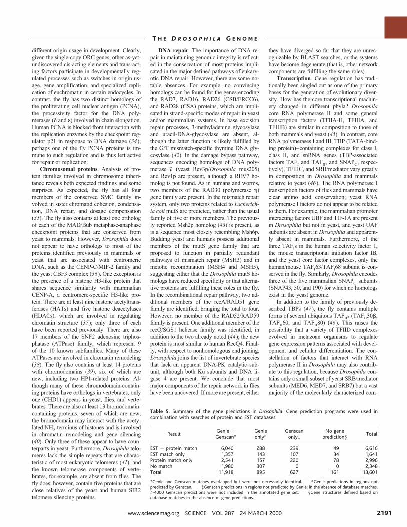
Table 5. Summary of the gene predictions in Drosophila. Gene prediction programs were used incombination with searches of protein and EST databases.
ResultGenie 1Genscan*
Genieonly†
Genscanonly‡
No geneprediction§
Total
EST 1 protein match 6,040 288 239 49 6,616EST match only 1,357 143 107 34 1,641Protein match only 2,541 157 220 78 2,996No match 1,980 307 0 0 2,348Total 11,918 895 627 161 13,601
*Genie and Genscan matches overlapped but were not necessarily identical. †Genie predictions in regions notpredicted by Genscan. ‡Genscan predictions in regions not predicted by Genie; in the absence of database matches,.4000 Genscan predictions were not included in the annotated gene set. §Gene structures defined based ondatabase matches in the absence of gene predictions.
www.sciencemag.org SCIENCE VOL 287 24 MARCH 2000 2191
T H E D R O S O P H I L A G E N O M E
ponents of mammalian coactivator complexessuch as ARC/DRIP/TRAP.
Gene regulation. On the basis of similar-ity to known proteins, Drosophila appears toencode about 700 transcription factors, abouthalf of which are zinc-finger proteins. Bycontrast, the worm has about 500 transcrip-tion factors, fewer than one-third of which arezinc-finger proteins (29). Two additionalclasses play key roles in regulation: thehomeodomain-containing and nuclear hor-mone receptor–type transcription factors.
Homeodomain-containing proteins con-trol a wide variety of developmental pro-cesses. Twenty-two new homeodomain-
containing proteins were uncovered in ouranalysis, bringing the total to more than100. Ten of these were members of thepaired-box PRX superclass (48), some withknown vertebrate homologs: short stature ho-meobox 2 (SHOX), cartilage homeoprotein 1(CART), and the two retina-specific proteins(VSX-1 and VSX-2) of goldfish. New mem-bers were also found in the LIM and TGIFclass. The two new LIM members contain ahomeobox and two copies of the LIM motif; thetwo new TGIF members occur as a local tan-dem duplication on the right arm of chromo-some 2. We also found single new members ofthe NK-2, muscle-specific homeobox, proline-
rich homeodomain (PRH), and BarH classes.The new fly gene encoding NK-2 is a cognateof the gene encoding the NKX-5.1 mouse pro-tein. The new fly gene encoding muscle-specif-ic homeobox is most similar to the gene encod-ing the MSX-1 mouse protein involved incraniofacial morphogenesis. The new fly geneencoding PRH is most similar to a mouse geneexpressed in myeloid cells. The remaining ho-meodomain-containing proteins are orphans:One has similarity to the human H6 proteininvolved in craniofacial development, and an-other to HB9, a protein required for normaldevelopment of the pancreas.
Nuclear hormone receptors (NRs) are
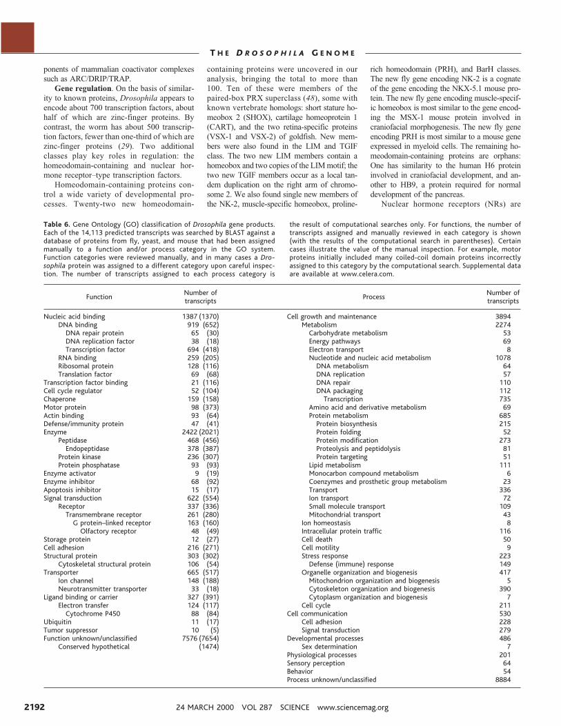
Table 6. Gene Ontology (GO) classification of Drosophila gene products.Each of the 14,113 predicted transcripts was searched by BLAST against adatabase of proteins from fly, yeast, and mouse that had been assignedmanually to a function and/or process category in the GO system.Function categories were reviewed manually, and in many cases a Dro-sophila protein was assigned to a different category upon careful inspec-tion. The number of transcripts assigned to each process category is
the result of computational searches only. For functions, the number oftranscripts assigned and manually reviewed in each category is shown(with the results of the computational search in parentheses). Certaincases illustrate the value of the manual inspection. For example, motorproteins initially included many coiled-coil domain proteins incorrectlyassigned to this category by the computational search. Supplemental dataare available at www.celera.com.
FunctionNumber oftranscripts
ProcessNumber oftranscripts
Nucleic acid binding 1387 (1370) Cell growth and maintenance 3894DNA binding 919 (652) Metabolism 2274
DNA repair protein 65 (30) Carbohydrate metabolism 53DNA replication factor 38 (18) Energy pathways 69Transcription factor 694 (418) Electron transport 8
RNA binding 259 (205) Nucleotide and nucleic acid metabolism 1078Ribosomal protein 128 (116) DNA metabolism 64Translation factor 69 (68) DNA replication 57
Transcription factor binding 21 (116) DNA repair 110Cell cycle regulator 52 (104) DNA packaging 112Chaperone 159 (158) Transcription 735Motor protein 98 (373) Amino acid and derivative metabolism 69Actin binding 93 (64) Protein metabolism 685Defense/immunity protein 47 (41) Protein biosynthesis 215Enzyme 2422 (2021) Protein folding 52
Peptidase 468 (456) Protein modification 273Endopeptidase 378 (387) Proteolysis and peptidolysis 81
Protein kinase 236 (307) Protein targeting 51Protein phosphatase 93 (93) Lipid metabolism 111
Enzyme activator 9 (19) Monocarbon compound metabolism 6Enzyme inhibitor 68 (92) Coenzymes and prosthetic group metabolism 23Apoptosis inhibitor 15 (17) Transport 336Signal transduction 622 (554) Ion transport 72
Receptor 337 (336) Small molecule transport 109Transmembrane receptor 261 (280) Mitochondrial transport 43
G protein–linked receptor 163 (160) Ion homeostasis 8Olfactory receptor 48 (49) Intracellular protein traffic 116
Storage protein 12 (27) Cell death 50Cell adhesion 216 (271) Cell motility 9Structural protein 303 (302) Stress response 223
Cytoskeletal structural protein 106 (54) Defense (immune) response 149Transporter 665 (517) Organelle organization and biogenesis 417
Ion channel 148 (188) Mitochondrion organization and biogenesis 5Neurotransmitter transporter 33 (18) Cytoskeleton organization and biogenesis 390
Ligand binding or carrier 327 (391) Cytoplasm organization and biogenesis 7Electron transfer 124 (117) Cell cycle 211
Cytochrome P450 88 (84) Cell communication 530Ubiquitin 11 (17) Cell adhesion 228Tumor suppressor 10 (5) Signal transduction 279Function unknown/unclassified 7576 (7654) Developmental processes 486
Conserved hypothetical (1474) Sex determination 7Physiological processes 201Sensory perception 64Behavior 54Process unknown/unclassified 8884
24 MARCH 2000 VOL 287 SCIENCE www.sciencemag.org2192
T H E D R O S O P H I L A G E N O M E
sequence-specific, ligand-dependent tran-scription factors that contribute to physio-logical homeostasis by functioning as bothtranscriptional activators and repressors.Examination of the fly genome revealedonly four additional NR members, bringingthe total to 20. In contrast, the NR familyrepresents the most abundant class of tran-scriptional regulators in the worm: Morethan 200 member genes have been de-scribed. One of the newly identified flyNRs possesses a new P-box element (Cys-Asp-Glu-Cys-Ser-Cys-Phe-Phe-Arg-Arg),which confers DNA binding specificity,bringing to 76 the number of P-boxesidentified to date in all species. A search ofthe Drosophila genome failed to identifyany homologs to the mammalian p160 genefamily of NR coactivator proteins. SMRTER,despite weak similarity to the mammaliancorepressors SMRT and N-CoR, appears tobe the only close relative in Drosophila.
Translation and RNA processing. Al-though the structure of the ribosome has beenwell worked out, it has become apparent thatmany ribosomal proteins are multifunctionaland are involved in processes as disparate asDNA repair and iron-binding (49). There hasbeen an enormous genetic investigation of theconsequences of changes in expression levelof Drosophila ribosomal proteins (the Minutephenotype) (50); the identification and map-ping of the complete set presented here willprovide the basis for in-depth dissections oftheir functions and disease roles.
Most genes encoding general translationfactors are present in only one copy in theDrosophila genome, as they are in other ge-nomes studied to date; however, we discov-ered six genes encoding proteins highly sim-ilar to the messenger RNA (mRNA) cap-binding protein eIF4E. These may add com-plexity to regulation of cap-dependenttranslation, which is central to cellulargrowth control. Caenorhabditis elegans hasthree eIF4E isoforms, which were hypothe-sized to be necessary because trans-splicedmRNAs possess a different cap structure thando other mRNAs (51); however, Drosophiladoes not have trans-spliced mRNAs. The ac-tivity of eIF4E is regulated by an inhibitorprotein, 4E-BP. The Drosophila genome con-tains only a single gene encoding 4E-BP; incontrast, mammals have at least three 4E-BPisoforms but perhaps fewer eIF4E isoformsthan do flies. Of the more than 200 RNA-binding proteins identified, the most frequentstructural classes are RRM proteins (114),DEAD- or DExH-box helicases (58), andKH-domain proteins (31). This distribution issimilar to that observed in the C. elegansgenome. These structural motifs are some-times found in proteins for which experimen-tal evidence indicates a function in DNA,rather than RNA, binding. Overall, the trans-
lational machinery appears well conservedthroughout the eukaryotes.
The process of nonsense-mediated decay(52), the accelerated decay of mRNAs thatcannot be translated throughout their entirelength, has been genetically characterized inyeast and C. elegans but not in Drosophila. Wefound homologs of UPF1/SMG-2, SMG-1, andSMG-7 in the Drosophila genome, indicatingthat this process is conserved in flies.
Of particular interest are genes for compo-nents of the minor, or U12, spliceosome (53).Such introns are known in mammals, Drosoph-ila, and Arabidopsis, but not C. elegans. Usingconservative criteria (including a perfect matchto the U12 consensus 59 splice site for nucleo-tides 2 to 7, TATCCT), we found one intronthat appears to be of the U12 type per 1000genes. As expected, the minor spliceosomesnRNAs U12, U4atac, and U6atac are presentin the Drosophila genome. However, neitherU11 nor the U11-associated 35-kD protein (54)could be identified in the sequence. It is possi-ble that these components of the minor spliceo-some are less well conserved, or that the minorspliceosome in Drosophila does not containthem.
Cytochrome P450. The cytochrome P450monooxygenases (CYPs) are a large and an-cient superfamily of proteins that carry outmultiple reactions to enable organisms to ridthemselves of foreign compounds. HumanCYP2D6, for example, influences the metab-olism of beta blockers, antidepressants, anti-psychotics, and codeine, and insect CYPsfunction in the synthesis or degradation ofhormones and pheromones and in the metab-olism of natural and synthetic toxins, includ-ing insecticides (55). We found 90 P450 flygenes, of which four are pseudogenes, a fig-ure that is comparable to the 80 CYPs of C.elegans. These 90 genes, some of which areclustered, are divided among 25 families, fiveof which are found in Lepidoptera, Co-leoptera, Hymenoptera, Orthoptera, andIsoptera. However, more than half of the 90genes belong to only two families, CYP4 andCYP6, the former family shared with verte-brates. CYP51, used in making cholesterol inanimals and related molecules in plants andfungi, is absent from both the fly and wormgenomes; it is well known that the fly mustobtain cholesterol from its diet. A compre-hensive collection of phylogenetically di-verse CYP sequences is available (56).
Solute transport. Solute transporterscontribute to the most basic properties ofliving systems, such as establishment of cellpotential or generation of ATP; in highereukaryotes, these proteins help mediate ad-vanced functions such as behavior, learning,and memory. Hydropathy analyses predictthat 20% of the gene products in Drosophilareside in cellular membranes, having four ormore hydrophobic a helices (57). A consid-
erable fraction of these proteins (657, or 4%)are dedicated to ion and metabolite move-ment. More than 80% of the annotated trans-porters are new to Drosophila and were iden-tified by similarity to proteins characterizedin other eukaryotes. The largest families aresugar permeases, mitochondrial carrier pro-teins, and the ATP-binding cassette (ABC)transporters, with 97, 38, and 48 genes, re-spectively; these families are also the mostcommon in yeast and C. elegans (29). Also ofnote are three families of anion transportersthat mediate flux of sulfate, inorganic phos-phate, and iodide. Na1-anion transporters,with 17 members, are particularly abundantrelative to worm and yeast. Although individ-ual members of these families have beeninvestigated—for example, the mitochondrialcarrier protein COLT required for gas-fillingof the tracheal system (58) and the ABCtransporters associated with eye pigment dis-tribution (59)—the variety and number oftransporters within each family are impres-sive. These data lay the foundation for under-standing global transport processes critical toDrosophila physiology and development.
Metabolic processes. The biosyntheticnetworks of the fly are remarkably completecompared to those of many different pro-karyotes and to yeast, in which key enzymesof various pathways may be missing (60). Asin vertebrates, many fly enzymes are encodedby multiple genes. Two families are notewor-thy because of their size. The triacylglycerollipases are encoded by 31 genes and meritconsideration in investigations of lipolysisand energy storage and redistribution. In ad-dition, there are 32 genes encoding uridinediphosphate (UDP) glycosyltransferases,which participate in the production of sterolglycosides and in the biodegradation of hy-drophobic compounds. Several UDP glyco-syltransferase genes are highly expressed inthe antennae and may have roles in olfaction.In vertebrates, these enzymes are critical todrug clearance and detoxification (61). A ma-jor challenge will be to determine whether thenumber of these proteins present in the ge-nome is correlated with the importance andcomplexity of the regulatory events involvedin any given enzymatic reaction.
Iron (Fe) is both essential for and toxicto for all living things, and metazoan ani-mals use similar strategies for obtaining,transporting, storing, and excreting iron.Three findings from the analysis of thegenome shed light on the underlying com-mon mechanisms that have escaped atten-tion in the past. First, a third ferritin genehas been found that probably encodes asubunit belonging to a cytosolic ferritin, thepredominant type in vertebrates. This find-ing indicates that intracellular iron storagemechanisms in flies might be very similarto those in vertebrates. Subunits of the
www.sciencemag.org SCIENCE VOL 287 24 MARCH 2000 2193
T H E D R O S O P H I L A G E N O M E
predominant secreted ferritins in insects areencoded by two highly expressed autosom-al genes (62). Second, the dipteran trans-ferrins studied so far appear to play antibi-otic rather than iron-transport roles; onesuch transferrin was previously character-ized in Drosophila (63). We have nowidentified two additional transferrins. Theconservation of iron-binding residues andCOOH-terminal hydrophobic sequences inthese new transferrins suggests that theyare homologs of the human melanotrans-ferrin p97. The latter is anchored to thecells and mediates iron uptake indepen-dently from the main vertebrate pathwaythat involves serum transferrin and its re-ceptor (64 ). Third, proteins homologous tovertebrate transferrin receptors appear to beabsent from the fly. Thus, the Drosophilahomologs of the vertebrate melanotrans-ferrin could mediate the main insect path-way for cellular uptake of iron and possiblyof other metal and nonmetal small ligands.This appears to be an ancestral mechanism,and the exploration of these findings shouldbe crucial in bringing together what hasseemed to be divergent iron homeostasisstrategies in vertebrates and insects.
This initial look at the genomic basis ofthe fly’s fundamental biochemical pathwaysreveals that its biosynthetic networks are fair-ly consistent with those of worm and human.On the other hand, there are a number of newfindings. The large diversity of transcriptionfactors, including several hundred zinc-fingerproteins and novel homeodomain-containingproteins and nuclear hormone receptors, islikely related to the substantial regulatory
complexity of the fly. In addition, many ofthe genes involved in core processes are sin-gle-copy genes and thus provide startingpoints for detailed studies of phenotype, freeof the complications of genetically redundantrelatives.
Concluding RemarksGenome assembly relied on the use of severaltypes of data, including clone-based se-quence, whole-genome sequence from librar-ies with three insert sizes, and a BAC-basedSTS content map. The combination of theseresources resulted in a set of ordered contigsspanning nearly all of the euchromatic regionon each chromosome arm. We are takingadvantage of the cloned DNA available fromboth the clone-based and whole-genome sub-clones to fill the gaps between contigs; 331have been filled, and the remainder are inprogress.
It is useful to consider the relative con-tributions of the various data types to thefinished product with respect to how simi-lar programs might be carried out in thefuture. The BAC end-sequences and STScontent map provided the most informativelong-range sequence-based information atthe lowest cost. Both BAC ends and STSmap were necessary to link scaffolds tochromosomal locations. A higher density ofBAC end-sequences, from libraries pro-duced with a larger diversity of restrictionenzymes (or even from a random-shear li-brary), would have resulted in larger scaf-folds at lower shotgun sequence coverage;this is our primary recommendation forfuture projects. Although the clone-baseddraft sequence data did not result in a mark-edly different extent of scaffold coveragecompared to assembly without the clone-based data, they were useful in the resolu-tion of repeated sequences, particularly inthe transition zones between euchromatinand centric heterochromatin. In terms ofsequence coverage, adequate scaffold sizewas obtained with whole-genome sequencecoverage as low as 6.53 (11). The assem-bly algorithm did not take any specificadvantage of the fact that each draft se-quence read from a BAC clone came froma defined region of the genome. Addingthis feature could mean that adequate ge-nome assembly could be obtained at lowerwhole-genome sequence coverage. Conti-guity and scaffold size continued to in-crease with increased coverage, and so adecision to proceed with additional se-quencing versus more directed gap closureshould be driven by available resources.
The assembled sequence has allowed afirst look at the overall Drosophila genomestructure. As previously suspected, there isno clear boundary between euchromatinand heterochromatin. Rather, over a region
of ;1 Mb, there is a gradual increase in thedensity of transposable elements and otherrepeats, to the point that the sequence isnearly all repetitive. However, there areclearly genes within heterochromatin, andwe suspect that most of our 3.8 Mb ofunmapped scaffolds represent such genes,both near the centromeres and on the Ychromosome (which is almost entirely het-erochromatic). Access to these sequenceswas an unexpected benefit of the WGSapproach.
The genome sequence and the set of 13,601predicted genes presented here are consideredRelease 1. Both will evolve over time as addi-tional sequence gaps are closed, annotations areimproved, cDNAs are sequenced, and genes arefunctionally characterized. The diversity of pre-dicted genes and gene products will serve as theraw material for continued experimental workaimed at unraveling the molecular mechanismsunderlying development, behavior, aging, andmany other processes common to metazoansfor which Drosophila is such an excellentmodel.
References and Notes1. G. L. G. Miklos and G. M. Rubin, Cell 86, 521
(1996).2. A. S. Spradling et al., Genetics 153, 135 (1999).3. M. Ashburner et al., Genetics 153, 179 (1999).4. J. C. Venter et al., Science 280, 1540 (1998).5. G. M. Rubin and E. Lewis, Science 287, 2216 (2000).6. D. L. Hartl et al., Trends Genet. 8, 70 (1992).7. R. D. Fleischmann et al., Science 269, 496 (1995);
C. M. Fraser and R. D. Fleischmann, Electrophoresis18, 1207 (1997).
8. J. L. Weber and E. W. Myers, Genome Res. 7, 409(1997).
9. J. C. Venter, H. O. Smith, L. Hood, Nature 381, 364(1996).
10. R. Hoskins et al., Science 287, 2271 (2000).11. E. W. Myers et al., Science 287, 2196 (2000).12. A number of methods were used to close gaps.
Whenever possible, gaps were localized to a chro-mosome region and a spanning genomic clone wasidentified. When a spanning clone could be identi-fied, it was used as a template for sequencing. Thesequencing approach was determined by the gapsize. For gaps smaller than 1 kb, BAC templateswere sequenced directly with custom primers. Forgaps larger than 1 kb, 3-kb plasmids or M13 clonesfrom the clone-based draft sequencing were se-quenced by directed methods, or 10-kb plasmidsfrom the WGS sequencing project were sequencedby random transposon-based methods. If no 3-kbor 10-kb plasmid could be identified, PCR productswere amplified from BAC clones or genomic DNAand end-sequenced directly with the PCR primers.
13. K. S. Weiler and B. T. Wakimoto, Annu. Rev. Genet.29, 577 (1995); S. Henikoff, Biochem. Biophys. Acta1470, 1 (2000); S. Pimpinelli et al., Proc. Natl. Acad.Sci. U.S.A. 92, 3804 (1995); A. R. Lohe, A. J. Hilliker,P. A. Roberts, Genetics 134, 1149 (1993).
14. G. L. G. Miklos, M. Yamamoto, J. Davies, V. Pirrotta,Proc. Natl. Acad. Sci U.S.A. 85, 2051 (1988).
15. See ftp.ebi.ac.uk/pub/databases/edgp/sequence_sets/nuclear_cds_set.embl.v2.9.Z.
16. The genes found in unscaffolded sequence wereSu(Ste) (FlyBase identifier FBgn0003582) on the Ychromosome, His1 (FBgn0001195) and His4(FBgn0001200) (histone genes were screened outbefore assembly), rbp13 (FBgn0014016), and idr(FBgn0020850).
17. C. Burge and S. Karlin, J. Mol. Biol. 268, 78 (1997).18. M. G. Reese, D. Kulp, H. Tammana, D. Haussler,
Genome Res., in press.
Fig. 4. Coding content of the fly genome. Eachpredicted gene in the genome is depicted as abox color-coded by similarity to genes frommammals, C. elegans, and S. cerevisiae. A leg-end appears at the end of each chromosomearm describing the components of each panel.In order from the top, they are (A) scale inmegabases, (B) polytene chromosome divi-sions, (C) GC content in a range from 25 to65%, (D) transposable elements, and genes onthe (E) plus and (F) minus strands. The width ofeach gene element represents the total genom-ic length of the transcription unit. The height ofeach gene element represents EST coverage:The shortest boxes have no EST matches, me-dium-size boxes have 1 to 12 EST matches, andthe tallest boxes have 13 or more EST matches.The color code for sequence similarity appearson each side of the fold-out figure. The graphicsfor this figure were prepared using gff2ps (68).Each gene has been assigned a FlyBase identi-fier (FBgn) in addition to the Celera identifier(CT#). Access to supporting information oneach gene is available through FlyBase athttp://flybase.bio.indiana.edu. These data arealso available through a graphical viewing toolat FlyBase (http://flybase.bio.indiana.edu) andCelera (www.celera.com), with additional sup-porting information.
24 MARCH 2000 VOL 287 SCIENCE www.sciencemag.org2194
T H E D R O S O P H I L A G E N O M E
19. Sequence contigs were searched against publiclyavailable sequence at the DNA level and as six-frame translations against public protein sequencedata. DNA searches were against the invertebrate(INV) division of GenBank, a set of 80,000 ESTsequences produced at BDGP assembled to pro-duce consensus sequences (21), and a set of cu-rated Drosophila protein-coding genes prepared bythree of the authors (M. Ashburner, L. Bayraktaro-glu, and P. V. Benos) (15 ). Protein searches wereperformed against this set of curated protein se-quences and against the nonredundant protein da-tabase available at the National Center for Bio-technology Information. Initial searches were per-formed with a version of BLAST2 (25), optimizedfor the Compaq Alpha architecture. Additional pro-cessing of each query-subject pair was performedto improve the alignments. All BLAST results hav-ing an expectation score of ,1 3 1024 were thenprocessed on the basis of their high-scoring pair(HSP) coordinates on the contig to remove redun-dant hits, retaining hits that supported possiblealternative splicing. This procedure was performedseparately by hits to particular organisms so as notto exclude HSPs that support the same gene struc-ture. Sequences producing BLAST hits judged to beinformative, nonredundant, and sufficiently similarto the contig sequence were then realigned to thecontig with Sim4 [L. Florea, G. Hartzell, Z. Zhang,G. M. Rubin, W. Miller, Genome Res. 8, 967 (1998)]for ESTs, and with Lap [X. Huang, M. D. Adams, H.Zhou, A. R. Kerlavage, Genomics 46, 37 (1995)] forproteins. Because both of these algorithms takesplicing into account, the resulting alignments usu-ally respect intron-exon boundaries and thus facil-itate human annotation. Some regions of the ge-nome may be underannotated because the bulk ofthe annotation work was done on an earlier assem-bly version. Continued updates will be availablethrough FlyBase.
20. M. G. Reese, G. Hartzell, N. L. Harris, U. Ohler, S. E.Lewis, Genome Res., in press.
21. G. M. Rubin et al., Science 287, 2222 (2000).22. See the Gene Ontology Web site (www.geneontology.
org).23. See the Saccharomyces Genome Database Web site
(http://genome-www.stanford.edu/Saccharomyces).24. D. Allen and J. Blake, Mouse Genome Informatics
(www.informatics.jax.org).25. S. F. Altschul et al., Nucleic Acids Res. 25, 3389
(1997).26. S. M. Mount et al., Nucleic Acids Res. 20, 4255
(1992).27. The C. elegans Sequencing Consortium, Science 282,
2012 (1998).28. X. Lin et al., Nature 402, 761 (1999).29. G. M. Rubin et al., Science 287, 2204 (2000).30. A. Dutta and S. P. Bell, Annu. Rev. Cell Dev. Biol. 13,
293 (1997).31. I. Chesnokov, M. Gossen, D. Remus, M. Botchan,
Genes Dev. 13, 1288 (1999).32. G. Feger, Gene 227, 149 (1999).33. D. T. Pak et al., Cell 97, 311 (1997); J. Rohrbough, S.
Pinto, R. M. Mihalek, T. Tully, K. Broadie, Neuron 23,55 (1999).
34. S. Waga, G. J. Hannon, D. Beach, B. Stillman, Nature369, 574 (1994); H. Flores-Rozas et al., Proc. Natl.Acad. Sci. U.S.A. 91, 8655 (1994).
35. R. Jessberger, C. Frei, S. M. Gasser, Curr. Opin. Genet.Dev. 8, 254 (1998); T. Hirano, Curr. Opin. Genet. Dev.10, 317 (1998); A. V. Strunnikov, Trends Cell Biol. 8,454 (1998).
36. R. Saffery et al., Hum. Mol. Genet. 9, 175 (2000); J. M.Craig, W. C. Earnshaw, P. Vagnarelli, Exp. Cell Res.246, 249 (1999); R. Saffery et al., Chromosome Res.7, 261 (1996).
37. R. Belotserkovskaya and S. L. Berger, Crit. Rev. Eu-karyotic Gene Expr. 9, 221 (1999).
38. J. A. Eisen, K. S. Sweder, P. C. Hanawalt, Nucleic AcidsRes. 23, 2715 (1995); K. J. Pollard and C. L. Peterson,Bioessays 20, 771 (1998).
39. E. V. Koonin, S. Zhou, J. C. Lucchesi, Nucleic Acids Res.23, 4229 (1995).
40. F. Jeanmougin et al., Trends Biochem. Sci. 22, 151
(1997); F. Winston and C. D. Allis, Nature Struct. Biol.6, 601 (1999).
41. R. W. Levis, Mol. Gen. Genet. 236, 440 (1993); H.Biessmann and J. M. Mason, Chromosoma 106, 63(1997).
42. P. Gallinari and J. Jiricny, Nature 383, 735 (1996).43. B. Flores and W. Engels, Proc. Natl. Acad. Sci. U.S.A.
96, 2964 (1999).44. K. Kusano, M. E. Berres, W. R. Engels, Genetics 151,
1027 (1999); J. J. Sekelsky, M. H. Brodsky, G. M.Rubin, R. S. Hawley, Nucleic Acids Res. 27, 3762(1999).
45. M. Hampsey, Microbiol. Mol. Biol. Rev. 62, 465(1998); R. H. Reeder, Prog. Nucleic Acid Res. Mol.Biol. 62, 293 (1999); I. M. Willis, Eur. J. Biochem. 212,1 (1993).
46. T. I. Lee and R. A. Young, Genes Dev. 12, 1398 (1998);M. Hampsey and D. Reinberg, Curr. Opin. Genet. Dev.9, 132 (1999).
47. M. D. Rabenstein, S. Zhou, J. T. Lis, R. Tjian, Proc. Natl.Acad. Sci. U.S.A. 96, 4791 (1999).
48. D. Duboule, Ed., Guidebook to the Homeobox Genes(Oxford Univ. Press, New York, 1994).
49. I. G. Wool, Trends Biochem. Sci. 21, 164 (1996).50. A. Lambertsson, Adv. Genet. 38, 69 (1998).51. M. Jankowska-Anyszka et al., J. Biol. Chem. 273,
10538 (1998).52. M. R. Culbertson, Trends Genet. 15, 74 (1999).53. C. Burge, T. Tuschl, P. Sharp, in The RNA World, R.
Gesteland, T. Cech, J. Atkins, Eds. (Cold Spring HarborLaboratory Press, Cold Spring Harbor, NY, ed. 2,1999).
54. C. L. Will, C. Schneider, R. Reed, R. Luhrmann, Science284, 2003 (1999).
55. R. Feyereisen, Annu. Rev. Entomol. 44, 507 (1999).56. See D. Nelson’s Web site (http://drnelson.utmem.
edu/CytochromeP450.html).57. G. von Heijne, J. Mol. Biol. 225, 487 (1992).58. K. Hartenstein et al., Genetics 147, 1755 (1997).59. R. G. Tearle, J. M. Belote, M. McKeown, B. S. Baker,
A. J. Howells, Genetics 122, 595 (1989).60. R. Maleszka, Microbiology 143, 1781 (1997).61. Q. Wang, G. Hasan, C. W. Pikielny, J. Biol. Chem. 274,
10309 (1999).62. B. C. Dunkov and T. Georgieva, DNA Cell Biol. 18, 937
(1999).63. T. Yoshiga et al., Eur. J. Biochem. 260, 414 (1999).64. M. L. Kennard et al., EMBO J. 14, 4178 (1995).65. High molecular weight genomic DNA was prepared
from nuclei isolated [C. D. Shaffer, J. M. Wuller,S. C. R. Elgin, Methods Cell Biol. 44, 185 (1994)]from 2.59 g of embryos of an isogenic y; cn bw spstrain [B. J. Brizuela et al., Genetics 137, 803(1994)]. The genomic DNA was randomly sheared,end-polished with Bal31 nuclease/ T4 DNA poly-merase, and carefully size-selected on 1% low-melting-point agarose. After ligation to BstX1adaptors, genomic fragments were inserted intoBstX1-linearized plasmid vector. Libraries of 1.8 60.2 kb were cloned in a high-copy pUC18 deriva-tive, and libraries of 9.8 6 1.0, 10.5 6 1.0, and11.5 6 1.0 kbp were cloned in a medium-copypBR322 derivative. High-throughput methods in
384-well format were implemented for plasmidgrowth, alkaline lysis plasmid purification, and ABIBig Dye Terminator DNA sequencing reactions.Sequence reads from the genomic libraries weregenerated over a 4-month period using 300 DNAanalyzers (ABI Prism 3700). These reads representmore than 123 coverage of the 120-Mbp euchro-matic portion of the Drosophila genome ( Table 1).Base-calling was performed using 3700 Data Col-lection (PE Biosystems) and sequence data weretransferred to a Unix computer environment forfurther processing. Error probabilities were as-signed to each base with TraceTuner software de-veloped at Paracel Inc. (www.paracel.com). Thepredicted error probability was used to trim eachsequence read such that the overall accuracy ofeach trimmed read was predicted to be .98.5%and no single 50-bp region was less than 97%accurate. The efficacy of TraceTuner and the trim-ming algorithm was demonstrated by comparingtrimmed sequence reads to high-quality finishedsequence data from BDGP (Fig. 2).
66. For clone-based genomic sequencing, BAC, P1, andcosmid DNAs were prepared by alkaline lysis pro-cedures and purified by CsCl gradient ultracentrif-ugation. DNA was randomly sheared and size-selected on LMP agarose for fragments in the 3-kbrange for plasmids and in the 2-kb range for M13clones. After blunt-ending with T4 DNA polymer-ase, plasmids were generated by ligation to BstX1adaptors and insertion into BstX1-linearizedpOT2A vector. M13 clones were generated usingthe double-adaptor protocol [B. Andersson et al.,Anal. Biochem. 236, 107 (1996)]. Plasmid sequenc-ing templates were prepared by alkaline lysis (Qia-gen) or by PCR, and M13 templates were preparedusing the sodium perchlorate– glass fiber filtertechnique [B. Andersson et al., Biotechniques 20,1022 (1996)]. Paired end-sequences of 3-kb plas-mid subclones were generated (principally) withABI Big Dye Terminator chemistry on ABI 377 slabgel or ABI 3700 capillary sequencers. AdditionalM13 subclone sequence was generated usingBODIPY dye-labeled primers. Procedures for finish-ing sequence to high quality at LBNL were asdescribed (3).
67. M.-T. Yamamoto et al., Genetics 125, 821 (1990).68. J. F. Abril and R. Guigo, Bioinformatics, in press.69. A. Peter et al., in preparation.70. J. Locke, L. Podemski, N. Aippersbach, H. Kemp, R.
Hodgetts, in preparation.71. The many participants from academic institutions are
grateful for their various sources of support. Wethank B. Thompson and his staff for the excellentlaboratories and work environment, M. Peterson andhis team for computational support, and V. DiFrancesco, S. Levy, K. Chaturvedi, D. Rusch, C. Yan,and V. Bonazzi for technical discussions and thought-ful advice. We are indebted to R. Guigo and to E.Lerner of Aquent Partners for assistance with illus-trations. The work described was funded by CeleraGenomics, the Howard Hughes Medical Institute, andNIH grant P50-HG00750 (G.M.R.).
www.sciencemag.org SCIENCE VOL 287 24 MARCH 2000 2195
T H E D R O S O P H I L A G E N O M E
Related Documents