r Human Brain Mapping 35:227–237 (2014) r The (Eigen)Value of Diffusion Tensor Imaging to Investigate Depression After Traumatic Brain Injury Jerome J. Maller, 1 * Richard H.S. Thomson, 1 Kerstin Pannek, 2 Stephen E. Rose, 3 Neil Bailey, 1 Philip M. Lewis, 4 and Paul B. Fitzgerald 1 1 Monash Alfred Psychiatry Research Centre, The Alfred & Monash University School of Psychology and Psychiatry, Melbourne, Victoria, Australia 2 The University of Queensland, Centre for Advanced Imaging, Centre for Clinical Research, School of Medicine, Brisbane, Queensland, Australia 3 Centre for Magnetic Resonance, Centre for Clinical Research, Centre for Medical Diagnostic Technologies in Queensland University of Queensland, Brisbane, Queensland, Australia 4 Department of Neurosurgery, Alfred Hospital, Melbourne, Victoria, Australia r r Abstract: Background: Many people with a traumatic brain injury (TBI), even mild to moderate, will develop major depression (MD). Recent studies of patients with MD suggest reduced fractional anisotropy (FA) in dorsolateral prefrontal cortex (DLPFC), temporal lobe tracts, midline, and capsule regions. Some of these pathways have also been found to have reduced FA in patients with TBI. It is unknown whether the pathways implicated in MD after TBI are similar to those with MD without TBI. This study sought to investigate whether there were specific pathways unique to TBI patients who develop MD. Methods: A sample of TBI-MD subjects (N ¼ 14), TBI-no-MD subjects (N ¼ 12), MD-no-TBI (N ¼ 26), and control sub- jects (no TBI or MD, N ¼ 23), using a strict measurement protocol underwent psychiatric assessments and diffusion tensor brain Magnetic Resonance Imaging (MRI). Results: The findings of this study indicate that (1) TBI patients who develop MD have reduced axial diffusivity in DLPFC, corpus callosum (CC), and nu- cleus accumbens white matter tracts compared to TBI patients who do not develop MD and (2) MD patients without a history of TBI have reduced FA along the CC. We also found that more severe MD relates to altered radial diffusivity. Conclusions: These findings suggest that compromise to specific white matter pathways, including both axonal and myelination aspects, after a mild TBI underlie the susceptibility of these patients developing MD. Hum Brain Mapp 35:227–237, 2014. V C 2012 Wiley Periodicals, Inc. Key words: depression; magnetic resonance imaging; diffusion; dorsolateral prefrontal cortex; longitudinal fasciculus; white matter r r INTRODUCTION Previous volumetric investigations of patients with major depression (MD) have revealed smaller volumes compared with controls in areas such as the hippocampus, frontal lobe, and corpus callosum [CC; Geuze et al., 2005; Lacerda et al., 2005; Maller et al., 2007]. Similar regions have also been found to be reduced in volume in patients who have sustained a mild to moderate traumatic brain *Correspondence to: Jerome J. Maller, Monash Alfred Psychiatry Research Centre, The Alfred & Monash University School of Psy- chology and Psychiatry, Melbourne Victoria, Australia. Level 1—Old Baker Building PO Box 315 Prahran 3181, Commercial Road Melbourne, Victoria, Australia. E-mail: [email protected] Received for publication 5 October 2011; Revised 21 June 2012; Accepted 10 July 2012 DOI: 10.1002/hbm.22171 Published online 24 September 2012 in Wiley Online Library (wileyonlinelibrary.com). V C 2012 Wiley Periodicals, Inc.

Welcome message from author
This document is posted to help you gain knowledge. Please leave a comment to let me know what you think about it! Share it to your friends and learn new things together.
Transcript
r Human Brain Mapping 35:227–237 (2014) r
The (Eigen)Value of Diffusion Tensor Imaging toInvestigate Depression After Traumatic
Brain Injury
Jerome J. Maller,1* Richard H.S. Thomson,1 Kerstin Pannek,2
Stephen E. Rose,3 Neil Bailey,1 Philip M. Lewis,4 and Paul B. Fitzgerald1
1Monash Alfred Psychiatry Research Centre, The Alfred & Monash University School of Psychologyand Psychiatry, Melbourne, Victoria, Australia
2The University of Queensland, Centre for Advanced Imaging, Centre for Clinical Research,School of Medicine, Brisbane, Queensland, Australia
3Centre for Magnetic Resonance, Centre for Clinical Research, Centre for Medical DiagnosticTechnologies in Queensland University of Queensland, Brisbane, Queensland, Australia
4Department of Neurosurgery, Alfred Hospital, Melbourne, Victoria, Australia
r r
Abstract: Background: Many people with a traumatic brain injury (TBI), even mild to moderate, willdevelop major depression (MD). Recent studies of patients with MD suggest reduced fractional anisotropy(FA) in dorsolateral prefrontal cortex (DLPFC), temporal lobe tracts, midline, and capsule regions. Some ofthese pathways have also been found to have reduced FA in patients with TBI. It is unknown whether thepathways implicated in MD after TBI are similar to those with MD without TBI. This study sought toinvestigate whether there were specific pathways unique to TBI patients who develop MD. Methods: Asample of TBI-MD subjects (N ¼ 14), TBI-no-MD subjects (N ¼ 12), MD-no-TBI (N ¼ 26), and control sub-jects (no TBI or MD, N ¼ 23), using a strict measurement protocol underwent psychiatric assessments anddiffusion tensor brain Magnetic Resonance Imaging (MRI). Results: The findings of this study indicate that(1) TBI patients who develop MD have reduced axial diffusivity in DLPFC, corpus callosum (CC), and nu-cleus accumbens white matter tracts compared to TBI patients who do not develop MD and (2) MDpatients without a history of TBI have reduced FA along the CC. We also found that more severe MDrelates to altered radial diffusivity. Conclusions: These findings suggest that compromise to specific whitematter pathways, including both axonal and myelination aspects, after a mild TBI underlie the susceptibilityof these patients developing MD. Hum Brain Mapp 35:227–237, 2014. VC 2012 Wiley Periodicals, Inc.
Key words: depression; magnetic resonance imaging; diffusion; dorsolateral prefrontal cortex;longitudinal fasciculus; white matter
r r
INTRODUCTION
Previous volumetric investigations of patients withmajor depression (MD) have revealed smaller volumescompared with controls in areas such as the hippocampus,
frontal lobe, and corpus callosum [CC; Geuze et al., 2005;
Lacerda et al., 2005; Maller et al., 2007]. Similar regions
have also been found to be reduced in volume in patients
who have sustained a mild to moderate traumatic brain
*Correspondence to: Jerome J. Maller, Monash Alfred PsychiatryResearch Centre, The Alfred & Monash University School of Psy-chology and Psychiatry, Melbourne Victoria, Australia. Level1—Old Baker Building PO Box 315 Prahran 3181, CommercialRoad Melbourne, Victoria, Australia. E-mail: [email protected]
Received for publication 5 October 2011; Revised 21 June 2012;Accepted 10 July 2012
DOI: 10.1002/hbm.22171Published online 24 September 2012 in Wiley Online Library(wileyonlinelibrary.com).
VC 2012 Wiley Periodicals, Inc.
injury (TBI), as a result of the impact of the brain against
the inside of the skull which leads to stretching and thin-
ning of the white matter, often referred to as diffuse axonal
injury [DAI; Beauchamp et al., 2009; Tasker et al., 2005].
Mild TBI (mTBI) cases constitute � 85% of reported TBI
cases [Bazarian et al., 2005]. Although this finding is not
always consistent, it may be the result of studies poten-
tially being confounded by age, sex, and other demo-
graphic factors as well as heterogeneous protocols
developed for estimating volumetric values [von Gunten
et al., 2000]. This could also result from not taking into
account behavioral conditions such as MD which has been
reported in up to 77% of patients post-TBI [Alderfer et al.,
2005; Gordon et al., 2006; Jorge, 2005; Silver et al., 2009].
The few volumetric studies that have considered post-TBI
MD have found reduced volume in prefrontal and tempo-
ral structures such as the dorsolateral prefrontal cortex
(DLPFC) and orbitofrontal cortex, cingulate gyrus, and hip-
pocampus [e.g., Chen et al., 2008; Hudak et al., 2011; Jorge
et al., 2004], when compared with TBI-no-MD or controls.Diffusion tensor imaging [DTI; Basser et al., 1994] is a
technique that is more sensitive to axonal damage thanconventional structural MR imaging [Kennedy et al., 2009;Provenzale, 2011]. White matter integrity can be assessedusing the fractional anisotropy (FA) index, a valuebetween 0 and 1 with higher value indicating greater ani-sotropy and hence white matter integrity. We recentlyreviewed the literature of published articles reporting FAvalues in samples of patients with TBI or MD [Malleret al., 2011] and found no studies that used DTI to investi-gate depression post-TBI, although there were some com-mon brain regions identified between the TBI/DTI andMD/DTI studies, including fronto-temporal connections,CC, right parietal lobe, and structures contained withinthe basal ganglia. The internal capsule was commonlyreported to have significantly reduced FA.
During processing of diffusion tensor magnetic Reso-nance Imaging (MRI) data we estimate a tensor, D, thatinherently contains intravoxel structural and dynamic infor-mation [Basser et al., 1994]. The eigenvalues of D are the dif-fusion coefficients in these orthotropic directions. Of thethree eigenvalues (k) measured by the DTI technique, theprincipal eigenvalue k1 (also referred to as axial or paralleldiffusivity) is thought to relate more to axonal morphologyand degradation rather than myelin injury, which relatesmore to radial diffusivity [average of the second and thirdeigenvalues; Li et al., 2011; Song et al., 2002]. As MD out-comes of TBI are currently thought of as system-diseases,rather than limited to a single region of interest, it is sensibleto investigate white matter integrity in this population on awhole-brain basis. Therefore, we sought to use a consistentimaging protocol and FA and eigenvalues estimation proce-dure in a sample of subjects with mTBI who developed MD,subjects with mTBI who did not develop MD, subjects withMD with no history of TBI, and controls with no history of
MD or TBI, to examine whether regions of different FA anddiffusivity values are consistent with our review.
Our specific aims and hypotheses were
1. To investigate whether there is a specific relationshipbetween changes in structural and functional integrityand the presence of post-TBI depression (Hypothesis1: Patients with post-TBI depression will display sig-nificantly reduced anisotropy in prefrontal cortical–sub cortical white matter tracts compared withpatients post-TBI who have no depression);
2. To investigate whether changes in structural andfunctional integrity are specific to post-TBI depression(Hypothesis 2: Patients with post-TBI depression willdisplay significantly reduced anisotropy in prefrontalcortical–sub cortical white matter tracts comparedwith patients with depression not related to TBI);
3. To explore the structural and functional integrity spe-cific to depression without TBI (Hypothesis 3: Patientswith depression not related to TBI will display signifi-cantly reduced anisotropy in prefrontal cortical - subcortical white matter tracts compared to healthy con-trols with no history of TBI or depression); and
4. To explore the functional implications of changes inintegrity (Hypothesis 4: There will be a correlationbetween anisotropy in prefrontal cortical–sub corticalwhite matter tracts and depressive symptoms inpatients with post-TBI depression).
MATERIALS AND METHODS
Subjects
A total of 78 subjects were recruited. 26 patients withMD without a history of TBI (MD-no-TBI), 15 patients witha history of mTBI who developed MD (TBI-MD), 12patients with a history of mTBI who did not develop MD(TBI-no-MD), and 25 controls were included in this study.All participants underwent psychiatric and MRI assess-ments (Table I). Patients were recruited through publicnotices and through the clinical services of the Alfred Hos-pital, Melbourne, Victoria and were required to have nohistory of pre-TBI depression and TBI-MD patients wererequired to have developed MD between 6 weeks and 12months post-TBI. All subjects with TBI were required tohave a Glasgow Coma Sale score of 13 or 14 on ambulancearrival at the accident scene. Loss of consciousness amongmTBI subjects ranged from 15 min to 2 h, and post-trau-matic amnesia ranged from 10 min to 48 h, according toformal hospital records. One TBI patient had a very largehead whose whole brain could not fit into the MRI field ofview, hence their data were excluded from analyses (reduc-ing the number of TBI-MD subjects to 14). Another TBIparticipant had a low volume subarachnoid hemorrhageover the left parietal lobe. Their data were included as theirwhite matter skeleton registered well during the TBSS
r Maller et al. r
r 228 r

preprocessing steps with no misalignments in the region ofthe hemorrhage. No participants had destructive whitematter lesions on FLAIR images. All patients underwentCT scanning in the acute phase with all but one patienthaving a negative finding (as described above).
Twenty-five controls were recruited from notices andword of mouth. Two control scans were discarded due toincidental abnormalities, hence total control sample sizewas 23 (nine males). All MD patients (the MD only andTBI-MDD groups) were required to have a diagnosis ofmajor depressive disorders made by a treating psychiatristand confirmed with the Mini-International Neuropsychiat-ric Interview [MINI; Sheehan et al., 1998] and a score of atleast 16 on the Montgomery-Asberg Depression RatingScale [MADRS; Montgomery and Asberg, 1979]. Exclusioncriteria for controls included a current or previous DSM-IV [SCID; First et al., 2001] axis I diagnoses, current activemedical problem, and subjects were required to have noknown neurological disease or a contraindication to MRIscanning. In addition, control subjects were required tohave no history of psychiatric illness. All subjects providedwritten informed consent on a form approved by theAlfred Human Subjects Research and Ethics Committee.
Image Acquisition
A 1.5 T GE Signa Imaging System (General ElectricMedical Systems, Milwaukee, WI) was used to acquirecontiguous AC-PC aligned sagittal SPGR T1-weightedsequence (TR ¼ 100, TE ¼ 450, matrix size ¼ 224 � 224,NEX ¼ 1, slice thickness ¼ 1.4 mm), following a T2-FLAIR.A diffusion-based sequence was then acquired (slice thick-ness ¼ 3 mm, in-plane resolution ¼ 0.90 mm2, TR/TE ¼17 s/86.6 ms, b value ¼ 1,000 mm2/s along 12 noncollineardirections, and two images at b ¼ 0, NEX ¼ 1). All scanfiles were deidentified so that FA and eigenvalue analysesoccurred blind to subject group.
Image Processing and Analysis
Diffusion-weighted images were corrected for eddy cur-rent distortions using tools provided with FMRIB’s Diffu-sion Toolbox (FDT, part of FMRIB Software Library FSL;[Smith et al., 2004]; http://www.fmrib.ox.ac.uk/fsl; ver-
sion 4.1). The diffusion tensor, its three eigenvalues, andFA images were then calculated using FSL. Voxel-wiseanalysis was performed using TBSS [Smith et al., 2006].
All participants’ FA images were registered nonlinearly toFMRIB’s FA template (supplied with FSL). The results ofthis registration step were visually checked for accuracy foreach participant. Using the average of all participants’ regis-tered FA images, an FA ‘‘skeleton’’ was generated whichbest represents the centers of all white matter tracts commonto all participants’ brains [Smith et al., 2006]. An FA thresh-old of 0.2 was applied to the resulting skeleton. Each partici-pant’s aligned FA and eigenvalue images were thenprojected onto this common FA skeleton. The resultingimages were analyzed using the permutation-based non-parametric statistical technique ‘‘randomize’’ [Nichols andHolmes, 2002]. A total of 5,000 random permutations wereapplied for each analysis. Differences between groups wereassessed using a two-tailed test. P-values are reported at the0.05 significance level, corrected for multiple comparisonsusing threshold-free cluster enhancement [Nichols andHolmes, 2002]. Analysis of radial eigenvalues (k2/3) willidentify anatomical locations within white matter tractswhere there is increased diffusion of water perpendicular tothe principal axonal direction (k1); such a measure may bet-ter highlight axonal injury within white matter pathways.
In TBI patients who developed MD, the white matterpathways that significantly correlated with a clinical mea-sure of the MD severity (MADRS score) were identified.This offers an alternative approach for identifying theaffected white matter pathways that relate to the develop-ment of MD after TBI. Hence, a regression analysis wasperformed, correlating diffusivity measures with the sever-ity of depression using age as a confounding regressor. Toassist with localization of white matter tracts for all TBSSanalyses, the MRI Atlas of Human White Matter [Moriet al., 2005] was consulted.
Statistical Analysis of Demographic Data
All demographic data were statistically analyzed usingSPSS for Windows version 19.0 (SPSS, Chicago, IL). Analyseswere two-tailed and evaluated for significance at the 0.05alpha level. Simple t-tests and analysis of variance (ANOVA)were used to compare demographics between groups.
TABLE I. Demographics of the four groups included in the DTI analyses
Variable
Group
TBI-MD TBI-no-MD MD-no-TBI Control
Number (M:F) 14 (6:8) 12 (10:2) 26 (17:9) 23 (9:14)Age mean (SD) 48.00 (9.92) 33.08 (12.69) 44.08 (12.99) 38.35 (13.00)MADRS mean (SD) 28.77 (7.68)
Range: 16–432.25 (2.38)Range: 0–6
32.27 (4.11)Range: 25–39
N/A
MADRS, Montgomery–Asberg Depression Rating Scale; MD, major depression; N/A, not applica-ble; TBI, traumatic brain injury.
r Diffusion Tensor Imaging, TBI, and Depression r
r 229 r
RESULTS
Demographics
There were more males in the MD-no-TBI and TBI-no-MD groups (P ¼ 0.04). The ANOVA revealed a significantdifference between the mean age of subjects in the TBI-MD and TBI-no-MD groups (P ¼ 0.035), therefore age wascovaried for in the TBSS analysis between these twogroups. Time since injury varied from 6 weeks to 10 years.The number of medications that each MD subject was tak-ing was between three and eight, and length of depressionranged from 1 year to 50 years. Preliminary TBSS analysesof medications (number) and length of depression (years)demonstrated that these two variables were not related toFA or its constituents.
TBSS Differences in FA and Eigenvalues
All TBI vs. controls
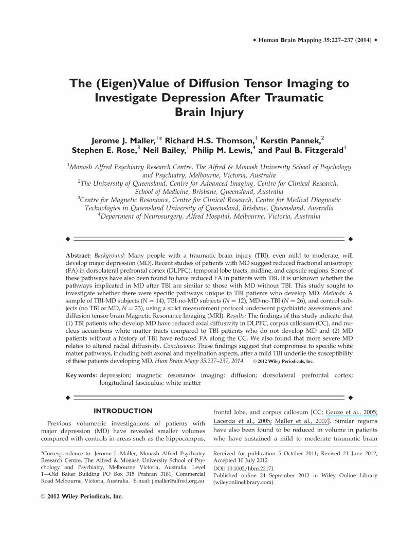
An exploration of the differences in diffusivity betweenall TBI subjects (N ¼ 26) and controls (N ¼ 23) was firstperformed to determine whether this patient populationwas consistent with previous DTI mTBI research. Althoughreduced FA in the TBI sample was not statistically signifi-cant (P ¼ 0.10, corrected), radial diffusivity was greater inthe TBI sample at the P ¼ 0.05 level (corrected) acrosswidespread areas including the CC, bilateral PFC, superiorlongitudinal fasciculi, and right internal capsule (Fig. 1).
TBI-MD vs. TBI-no-MD
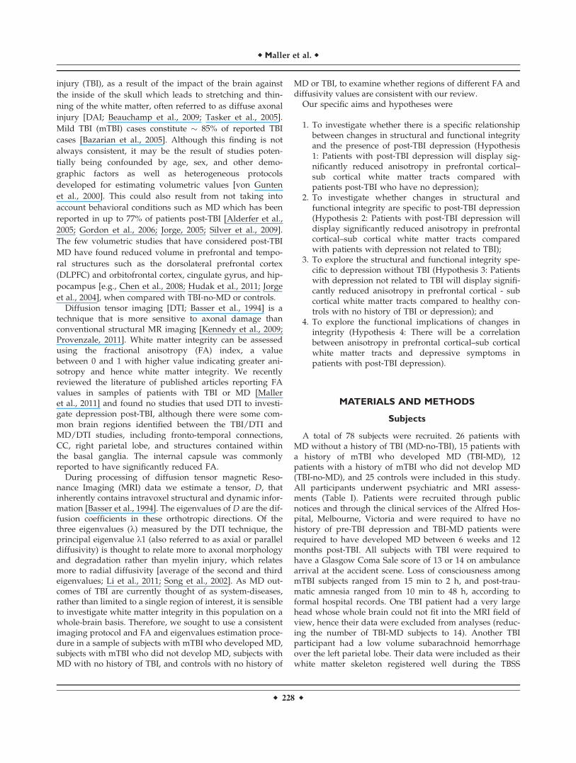
We identified a decrease in axial diffusivity (P < 0.007)in the bilateral DLPFCs, anterior CC, right nucleus accum-
bens, right internal capsule, right inferior and superiorlongitudinal fasciculi, and brainstem for TBI-MD patientscompared with TBI-no-MD patients (Fig. 2 and Table II).This indicates that there is widespread white matter dam-age unique to the development of MD after a TBI. FA andradial diffusivity did not differ significantly betweengroups.
TBI-MD vs. MD-no-TBI
We found no significant differences between TBI-MDand MD-no-TBI. There were reductions in FA in the MD-TBI group compared with the MD-no-TBI group, althoughthese differences between groups did not reach signifi-cance at the 0.05 level for the corrected analyses. Notrends were found for non-FA values.
MD-no-TBI vs. controls
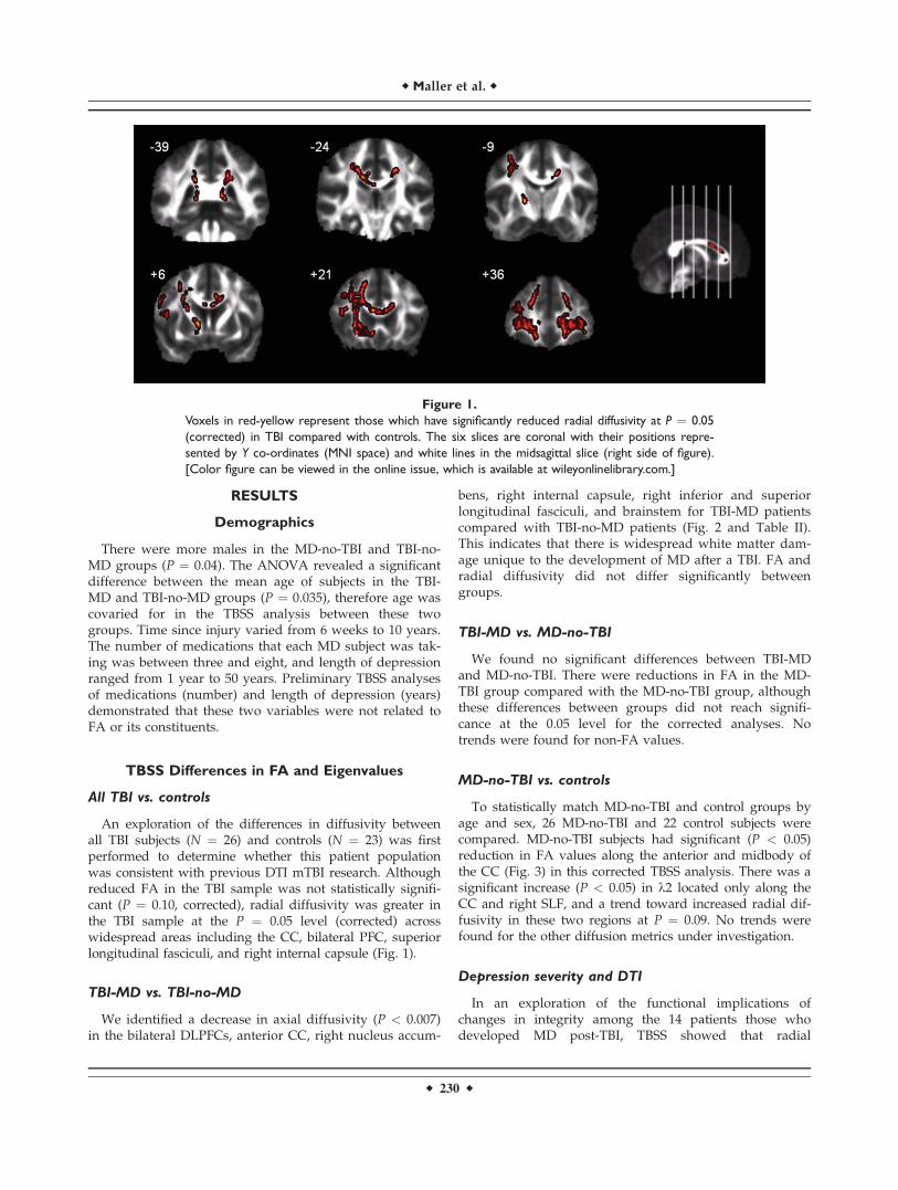
To statistically match MD-no-TBI and control groups byage and sex, 26 MD-no-TBI and 22 control subjects werecompared. MD-no-TBI subjects had significant (P < 0.05)reduction in FA values along the anterior and midbody ofthe CC (Fig. 3) in this corrected TBSS analysis. There was asignificant increase (P < 0.05) in k2 located only along theCC and right SLF, and a trend toward increased radial dif-fusivity in these two regions at P ¼ 0.09. No trends werefound for the other diffusion metrics under investigation.
Depression severity and DTI
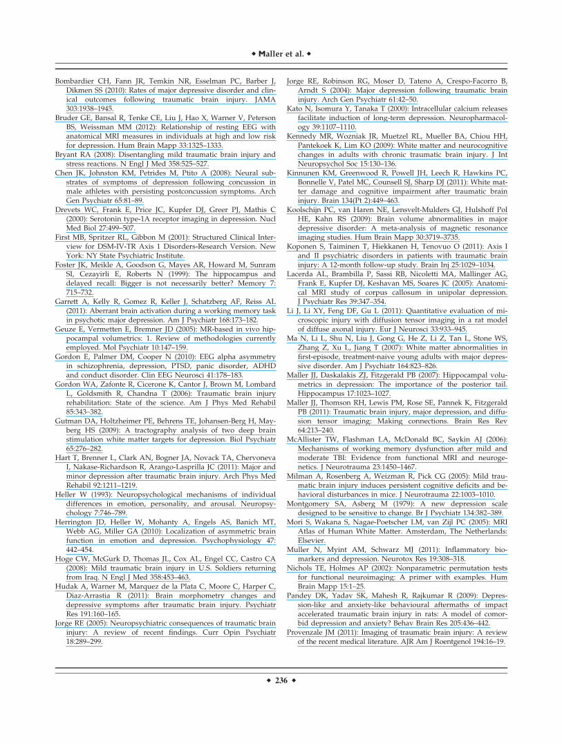
In an exploration of the functional implications ofchanges in integrity among the 14 patients those whodeveloped MD post-TBI, TBSS showed that radial
Figure 1.
Voxels in red-yellow represent those which have significantly reduced radial diffusivity at P ¼ 0.05
(corrected) in TBI compared with controls. The six slices are coronal with their positions repre-
sented by Y co-ordinates (MNI space) and white lines in the midsagittal slice (right side of figure).
[Color figure can be viewed in the online issue, which is available at wileyonlinelibrary.com.]
r Maller et al. r
r 230 r

diffusivity was significantly correlated with MADRS scores(P < 0.05) along the CC, bilateral nucleus accumbens,bilateral internal capsule, bilateral cingulated gyri, supe-rior longitudinal fasciculi, and brainstem. At a significancelevel of P ¼ 0.028 (corrected) the k3 TBSS analysis showedvoxels along these same white matter pathways (Fig. 4).Although the corrected analyses for axial diffusivity, k2,and mean diffusivity did not reach significance, they all
suggested a trend of voxels of reduced values beinglocated along only the CC.
DISCUSSIONS
This study is the first to investigate the development ofMD after mild traumatic brain injury (mTBI) using DTI. It
TABLE II. Regions of different FA and non-FA values as identified by TBSS
TBI-MD vs.TBI (no-MD)
MD (no-TBI)vs. TBI-MD
MD (no-TBI)vs. controls
MADRS(TBI-MD)
Region FAa Axialb Axialc FAb k2b Radiald k3b Radialb Analysis frequency Frequency order
PFC (R) * * 2 3PFC (L) * 1 4Cing (R) * * * 2 3Cing (L) * * 1 4CC * * * * * * 3 2IC (R) * * * * 3 2Nucl Acc (R) * * * 2 3Nucl Acc (L) * 1 4SLF (R) * * * * * * * 4 1SLF (L) * * 1 4ILF (R) * 1 4Brainstem * * * 2 3
‘‘*’’ denotes that the region had significantly different values as identified by TBSS between the two groups compared.aP ¼ 0.14 (corrected).bP < 0.05 (corrected).cP ¼ 0.17 (corrected).dP ¼ 0.09 (corrected).CC, corpus callosum; FA, fractional anisotropy; IC, internal capsule; ILF, inferior longitudinal fasciculus; L, left; k, eigenvalue; MD,major depression; Mean Diff, mean diffusivity; Nucl Acc, nucleus accumbens; PFC, prefrontal cortex; R, right; SLF, superior longitudinalfasciculus; TBI, traumatic brain injury; Axial, axial diffusivity; Radial, radial diffusivity.
Figure 2.
Reduced axial diffusivity voxels (red-yellow) at P ¼ 0.007 (corrected) in those with post-TBI MD
compared with TBI-no-MD. Numbers represent Y co-ordinates. [Color figure can be viewed in
the online issue, which is available at wileyonlinelibrary.com.]
r Diffusion Tensor Imaging, TBI, and Depression r
r 231 r

reports the finding of reduced FA and axial diffusivity val-ues in the CC (specifically the anterior aspect) and rightsuperior longitudinal fasciculus in MD patients without ahistory of TBI and further reduced FA in this region andbilateral PFCs in those who developed MD after a TBI.
Furthermore, significantly increased k3 was seen alongwhite matter pathways directly related to regions impli-cated in fronto-temporal models of MD and consistentwith those elucidated from our recent review [Malleret al., 2011].
Figure 3.
TBSS results of MD (N ¼ 26) compared against controls (N ¼ 22) at P ¼ 0.05 (corrected). A:
FA and B: k2. Numbers represent Y co-ordinates. [Color figure can be viewed in the online
issue, which is available at wileyonlinelibrary.com.]
Figure 4.
TBSS corrected analyses of correlations between MADRS scores
in patients with post-TBI MD and radial diffusivity (P ¼ 0.05,
corrected). The top row of coronal slices from posterior (left of
screen) to anterior (right of screen) and midsagittal slice, illus-
trates the locations of voxels (red-yellow) which were significant
in this analysis. The three-dimensional rendering of this set of
significant voxels from an anterior-dorsal perspective is pre-
sented on the left of the figure, and on the right is a representa-
tion of those voxels from a right-side dorso-lateral perspective.
Numbers represent Y co-ordinates. A ¼ anterior; P ¼ poste-
rior; I ¼ inferior; S ¼ superior. [Color figure can be viewed in
the online issue, which is available at wileyonlinelibrary.com.]
r Maller et al. r
r 232 r

Consistent with previous mTBI research, there were anumber of regions with reduced white matter integrity insubjects with mTBI, when compared with those without ahistory of TBI (or MD), including the prefrontal corticesand midline tracts. To investigate Aim 1 (whether there isa specific relationship between changes in structural andfunctional integrity and the presence of post-TBI depres-sion), post-TBI MD and TBI participants with no depressivesyndrome were initially studied. Significant microstructuralchanges in the frontal-parietal and fronto-temporal whitematter tracts were investigated by the voxelwise compari-son of FA and non-FA measures between the two groups.In an exploration of Aims 2 and 3 (to investigate whetherchanges in structural and functional integrity are specific topost-TBI depression, and to investigate the structural andfunctional integrity specific to depression without TBI), wefound that those with MD-no-TBI had widespread reduc-tion in FA (and axial diffusivity), mostly along the sametracts identified in the TBSS analysis between TBI-MD andTBI-no-MD.
Depression is common after a TBI [Silver et al., 2009].Hudak et al. [2011] recently reported depressive symptomsin post-TBI patients to significantly correlate with volumet-rics of three brain regions (left rostral anterior cingulateand bilateral orbitofrontal cortex); regions reported by pre-vious studies of volumetric assessment in patients withdepression post-TBI [Chen et al., 2008; Jorge, 2005; Jorgeet al., 2004] include the PFC, DLPFC, hippocampus, andanterior cingulate. As these areas have also been impli-cated by a number of studies linking depression notrelated to TBI (reviewed by Koolschijn et al. [2009]), theysupport a common link between spontaneous and non-spontaneous (e.g., post-TBI) MD models; that is, involve-ment of fronto-limbic pathways. However, structure sizedoes not necessarily reflect underlying function or integ-rity [e.g. Foster et al., 1999]. Hence, this study is the firstto investigate post-TBI MD in the context of function byusing measures of diffusion as biomarkers. We foundreduced FA and axial diffusivity among similar regions tothose reported from volumetric studies, but we also foundreduced anisotropy in the CC, an area commonly reportedas reduced in MD patients and TBI patients. The right in-ferior and superior longitudinal fasciculi were also impli-cated in the TBSS results, which is consistent with otherstudies which have found similar regions, and only on theright, to have reduced FA and/or axial diffusivity in MDor post-TBI in human and animal models [e.g., Kinnunenet al., 2011; Li et al., 2011]. More importantly, we foundFA in post-TBI MD, when compared with post-TBI with-out MD, was most reduced (as supported by the axial dif-fusivity reduction and MD difference) in two of the mostwidely-reported regions reported as relevant to the devel-opment of MD, which are the CC and DLPFCs. The CChas been reported in many publications as reduced in sizeamong post-TBI patients [Bigler and Maxwell, 2011] andMD patients without a history of TBI [Maller et al., 2011].Furthermore, we found the internal capsule and nucleus
accumbens to have significantly reduced axial diffusivityin TBI-MD when compared against TBI-no-MD. This issupported by a main analysis which also found MD sever-ity to significantly relate to radial diffusivity of the internalcapsule and nucleus accumbens. These are important find-ings as they are consistent with DTI studies in deep brainstimulation (DBS) MD patients in which the nucleusaccumbens (via the internal capsule) is often the target ofthe electrodes. [Gutman et al., 2009] showed with DTI thatthe targets for DBS when used to treat depression havedistinct and widespread projections to frontal and tempo-ral poles as well as the cingulate, thalamus, hypothalamus,nucleus accumbens, and brainstem, which was also impli-cated in our TBSS results; these are regions implicated inantidepressant response mechanisms [Airan et al., 2007;Schlaepfer et al., 2008; Tanis et al., 2007]. Very similartracts were identified as having reduced FA in a separatestudy of MD subjects [Zou et al., 2008] which reportedreduced FA restricted to the internal capsule (and parietalregion). This is relevant because DBS is used for MD onlyin patients whose MD is highly treatment-resistant (i.e.,nonresponsive to medication, electroconvulsive therapy,and often other forms of neurostimulation as well). Thatwe found this region to be of reduced integrity in thosewho develop MD after TBI, and related to the severity oftheir MD, is consistent with the anecdotal findings of thesepatients’ MD often being treatment-resistant.
The final set of analyses found that diffusivity valueswere related to MD severity in regions reported by the lit-erature in other groups of MD patients, as recentlyreviewed [Maller et al., 2011]. Specifically, the CC, medialfrontal gyri, nucleus accumbens, cingulate bundles, inter-nal capsules, brainstem, and longitudinal fasciculi (TableII). These are among the majority of regions identified infronto-temporal models of MD. Importantly, the radial dif-fusivity values were implicated. This finding could suggestan issue relating to myelination rather than axonalintegrity, which could in turn suggest that a degenerativeprocess in addition to DAI is the precursor for the devel-opment of MD after mTBI. Although it is difficult to spec-ulate about whether increased radial diffusivity relates toDAI or a myelination problem, DAI probably leads toWallerian degeneration of multiple neural circuits, whichwill involve cortical degeneration (atrophy) at some stage.
The decrease in FA and axial diffusivity along the rightsuperior longitudinal fasciculus in subjects with MD post-TBI relative to TBI patients who did not develop MD, aswell as in those with MD (no TBI) compared withmatched controls (with no history of MD or TBI) is con-sistent with the literature. For example, the low FA regionsreported in a DTI study of post-TBI patients [Salmondet al., 2006] are very similar to those found in a study ofpatients with first-episode treatment-naıve MD [Ma et al.,2007] and from a group of patients with first-episoderemitted depression [Yuan et al., 2007], which include theright parietal lobe and right frontal gyri. Furthermore,Alexopoulos et al. [2008] found similar regions to have
r Diffusion Tensor Imaging, TBI, and Depression r
r 233 r

reduced FA in a group of MD patients who did notachieve remission.
That axial and radial diffusivity analyses showed highersignificance levels suggest that compared with FA, theyare more sensitive markers of DAI. It is also consistentwith rat studies [e.g., Li et al., 2011; Song et al., 2002]which tracked the changes in FA and its eigenvalueconstituents from the acute phase to months afterwardvalidated by histological investigation such as optical im-munochemical staining. The group differences in TBI-MDvs. TBI-no-MD were in axial diffusivity, whereas MDseverity was significantly related to radial diffusivity.Collectively, these results suggest that MD is related toboth types of reduced white matter integrity, i.e., fromboth radial and axial perspectives.
Although the underpinnings of the difference(s) betweenlambda 2 and lambda 3 are currently unexplored, theresults of our study and others [e.g., Thomalla et al., 2004]which report such examples of where only one of the twoconstituents of radial diffusivity is significant whilst theother is not, suggest that lambda 2 and 3 have differentmeanings, i.e., they do not represent identical aspects of ra-dial diffusivity. We also performed separate lambda 2 andlambda 3 analyses to investigate whether radial diffusivityper se was related to TBI and/or depression, hence to ac-complish this aim comprehensively we analyzed not onlythe radial diffusivity conglomerate but its constituents aswell. That is, we wanted to investigate whether either of itsconstituents was significant even if the conglomerate wasnot. Our results suggest that lambda 2 and 3 are not simplereflections of one another, but rather, tell different parts ofthe radial diffusivity story, which may be found in futureresearch to represent quite distinct aspects of FA which mayrepresent discrete white matter damage and/or are the ba-sis for targeted treatment and therapy.
TBSS is a widely used program specifically designed tocompare groups on a voxel-by-voxel basis for differencesin FA and its constituents. The program’s authors suggestthat it should not be used on scans of brains that have sus-tained major damage as the white matter skeletons ofthose patients may not align accurately with that of thetemplate. This study was consistent with this advice asonly one of our patients had a positive CT finding whichwas very minor, superficial, and distal to white matter,hence unlikely to influence the white matter skeleton regis-trations (this was confirmed by visual checks for accurateregistration of each subject’s white matter skeleton beforegroup analyses proceeded).
Chronic primary or secondary dysfunction in dopaminer-gic, noradrenergic, and serotonergic systems seem to be rel-atively common consequences of TBI [Arciniegas andSilver, 2006; McAllister et al., 2006]. It is well establishedthat these systems are also compromised in MD patients[Bennett, 2010; Drevets et al., 2000; Muller et al., 2011; Wag-ner et al., 2010]. In humans, MD is a common sequelae ofTBI [Bombardier et al., 2010], even when mild [Ryan andWarden, 2003]. The symptom overlap between MD and the
common ‘‘post concussion syndrome’’ is substantial [Bryant,2008; Hoge et al., 2008]. Furthermore, there is evidence thatlate-life MD can be precipitated by accumulated small silentcerebral infarctions that appear as white matter hyperinten-sities on MRI scans [Alexopoulos et al., 1997; Sheline et al.,2010; Thomas et al., 2002]. The overlap between brainregions implicated in traumatic and nontraumatic MD isdiscussed in a recent comprehensive review by Wager-Smith and Markou [2011]. The authors propose stress-induced ‘‘microdamage’’ to the brain as a putative triggerfor subsequent cellular repair and neuroinflammation lead-ing to symptoms of depression. In susceptible individuals,impaired cellular repair mechanisms may lead to a chroniccycle of neuroinflammation, presenting as persistentdepression. As examples, they offer some preliminary stud-ies in experimental animals, which have noted that depres-sive-like behaviors follow traumatic or ischemic braininjury, although the studies are not well controlled for con-founds [Kato et al., 2000; Milman et al., 2005; Pandey et al.,2009; Shapira et al., 2007]. Moreover, they describe previousfindings of reduced hippocampal volume in depression, forwhich there is evidence the degree of volume loss is relatedto the number of depressive episodes experienced. Thisagain supports the theory that volume loss, secondary tostress-induced cellular damage, precipitates depressivesymptoms. This general hypothesis fits well with our find-ings, wherein we describe reduced FA (indicating neuronaldamage) in CC, DLPFC, and basal ganglia in both the TBI-MD and MD-no-TBI groups alike.
It is notable that there was some predominance of right-sided changes in the depression analyses. The implicationof right-sided involvement in depression post-TBI is consist-ent with much of the existing MRI and EEG depression liter-ature. For EEG, depression is associated with a shift towardgreater right than left activity, consistent with findings forunpleasant emotion [Herrington et al., 2010]. It was recentlyreported that in subjects at high-risk for developing depres-sion, alpha power correlated inversely with cortical thick-ness particularly over the right posterior region [Bruderet al., 2012] and another group [Shankman et al., 2011]recently found relatively lower right posterior alpha activityin patients with melancholic depression. In general, alphapower is found to be reduced and fMRI activation moreright-lateralized in patients with depression [Gordon et al.,2010; Saletu et al., 2010] depression. That is, there is oftenleft hemisphere underactivation at rest and right hemi-sphere overactivation during tasks. In an fMRI study, forexample, Garrett et al. [2011] found patients with MD toshow hyperactivation in the right temporoparietal regionassociated with orienting to unexpected stimuli. Heller’s[1993] model of emotion processing predicts that depressionwould be related to greater right than left frontal neural ac-tivity, combined with low levels of right temporo-parietalactivity. In the context of structural MRI, a reduction of righthippocampal-entorhinal cortex volume with voxel-basedmorphometry in patients with depression is often reported[e.g., Ahdidan et al., 2011; Bell-McGinty et al., 2002] and
r Maller et al. r
r 234 r

relative to the left hemisphere, depressed individuals havesmaller right-parietal viable brain volumes than nonde-pressed individuals [Schonberger et al., 2011]. In general,there seems to be something specific about right-sidedchanges and the development of depression post-TBI, andalthough less likely, it is possible that developing depres-sion leads to greater right-sided dysfunction. Only a studyexamining subjects before and after a TBI can suitablyaddress this question.
A limitation of this study is that the time since injury wasvery broad; previous research indicates FA is most reducedduring the acute post-TBI phase, hence, it is possible thatpatients whose TBI was more recent had the most reducedwhite matter integrity along the tracts revealed to havereduced white matter integrity, when compared with sub-jects without MD. Longitudinal research is required tounderstand how these pathways are affected over time andwhether deficits accumulate or ameliorate over time. As TBIsubjects were recruited and not prospectively retained,there must be unevenness in data collection with regards today-of-injury clinical information. It is possible that 48 h oftruly documented post-traumatic amnesia would not be amild TBI, but move it into the moderate range. Likewise,loss of consciousness of greater than 1 h, for some wouldmove the injury into the moderate category. Depending onhow many these clinical features apply to, there may needto be a modification in using the mild TBI moniker in fol-low-up studies of these patients. It is likely, however, thatthe use of more homogeneous groups in terms of the mech-anism of injury would produce clearer or more consistentresults. Finally, without longitudinal data, it is difficult tomake a cause and effect judgment as to the relationshipbetween white matter changes and symptoms. Injuryrelated loss of white matter integrity may cause depression.However, it is also possible that the development of adepressive illness, through other mechanisms, may result inreduced intercortical connectivity which over time reduceswhite matter integrity. This study enrolled a greater portionof women in the TBI-MD than the TBI-no-MD, and althoughthere is a higher incidence of depression in women, the gen-der difference was not addressed as this was beyond thescope of the study’s aims. This is an area which we aim toaddress in future investigations.
Although this study focused on MD, other psychiatricdisturbances have also been related to TBI. For example,Hart et al. [2011] reported minor depression and MD toeach be present in approximately a quarter of their TBIpatients (N ¼ 1,570), and [Koponen et al., 2011] recentlyreported both Axis I and II psychiatric disorders to becommon among patients with TBI. Our future investiga-tions will consider this.
CONCLUSION
Previous DTI studies of TBI patients (whether TBSS orother techniques were applied) have not considered the
degree of depression among their samples, hence, this isthe first study to attempt an investigation of the develop-ment of MD after TBI in the context of anisotropy. Consist-ent with a recent review, we have shown in mild TBIpatients that the development of MD is related to compro-mised FA and axial and radial diffusivity in the prefrontal,CC, cingulated, internal capsule, and parietal regions, sup-porting models of MD without TBI. Reduced integrity inthese regions may therefore be a biomarker for the devel-opment of MD post-TBI. Future studies should addressthis important issue.
ACKNOWLEDGMENTS
The authors thank all the patients and volunteers whoagreed to participate in this study, Research Nurses at theMonash Alfred Psychiatry Research Centre, and the staffof the MRI facility at the Alfred Hospital, Melbourne,Victoria.
REFERENCES
Ahdidan J, Hviid LB, Chakravarty MM, Ravnkilde B, Rosenberg R,Rodell A, Stodkilde-Jorgensen H, Videbech P (2011): Longitudi-nal MR study of brain structure and hippocampus volume inmajor depressive disorder. Acta Psychiatr Scand 123:211–219.
Airan RD, Meltzer LA, Roy M, Gong Y, Chen H, Deisseroth K(2007): High-speed imaging reveals neurophysiological links tobehavior in an animal model of depression. Science 317:819–823.
Alderfer BS, Arciniegas DB, Silver JM (2005): Treatment of depres-sion following traumatic brain injury. J Head Trauma Rehabil20:544–562.
Alexopoulos GS, Meyers BS, Young RC, Campbell S, SilbersweigD, Charlson M (1997): ‘‘Vascular depression’’ hypothesis. ArchGen Psychiatr 54:915–922.
Alexopoulos GS, Murphy CF, Gunning-Dixon FM, Latoussakis V,Kanellopoulos D, Klimstra S, Lim KO, Hoptman MJ (2008):Microstructural white matter abnormalities and remission ofgeriatric depression. Am J Psychiatr 165:238–244.
Arciniegas DB, Silver JM (2006): Pharmacotherapy of posttrau-matic cognitive impairments. Behav Neurol 17:25–42.
Basser PJ, Mattiello J, LeBihan D (1994): MR diffusion tensor spec-troscopy and imaging. Biophys J 66:259–267.
Bazarian JJ, McClung J, Shah MN, Cheng YT, Flesher W, Kraus J(2005): Mild traumatic brain injury in the United States, 1998–2000. Brain Inj 19:85–91.
Beauchamp MH, Anderson VA, Catroppa C, Maller JJ, Godfrey C,Rosenfeld JV, Kean M (2009): Implications of reduced callosalarea for social skills after severe traumatic brain injury in chil-dren. J Neurotrauma 26:1645–1654.
Bell-McGinty S, Butters MA, Meltzer CC, Greer PJ, Reynolds CFIII, Becker JT. (2002): Brain morphometric abnormalities ingeriatric depression: Long-term neurobiological effects of ill-ness duration. Am J Psychiatr 159:1424–1427.
Bennett MR (2010): Synapse regression in depression: The role of5-HT receptors in modulating NMDA receptor function andsynaptic plasticity. Aust NZ J Psychiatr 44:301–308.
Bigler ED, Maxwell WL (2011): Neuroimaging and neuropathol-ogy of TBI. NeuroRehabilitation 28:63–74.
r Diffusion Tensor Imaging, TBI, and Depression r
r 235 r
Bombardier CH, Fann JR, Temkin NR, Esselman PC, Barber J,Dikmen SS (2010): Rates of major depressive disorder and clin-ical outcomes following traumatic brain injury. JAMA303:1938–1945.
Bruder GE, Bansal R, Tenke CE, Liu J, Hao X, Warner V, PetersonBS, Weissman MM (2012): Relationship of resting EEG withanatomical MRI measures in individuals at high and low riskfor depression. Hum Brain Mapp 33:1325–1333.
Bryant RA (2008): Disentangling mild traumatic brain injury andstress reactions. N Engl J Med 358:525–527.
Chen JK, Johnston KM, Petrides M, Ptito A (2008): Neural sub-strates of symptoms of depression following concussion inmale athletes with persisting postconcussion symptoms. ArchGen Psychiatr 65:81–89.
Drevets WC, Frank E, Price JC, Kupfer DJ, Greer PJ, Mathis C(2000): Serotonin type-1A receptor imaging in depression. NuclMed Biol 27:499–507.
First MB, Spritzer RL, Gibbon M (2001): Structured Clinical Inter-view for DSM-IV-TR Axis 1 Disorders-Research Version. NewYork: NY State Psychiatric Institute.
Foster JK, Meikle A, Goodson G, Mayes AR, Howard M, SunramSI, Cezayirli E, Roberts N (1999): The hippocampus anddelayed recall: Bigger is not necessarily better? Memory 7:715–732.
Garrett A, Kelly R, Gomez R, Keller J, Schatzberg AF, Reiss AL(2011): Aberrant brain activation during a working memory taskin psychotic major depression. Am J Psychiatr 168:173–182.
Geuze E, Vermetten E, Bremner JD (2005): MR-based in vivo hip-pocampal volumetrics: 1. Review of methodologies currentlyemployed. Mol Psychiatr 10:147–159.
Gordon E, Palmer DM, Cooper N (2010): EEG alpha asymmetryin schizophrenia, depression, PTSD, panic disorder, ADHDand conduct disorder. Clin EEG Neurosci 41:178–183.
Gordon WA, Zafonte R, Cicerone K, Cantor J, Brown M, LombardL, Goldsmith R, Chandna T (2006): Traumatic brain injuryrehabilitation: State of the science. Am J Phys Med Rehabil85:343–382.
Gutman DA, Holtzheimer PE, Behrens TE, Johansen-Berg H, May-berg HS (2009): A tractography analysis of two deep brainstimulation white matter targets for depression. Biol Psychiatr65:276–282.
Hart T, Brenner L, Clark AN, Bogner JA, Novack TA, ChervonevaI, Nakase-Richardson R, Arango-Lasprilla JC (2011): Major andminor depression after traumatic brain injury. Arch Phys MedRehabil 92:1211–1219.
Heller W (1993): Neuropsychological mechanisms of individualdifferences in emotion, personality, and arousal. Neuropsy-chology 7:746–789.
Herrington JD, Heller W, Mohanty A, Engels AS, Banich MT,Webb AG, Miller GA (2010): Localization of asymmetric brainfunction in emotion and depression. Psychophysiology 47:442–454.
Hoge CW, McGurk D, Thomas JL, Cox AL, Engel CC, Castro CA(2008): Mild traumatic brain injury in U.S. Soldiers returningfrom Iraq. N Engl J Med 358:453–463.
Hudak A, Warner M, Marquez de la Plata C, Moore C, Harper C,Diaz-Arrastia R (2011): Brain morphometry changes anddepressive symptoms after traumatic brain injury. PsychiatrRes 191:160–165.
Jorge RE (2005): Neuropsychiatric consequences of traumatic braininjury: A review of recent findings. Curr Opin Psychiatr18:289–299.
Jorge RE, Robinson RG, Moser D, Tateno A, Crespo-Facorro B,Arndt S (2004): Major depression following traumatic braininjury. Arch Gen Psychiatr 61:42–50.
Kato N, Isomura Y, Tanaka T (2000): Intracellular calcium releasesfacilitate induction of long-term depression. Neuropharmacol-ogy 39:1107–1110.
Kennedy MR, Wozniak JR, Muetzel RL, Mueller BA, Chiou HH,Pantekoek K, Lim KO (2009): White matter and neurocognitivechanges in adults with chronic traumatic brain injury. J IntNeuropsychol Soc 15:130–136.
Kinnunen KM, Greenwood R, Powell JH, Leech R, Hawkins PC,Bonnelle V, Patel MC, Counsell SJ, Sharp DJ (2011): White mat-ter damage and cognitive impairment after traumatic braininjury. Brain 134(Pt 2):449–463.
Koolschijn PC, van Haren NE, Lensvelt-Mulders GJ, Hulshoff PolHE, Kahn RS (2009): Brain volume abnormalities in majordepressive disorder: A meta-analysis of magnetic resonanceimaging studies. Hum Brain Mapp 30:3719–3735.
Koponen S, Taiminen T, Hiekkanen H, Tenovuo O (2011): Axis Iand II psychiatric disorders in patients with traumatic braininjury: A 12-month follow-up study. Brain Inj 25:1029–1034.
Lacerda AL, Brambilla P, Sassi RB, Nicoletti MA, Mallinger AG,Frank E, Kupfer DJ, Keshavan MS, Soares JC (2005): Anatomi-cal MRI study of corpus callosum in unipolar depression.J Psychiatr Res 39:347–354.
Li J, Li XY, Feng DF, Gu L (2011): Quantitative evaluation of mi-croscopic injury with diffusion tensor imaging in a rat modelof diffuse axonal injury. Eur J Neurosci 33:933–945.
Ma N, Li L, Shu N, Liu J, Gong G, He Z, Li Z, Tan L, Stone WS,Zhang Z, Xu L, Jiang T (2007): White matter abnormalities infirst-episode, treatment-naive young adults with major depres-sive disorder. Am J Psychiatr 164:823–826.
Maller JJ, Daskalakis ZJ, Fitzgerald PB (2007): Hippocampal volu-metrics in depression: The importance of the posterior tail.Hippocampus 17:1023–1027.
Maller JJ, Thomson RH, Lewis PM, Rose SE, Pannek K, FitzgeraldPB (2011): Traumatic brain injury, major depression, and diffu-sion tensor imaging: Making connections. Brain Res Rev64:213–240.
McAllister TW, Flashman LA, McDonald BC, Saykin AJ (2006):Mechanisms of working memory dysfunction after mild andmoderate TBI: Evidence from functional MRI and neuroge-netics. J Neurotrauma 23:1450–1467.
Milman A, Rosenberg A, Weizman R, Pick CG (2005): Mild trau-matic brain injury induces persistent cognitive deficits and be-havioral disturbances in mice. J Neurotrauma 22:1003–1010.
Montgomery SA, Asberg M (1979): A new depression scaledesigned to be sensitive to change. Br J Psychiatr 134:382–389.
Mori S, Wakana S, Nagae-Poetscher LM, van Zijl PC (2005): MRIAtlas of Human White Matter. Amsterdam, The Netherlands:Elsevier.
Muller N, Myint AM, Schwarz MJ (2011): Inflammatory bio-markers and depression. Neurotox Res 19:308–318.
Nichols TE, Holmes AP (2002): Nonparametric permutation testsfor functional neuroimaging: A primer with examples. HumBrain Mapp 15:1–25.
Pandey DK, Yadav SK, Mahesh R, Rajkumar R (2009): Depres-sion-like and anxiety-like behavioural aftermaths of impactaccelerated traumatic brain injury in rats: A model of comor-bid depression and anxiety? Behav Brain Res 205:436–442.
Provenzale JM (2011): Imaging of traumatic brain injury: A reviewof the recent medical literature. AJR Am J Roentgenol 194:16–19.
r Maller et al. r
r 236 r

Ryan LM, Warden DL (2003): Post concussion syndrome. Int RevPsychiatr 15:310–316.
Saletu B, Anderer P, Saletu-Zyhlarz GM (2010): EEG topographyand tomography (LORETA) in diagnosis and pharmacotherapyof depression. Clin EEG Neurosci 41:203–210.
Salmond CH, Menon DK, Chatfield DA, Williams GB, Pena A,Sahakian BJ, Pickard JD (2006): Diffusion tensor imaging inchronic head injury survivors: Correlations with learning andmemory indices. Neuroimage 29:117–124.
Schlaepfer TE, Cohen MX, Frick C, Kosel M, Brodesser D, AxmacherN, Joe AY, Kreft M, Lenartz D, Sturm V (2008): Deep brain stimu-lation to reward circuitry alleviates anhedonia in refractory majordepression. Neuropsychopharmacology 33:368–377.
Schonberger M, Ponsford J, Reutens D, Beare R, Clarke D, O’Sulli-van R (2011): The relationship between mood disorders and MRIfindings following traumatic brain injury. Brain Inj 25:543–550.
Shankman SA, Sarapas C, Klein DN (2011): The effect of pre- vs.post-reward attainment on EEG asymmetry in melancholicdepression. Int J Psychophysiol 79:287–295.
Shapira M, Licht A, Milman A, Pick CG, Shohami E, Eldar-Finkel-man H (2007): Role of glycogen synthase kinase-3beta in earlydepressive behavior induced by mild traumatic brain injury.Mol Cell Neurosci 34:571–577.
Sheehan DV, Lecrubier Y, Sheehan KH, Amorim P, Janavs J,Weiller E, Hergueta T, Baker R, Dunbar GC (1998): TheMini-International Neuropsychiatric Interview (M.I.N.I.): Thedevelopment and validation of a structured diagnostic psychi-atric interview for DSM-IV and ICD-10. J Clin Psychiatr59(Suppl 20):22–33; quiz 34-57.
Sheline YI, Pieper CF, Barch DM, Welsh-Bohmer K, McKinstryRC, MacFall JR, D’Angelo G, Garcia KS, Gersing K, Wilkins C,Taylor W, Steffens DC, Krishnan RR, Doraiswamy PM (2010):Support for the vascular depression hypothesis in late-lifedepression: Results of a 2-site, prospective, antidepressanttreatment trial. Arch Gen Psychiatr 67:277–285.
Silver JM, McAllister TW, Arciniegas DB (2009): Depression andcognitive complaints following mild traumatic brain injury.Am J Psychiatr 166:653–661.
Smith SM, Jenkinson M, Woolrich MW, Beckmann CF, BehrensTE, Johansen-Berg H, Bannister PR, De Luca M, Drobnjak I,Flitney DE, Niazy RK, Saunders J, Vickers J, Zhang Y, De Ste-fano N, Brady JM, Matthews PM (2004): Advances in func-tional and structural MR image analysis and implementationas FSL. Neuroimage 23(Suppl 1):S208–S219.
Smith SM, Jenkinson M, Johansen-Berg H, Rueckert D, NicholsTE, Mackay CE, Watkins KE, Ciccarelli O, Cader MZ, Mat-thews PM, Behrens TE (2006): Tract-based spatial statistics:Voxelwise analysis of multi-subject diffusion data. Neuroimage31:1487–1505.
Song SK, Sun SW, Ramsbottom MJ, Chang C, Russell J, Cross AH(2002): Dysmyelination revealed through MRI as increased ra-dial (but unchanged axial) diffusion of water. Neuroimage17:1429–1436.
Tanis KQ, Newton SS, Duman RS (2007): Targeting neurotrophic/growth factor expression and signaling for antidepressant drugdevelopment. CNS Neurol Disord Drug Targets 6:151–160.
Tasker RC, Salmond CH, Westland AG, Pena A, Gillard JH, Saha-kian BJ, Pickard JD (2005): Head circumference and brain andhippocampal volume after severe traumatic brain injury inchildhood. Pediatr Res 58:302–308.
Thomas AJ, Perry R, Barber R, Kalaria RN, O’Brien JT (2002):Pathologies and pathological mechanisms for white matterhyperintensities in depression. Ann NY Acad Sci 977:333–339.
Thomalla G, Glauche V, Koch MA, Beaulieu C, Weiller C,Rother J (2004): Diffusion tensor imaging detects early Waller-ian degeneration of the pyramidal tract after ischemic stroke.Neuroimage 22:1767–1774.
von Gunten A, Fox NC, Cipolotti L, Ron MA (2000): A volumetricstudy of hippocampus and amygdala in depressed patientswith subjective memory problems. J Neuropsychiatr Clin Neu-rosci 12:493–498.
Wager-Smith K, Markou A (2011): Depression: A repair response tostress-induced neuronal microdamage that can grade into a chronicneuroinflammatory condition? Neurosci Biobehav Rev 35:742–764.
Wagner G, Koch K, Schachtzabel C, Sobanski T, Reichenbach JR,Sauer H, Schlosser RG (2010): Differential effects of serotoner-gic and noradrenergic antidepressants on brain activity duringa cognitive control task and neurofunctional prediction oftreatment outcome in patients with depression. J PsychiatrNeurosci 35:247–257.
Yuan Y, Zhang Z, Bai F, Yu H, Shi Y, Qian Y, Zang Y, Zhu C, LiuW, You J (2007): White matter integrity of the whole brain isdisrupted in first-episode remitted geriatric depression. Neuro-report 18:1845–1849.
Zou K, Huang X, Li T, Gong Q, Li Z, Ou-Yang L, Deng W, ChenQ, Li C, Ding Y, Sun X (2008): Alterations of white matter in-tegrity in adults with major depressive disorder: A magneticresonance imaging study. J Psychiatr Neurosci 33:525–530.
r Diffusion Tensor Imaging, TBI, and Depression r
r 237 r
Related Documents


