Citation: Kim, H.; Lee, S. The Efficacy of Pain Neuroscience Education on Active Rehabilitation Following Arthroscopic Rotator Cuff Repair: A CONSORT-Compliant Prospective Randomized Single-Blind Controlled Trial. Brain Sci. 2022, 12, 764. https://doi.org/ 10.3390/brainsci12060764 Academic Editor: Alex Green Received: 2 May 2022 Accepted: 9 June 2022 Published: 10 June 2022 Publisher’s Note: MDPI stays neutral with regard to jurisdictional claims in published maps and institutional affil- iations. Copyright: © 2022 by the authors. Licensee MDPI, Basel, Switzerland. This article is an open access article distributed under the terms and conditions of the Creative Commons Attribution (CC BY) license (https:// creativecommons.org/licenses/by/ 4.0/). brain sciences Article The Efficacy of Pain Neuroscience Education on Active Rehabilitation Following Arthroscopic Rotator Cuff Repair: A CONSORT-Compliant Prospective Randomized Single-Blind Controlled Trial Hyunjoong Kim 1 and Seungwon Lee 2, * 1 Department of Physical Therapy, Graduate School of Sahmyook University, 815, Hwarang-ro, Nowon-gu, Seoul 01795, Korea; [email protected] 2 Department of Physical Therapy, College of Health and Welfare, Sahmyook University, 815, Hwarang-ro, Nowon-gu, Seoul 01795, Korea * Correspondence: [email protected] Abstract: Pain neuroscience education (PNE), a modern educational therapy, has been reported to be effective in pain control by reducing fear of movement. This study investigated the effects of additional PNE on a physical therapy rehabilitation protocol (PTRP) following arthroscopic rotator cuff repair (ARCR). In this single-blind, randomized controlled trial, 34 patients who had undergone ARCR were randomly allocated (1:1) into two groups: PNE (PTRP plus PNE) and PTRP. PTRP was performed five times a week, for four weeks, 115 min per session (physical agents, manual therapy, and exercises), and PNE was performed twice at the beginning (face-to-face PNE) and end (non-face-to-face) of the PTRP. The outcome measures were measured four times for pain intensity, pain cognition, and shoulder function; two times for a range of motion; and once for satisfaction. No significant difference in pain intensity was observed between the groups. However, in pain cognition, the Tampa Scale for Kinesiophobia avoidance showed a significant interaction between time and group, and PNE showed a higher effect size than PTRP in the post-test and follow-up in several variables. In conclusion, the significant improvement in avoidance in postoperative rehabilitation suggests that there is a partially positive benefit in terms of pain, range of motion, and shoulder function in ARCR patients. Keywords: pain neuroscience education; rotator cuff repair; postoperative rehabilitation; physi- cal therapy 1. Introduction Disorders of the rotator cuff and muscles around the shoulder, which are consid- ered the most common causes of shoulder pain, are also the most common causes of musculoskeletal pain. In addition, rotator cuff disorders, including rotator cuff tears, are considered degenerative diseases, and their incidence increases with age [1]. Primary care for patients with shoulder pain and rotator cuff tear mainly involves changes in usual activ- ities, use of analgesics, corticosteroid injections, and physical therapy [2,3]. If conservative treatment fails, surgical treatment may be considered [4,5]. After arthroscopic rotator cuff repair (ARCR), various rehabilitation protocols have been proposed; in principle, the postoperative rehabilitation phase proceeds according to the tendon healing process. Based on recent research trends, early and delayed rehabilita- tion are considered controversial [6], and approaches for them are still limited [7,8]. The existing general rehabilitation protocol minimizes active motion for 4 weeks after surgery and focuses on conscious muscle control of the upper extremities and trunk [9,10]. However, in clinical settings, patients cannot easily control their pain. It has been reported that pain neuroscience education (PNE), a modern educational therapy method, Brain Sci. 2022, 12, 764. https://doi.org/10.3390/brainsci12060764 https://www.mdpi.com/journal/brainsci

Welcome message from author
This document is posted to help you gain knowledge. Please leave a comment to let me know what you think about it! Share it to your friends and learn new things together.
Transcript
Citation: Kim, H.; Lee, S. The
Efficacy of Pain Neuroscience
Education on Active Rehabilitation
Following Arthroscopic Rotator Cuff
Repair: A CONSORT-Compliant
Prospective Randomized
Single-Blind Controlled Trial. Brain
Sci. 2022, 12, 764. https://doi.org/
10.3390/brainsci12060764
Academic Editor: Alex Green
Received: 2 May 2022
Accepted: 9 June 2022
Published: 10 June 2022
Publisher’s Note: MDPI stays neutral
with regard to jurisdictional claims in
published maps and institutional affil-
iations.
Copyright: © 2022 by the authors.
Licensee MDPI, Basel, Switzerland.
This article is an open access article
distributed under the terms and
conditions of the Creative Commons
Attribution (CC BY) license (https://
creativecommons.org/licenses/by/
4.0/).
brainsciences
Article
The Efficacy of Pain Neuroscience Education on ActiveRehabilitation Following Arthroscopic Rotator Cuff Repair:A CONSORT-Compliant Prospective Randomized Single-BlindControlled TrialHyunjoong Kim 1 and Seungwon Lee 2,*
1 Department of Physical Therapy, Graduate School of Sahmyook University, 815, Hwarang-ro, Nowon-gu,Seoul 01795, Korea; [email protected]
2 Department of Physical Therapy, College of Health and Welfare, Sahmyook University, 815, Hwarang-ro,Nowon-gu, Seoul 01795, Korea
* Correspondence: [email protected]
Abstract: Pain neuroscience education (PNE), a modern educational therapy, has been reported tobe effective in pain control by reducing fear of movement. This study investigated the effects ofadditional PNE on a physical therapy rehabilitation protocol (PTRP) following arthroscopic rotatorcuff repair (ARCR). In this single-blind, randomized controlled trial, 34 patients who had undergoneARCR were randomly allocated (1:1) into two groups: PNE (PTRP plus PNE) and PTRP. PTRPwas performed five times a week, for four weeks, 115 min per session (physical agents, manualtherapy, and exercises), and PNE was performed twice at the beginning (face-to-face PNE) and end(non-face-to-face) of the PTRP. The outcome measures were measured four times for pain intensity,pain cognition, and shoulder function; two times for a range of motion; and once for satisfaction. Nosignificant difference in pain intensity was observed between the groups. However, in pain cognition,the Tampa Scale for Kinesiophobia avoidance showed a significant interaction between time andgroup, and PNE showed a higher effect size than PTRP in the post-test and follow-up in severalvariables. In conclusion, the significant improvement in avoidance in postoperative rehabilitationsuggests that there is a partially positive benefit in terms of pain, range of motion, and shoulderfunction in ARCR patients.
Keywords: pain neuroscience education; rotator cuff repair; postoperative rehabilitation; physi-cal therapy
1. Introduction
Disorders of the rotator cuff and muscles around the shoulder, which are consid-ered the most common causes of shoulder pain, are also the most common causes ofmusculoskeletal pain. In addition, rotator cuff disorders, including rotator cuff tears, areconsidered degenerative diseases, and their incidence increases with age [1]. Primary carefor patients with shoulder pain and rotator cuff tear mainly involves changes in usual activ-ities, use of analgesics, corticosteroid injections, and physical therapy [2,3]. If conservativetreatment fails, surgical treatment may be considered [4,5].
After arthroscopic rotator cuff repair (ARCR), various rehabilitation protocols havebeen proposed; in principle, the postoperative rehabilitation phase proceeds according tothe tendon healing process. Based on recent research trends, early and delayed rehabilita-tion are considered controversial [6], and approaches for them are still limited [7,8]. Theexisting general rehabilitation protocol minimizes active motion for 4 weeks after surgeryand focuses on conscious muscle control of the upper extremities and trunk [9,10].
However, in clinical settings, patients cannot easily control their pain. It has beenreported that pain neuroscience education (PNE), a modern educational therapy method,
Brain Sci. 2022, 12, 764. https://doi.org/10.3390/brainsci12060764 https://www.mdpi.com/journal/brainsci

Brain Sci. 2022, 12, 764 2 of 14
is effective in pain control by reducing fear of movement based on an understanding ofneurophysiology [11]. Studies on PNE are mainly based on the beliefs and cognitionsof patients about chronic pain that affect the patient’s pain experience and treatmentoutcomes [12,13]. However, recent research trends have changed to identify the potentialbenefits in non-chronic pain conditions.
We hypothesized that education could potentially reduce the likelihood of developingchronic pain and disability for patients with acute, subacute, preoperative, and prior painexperiences (healthy individuals). In a related study conducted in the United States, patientswho received preoperative PNE before their back surgery and total knee arthroplastyshowed significant improvement in health at six months, one year, and three years offollow-up compared to those who did not receive preoperative PNE [14–16].
This study aimed to investigate the effect of additional PNE on pain intensity, pain cog-nition, range of motion, shoulder function, and treatment satisfaction in the rehabilitationprotocol after ARCR.
2. Materials and Methods2.1. Study Design
This study was a two-arm, parallel, single-blind randomized controlled trial witha longitudinal prospective design. The study was conducted from February to August2021, and the protocol was registered in January (ClinicalTrial.gov.: NCT0475311). Figure 1shows the data collection and research procedures.
Brain Sci. 2022, 12, 764 2 of 14
However, in clinical settings, patients cannot easily control their pain. It has been re‐
ported that pain neuroscience education (PNE), a modern educational therapy method, is ef‐
fective in pain control by reducing fear of movement based on an understanding of neuro‐
physiology [11]. Studies on PNE are mainly based on the beliefs and cognitions of patients
about chronic pain that affect the patient’s pain experience and treatment outcomes [12,13].
However, recent research trends have changed to identify the potential benefits in non‐chronic
pain conditions.
We hypothesized that education could potentially reduce the likelihood of developing
chronic pain and disability for patients with acute, subacute, preoperative, and prior pain ex‐
periences (healthy individuals). In a related study conducted in the United States, patients
who received preoperative PNE before their back surgery and total knee arthroplasty showed
significant improvement in health at six months, one year, and three years of follow‐up com‐
pared to those who did not receive preoperative PNE [14,15,16].
This study aimed to investigate the effect of additional PNE on pain intensity, pain cog‐
nition, range of motion, shoulder function, and treatment satisfaction in the rehabilitation pro‐
tocol after ARCR.
2. Materials and Methods
2.1. Study Design
This study was a two‐arm, parallel, single‐blind randomized controlled trial with a
longitudinal prospective design. The study was conducted from February to August 2021,
and the protocol was registered in January (ClinicalTrial.gov.: NCT0475311). Figure 1
shows the data collection and research procedures.
Figure 1. Schematic of the experimental design. ARCR: arthroscopic rotator cuff repair; PNE: pain
neuroscience education; POD: postoperative day; PTRP: physical therapy rehabilitation protocol.
2.2. Participants and Ethics
This study included patients admitted to The Better Hospital (Gwangju, Republic of
Korea) for postoperative rehabilitation after ARCR. Potential participants were recruited
autonomously through the sports rehabilitation center bulletin board. Assessment for el‐
igibility was based on some inclusion and exclusion criteria [17,18].
2.2.1. Inclusion Criteria
Adults aged > 18 years;
Four weeks after ARCR;
Willing to participate in the study.
2.2.2. Exclusion Criteria
Older adults (age > 65 years);
Unable to receive education remotely;
Additional tendon augmentation in ARCR;
Figure 1. Schematic of the experimental design. ARCR: arthroscopic rotator cuff repair; PNE: painneuroscience education; POD: postoperative day; PTRP: physical therapy rehabilitation protocol.
2.2. Participants and Ethics
This study included patients admitted to The Better Hospital (Gwangju, Republic ofKorea) for postoperative rehabilitation after ARCR. Potential participants were recruitedautonomously through the sports rehabilitation center bulletin board. Assessment foreligibility was based on some inclusion and exclusion criteria [17,18].
2.2.1. Inclusion Criteria
• Adults aged >18 years;• Four weeks after ARCR;• Willing to participate in the study.
2.2.2. Exclusion Criteria
• Older adults (age > 65 years);• Unable to receive education remotely;• Additional tendon augmentation in ARCR;• History of surgery on the same shoulder before ARCR;• Osteoarthritis findings in the shoulder joint;
Brain Sci. 2022, 12, 764 3 of 14
• Mental health and cognitive problems to the extent that they cannot understand theguidelines for assessment and/or intervention.
2.2.3. Ethics
Before starting the study, the researcher (H.K.) directly explained the purpose, signifi-cance, importance, and procedure of the study to all participants and provided informationon the risks and inconveniences that may occur during the experiment and the risk pre-vention plan. A sufficient explanation was also provided in writing. Subsequently, theparticipant filled out the informed consent form (ICF). The ICF avoided medical terminol-ogy and was written in an easy-to-understand manner for participants. In addition, theconfidentiality and anonymity of the participants’ personal information were guaranteed,and the researcher provided answers to the research participants’ questions at all times.The participants were informed that they could withdraw from participating in the studyat any time.
All participants were informed of the purpose and procedures of the ethical standardsof the Declaration of Helsinki before the study. The institutional review board of SahmyookUniversity approved this study (2-1040781-A-N-012021010HR).
2.3. Sample Size
Sample sizes were calculated using different values from the simple shoulder test(SST) in the study by Mazzocca et al. [19]. Calculations were performed using G*power 3.1(Franz Faul, University Kiel, Germany). The settings configured in the software were effectsize f(v) = 0.23, power = 0.80, number of groups = 2, measure = 4. The required number ofparticipants was calculated to be 28. Thirty-four participants were recruited, taking intoaccount dropouts from the total number of participants.
2.4. Randomization and Blinding
The enrolled participants were randomly allocated to two equal-sized blocks for PNEand the physical therapy rehabilitation protocol (PTRP) using a random allocation software(Isfahan University, Isfahan, Iran). Additionally, the identification code was randomlygenerated using two digits. The single-blind interventions were scheduled differently. Theeducational group was held in the hospital cafeteria on a separate schedule. However,the assessor (H.K.) was not blinded during the four tests carried out (baseline, mid-test,post-test, and follow-up).
2.5. Intervention
The two groups were subjected to PNE (PNE plus PTRP) and PTRP. As shown inFigure 1, PNE was performed twice (face-to-face and non-face-to-face) for 30 min each, andPTRP was performed five times a week for four weeks, 115 min per session (physical agent:35 min; manual therapy: 30 min; exercises: 50 min).
2.5.1. Pain Neuroscience Education
PNE aims to reconceptualize pain perception from a biomedical or structural modelto a biopsychosocial pain model through education on the neurophysiological aspects ofpain. Participants assigned to the PNE group underwent a baseline assessment, followedby pain education by a physical therapist (H.K.) in groups of 1–4 patients. The educationalcontent of PNE consisted of the following [15,16,20,21]:
• Definition and types of pain;• Neurophysiology of pain;• No reference to anatomical or pathoanatomical models;• No discussion of emotional or behavioral aspects of pain;• Nociception and nociceptive pathways;• Mechanism of pain control;• Peripheral sensitization;

Brain Sci. 2022, 12, 764 4 of 14
• Pain alarm system;• Central sensitization;• Plasticity of the nervous system;• Treatment cases in pain neuroscience education;• Shoulder biomechanics;• Arthroscopy rotator cuff repair details and procedure.
The program was constructed through a meta-analysis on the effect of PNE on pain andkinesiophobia, which we previously reported [11]. In face-to-face PNE, friends and familywere allowed to accompany the patient in the hospital cafeteria to reduce the burden ofeducation and improve fidelity [22]. The PNE was conducted using audiovisual materialscentered on easy-to-understand metaphors and images for 30 min and a summarizedPNE handout was distributed. Non-face-to-face education was performed by providingindividual video links after 4 weeks of PTRP.
2.5.2. Physical Therapy Rehabilitation Protocol
The rehabilitation protocol in Table 1 was designed to evaluate the effectiveness ofpostoperative rehabilitation in patients [23–25]. The rehabilitation protocol was a physicaltherapy intervention that consisted of physical agents, manual therapy, and exercise. As inour previous study, five sessions a week with 115 min per session (physical agent: 35 min;manual therapy: 30 min; exercises: 50 min) were conducted for four weeks.
Table 1. Physical therapy rehabilitation protocol.
Types Component Dosage
Physical agentsSuperficial heat therapy 15 min per session, 5 sessions per week
Microwave therapy 5 min per session, 5 sessions per weekTENS 15 min per session, 5 sessions per week
Manualtherapy
Soft tissue mobilization 20 min per session, 5 sessions per weekJoint mobilization 10 min per session, 5 sessions per week
ExercisesROM exercise 30 min per session, 5 sessions per week
Therapeutic exercise 20 min per session, 5 sessions per weekCPM, continuous passive motion; POD, postoperative day; ROM, range of motion; TENS, transcutaneous electricalnerve stimulation.
2.6. Outcomes
Variables were measured four times at two-week intervals from the 4th to the 10thweek since postoperative day (POD) (Figure 1). An assessor divided the measured variableslargely into evaluations and self-report questionnaires. Mid-test (POD 6 wk) and follow-up(POD 10 wk) were measured using self-report questionnaires.
2.6.1. Pain Intensity
The primary outcome measured in this study was pain intensity. Pain intensity wasevaluated by dividing pain into usual and worst pain. The numeric pain rating scale (NPRS)consists of 11 points, ranging from 0 (no pain) to 10 (most severe pain imaginable) [26].NPRS has a high score in test–retest reliability (intraclass correlation coefficient (ICC) = 0.74)and a minimal clinically important difference (MCID) of 1.1 points [26,27].
2.6.2. Pain Cognition
Pain cognition was measured using the Korean version of the Pain CatastrophizingScale (K-PCS) and the Tampa Scale for Kinesiophobia (TSK-11). The K-PCS is a ques-tionnaire assessment tool that evaluates catastrophic thoughts and emotions related topain [28,29]. On a 5-point scale with a range of 13 items, a score of 0 means “never” and a
Brain Sci. 2022, 12, 764 5 of 14
score of 4 means “always”. A high score indicates the severity of pain [30]. The minimaldetectable change (MDC) for the PCS is 9.1 [31].
The TSK-11 is a questionnaire assessment tool used to evaluate kinesiophobia, whichrefers to a fear of movement [32,33]. On a 4-point scale of 11 items, 1 point means “totallyagree” and 4 means “completely disagree”. The higher the score, the greater the avoidanceresponse due to the fear of movement [34]. The MDC of TSK-11 is 5.64 [35].
2.6.3. Range of Motion
Shoulder joint range of motion (ROM) was measured according to internationalguidelines using a goniometer [36]. ROM measurement using a goniometer has excellentintra-inspector reliability (ICC = 0.91–0.99) [37]. Active flexion, scaption flexion, abduction,horizontal adduction, external rotation, and internal rotation were measured [18].
2.6.4. Shoulder Function
Shoulder function was evaluated using the Korean version of the disabilities of thearm, shoulder, and hand (K-DASH), the Korean version of the shoulder pain and disabilityindex (K-SPADI), and SST as self-report questionnaires.
The K-DASH is a widely used tool for evaluating patients with shoulder joint disor-ders [38]. It consists of 30 items and the score ranges from 0 to 100, with 0 indicating nodisability. The reported ICC (2, 1) is 0.96 [39,40] and the MCID is 10.2 points [41].
The K-SPADI measures the level of disability perceived by the patient. It consists of asubscale of five items measuring pain and a subscale of eight items measuring disability.The total score is 100 points; the higher the score, the greater the disability [42–44]. TheSPADI has a test–retest reliability (ICC) of 0.89 [45], and the reported MCID ranges from 8to 13.2 [41,42,46].
The SST consists of daily life-related items regarding the shoulder joint. It consists of12 items with “yes” or “no” responses. A higher score indicates shoulder joint dysfunctionin a state in which physical performance is impossible. The inter-rater reliability (r) of theSST is 0.85 [47], and the MCID is 2 points [48].
2.6.5. Treatment Satisfaction
The method introduced by Tashjian et al. [49] was used to measure treatment satisfac-tion using the visual analog scale (VAS) for patients with rotator cuff repair. Participantsmarked their experience ranging from “not at all satisfied” to “very satisfied” on a 10 cmline. Satisfaction was evaluated only at the end of the 4-week intervention.
2.7. Data Analysis
All statistical analyses were performed using IBM SPSS Statistics version 25.0 (IBMCorp., Armonk, NY, USA). To test homogeneity for the two parallel arms, a chi-squaretest was performed for categorical variables, and an independent t-test was performed forcontinuous variables. The general characteristics of the participants were expressed usingdescriptive statistics, and two-way repeated-measures analysis of variance was performedto determine the differences between groups according to the time of measurement. Anindependent t-test was performed to determine when the interaction between groupsappeared over time. When an interaction was detected, a post hoc test was performedusing the Bonferroni test. To analyze the effect size of variables for each group, Cohen’sd was used when only two measurements were performed, and partial eta squared (η2
p)was used when four measurements were performed [50]. All statistical significance levels(α) were 0.05, and in the post hoc test, they were set to 0.0125 according to the numberof measurements.

Brain Sci. 2022, 12, 764 6 of 14
3. Results
Figure 2 shows a flowchart of this study based on the Consolidated Standards ofReporting Trials (CONSORT) guidelines. Forty-nine potential participants were screened,and fifteen participants were excluded. All 34 enrolled participants were analyzed withoutany dropouts.
Brain Sci. 2022, 12, 764 6 of 14
3. Results
Figure 2 shows a flowchart of this study based on the Consolidated Standards of Re‐
porting Trials (CONSORT) guidelines. Forty‐nine potential participants were screened,
and fifteen participants were excluded. All 34 enrolled participants were analyzed with‐
out any dropouts.
Figure 2. CONSORT flow diagram. CONSORT: Consolidated Standards of Reporting Trials; PNE:
pain neuroscience education; PTRP: physical therapy rehabilitation protocol.
3.1. General Characteristics of the Participants
Table 2 presents the participants’ general characteristics. There were no significant
differences between the groups in terms of sex, affected side, age, height, weight, and
body mass index (p < 0.05). However, in ARCR, there were significant differences between
groups in capsular release and biceps tenodesis (p < 0.05).
Table 2. General characteristics of the participants.
Variables PNE (n = 17) PTRP (n = 17)
X2/t Mean ± SD Mean ± SD
General characteristics
Sex (male, %) 7 (41.18) 6 (35.29) 0.125
Affected side (Left, %) 6 (35.29) 7 (41.18) 0.125
Age (years) 51.12 ± 5.64 51.82 ± 4.85 −0.391
Height (cm) 163.59 ± 7.53 162.18 ± 6.07 0.601
Weight (kg) 63.88 ± 8.87 64.82 ± 9.38 −0.301
BMI (kg/m2) 23.79 ± 2.10 24.55 ± 2.42 −0.971
Arthroscopic rotator cuff repair
Supraspinatus (n) 7 9 0.472
Subscapularis (n) 12 10 0.515
Figure 2. CONSORT flow diagram. CONSORT: Consolidated Standards of Reporting Trials; PNE:pain neuroscience education; PTRP: physical therapy rehabilitation protocol.
3.1. General Characteristics of the Participants
Table 2 presents the participants’ general characteristics. There were no significantdifferences between the groups in terms of sex, affected side, age, height, weight, andbody mass index (p < 0.05). However, in ARCR, there were significant differences betweengroups in capsular release and biceps tenodesis (p < 0.05).
Table 2. General characteristics of the participants.
VariablesPNE (n = 17) PTRP (n = 17)
X2/tMean ± SD Mean ± SD
General characteristicsSex (male, %) 7 (41.18) 6 (35.29) 0.125
Affected side (Left, %) 6 (35.29) 7 (41.18) 0.125Age (years) 51.12 ± 5.64 51.82 ± 4.85 −0.391Height (cm) 163.59 ± 7.53 162.18 ± 6.07 0.601Weight (kg) 63.88 ± 8.87 64.82 ± 9.38 −0.301
BMI (kg/m2) 23.79 ± 2.10 24.55 ± 2.42 −0.971
Brain Sci. 2022, 12, 764 7 of 14
Table 2. Cont.
VariablesPNE (n = 17) PTRP (n = 17)
X2/tMean ± SD Mean ± SD
Arthroscopic rotator cuff repairSupraspinatus (n) 7 9 0.472Subscapularis (n) 12 10 0.515
Capsular release (n) 7 13 4.371 *Biceps tenodesis (n) 16 11 4.497 *
SAD (n) 17 17 -BMI, body mass index; PNE, pain neuroscience education; PTRP, physical therapy rehabilitation protocol; SAD,subacromial decompression; SD, standard deviation. * p < 0.05, statistically significant difference.
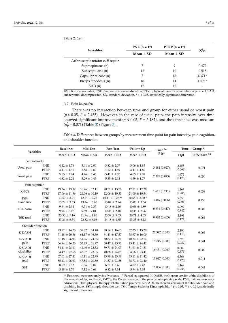
3.2. Pain Intensity
There was no interaction between time and group for either usual or worst pain(p > 0.05, F = 2.455). However, in the case of usual pain, the pain intensity over timeshowed significant improvement (p < 0.05, F = 3.182), and the effect size was medium(η2
p = 0.071) (Table 3) (Figure 3).
Table 3. Differences between groups by measurement time point for pain intensity, pain cognition,and shoulder function.
VariablesBaselines Mid-Test Post-Test Follow-Up Time (a)
F (p)
Time × Group (a)
Mean ± SD Mean ± SD Mean ± SD Mean ± SD F (p) Effect Size (b)
Pain intensity
Usual painPNE 4.12 ± 1.76 3.41 ± 2.00 3.82 ± 2.07 3.06 ± 1.85
3.182 (0.027) 2.455(0.068) 0.071
PTRP 3.41 ± 1.46 3.88 ± 1.80 4.12 ± 1.69 3.41 ± 1.80
Worst painPNE 5.65 ± 2.64 4.76 ± 2.46 5.41 ± 2.37 4.65 ± 2.09
2.399 (0.073) 1.672(0.178) 0.050
PTRP 4.82 ± 2.24 5.29 ± 1.45 5.35 ± 2.12 4.59 ± 1.77
Pain cognition
K-PCSPNE 19.24 ± 13.37 18.76 ± 13.11 20.71 ± 13.78 17.71 ± 12.28
1.611 (0.211) 1.267(0.286) 0.038
PTRP 17.06 ± 11.36 21.06 ± 10.19 22.06 ± 10.35 21.00 ± 10.34
TSK-avoidance
PNE 13.59 ± 3.24 12.24 ± 2.73 10.41 ± 3.26 ** 10.65 ± 3.00 *4.469 (0.006) 5.650
(0.001) 0.150PTRP 13.29 ± 3.53 13.24 ± 3.60 13.82 ± 2.74 13.00 ± 3.34
TSK-harmPNE 9.94 ± 2.14 9.71 ± 2.37 10.18 ± 2.40 10.06 ± 1.89
0.931 (0.417) 0.097(0.942) 0.003
PTRP 9.94 ± 3.07 9.59 ± 2.81 10.35 ± 2.18 10.35 ± 2.96
TSK-totalPNE 23.53 ± 5.16 21.94 ± 4.90 20.59 ± 5.53 20.71 ± 4.65
0.982 (0.405) 2.191(0.121) 0.064
PTRP 23.24 ± 6.34 22.82 ± 6.06 24.18 ± 4.65 23.35 ± 6.13
Shoulder function
K-DASHPNE 73.92 ± 14.75 59.02 ± 14.40 58.14 ± 16.63 52.35 ± 15.29
22.342 (0.000) 2.190(0.118) 0.064
PTRP 71.18 ± 20.36 64.17 ± 16.30 64.41 ± 17.57 58.97 ± 16.00K-SPADI
-painPNE 61.18 ± 26.95 51.06 ± 24.65 50.82 ± 24.21 40.24 ± 22.54
15.245 (0.000) 1.370(0.257) 0.041
PTRP 56.94 ± 26.26 53.29 ± 21.77 50.47 ± 23.92 45.41 ± 24.42K-SPADI
-disabilityPNE 54.41 ± 28.11 41.40 ± 22.52 39.71 ± 24.03 31.91 ± 21.31
16.431 (0.000) 0.080(0.971) 0.002
PTRP 54.49 ± 27.68 43.97 ± 23.35 40.88 ± 24.89 34.56 ± 23.41
K-SPADI-total
PNE 57.01 ± 27.41 45.11 ± 22.79 43.98 ± 23.58 35.11 ± 21.4217.917 (0.000) 0.366
(0.778) 0.011PTRP 55.43 ± 26.83 47.56 ± 20.80 44.57 ± 23.58 38.73 ± 23.40
SSTPNE 8.59 ± 2.92 6.06 ± 1.82 6.71 ± 3.46 4.82 ± 2.43
16.056 (0.000) 1.469(0.236) 0.044
PTRP 8.18 ± 1.70 7.12 ± 1.69 6.82 ± 3.34 5.94 ± 3.03
(a) Repeated-measures analysis of variance, (b) Partial eta squared. K-DASH, the Korean version of the disabilities ofthe arm, shoulder, and hand; K-PCS, the Korean version of the pain catastrophizing scale; PNE, pain neuroscienceeducation; PTRP, physical therapy rehabilitation protocol; K-SPADI, the Korean version of the shoulder pain anddisability index; SST, simple shoulder test; TSK, Tampa Scale for Kinesiophobia. * p < 0.05, ** p < 0.01, statisticallysignificant difference.
Brain Sci. 2022, 12, 764 8 of 14Brain Sci. 2022, 12, 764 8 of 14
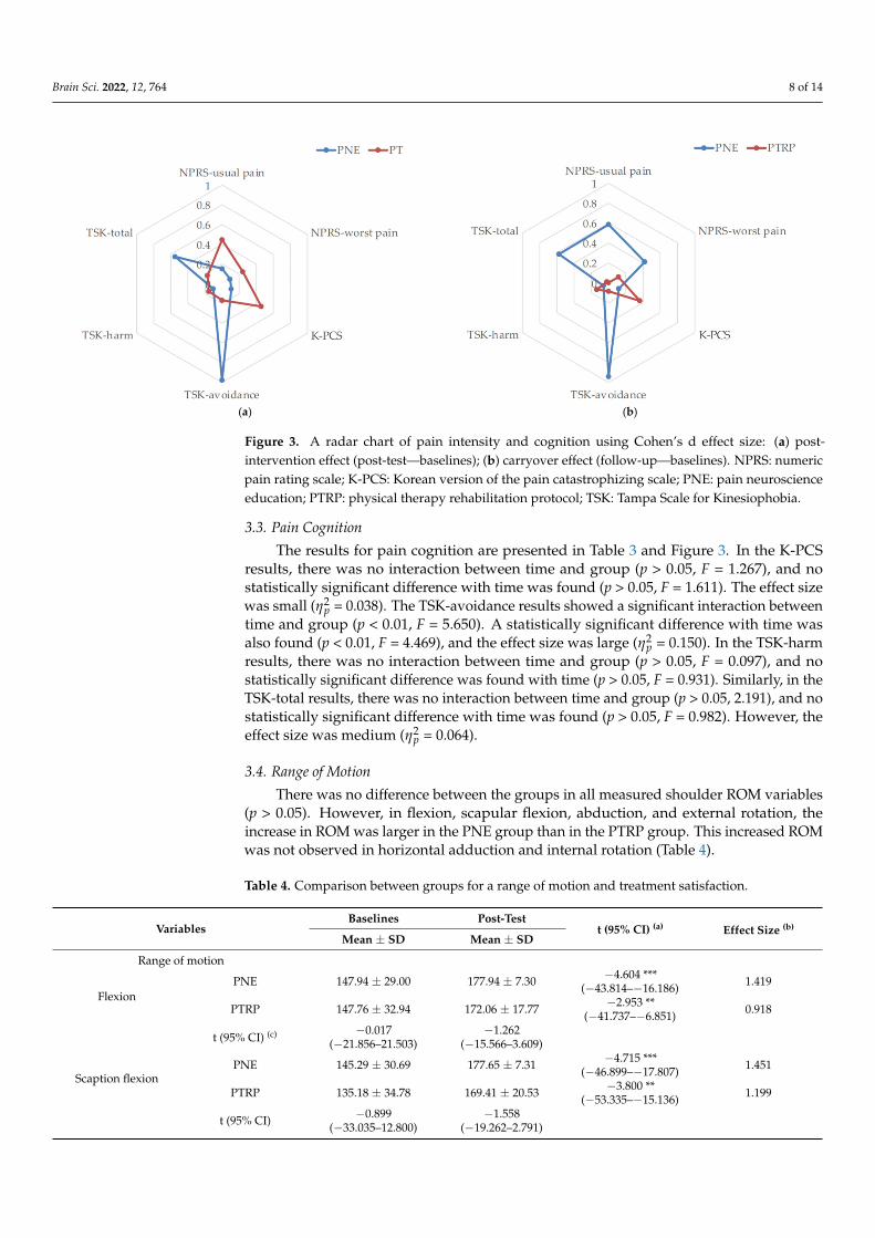
(a) (b)
Figure 3. A radar chart of pain intensity and cognition using Cohen’s d effect size: (a) post‐interven‐
tion effect (post‐test—baselines); (b) carryover effect (follow‐up—baselines). NPRS: numeric pain
rating scale; K‐PCS: Korean version of the pain catastrophizing scale; PNE: pain neuroscience edu‐
cation; PTRP: physical therapy rehabilitation protocol; TSK: Tampa Scale for Kinesiophobia.
3.3. Pain Cognition
The results for pain cognition are presented in Table 3 and Figure 3. In the K‐PCS
results, there was no interaction between time and group (p > 0.05, F = 1.267), and no sta‐
tistically significant difference with time was found (p > 0.05, F = 1.611). The effect size was
small ( = 0.038). The TSK‐avoidance results showed a significant interaction between
time and group (p < 0.01, F = 5.650). A statistically significant difference with time was also
found (p < 0.01, F = 4.469), and the effect size was large ( = 0.150). In the TSK‐harm results, there was no interaction between time and group (p > 0.05, F = 0.097), and no sta‐
tistically significant difference was found with time (p > 0.05, F = 0.931). Similarly, in the
TSK‐total results, there was no interaction between time and group (p > 0.05, 2.191), and
no statistically significant difference with time was found (p > 0.05, F = 0.982). However,
the effect size was medium ( = 0.064).
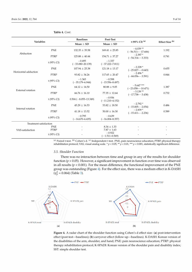
3.4. Range of Motion
There was no difference between the groups in all measured shoulder ROM variables
(p > 0.05). However, in flexion, scapular flexion, abduction, and external rotation, the in‐
crease in ROM was larger in the PNE group than in the PTRP group. This increased ROM
was not observed in horizontal adduction and internal rotation (Table 4).
Table 4. Comparison between groups for a range of motion and treatment satisfaction.
Variables Baselines Post‐Test
t (95% CI) (a) Effect Size (b) Mean ± SD Mean ± SD
Range of motion
Flexion
PNE 147.94 ± 29.00 177.94 ± 7.30 −4.604 *** (−43.814–
−16.186) 1.419
PTRP 147.76 ± 32.94 172.06 ± 17.77 −2.953 ** (−41.737–
−6.851) 0.918
t (95% CI) (c) −0.017 (−21.856–21.503) −1.262 (−15.566–
3.609)
Scaption flex‐
ion
PNE 145.29 ± 30.69 177.65 ± 7.31 −4.715 *** (−46.899–
−17.807) 1.451
PTRP 135.18 ± 34.78 169.41 ± 20.53 −3.800 ** (−53.335–
−15.136) 1.199
t (95% CI) −0.899 (−33.035–12.800) −1.558 (−19.262–
2.791)
Figure 3. A radar chart of pain intensity and cognition using Cohen’s d effect size: (a) post-intervention effect (post-test—baselines); (b) carryover effect (follow-up—baselines). NPRS: numericpain rating scale; K-PCS: Korean version of the pain catastrophizing scale; PNE: pain neuroscienceeducation; PTRP: physical therapy rehabilitation protocol; TSK: Tampa Scale for Kinesiophobia.
3.3. Pain Cognition
The results for pain cognition are presented in Table 3 and Figure 3. In the K-PCSresults, there was no interaction between time and group (p > 0.05, F = 1.267), and nostatistically significant difference with time was found (p > 0.05, F = 1.611). The effect sizewas small (η2
p = 0.038). The TSK-avoidance results showed a significant interaction betweentime and group (p < 0.01, F = 5.650). A statistically significant difference with time wasalso found (p < 0.01, F = 4.469), and the effect size was large (η2
p = 0.150). In the TSK-harmresults, there was no interaction between time and group (p > 0.05, F = 0.097), and nostatistically significant difference was found with time (p > 0.05, F = 0.931). Similarly, in theTSK-total results, there was no interaction between time and group (p > 0.05, 2.191), and nostatistically significant difference with time was found (p > 0.05, F = 0.982). However, theeffect size was medium (η2
p = 0.064).
3.4. Range of Motion
There was no difference between the groups in all measured shoulder ROM variables(p > 0.05). However, in flexion, scapular flexion, abduction, and external rotation, theincrease in ROM was larger in the PNE group than in the PTRP group. This increased ROMwas not observed in horizontal adduction and internal rotation (Table 4).
Table 4. Comparison between groups for a range of motion and treatment satisfaction.
VariablesBaselines Post-Test
t (95% CI) (a) Effect Size (b)
Mean ± SD Mean ± SD
Range of motion
FlexionPNE 147.94 ± 29.00 177.94 ± 7.30 −4.604 ***
(−43.814–−16.186) 1.419
PTRP 147.76 ± 32.94 172.06 ± 17.77 −2.953 **(−41.737–−6.851) 0.918
t (95% CI) (c) −0.017(−21.856–21.503)
−1.262(−15.566–3.609)
Scaption flexionPNE 145.29 ± 30.69 177.65 ± 7.31 −4.715 ***
(−46.899–−17.807) 1.451
PTRP 135.18 ± 34.78 169.41 ± 20.53 −3.800 **(−53.335–−15.136) 1.199
t (95% CI) −0.899(−33.035–12.800)
−1.558(−19.262–2.791)
Brain Sci. 2022, 12, 764 9 of 14
Table 4. Cont.
VariablesBaselines Post-Test
t (95% CI) (a) Effect Size (b)
Mean ± SD Mean ± SD
AbductionPNE 132.35 ± 35.58 169.41 ± 25.85 −4.039 **
(−56.511–−17.606) 1.192
PTRP 125.88 ± 40.44 154.71 ± 37.27 −2.397 *(−54.314–−3.333) 0.741
t (95% CI) −0.495(−33.080–20.139)
−1.337(−37.222–7.811)
Horizontal adductionPNE 107.94 ± 25.38 121.18 ± 13.17 −2.228 *
(−25.827–−0.643) 0.655
PTRP 93.82 ± 34.26 117.65 ± 20.47 −2.496 *(−44.056–−3.591) 0.844
t (95% CI) −1.365(−35.179–6.944)
−0.598(−15.556–8.497)
External rotationPNE 64.12 ± 14.50 80.88 ± 9.05 −5.647 ***
(−23.058–−10.471) 1.387
PTRP 66.76 ± 16.10 77.35 ± 12.64 −3.139 **(−17.738–−3.438) 0.732
t (95% CI) 0.504 (−8.055–13.349) −0.936(−11.210–4.152)
Internal rotationPNE 45.29 ± 16.53 53.82 ± 18.50 −2.792 *
(−15.005–−2.054) 0.486
PTRP 41.18 ± 13.52 50.00 ± 16.30 −2.839 *(−15.411–−2.236) 0.589
t (95% CI) −0.795(−14.670–6.435)
−0.639(−16.004–8.357)
Treatment satisfaction
VAS-satisfactionPNE - 8.34 ± 1.33 - -PTRP - 7.87 ± 1.63 - -
t (95% CI) - −0.922(−1.511–0.569) - -
(a) Paired t-test. (b) Cohen’s d. (c) Independent t-test. PNE, pain neuroscience education; PTRP, physical therapyrehabilitation protocol; VAS, visual analog scale. * p < 0.05, ** p < 0.01, *** p < 0.001, statistically significant difference.
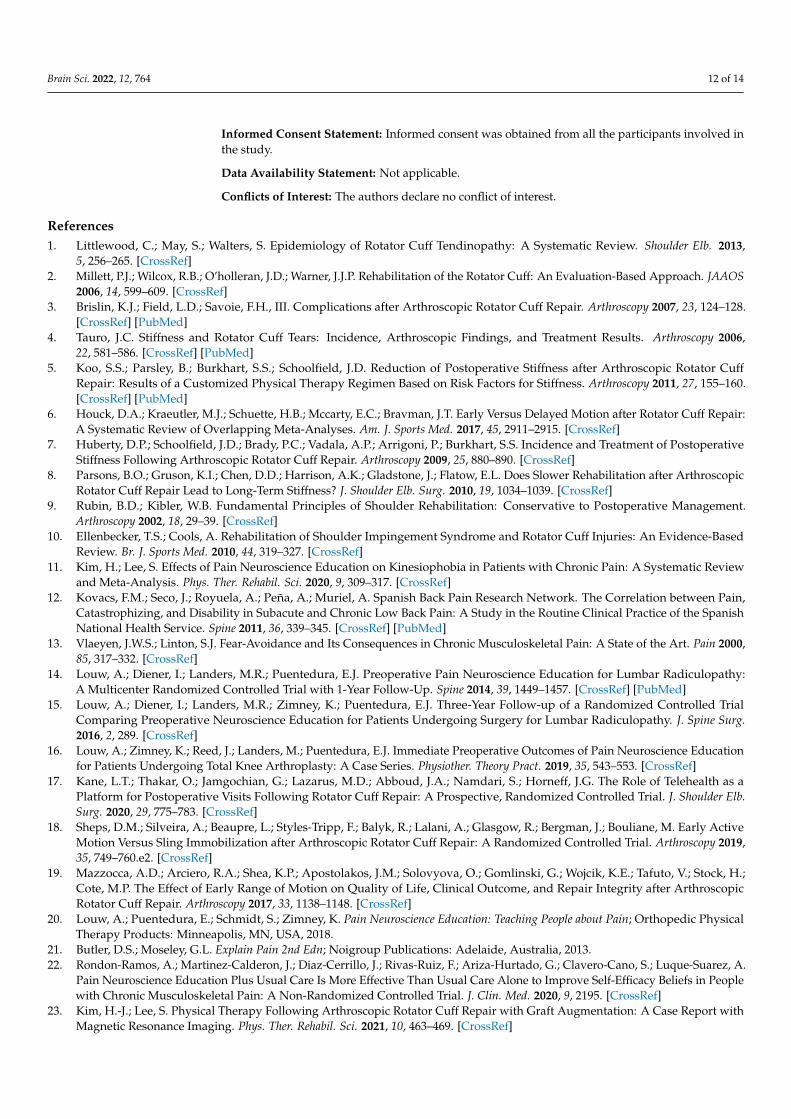
3.5. Shoulder Function
There was no interaction between time and group in any of the results for shoulderfunction (p > 0.05). However, a significant improvement in function over time was observedin all results (p < 0.001). For the mean difference, the functional improvement of the PNEgroup was outstanding (Figure 4). For the effect size, there was a medium effect in K-DASH(η2
p = 0.064) (Table 3).
Brain Sci. 2022, 12, 764 9 of 14
Abduction
PNE 132.35 ± 35.58 169.41 ± 25.85 −4.039 ** (−56.511–
−17.606) 1.192
PTRP 125.88 ± 40.44 154.71 ± 37.27 −2.397 * (−54.314–
−3.333) 0.741
t (95% CI) −0.495 (−33.080–20.139) −1.337 (−37.222–
7.811)
Horizontal
adduction
PNE 107.94 ± 25.38 121.18 ± 13.17 −2.228 * (−25.827–
−0.643) 0.655
PTRP 93.82 ± 34.26 117.65 ± 20.47 −2.496 * (−44.056–
−3.591) 0.844
t (95% CI) −1.365 (−35.179–6.944) −0.598 (−15.556–
8.497)
External rota‐
tion
PNE 64.12 ± 14.50 80.88 ± 9.05 −5.647 *** (−23.058–
−10.471) 1.387
PTRP 66.76 ± 16.10 77.35 ± 12.64 −3.139 ** (−17.738–
−3.438) 0.732
t (95% CI) 0.504 (−8.055–13.349) −0.936 (−11.210–
4.152)
Internal rota‐
tion
PNE 45.29 ± 16.53 53.82 ± 18.50 −2.792 * (−15.005–
−2.054) 0.486
PTRP 41.18 ± 13.52 50.00 ± 16.30 −2.839 * (−15.411–
−2.236) 0.589
t (95% CI) −0.795 (−14.670–6.435) −0.639 (−16.004–
8.357)
Treatment satisfaction
VAS‐satisfac‐
tion
PNE ‐ 8.34 ± 1.33 ‐ ‐
PTRP ‐ 7.87 ± 1.63 ‐ ‐
t (95% CI) ‐ −0.922 (−1.511–0.569) ‐ ‐ (a) Paired t‐test. (b) Cohen’s d. (c) Independent t‐test. PNE, pain neuroscience education; PTRP, phys‐
ical therapy rehabilitation protocol; VAS, visual analog scale. * p < 0.05, ** p < 0.01, *** p < 0.001,
statistically significant difference.
3.5. Shoulder Function
There was no interaction between time and group in any of the results for shoulder
function (p > 0.05). However, a significant improvement in function over time was ob‐
served in all results (p < 0.001). For the mean difference, the functional improvement of
the PNE group was outstanding (Figure 4). For the effect size, there was a medium effect
in K‐DASH ( = 0.064) (Table 3).
(a) (b)
Figure 4. A radar chart of the shoulder function using Cohen’s d effect size: (a) post‐intervention
effect (post‐test—baselines); (b) carryover effect (follow‐up—baselines). K‐DASH: Korean version
of the disabilities of the arm, shoulder, and hand; PNE: pain neuroscience education; PTRP: physical
therapy rehabilitation protocol; K‐SPADI: Korean version of the shoulder pain and disability index;
SST: simple shoulder test.
Figure 4. A radar chart of the shoulder function using Cohen’s d effect size: (a) post-interventioneffect (post-test—baselines); (b) carryover effect (follow-up—baselines). K-DASH: Korean version ofthe disabilities of the arm, shoulder, and hand; PNE: pain neuroscience education; PTRP: physicaltherapy rehabilitation protocol; K-SPADI: Korean version of the shoulder pain and disability index;SST: simple shoulder test.

Brain Sci. 2022, 12, 764 10 of 14
3.6. Treatment Satisfaction
On comparing treatment satisfaction in the post-test, the difference in the average scorewas 0.47, indicating no statistically significant difference (p > 0.05, t = −0.922) (Table 4).
4. Discussion
PNE, which has previously been studied only in central sensitization in chronic pain,was studied for use in subacute conditions for the first time. We recognized that difficultyin pain control and functional improvement in postoperative rehabilitation is a chronicproblem. Therefore, we investigated the beneficial effects of additional PNE on painintensity, pain cognition, range of motion, shoulder function, and treatment satisfactionduring postoperative rehabilitation following ARCR. Our results were generally better thanthose of a previous study [23–25] that investigated the effect of PTRP alone. Additionally,a significant interaction between time and group was found in the avoidance subscale ofpain cognition (F = 5.650, η2
p = 0.150).The primary outcome measure was pain intensity, and no interaction between time
and group was observed with regard to this (p > 0.05). There was no significant decreasein PTRP and PTRP plus PNE compared to the reported MCID (1.1 points) in previousstudies. However, the effect size was found to be medium only for usual pain (η2
p = 0.071).These results may be useful for postoperative pain control. In a digitally assisted versusconventional home-based rehabilitation study for rehabilitation following ARCR, painwas maintained or increased for up to 12 weeks [51]. In addition, when comparing ourresults with those of a study that used steroid injections for pain control, our results weremuch higher at 6 weeks [52]. These results are interpreted as uncontrolled pain during thetissue healing process because POD 4 week is when the proliferative stage ends and thematuration and remodeling stage begins [53].
In this study, no interaction between time and group was found for any of the variablesexcept TSK-avoidance in pain cognition. However, the PTRP and the additional PNEshowed superior results compared to other studies, in pain cognition except for PCS andTSK-harm showed a larger effect size than the PTRP-only group in the results of theindividual analysis of each variable (Table 4) (Figures 3 and 4). Similarly, the results ofshoulder function were not statistically significant (p < 0.05), but additional PNE showeda larger effect size on the improvement of shoulder function. These results were inferredfrom TSK-avoidance, which showed the only significant difference for measured variables.Although there was no significant difference in pain intensity, additional PNE showedbetter overall scores for shoulder function and active ROM (shoulder flexion, scaptionflexion, abduction, and external rotation). This is because the event of surgery enhances fearof pain, and because pain is caused by tissue damage, a certain amount of time is requiredfor pain control. Therefore, shoulder active motion and function were further improvedin patients who received pain education; less avoidance of fear and low awareness ofpain-induced disability were reported [54]. In addition, considering that the levels of fearavoidance and pain interference are risk factors for sustaining pain [55], it can be inferredfrom the results obtained that additional PNE is more beneficial in the follow-up of painintensity (Figure 4).
This is the first randomized controlled trial to perform PNE for postoperative painother than central sensitization. When compared to PTRP, no significant difference wasfound in all variables except TSK-avoidance. However, when the effect sizes were com-pared, additional PNE showed more positive results for most variables. Therefore, it isworth analyzing the relationship between avoidance and other variables. To relieve thefear of movement in active rehabilitation by starting active motion, our PNE had additionalinformation on ARCR and shoulder biomechanics. This was based on the pain–tension–fear cycle reported in other pain theories [56,57]. In other words, PNE is essential inpostoperative rehabilitation to control increased pain due to fear of movement during theimmobilization phase after ARCR. This is because physiological arousal owing to auto-nomic nervous system activation affects pain fear and avoidance behavior, contributing

Brain Sci. 2022, 12, 764 11 of 14
to the maintenance of chronic pain [58]. In addition, in a functional magnetic resonanceimaging study of patients with chronic neck pain and healthy adults, the right dorsolateralprefrontal cortex, an emotional factor of pain, the anterior insular cortex, and increasedfunctional connectivity were significantly related to an increase in TSK [59,60]. The insularcortex is responsible for various higher-order cognitive processes and information relatedto the state of the body during other emotional processes [61]. It has been reported that dis-comfort caused by tonic pain is encoded in the insular cortex and contributes to persistentpain [62]. This is consistent with the contribution of the insular cortex to the constructionof a unique signature/fingerprint of pain experience [63].
Therefore, we assumed that the pain caused by avoidance from fear of movementwas a tonic pain due to tension. Based on the association between the insular cortex andTSK in this cycle, this was thought to contribute to the pain experience. Although therewas no clear difference between the control group (PTRP) and the PNE group, there was asignificant difference in avoidance, a subscale of TSK, and a high effect size was observedin other variables (excluding horizontal adduction and internal rotation).
Our study has several limitations. First, an in-depth analysis was not conductedbecause the measurement of central sensitization suitable for pain education and psy-chological measurement after surgery were not performed. Second, a long-term study isneeded because the intervention and follow-up periods in the postoperative rehabilita-tion study were relatively short. Third, the TSK-11 and SST were directly translated andperformed by the researcher. Fourth, it may be difficult for some to understand PNE bylistening alone, so a tool is needed to check the level of understanding of the educationalcontent. In future studies, it will be necessary to add educational content suitable forvarious patient groups and each condition, as well as PNE for postoperative patients.
As a clinical implication of the results of this study, this was the first study in whichit was confirmed that PNE for neuroplastic pain control in postoperative pain (acute orsubacute pain) control can partially contribute. Postoperative joint immobilization orpassive range of motion exercise is required in the maximum protective phase (acutecondition) after surgery. Although the period differs depending on the affected tissue,in rehabilitation, an active range of motion exercise that requires voluntary movementafter a certain period is more effective for tissue healing. However, the pain experiencecaused by postoperative pain may further affect the pain–tension–fear cycle, contributingto an increase in pain. Therefore, postoperative PNE could be provided as an additionaltreatment option for the improvement of chronically progressive pain and decreasedfunction in postoperative patients.
5. Conclusions
In conclusion, we performed a randomized controlled trial to compare the effectof additional PNE on ARCR patients with PTRP in active rehabilitation after a certainimmobilization phase. Our results showed a decrease in TSK avoidance compared toPTRP after 4 weeks of intervention and in the follow-up evaluation two weeks later.Furthermore, additional PNE showed better effect sizes in pain, range of motion, andshoulder function in ARCR patients. We propose, for the first time, the potential impact ofPNE on postoperative rehabilitation.
Author Contributions: Conceptualization, H.K.; methodology, H.K. and S.L.; software, H.K.; vali-dation, S.L.; formal analysis, H.K. and S.L.; investigation, H.K.; resources, H.K.; data curation, H.K.and S.L.; writing—original draft preparation, H.K.; writing—review and editing, S.L.; supervision,S.L.; project administration, H.K. All authors have read and agreed to the published version ofthe manuscript.
Funding: This study received no external funding.
Institutional Review Board Statement: The study was conducted in accordance with the guidelinesof the Declaration of Helsinki and approved by the Institutional Review Board (IRB) of SahmyookUniversity (IRB No. 2-1040781-A-N-012021010HR).
Brain Sci. 2022, 12, 764 12 of 14
Informed Consent Statement: Informed consent was obtained from all the participants involved inthe study.
Data Availability Statement: Not applicable.
Conflicts of Interest: The authors declare no conflict of interest.
References1. Littlewood, C.; May, S.; Walters, S. Epidemiology of Rotator Cuff Tendinopathy: A Systematic Review. Shoulder Elb. 2013,
5, 256–265. [CrossRef]2. Millett, P.J.; Wilcox, R.B.; O’holleran, J.D.; Warner, J.J.P. Rehabilitation of the Rotator Cuff: An Evaluation-Based Approach. JAAOS
2006, 14, 599–609. [CrossRef]3. Brislin, K.J.; Field, L.D.; Savoie, F.H., III. Complications after Arthroscopic Rotator Cuff Repair. Arthroscopy 2007, 23, 124–128.
[CrossRef] [PubMed]4. Tauro, J.C. Stiffness and Rotator Cuff Tears: Incidence, Arthroscopic Findings, and Treatment Results. Arthroscopy 2006,
22, 581–586. [CrossRef] [PubMed]5. Koo, S.S.; Parsley, B.; Burkhart, S.S.; Schoolfield, J.D. Reduction of Postoperative Stiffness after Arthroscopic Rotator Cuff
Repair: Results of a Customized Physical Therapy Regimen Based on Risk Factors for Stiffness. Arthroscopy 2011, 27, 155–160.[CrossRef] [PubMed]
6. Houck, D.A.; Kraeutler, M.J.; Schuette, H.B.; Mccarty, E.C.; Bravman, J.T. Early Versus Delayed Motion after Rotator Cuff Repair:A Systematic Review of Overlapping Meta-Analyses. Am. J. Sports Med. 2017, 45, 2911–2915. [CrossRef]
7. Huberty, D.P.; Schoolfield, J.D.; Brady, P.C.; Vadala, A.P.; Arrigoni, P.; Burkhart, S.S. Incidence and Treatment of PostoperativeStiffness Following Arthroscopic Rotator Cuff Repair. Arthroscopy 2009, 25, 880–890. [CrossRef]
8. Parsons, B.O.; Gruson, K.I.; Chen, D.D.; Harrison, A.K.; Gladstone, J.; Flatow, E.L. Does Slower Rehabilitation after ArthroscopicRotator Cuff Repair Lead to Long-Term Stiffness? J. Shoulder Elb. Surg. 2010, 19, 1034–1039. [CrossRef]
9. Rubin, B.D.; Kibler, W.B. Fundamental Principles of Shoulder Rehabilitation: Conservative to Postoperative Management.Arthroscopy 2002, 18, 29–39. [CrossRef]
10. Ellenbecker, T.S.; Cools, A. Rehabilitation of Shoulder Impingement Syndrome and Rotator Cuff Injuries: An Evidence-BasedReview. Br. J. Sports Med. 2010, 44, 319–327. [CrossRef]
11. Kim, H.; Lee, S. Effects of Pain Neuroscience Education on Kinesiophobia in Patients with Chronic Pain: A Systematic Reviewand Meta-Analysis. Phys. Ther. Rehabil. Sci. 2020, 9, 309–317. [CrossRef]
12. Kovacs, F.M.; Seco, J.; Royuela, A.; Peña, A.; Muriel, A. Spanish Back Pain Research Network. The Correlation between Pain,Catastrophizing, and Disability in Subacute and Chronic Low Back Pain: A Study in the Routine Clinical Practice of the SpanishNational Health Service. Spine 2011, 36, 339–345. [CrossRef] [PubMed]
13. Vlaeyen, J.W.S.; Linton, S.J. Fear-Avoidance and Its Consequences in Chronic Musculoskeletal Pain: A State of the Art. Pain 2000,85, 317–332. [CrossRef]
14. Louw, A.; Diener, I.; Landers, M.R.; Puentedura, E.J. Preoperative Pain Neuroscience Education for Lumbar Radiculopathy:A Multicenter Randomized Controlled Trial with 1-Year Follow-Up. Spine 2014, 39, 1449–1457. [CrossRef] [PubMed]
15. Louw, A.; Diener, I.; Landers, M.R.; Zimney, K.; Puentedura, E.J. Three-Year Follow-up of a Randomized Controlled TrialComparing Preoperative Neuroscience Education for Patients Undergoing Surgery for Lumbar Radiculopathy. J. Spine Surg.2016, 2, 289. [CrossRef]
16. Louw, A.; Zimney, K.; Reed, J.; Landers, M.; Puentedura, E.J. Immediate Preoperative Outcomes of Pain Neuroscience Educationfor Patients Undergoing Total Knee Arthroplasty: A Case Series. Physiother. Theory Pract. 2019, 35, 543–553. [CrossRef]
17. Kane, L.T.; Thakar, O.; Jamgochian, G.; Lazarus, M.D.; Abboud, J.A.; Namdari, S.; Horneff, J.G. The Role of Telehealth as aPlatform for Postoperative Visits Following Rotator Cuff Repair: A Prospective, Randomized Controlled Trial. J. Shoulder Elb.Surg. 2020, 29, 775–783. [CrossRef]
18. Sheps, D.M.; Silveira, A.; Beaupre, L.; Styles-Tripp, F.; Balyk, R.; Lalani, A.; Glasgow, R.; Bergman, J.; Bouliane, M. Early ActiveMotion Versus Sling Immobilization after Arthroscopic Rotator Cuff Repair: A Randomized Controlled Trial. Arthroscopy 2019,35, 749–760.e2. [CrossRef]
19. Mazzocca, A.D.; Arciero, R.A.; Shea, K.P.; Apostolakos, J.M.; Solovyova, O.; Gomlinski, G.; Wojcik, K.E.; Tafuto, V.; Stock, H.;Cote, M.P. The Effect of Early Range of Motion on Quality of Life, Clinical Outcome, and Repair Integrity after ArthroscopicRotator Cuff Repair. Arthroscopy 2017, 33, 1138–1148. [CrossRef]
20. Louw, A.; Puentedura, E.; Schmidt, S.; Zimney, K. Pain Neuroscience Education: Teaching People about Pain; Orthopedic PhysicalTherapy Products: Minneapolis, MN, USA, 2018.
21. Butler, D.S.; Moseley, G.L. Explain Pain 2nd Edn; Noigroup Publications: Adelaide, Australia, 2013.22. Rondon-Ramos, A.; Martinez-Calderon, J.; Diaz-Cerrillo, J.; Rivas-Ruiz, F.; Ariza-Hurtado, G.; Clavero-Cano, S.; Luque-Suarez, A.
Pain Neuroscience Education Plus Usual Care Is More Effective Than Usual Care Alone to Improve Self-Efficacy Beliefs in Peoplewith Chronic Musculoskeletal Pain: A Non-Randomized Controlled Trial. J. Clin. Med. 2020, 9, 2195. [CrossRef]
23. Kim, H.-J.; Lee, S. Physical Therapy Following Arthroscopic Rotator Cuff Repair with Graft Augmentation: A Case Report withMagnetic Resonance Imaging. Phys. Ther. Rehabil. Sci. 2021, 10, 463–469. [CrossRef]
Brain Sci. 2022, 12, 764 13 of 14
24. Kim, H.-J.; Choi, W.; Jung, J.-H.; Park, S.-G.; Joo, Y.-L.; Lee, S. Effect of Extracorporeal Shock Wave Therapy after ArthroscopicRotator Cuff Repair. 2021. Available online: https://ClinicalTrials.gov/show/NCT04848103 (accessed on 13 April 2022).
25. Kim, H.; Lee, S. Postoperative Rehabilitation Protocol Following Arthroscopic Rotator Cuff Repair: A Prospective Single-ArmPragmatic Interventional Study. Medicina 2022, 58, 729. [CrossRef]
26. Michener, L.A.; Snyder, A.R.; Leggin, B.G. Responsiveness of the Numeric Pain Rating Scale in Patients with Shoulder Pain andthe Effect of Surgical Status. J. Sport Rehabil. 2011, 20, 115–128. [CrossRef] [PubMed]
27. Mintken, P.E.; Glynn, P.; Cleland, J.A. Psychometric Properties of the Shortened Disabilities of the Arm, Shoulder, and HandQuestionnaire (Quickdash) and Numeric Pain Rating Scale in Patients with Shoulder Pain. J. Shoulder Elb. Surg. 2009,18, 920–926. [CrossRef]
28. Sullivan, M.J.; Bishop, S.R.; Pivik, J. The Pain Catastrophizing Scale: Development and Validation. Psychol. Assess. 1995,7, 524. [CrossRef]
29. Cho, S.; Kim, H.-Y.; Lee, J.-H. Validation of the Korean Version of the Pain Catastrophizing Scale in Patients with ChronicNon-Cancer Pain. Qual. Life Res. 2013, 22, 1767–1772. [CrossRef]
30. García-Campayo, J.; Rodero, B.; Del Hoyo, Y.L.; Luciano, J.V.; Alda, M.; Gili, M. Validation of a Spanish Language Version of thePain Self-Perception Scale in Patients with Fibromyalgia. BMC Musculoskelet. Disord. 2010, 11, 255. [CrossRef]
31. George, S.Z.; Valencia, C.; Beneciuk, J.M. A Psychometric Investigation of Fear-Avoidance Model Measures in Patients withChronic Low Back Pain. J. Orthop. Sports 2010, 40, 197–205. [CrossRef]
32. Goubert, L.; Crombez, G.; Van Damme, S.; Vlaeyen, J.W.; Bijttebier, P.; Roelofs, J. Confirmatory Factor Analysis of the TampaScale for Kinesiophobia: Invariant Two-Factor Model across Low Back Pain Patients and Fibromyalgia Patients. Clin. J. Pain 2004,20, 103–110. [CrossRef]
33. Roelofs, J.; Peters, M.L.; McCracken, L.; Vlaeyen, J. The Pain Vigilance and Awareness Questionnaire (Pvaq): Further PsychometricEvaluation in Fibromyalgia and Other Chronic Pain Syndromes. Pain 2003, 101, 299–306. [CrossRef]
34. Gómez-Pérez, L.; López-Martínez, A.E.; Ruiz-Párraga, G.T. Psychometric Properties of the Spanish Version of the Tampa Scale forKinesiophobia (Tsk). J. Pain 2011, 12, 425–435. [CrossRef] [PubMed]
35. Kikuchi, N.; Matsudaira, K.; Sawada, T.; Oka, H. Psychometric Properties of the Japanese Version of the Tampa Scale forKinesiophobia (Tsk-J) in Patients with Whiplash Neck Injury Pain and/or Low Back Pain. J. Orthop. Sci. 2015, 20, 985–992.[CrossRef] [PubMed]
36. Norkin, C.C.; White, D.J. Measurement of Joint Motion: A Guide to Goniometry; FA Davis: Philadelphia, PA, USA, 2016.37. Mullaney, M.J.; McHugh, M.P.; Johnson, C.P.; Tyler, T.F. Reliability of Shoulder Range of Motion Comparing a Goniometer to a
Digital Level. Physiother. Theory Pract. 2010, 26, 327–333. [CrossRef] [PubMed]38. Homsi, C.; Bordalo-Rodrigues, M.; Da Silva, J.J.; Stump, X.M. Ultrasound in Adhesive Capsulitis of the Shoulder: Is Assessment
of the Coracohumeral Ligament a Valuable Diagnostic Tool? Skelet. Radiol. 2006, 35, 673–678. [CrossRef] [PubMed]39. Beaton, D.E.; Katz, J.N.; Fossel, A.H.; Wright, J.G.; Tarasuk, V.; Bombardier, C. Measuring the Whole or the Parts? Validity,
Reliability, and Responsiveness of the Disabilities of the Arm, Shoulder and Hand Outcome Measure in Different Regions of theUpper Extremity. J. Hand Ther. 2001, 14, 128–142. [CrossRef]
40. Lee, J.-Y.; Lim, J.-Y.; Oh, J.H.; Ko, Y.-M. Cross-Cultural Adaptation and Clinical Evaluation of a Korean Version of the Disabilitiesof Arm, Shoulder, and Hand Outcome Questionnaire (K-Dash). J. Shoulder Elb. Surg. 2008, 17, 570–574. [CrossRef]
41. Schmitt, J.S.; Di Fabio, R.P. Reliable Change and Minimum Important Difference (Mid) Proportions Facilitated Group Responsive-ness Comparisons Using Individual Threshold Criteria. J. Clin. Epidemiol. 2004, 57, 1008–1018. [CrossRef]
42. Paul, A.; Lewis, M.; Shadforth, M.F.; Croft, P.R.; Van Der Windt, D.A.W.M.; Hay, E.M. A Comparison of Four Shoulder-SpecificQuestionnaires in Primary Care. Ann. Rheum. Dis. 2004, 63, 1293–1299. [CrossRef]
43. Roach, K.E.; Budiman-Mak, E.; Songsiridej, N.; Lertratanakul, Y. Development of a Shoulder Pain and Disability Index. ArthritisRheumatol. 1991, 4, 143–149. [CrossRef]
44. Choi, Y.; Park, J.W.; Noh, S.; Kim, M.S.; Park, Y.H.; Sung, D.H. Reliability, Validity, and Responsiveness of the Korean Version ofthe Shoulder Disability Questionnaire and Shoulder Rating Questionnaire. Ann. Rehabil. Med. 2015, 39, 705. [CrossRef]
45. Tveitå, E.K.; Sandvik, L.; Ekeberg, O.M.; Juel, N.G.; Bautz-Holter, E. Factor Structure of the Shoulder Pain and Disability Index inPatients with Adhesive Capsulitis. BMC Musculoskelet. Disord. 2008, 9, 1–7. [CrossRef] [PubMed]
46. Williams, J.W., Jr.; Holleman, D.R., Jr.; Simel, D. Measuring Shoulder Function with the Shoulder Pain and Disability Index.J. Rheumatol. 1995, 22, 727–732. [PubMed]
47. Hsu, J.E.; Russ, S.M.; Somerson, J.S.; Tang, A.; Warme, W.J.; Matsen, F.A., III. Is the Simple Shoulder Test a Valid OutcomeInstrument for Shoulder Arthroplasty? J. Shoulder Elbow Surg. 2017, 26, 1693–1700. [CrossRef] [PubMed]
48. Tashjian, R.Z.; Deloach, J.; Green, A.; Porucznik, C.A.; Powell, A.P. Minimal Clinically Important Differences in Ases and SimpleShoulder Test Scores after Nonoperative Treatment of Rotator Cuff Disease. JBJS 2010, 92, 296–303. [CrossRef] [PubMed]
49. Tashjian, R.Z.; Bradley, M.P.; Tocci, S.; Rey, J.; Henn, R.F.; Green, A. Factors Influencing Patient Satisfaction after Rotator CuffRepair. J. Shoulder Elb. Surg. 2007, 16, 752–758. [CrossRef]
50. Cohen, J. A Power Primer. Psychol. Bull. 1992, 112, 155. [CrossRef]51. Correia, F.D.; Molinos, M.; Luís, S.; Carvalho, D.; Carvalho, C.; Costa, P.; Seabra, R.; Francisco, G.; Bento, V.; Lains, J. Digitally
Assisted Versus Conventional Home-Based Rehabilitation after Arthroscopic Rotator Cuff Repair: A Randomized ControlledTrial. Am. J. Phys. Med. Rehabil. 2021, 101, 237. [CrossRef]
Brain Sci. 2022, 12, 764 14 of 14
52. Ha, J.-W.; Kim, H.; Kim, S.H. Effects of Steroid Injection During Rehabilitation after Arthroscopic Rotator Cuff Repair. Clin.Shoulder Elb. 2021, 24, 166. [CrossRef]
53. Conti, M.; Garofalo, R.; Rose, G.D.; Massazza, G.; Vinci, E.; Randelli, M.; Castagna, A. Post-Operative Rehabilitation after SurgicalRepair of the Rotator Cuff. Musculoskelet. Surg. 2009, 93, 55–63. [CrossRef]
54. Fletcher, C.; Bradnam, L.; Barr, C. The Relationship between Knowledge of Pain Neurophysiology and Fear Avoidance in Peoplewith Chronic Pain: A Point in Time, Observational Study. Physiother. Theory Pract. 2016, 32, 271–276. [CrossRef]
55. Smith, P.J.A.; Russo, P.L.; Santayana, P.N. Fear Avoidance Predicts Persistent Pain in Young Adults with Low Back Pain:A Prospective Study. J. Orthop. Sports Phys. Ther. 2021, 51, 383–391. [CrossRef] [PubMed]
56. Rakhshaee, Z. Effect of Three Yoga Poses (Cobra, Cat and Fish Poses) in Women with Primary Dysmenorrhea: A RandomizedClinical Trial. J. Pediatr. Adolesc. Gynecol. 2011, 24, 192–196. [CrossRef] [PubMed]
57. Ben-Menachem, M. Treatment of Dysmenorrhea: A Relaxation Therapy Program. Int. J. Gynaecol. Obstet. 1980, 17, 340–342.[CrossRef] [PubMed]
58. Norton, P.J.; Asmundson, G.J. Amending the Fear-Avoidance Model of Chronci Pain: What Is the Role of Physiological Arousal?Behav. Ther. 2003, 34, 17–30. [CrossRef]
59. Ihara, N.; Wakaizumi, K.; Nishimura, D.; Kato, J.; Yamada, T.; Suzuki, T.; Hashiguchi, S.; Terasawa, Y.; Kosugi, S.; Morisaki, H.Aberrant Resting-State Functional Connectivity of the Dorsolateral Prefrontal Cortex to the Anterior Insula and Its Associationwith Fear Avoidance Belief in Chronic Neck Pain Patients. PLoS ONE 2019, 14, e0221023. [CrossRef]
60. Apkarian, A.V.; Bushnell, M.C.; Treede, R.-D.; Zubieta, J.-K. Human Brain Mechanisms of Pain Perception and Regulation inHealth and Disease. Eur. J. Pain 2005, 9, 463–484. [CrossRef]
61. Gogolla, N. The Insular Cortex. Curr. Biol. 2017, 27, R580–R586. [CrossRef]62. Schreckenberger, M.; Siessmeier, T.; Viertmann, A.; Landvogt, C.; Buchholz, H.-G.; Rolke, R.; Treede, R.-D.; Bartenstein, P.;
Birklein, F. The Unpleasantness of Tonic Pain Is Encoded by the Insular Cortex. Neurology 2005, 64, 1175–1183. [CrossRef]63. Starr, C.J.; Sawaki, L.; Wittenberg, G.; Burdette, J.H.; Oshiro, Y.; Quevedo, A.S.; Coghill, R.C. Roles of the Insular Cortex in the
Modulation of Pain: Insights from Brain Lesions. J. Neurosci. 2009, 29, 2684–2694. [CrossRef]
Related Documents