Cancer Chemother Pharmacol (1996) 37:479-485 9 Springer-Verlag 1996 Linda Hogarth. Martin English. Lisa Price. Ruth Wyllie Andrew D.J. Pearson .Andrew G. Hall The effect of treatment with high dose melphalan, cisplatin or carboplatin on levels of glutathione in plasma, erythrocytes, mononuclear cells and urine Received: 6 October 1994/Accepted: 22 May 1995 Abstract Glutathione (GSH) has been implicated as an important factor in the detoxification of many elec- trophilic xenobiotics, including agents used in cytotoxic chemotherapy. Maintenance of high levels of GSH in normal tissues is believed to be important in the prevention of drug-induced toxicity. Previous stud- ies have indicated that exposure of cells to some toxic electrophiles both in vitro and in vivo can cause a tem- porary decrease in intracellular levels of GSH. In this paper we report that in a series of 22 children and young adults treated with high dose melphalan (ten courses studied, all 200 mg/mZ), cisplatin (eight courses, 80-104mg/m 2) or carboplatin (seven courses, 507- 750 mg/m 2) there was no significant alteration in the level of plasma, erythrocyte or urine GSH in the period immediately following drug administration. Fluctu- ations in the level of GSH in mononuclear cells were observed in some patients but did not follow any con- sistent pattern and were similar to those observed in a series of nine normal adult controls over the same time course. These results suggest that for melphalan, cisplatin and carboplatin, drug-GSH adduct formation is insufficient to cause a measurable decrease in in- tracellular GSH levels in normal peripheral haema- topoietic cells during the course of treatment. Key words Glutathione" Cytotoxic chemotherapy" Cisplatin 9Carboplatin 9Melphalan 9Nephrotoxicity L. Hogarth" A.G. Hall (~) Leukaemia Research Fund Laboratory, 4th Floor, Cookson Building, Medical School, Framlington Place, Newcastle upon Tyne, UK, NE2 4HH M. English" L. Price" R. Wyllie 9A.D.J. Pearson Department of Child Health, University of Newcastle upon Tyne, UK Introduction Glutathione (GSH) is the most abundant non-protein intracellular thiol and, as such, is an essential compon- ent of the system which maintains a reducing environ- ment within the cell. In addition, GSH is involved in the detoxification of a wide range of electrophilic xenobiotics through the formation of inactive conju- gate~, which may be subsequently metabolised and excreted as mercapturic acids [1]. An elevation in the level of cytoplasmic GSH has been implicated in the development of tumour resistance to a number of drugs used in the treatment of cancer, including the bifunc- tional alkylating agent, melphalan [2], and the plati- num compound, cisplatin [-3], and also appears to play an important role in the protection of normal tissues against drug-induced damage, including cisplatin- induced nephrotoxicity. Administration of glutathione or glutathione esters has been shown to reduce cis- platin-induced kidney damage in both animal [-4] and clinical [-5,6] studies. Conversely, depletion of GSH by the administration of buthionine sulfoximine aggra- vates cisplatin-induced nephrotoxicity in mice [-7] and young rats [8]. For several compounds, such as menadione [-9] and diethyl maleate [10], the formation of GSH adducts can lead to a significant decrease in intracellular GSH, which, in turn, may allow cellular damage through the unopposed action of oxygen free radicals. It has been shown that exposure of kidney cortical slices [11] or a mouse mammary tumour cell line [12] to cisplatin in vitro results in glutathione depletion but the effects of this drug on GSH levels in vivo have not been studied. Treatment of mice with cyclophosphamide has been shown to cause an initial reduction in GSH levels in the bone marrow, followed 5-7 days later by a rebound increase [13]. It has been suggested that this phenom- enon may underlie the observation that "priming" mice with a low dose of cyclophosphamide will protect them

Welcome message from author
This document is posted to help you gain knowledge. Please leave a comment to let me know what you think about it! Share it to your friends and learn new things together.
Transcript
Cancer Chemother Pharmacol (1996) 37:479-485 �9 Springer-Verlag 1996
Linda Hogarth. Martin English. Lisa Price. Ruth Wyllie Andrew D.J. Pearson .Andrew G. Hall
The effect of treatment with high dose melphalan, cisplatin or carboplatin on levels of glutathione in plasma, erythrocytes, mononuclear cells and urine
Received: 6 October 1994/Accepted: 22 May 1995
Abstract Glutathione (GSH) has been implicated as an important factor in the detoxification of many elec- trophilic xenobiotics, including agents used in cytotoxic chemotherapy. Maintenance of high levels of GSH in normal tissues is believed to be important in the prevention of drug-induced toxicity. Previous stud- ies have indicated that exposure of cells to some toxic electrophiles both in vitro and in vivo can cause a tem- porary decrease in intracellular levels of GSH. In this paper we report that in a series of 22 children and young adults treated with high dose melphalan (ten courses studied, all 200 mg/mZ), cisplatin (eight courses, 80-104mg/m 2) or carboplatin (seven courses, 507- 750 mg/m 2) there was no significant alteration in the level of plasma, erythrocyte or urine GSH in the period immediately following drug administration. Fluctu- ations in the level of GSH in mononuclear cells were observed in some patients but did not follow any con- sistent pattern and were similar to those observed in a series of nine normal adult controls over the same time course. These results suggest that for melphalan, cisplatin and carboplatin, drug-GSH adduct formation is insufficient to cause a measurable decrease in in- tracellular GSH levels in normal peripheral haema- topoietic cells during the course of treatment.
Key words Glutathione" Cytotoxic chemotherapy" Cisplatin �9 Carboplatin �9 Melphalan �9 Nephrotoxicity
L. Hogarth" A.G. Hall ( ~ ) Leukaemia Research Fund Laboratory, 4th Floor, Cookson Building, Medical School, Framlington Place, Newcastle upon Tyne, UK, NE2 4HH
M. English" L. Price" R. Wyllie �9 A.D.J. Pearson Department of Child Health, University of Newcastle upon Tyne, UK
Introduction
Glutathione (GSH) is the most abundant non-protein intracellular thiol and, as such, is an essential compon- ent of the system which maintains a reducing environ- ment within the cell. In addition, GSH is involved in the detoxification of a wide range of electrophilic xenobiotics through the formation of inactive conju- gate~, which may be subsequently metabolised and excreted as mercapturic acids [1]. An elevation in the level of cytoplasmic GSH has been implicated in the development of tumour resistance to a number of drugs used in the treatment of cancer, including the bifunc- tional alkylating agent, melphalan [2], and the plati- num compound, cisplatin [-3], and also appears to play an important role in the protection of normal tissues against drug-induced damage, including cisplatin- induced nephrotoxicity. Administration of glutathione or glutathione esters has been shown to reduce cis- platin-induced kidney damage in both animal [-4] and clinical [-5, 6] studies. Conversely, depletion of GSH by the administration of buthionine sulfoximine aggra- vates cisplatin-induced nephrotoxicity in mice [-7] and young rats [8].
For several compounds, such as menadione [-9] and diethyl maleate [10], the formation of GSH adducts can lead to a significant decrease in intracellular GSH, which, in turn, may allow cellular damage through the unopposed action of oxygen free radicals. It has been shown that exposure of kidney cortical slices [11] or a mouse mammary tumour cell line [12] to cisplatin in vitro results in glutathione depletion but the effects of this drug on GSH levels in vivo have not been studied.
Treatment of mice with cyclophosphamide has been shown to cause an initial reduction in GSH levels in the bone marrow, followed 5-7 days later by a rebound increase [13]. It has been suggested that this phenom- enon may underlie the observation that "priming" mice with a low dose of cyclophosphamide will protect them
480
against a subsequent high dose [14], an approach which has been exploited clinically in patients receiving high dose melphalan [-15].
Lind et al. [16] demonstrated that treatment of lym- phoid cells in vitro with 4-hydroperoxifosfamide or chloroacetaldehyde caused a marked reduction in GSH levels, presumably due to the formation of adducts with the drug or its metabolites. A similar depletion was observed in the lymphocytes of a patient undergoing treatment with ifosfamide, but the effect of other elec- trophilic drugs on GSH levels in patients has not been extensively reported.
We have previously reported that patients treated with a combination of high dose melphalan and car- boplatin prior to bone marrow transplantation experi- enced severe nephrotoxicity [-17]. This was unexpected in view of the fact that these drugs are associated with minimal nephrotoxicity when administered alone, and may be related to the dose intensity of the schedule employed, as renal toxicity was not observed when the same drugs were given over a longer period [18]. In view of the evidence that nephrotoxicity associated with cisplatin has been linked with GSH depletion in renal tubular cells, we postulated that the combination of melphalan and carboplatin may produce toxicity by an additive effect on intracellular GSH levels. In order to test this hypothesis, and to establish if drugs apart from the oxazophophorenes may affect GSH homeo- stasis, we studied the effect of chemotherapy on GSH
levels in the peripheral mononuclear cells, erythrocytes, plasma and urine of patients undergoing treatment with cisplatin, carboplatin or melphalan.
Materials and methods
Patients and samples
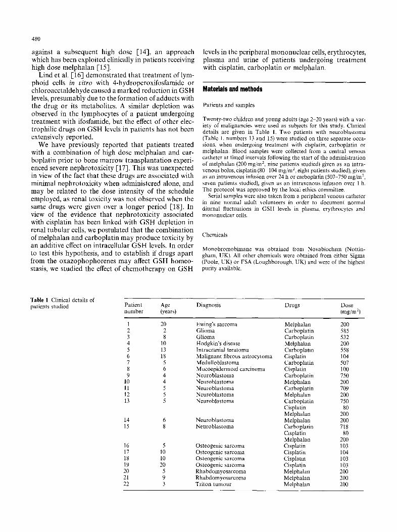
Twenty-two children and young adults (age 2-20 years) with a var- iety of malignancies were used as subjects for this study. Chnical details are given in Table 1. Two patients with neuroblastoma (Table 1, numbers 13 and 15) were studied on three separate occa- sions, when undergoing treatment with cisplatin, carboplatin or melphalan. Blood samples were collected from a central venous catheter at timed intervals following the start of the administration of melphalan (200 mg/m 2, nine patients studied) given as an intra- venous bolus, cisplatin (80-104 mg/m 2, eight patients studied), given as an intravenous infusion over 24 h or carboplatin (507-750 mg/m 2, seven patients studied), given as an intravenous infusion over 1 h. The protocol was approved by the local ethics committee.
Serial samples were also taken from a peripheral venous catheter in nine normal adult volunteers in order to document normal diurnal fluctuations in GSH levels in plasma, erythrocytes and mononuclear cells.
Chemicals
Monobromobimane was obtained from Novabiochem (Nottin- gham, UK). All other chemicals were obtained from either Sigma (Poole, UK) or FSA (Loughborough, UK) and were of the highest purity available.
Table 1 Clinical details of patients studied Patient Age Diagnosis Drugs Dose
number (years) (mg/m 2)
1 20 Ewing's sarcoma Melphalan 200 2 2 Glioma Carboplatin 585 3 8 Glioma Carboplatin 532 4 10 Hodgkin's disease Melphalan 200 5 13 Intracranial teratoma Carboplatm 558 6 18 Malignant fibrous astrocytoma Cisplatin 104 7 5 Medulloblastoma Carboplatin 507 8 6 Mucoepidermold carcinoma Cisplatin 100 9 4 Neuroblastoma Carboplatin 750
10 4 Neuroblastoma Melphalan 200 11 5 Neuroblastoma Carboplatin 709 12 5 Neuroblastoma Melphalan 200 13 5 Neuroblastoma Carboplatin 750
Cisplatin 80 Melphalan 200
14 6 Neuroblastoma Melphalan 200 15 8 Neuroblastoma Carboplatin 718
Clsplatin 80 Melphalan 200
16 5 Osteogenic sarcoma Clsplatin 103 17 10 Osteogenic sarcoma Cisplatin 104 18 10 Osteogenic sarcoma Cisplatln 103 19 20 Osteogenic sarcoma Cisplatin 103 20 5 Rhabdomyosarcoma Melphalan 200 21 9 Rhabdomyosarcoma Melphalan 200 22 3 Triton tumour Melphalan 200
Determination of glutathlone levels
GSH levels in erythrocytes, mononuclear cells and plasma were measured by the method of Colgreave and Moldeus [19] with minor alterations. Samples (5 ml) were collected into pre-cooled tubes containing lithium heparin and processed immediately to minimise errors caused by GSH oxidation or breakdown [20]. For the analy- sis of plasma GSH, 0.5 ml of whole blood was centrifuged at 13 000 rpm for 1 min in a microcentrifuge. One hundred and fifty microlitres of plasma was added to an equal volume of 8 mM monobromobimane in 50 mM N-ethylmaleimide, pH 8.0. Fifteen microlitres of 100% (w/v) trichloroacetic acid (TCA) was added after 5 min Incubation in the dark and the sample stored at - 80~ prior to analysis. Results
For the measurement of erythrocyte GSH, 0.4 ml of whole blood was mixed with an equal volume of phosphate-buffered saline (PBS) and passed down a 3-ml column of a slurry consisting of 50% (w/w) microcrystalline cellulose and 50% a-cellulose in order to remove white blood cells. Cells were washed in 20 ml of PBs and resus- pended in 10ml of PBS. An aliquot was mixed with mono- bromobimane and TCA as described above. Cells were counted using a Coulter counter.
For the measurement of GSH in mononuclear cells, whole blood was separated over Lymphoprep (Nycomed, Oslo, Norway) and washed in PBS. Red cells were lysed by incubation in a solution containing 0.83% (w/v) NH4CI, 0.037% (w/v) EDTA and 0.1% KHCO 3 for 4 min prior to resuspension in 0.4 ml of PBS. Cells were counted using a haemocytometer and viability assessed by Trypan Blue exclusion. The suspension was mixed with monobromobimane and TCA as described above.
For the estimation of the amount of derivitised GSH, samples were centrifuged at 13 000 rpm for 10 min using a microcentrifuge and 25 lal aliquots of the supernatant applied to a 5 gm Spherisorb ODS column (4.6x 150mm, Jones Chromatography, Hengoed, UK) equilibrated with 10% acetonitrile, 0.25% acetic acid (solvent A). High pressure liquid chromatography (HPLC) was performed using a Waters model 625 pump and system controller (Millipore, Watford, UK). Peaks were detected using a Waters model 470 fluorescence detector set at an excitation wavelength of 394 nm and measuring at 480 nm. The flow rate was 1 ml/min. After 5-rain isocratic flow, elutlon of derivitised GSH was achieved using 75% acetonltrlle, 0.25% acetic acid (solvent B) using a convex gradient (curve 6) over 8 rain followed by a 4-rain flow with solvent B. GSH concentrations were estimated from a calibration curve generated using reduced glutathione mixed with 1.5 mM dlthioth- reitol. The lower limit of detection using this assay was found to be 0.5 lamol/1.
Monobromoblmane derivitisation could not be used for the deter- mination of levels of GSH in urine due to the presence of interfering peaks during HPLC. For these samples orthophthaldehyde (OPA) was used as described by Neuschwander-Tetri and Roll [21], to Subjects measure total (1.e. oxidised and reduced) GSH. A 100-gl portion of urine was mixed with an equal volume of 25 mM dithiothreitol and 50 gl of 0.1 M TRIS-HC1 pH 8.5. After incubation for 30 min on ice, proteins were precipitated by the a~dditlon of 750 p.1 of 2.5% (w/v) sulphosalicylic acid and samples centrifuged for 5 min at 13 000 rpm in a microcentrifuge. A 200-gl aliquot of the supernatant was mixed with an equal volume of 32 mM OPA in 0.4 M potassium borate
481
pH 9.9. After incubation at room temperature for 1 mm 2 ml of 100 mM sodium phosphate pH 7 was added and the samples cenm- fuged for 10 min at 13 000 rpm in a mlcrocentrlfuge. A 100-p.l sample of the supernatant was applied to a 5-gm Sphensorb ODS column (4.6 x 150 ram, Jones Chromatography, Hengoed, UK) equilibrated in 0 15 M sodium acetate pH 7 with 6% (v/v) methanol Denvltlsed products were eluted with a 6 100% linear gradient of methanol using the HPLC apparatus described above Peaks were detected using an excitation wavelength of 338 nm and an enusslon wavelength of 425 nm. The lower limit of detection for this assay ~as found to be 10 gmol/1.
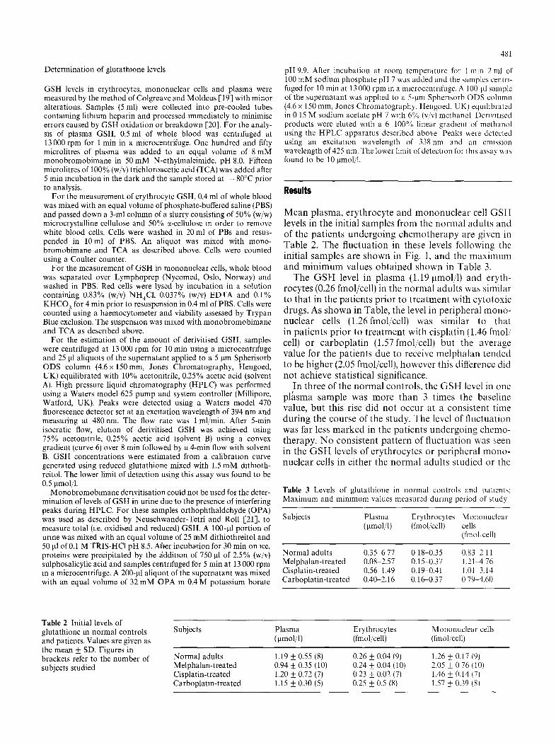
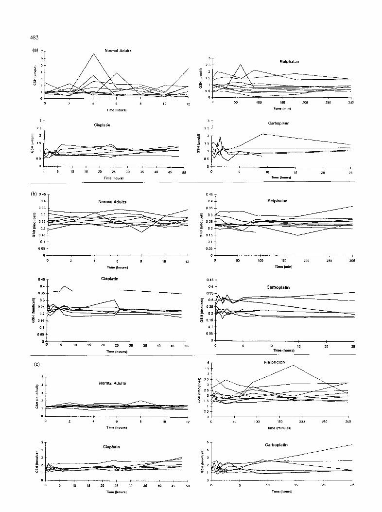
Mean plasma, erythrocyte and mononuclear cell GSH levels in the initial samples from the normal adults and of the patients undergoing chemotherapy are given in Table 2. The fluctuation in these levels following the initial samples are shown in Fig. 1, and the maximum and minimum values obtained shown in Table 3.
The GSH level in plasma (1.19 ~tmol/1) and eryth- rocytes (0.26 fmol/cell) in the normal adults was similar to that in the patients prior to treatment with cytotoxic drugs. As shown in Table, the level in peripheral mono- nuclear cells (1.26fmol/cell) was similar to that in patients prior to treatment with cisplatin (1.46 fmol/ cell) or carboplatin (1.57 fmol/cell) but the average value for the patients due to receive melphalan tended to be higher (2.05 fmol/cell), however this difference did not achieve statistical significance.
In three of the normal controls, the GSH level in one pl~tsma sample was more than 3 times the baseline value, but this rise did not occur at a consistent time during the course of the study. The level of fluctuation was far less marked in the patients undergoing chemo- therapy. No consistent pattern of fluctuation was seen in the GSH levels of erythrocytes or peripheral mono- nuclear cells in either the normal adults studied or the
Table 3 Levels of glutathione in normal controls and patients; Maximum and minimum values measured during period of study
Plasma Erythrocytes Mononuclear (~tmol/1) (fmol/cell) cells
(fmol/cell)
Normal adults 0.35 6 77 0 18-0.35 0.83 2 11 Melphalan-treated 0.08 2.57 0.15-0.37 1.21 476 Cisplatin-treated 0.56 1 .49 0.19-0.41 1.01 3.14 Carboplatin-treated 0.40-2.16 0.16-0.37 0 79 4.60
Table 2 Initial levels of glutathione in normal controls and patients. Values are given as the mean + SD. Figures in brackets refer to the number of subjects studied
Subjects Plasma (gmol/1)
Erythrocytes Mononuclear cells (fmol/cell) (fmol/cell)
Normal adults 1.19 _+ 0.55 (8) 0.26 _+ 0.04 (9) 1.26 + 0.17 (9) Melphalan-treated 0.94 _ 0.35 (10) 0.24 _+ 0.04 (10) 2.05 _+ 0 76 (10) Cisplatin-treated 1.20 _+ 0.72 (7) 0 23 _ 0.02 (7) 1.46 _+ 0.14 (7) Carboplatln-treated 1.15 _+ 0.30 (5) 0.25 _+ 0.5 (8) 1.57 + 0.39 (8)
482
(a) 'l 61
E 4
~ 3 ~ 2
3 ,
25
_~ 2
_= 15
O5
Normal A d u l t s
I I I I ~
2 4 6 8 10 12
Time {hours)
Cisp la t in
I I I I / I I I I
5 10 15 20 25 30 35 40 45
Time ihoutsl
I
5O
3 t M e l p h a l a n
2
0 5
o I I T I I I I 0 50 lao 150 200 250 300
Time (rain)
3
2 5
~ 2
~ 1 5
,
O5
0
Cafbop la t in
I 115 ( I I0 20 25
Time (hoursl
(b) o,s T
0o:; Norma,,ou,,s -o3 i 025
0.2
L~ 015
O0
0 2 4 6 8 10 12
Time (hours)
0 45
0.4
0 35
03 .=. 025
02
015
01
0 05 �9
0 0
C i s p l a t i n
1~0 1'5 ' i , , i 20 25 30 35 40 45
Time (hours)
50
13 45 -
0 4 -
0 35 -
0 3 -
0 25 -
0 2 -
015 -
0 1 -
0.05
M e i p h a l a n
~ : ~ ~ . . . . .
J r
_ f
I ] [
100 150 200
Time (rain)
J
250
d
300
0 45
O4
0,35
~ 0 3 u ~ 0 2 5 �9
~ 0 2
0.15,
0 1 ,
0 05 �9
0
0
C a r b o p i a t i n
[ I r ~ I
5 10 15 20 25 Time (hours)
(c)
5 T
4 J- Norma l A d u l l s
I I I ] I
0 2 4 6 8 10
Time (hourS)
5
C i s p l a t i n
o g i -
0 I t I I I I ~ I l
15 20 25 30 35 40 45 50 0 5 10
Time (hours)
5
4 5 .
4 -
3 5 -
~ 2 5 -
= 2 -
1
O5
0
M e l p n a l a n
I I I I I I
50 100 150 200 250 300
Time (mlnules)
5
I
/ J
I i I
0 5 10 15 20 25
Time (hours)
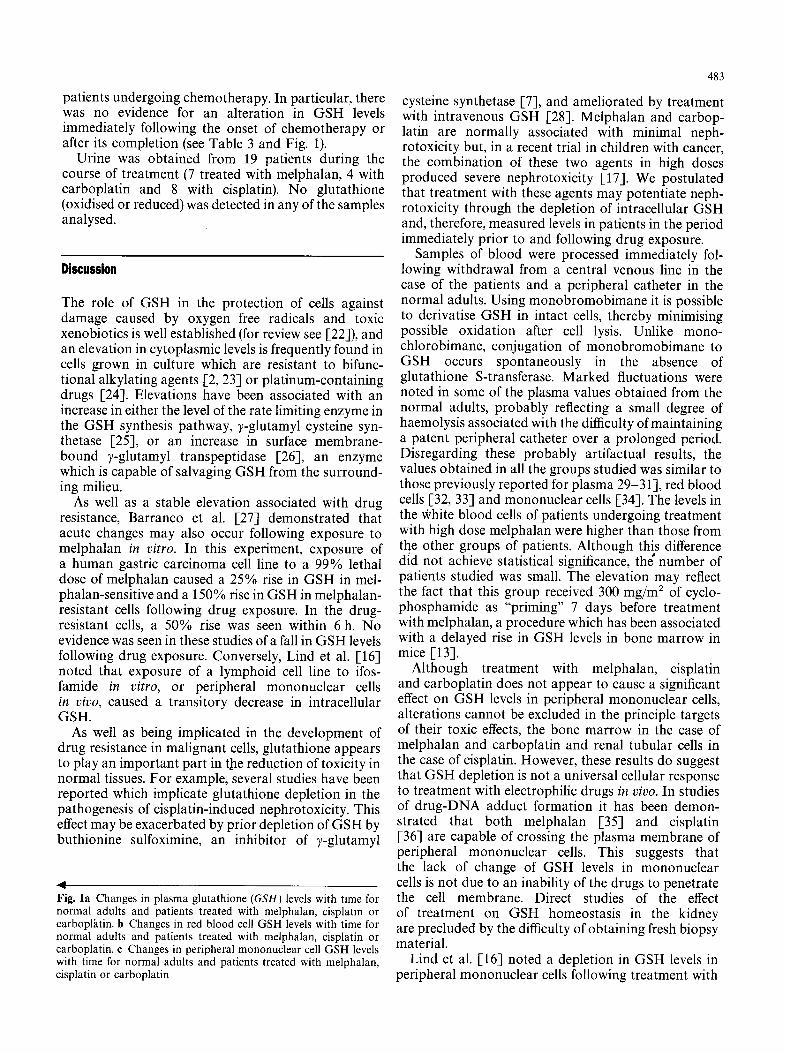
patients undergoing chemotherapy. In particular, there was no evidence for an alteration in GSH levels immediately following the onset of chemotherapy or after its completion (see Table 3 and Fig. 1).
Urine was obtained from 19 patients during the course of treatment (7 treated with melphalan, 4 with carboplatin and 8 with cisplatin). No glutathione (oxidised or reduced) was detected in any of the samples analysed.
Discussion
The role of GSH in the protection of cells against damage caused by oxygen free radicals and toxic xenobiotics is well established (for review see [22]), and an elevation in cytoplasmic levels is frequently found in cells grown in culture which are resistant to bifunc- tional alkylating agents [2, 23] or platinum-containing drugs [24]. Elevations have been associated with an increase in either the level of the rate limiting enzyme in the GSH synthesis pathway, 7-glutamyl cysteine syn- thetase [25], or an increase in surface membrane- bound 7-glutamyl transpeptidase [26], an enzyme which is capable of salvaging GSH from the surround- ing milieu.
As well as a stable elevation associated with drug resistance, Barranco et al. [27] demonstrated that acute changes may also occur following exposure to melphalan in vitro. In this experiment, exposure of a human gastric carcinoma cell line to a 99% lethal dose of melphalan caused a 25% rise in GSH in mel- phalan-sensitive and a 150% rise in GSH in melphalan- resistant cells following drug exposure. In the drug- resistant cells, a 50% rise was seen within 6 h. No evidence was seen in these studies of a fall in GSH levels following drug exposure. Conversely, Lind et al. [16] noted that exposure of a lymphoid cell line to ifos- famide in vitro, or peripheral mononuclear cells in vivo, caused a transitory decrease in intracellular GSH.
As well as being implicated in the development of drug resistance in malignant cells, glutathione appears to play an important part in tJae reduction of toxicity in normal tissues. For example; several studies have been reported which implicate glutathione depletion in the pathogenesis of cisplatin-induced nephrotoxicity. This effect may be exacerbated by prior depletion of GSH by buthionine sulfoximine, an inhibitor of 7-glutamyl
Fig. la Changes in plasma glutathione (GSH) levels with time for normal adults and patients treated with melphalan, cisplatm or carbopl~ttin, b Changes in red blood cell GSH levels with time for normal adults and patients treated with melphalan, cisplatin or carboplatin, c Changes in peripheral mononuclear cell GSH levels with time for normal adults and patients treated with melphalan, cisplatin or carboplatin
483
cysteine synthetase [7], and ameliorated by treatment with intravenous GSH [28]. Melphalan and carbop- latin are normally associated with minimal neph- rotoxicity but, in a recent trial in children with cancer, the combination of these two agents in high doses produced severe nephrotoxicity [17]. We postulated that treatment with these agents may potentiate neph- rotoxicity through the depletion of intracellular GSH and, therefore, measured levels in patients in the period immediately prior to and following drug exposure.
Samples of blood were processed immediately fol- lowing withdrawal from a central venous line in the case of the patients and a peripheral catheter in the normal adults. Using monobromobimane it is possible to derivatise GSH in intact cells, thereby minimising possible oxidation after cell lysis. Unlike mono- chlorobimane, conjugation of monobromobimane to GSH occurs spontaneously in the absence of glutathione S-transferase. Marked fluctuations were noted in some of the plasma values obtained from the normal adults, probably reflecting a small degree of haemolysis associated with the difficulty of maintaining a patent peripheral catheter over a prolonged period. Disregarding these probably artifactual results, the values obtained in all the groups studied was similar to those previously reported for plasma 29-31], red blood cells [32, 33] and mononuclear cells [34]. The levels in the ~r blood cells of patients undergoing treatment with high dose melphalan were higher than those from the other groups of patients. Although this difference did not achieve statistical significance, the" number of patients studied was small. The elevation may reflect the fact that this group received 300 mg/m 2 of cyclo- phosphamide as "priming" 7 days before treatment with melphalan, a procedure which has been associated with a delayed rise in GSH levels in bone marrow in mice [13].
Although treatment with melphalan, cisplatin and carboplatin does not appear to cause a significant effect on GSH levels in peripheral mononuclear cells, alterations cannot be excluded in the principle targets of their toxic effects, the bone marrow in the case of melphalan and carboplatin and renal tubular cells in the case of cisplatin. However, these results do suggest that GSH depletion is not a universal cellular response to treatment with electrophilic drugs in vivo. In studies of drug-DNA adduct formation it has been demon- strated that both melphalan [35] and cisplatin [36] are capable of crossing the plasma membrane of peripheral mononuclear cells. This suggests that the lack of change of GSH levels in mononuclear cells is not due to an inability of the drugs to penetrate the cell membrane. Direct studies of the effect of treatment on GSH homeostasis in the kidney are precluded by the difficulty of obtaining fresh biopsy material.
Lind et al. [16] noted a depletion in GSH levels in peripheral mononuclear cells following treatment with
484
ifosfamide, a drug associated with considerable nephrotoxicity. The dose of drug used (5 g /m 2) and, thus, the amount of metabolites produced in this pa- tient, was much greater than that used in patients treated with cisplatin, melphalan or carboplatin. On stoichiometric grounds, depletion of GSH secondary to adduct formation is less likely to be measurable in the subjects of our study than in those treated with oxazophosphorenes.
Treatment of experimental animals with potent inhibitors of 7-glutamyl transpeptidase activity has been shown to cause a marked increase in levels of GSH in the urine due to a decrease in tubular reabsorption [37]. Although cisplatin has been shown to act as an inhibitor of 7-glutamyl transpeptidase, both in vitro and in vivo [38], we could demonstrate no increase in GSH loss in patients undergoing treatment with the drug.
In summary, in this study we have demonstrated that peripheral mononuclear and red blood cells are able to maintain normal levels of cytoplasmic GSH levels fol- lowing exposure to cisplatin, carboplatin or high dose melphalan in children and young adults undergoing treatment for cancer. In addition, such treatment has no detectable effect on the ability of the kidney to reabsorb filtered GSH. These results suggest that these drugs do not have a generalised effect on GSH homeo- stasis, although effects on specific organs such as the kidney cannot be excluded.
Acknowledgements The authors wish to acknowledge the support of the Neuroblastoma Society (L.H.), the North of England Children's Cancer Research Fund (M.E. and L.P.), the North of England Cancer Research Campaign (R.W.) and the Leukaemia Research Fund (A.G.H).
References
1. Stevens JL, Jones DP (1989) The mercapturic acid pathway: biosynthesis, intermediary metabolism, and physiological dispo- sition. In: Dolphin D, Avramovic O, Poulson R (eds) Glutathione. Chemical, biochemical, and medical aspects. Wiley, New York, p 45
2. Ahmad S, Okine L, Le B, Najarian P, Vistica DT (1987) Elev- ation of glutathione in phenylalanine mustard-resistant murine L1210 leukemia cells. J Biol Chem 262:15048
3. Hospers GA, Mulder NH, Jong B de, Ley L de, Uges DR, Fichtinger Schepman AM, Scheper RJ, Vries EG de (1988) Characterization of a human small cell lung carcinoma cell line with acquired resistance to eis-dlamminedichloroplatinum(II) in vitro. Cancer Res 48:6803
4. Zunino F, Tofanettl O, Besati A, Cavalletti E, Savi G (1983) Protective effect of reduced glutathione against cis-dich- lorodiammine platinum (II)-lnduced nephrotoxicity and lethal toxicity. Tumori 69: 105.
5. Orlana S, Bohm S, Spatti G, Zunino F, Di Re F (1987) A pre- hminary clinical experience with reduced glutathione as protec- tor against cisplatin toxicity. Tumori 73:337
6. Anderson ME, Naganuma A, Meister A (1990) Protection against cisplatin toxicity by administration of glutathione ester FASEB J 4:3251
7. Ishikawa M, Takayanagi Y, Sasaki K (1990) The deleterious effect of buthionlne sulfoximlne, a glutathione-depleting agent, on the cisplatin toxicity in mice. Jpn J Pharmacol 52:652
8. Appenroth D, Winnefeld K (1993) Role of glutathione for cis- platin nephrotoxicity in young and adult rats. Ren Fail 15:135
9. Chang M, Shi M, Forman HJ (1992) Exogenous glutathione protects endothelial cells from menadione toxicity. Am J Physiol 262:L637
10. Rajpert De Meyts E, Shi M, Chang M, Roblson TW, Groffen J, Heisterkamp N, Forman HJ (1992) Transfection with gamma- glutamyl transpeptldase enhances recovery from glutathione depletion using extracellular glutathione. Toxicol Appl Pharma- col 114:56
11. Nakano S, Gemba M (1989) Potentiation of cisplatin-induced lipid peroxidation in kidney cortical slices by glutathlone de- pletion. Jpn J Pharmacol 50:87
12. Muller MR, Wright KA, Twentyman PR (1991) Differential properties of cisplatin and tetraplatin with respect to cytotoxic- ity and perturbation of cellular glutathione levels. Cancer Chemother Pharmacol 28:273
13. Adams DJ, Carmichael J, Wolf CR (1985) Altered mouse bone marrow glutathione and glutathione transferase levels in res- ponse to cytotoxins. Cancer Res 45:1669
14. Millar JL, Hudspith BN, Blackett NM (1975) Reduced lethality in mace receiving a combined dose of cyclophosphamide and busulfan. Br J Cancer 32:193
15. Hedley DW, Millar JL, McElwain TJ, Gordon MY (1978) Acceleration of bone-marrow recovery by pre-treatment with cyclophosphamide in patients receiving high-dose melphalan. Lancet 2:966
16. Lind MJ, McGown AT, Hadfield JA, Thatcher N, Crowther D, Fox BW (1989) The effect of ifosfamlde and its metabolites on intracellular glutathione levels in vitro and in vivo. Biochem Pharmacol 38:1835
17. Gordon SJ, Pearson ADJ, Reid MM, Craft AW (1992) Toxicity of single-day high-dose vincristine, melphalan, etoposide and carboplatin consolidation with autologous bone marrow rescue in advanced neuroblastoma. Eur J Cancer 28A: 1319
18. Corbett R, Pinkerton R, Pritchard J, Mellor S, Lewis I, King- ston J, McElwain T (1992) Pilot study of high-dose vincristine, etoposide, carboplatin and melphalan with autologous bone marrow rescue in advanced neuroblastoma. Eur J Cancer 28A: 1324
19. Colgreave IA, Moldeus P (1986) Methodologies for the applica- tion of monobromobimane to the simultaneous analysis of soluble and protein thiol components of biological systems. J Biochem Baophys Methods 13:231
20. Anderson ME (1985) Determination of glutathione and glutathione disulfide in biological samples. Methods Enzymol 113:548
21. Neuschwander-Tetri BA, Roll FJ (1989) Glutathione measure- ment by high-performance liquid chromatography separation and fluorometric detection of the glutathione-orthophthal- dehyde adduct. Anal Biochem 179:236
22. Russo A, Carmachael J, Friedman N, DeGraff W, Tochner Z, Glatstein E, Mitchell JB (1986) The roles of intracellular glutathione in antineoplastic chemotherapy. Int J Radiat Oncol Biol Phys 12:1347
23. Ripple M, Mulcahy RT, Wilding G (1993) Characterisncs of the glutathione/glutathione-S-transferase detoxification system in melphalan resistant human prostate cancer cells. J Urol 150:209
24. Mistry P, Kelland LR, Abel G, Sidhar S, Harrap KR (1991) The relationships between glutathione, glutathione-S-transferase and cytotoxicity of platinum drugs and melphalan in eight human ovarian carcinoma cell lines. Br J Cancer 64:215
25. Bailey HH, Gipp JJ, Ripple M, Wilding G, Mulcahy RT (1992) Increase in gamma-glutamylcysteine synthetase activity and steady-state messenger RNA levels in melphalan-resistant DU- 145 human prostate carcinoma cells expressing elevated glutathione levels. Cancer Res 52:5115
485
26. Ahmad S, Okine L, Wood R, Aljian J, Vistica DT (1987) Gamma-glutamyl transpeptidase (gamma-GT) and mainten- ance of thiol pools in tumor ceils resistant to alkylating agents. J Cell Physlol 131:240
27. Barranco SC, Townsend CMJ, Weintraub B, Beasley EG, Mac- Lean KK, Shaeffer J, Liu NH, Schellenberg K (1990) Changes in glutathione content and resistance to antlcancer agents in hu- man stomach cancer cells induced by treatments with melphalan in vitro. Cancer Res 50:3614
28. Zunino F, Pratesi G, Micheloni A, Cavalletti E, Sala F, Tofanetti O (1989) Protective effect of reduced glutathione against cisplatin-induced renal and systemic toxicity and its influence on the therapeutic activity of the antltumor drug. Chem Biol Interact 70:89
29. Beutler E, Gelbart T (1985) Plasma glutathione in health and in patients with malignant disease. J Lab Clin Med 105:581
30. Svardal AM, Mansoor MA, Ueland PM (1990) Deter- minatlon of reduced, oxidized, and protein-bound glutathione in human plasma with precolumn derivatlzation with monobromobimane and liquid chromatography. Anal Blochem 184:338
31. Mansoor MA, Svardal AM, Ueland PM (1992) Determination of the in vivo redox status of cysteine, cysteinylglycine, homocys- telne, and glutathione in human plasma. Anal Biochem 200:218
32. Chauhan DP, Gupta PH, Nampoothiri MRN, Singhal PC, Chugh KS, Nair CR (1982) Determinat'ion of erythrocyte dis- mutase, catalase, glucose-6-phosphate dehydrogenase, reduced glutathione and malonyldialdehyde in uremia. Clin Chim Acta 123:153
33. Nishiyama J, Kuninori T (1984) Assay of biological thiols by a combination of high-performance liquid chromatography and post column reaction with 6,6'-dithiodinIcotinic acid. Anal Bio- chem 138:95
34. Kosower NS, Kosower EM (1978) The glutathione status of cells. Int Rev Cytol 54:109
35. Tilby M J, Newell DR, Vlner C, Selby PJ, Dean CJ (1993) Appli- cation of a sensitive immunoassay to the study of DNA adducts formed in peripheral blood mononuclear cells of patients under- going high-dose melphalan therapy. Eur J Cancer 29A: 681
36. Tilby M J, Johnson C, Knox RJ, Cordell J, Roberts J J, Dean CJ (1991) Sensitive detection of DNA modifications induced by cisplatin and carboplatln m vitro and in vivo using a monoclonal antibody. Cancer Res 51:123
37. Meister A (1989) Metabolism and function of glutathione. In: Dolphin D, Poulson R, Avramovic O (eds) Glutathione: chem- ical, biochemical and medical aspects. Wiley, New York, p 367
38. Dedon PC, Borch RF (1987) Characterization of the reactions of platinum antitumor agents with biologic and nonbiologic sul- fur-containing nucleophiles. Biochem Pharmacol 36:1955
Related Documents