The effect of biophysical attributes of the ocular trabecular meshwork associated with glaucoma on the cell response to therapeutic agents Clayton T. McKee 1 , Joshua A. Wood 1 , Nihar M. Shah 1 , Marion E. Fischer 1 , Christopher M. Reilly 1,2 , Christopher J. Murphy 1,3 , and Paul Russell 1,* 1 Department of Surgical and Radiological Sciences, School of Veterinary Medicine, University of California Davis, Davis, USA 2 Department of Pathology, Microbiology, and Immunology, School of Veterinary Medicine, University of California Davis, Davis, USA 3 Department of Ophthalmology and Vision Science, School of Medicine, University of California Davis, Davis, USA Abstract Glaucoma is a devastating neurodegenerative disease, which can lead to vision loss and is associated with irreversible damage to retinal ganglion cells. Although the mechanism of disease onset remains unknown, we have recently demonstrated that the stiffness of the ocular trabecular meshwork (HTM) increases dramatically in human donor eyes with a history of glaucoma. Here we report that polyacrylamide hydrogels, which mimic the compliant conditions of normal and glaucomatous HTM, profoundly modulate cytoskeletal dynamics and the elastic modulus of the overlying HTM cells. Substratum compliance also modulates HTM cell response to Latrunculin- B, a cytoskeletal disrupting agent currently in human clinical trials for the treatment of glaucoma. Additionally, we observed a compliance-dependent rebound effect of Latrunculin-B with an unexpected increase in HTM cell elastic modulus being observed upon withdrawal of the drug. The results predict that cytoskeletal disrupting drugs may be more potent in advanced stages of glaucoma. INTRODUCTION Historically, laboratory investigations of cellular behaviors have been conducted on flat, rigid substrates such as glass or polystyrene. These substrates present cells with static, biophysical cues that differ greatly from those found in the human body. Within the body, cells interact with dynamic compliant environments that can change with aging [1,2] or during the progression of diseases such as cancer [3,4], atherosclerosis [5] and fibrosis [6]. Compliance of the substratum has also been shown to influence a wide range of fundamental cell behaviors including cell morphology [7,8], migration [7-10], proliferation [11,12] and differentiation [12-14]. © 2010 Elsevier Ltd. All rights reserved. * Corresponding Author. [email protected]. Publisher's Disclaimer: This is a PDF file of an unedited manuscript that has been accepted for publication. As a service to our customers we are providing this early version of the manuscript. The manuscript will undergo copyediting, typesetting, and review of the resulting proof before it is published in its final citable form. Please note that during the production process errors may be discovered which could affect the content, and all legal disclaimers that apply to the journal pertain. NIH Public Access Author Manuscript Biomaterials. Author manuscript; available in PMC 2012 March 1. Published in final edited form as: Biomaterials. 2011 March ; 32(9): 2417–2423. doi:10.1016/j.biomaterials.2010.11.071. NIH-PA Author Manuscript NIH-PA Author Manuscript NIH-PA Author Manuscript

Welcome message from author
This document is posted to help you gain knowledge. Please leave a comment to let me know what you think about it! Share it to your friends and learn new things together.
Transcript
The effect of biophysical attributes of the ocular trabecularmeshwork associated with glaucoma on the cell response totherapeutic agents
Clayton T. McKee1, Joshua A. Wood1, Nihar M. Shah1, Marion E. Fischer1, Christopher M.Reilly1,2, Christopher J. Murphy1,3, and Paul Russell1,*1 Department of Surgical and Radiological Sciences, School of Veterinary Medicine, University ofCalifornia Davis, Davis, USA2 Department of Pathology, Microbiology, and Immunology, School of Veterinary Medicine,University of California Davis, Davis, USA3 Department of Ophthalmology and Vision Science, School of Medicine, University of CaliforniaDavis, Davis, USA
AbstractGlaucoma is a devastating neurodegenerative disease, which can lead to vision loss and isassociated with irreversible damage to retinal ganglion cells. Although the mechanism of diseaseonset remains unknown, we have recently demonstrated that the stiffness of the ocular trabecularmeshwork (HTM) increases dramatically in human donor eyes with a history of glaucoma. Herewe report that polyacrylamide hydrogels, which mimic the compliant conditions of normal andglaucomatous HTM, profoundly modulate cytoskeletal dynamics and the elastic modulus of theoverlying HTM cells. Substratum compliance also modulates HTM cell response to Latrunculin-B, a cytoskeletal disrupting agent currently in human clinical trials for the treatment of glaucoma.Additionally, we observed a compliance-dependent rebound effect of Latrunculin-B with anunexpected increase in HTM cell elastic modulus being observed upon withdrawal of the drug.The results predict that cytoskeletal disrupting drugs may be more potent in advanced stages ofglaucoma.
INTRODUCTIONHistorically, laboratory investigations of cellular behaviors have been conducted on flat,rigid substrates such as glass or polystyrene. These substrates present cells with static,biophysical cues that differ greatly from those found in the human body. Within the body,cells interact with dynamic compliant environments that can change with aging [1,2] orduring the progression of diseases such as cancer [3,4], atherosclerosis [5] and fibrosis [6].Compliance of the substratum has also been shown to influence a wide range of fundamentalcell behaviors including cell morphology [7,8], migration [7-10], proliferation [11,12] anddifferentiation [12-14].
© 2010 Elsevier Ltd. All rights reserved.*Corresponding Author. [email protected]'s Disclaimer: This is a PDF file of an unedited manuscript that has been accepted for publication. As a service to ourcustomers we are providing this early version of the manuscript. The manuscript will undergo copyediting, typesetting, and review ofthe resulting proof before it is published in its final citable form. Please note that during the production process errors may bediscovered which could affect the content, and all legal disclaimers that apply to the journal pertain.
NIH Public AccessAuthor ManuscriptBiomaterials. Author manuscript; available in PMC 2012 March 1.
Published in final edited form as:Biomaterials. 2011 March ; 32(9): 2417–2423. doi:10.1016/j.biomaterials.2010.11.071.
NIH
-PA Author Manuscript
NIH
-PA Author Manuscript
NIH
-PA Author Manuscript
Recently, it has been reported that the compliance of the Human Trabecular Meshwork(HTM) is significantly decreased during the progression of glaucoma [15]. Glaucoma is adevastating neurodegenerative disease associated with irreversible damage of the opticnerve, which often leads to vision loss [16]. Although the mechanisms responsible for theonset of the disease remain unknown, glaucoma is commonly associated with increased IOP[17]. In humans, IOP is primarily maintained by outflow of aqueous humor through thetrabecular meshwork [18-21] and is often elevated in patients with glaucoma, due to changesin the facility of aqueous outflow [22]. The results by Last et.al.,(15) suggest that thechanges in stiffness of the HTM during the progression of glaucoma may influence theoutflow facility of aqueous humor and therefore IOP. The HTM (Fig. 1) is a complex, three-dimensional structure comprised of trabecular meshwork cells and associated extracellularmatrix comprised of interwoven collagen beams and perforated sheets [21]. The tissuereversibly deforms with normal physiological events such as accommodation and blinking[23,24] and therefore provides transient biophysical cues in the form of topography [25] andcompliance [15], to human trabecular meshwork cells adhered within the meshwork.
In aggregate, findings on cellular response to substrate compliance suggest that alterations inmeshwork compliance associated with glaucoma may also alter HTM cell physiology andsubsequent response to therapeutic agents, which target the HTM to reduce IOP. Here, wereport that alterations in substratum compliance that mimics the changes associated withglaucoma modulate the compliance of adhered HTM cells. Additionally, we present studiesdemonstrating that alterations in substratum compliance alter the response of HTM cells todrugs aimed at lowering IOP via disruption of the actin cytoskeleton.
All current clinical treatments for glaucoma attempt to slow the progression of the diseaseby lowering intraocular pressure [26], by either decreasing aqueous humor production orincreasing aqueous humor outflow. Therapeutic compounds administered include betablockers, prostaglandin analogues, alpha-adrenergic agonists, carbonic anhydrase inhibitorsor a combination of these drugs [16]. Recently, Latrunculin B (Lat-B), a compound thatinduces reversible disruption of the actin cytoskeleton of cells [27], has been shown toincrease aqueous humor outflow and decrease IOP [28-30]. Although disruption of the actinnetwork has been proposed as the means by which IOP is lowered, changes in HTMarchitecture and mechanics that may regulate IOP have not been fully investigated. Theimmediate effect of Lat-B exposure on cell mechanics has been studied on a number ofdifferent cell types and results in a decrease in the elastic modulus of the cell by actindepolymerization [31,32]. Previous studies have been conducted on flat glass or plasticsubstrates with elastic moduli in the giga-pascal range. HTM cells never interact withsubstrates having such low compliance in vivo. The effect of substrate compliance on themechanical response of a cell to Lat-B exposure has not been extensively investigated andthe effect of substratum compliance on actin repolymerization and associated changes incellular mechanics after exposure has not been defined. HTM cell recovery after exposure toLat-B and the influence of substrate compliance are particularly important in the context ofclinical treatment of glaucoma. A single application of Lat-B does not result in a long-termdecrease in IOP, requiring repeated administration to maintain lowered IOP.
To better understand how HTM cells respond to and recover from Lat-B exposure in normaland glaucomatous trabecular meshwork, we have used an atomic force microscope (AFM)and fluorescence microscopy to study the disassembly and repolymerization of the actincytoskeleton on polyacrylamide (PA) hydrogels that mimic the compliance of normal andglaucomatous HTM. Additionally, hard glass substrates were used to demonstrate how anextremely stiff surface, typical of standard cell culture substrates, could influenceinterpretation of data obtained from HTM cells.
McKee et al. Page 2
Biomaterials. Author manuscript; available in PMC 2012 March 1.
NIH
-PA Author Manuscript
NIH
-PA Author Manuscript
NIH
-PA Author Manuscript
MATERIALS and METHODSHTM Cell Line and Fluorescent Staining Reagents
Primary HTM cells were obtained from corneal buttons from donors with no history ofocular diseases and that were not suitable for transplant. HTM cells were isolated aspreviously described[33], and cultured in DMEM/F12 (HyClone, Fisher Scientific,Waltham, MA) medium with 10% fetal bovine serum (Atlanta Biologicals, Lawrenceville,GA. All new primary cultures were incubated for four days with 0.1uM dexamethasone, andonly cells that had increased expression of myocilin (a marker for HTM cell identity) wereused for this study. All studies were conducted using cells before the 7th passage. NormalHTM cells were employed because growth of HTM cells from glaucomatous individuals isgenerally not robust enough for cell culture experiments [34,35]. Cells were plated ontoglass surfaces at 100,000 cells per surface. A similar number of cells were plated onto thebiomimetic PA hydrogels. The cells were allowed to attach to the surfaces for 12 hours priorto the start of the experiment. Fluorescent images of the actin cytoskeleton were obtainedfrom fixed cells stained with phalloidin-AlexaFluor 568 (Invitrogen, Carlsbad, CA).Fluorescent images of the cell membrane were obtained from live cells stained with wheatgerm agglutinin-AlexaFluor 488 (Invitrogen).
Biomimetic Polyacrylamide Hydrogel SynthesisWe have reported the average Young's modulus of the human trabecular meshwork to be4.0±2.2 kPa for normal tissue and 80.0±32.5 kPa for glaucomatous tissue [15]. Using thesevalues as a guide we prepared two different PA gels that mimicked the compliant conditionsof normal (homeomimetic, HM) and glaucomatous (pathomimetic, PM) tissue.Polyacrylamide gels, formed by free-radical polymerization, have been widely used in thestudy of substrate compliance on cell behavior because the compliance can be easily tunedby altering the cross-linker density [7,14]. In addition, the surfaces of the PA gels can befunctionalized to ensure cell adhesion and survival [7].
All pre-gel monomer solutions were prepared in 50 ml conical tubes. For the homeomimetictissue polyacrylamide hydrogels (HM), 1.1 ml of a pre-mixed solution of acrylamide (Am)and N,N’-methylenebisacrylamide (BIS) (Am:BIS 29:1, 40%, Fisher Scientific), and 400 μlof (3-acrylamidepropyl)trimethylammonium chloride (API, 75% w/w, Sigma Aldrich, St.Louis, MO) were dissolved in 8.5 ml of ultrapure water (Millipore, Billerica, MA). To createpathomimetic substrates that approximate the compliance of glaucomatous meshwork (PM),an Am:BIS solution(18:1) was prepared by dissolving 3.554 g of Am (MP Biomedical,Solon, OH), 192.7 mg of BIS (Sigma), and 400 μl of API in 9.6 ml of ultrapure water.Dissolved oxygen is known to interfere with this type of free radical polymerization. Theabove solutions were kept at room temperature for one hour, prior to the addition ofpolymerization initiator and catalyst, to obtain repeatable results. Once equilibrated, 200 μlof 10% w/v solution of ammonium persulfate (Fisher Scientific) and 30 μl oftetramethylethylenediamine (Sigma) were added. These solutions were gently swirled in theconical tubes for 10-15 seconds and quickly poured into empty Criterion gel castingcassettes (1 mm thick, Bio-Rad, Hercules, CA). These containers are closed to theatmosphere except for a thin strip at the top of the cassette. After 60 minutes, the cassetteswere cracked open, and substrates were cut using a 3/8 inch diameter round punch. The topcentimeter of gels, closest to atmosphere, was discarded due to inconsistency in compliancevalues with the rest of the gel. To remove unreacted reagents, the cut PA gels were placed inpolystyrene dishes and rinsed three times in 1x PBS (HyClone, Fisher Scientific). The gelswere then sterilized in PBS with short wavelength (280 nm) UV light for 30 minutes.Finally, the PBS was replaced with fresh, sterile PBS and the substrates were stored in aCO2 incubator at 37°C for at least 24 hours to attain their final equilibrium swelling.
McKee et al. Page 3
Biomaterials. Author manuscript; available in PMC 2012 March 1.
NIH
-PA Author Manuscript
NIH
-PA Author Manuscript
NIH
-PA Author Manuscript
The PA gels were then adhered to UV-cleaned, glass-bottom petri dishes (World PrecisionInstruments, Sarasota, FL), incubated in HTM medium for 12 hours, and then coated with amixture of fibronectin and collagen (FNC coating mix, Athena Environmental Sciences,Baltimore, MD) for 10 minutes prior to cell seeding. HTM cells were added to these dishesin media and were allowed to incubate overnight to ensure proper cell adhesion to the FNC-coated PA gels. Immediately prior to AFM measurements, the HTM media was rinsed awaywith PBS. This was done because fetal bovine serum is known to inactivate Lat-B [36].
Contact MechanicsThe contact mechanics of the PA substrates and the HTM cells were studied with twoMFP-3D atomic force microscopes (Asylum Research, Santa Barbara, CA) interfaced witheither an Olympus (FluoView 1000 laser scanning confocal microscope, Olympus America,Center Valley, PA) or Zeiss (Axio Observer A1, Carl Zeiss, Thornwood, NY) invertedmicroscope. No differences in contact mechanics of the samples were observed between thetwo instruments. The AFM probes in all of these studies were silicon nitride cantilevers(PNP-TR-50, k = 60 pN/nm, NanoAndMore, Lady's Island, SC) with a square pyramid tipincorporated at the free end. For each experiment, the actual spring constant of the cantileverwas determined by monitoring the amplitude of the lever's thermal vibration at resonanceand applying the equipartition theorem [37]. All probe indentation measurements wereobtained over the central region of the cell where the nucleus was present. Phase contrastimaging demonstrates alignment (Fig.2).
Compliance of the HTM cells and PA gels was quantified by fitting the force, generated bythe indenting probe, vs. the depth of indentation with equation 1 [38].
Eq.1
Where α is the half-angle opening of the square pyramid tip (35°), ν is Poisson's ratio, E isYoung's modulus and δ is the penetration depth. By using Eq.1, we have assumed that theHTM cells and PA gels were perfectly elastic, isotropic, and infinitely thick and that thesquare pyramid indenter was a rigid cone. A Poisson ratio of 0.5 (incompressible) for boththe cell and polyacrylamide gels was assigned, since both samples were well hydrated. Thisallows a solution for Young's modulus of the sample.
Application of Eq. 1 is not straightforward, as both the cells and gels were not perfectlyelastic or isotropic, they were viscoelastic and anisotropic, and they were also not infinitelythick. However, in the limit of small indentations both the cells and gels were well describedby Eq.1. The method proposed by Mahaffey et al., [39] was applied to quantitatively definethe indentation depth over which these viscoelastic materials behave as elastic bodies. Thisis accomplished by plotting experimental values of E vs. δ during tip indentation and notingthe penetration depth over which E remains constant. In addition to defining the region ofelastic response, defining the exact position of contact between the probe and cell or gel canintroduce error in determining E. To help minimize this, very weak cantilever springs withmanufacturers’ spring constants of 60 pN/nm were used. Finally, many curves wereaveraged together to produce a mean and standard deviation force vs. indentation plots (Fig.3). The main graph shows 25 different indentation vs. force curves (grey dots) measured atmultiple positions on a PA gel. Contact was defined by visually noting where the cantileverdeviates from a forward extrapolation of zero deflection. From these curves we generated asingle curve of the average and two additional curves of the positive and negative standarddeviations (black line with error bars, and open circles in the inset). A linear least-squares fit
McKee et al. Page 4
Biomaterials. Author manuscript; available in PMC 2012 March 1.
NIH
-PA Author Manuscript
NIH
-PA Author Manuscript
NIH
-PA Author Manuscript
of the data using Eq.1 (black lines) was then applied to the elastic region of these threecurves to give an average and standard deviation of Young's modulus.
For each cell experiment, 5-7 cells were probed at ~2 um/s, with 5 indentation curves oneach cell. Drive speeds were approximate due to the fact that the free end of cantileverslowed down once contact was made and the probe began to indent the sample. Young'smodulus of the HTM cells was initially measured before Lat-B exposure. All forcemeasurements were conducted in PBS. The cells were then exposed to a 0.2 uM Lat-Bsolution for 30 minutes. The concentration of Lat-B was based on previous studiesexamining the effects of lat-B on IOP [30]. At the end of this dose period, the Lat-B wasrinsed away and the cells recovered in HTM medium. Young's modulus data were measuredat 90 and 270 minutes (in PBS) post removal of Lat-B. All experiments were done intriplicate using primary HTM cells from three different donors.
RESULTSCell Morphology and Response to Exposure and Recovery from Latrunculin B
The morphology of HTM cells adhered to glass and the glaucomatous (pathomimetic)polyacrylamide gel (PM, 92.2±10.4 kPa) substrates were predominantly elongated withpronounced asymmetry. On the homeomimetic polyacrylamide gel (HM, 4.0±1.5 kPa), thecells were predominantly adhered in a radially symmetric fashion and were considerablyrounder in appearance (Fig.5). When exposed to Lat-B, HTM cells on glass and PMsubstrates responded in a similar fashion to other cell types previously studied [27,32,40].Lat-B disrupted the dynamic process of actin filament maintenance and the actinpolymerization process rapidly shifted towards net depolymerization. Confocal imagingshowed this resulted in a dramatic change in cell shape (Fig.4), demonstrating that actinfilaments play a central role in HTM cell morphology. Figure 4 highlights an interestingfinding observed with Lat-B treatment. The confocal fluorescent emission images wereobtained before and at 30 minutes of exposure to Lat-B on glass by staining live cells withwheat germ agglutinin, a lectin that adheres to the cell membrane. As the cell retractedduring drug treatment, it adhered to the glass with delicate processes. Z-stack imagesshowed that these processes helped to anchor the retracting cell to the underlying glass.These strands resembled retraction fibers observed during mitosis[41]. Similar cable-likefilaments have also been reported by Evanko et. al., [42]. A significant number of cellsdetached from the glass and HM substrates during subsequent rinsing steps, suggesting thesewere very weak adhesion points.
After rinsing the Lat-B from the dish, the cells very quickly began to repolymerize actinfilaments, and they returned to their pre-exposure morphology within 60-90 minutes. Thedramatic changes in cell morphology observed on glass and PM substrates were not aspronounced for HTM cells adhered to HM substrates as a result of exposure to, or recoveryfrom Lat-B.
Effect of Substrate Modulus on Cell Modulus Before and During Recovery FromLatrunculin B
Prior to treatment with Lat-B, HTM cell compliance was proportional to the compliance ofthe underlying substrate. On glass with GPa modulus, Young's modulus of the HTM cellswas 2.7±0.7 kPa. The elastic modulus of HTM cells grown on pathomimetic substrates was1.8±0.5 kPa and was approximately 12% higher than cells grown on homeomimeticsubstrates 1.6±0.2 kPa (Table 1). Differences in compliance and cytoskeletal architecturebetween cells on pathomimetic and homeomimetic surfaces were also observed, andsignificantly more pronounced, upon recovery from Lat-B exposure. Fluorescent images of
McKee et al. Page 5
Biomaterials. Author manuscript; available in PMC 2012 March 1.
NIH
-PA Author Manuscript
NIH
-PA Author Manuscript
NIH
-PA Author Manuscript

the actin cytoskeleton confirmed that HTM cells on glass and the PM substrates producedsignificantly more actin stress fibers than cells adhered to the more compliant HM substrate(Fig.5). The greater degree of actin polymerization contributed to increased cell rigidity(increased Young's modulus). The fluorescent images also demonstrated HTM cells on theHM substrates were more rounded in appearance pre-dose and had fewer actin stress fibers.Loss of stress fibers with Lat-B exposure did not alter cell morphology as dramatically asthe cells on the other two substrates.
The modulus of HTM cells at 30 minutes of Lat-B exposure was difficult to obtain, since thecontracted cells offered almost no resistance to the indenting probe and the initial point ofcontact could not be reliably determined. However, when compared to pre-exposure levels,the cell resistance to deformation at 30 minutes of Lat-B exposure was substantiallydecreased. The primary observation for the response of HTM cells to Lat-B exposure andrecovery is demonstrated in Figure 6 (on glass). During recovery, HTM cell modulusinitially increased and then decreased back to pre-exposure levels by 270 minutes. Forexample, on glass, Young's modulus of the HTM cells before drug exposure was 2.8±0.5kPa (circles). At 90 minutes after removal of Lat-B, the cell modulus increased to 10.1±2.4kPa (squares) and then decreased back to 2.3±0.2 kPa (triangles) by 270 minutes ofrecovery. A similar trend was observed for both the PM and HM substrates. The magnitudeof the differences in cell stiffness from before exposure and at 90 minutes of recoveryincreased proportional with substrate stiffness. Importantly, this trend was found at all timepoints, pre and post exposure to Lat-B, with a maximum percentage difference ofapproximately 54% (PM to HM) at 90 minutes of recovery. The combined results for HTMcells on glass and the compliant substrates mimicking glaucomatous and healthy tissue areshown in Table 1.
DISCUSSIONRelationship Between Cell Morphology and Modulus to Glaucoma
The actin cytoskeleton of HTM cells plays an important role in cell morphology and elasticmodulus. As shown in Fig.4, polymeric actin, not intermediate filaments or tubulin, was theprimary intracellular structure that maintained adhered HTM cell architecture. This actinnetwork interfaces with the underlying substrate through focal adhesion complexes [43,44],and therefore the dynamic nature of HTM cell architecture and modulus was regulated, inpart, by the biophysical cue of substrate compliance. As the elastic modulus of the substrateincreased (became stiffer), the number of actin stress fibers within the cell increased, whichled to an increased elastic modulus of the cell. This trend has also been reported forepithelial cells and fibroblasts on collagen coated PA gels [7] of varying stiffness. Theelastic modulus of the HTM cells on our most rigid substrate, glass, was very similar toresults found for endothelial cells from Schlemm's canal on rigid tissue culture polystyrene(TCP) dishes [45]. However, both glass and TCP have elastic modulus values in the GParange. Such values are never present in the eye, and therefore present an unnaturalbiophysical cue to adhered cells, which stimulates actin polymerization. In addition, as themodulus of the substrate increased, the morphology of the cell changed from a roundedsymmetric shape to an elongated asymmetric one. This is a clear indication that HTM cellsare capable of detecting and responding to the changing extracellular biophysical cue ofcompliance associated with glaucoma.
HTM cell response to exposure and removal of Latrunculin BSubstrate induced changes in the actin network of adhered HTM cells influenced the cell'sresponse to Lat-B exposure. For both the glass and stiff (pathomimetic) polyacrylamidesubstrates, exposure to Lat-B induced dramatic changes in cell morphology due to disruption
McKee et al. Page 6
Biomaterials. Author manuscript; available in PMC 2012 March 1.
NIH
-PA Author Manuscript
NIH
-PA Author Manuscript
NIH
-PA Author Manuscript
of the more pronounced actin networks these cells contained. For HTM cells adhered to thevery low modulus homeomimetic substrate, Lat-B had very little effect on cell morphology,due, at least in part, to the lesser amount of polymerized actin present within the cell prior toexposure. This finding suggests that the impact of treatment with Lat-B would be mostpronounced in glaucomatous eyes possessing a stiffer meshwork. Additionally, topicalapplication of Lat-B results in a wide array of cell types (e.g. corneal epithelial, stromal andendothelial cells, conjunctival cells) being exposed to the drug on its way to the meshwork.Cells adhered to very compliant substrates (e.g. corneal cells adhered to basementmembranes with an elastic modulus of approximately 7.5 kPa [46]) would be affected to amuch lesser degree than glaucomatous HTM cells attached to a much stiffer substrate. It alsosuggests that the impact of Lat-B on the actin network of “off target” cells could be affectedby alteration in substratum compliance associated with disease states (e.g. fibrosis) andtherapeutic interventions (e.g. corneal collagen cross-linking using riboflavin [47]).
The elastic modulus of HTM cells did not simply increase back to the observed compliantcondition before exposure to Lat-B. During recovery, the elastic modulus of the cellsinitially increased above pre-dose values and then decreased back to a pre-dose elastic stateby 270 minutes after Lat-B removal. Although this effect was observed for all threesubstrates, it was more pronounced on the substrates with the higher elastic modulus. Thedifference in cell elastic modulus between cells pre-exposure and cells at 90 minutes ofrecovery increased with increasing elastic modulus of the underlying substrate. As a control,the elastic modulus of HTM cells was additionally measured at 2, 4, 12, 24, and 120 hoursfrom initial seeding, and no change in cell modulus was observed in the absence of exposureto Lat-B.
In both human and cynomolgus macaque eyes, application of Lat-B has been shown toincrease aqueous outflow and lower IOP in glaucomatous eyes [29,30]. This demonstratesthat the actin network of HTM cells plays a role in regulating IOP. Morphological changesin Lat-B treated eyes suggest that the increased outflow is due to an increased space betweenthe inner wall of Schlemm's canal and the trabecular meshwork[48], termed thejuxtacanalicular region (JXT). We have shown that disruption of actin stress fibers decreasesthe modulus of the HTM cells (i.e. a “softer” cell) and that this disruption is dependent onthe disease state of the underlying substrate. These stress fibers influence the mechanicalproperties of the HTM through cell focal adhesions, which are capable of deformingcompliant substrates[49,50]. The loss of cell-induced contractile loads upon exposure to Lat-B may therefore lead to the previously observed expansions of the JXT, by a concomitantsoftening of the HTM.
CONCLUSIONGlaucoma is associated with alterations in the intrinsic biophysical attributes of the HTM.Changes in HTM compliance, in turn, are reflected by changes in HTM cell cytoskeletaldynamics. The relationship between substrate compliance and cell actin polymerizationinfluences the response of HTM cells to the actin cytoskeleton-disrupting drug LatrunculinB. HTM cells were significantly more responsive to Lat-B when adhered to stiffer substratesdue to the increased number of actin fibers, present before dose. As the meshwork stiffens,the adhered cells also stiffen due to increased actin stress fibers. Interestingly, there is arebound effect of Lat-B application with an unexpected increase in cell modulus when thedrug effect diminished. The modulus returned to pre-exposure levels in around four hoursafter this increase was observed. These data show the influence of substrate compliance oncellular compliance and response to therapeutic agents in relationship to disease states. Theresults have also led us to speculate on the mechanism by which Lat-B decreases IOP.
McKee et al. Page 7
Biomaterials. Author manuscript; available in PMC 2012 March 1.
NIH
-PA Author Manuscript
NIH
-PA Author Manuscript
NIH
-PA Author Manuscript

AcknowledgmentsThe National Eye Institute IPA, 3UCD10-0076 and NIH, 1R01EY016134 funded this work.
References1. Lai-Fook SJ, Hyatt RE. Effects of age on elastic moduli of human lungs. J Appl Physiol 2000;89(1):
163–168. [PubMed: 10904048]2. Erpelding TN, Hollman KW, O'Donnell M. Mapping age-related elasticity changes in porcine lenses
using bubble-based acoustic radiation force. Exp Eye Res 2007;84(2):332–341. [PubMed:17141220]
3. Samani A, Zubovits J, Plewes D. Elastic moduli of normal and pathological human breast tissues: aninversion-technique-based investigation of 169 samples. Phys Med Biol 2007;52(6):1565–1576.[PubMed: 17327649]
4. Krouskop TA, Wheeler TM, Kallel F, Garra BS, Hall T. Elastic moduli of breast and prostate tissuesunder compression. Ultrason Imaging 1998;20(4):260–274. [PubMed: 10197347]
5. Matsumoto T, Abe H, Ohashi T, Kato Y, Sato M. Local elastic modulus of atherosclerotic lesions ofrabbit thoracic aortas measured by pipette aspiration method. Physiol Meas 2002;23(4):635–648.[PubMed: 12450265]
6. Yeh WC, Li PC, Jeng YM, Hsu HC, Kuo PL, Li ML, et al. Elastic modulus measurements of humanliver and correlation with pathology. Ultrasound Med Biol 2002;28(4):467–474. [PubMed:12049960]
7. Pelham RJ Jr. Wang Y. Cell locomotion and focal adhesions are regulated by substrate flexibility.Proc Natl Acad Sci U S A 1997;94(25):13661–13665. [PubMed: 9391082]
8. Isenberg BC, Dimilla PA, Walker M, Kim S, Wong JY. Vascular smooth muscle cell durotaxisdepends on substrate stiffness gradient strength. Biophys J 2009;97(5):1313–1322. [PubMed:19720019]
9. Chen CC, Hsieh PCH, Wang GM, Chen WC, Yeh ML. The influence of surface morphology andrigidity on the substrata on cell motility. Mater Lett 2009;63(21):1872–1875.
10. Wong JY, Velasco A, Rajagopalan P, Pham Q. Directed movement of vascular smooth musclecells on gradient-compliant hydrogels. Langmuir 2003;19(5):1908–1913.
11. Liao SW, Yu TB, Guan Z. De novo design of saccharide-peptide hydrogels as synthetic scaffoldsfor tailored cell responses. J Am Chem Soc 2009;131(48):17638–17646. [PubMed: 19908839]
12. Leipzig ND, Shoichet MS. The effect of substrate stiffness on adult neural stem cell behavior.Biomaterials 2009;30(36):6867–6878. [PubMed: 19775749]
13. Banerjee A, Artha M, Choudhary S, Ashton RS, Bhatia SR, Schaffer DV, et al. The influence ofhydrogel modulus on the proliferation and differnetiation of encapsulated neural stem cells.Biomaterials 2009;30(27):4695–4699. [PubMed: 19539367]
14. Discher DE, Janmey P, Wang YL. Tissue cells feel and respond to the stiffness of their substrate.Science 2005;310(5751):1139–1143. [PubMed: 16293750]
15. Last JA, Pan T, Ding Y, Reilly CM, Keller K, Acott TS, Fautsch MP, Murphy CJ, Russell P.Elastic modulus determination of normal and glaucomatous human trabecular meshwork. InvestOphth Vis Sci. In Press.
16. Epstein, DL. Chandler and Grant's Glaucoma. 4th ed. Lippincott Williams & Wilkins; 1997.17. Johnson M, Chan D, Read AT, Christensen C, Sit A, Ethier CR. The pore density in the inner wall
endothelium of Schlemm's canal of glaucomatous eyes. Invest Ophth Vis Sci 2002;43(9):2950–2955.
18. Tamm ER. The trabecular meshwork outflow pathways: structural and functional aspects. Exp EyeRes 2009;88(4):648–655. [PubMed: 19239914]
19. Tektas OY, Lutjen-Drecoll E. Structural changes of the trabecular meshwork in different kinds ofglaucoma. Exp Eye Res 2009;88(4):769–775. [PubMed: 19114037]
20. Overby DR, Stamer WD, Johnson M. The changing paradigm of outflow resistance generation:towards synergistic models of the JCT and inner wall endothelium. Exp Eye Res 2009;88(4):656–670. [PubMed: 19103197]
McKee et al. Page 8
Biomaterials. Author manuscript; available in PMC 2012 March 1.
NIH
-PA Author Manuscript
NIH
-PA Author Manuscript
NIH
-PA Author Manuscript
21. Johnson M. What controls aqueous humour outflow resistance? Exp Eye Res 2006;82(4):545–557.[PubMed: 16386733]
22. Grant WM. Clinical measurements of aqueous outflow. AMA Arch Ophthalmol 46(2):113–131.195. [PubMed: 14856471]
23. Green K, Luxenberg MN. Consequences of eyelid squeezing on intra-ocular pressure. Am JOphthalmol 1979;88(6):1072–1077. [PubMed: 517612]
24. Miller D. Pressure of lid on eye. Arch Ophthalmol 1967;78(3):328–&. [PubMed: 6040008]25. Russell P, Gasiorowski JZ, Nealy PF, Murphy CJ. Response of human trabecular meshwork cells
to topographic cues on the nanoscale level. Invest Ophth Vis Sci 2008;49(2):629–635.26. Heijl A, Leske MC, Bengtsson B, Hyman L, Hussein M. Reduction of intraocular pressure and
glaucoma progression: results from the early manifest glaucoma trial. Arch Ophthalmol2002;120(10):1268–1279. [PubMed: 12365904]
27. Spector I, Shochet NR, Kashman Y, Groweiss A. Latrunculins: novel marine toxins that disruptmicrofilament organization in cultured cells. Science 1983;219(4584):493–495. [PubMed:6681676]
28. Tian B, Geiger B, Epstein DL, Kaufman PL. Cytoskeletal involvement in the regulation of aqueoushumor outflow. Invest Ophth Vis Sci 2000;41(3):619–623.
29. Peterson JA, Tian B, Geiger B, Kaufman PL. Effect of latrunculin-B on outflow facility inmonkeys. Exp Eye Res 2000;70(3):307–313. [PubMed: 10712817]
30. Ethier CR, Read AT, Chan DW. Effects of latrunculin-B on outflow facility and trabecularmeshwork structure in human eyes. Invest Ophth Vis Sci 2006;47(5):1991–1998.
31. Radmacher M, Rotsch C. Drug-induced changes of cytoskeletal structure and mechanics infibroblasts: An atomic force microscopy study. Biophys J 2000;78(1):520–535. [PubMed:10620315]
32. Wakatsuki T, Schwab B, Thompson NC, Elson EL. Effects of cytochalasin D and latrunculin B onmechanical properties of cells. J Cell Sci 2001;114(5):1025–1036. [PubMed: 11181185]
33. Rhee DJ, Tamm ER, Russell P. Donor corneoscleral buttons: a new source of trabecular meshworkfor research. Exp Eye Res 2003;77:749–746. [PubMed: 14609563]
34. Tschumper RC, Johnson DH. Trabecular meshwork cellularity-differences between fellow eyes.Invest Ophth Vis Sci 1990;31(7):1327–1331.
35. Gasiorowski JZ, Russell P. Biological properties of trabecular meshwork cells. Exp Eye Res2009;88(4):671–675. [PubMed: 18789927]
36. Spector I, Shochet NR, Blasberger D, Kashman Y. Latrunculins--novel marine macrolides thatdisrupt microfilament organization and affect cell growth: I. Comparison with cytochalasin D. CellMotil Cytoskel 1989;13(3):127–144.
37. Hutter JL, Bechhoefer J. Calibration of atomic force microscope tips. Rev Sci Instrum1993;64(11):3342–3342.
38. Love AEH. Boussingesq's problem for a rigid cone. Q J Math 1939;10:161–175.39. Mahaffy RE, Shih CK, MacKintosh FC, Kas J. Scanning probe-based frequency-dependent
microrheology of polymer gels and biological cells. Phys Rev Lett 2000;85(4):880–883. [PubMed:10991422]
40. Coue M, Brenner SL, Spector I, Korn ED. Inhibition of actin polymerization by latrunculin-A.Febs Lett 1987;213(2):316–318. [PubMed: 3556584]
41. Sanger JM, Reingold AM, Sanger JW. Cell surface changes during mitosis and cytokinesis ofepithelial cells. Cell Tissue Res 1984;237(3):409–417. [PubMed: 6488284]
42. Evanko SP, Potter-Perigo S, Johnson PY, Wight TN. Organization of hyaluronan and versican inthe extracellular matrix of human fibroblasts treated with the viral mimetic poly I:C. J HistochemCytochem 2009;57(11):1041–1060. [PubMed: 19581629]
43. Lazarides E, Burridge K. Alpha-actinin: immunofluorescent localization of a muscle structuralprotein in nonmuscle cells. Cell 1975;6(3):289–298. [PubMed: 802682]
44. Burridge K, Chrzanowska-Wodnicka M. Focal adhesions, contractility, and signaling. Annu RevCell Dev Biol 1996;12:463–518. [PubMed: 8970735]
McKee et al. Page 9
Biomaterials. Author manuscript; available in PMC 2012 March 1.
NIH
-PA Author Manuscript
NIH
-PA Author Manuscript
NIH
-PA Author Manuscript

45. Zeng D, Juzkiw T, Read AT, Chan DW, Glucksberg MR, Ethier CR, et al. Young's modulus ofelasticity of Schlemm's canal endothelial cells. Biomech Model Mechanobiol 2010;9(1):19–33.[PubMed: 19387710]
46. Last JA, Liliensiek SJ, Nealey PF, Murphy CJ. Determining the mechanical properties of humancorneal basement membranes with atomic force microscopy. J Struct Biol 2009;167(1):19–24.[PubMed: 19341800]
47. Kymionis G, Portaliou D. Corneal crosslinking with riboflavin and UVA for the treatment ofkeratoconus. J Cataract Refr Surg 2007;33(7):1143–1144.
48. Sabanay I, Tian B, Gabelt BT, Geiger B, Kaufman PL. Latrunculin B effects on trabecularmeshwork and corneal endothelial morphology in monkeys. Exp Eye Res 2006;82(2):236–246.[PubMed: 16054137]
49. Harris AK, Wild P, Stopak D. Silicone rubber substrata: a new wrinkle in the study of celllocomotion. Science 1980;208(4440):177–179. [PubMed: 6987736]
50. Burridge K. Are stress fibres contractile? Nature 1981;294(5843):691–692. [PubMed: 7198718]
McKee et al. Page 10
Biomaterials. Author manuscript; available in PMC 2012 March 1.
NIH
-PA Author Manuscript
NIH
-PA Author Manuscript
NIH
-PA Author Manuscript

FIGURE 1.Human Trabecular Meshwork. The HTM is located at the inner corneoscleral junction, nearthe base of the iris. It is composed of collagen beams (blue) lined by trabecular meshworkcells forming a 3-dimensional “sieve”-like network. This structure is responsible fordetermining outflow facility, as it directly abuts Schlemm's canal. Ciliary muscle fibers(stained red) insert on the posterior aspect of the meshwork. Masson's trichrome stain, scalebar 200 μm.
McKee et al. Page 11
Biomaterials. Author manuscript; available in PMC 2012 March 1.
NIH
-PA Author Manuscript
NIH
-PA Author Manuscript
NIH
-PA Author Manuscript
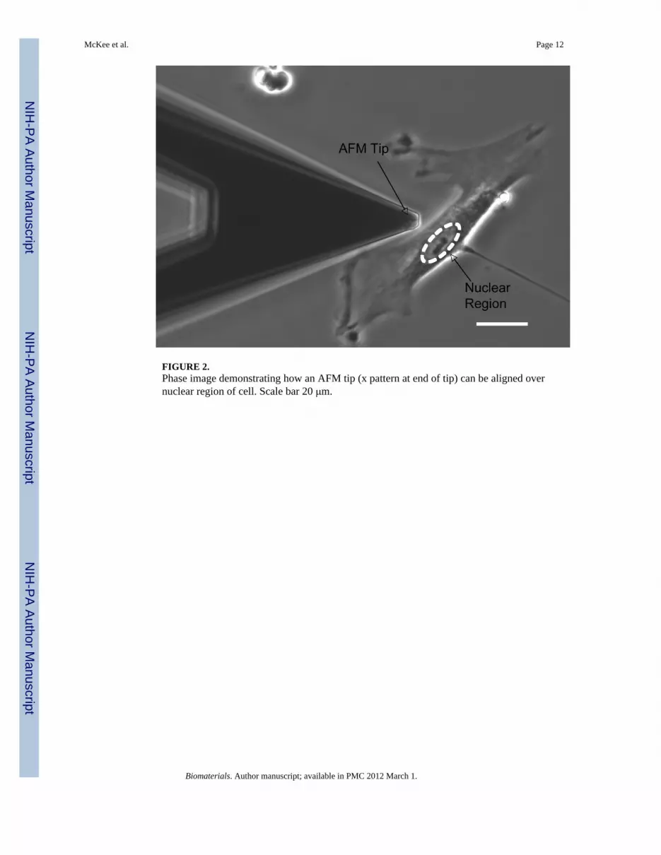
FIGURE 2.Phase image demonstrating how an AFM tip (x pattern at end of tip) can be aligned overnuclear region of cell. Scale bar 20 μm.
McKee et al. Page 12
Biomaterials. Author manuscript; available in PMC 2012 March 1.
NIH
-PA Author Manuscript
NIH
-PA Author Manuscript
NIH
-PA Author Manuscript
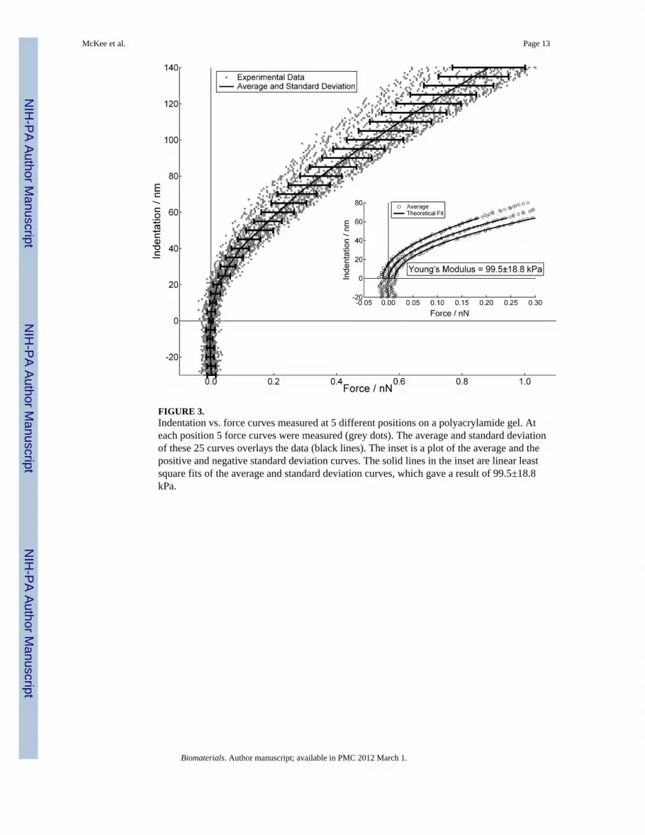
FIGURE 3.Indentation vs. force curves measured at 5 different positions on a polyacrylamide gel. Ateach position 5 force curves were measured (grey dots). The average and standard deviationof these 25 curves overlays the data (black lines). The inset is a plot of the average and thepositive and negative standard deviation curves. The solid lines in the inset are linear leastsquare fits of the average and standard deviation curves, which gave a result of 99.5±18.8kPa.
McKee et al. Page 13
Biomaterials. Author manuscript; available in PMC 2012 March 1.
NIH
-PA Author Manuscript
NIH
-PA Author Manuscript
NIH
-PA Author Manuscript
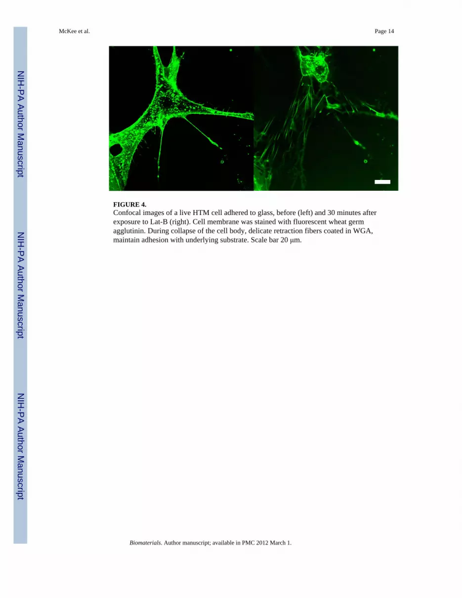
FIGURE 4.Confocal images of a live HTM cell adhered to glass, before (left) and 30 minutes afterexposure to Lat-B (right). Cell membrane was stained with fluorescent wheat germagglutinin. During collapse of the cell body, delicate retraction fibers coated in WGA,maintain adhesion with underlying substrate. Scale bar 20 μm.
McKee et al. Page 14
Biomaterials. Author manuscript; available in PMC 2012 March 1.
NIH
-PA Author Manuscript
NIH
-PA Author Manuscript
NIH
-PA Author Manuscript
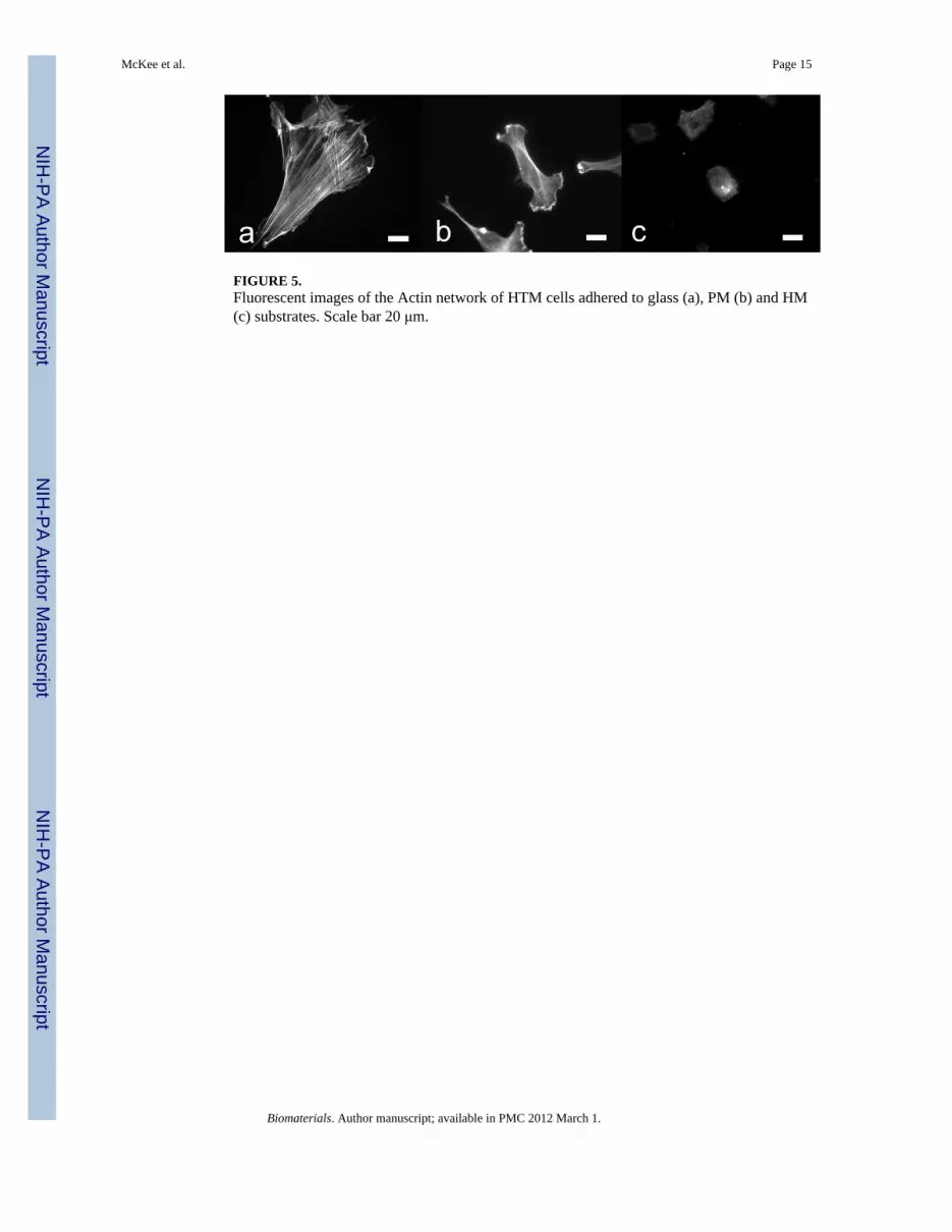
FIGURE 5.Fluorescent images of the Actin network of HTM cells adhered to glass (a), PM (b) and HM(c) substrates. Scale bar 20 μm.
McKee et al. Page 15
Biomaterials. Author manuscript; available in PMC 2012 March 1.
NIH
-PA Author Manuscript
NIH
-PA Author Manuscript
NIH
-PA Author Manuscript

FIGURE 6.Indentation vs. force curves measured on HTM cells on glass before dose (circles), and 90and 270 minutes after exposure and removal of Lat-B (squares and triangles respectively).HTM cell resistance to deformation increased ~ 4 times at 90 minutes, and then decreasedback to the pre-dosed cell modulus within 270 minutes of exposure.
McKee et al. Page 16
Biomaterials. Author manuscript; available in PMC 2012 March 1.
NIH
-PA Author Manuscript
NIH
-PA Author Manuscript
NIH
-PA Author Manuscript

NIH
-PA Author Manuscript
NIH
-PA Author Manuscript
NIH
-PA Author Manuscript
McKee et al. Page 17
TABLE 1
HTM Cell modulus before exposure to Latrunculin B, and 90 and 270 minutes after exposure and removal ofLatrunculin B. Cell modulus on both glass and pathomimetic substrate was significantly increased at 90minutes of actin repolymerization. Within 270 minutes, cells have returned to pre-dose modulus values.
HTM Cell Modulus on Substrates (kPa)
Condition Glass1 Pathomimetic2 Homeomimetic3
Pre-dose 2.7±0.7 1.8±0.5 1.6±0.2
90 min. rec. 11.0±2.3 4.0±1.8 2.3±0.2
270 min. rec. 2.3±0.2 2.0±0.4 1.7±0.5
1Elastic modulus, E, in GPa range
2E = 92±10 kPa
3E = 4.0±1.5 kPa
Biomaterials. Author manuscript; available in PMC 2012 March 1.
Related Documents