Resveratrol prevents the expression of glaucoma markers induced by chronic oxidative stress in trabecular meshwork cells Coralia Luna, Guorong Li, Paloma B Liton, Jianming Qiu, David L. Epstein, Pratap Challa, and Pedro Gonzalez Duke Eye Center, Duke University Medical Center Box 3802, Durham, NC 27710, NC, USA Abstract Elevated intraocular pressure (IOP) constitutes the best characterized risk for primary open-angle glaucoma (POAG). Elevated IOP is believed to result from an increase in aqueous humor outflow resistance at the level of the trabecular meshwork (TM)/ Schlemm's canal (SC). Malfunction of the TM in POAG is associated with the expression of markers for inflammation, cellular senescence, oxidative damage, and decreased cellularity. Current POAG treatments rely on lowering IOP, but there is no therapeutic approach available to delay the loss of function of the TM in POAG patients. We evaluated the effects of chronic administration of the dietary supplement resveratrol on the expression of markers for inflammation, oxidative damage, and cellular senescence in primary TM cells subjected to chronic oxidative stress (40% O 2 ). Resveratrol treatment effectively prevented increased production of intracellular reactive oxygen species (iROS) and inflammatory markers (IL1α, IL6, IL8, and ELAM-1), and reduced expression of the senescence markers sa-β-gal, lipofuscin, and accumulation of carbonylated proteins. Furthermore, resveratrol exerted antiapoptotic effects that were not associated with a decrease in cell proliferation. These results suggest that resveratrol could potentially have a role in preventing the TM tissue abnormalities observed in POAG. Keywords Resveratrol; oxidative stress; glaucoma; trabecular meshwork 1. Introduction Primary Open-Angle Glaucoma (POAG) is an age-related-disease affecting millions of people worldwide. This optic neuropathy is characterized by loss of retinal ganglion cell axons and remodeling of the optic nerve head that is accompanied by progressive visual field loss that can result in irreversible blindness. The main risk factor for POAG is elevated intraocular pressure (IOP) and lowering IOP is currently the only therapeutic approach available to delay the progression of the disease (Armaly, et al., 1980; Leske, et al., 2003). The trabecular meshwork (TM) is the tissue responsible for draining most of the aqueous humor from the anterior chamber of the eye, and the increase in IOP observed in POAG is believed to result from an increase in resistance to aqueous humor outflow at the level of the TM/ Schlemm's canal (SC) (Maepea and Bill, 1992; Moses, 1977). Corresponding author information: Tel.: 1 919 6815995; fax: 1 919 6848983, E-mail address: E-mail: [email protected]. Publisher's Disclaimer: This is a PDF file of an unedited manuscript that has been accepted for publication. As a service to our customers we are providing this early version of the manuscript. The manuscript will undergo copyediting, typesetting, and review of the resulting proof before it is published in its final citable form. Please note that during the production process errors may be discovered which could affect the content, and all legal disclaimers that apply to the journal pertain. NIH Public Access Author Manuscript Food Chem Toxicol. Author manuscript; available in PMC 2010 January 1. Published in final edited form as: Food Chem Toxicol. 2009 January ; 47(1): 198–204. doi:10.1016/j.fct.2008.10.029. NIH-PA Author Manuscript NIH-PA Author Manuscript NIH-PA Author Manuscript

Welcome message from author
This document is posted to help you gain knowledge. Please leave a comment to let me know what you think about it! Share it to your friends and learn new things together.
Transcript
Resveratrol prevents the expression of glaucoma markersinduced by chronic oxidative stress in trabecular meshwork cells
Coralia Luna, Guorong Li, Paloma B Liton, Jianming Qiu, David L. Epstein, Pratap Challa,and Pedro GonzalezDuke Eye Center, Duke University Medical Center Box 3802, Durham, NC 27710, NC, USA
AbstractElevated intraocular pressure (IOP) constitutes the best characterized risk for primary open-angleglaucoma (POAG). Elevated IOP is believed to result from an increase in aqueous humor outflowresistance at the level of the trabecular meshwork (TM)/ Schlemm's canal (SC). Malfunction of theTM in POAG is associated with the expression of markers for inflammation, cellular senescence,oxidative damage, and decreased cellularity. Current POAG treatments rely on lowering IOP, butthere is no therapeutic approach available to delay the loss of function of the TM in POAG patients.We evaluated the effects of chronic administration of the dietary supplement resveratrol on theexpression of markers for inflammation, oxidative damage, and cellular senescence in primary TMcells subjected to chronic oxidative stress (40% O2). Resveratrol treatment effectively preventedincreased production of intracellular reactive oxygen species (iROS) and inflammatory markers(IL1α, IL6, IL8, and ELAM-1), and reduced expression of the senescence markers sa-β-gal,lipofuscin, and accumulation of carbonylated proteins. Furthermore, resveratrol exerted antiapoptoticeffects that were not associated with a decrease in cell proliferation. These results suggest thatresveratrol could potentially have a role in preventing the TM tissue abnormalities observed inPOAG.
KeywordsResveratrol; oxidative stress; glaucoma; trabecular meshwork
1. IntroductionPrimary Open-Angle Glaucoma (POAG) is an age-related-disease affecting millions of peopleworldwide. This optic neuropathy is characterized by loss of retinal ganglion cell axons andremodeling of the optic nerve head that is accompanied by progressive visual field loss thatcan result in irreversible blindness. The main risk factor for POAG is elevated intraocularpressure (IOP) and lowering IOP is currently the only therapeutic approach available to delaythe progression of the disease (Armaly, et al., 1980; Leske, et al., 2003). The trabecularmeshwork (TM) is the tissue responsible for draining most of the aqueous humor from theanterior chamber of the eye, and the increase in IOP observed in POAG is believed to resultfrom an increase in resistance to aqueous humor outflow at the level of the TM/ Schlemm'scanal (SC) (Maepea and Bill, 1992; Moses, 1977).
Corresponding author information: Tel.: 1 919 6815995; fax: 1 919 6848983, E-mail address: E-mail: [email protected]'s Disclaimer: This is a PDF file of an unedited manuscript that has been accepted for publication. As a service to our customerswe are providing this early version of the manuscript. The manuscript will undergo copyediting, typesetting, and review of the resultingproof before it is published in its final citable form. Please note that during the production process errors may be discovered which couldaffect the content, and all legal disclaimers that apply to the journal pertain.
NIH Public AccessAuthor ManuscriptFood Chem Toxicol. Author manuscript; available in PMC 2010 January 1.
Published in final edited form as:Food Chem Toxicol. 2009 January ; 47(1): 198–204. doi:10.1016/j.fct.2008.10.029.
NIH
-PA Author Manuscript
NIH
-PA Author Manuscript
NIH
-PA Author Manuscript
The specific mechanisms leading to the failure of the TM to maintain normal levels of outflowresistance in POAG are not completely understood. However, it is known that the TM fromglaucoma donors is characterized by the sustained activation of a stress response that resultsin expression of pro-inflammatory markers such as ELAM-1 (Wang, et al., 2001). In acomparative analysis between control and POAG tissues, several genes associated withinflammation and an acute-phase response, including ELAM-1, were up-regulated (Liton, etal., 2006). Glaucomatous TM cells produce constitutively IL1α that has been shown to lead tothe up-regulation of ELAM-1 and other inflammatory mediators (Wang, et al., 2001; Zhang,et al., 2006). In addition, a polymorphism in IL1α that leads to increased IL1 expression hasbeen reported to be a risk factor for POAG (Wang, et al., 2006). Together with the sustainedactivation of a pro-inflammatory response, the TM from glaucoma donors is also known toshow some decrease in cellularity and an increase in the expression of the cellular senescencemarker sa-beta-galactosidase (sa-β-gal) (Alvarado, et al., 1984; Liton, et al., 2005), whichsuggests that apoptosis and cellular senescence may contribute to the loss of function of theTM.
Oxidative stress may also be a contributing factor in the observed alterations of the TM inglaucoma. Acute treatment of TM cells with H2O2 has been shown to induce the expressionof the glaucoma marker ELAM-1 (Zhou, et al., 2007) and chronic H2O2 treatment results in asustained activation of a stress response similar to that observed in TM cells from glaucomadonors (Li, 2007). Furthermore, oxidative stress has been suggested to contribute to the lossin cellularity in the TM by inducing apoptosis (Alvarado, et al., 1981; Alvarado, et al., 1984)and is also known to contribute to an increased expression of sa-β-gal (Caballero, et al.,2003). The TM is subjected to relatively high oxygen concentration from the aqueous humor,between 5% and 6% (Helbig, et al., 1993), higher than in most tissues (around 3%). Thepathogenic role of reactive oxygen species (ROS) in glaucoma is supported by additionalexperimental findings including the induction of TM degeneration and increase in resistanceto aqueous humor outflow by hydrogen peroxide (Kahn, et al., 1983; Nguyen, et al., 1988);reduction in the antioxidant capacities of the TM with age (De La Paz and Epstein, 1996) andpositive correlation between oxidative DNA damage in the TM with visual-field loss andincreased IOP (Sacca, et al., 2005). Recently, the increased expression of the oxidative stressmarker sPLA2-IIA has also been reported in association with glaucoma (Zhou, et al., 2007).
Although there is great deal of research aimed at preventing the loss of ganglion cells usingneuroprotective agents (Cheung, et al., 2008; Weber, et al., 2008), current treatments for POAGrely on lowering the IOP and, at the present, there is no therapy that can help to prevent ordelay the loss of function of the TM in POAG patients. Resveratrol, a naturally occurringpolyphenol found in berries, nuts, and red wine, can enhance stress resistance and extend thelife span of various organisms from yeast to vertebrates (Bauer, et al., 2004; Baur, et al.,2006; Howitz, et al., 2003; Valenzano, et al., 2006). It has been reported to exert anti-inflammatory, anti-oxidant, and anti-apoptotic effects, and there is experimental evidencesupporting the beneficial effects of resveratrol in preventing or slowing down a wide varietyof age related diseases (Baur and Sinclair, 2006; Cucciolla, et al., 2007; Holme and Pervaiz,2007; Kahn, et al., 1983). Therefore, we hypothesized that resveratrol may help to prevent thealterations induced by chronic oxidative stress in TM cells. To evaluate the potential ofresveratrol to protect TM function we analyzed the effects of chronic resveratrol treatment onthe expression of markers for inflammation, oxidative damage, and cellular senescence inprimary TM cells subjected to chronic oxidative stress, as well as on the levels of apoptosisinduced by acute oxidative injury.
Luna et al. Page 2
Food Chem Toxicol. Author manuscript; available in PMC 2010 January 1.
NIH
-PA Author Manuscript
NIH
-PA Author Manuscript
NIH
-PA Author Manuscript

2. Material and Methods2.1. Cell culture
Primary porcine TM cells were obtained from pig eyes, and cultured as previously described(Stamer, et al., 1995). Cell cultures were maintained at 37°C in 5% CO2 in media (low glucoseDulbecco's Modified Eagle Medium with L-glutamine, 110mg/mL sodium pyruvate, 10% fetalbovine serum, 100μM non-essential aminoacids, 100 units/mL penicillin, 100μg/mLstreptomicyn sulfate and 0.25μg/mL amphotericin B). All the reagents were obtained fromInvitrogen Corporation (Carlsbad, CA).
2.2. Chronic Oxidative stress and resveratrol treatmentFor most of the experiments, confluent primary porcine TM cells were submitted to chronictreatment with resveratrol (25μM in DMSO, from Sigma, Saint Louis, MO) or vehicle (1μlDMSO/mL cell culture media) every three days for 15 days. Cells under resveratrol or vehicletreatment were incubated under oxidative stress conditions (40% oxygen). Additional controlcultures were treated with vehicle and incubated at physiological oxygen concentration (5%).After 15 days cells were subjected to different assays.
2.3 Cytotoxicity AssayCytotoxicity was measured using the Cyto Tox 96® Non-Radioactive Cytotoxicity assay(Promega, Madison,WI) following the manufacturer's instructions. Briefly the release of lactatedehydrogenase (LDH) is converted into a red formazan product proportional to the number oflysed cells.
2.4. Senescence associated beta-galactosidase (sa-β-gal) activityActivity of sa-β-gal was measured by flow cytometry using the fluorogenic substrate C12FDG(Molecular Probes, Eugene, OR) as previously described (Fiering, et al., 1991). Briefly, cellswere incubated with 300μM of chloroquine for 30 minutes to modulate the intracellular pH.Cell cultures were then washed with phosphate buffered saline (PBS), trypsinized, resuspendedin PBS, and incubated for 5 minutes at 37°C in a water bath, mixed gently with 2mM of FDG,and incubated for 1 minute. The cell suspension was placed on ice, diluted 10 times with coldPBS, and incubated on ice until flow cytometry analysis (FACScaliber, Becton Dickinson, CA,USA). The average number of cells analyzed for each experiment was 10,000.
2.5. Cell proliferationCell proliferation was assayed using 10μM BrdU (5-bromo-2′-deoxyuridine, from Sigma, SantLouis, MO). Briefly, after two weeks of treatment, cells in 6 well plates were trypsinized andseeded in 10cm plates, incubated 48 hours and labeled with BrDu for 5 hours. After labeling,cells were trypsinized and fixed in cold methanol overnight. Cells were washed in PBS/BSA,denaturated with 2M HCl, neutralized with 0.1M sodium borate, and hybridized with antiBrdU-FITC antibody (Santa Cruz Biothecnology, Santa Cruz, CA), and resuspended inpropidium iodine (10μg/mL) containing 300μg/mL RNase (Sigma, MO, USA). BrdUincorporation was calculated from 10,000 cells by flow cytometry.
2.6. Analysis of autofluorescenceCell autofluorescence (lipofuscin-like or ceroid-like material) was determined by flowcytometry at 563-607nm. The average number of cells analyzed for each experiment was10,000.
Luna et al. Page 3
Food Chem Toxicol. Author manuscript; available in PMC 2010 January 1.
NIH
-PA Author Manuscript
NIH
-PA Author Manuscript
NIH
-PA Author Manuscript

2.7. Intracellular Reactive Oxygen Species (iROS) measurementCells were loaded with 10μM of 2′7′-dichlorofluorescein diacetate (DCFDA) (Calbiochem, LaJolla, CA) incubated for 30 minutes in PBS and washed with PBS. This was followed byincubation for 20 minutes in media and trypsinization. Finally, the cells were collected in PBSand kept on ice until analyzed by flow cytometry. The average number of cells analyzed foreach experiment was 10,000.
2.8. RNA extractionFor RNA extraction, porcine primary TM cells were washed with PBS and immediatelysubmerged in RNA-later (Ambion Inc, Austin, TX). Total RNA was then isolated usingRNeasy kit (Qiagen Inc. Valencia, CA) according to the manufacturer's instructions and treatedwith DNase. RNA yields were determined using RiboGreen fluorescent dye (Molecular ProbesInc. Eugene, OR).
2.9. Quantitative real-time PCR (RT-PCR)First strand cDNA was synthesized from total RNA (1μg) by reverse transcription using oligo-dT and Superscript II reverse transcriptase (Invitrogen, Carlsbad, CA) according tomanufacturer's instructions. RT-PCR reactions were performed in 20μL mixture containing1μL of the cDNA preparation, 1X iQ SYBR Green Supermix (Bio-Rad, Hercules, CA), usingthe following PCR parameters: 95°C for 5 minutes followed by 50 cycles of 95°C for 15seconds, 65°C for 15 seconds, and 72°C for 15 seconds. The fluorescence threshold value (Ct)was calculated using the iCycle system software. The absence of nonspecific products wasconfirmed by both analysis of the melting curves and electrophoresis in 3% Super acrylAgarosegels. β-actin was used as an internal standard of mRNA expression. Primers for RT-PCR weredesigned using Primer3 (v. 0.4.0) software (Rozen, 2000) and are showed in Table 1.
2.10. Protein extraction and protein carbonylation assayFor protein extraction cells were washed in PBS and lysated in 1X RIPA buffer, and proteinconcentration was determined using Micro BCA Protein Assay Kit (Pierce, Rockford, IL). TheOxyBlot™ Protein Oxidation Detection Kit (Chemicon, Temecula, CA) was employed for thecomparative analysis of total protein carbonylation according to the manufacturer'sinstructions. Briefly, 2,4-dinitrophenylhy-drazine (DNPH) derivatization was carried outfollowed by SDS-PAGE under reducing conditions and blots were incubated with anti-DNPantibody. Blots were developed using a chemiluminescence detection system (ECL-Plus fromAmersham, Buckinghamshire, UK). The comparison between the samples was calculatedusing the ratio between densitometric values of the total oxyblot bands and the total proteinsstained with Commassie Blue on the same blot.
2.11. DNA damage assayDNA damage quantification kit (Dojindo, Gaithersburg, MD) detects DNA modifications thatresult in the formation of aldehyde groups, which are the open ring form of the abasic sites(AP sites). Briefly, an excess of Aldehyde Reactive Probe (ARP) conjugated to avidin wasincubated with DNA samples. After labeling, the AP sites were quantified by a colorimetricreaction using anti avidin antibody conjugated to peroxidase, following the manufacturer'sinstructions.
2.12. Proteasome activity assayChymotrypsin-like (CT-L), trypsin-like (T-L), and caspase-like (PGPH) activities of theproteasome were assayed using the fluorogenic peptides (from Sigma, Sant Louis, MO) N-Succinyl-Leu-Leu-Val-Tyr-AMC (Suc-LLVY-AMC at 70μM), Z-Leu-Leu-Glu-AMC
Luna et al. Page 4
Food Chem Toxicol. Author manuscript; available in PMC 2010 January 1.
NIH
-PA Author Manuscript
NIH
-PA Author Manuscript
NIH
-PA Author Manuscript

(ZLLG-AMC at 70μM) and Boc-Leu-Arg-Arg-AMC (BOLAA at 200μM). Briefly, cells werelysated (20mM Tris–HCl, pH 7.5; 10% glycerol; 5mM ATP; 0.2% Np40) and 20μg of proteinextract diluted in assay buffer (50mM Hepes/ KOH pH 7.5). Assays were carried out at 37°Cfor 30 minutes. The fluorescence of the samples was evaluated using a BIO-TEK Synergy HTspectrofluorimeter at excitation/emission wavelengths of 360/460. Activity was determined asthe difference between total activity of crude extracts and the remaining activity with 20μMMG132 (Sigma, St Louis, MO), a proteasome inhibitor.
2.13. Apoptotic analysisApoptosis was measured using the Vybrant Apoptosis Assay kit (Invitrogen, Eugene, OR).Briefly, apoptosis was induced using several concentrations of H2O2 (0, 200, 400 and800μM). Cells were treated with 25μM of resveratrol or vehicle 24 hours before H2O2 treatmentand during the treatment. Cells were trypsinized four hours after H2O2 treatment, washed withcold PBS, resuspended in PBS with YO-PRO and propidium iodine dyes, incubated on ice for30 minutes, and analyzed by flow cytometry. The average number of cells analyzed for eachexperiment was 10,000.
2.14. Statistical analysisResults are expressed as mean value ± SD in at least three independent experiments. The SASSoftware package (SAS/STAT, version 8, Cary, NC: SAS Institute Inc., 1999) was used forthe statistical analysis. P-values were calculated by a non paired two sided t-test and by thenon parametric test Kruskal-Wallis (that is the same as the Wilcoxon test when there are onlytwo groups). In the figures “*” denoted p-values based on t-test and “#” p-values based on theKruskal-Wallis test.
3. Results3.1. Cytotoxicity of chronic treatment with resveratrol in TM cells
In order to evaluate potential cytotoxic effects of resveratrol, TM cell cultures incubated atphysiologic oxygen concentration (5%) were subjected to chronic treatment with differentconcentrations of resveratrol or vehicle for 15 days. A single treatment with high concentrationsof resveratrol (200 and 400μM) resulted in extensive cell death in less than 48 hours. However,chronic treatment with resveratrol at concentrations of 100μM or lower did not show anyincrease in cytotoxity compared to the vehicle treated controls. Treatment with resveratrol atconcentrations of 50 and 100μM resulted in a small but significant decrease in cytotoxicitycompared to control cultures treated with vehicle (Figure 1).
3.2. Resveratrol inhibits intracellular ROS productionTo evaluate the effects of resveratrol on the induction of intracellular ROS production mediatedby chronic oxidative stress, confluent primary cultures of porcine TM cells were subjected tochronic oxidative stress (15 days at 40% oxygen) and chronic treatment with resveratrol orvehicle (every three days during 15 days). Non-stressed control cultures were incubated inparallel at 5% oxygen concentration. The induction of endogenous ROS production in thismodel was significantly decreased by resveratrol treatment (4 fold) compared to cells treatedwith vehicle. The amount of intracellular ROS in resveratrol-treated cells under oxidative stresswas similar to that of non-stressed control cells incubated at physiological oxygenconcentration (5%) (Figure 2).
3.3. Resveratrol reduces the activation of inflammatory markersThe induction of mRNA expression of the inflammatory markers IL1α, IL6, IL8 and ELAM-1after chronic oxidative stress was significantly inhibited by chronic treatment with resveratrol.
Luna et al. Page 5
Food Chem Toxicol. Author manuscript; available in PMC 2010 January 1.
NIH
-PA Author Manuscript
NIH
-PA Author Manuscript
NIH
-PA Author Manuscript

As shown in figure 3, resveratrol inhibited the induction of these inflammatory markers insamples incubated at 40% oxygen.
3.4. Resveratrol reduces the accumulation of senescence markersCells treated with vehicle and incubated under oxidative stress showed a significant increasein autofluorescence (2.4 fold) and sa-β-gal (1.8 fold) when compared to cells in the samecondition treated with resveratrol. Cells under oxidative stress, treated with vehicle orresveratrol, showed higher levels of autofluorescence (3.1 and 1.2 fold respectively) and sa-β-gal (3.5 and 1.9 fold respectively) when compared to cells incubated at more physiologicaloxygen concentration (Figure 4A and 4B).
3.5. Decreased accumulation of carbonylated proteins with no change in proteasomalactivity after resveratrol treatment
The accumulation of carbonylated proteins induced by oxidative stress was significantly lowerin resveratrol-treated samples compared to samples treated with vehicle under oxidative stress,as well as to that in cells incubated at physiological oxygen concentrations (Figures 5A, B andC). However, this decrease in oxidized proteins was not associated with an increase inproteasomal activity. The three proteolytic activities of the proteasome (chymotrypsin-like,trypsin-like and caspase-like activity) showed decreased activity in both resveratrol treated andnon-treated cells incubated at 40% oxygen compared to the non-stressed controls incubated atphysiologic oxygen concentrations (Figure 5D).
3.6. Resveratrol protects against apoptosis in an acute model of oxidative stressCells treated with resveratrol showed protection against apoptosis after acute oxidative stress(200, 400, and 800μM of H2O2), when compared to cells treated with vehicle. Cells treatedwith vehicle showed a linear correlation between H2O2 concentration and apoptosis; cellstreated with resveratrol exhibited protection against apoptosis in all H2O2 concentrations(Figure 6).
3.7. Proliferation and DNA damage are not significantly affected by resveratrol treatment ina chronic model of oxidative stress
Resveratrol treatment did not result in significant changes on proliferation measured by BrDUincorporation when compared to cells treated with vehicle (Figure 7A). Similarly, nosignificant changes were observed in the amount of DNA damage under oxidative stress. Cellstreated with vehicle or resveratrol under oxidative stress showed similar levels of DNA damagethat were higher than those in cells incubated at physiological oxygen concentration (Figure7B).
4. DiscussionOur results demonstrated that chronic treatment of cultured TM cells with resveratroleffectively prevented the upregulation of markers induced by oxidative stress.
Resveratrol has been shown to reduce inflammation in vivo via inhibition of cyclooxygenase-2and NF-κB activities (Holmes-McNary and Baldwin, 2000; Martin, et al., 2006; Tsai, et al.,1999). However, we have recently reported that the induction of the inflammatory markersIL1α, IL-6, IL-8, and ELAM-1 in TM cells subjected to chronic oxidative stress is dependenton the activation of iROS generated by the mitochondria. This increase in iROS is notdependent on cyclooxygenase and is responsible for the chronic activation of NF-κB. One ofthe more clear effects of chronic treatment with resveratrol in TM cells was the inhibition ofiROS production that results from oxidative stress. Therefore the inhibitory effect of resveratrol
Luna et al. Page 6
Food Chem Toxicol. Author manuscript; available in PMC 2010 January 1.
NIH
-PA Author Manuscript
NIH
-PA Author Manuscript
NIH
-PA Author Manuscript

on the production of iROS may be responsible for the concomitant inhibition of the sustainedstress response. Although resveratrol has been shown to induce the production of iROS in somecancer cell lines (Heiss, et al., 2007; Shankar, et al., 2007), our results are consistent with thosereported in other primary cells including HUVEC cells (Chow, et al., 2007; Ungvari, et al.,2007), primary cortical astrocytes (de Almeida, et al., 2007; de Almeida, et al., 2007),RAW264.7 macrophages (Ciz, et al., 2007; Jang, et al., 1999), RPE cells (King, et al., 2005),and dopaminergic neurons (Okawara, et al., 2007).
Chronic treatment of TM cells with resveratrol also resulted in a significant decrease in theaccumulation of fluorescent pigments, carbonylated proteins, and the expression of the cellularsenescence marker sa-β-gal, that are induced by oxidative stress. A decrease in lipofuscin andsa-β-gal upon resveratrol treatment has also been observed in the short-lived fish N. furzeri. Inthis small vertebrate resveratrol treatment not only reduces the expression of senescencemarkers but increases life span and decreases aggregated protein in elderly fish brains(Valenzano and Cellerino, 2006; Valenzano, et al., 2006). In our model, treatment withresveratrol did not prevent the loss of proteasome activity induced by oxidative stress.Therefore, the observed decrease in accumulation of oxidized proteins and fluorescentpigments is not likely to result from increased proteasomal degradation of damaged proteins,but rather from other factors such as resveratrol ROS scavenging properties (Ciz, et al.,2007; Lorenz, et al., 2003); an increase in antioxidant enzymes (Baur and Sinclair, 2006;Karaoglan, et al., 2008; Robb, et al., 2007; Ungvari, et al., 2007); activation of autophagypathways (Opipari, et al., 2004; Trincheri, et al., 2007); and/or the observed inhibition of iROSproduction (Chow, et al., 2007; de Almeida, et al., 2007; Jha, et al., 2008; King, et al., 2005).
In addition to the protective effects in our chronic oxidative stress model, resveratrol alsodemonstrated a significant anti-apoptotic effect after acute oxidative injury. Resveratrol hasreceived a great deal of attention because of its pro-apoptotic effects in cancer cells (Kaindl,et al., 2007; Kalra, et al., 2007; Park, et al., 2007; van Ginkel, et al., 2007). However, its anti-apoptotic effects in several primary cell lines have been well documented (Csaki, et al.,2008; Jha, et al., 2008; Kumar, et al., 2007). Interestingly, our results showed no negativeeffects of chronic resveratrol treatment on the proliferation of TM cells. This is somewhatsurprising, since resveratrol has been reported to exert antiproliferative effects, mainly incancer cells, but also in other non-cancerous cell types (Lee and Moon, 2005; Poussier, et al.,2005). RPE cells treated with resveratrol at pre-confluence levels (80%) showed a significantdecrease in proliferation that was not observed when they were confluent (King, et al., 2005).In our experimental model, cells were treated at confluence levels and this difference mightexplain our observed effect of resveratrol on proliferation. Since the decrease in cellularity ofthe TM associated with aging and POAG has been hypothesized to result, at least in part, fromoxidative damage, our results suggest that chronic treatment with resveratrol could be effectivein delaying the loss of cells in the TM by acting to prevent apoptosis without inhibiting cellproliferation. One important point that deserves future investigation is whether resveratrol canreverse, at least in part, some of the effects of chronic oxidative stress. Preliminary data suggestthat short-term treatment with resveratrol may not be helpful to reverse the damage that hasalready been produced (data not showed). However, a definitive answer to this question willrequire longer term experiments aimed at evaluating the effects of resveratrol treatment on theprogression of oxidative damage for an extended period of time after chronic oxidative injury.
In conclusion, the inhibition of iROS production by resveratrol may help to prevent theinduction of inflammatory and senescence markers in TM cells after chronic oxidative stress,as well as possibly prevent the decrease in cellularity observed in the TM of POAG patients.These results suggest that resveratrol could potentially have a novel role in preventing ordelaying some of the abnormalities of the TM observed in POAG.
Luna et al. Page 7
Food Chem Toxicol. Author manuscript; available in PMC 2010 January 1.
NIH
-PA Author Manuscript
NIH
-PA Author Manuscript
NIH
-PA Author Manuscript
AcknowledgementsThis work was supported by NEI EY01894, NEI EY016228, and NEI EY 014209NEI EY05722, and Research toPrevent Blindness.
The authors thank Thusita Dissanayake (Flow Cytometry Facility, Duke University) for his help in the flow cytometryanalyses.
BibliographyAlvarado J, Murphy C, Polansky J, Juster R. Age-related changes in trabecular meshwork cellularity.
Invest Ophthalmol Vis Sci 1981;21:714–727. [PubMed: 7298275]Alvarado J, Murphy C, Juster R. Trabecular meshwork cellularity in primary open-angle glaucoma and
nonglaucomatous normals. Ophthalmology 1984;91:564–579. [PubMed: 6462622]Armaly MF, Krueger DE, Maunder L, Becker B, Hetherington J Jr, Kolker AE, Levene RZ, Maumenee
AE, Pollack IP, Shaffer RN. Biostatistical analysis of the collaborative glaucoma study. I. Summaryreport of the risk factors for glaucomatous visual-field defects. Arch Ophthalmol 1980;98:2163–2171.[PubMed: 7447768]
Bauer JH, Goupil S, Garber GB, Helfand SL. An accelerated assay for the identification of lifespan-extending interventions in Drosophila melanogaster. Proc Natl Acad Sci U S A 2004;101:12980–12985. [PubMed: 15328413]
Baur JA, Pearson KJ, Price NL, Jamieson HA, Lerin C, Kalra A, Prabhu VV, Allard JS, Lopez-Lluch G,Lewis K, Pistell PJ, Poosala S, Becker KG, Boss O, Gwinn D, Wang M, Ramaswamy S, Fishbein KW,Spencer RG, Lakatta EG, Le Couteur D, Shaw RJ, Navas P, Puigserver P, Ingram DK, de Cabo R,Sinclair DA. Resveratrol improves health and survival of mice on a high-calorie diet. Nature2006;444:337–342. [PubMed: 17086191]
Baur JA, Sinclair DA. Therapeutic potential of resveratrol: the in vivo evidence. Nat Rev Drug Discov2006;5:493–506. [PubMed: 16732220]
Caballero M, Liton PB, Epstein DL, Gonzalez P. Proteasome inhibition by chronic oxidative stress inhuman trabecular meshwork cells. Biochem Biophys Res Commun 2003;308:346–352. [PubMed:12901875]
Cheung W, Guo L, Cordeiro MF. Neuroprotection in glaucoma: drug-based approaches. Optom Vis Sci2008;85:406–416. [PubMed: 18521010]
Chow SE, Hshu YC, Wang JS, Chen JK. Resveratrol attenuates oxLDL-stimulated NADPH oxidaseactivity and protects endothelial cells from oxidative functional damages. J Appl Physiol2007;102:1520–1527. [PubMed: 17194732]
Ciz M, Pavelkova M, Gallova L, Kralova J, Kubala L, Lojek A. The influence of wine polyphenols onreactive oxygen and nitrogen species production by rat macrophages RAW 264.7. Physiol Res.2007PMID: 17465695
Csaki C, Keshishzadeh N, Fischer K, Shakibaei M. Regulation of inflammation signalling by resveratrolin human chondrocytes in vitro. Biochem Pharmacol 2008;75:677–687. [PubMed: 17959154]
Cucciolla V, Borriello A, Oliva A, Galletti P, Zappia V, Ragione FD. Resveratrol: From Basic Scienceto the Clinic. Cell Cycle 2007;6:2495–2510. [PubMed: 17726376]
de Almeida LM, Pineiro CC, Leite MC, Brolese G, Leal RB, Gottfried C, Goncalves CA. ProtectiveEffects of Resveratrol on Hydrogen Peroxide Induced Toxicity in Primary Cortical AstrocyteCultures. Neurochem Res 2007;33:8–15. [PubMed: 17594518]
de Almeida LM, Pineiro CC, Leite MC, Brolese G, Tramontina F, Feoli AM, Gottfried C, GoncalvesCA. Resveratrol increases glutamate uptake, glutathione content, and S100B secretion in corticalastrocyte cultures. Cell Mol Neurobiol 2007;27:661–668. [PubMed: 17554623]
De La Paz MA, Epstein DL. Effect of age on superoxide dismutase activity of human trabecularmeshwork. Invest Ophthalmol Vis Sci 1996;37:1849–1853. [PubMed: 8759353]
Fiering SN, Roederer M, Nolan GP, Micklem DR, Parks DR, Herzenberg LA. Improved FACS-Gal: flowcytometric analysis and sorting of viable eukaryotic cells expressing reporter gene constructs.Cytometry 1991;12:291–301. [PubMed: 1905992]
Luna et al. Page 8
Food Chem Toxicol. Author manuscript; available in PMC 2010 January 1.
NIH
-PA Author Manuscript
NIH
-PA Author Manuscript
NIH
-PA Author Manuscript

Heiss EH, Schilder YD, Dirsch VM. Chronic Treatment with Resveratrol Induces Redox Stress- andAtaxia Telangiectasia-mutated (ATM)-dependent Senescence in p53-positive Cancer Cells. J BiolChem 2007;282:26759–26766. [PubMed: 17626009]
Helbig H, Hinz JP, Kellner U, Foerster MH. Oxygen in the anterior chamber of the human eye. Ger JOphthalmol 1993;2:161–164. [PubMed: 8334391]
Holme AL, Pervaiz S. Resveratrol in cell fate decisions. J Bioenerg Biomembr 2007;39:59–63. [PubMed:17308975]
Holmes-McNary M, Baldwin AS Jr. Chemopreventive properties of trans-resveratrol are associated withinhibition of activation of the IkappaB kinase. Cancer Res 2000;60:3477–3483. [PubMed: 10910059]
Howitz KT, Bitterman KJ, Cohen HY, Lamming DW, Lavu S, Wood JG, Zipkin RE, Chung P,Kisielewski A, Zhang LL, Scherer B, Sinclair DA. Small molecule activators of sirtuins extendSaccharomyces cerevisiae lifespan. Nature 2003;425:191–196. [PubMed: 12939617]
Jang DS, Kang BS, Ryu SY, Chang IM, Min KR, Kim Y. Inhibitory effects of resveratrol analogs onunopsonized zymosan-induced oxygen radical production. Biochem Pharmacol 1999;57:705–712.[PubMed: 10037457]
Jha RK, Yong MQ, Chen SH. The protective effect of resveratrol on the intestinal mucosal barrier in ratswith severe acute pancreatitis. Med Sci Monit 2008;14:BR14–19. [PubMed: 18160933]
Kahn MG, Giblin FJ, Epstein DL. Glutathione in calf trabecular meshwork and its relation to aqueoushumor outflow facility. Invest Ophthalmol Vis Sci 1983;24:1283–1287. [PubMed: 6885312]
Kaindl U, Eyberg I, Rohr-Udilova N, Heinzle C, Marian B. The dietary antioxidants resveratrol andquercetin protect cells from exogenous pro-oxidative damage. Food Chem Toxicol. 2007PMID:17936464
Kalra N, Roy P, Prasad S, Shukla Y. Resveratrol induces apoptosis involving mitochondrial pathways inmouse skin tumorigenesis. Life Sci 2007;82:348–358. [PubMed: 18201729]
Karaoglan A, Akdemir O, Barut S, Kokturk S, Uzun H, Tasyurekli M, Colak A. The effects of resveratrolon vasospasm after experimental subarachnoidal hemorrhage in rats. Surg Neurol. 2008PMID:18207513
King RE, Kent KD, Bomser JA. Resveratrol reduces oxidation and proliferation of human retinal pigmentepithelial cells via extracellular signal-regulated kinase inhibition. Chem Biol Interact 2005;151:143–149. [PubMed: 15698585]
Kumar A, Kaundal RK, Iyer S, Sharma SS. Effects of resveratrol on nerve functions, oxidative stress andDNA fragmentation in experimental diabetic neuropathy. Life Sci 2007;80:1236–1244. [PubMed:17289084]
Lee B, Moon SK. Resveratrol inhibits TNF-alpha-induced proliferation and matrix metalloproteinaseexpression in human vascular smooth muscle cells. J Nutr 2005;135:2767–2773. [PubMed:16317118]
Leske MC, Heijl A, Hussein M, Bengtsson B, Hyman L, Komaroff E. Factors for glaucoma progressionand the effect of treatment: the early manifest glaucoma trial. Arch Ophthalmol 2003;121:48–56.[PubMed: 12523884]
Li G, Luna C, Liton P, Navarro I, Epstein DL, Gonzalez P. Sustained stress response after oxidative stressin trabecular meshwork cells. Mol Vis 2007;13:2282–2288. [PubMed: 18199969]
Liton PB, Challa P, Stinnett S, Luna C, Epstein DL, Gonzalez P. Cellular senescence in the glaucomatousoutflow pathway. Exp Gerontol 2005;40:745–748. [PubMed: 16051457]
Liton PB, Luna C, Challa P, Epstein DL, Gonzalez P. Genome-wide expression profile of humantrabecular meshwork cultured cells, nonglaucomatous and primary open angle glaucoma tissue. MolVis 2006;12:774–790. [PubMed: 16862071]
Lorenz P, Roychowdhury S, Engelmann M, Wolf G, Horn TF. Oxyresveratrol and resveratrol are potentantioxidants and free radical scavengers: effect on nitrosative and oxidative stress derived frommicroglial cells. Nitric Oxide 2003;9:64–76. [PubMed: 14623172]
Maepea O, Bill A. Pressures in the juxtacanalicular tissue and Schlemm's canal in monkeys. Exp EyeRes 1992;54:879–883. [PubMed: 1521580]
Martin AR, Villegas I, Sanchez-Hidalgo M, de la Lastra CA. The effects of resveratrol, a phytoalexinderived from red wines, on chronic inflammation induced in an experimentally induced colitis model.Br J Pharmacol 2006;147:873–885. [PubMed: 16474422]
Luna et al. Page 9
Food Chem Toxicol. Author manuscript; available in PMC 2010 January 1.
NIH
-PA Author Manuscript
NIH
-PA Author Manuscript
NIH
-PA Author Manuscript

Moses RA. The effect of intraocular pressure on resistance to outflow. Surv Ophthalmol 1977;22:88–100. [PubMed: 335549]
Nguyen KP, Chung ML, Anderson PJ, Johnson M, Epstein DL. Hydrogen peroxide removal by the calfaqueous outflow pathway. Invest Ophthalmol Vis Sci 1988;29:976–981. [PubMed: 3372170]
Okawara M, Katsuki H, Kurimoto E, Shibata H, Kume T, Akaike A. Resveratrol protects dopaminergicneurons in midbrain slice culture from multiple insults. Biochem Pharmacol 2007;73:550–560.[PubMed: 17147953]
Opipari AW Jr, Tan L, Boitano AE, Sorenson DR, Aurora A, Liu JR. Resveratrol-inducedautophagocytosis in ovarian cancer cells. Cancer Res 2004;64:696–703. [PubMed: 14744787]
Park JW, Woo KJ, Lee JT, Lim JH, Lee TJ, Kim SH, Choi YH, Kwon TK. Resveratrol induces pro-apoptotic endoplasmic reticulum stress in human colon cancer cells. Oncol Rep 2007;18:1269–1273.[PubMed: 17914584]
Poussier B, Cordova AC, Becquemin JP, Sumpio BE. Resveratrol inhibits vascular smooth muscle cellproliferation and induces apoptosis. J Vasc Surg 2005;42:1190–1197. [PubMed: 16376213]
Robb EL, Page MM, Wiens BE, Stuart JA. Molecular mechanisms of oxidative stress resistance inducedby resveratrol: Specific and progressive induction of MnSOD. Biochem Biophys Res Commun2007;367:406–412. [PubMed: 18167310]
Rozen, S.; S, H. Primer3 on the WWW for general users and for biologist programmers. In: Krawetz, S.;M, S., editors. Bioinformatics Methods and Protocols: Methods in Molecular Biology. Totowa, NJ:Humana Press; 2000. p. 365-386.
Sacca SC, Pascotto A, Camicione P, Capris P, Izzotti A. Oxidative DNA damage in the human trabecularmeshwork: clinical correlation in patients with primary open-angle glaucoma. Arch Ophthalmol2005;123:458–463. [PubMed: 15824217]
Shankar S, Siddiqui I, Srivastava RK. Molecular mechanisms of resveratrol (3,4,5-trihydroxy-trans-stilbene) and its interaction with TNF-related apoptosis inducing ligand (TRAIL) in androgen-insensitive prostate cancer cells. Mol Cell Biochem 2007;304:273–285. [PubMed: 17636462]
Stamer WD, Seftor RE, Snyder RW, Regan JW. Cultured human trabecular meshwork cells expressaquaporin-1 water channels. Curr Eye Res 1995;14:1095–1100. [PubMed: 8974838]
Trincheri NF, Follo C, Nicotra G, Peracchio C, Castino R, Isidoro C. Resveratrol-induced ApoptosisDepends on the Lipid Kinase Activity of Vps34 and on the Formation of Autophagolysosomes.Carcinogenesis 2007;29:381–389. [PubMed: 18048384]
Tsai SH, Lin-Shiau SY, Lin JK. Suppression of nitric oxide synthase and the down-regulation of theactivation of NFkappaB in macrophages by resveratrol. Br J Pharmacol 1999;126:673–680.[PubMed: 10188978]
Ungvari Z, Orosz Z, Rivera A, Labinskyy N, Xiangmin Z, Olson S, Podlutsky A, Csiszar A. Resveratrolincreases vascular oxidative stress resistance. Am J Physiol Heart Circ Physiol 2007;292:H2417–2424. [PubMed: 17220179]
Valenzano DR, Cellerino A. Resveratrol and the pharmacology of aging: a new vertebrate model tovalidate an old molecule. Cell Cycle 2006;5:1027–1032. [PubMed: 16687936]
Valenzano DR, Terzibasi E, Genade T, Cattaneo A, Domenici L, Cellerino A. Resveratrol prolongslifespan and retards the onset of age-related markers in a short-lived vertebrate. Curr Biol2006;16:296–300. [PubMed: 16461283]
van Ginkel PR, Sareen D, Subramanian L, Walker Q, Darjatmoko SR, Lindstrom MJ, Kulkarni A, AlbertDM, Polans AS. Resveratrol inhibits tumor growth of human neuroblastoma and mediates apoptosisby directly targeting mitochondria. Clin Cancer Res 2007;13:5162–5169. [PubMed: 17785572]
Wang CY, Shen YC, Lo FY, Su CH, Lee SH, Lin KH, Tsai HY, Kuo NW, Fan SS. Polymorphism in theIL-1alpha (-889) locus associated with elevated risk of primary open angle glaucoma. Mol Vis2006;12:1380–1385. [PubMed: 17149369]
Wang N, Chintala SK, Fini ME, Schuman JS. Activation of a tissue-specific stress response in the aqueousoutflow pathway of the eye defines the glaucoma disease phenotype. Nat Med 2001;7:304–309.[PubMed: 11231628]
Weber A, Harman CD, Viswanathan S. Effects of optic nerve injury, glaucoma, and neuroprotection onthe survival, structure, and function of ganglion cells in the mammalian retina. J Physiol. 2008
Luna et al. Page 10
Food Chem Toxicol. Author manuscript; available in PMC 2010 January 1.
NIH
-PA Author Manuscript
NIH
-PA Author Manuscript
NIH
-PA Author Manuscript
Zhang X, Schroeder A, Callahan EM, Coyle BM, Wang N, Erickson KA, Schuman JS, Fini ME.Constitutive signalling pathway activity in trabecular meshwork cells from glaucomatous eyes. ExpEye Res 2006;82:968–973. [PubMed: 16516195]
Zhou Q, Liu YQ, Zhao JL, Zhang H. Effects of oxidative stress on the expression of endothelial-leukocyteadhesion molecule-1 in porcine trabecular meshwork cells. Zhongguo Yi Xue Ke Xue Yuan XueBao 2007;29:394–397. [PubMed: 17633469]
Luna et al. Page 11
Food Chem Toxicol. Author manuscript; available in PMC 2010 January 1.
NIH
-PA Author Manuscript
NIH
-PA Author Manuscript
NIH
-PA Author Manuscript

Figure 1.Cytotoxic effects of chronic treatment with resveratrol in TM cells incubated at physiologicaloxygen concentration (5%). Cytotoxicity was evaluated using Cyto Tox 96 assay after 15 daysof chronic treatment with different concentrations of resveratrol (12.5 to 400μM) or vehicle.A single treatment with high concentrations of resveratrol (200 and 400μM) resulted inextensive cell death in less than 48 hours. Since these cultures did not survive a chronictreatment for 15 days, cytotoxicity data for these concentrations is not included in the figure(N=3, t-test *P ≤ 0.05, # Kruskal-Wallis test P ≤ 0.049).
Luna et al. Page 12
Food Chem Toxicol. Author manuscript; available in PMC 2010 January 1.
NIH
-PA Author Manuscript
NIH
-PA Author Manuscript
NIH
-PA Author Manuscript
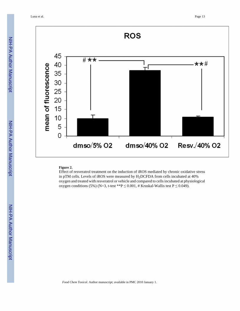
Figure 2.Effect of resveratrol treatment on the induction of iROS mediated by chronic oxidative stressin pTM cells. Levels of iROS were measured by H2DCFDA from cells incubated at 40%oxygen and treated with resveratrol or vehicle and compared to cells incubated at physiologicaloxygen conditions (5%) (N=3, t-test **P ≤ 0.001, # Kruskal-Wallis test P ≤ 0.049).
Luna et al. Page 13
Food Chem Toxicol. Author manuscript; available in PMC 2010 January 1.
NIH
-PA Author Manuscript
NIH
-PA Author Manuscript
NIH
-PA Author Manuscript

Figure 3.Effect of resveratrol treatment on the expression of inflammatory markers induced by chronicoxidative stress. The changes in mRNA expression of IL1α, IL6, IL8 and ELAM-1 inducedby incubation at 40% oxygen compared to control cultures incubated at 5% oxygen weremeasured by Real Time- PCR in cells treated with either resveratrol or dmso. β-actin was usedas an internal control (N=3, t-test ** P ≤ 0.001; # Kruskal-Wallis test P ≤ 0.049).
Luna et al. Page 14
Food Chem Toxicol. Author manuscript; available in PMC 2010 January 1.
NIH
-PA Author Manuscript
NIH
-PA Author Manuscript
NIH
-PA Author Manuscript

Figure 4.Effect of resveratrol treatment on the expression of the senescence markers autofluorescence(A) and sa-β-gal (B). Autofluorescence and sa-β-gal activity were determined by flowcytometry in cells subjected to chronic oxidative stress (40% oxygen) treated with eitherresveratrol or vehicle and compared to cells incubated at physiological oxygen concentration(N=3, t-test, * P ≤ 0.05, ** P ≤ 0.001; # Kruskal-Wallis test P ≤ 0.049).
Luna et al. Page 15
Food Chem Toxicol. Author manuscript; available in PMC 2010 January 1.
NIH
-PA Author Manuscript
NIH
-PA Author Manuscript
NIH
-PA Author Manuscript
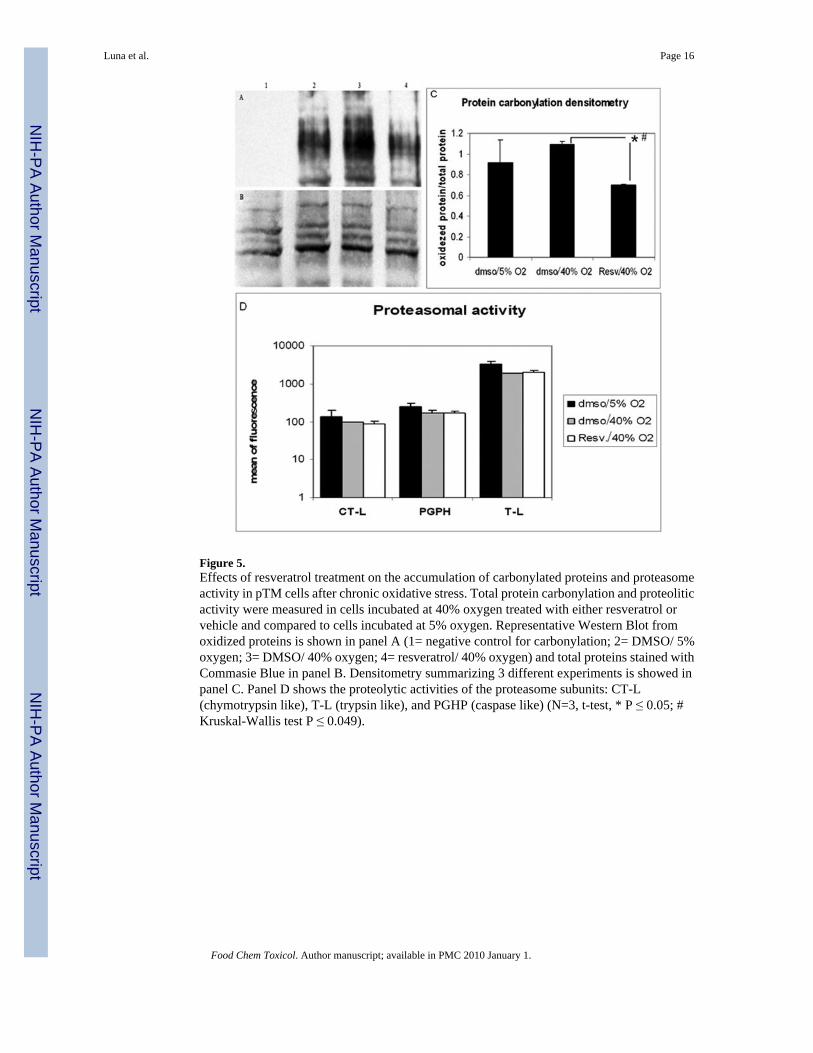
Figure 5.Effects of resveratrol treatment on the accumulation of carbonylated proteins and proteasomeactivity in pTM cells after chronic oxidative stress. Total protein carbonylation and proteoliticactivity were measured in cells incubated at 40% oxygen treated with either resveratrol orvehicle and compared to cells incubated at 5% oxygen. Representative Western Blot fromoxidized proteins is shown in panel A (1= negative control for carbonylation; 2= DMSO/ 5%oxygen; 3= DMSO/ 40% oxygen; 4= resveratrol/ 40% oxygen) and total proteins stained withCommasie Blue in panel B. Densitometry summarizing 3 different experiments is showed inpanel C. Panel D shows the proteolytic activities of the proteasome subunits: CT-L(chymotrypsin like), T-L (trypsin like), and PGHP (caspase like) (N=3, t-test, * P ≤ 0.05; #Kruskal-Wallis test P ≤ 0.049).
Luna et al. Page 16
Food Chem Toxicol. Author manuscript; available in PMC 2010 January 1.
NIH
-PA Author Manuscript
NIH
-PA Author Manuscript
NIH
-PA Author Manuscript

Figure 6.Protective effect of resveratrol against apoptosis induced by acute oxidative challenge. Cellstreated with resveratrol or vehicle were subjected to different concentrations of H2O2 (200,400, and 800μM) for 4 hours and assayed for apoptosis (N=3, t-test, ** P ≤ 0.001; # Kruskal-Wallis test P ≤ 0.049).
Luna et al. Page 17
Food Chem Toxicol. Author manuscript; available in PMC 2010 January 1.
NIH
-PA Author Manuscript
NIH
-PA Author Manuscript
NIH
-PA Author Manuscript
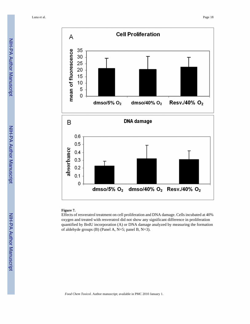
Figure 7.Effects of resveratrol treatment on cell proliferation and DNA damage. Cells incubated at 40%oxygen and treated with resveratrol did not show any significant difference in proliferationquantified by BrdU incorporation (A) or DNA damage analyzed by measuring the formationof aldehyde groups (B) (Panel A, N=5; panel B, N=3).
Luna et al. Page 18
Food Chem Toxicol. Author manuscript; available in PMC 2010 January 1.
NIH
-PA Author Manuscript
NIH
-PA Author Manuscript
NIH
-PA Author Manuscript

NIH
-PA Author Manuscript
NIH
-PA Author Manuscript
NIH
-PA Author Manuscript
Luna et al. Page 19
Table 1
Name Genebank Start position Forward 5′-3′ Reverse 5′-3′
Acc #
IL-1α NM_14029 842 AAGTGTTGACAGGCCGTATG TACCAGACTTCGCTCCCTCT
ELAM-1 NM_214268 697 CCCATGGAACACAACCTGTGCATT AGCTTTACACGTTGGCTTCTTGCC
IL-8 NM_213867 92 AAACTGGCTGTTGCCTTCTT ATTTATGCACTGGCATCGAA
IL-6 NM_214399 365 GCTTCCAATCTGGGTTCAAT CTAATCTGCACAGCCTCGAC
β-actin AY550069 1063 AAGATCAAGATCATCGCGCCTCCA TGGAATGCAACTAACAGTCCGCCT
Food Chem Toxicol. Author manuscript; available in PMC 2010 January 1.
Related Documents


![Qualitative changes in fetal trabecular meshwork fibers … · human fetal eyes (8–36 weeks of gestation), Ruano-Gil et al. [9] also hypothesized that mechanical forces induced](https://static.cupdf.com/doc/110x72/5b94d13909d3f2e5688daf79/qualitative-changes-in-fetal-trabecular-meshwork-fibers-human-fetal-eyes-836.jpg)