THE CONTRIBUTION OF COMMON AND RARE VARIANTS TO THE COMPLEX GENETICS OF PSYCHIATRIC DISORDERS Dissertation zur Erlangung des naturwissenschaftlichen Doktorgrades der Julius-Maximilians-Universität Würzburg vorgelegt von Sandra Schulz geboren am 29. April 1981 in Nürnberg Würzburg, 2010

Welcome message from author
This document is posted to help you gain knowledge. Please leave a comment to let me know what you think about it! Share it to your friends and learn new things together.
Transcript
THE CONTRIBUTION OF COMMON AND RARE
VARIANTS TO THE COMPLEX GENETICS OF
PSYCHIATRIC DISORDERS
Dissertation zur Erlangung des
naturwissenschaftlichen Doktorgrades
der Julius-Maximilians-Universität Würzburg
vorgelegt von
Sandra Schulz
geboren am 29. April 1981 in Nürnberg
Würzburg, 2010

Eingereicht am: ……………………………………………………………………………………....
Mitglieder der Promotionskomission:
Vorsitzender: …………………………………………………………………………………………
Gutachter: ……………………………………………………………………………………………
Gutachter: ……………………………………………………………………………………………
Tag des Promotionskolloquiums: ………………………………………………………………...
Doktorurkunde ausgehändigt am : ………………………………………………………………

III
The present work was accomplished within the Graduate Programme 1156 “From Synaptic Plasticity
to Behavioural Modulation in Genetic Model Organisms” (Speaker: Prof. Dr. M. Heisenberg) of the
International Graduate School of Life Sciences in the Department of Psychiatry, Psychosomatics and
Psychotherapy of the Julius-Maximilians University, Würzburg from August 2005 until July 2009 under
supervision of Prof. Dr. K.P. Lesch.
Dekan: Prof. Dr. Martin Müller
Lehrstuhl für Pharmazeutische Biologie
der Julius-Maximilians-Universität Würzburg
Julius-von-Sachs-Platz 2, 97082 Würzburg
Erstgutachter: Prof. Dr. Klaus-Peter Lesch
Klinik für Psychiatrie, Psychosomatik und
Psychotherapie
der Julius-Maximilians-Universität Würzburg
Füchsleinstrasse 15, 97080 Würzburg
Zweitgutachter: PH Dr. Bertram Gerber
Lehrstuhl für Genetik und Neurobiologie
der Julius-Maximilians-Universität Würzburg
Biozentrum, Am Hubland, 97074 Würzburg
Kooperationspartner: PH Dr. Reinhard Ullmann
Lehrstuhl für Molekulare Zytogenetik
des Max-Planck-Instituts für Molekulare Genetik
Ihnestrasse 63-73, 14195 Berlin
Chapter A INDEX
IV
A. INDEX
A INDEX IV
B LIST OF SCIENTIFIC PUBLICATIONS IX
C LECTURES X
D PRESENTATIONS AT CONFERENCES XI
E CURRICULUM VITAE XII
F ABSTRACT XIV
G ZUSAMMENFASSUNG XVI
I. INTRODUCTION
1. ATTENTION-DEFICIT/HYPERACTIVITY DISORDER (ADHD) 1
1.1. CLINICAL PHENOTYPE 1
1.2. TREATMENT 2
1.3. NEUROBIOLOGICAL FUNDAMENTALS 2
PREFRONTAL CORTEX 4
DORSAL ANTERIOR CINGULATED CORTEX 4
STRIATUM 4
CEREBELLUM 5
CORPUS CALLOSUM 5
2. CANDIDATE GENES 6
2.1. DOPAMINERGIC SYSTEM 6
2.2. DOPAMINERGIC GENES 11
DOPAMINE TRANSPORTER 1 11
DOPAMINE RECEPTOR 1 11
DOPAMINE RECEPTOR 4 12

Chapter A INDEX
V
DOPAMINE RECEPTOR 5 13
DOPAMINE Β-HYDROXYLASE 13
2.3. NORADRENERGIC SYSTEM 14
NOREPINEPHRINE TRANSPORTER 17
ADRENERGIC RECEPTOR 2A 17
2.4. SEROTONERGIC SYSTEM 18
2.5. SEROTONERGIC GENES 21
SEROTONIN TRANSPORTER 21
SEROTONIN RECEPTOR 1B 21
TRYPTOPHAN HYDROXYLASE 2 22
2.6. NEUROPEPTIDES 22
NEUROPEPTIDE Y 22
LATROPHILIN 3 23
2.7. OTHER CANDIDATE GENES 24
MONOAMINE OXIDASE ISOENZYME A 24
SYNAPTOSOMAL ASSOCIATED PROTEIN 25 24
3. MEGALOENCEPHALIC LEUKOENCEPHALOPATHY WITH 25
SUBCORTICAL CYSTS
3.1. CLINICAL FEATURE 25
3.2. FINDINGS 25
II. MATERIAL & METHODS
1. MATERIAL 28
2. METHODS 41
2.1. BASAL MOLECULAR GENETIC METHODS 41
POLYMERASE-CHAIN REACTION 41
Chapter A INDEX
VI
REVERSE TRANSCRIPTASE POLYMERASE-CHAIN 43
REACTION OLIGONUCLEOTIDE PRIMER
AGAROSE GEL ELECTROPHORESIS 45
DNA PRECIPITATION 45
DNA CUTTING BY RESTRICTION ENDONUCLEASES 45
2.2. IN SITU HYBRIDIZATION 46
2.3. IMMUNOHISTOCHEMISTRY 48
2.4. ARRAY COMPARATIVE GENOMIC HYBRIDIZATION 50
(ARRAY CGH)
2.5. HIGH THROUGHPUT SNP GENOTYPING USING 58
MALDI-TOF MASS SPECTROMETRY
2.6. TARGETING VECTOR CONSTRUCTION FOR 63
KNOCKOUT MICE
LIGATION 64
TRANSFORMATION 65
SELECTION OF POSITIVE CLONES VIA COLONY 66
SCREENING
ELECTROPORATION 67
III. RESULTS
1. GENOMIC COPY NUMBER VARIATIONS IN ADHD 68
1.1. ARRAY COMPARATIVE GENOMIC HYBRIDIZATION 68
1.2. PHENOTYPE OF THE 7Q15 DUPLICATION IN A 74
MULTIGENERATIONAL PEDIGREE
2. LINKAGE ANALYSES 81
2.1. GLUCOSETRANSPORTER 3 AND 6 81
2.2. GENOTYPING OF PLEKHB1, RAB6A AND PDE4D 84
2.3. THE SYNAPYIC VESICLE PROTEIN 2C 92

Chapter A INDEX
VII
3. IMMUNOHISTOCHEMICAL ANALYSIS OF LPHN3 96
3.1. REGIONAL DISTRIBUTION OF LPHN3 mRNA IN THE 96
MURINE BRAIN USING ISH
3.2. CELLULAR AND REGIONAL DISTRIBUTION PATTERN 98
OF LPHN3 PROTEIN IN HUMAN AND MURINE BRAIN
SECTIONS
4. RESEARCHES IN MLC 100
4.1. GENOTYPING OF MLC1 POLYMORPHISMS FOR 100
ASSOCIATIONS WITH PERIODIC CATATONIA
4.2. MLC1KNOCKOUT PLASMID VECTOR 103
IV. DISCUSSION
1. NEW ADHD CANDIDATE GENES BY ARRAY CGH 106
1.1. NEUROPEPTIDE Y 106
1.2. GLUCOSETRANSPORTER 3 AND 6 109
1.3. CUB AND SUSHIE MULTIBLE DOMAINS 1 112
1.4. BUTYRYLCHOLINESTERASE 112
1.5. PLEKHB1, RAB6A AND PDE4D 114
1.6. SYNAPTIC VESICLE PROTEIN 2C 116
1.7. FURTHER CANDIDATE GENES 117
2. DISTRIBUTION OF LPHN3 mRNA IN CNS 118
3. NEW FINDINGS OF MLC 119
3.1. MLC1 POLYMORPHISMS ARE ASSOCIATED WITH 119
PERIODIC CATATONIA
3.2. GENERATION OF A KNOCKOUT MOUSE BY GENE 121
TARGETING

Chapter A INDEX
VIII
V. APPENDIX
1. REFERENCES 122
2. LIST OF FIGURES AND TABLES 137
3. LIST OF ABBREVIATIONS 141
4. ACKNOWLEDGEMENT 149
5. DECLARATION / ERKLÄRUNG 150
Chapter B LIST OF SCIENTIFIC PUBLICATIONS
IX
B. LIST OF SCIENTIFIC PUBLICATIONS
1. Selch S, Strobel A, Haderlein J, Meyer J, Jacob CP, Schmitt A, Lesch KP, Reif A.
(2007). “MLC1 polymorphisms are specifically associated with periodic
catatonia, a subgroup of chronic schizophrenia.” Biol Psychiatry 61 (10): 1211-4.
2. Veenema AH, Reber SO, Selch S, Obermeier F, Neumann ID. (2008). “Early life
stress enhances the vulnerability to chronic psychosocial stress and
experimental colitis in adult mice.” Endocrinology 149 (6): 2727-36.
3. Lesch KP*, Selch S*, Renner TJ*, Jacob C, Nguyen TT, Romanos M, Shoichet S,
Dempfle A, Heine M, Boreatti-Hümmer A, Walitza S, Romanos J, Zerlaut H, Allolio B,
Fassnacht M, Wultsch T, Reif A, Schäfer H, Warnke A, Ropers HH, Ullmann R.
(2010) “Genome-wide copy number variation analysis in ADHD: association
with neuropeptide Y gene dosage in an extended pedigree.” Mol Psychiatry
(Epub ahead of print)
* Equal contribution
Chapter C LECTURES
X
C. LECTURES
1. Selch S. (Dec 2005) “Behavioral Phenotyping.” 2nd Würzburg Brain and Behaviour
Days: A critical evaluation of available method, meeting of the Graduate College
(GRK) 1156 “From Synaptic Plasticity to Behavioural Modulation in Genetic Model
Organisms” within the International Graduate School of Life Science.
2. Selch S. (Apr 2007) “A genomwide duplication and deletion analysis on patients
with ADHD.“ 4th Würzburg Brain and Behaviour Days: Presentation of the latest
results, meeting of the Graduate College (GRK) 1156: “From Synaptic Plasticity to
Behavioural Modulation in Genetic Model Organisms” within the International
Graduate School of Life Science.
3. Selch S. (May 2007) “Untersuchungen zu ADHS mit Hilfe des Microarray-based
comparative genomic hybridization (a-CGH).” Scientific neurobiological meeting,
Department of Psychiatry, Psychosomatics and Psychotherapy, University of
Würzburg.
4. Selch S. (Dec 2007) “Molekularbiologische Untersuchungen zu MLC1 – ein
Kandidatengen für Schizophrenie.“ Scientific neurobiological meeting, Department
of Psychiatry, Psychosomatics and Psychotherapy, University of Würzburg.

Chapter D PRESENTATIONS AT CONFERENCES
XI
D. PRESENTATIONS AT CONFERENCES
1. Selch S, Fritzen S, Schmitt A, Lesch KP, Reif A. (Poster) “Neural stem cell
proliferation is significantly reduced in schizophrenic, but not in affective
psychoses.” (2005) FENS (Federation of European Neuroscience), Vienna, Austria
2. Selch S, Lesch KP, Romanos M, Walitza S, Hemminger U, Warnke A, Romanos J,
Renner T, Jacob C, Ropers HH, Ullmann R. (Poster) “A genomwide duplication-
and deletion analysis on patients with ADHD.” (2007) ECNP (European College of
Neuropharmacology) workshop in neuropsychopharmacology for young scientists,
Nice, France
3. Selch S, Kreutzfeldt M, Hall FS, Perona M, Ortega G, Hofmann M, Nietzer S, Sora I,
Uhl GR, Lesch KP, Gerlach M, Grünblatt E, Schmitt A. (Poster) “ADHD and
Latrophilin3: Are there reasons to pay attention?” (2008) FENS (Federation of
European Neuroscience), Geneva, Switzerland

Chapter E CURRICULUM VITAE
XII
E. CURRICULUM VITAE
Personal data
Name Sandra Schulz, nee Selch
Date of birth April 29 1981 in Nuremberg, Germany
Citizenship German
Permanent Residence Canadian
Marital status married, no children
Professional career
Since 08/2005 PhD program at the Department of Psychiatry,
Psychosomatics and Psychotherapy, Julius-Maximilians
University of Würzburg
(Supervisor: Prof. Dr. Klaus-Peter Lesch)
PhD thesis: “The contribution of common and rare
variants to the complex genetics of psychiatric
disorders.”
04/2006 – 06/2006 Research fellowship at the Max-Planck Institute,
Department for Human Molecular Genetics, Berlin, Germany
08/2005 – 07/2008 PhD student fellowship of the DFG
Chapter E CURRICULUM VITAE
XIII
Graduiertenkolleg (GRK 1156): “From Synaptic Plasticity to
Behavioural Modulation in Genetic Model Organisms” within the
International Graduate School of Life Science
Education
04/2005 Diploma in Biology
08/2004 – 04/2005 Diploma thesis at the Department of Behavioural and
Molecular Neuroendocrinology, University of
Regensburg, Germany: “Einfluss von unmittelbar postnatalen
Stress auf die adulte Stressvulnerabilität und den Schweregrad
einer akuten DSS-induzierten Colitis bei C57BL/6 Mäusen.”
Supervisor: Prof. Dr. Inga Neumann
10/2000 – 04/2005 Study of Biology, University of Regensburg,
Germany
07/2000 University entrance diploma (Abitur)
09/1991 – 07/2000 Max-Reger-Gymnasium Amberg, Germany

Chapter F ABSTRACT
XIV
F. ABSTRACT
Attention deficit/hyperactivity disorder (ADHD), one of the most frequent childhood-onset,
chronic and lifelong neurodevelopmental diseases, affects 5 - 10% of school – aged children
and adolescents, and 4% of adults. The classified basic symptoms are - according to the
diagnostic system DSM-VI - inattentiveness, impulsivity and hyperactivity. Also daily life of
patients is impaired by learning problems, relationship crises, conflicts with authority and
unemployment, but also comorbidities like sleep - and eating problems, mood - and anxiety
disorders, depression and substance abuse disorders are frequently observed. Although
several twin and family studies have suggested heritability of ADHD, the likely involvement of
multiple genes and environmental factors has hampered the elucidation of its etiology and
pathogenesis. Due to the successful medication of ADHD with dopaminergic drugs like
methylphenidate, up to now, the search for candidate genes has mainly focused on the
dopaminergic and - because of strong interactions - the serotonergic system, including the
already analyzed candidate genes DAT1, DRD4 and 5, DBH or 5-HTTLPR.
Recently, DNA copy number changes have been implicated in the development of a number
of neurodevelopmental diseases and the analysis of chromosomal gains and losses by Array
Comparative Genomic Hybridization (Array CGH) has turned out a successful strategy to
identify disease associated genes. Here we present the first systematic screen for
chromosomal imbalances in ADHD using sub-megabase resolution Array CGH.
To detect micro-deletions and -duplications which may play a role in the pathogenesis of
ADHD, we carried out a genome-wide screen for copy number variations (CNVs) in a cohort
of 99 children and adolescents with severe ADHD. Using high-resolution aCGH, a total of 17
potentially syndrome-associated CNVs were identified. The aberrations comprise four
deletions and 13 duplications with approximate sizes ranging from 110 kb to 3 Mb. Two
CNVs occurred de novo and nine were inherited from a parent with ADHD, whereas five are
transmitted by an unaffected parent. Candidates include genes expressing acetylcholine-
metabolising butyrylcholinesterase (BCHE), contained in a de novo chromosome 3q26.1
deletion, and a brain-specific pleckstrin homology domain-containing protein (PLEKHB1),
with an established function in primary sensory neurons, in two siblings carrying a 11q13.4
duplication inherited from their affected mother. Other genes potentially influencing ADHD-
related psychopathology and involved in aberrations inherited from affected parents are the

Chapter F ABSTRACT
XV
genes for the mitochondrial NADH dehydrogenase 1 alpha subcomplex assembly factor 2
(NDUFAF2), the brain-specific phosphodiesterase 4D isoform 6 (PDE4D6), and the neuronal
glucose transporter 3 (SLC2A3). The gene encoding neuropeptide Y (NPY) was included in a
~3 Mb duplication on chromosome 7p15.2-15.3, and investigation of additional family
members showed a nominally significant association of this 7p15 duplication with increased
NPY plasma concentrations (empirical FBAT, p = 0.023). Lower activation of the left ventral
striatum and left posterior insula during anticipation of large rewards or losses elicited by
fMRI links gene dose-dependent increases in NPY to reward and emotion processing in
duplication carriers. Additionally, further candidate genes were examined via Matrix assisted
laser desorption/ionization time of flight mass spectrometry (MALDI-TOF MS). This method
enables the analysis of SNPs directly from human genomic DNA without the need for initial
target amplification by PCR.
All these findings implicate CNVs of behavior-related genes in the pathogenesis of ADHD
and are consistent with the notion that both frequent and rare variants influence the
development of this common multifactorial syndrome.
The second part of this work concentrates on MLC1, a gene associated with
Megalencephalic leukoencephalopathy with subcortical cysts, located on chromosome
22q13.33. To get more insight in the disease itself, a targeting vector for a conditional
knockout mouse was constructed using homologous recombination.
Furthermore, MLC1 has been suggested as a risk gene for schizophrenia, especially the
periodic catatonia subtype. An initially identified missense mutation was found to be
extremely rare in other patient cohorts; however, a recent report again argued for an
association of two intronic MLC1 SNPs with schizophrenia and bipolar disorder. A case-
control study of these polymorphisms as well as SNPs in the transcriptional control region of
MLC1 was conducted in 212 chronic schizophrenic patients, 56 of which suffered from
periodic catatonia, 106 bipolar patients, and 284 controls. Both intronic and promoter
polymorphisms were specifically and significantly associated with periodic catatonia but not
schizophrenia or bipolar disorder in general. A haplotype constructed from all polymorphisms
was also associated with periodic catatonia. The MLC1 variation is associated with periodic
catatonia; whether it constitutes a susceptibility or a modifier gene has to be determined.

Chapter G ZUSAMMENFASSUNG
XVI
G. ZUSAMMENFASSUNG
Aufmerksamkeitsdefizit/Hyperaktivitätssyndrom (ADHS) ist eine bereits im Kindesalter
beginnende, chronische und lebenslängliche psychische Krankheit, die zu 5 - 10% Kinder
und Jugendliche sowie zu 4% Erwachsene betrifft. Die klassifizierten Grundsyndrome sind
laut dem diagnostischen System DSM-IV Unaufmerksamkeit, Impulsivität und Hyperaktivität.
Auch der Alltag der Patienten ist aufgrund von Lernschwierigkeiten, Konflikten in der
Beziehung, Autoritätsproblemen und Arbeitslosigkeit beeinträchtigt. Zudem werden häufig
Komorbiditäten wie Schlaf- und Essprobleme, Stimmungs- und Angsterkrankungen,
Depressionen sowie Alkohol- und Drogenmissbrauch beobachtet. Obwohl Zwillings- und
Familienstudien auf die Vererbbarkeit von ADHS hinweisen, erschweren mehrere Gene und
Umweltfaktoren die Aufklärung der Ätiologie und Pathogenese. Aufgrund der erfolgreichen
Behandlung von ADHS mit dopaminergen Medikamenten wie Methylphenidat liegt der Fokus
bei der Suche nach neuen Kandidatengenen hauptsächlich beim dopaminergen und,
aufgrund der starken Interaktionen, beim serotonergen System, einschließlich der bereits
analysierten Gene DAT1, DRD4 und 5, DBH oder 5-HTTLPR.
Copy Number Changes sind in die Entstehung einer Vielzahl von Krankheiten mit einer
Störung der Entwicklung des zentralen Nervensystems impliziert. Die Analyse von
chromosomalen Deletionen oder Duplikationen durch Array Comparative Genomic
Hybridization (Array CGH) hat sich als eine erfolgreiche Strategie herausgestellt, um
krankheitsassoziierte Gene zu identifizieren. Diese Arbeit ist der erste systematische Screen
für den Nachweis von chromosomalem Ungleichgewicht bei ADHS mit Hilfe von Array CGH.
Um Mikrodeletionen und -duplikationen zu entdecken, die in der Pathogenese von ADHS
eine Rolle spielen könnten, haben wir einen genomweiten Screen für Copy Number
Variations (CNVs) an einer Gruppe mit 99 an ADHS erkrankten Kindern und Jugendlichen
durchgeführt. Durch Hochauflösungs-Array CGH wurden insgesamt 17 potentielle Syndrom
assoziierte CNVs identifiziert. Diese Aberrationen beinhalten vier Deletionen und 13
Duplikationen mit einer Größe von etwa 100 kb bis zu 3 Mb. Zwei CNVs sind de novo, neun
wurden von einem ebenfalls an ADHS erkrankten Elternteil vererbt und fünf von einem nicht
betroffenen Elter übertragen. Kandidatengene sind u. a. die Acetylcholin metabolisierende
Butyrylcholonesterase (BCHE), welche de novo in einer Deletion auf Chromosom 3q26.1
auftritt, und das Gehirn spezifische Pleckstrin homology domain-containing Protein
(PLEKHB1) mit einer bekannten Funktion in den primären sensorische Neuronen, welches

Chapter G ZUSAMMENFASSUNG
XVII
von der an ADHS erkrankten Mutter an zwei Geschwister in einer 11q13.4 Duplikation
vererbt wurde. Weitere Gene, die möglicherweise die Psychopathologie von ADHS
beeinflussen und von einem betroffenen Elternteil in einer Aberration vererbt wurden, sind
die Gene für die mitrochondriale NADH Dehydrogenase 1 Alpha Subcomplex Assembly
Factor 2 (NDUFAF2), die Gehirn spezifische Phosphodiesterase 4D Isoform 6 (PDE4D6)
und der neuronale Glukosetransporter 2 (SLC2A3). Das Gen, welches Neuropeptid Y (NPY)
codiert, wurde in einer ~3 Mb großen Duplikation auf Chromosom 7p15.2-15.3 gefunden.
Eine Untersuchung zusätzlicher Familienmitglieder zeigte eine nominell signifikante
Assoziation dieser 7q15 Duplikation mit einer gesteigerten NPY Plasmakonzentration
(empirischer FBAT, p = 0.023). Zusätzlich wurden weitere Kandidatengene durch Matrix-
unterstützte Laser-Desorption/Ionisation-Massenspektrometrie (MALDI-TOF MS) untersucht.
Diese Methode ermöglicht die Analyse von SNPs direkt von der humanen genomischen DNS
ohne vorherige Target Amplifikation durch PCR.
All diese Ergebnisse schließen CNVs von verhaltensverbundenen Genen in die
Pathogenese von ADHS mit ein und stimmen außerdem mit der These überein, dass
sowohl häufige wie auch seltene Variationen die Entwicklung dieses häufig auftretenden,
multifaktoriellen Syndroms beeinflussen.
Der zweite Teil dieser Arbeit beschäftigt sich mit dem Gen MLC1, das mit „Megalenzephaler
Leukoenzephalopathie mit subkortikalen Cysten“ assoziiert und auf Chromosom 22q13.33
lokalisiert ist. Um mehr Einblick in diese Krankheit zu erlangen wurde ein spezieller
Zielvektor für eine konditionale Knockout Maus durch homologe Rekombination erstellt.
Zusätzlich wird angenommen, dass MLC1 ein Risikogen für Schizophrenie sein könnte, v. a.
für den periodisch katatonischen Subtyp. Eine früher identifizierte Missense Mutation wurde
extrem selten in anderen Patientenkohorten gefunden. Ein kürzlich veröffentlichter Bericht
hingegen plädiert für eine Assoziation von zwei intronischen MLC1 SNPs mit Schizophrenie
und manisch-depressiver Erkrankung. Eine Fall-Kontroll-Studie über diese Polymorphismen
sowie über die SNPs der transkriptionalen Kontroll-Region von MLC1 wurde an 212
chronischen Schizophrenie-Patienten durchgeführt, von denen 56 an periodischer Katatonie
leiden und 106 manisch-depressiv waren, sowie an 284 Kontrollen. Sowohl die intronischen
Polymorphismen als auch die der Promotorregion waren spezifisch und signifikant mit
periodischer Katatonie assoziiert, allerdings nicht mit Schizophrenie oder manisch-
depressiver Erkrankung im Allgemeinen. Ein Haplotyp aus allen Polymorphismen konnte

Chapter G ZUSAMMENFASSUNG
XVIII
ebenfalls mit periodischer Katatonie assoziiert werden. Diese MLC1 Variation scheint somit
mit periodischer Katatonie verknüpft zu sein. Ob es ein Suszeptibilitäts- oder ein
Modifikatorgen darstellt, muss allerdings noch genauer bestimmt werden.

Chapter I Introduction
1
I. INTRODUCTION
1. ATTENTION-DEFICIT/HYPERACTIVITY DISORDER (ADHD)
1.1. CLINICAL PHENOTYPE
Attention-deficit/hyperactivity disorder (ADHD) belongs to the most common neurobehavioral
disorders with a childhood onset. It is characterized by the behavioral symptoms
hyperactivity, inattention and impulsivity (DSM-IV). By the recent diagnostic system DSM-IV
affected children are classified in three subtypes, the predominantly inattentive or
hyperactive-impulsive type as well as the combined type.
Inattention is a broad concept and involves much more than simply not paying attention for a
long period of time. The affected person also has persisting difficulties in the organization
and planning of tasks and following instructions, as well as working memory problems. Not
only one but the interaction of diverse, related cognitive functions falls in the category of
“inattention”. Impulsivity is characterized by abrupt and imprudent actions. These are mostly
precipitous and without assessment of possible risks. Consequently, the number of injuries is
higher-than-average in children with ADHD (Diagnosis 2000). Motor activity often appears
uncoordinated and handwriting is often not legible. Hyperactivity delineates an excess of
uncoordinated motor activity. Affected children often fidget with hands or feet, squirm in their
seat and/or have difficulty playing or engaging in leisure activities quietly. This motor activity
is one of the most conspicuous abnormalities of ADHD. In adulthood these symptoms are
often confined to a subjective feeling of agitation.
In children, as well as in adults, there is a high degree of co-morbidity. Children suffer
frequently from aggressive or antisocial behavior. Up to 20% of children with ADHD have a
conduct disorder, a pattern of repetitive behavior with symptoms of verbal and physical
aggression, destructive behavior or vandalism. Another 30 - 45% of the patients also have
oppositional defiant disorder (ODD) (Arcos-Burgos, Castellanos et al. 2004) which is
described as an ongoing, hostile, and defiant behavior towards authorities. Adolescents and
Chapter I Introduction
2
adults exhibit mainly anxiety and depressive disorders; substance abuse and alcoholism
come often along with antisocial personality disorder (Retz, Thome et al. 2002).
1.2. TREATMENT
In spite of the heterogeneous character of ADHD and still not clarified pathomechanisms,
psychostimulants like amphetamine or amoxetine have been applied for many years.
Amphetamines exert their behavioral effects by increasing the level of several key
neurotransmitters including serotonin, norepinephrine (NE) and dopamine (DA) in the brain.
Methylphenidate (MPH, known as ”Ritalin®”) i. e. increases the level of dopamine by partially
blocking the dopamine receptor. This inhibition blocks the reuptake of dopamine into the
presynaptic neuron, thereby increasing the amount of dopamine in the synaptic cleft.
Amphetamines also bind to the NE transporter (NET) and to the serotonin transporter
(SERT), but to a smaller amount than to the DA transporter (DAT).
Amoxetine is characterized by a different mode of action, as it is a selective NE reuptake
inhibitor increasing the concentration of NE in the prefrontal cortex, but not in the striatum.
Originally, amoxetine was used as an antidepressant but soon its effectiveness in the
treatment of ADHD emerged in controlled trials.
In summary, pharmacological effects depend on the relative concentration of DAT and NET
in the diverse brain regions. Indeed, the precise modes of action are still not clarified. So it is
possible that other neurotransmitter systems are equally involved by the impact of these
drugs.
1.3. NEUROBIOLOGICAL FUNDAMENTALS
A complex multigenetic etiology with a contribution of genes (see chap. 2) influencing
different neuronal functions and intermediate phenotypes are thought to form the genetic
basis of ADHD. Several brain areas, neurocircuits, and transmitter systems have been
implicated. Pharmacological and functional neuroimaging studies in human and animal

Chapter I Introduction
3
models have consistently linked the prefrontal/anterior cingulated cortex and various
connected association cortices to the modulation of attention, cognition, and motor response-
related processes as well as to those influencing executive and motor circuits or inhibit
behavior and decision making. A schematic image of the brain is shown in Fig. 1.
Fig. 1: Schematic picture of the human brain.
Brain structures that most frequently have been implicated in
ADHD are, amongst others, the prefrontal cortex or the cerebellum.
ADHD research showed that the brain regions with the most significant
decrease in brain activity were the superior prefrontal cortex and the
premotor cortex. (https://docyoung.com/adhd-science)

Chapter I Introduction
4
Prefrontal cortex
Of particular interest is the prefrontal cortex (PFC), especially the dorsolateral part. The PFC
uses representational knowledge, i. e. working memory, and attention as well as movement.
It is divided into three functional subgroups: the prefrontal (orbital, dorsolateral and mesial),
the premotor and motor regions (Fuster 1989). Patients with lesions in the PFC are easily
distracted, have poor concentration and organization and can be impulsive, because these
lesions impair the ability to sustain attention and reduce the ability to regulate sensory input
(Arnsten 2006). Therefore the PFC has particular relevance to ADHD; in support of this,
imaging studies indicated that ADHD patients often have smaller PFC volumes, mainly on
the right side (Casey, Castellanos et al. 1997; Sowell, Thompson et al. 2003). Furthermore,
nine independent MRI studies in children with ADHD detected a reduced prefrontal volume
either in the right or the left hemisphere (Seidman, Valera et al. 2005).
Dorsal anterior cingulated cortex
The dorsal anterior cingulated cortex (dACC) is located above the frontal lobe and exhibits
strong associations to the dorsolateral PFC, basal forebrain and the limbic structures. It
appears to play a role in complex rational cognitive processes, such as reward anticipation,
decision-making, modulation of emotional response (empathy and emotion), motivation,
problem solving and error detection (Bush, Vogt et al. 2002; Schneider, Retz et al. 2006).
There are some structural studies of dACC in ADHD. One study suggested a reduced
volume of the right posterior cingulate in children with ADHD (Overmeyer and Taylor 2000).
Several functional studies consistently argue for a hypoactivity of the dACC, especially in
adult patients (Schneider, Retz et al. 2006).
Striatum
The basal ganglia (putamen, pallidum, caudate nucleus) are essential for executive functions
(Dubois, Defontaines et al. 1995; Casey, Castellanos et al. 1997). On the one hand, the
striatum is an origin of dopaminergic synapses (Dougherty, Bonab et al. 1999) and dopamine
itself plays an important role in the regulation of striatal function. It is known that excitatory
drugs such as MPH increase extracellular dopamine in the striatum (Volkow, Fowler et al.
2002). On the other hand, an injury of the striatum seems to be associated with ADHD. Lou

Chapter I Introduction
5
has shown in 1996 for the first time that ADHD symptoms are associated with striatal
damage (Lou 1996). Experimental lesions of the striatum of mice lead to hyperactivity and
memory decline (Alexander, DeLong et al. 1986). If an uni - or bilateral volume reduction of
the nucleus caudatus could be one of the determining factors for the development of ADHD
is still under review (Seidman, Valera et al. 2005; Schneider, Retz et al. 2006). Until now no
evidence for basal ganglia volume reduction in adult ADHD has been reported. A possible
explanation is that differences between controls and ADHD disappear with increasing age
during brain development (Castellanos, Lee et al. 2002).
Cerebellum
Although the cerebellum was originally thought to be primarily involved in motor control, both
research and clinical findings show cerebellar involvement in many cognitive and affective
processes, which leads to an increased interest in ADHD research. Middleton & Strick
(Middleton and Strick 2001) have demonstrated cerebellar-cortical connections that provide
an anatomical substrate for a cerebellar-prefrontal circuit in the pathophysiology of ADHD.
Additionally, several groups studied the cerebellum in ADHD children. I. e. Castellanos
(Castellanos, Lee et al. 2002) compared regional brain volumes in male and female ADHD
patients and healthy controls. Mainly, the cerebellar volume was significantly smaller in
children with ADHD. Furthermore, the volumes were significantly and negatively correlated
with ratings of attentional problems. More recently, Durston (Durston, Hulshoff Pol et al.
2004) found smaller overall right cerebellar volumes in a group of 30 ADHD children.
Corpus callosum
The corpus callosum (CC), composed of mostly myelinated axons, connects homotypic
regions of the two cerebral hemispheres. Injury of callosal structures can lead to problems in
holding sustained attention with associated deficits in learning and memory (Schneider, Retz
et al. 2006). Abnormalities of the CC have been reported in a number of morphometric
studies of children with ADHD (Seidman, Valera et al. 2005). Because different measures
were used, the results cannot be easily compared. Nevertheless, fairly consistent evidence
indicates that abnormalities in ADHD children are found particularly in the posterior regions
linked to temporal and parietal cortices in the splenium (Seidman, Valera et al. 2005).

Chapter I Introduction
6
2. CANDIDATE GENES
Family, adoption, and twin studies revealed that ADHD is a highly heritable disorder
(h2 = 70 - 80%) (Thapar, Holmes et al. 1999) with a multifactorial pattern of inheritance most
likely due to multiple genes of small size effect. Twin studies support this hypothesis by
demonstrating a high concordance rate of 70% in monozygotic and 30% in dizygotic twins.
Furthermore, the worldwide prevalence is estimated to affect 5 - 10% of children and 4% of
adults (Biederman 2005). Genome-wide linkage analyses identified several susceptibility loci
on different chromosomes, like 4q13.2, 5q33.3, 11q22 or 17p11 (Arcos-Burgos, Castellanos
et al. 2004).
Due to the multiple character of ADHD it is also assumed that gene-gene as well as gene-
environment interactions have a role in this disorder. Environmental risk factors may include
perinatal and postnatal complications, low birth weight, maltreatment during childhood,
alcohol or cigarette consumption of the mother may exert influence on the development and
etiopathology of the disease (Banerjee, Middleton et al. 2007; Thapar, Langley et al. 2007).
There are many genes, which were analyzed with regard to ADHD, but only those showing
an association to the disorder are mentioned in the following chapters.
2.1. DOPAMINERGIC SYSTEM
Pharmacological and neuroimaging studies are consistently suggestive of the notion that
dopamine (DA) is one of the most important neurotransmitters in the etiology of ADHD. DA
has many functions in the central nervous system (CNS), including important roles in
behavior and cognition, motor activity, motivation and reward, sleep, mood, attention and
learning. Dopaminergic neurons are present in the ventral tegmental area (VTA) of the
midbrain. The dopaminergic neurons exist mainly in the substantia nigra and the ventral
tegmental area and project axons to large areas of the brain through the mesocortical,
mesolimbic, nigrostratial and tuberinfundibular pathway. Also in the vegetative peripheral
nervous system DA regulates the blood circulation of the viscera and influences the
extrapyramidal motor function.

Chapter I Introduction
7
DA is biosynthesized mainly by nervous tissue and the medulla of the adrenal glands. Its
biological precursor is the amino acid L-tyrosine, which is hydroxylated to L-
dihydroxyphenylalanine (L-DOPA) via the enzyme tyrosine-3-monooxygenase, also known
as tyrosine hydroylase. Afterwards L-DOPA is decarboxylated to DA by the aromatic L-amino
acid decarboxylase, which is often referred to as dopa decarboxylase. The whole reaction is
illustrated in Fig. 2.
Whereas DA fails to cross the blood brain barrier and hence is ineffective as therapy for
patients who have DA deficiencies (i.e. Parkinson’s disease), its amino acid precursor L-
DOPA is transported across this barrier and provides a substrate for DA synthesis (Ahlskog
2001). In neurons, DA is packaged after synthesis into vesicles, which are then released by
Ca2+-induced exocytosis into the synaptic cleft in response to a presynaptic action potential.
There it interacts with five different DA receptors DRD1-5 (see chap. 2.2.) (Fig. 3).

Chapter I Introduction
8
Fig. 2: The dopamine synthesis pathway.
Tyrosine is converted to L-dopa by the enzyme tyrosine
hydroxylase (TH), a reaction that also requires the TH cofactor
6-tetrahydrobiopterin (BH4). Guanosine triphosphate cyclohydrolase I
(GTPCHI) is the rate-limiting enzyme involved in BH4 synthesis.
Conversion of L-dopa to dopamine requires the enzyme aromatic acid
decarboxylase. (www.rpi.edu/~bellos/new_page_2.htm)

Chapter I Introduction
9
Fig. 3: Dopaminergic synapsis.
A message from one nerve cell to another is transmitted
with the help of different chemical transmitters. This
occurs at specific points of contract, synapses, between
the nerve cells. The chemical transmitter dopamine is
formed from the precursors tyrosine and L-dopa and is
stored in vesicles in the nerve endings. When a nerve
impulse causes the vesicle to empty, dopamine receptors
in the membrane of the receiving cell are influenced such
that the message is carried further into thecell.
(http://nobelprize.org/nobel_prizes/medicine/laureates/2000/press.html)
Chapter I Introduction
10
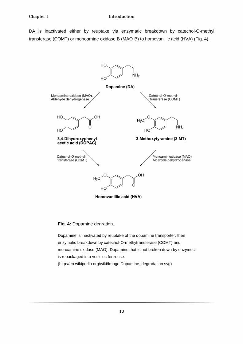
DA is inactivated either by reuptake via enzymatic breakdown by catechol-O-methyl
transferase (COMT) or monoamine oxidase B (MAO-B) to homovanillic acid (HVA) (Fig. 4).
Fig. 4: Dopamine degration.
Dopamine is inactivated by reuptake of the dopamine transporter, then
enzymatic breakdown by catechol-O-methytransferase (COMT) and
monoamine oxidase (MAO). Dopamine that is not broken down by enzymes
is repackaged into vesicles for reuse.
(http://en.wikipedia.org/wiki/Image:Dopamine_degradation.svg)

Chapter I Introduction
11
2.2. DOPAMINERGIC GENES
Dopamine transporter 1
Pharmacological agents, notably MPH, appear to exert therapeutic effects in ADHD by
increasing the functional availability of extracellular DA through inhibition of the DA
transporter (DAT1/SLC6A3) (Thapar, Langley et al. 2007). The membrane-spanning gene,
encoding 620 amino acids (aa), comprises 15 exons that span more than 52 kb of genomic
DNA on the human chromosome 5p15.33. DAT1 limits the duration of synaptic activity and
diffusion by reuptaking dopamine into neurons (Madras, Miller et al. 2005). It is expressed
selectively in all dopaminergic neurons in the substantia nigra and the ventral tegmental
area.
Most of the published association studies focus on a 40bp variable number tandem repeat
(VNTR) in the 3´-UTR (untranslated region) of SLC6A3, ranging from 1 to 13 repeats. The
VNTR may change DAT1 function, since it has been suggested to regulate gene expression
(Yang, Chan et al. 2007). In a recent study a positive association with the 10-repeat allele
and ADHD has been found (Yang, Chan et al. 2007). In line with that, linkage studies support
the DAT1 locus in ADHD (Friedel, Saar et al. 2007). However, results published hitherto are
equivocal and vary from no association (Brookes, Mill et al. 2006), a trend for association
(Maher, Marazita et al. 2002; Curran, Purcell et al. 2005) to a modest but significant
association (Faraone, Perlis et al. 2005).
Dopamine receptor 1
Once DA has been released, it binds to pre- and postsynaptic dopamine receptors (DRD1-5)
(Missale, Nash et al. 1998). As they belong to the class of metabotropic, G-protein-coupled
receptors, they modulate the activity of ion channels by second messenger cascades.
D1-like family receptors (DRD1 and DRD5) are coupled to the G-protein GS which
subsequently activates the adenylyl cyclase. DRD2, DRD3 and DRD4 belong to the D2-like
family of dopamine receptors which are coupled to the GI protein, thereby inhibiting adenylyl
cyclase and activating K+-channels (Missale, Nash et al. 1998).

Chapter I Introduction
12
DRD1, which is located at chromosome 5q35.2, is the most abundant dopamine receptor in
the CNS. It regulates neural growth and development and mediates behavioral responses.
Northern blot analyses and in-situ-hybridization demonstrated high expression in the
striatum, nucleus accumbens, and olfactory tubercle. No detectable product was amplified
from substantia nigra, kidney, heart or liver (Dearry, Gingrich et al. 1990). In a recently
published family-based ADHD study, strong evidence for linkage of a DRD1 haplotype with
inattentive, but not with impulsive/hyperactive symptoms was found (Misener, Luca et al.
2004). This haplotype contains four markers which span the whole gene. Bobb and
coworkers support this result: Although they could not replicate this association using a
family-based approach, they found a significantly higher frequency of these risk alleles in the
ADHD cases as compared to controls (Bobb, Addington et al. 2005).
Some animal models of ADHD refer to DRD1. The SHR rats (spontaneous hyperactive rat)
are generally considered to be a suitable genetic model for ADHD since they display
hyperactivity, impulsivity, poor stability of performance and poorly sustained attention
(Russell 2002). Postsynaptic D1 receptors were found to be up-regulated in the brains of five
and 15-week-old SHR. The fact that both D1 and D2 receptors (Kirouac and Ganguly 1993)
as well as DAT (Watanabe, Fujita et al. 1997) are increased in the striatum of
prehypertensive SHR can also be taken as evidence that changes in the DA function might
be involved in the pathogenesis of both the hypertension and behavioral characteristics of
the SHR (Russell 2002).
Dopamine receptor 4
The dopamine receptor gene DRD4 (chromosome 11p15.5), which spans 3 kb and
comprises four exons, is located primarilly in the hippocampus (HC), the frontal lobes and the
amygdala and shows a strong homology to DRD2 and 3. Both NE and DA are effective
agonists of DRD4.
The distribution of DRD4 mRNA in the brain, mainly in the fronto-subcortical network, argues
for a role in cognitive and emotional functions; functions implicated in the pathophysiology of
ADHD (Faraone, Doyle et al. 2001). Also various mutations in DRD4 were associated with
behavior phenotypes and ADHD. Population and family-based association studies focused
on a VNTR polymorphism in which alleles differ by the number of repeats of a 48 bp
sequence in exon 3. Several studies found an association of the 7-repeat allele with ADHD.
Chapter I Introduction
13
(Faraone, Doyle et al. 2001; Roman, Schmitz et al. 2001; Ding, Chi et al. 2002; Grady, Chi et
al. 2003; Li, Sham et al. 2006). However, it cannot be assumed if the presence of the
DRD4 7R allele is necessary or sufficient to cause ADHD.
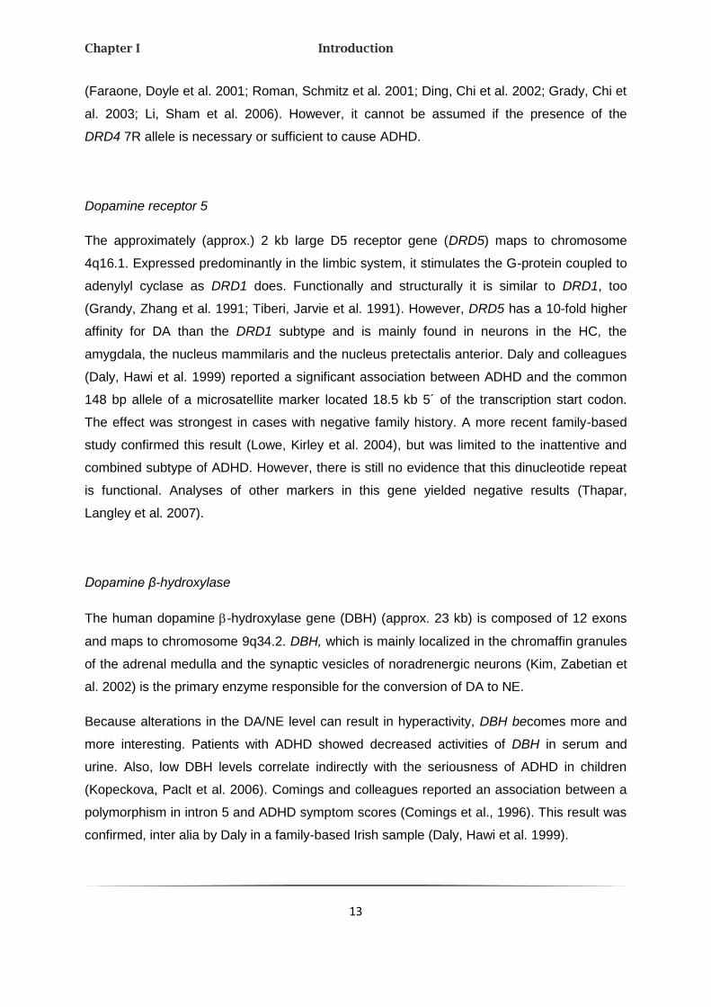
Dopamine receptor 5
The approximately (approx.) 2 kb large D5 receptor gene (DRD5) maps to chromosome
4q16.1. Expressed predominantly in the limbic system, it stimulates the G-protein coupled to
adenylyl cyclase as DRD1 does. Functionally and structurally it is similar to DRD1, too
(Grandy, Zhang et al. 1991; Tiberi, Jarvie et al. 1991). However, DRD5 has a 10-fold higher
affinity for DA than the DRD1 subtype and is mainly found in neurons in the HC, the
amygdala, the nucleus mammilaris and the nucleus pretectalis anterior. Daly and colleagues
(Daly, Hawi et al. 1999) reported a significant association between ADHD and the common
148 bp allele of a microsatellite marker located 18.5 kb 5´ of the transcription start codon.
The effect was strongest in cases with negative family history. A more recent family-based
study confirmed this result (Lowe, Kirley et al. 2004), but was limited to the inattentive and
combined subtype of ADHD. However, there is still no evidence that this dinucleotide repeat
is functional. Analyses of other markers in this gene yielded negative results (Thapar,
Langley et al. 2007).
Dopamine β-hydroxylase
The human dopamine -hydroxylase gene (DBH) (approx. 23 kb) is composed of 12 exons
and maps to chromosome 9q34.2. DBH, which is mainly localized in the chromaffin granules
of the adrenal medulla and the synaptic vesicles of noradrenergic neurons (Kim, Zabetian et
al. 2002) is the primary enzyme responsible for the conversion of DA to NE.
Because alterations in the DA/NE level can result in hyperactivity, DBH becomes more and
more interesting. Patients with ADHD showed decreased activities of DBH in serum and
urine. Also, low DBH levels correlate indirectly with the seriousness of ADHD in children
(Kopeckova, Paclt et al. 2006). Comings and colleagues reported an association between a
polymorphism in intron 5 and ADHD symptom scores (Comings et al., 1996). This result was
confirmed, inter alia by Daly in a family-based Irish sample (Daly, Hawi et al. 1999).
Chapter I Introduction
14
2.3. NORADRENERGIC SYSTEM
Noradrenergic drugs like despiramin and 2-adrenoreceptor agonists are often used to
relieve ADHD symptoms (Solanto 1998). Adrenaline, also known as epinephrine, is a
hormone and neurotransmitter, which belongs to the family of catecholamines. Adrenaline
was isolated and identified in 1895 by the Polish physiologist Napoleon Cybulski. As a “fight
or flight” hormone, adrenaline plays a central role in the short-term stress reaction and
mediates the rash appropriation of energy resources in emergency situations through
adrenergic receptors of the adrenal glands. The neurons of this biogenic amid were only
found in CNS, mainly in the medulla oblongata (Fig. 5).
Adrenaline is synthesized via methylation of the primary distal amine NE by the phenylamine
N-methyltransferase (PNMT) in the cytosol of adrenal medullary cells (Fig. 6). After its
release adrenaline is degraded via the enzymes MAO and COMT to metanephrine, HVA acid
and methoxy-4-hydroxyphenylethylenglycol (MOPEG).
Norepinephrine is a key neurotransmitter in both central and peripheral nervous systems
where it is released from noradrenergic neurons. The catecholamine regulates many
essential functions, including attention, memory, emotion, and autonomic functions (Kim,
Hahn et al. 2006). Also NE underlies the flight-or-fight response, it increases the heart rate,
triggers the release of glucose from energy stores, and increases the blood flow to skeletal
muscles via binding to adrenergic receptors. NE is synthesized from DA by DBH (see Fig. 6)
and released from the adrenal medulla into the blood as a hormone. Before the final -
oxidation it is transported into synaptic vesicles. Its inactivation occurs either enzymaticly
through the metabolites MAO and COMT or by a cellular reuptake into the presynapic cell.
Also, both catecholamines have no evident psychoactive effect in the brain. They are
consistently linked to ADHD, mainly due to its G-protein coupled adrenoreceptors, which are
expressed in different cell types.

Chapter I Introduction
15
Fig. 5: Noradrenergic system.
The cell bodies of most (nor-) adrenergic neurons lay in the
locus coeruleus. Approximately 3000 neurons of the locus coeruleus
are connected by axons which pervade all parts of the brain (red lines)
with billions of other neurons. Therefore (nor-) adrenergic neurons take
simultaneous parts in different brain functions and play an integral
part. Besides the locus coeruleus the area tegmentalis also harbors
(nor-) adrenergic nerve tracts (white lines).
(S.H. Snyder, Chemie der Psyche, Spektrum Verlag Heidelberg (1988)
Chapter I Introduction
16
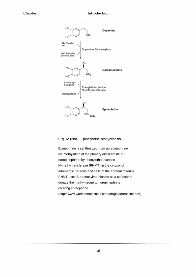
Fig. 6: (Nor-) Epinephrine biosynthesis.
Epinephrine is synthesized from norepinephrine
via methylation of the primary distal amine of
norepinephrine by phenylethanolamine
N-methyltransferase (PNMT) in the cytosol of
adrenergic neurons and cells of the adrenal medulla.
PNMT uses S-adenosylmethionine as a cofactor to
donate the methyl group to norepinephrine,
creating epinephrine.
(http://www.worldofmolecules.com/drugs/adrenaline.htm)

Chapter I Introduction
17
Norepinephrine transporter
The NE transporter (NET, SLC6A2) is a regulator of the NE homeostasis and primarily
responsible for the reuptake of NE into presynaptic nerve terminals (Kim, Hahn et al. 2006).
The human transporter, which spans approx. 45 kb and maps to chromosome 16q12.2, is
mainly expressed in the brainstem and adrenal glands and is sensitive against NET
inhibitors. These seem to be efficient in ADHD treatment (Biederman and Spencer 2000).
SNP and haplotype analyses in families with affected adults showed no association to ADHD
(Barr, Kroft et al. 2002; McEvoy, Hawi et al. 2002; Faraone, Perlis et al. 2005). Otherwise,
Kim and colleagues observed a significant association between the 3081 (A/T) polymorphism
and ADHD, suggesting that anomalous transcription factor-based repression of SLC6A2 may
increase the risk for the development of ADHD and other neuropsychiatric disorders (Kim,
Hahn et al. 2006).
Adrenergic receptor 2A
The G-protein coupled adrenergic receptors (ADR) specifically bind the endogenous
catecholamines adrenaline and NE. Due to their pharmacological and molecularbiological
nature they are divided into two classes: α1- and α2- adrenergic receptors are found in pre-
and postsynaptic neurons of the vegetative and central nervous system, where they inhibit
the transmitter release. β-adrenergic receptors, which are found in heart, smooth muscle and
fat tissue, are responsible for the regulation of the heart rate and smooth muscle relaxation.
The postsynaptic α2- adrenergic receptors (ADRA2) A, B and C are known to have a critical
role in regulating neurotransmitter release from adrenergic neurons as well as from
sympathetic nerves. To find out more about their function the neurotransmitter release in
mice in which the genes encoding the α2- adrenergic receptor subtype were disrupted, was
analyzed (Hein, Altman et al. 1999). Both ADRA1A and ADRA2C are determining factors for
the presynaptic neurotransmitter release of sympathetic and central noradrenergic neurons.
ADRA2A, a 3650 bp gene, which is located at chromosome 10q25.2, has no introns in
translated or in untranslated regions. The role of the noradrenergic system in ADHD is still
underlined. Researches in nonhuman primates demonstrated that NE can enhance the
cognitive functioning of the PFC through actions at α2-adrenergic receptors postjunctional to

Chapter I Introduction
18
noradrenergic terminals (Arnsten, Steere et al. 1996). Also in family-based and case-control
studies a strong association of the MspI polymorphism (1291 C G SNP) in the promoter
region of ADRA2A was found with the inattentive and combined subtype of ADHD (Halperin,
Newcorn et al. 1997; Comings, Gade-Andavolu et al. 1999). Schmitz (Schmitz, Denardin et
al. 2006) supported this thesis by demonstrating that homozygous subjects for the G allele
have an elevated risk for the inattentive subtype. Additional evidence for an involvement of
the noradrenergic system is that methylphenidate treatment improves the inattentive
symptoms in children and adolescents with ADHD (Polanczyk, Zeni et al. 2007; da Silva,
Pianca et al. 2008).
2.4. SEROTONERGIC SYSTEM
Because of the strong interaction between the dopaminergic and serotonergic neurosystem
as well as the therapeutic effects of serotonin reuptake inhibitors (SSRI), the serotonergic
system came to the focus of the researchers.
The neurotransmitter serotonin (5-HT), detected in 1948 by Irving Page, plays an important
role in the modulation of anger, aggression, sexuality, psychological processes and
metabolism. During stress 5-HT causes several changes in different brain areas (Fig. 7):
While the 5-HT level is increased in the cerebral cortex, its release is diminished in the
brainstem and diencephalon. Although it is not clarified if 5-HT deficiency in the brain causes
depression, bipolar or anxiety disorders, an enhancement of the 5-HT level leads to an
abatement of the symptoms. MAO-I and SSRIs enhance the 5-HT concentration in the brain,
which turns them to pharmacological useful antidepressants.

Chapter I Introduction
19
Fig. 7: The serotonergic system.
The serotonergic diffuse modulatory systems arise from the raphe nuclei.
The raphe nuclei are clustered along the midline of the brain stem and project
extensively to all levels of the CNS.
(http://aids.hallym.ac.kr/d/kns/tutor/medical/sero.html)
In the neuronal cytoplasm of liver, spleen and enterochromaffin cells of the intestinal
mucosa, 5-HT is synthesized from the amino acid L-tryptophan by a short metabolic pathway
consisting of two enzymes: tryptophan hydroxylase (TPH) and 5-HTP decarboxylase (DDC).
Because the indolamine cannot cross the blood-brain barrier, tryptophan and its metabolite
5-hydroxytryptophan (5-HTP), the direct precursor of 5-HT, attain the barrier by carrier
mediated transport or diffusion. Unbounded 5-HT is abolished by MAO-A and
aldehydhydrogenase to 5-hydroxyindoleacetic acid (5-HIAA), which is excreted in the urine.
An overview about synthesis and degradation is shown in Fig. 8.

Chapter I Introduction
20
Fig. 8: Pathway for the synthesis of serotonin from
tryptophan.
Serotonin is synthesized from the amino acid L-tryptophan by
the tryptophan hydroxylase (TPH) and the amino acid
decarboxylase (DDC). The TPH-mediated reaction is the
rate-limiting step in the pathway.
(http://en.wikipedia.org/wiki/Serotonin)

Chapter I Introduction
21
2.5. SEROTONERGIC GENES
Serotonin transporter
The human serotonin transporter gene SLC6A4, also known as SERT or 5-HTT, is mapped
to chromosome 12p11.1 - q12 and consists of 14 exons which span about 35 kb. SERT
seems to be one of the most analyzed genes in the psychiatric genetic with association to
many disorders and diagnosis. In the brain it arranges as an integral membrane protein the
reuptake of the released 5-HT from the synaptic cleft in neurons platelets and
enterochromaffin cells and determines the magnitude and duration of postsynaptic receptor-
mediated signaling (Lesch 1997). Furthermore SERT is the initial target for several
antidepressant and neurotoxins like ecstasy. The association between ADHD and SERT
exists mainly in the 44 bp insertion-/deletion polymorphism 5-HTTLPR in the 5´-flanking
promoter region (Seeger, Schloss et al. 2001) which consists of 14 (short “s”-) or 16 (long
“l”-form) repeats and builds the basis of many genetic association studies. The short version
of this allele results in decreased transporter expression (Lesch, Bengel et al. 1996).
Analysis of combined studies showed that ADHD children hold the l-allele and the
L/L-genotype above-average in comparison to healthy controls (Fisher, Francks et al. 2002;
Kent, Doerry et al. 2002; Retz, Thome et al. 2002).
Serotonin receptor 1B
The serotonin receptor 1B (HTR1B) encodes for the 5HT1B-receptor and maps to
chromosome 6q13. Specific evidences for a connection to ADHD were found in mice which
miss this receptor and show motor hyperactivity (Brunner, Buhot et al. 1999) and are
increasingly aggressive (Bouwknecht, Hijzen et al. 2001). Preclinical and clinical studies also
prove that serotonergic inputs may moderate DA´s effects on attention and
hyperactivity/impulsivity while HTR1B regulates DA release in the striatum, midbrain and
PFC (Smoller, Biederman et al. 2006). Further studies in and around the HTR1B-locus refer
to an association between this gene and ADHD (Hawi, Dring et al. 2002; Quist, Barr et al.
2003; Faraone, Perlis et al. 2005). Smoller (Smoller, Biederman et al. 2006) genotyped 21
SNPs in and around HTR1B in 12 multigenerational pedigrees with regard to ADHD. Only
three SNPs were nominally associated with the inattentive subtype.

Chapter I Introduction
22
Tryptophan hydroxylase 2
Primary it was assumed that the tryptophan hydroxylase gene (TPH) is widely distributed, but
then a second isoform, TPH2, was identified. This isoform is only expressed in the brain,
especially in serotonergic neurons of the raphe nuclei and formation reticularis. TPH2,
mapped to chromosome 12q21.1, is the rate-limiting enzyme in 5-HT synthesis. It catalyzes,
together with oxygen and tetrahydrobioptren as cosubstrates and iron as cofactor, the
hydroxylation from tryptophan to 5-hydroxytryptophan. Ko-mice showed a reduced
HT-production in brain and behavior abnormalities which are in accordance with human
depression or anxiety disorders (Beaulieu, Zhang et al. 2008). Furthermore, TPH2 is also in
humans the purpose of numerous phenotype studies in psychiatric disorders like ADHD. In
2005 Walitza and colleagues analyzed the effects of polymorphic variations in the TPH2
gene in 225 ADHD children out of 103 families. Two SNPs (rs4570625 and rs11178997)
revealed a trend towards an association to ADHD in a haplotype analysis (Walitza, Renner et
al. 2005). Sheehan established a significant association between diverse markers and HKS
(Sheehan, Lowe et al. 2005). Thus different polymorphisms of this gene, in the promoter
region and in introns are connected to ADHD.
2.6. NEUROPEPTIDES
Neuropeptides are released as second messengers by different neurons and affect either the
endocrine as neurosecretatory peptide hormones or paracrine as co-transmitters. They
depolarize or hyperpolarize other neurotransmitters not by binding to ion channels at the
postsynaptic membrane, but over receptors.
Neuropeptide Y
Neuropeptide Y (NPY) is a tyrosine-rich, highly conserved, 36 aa neuromodulatoring peptide
that has high structural similarity to peptide YY and pancreatic polypeptide. Since its
discovery in 1982 by Tatemoto (Tatemoto 1982) is has been characterized as one of the
most abundantly expressed peptides throughout the mammalian peripheral and central
nervous system mainly in the cortex, hippocampus, hypothalamus and metencephalon

Chapter I Introduction
23
(Chronwall, DiMaggio et al. 1985). Initial research discovered NPY´s effects on a large
number of neuroendocrine functions, circadian rhythms, stress response, central autonomic
functions, eating and drinking behaviors, and sexual and motor behavior (Wahlestedt, Ekman
et al. 1989; Westwood and Hanson 1999). Even behaviors related to neuropsychiatric
disorders (i.e. depression and schizophrenia) seem to be modified by NPY. The potent
neurotransmitter exerts its biological effects through at least five G-protein coupled receptors
termed Y1, Y2, Y4, Y5 and Y6 (Karl and Herzog 2007) and were frequently analyzed for
connection to neurological diseases including ADHD. In addition, NPY has been shown to
interact with neurotransmitter systems such as DA, γ-aminobutyric acid (GABA) and NE, and
co-localizes with several other neurotransmitters (Westwood and Hanson 1999). Because
the NPY-system is altered in many DA-associated psychotic diseases and moreover DA
plays a role (see chap. 2.2.), a connection to ADHD is most likely. Indeed, NPY was
implicated in ADHD only in one study: Oades detected that an elevated level of circulating
NPY as well as a decreased electrolyte excretion exists in ADHD children that may reflect a
common disturbance in metabolic homeostasis (Oades, Daniels et al. 1998). While it has
widely been investigated in the context of energy balance and body weight
regulation, NPY has recently not only been implicated in behavioral traits, particularly
negative emotionality and aggression (Raveh, Grunwald et al. 1993), but also in
several neuropsychiatric disorders including depression, panic disorder, bipolar
disorder, and schizophrenia (Koetzner and Woods 2002). A functional polymorphism
in the human NPY (Leu7Pro) resulting in increased NPY release from sympathetic
nerves is associated with characteristics of metabolic syndrome and it has been
suggested that the Pro7 allele is associated with an increased risk for alcohol
dependence, a common co-morbid disorder of ADHD (Manoharan, Kuznetsova et al.
2007).
Latrophilin 3
Currently three different isoforms of the latrophilin family are known, latrophilin (LPHN) 1, 2
and 3. The name came from its binding to α-latrotoxin (LTX), a potent presynaptic neurotoxin
from the venom of black widow spiders, which induces neurotransmitter and hormone
release by way of extracellular Ca2+-influx and cellular signal transduction pathways
(Erdogan, Chen et al. 2006). All isoforms are brain-specific chimeras of G-protein coupled,
Ca2+-independent receptors (GPCR) of the secretin/calitonin family and of cell adhesion

Chapter I Introduction
24
molecules (CAM) (Matsushita, Lelianova et al. 1999). Also latrophilins play an important role
both with cell adhesion and signal transduction. In a genome-wide linkage analysis was
shown that the region 4q13.1-13.2 (Arcos-Burgos, Castellanos et al. 2004) is connected with
ADHD and obsessive-compulsive disorder (OCD) (Jain, Palacio et al. 2007). Within this 40
Mb large region the gene LPHN3 was found to be associated with ADHD. Furthermore,
subsequent haplotype analyses identified a susceptibility locus inside exon 7 - 9 (413 kb) of
LPHN3 ((Arcos-Burgos, Jain et al.). The approx. 6 Mb large LPHN3, which consists of 24
exons, encodes for a 1249 aa protein. Unfortunately, the endogenous ligands are still
unknown for all three homologues.
2.7. OTHER CANDIDATE GENES
Monoamine oxidase isoenzyme A
Two monoamine oxidase isoenzymes MAO-A and MAO-B, lying in antipodal direction on the
X-chromosome, are mainly expressed in the outer membrane of mitochondria of neurons
and astroglia. Both oxidases catalyze the oxidative deamination of neurotransmitters and
monoamines. Man-made drugs which block MAOs, so-called monoamine oxidase inhibitors
(MAO-I), are applied more and more frequently as antidepressants.
Mutations in MAO-A, which exists of 15 exons and spans approx. 90 Mb, or a low MAO-A
activity were still associated with impulsive and criminal behavior (Chen, Holschneider et al.
2004). Based on different evidences of MAO-systems in the etiology and the course of
ADHD, Li and colleagues analyzed two polymorphisms in MAO-A and three in MAO-B (Li,
Kang et al. 2007). The results showed a significant association between both MAO-A
polymorphisms and ADHD in adolescents as well as between those and the
hyperactive/impulsive subtype.
Synaptosomal associated protein 25
The synaptosomal associated protein (SNAP-25), mapped to chromosome 20p11.2,
regulates membrane trafficking and is involved in the release of neurotransmitters as well as
the translocation of proteins to the cell membrane. Altered expression will have diffuse

Chapter I Introduction
25
effects on neuronal function. Interest in this gene has come from animal research. The
SNAP-25 deficient mouse mutant coloboma (CM/+) displays spontaneous motor
hyperactivity that is alleviated by stimulant medication (Barr, Feng et al. 2000; Mill, Curran et
al. 2002; Russell, Sagvolden et al. 2005; Thapar, Langley et al. 2007). The ko-mouse shows
therefore no hyperactivity (Washbourne, Thompson et al. 2002). In humans, evidence for an
association between SNAP-25 and ADHD is still not evident (Kustanovich, Merriman et al.
2003) because only a low accordance is denoted between numerous SNP analyses.
3. MEGALOENCEPHALIC LEUKOENCEPHALOPATHY WITH
SUBCORTICAL CYSTS
3.1. CLINICAL FEATURE
Megaloencephalic leucoencephalopathy with subcortical cysts (MLC) is characterized by
diffuse swelling of the white matter, large subcortical cysts, and megaencephaly with infantile
onset. As the disease progresses, the white matter swelling decreases and cerebral atrophy
ensues, while the subcortical cysts generally increase in size and number. The appearance
of subcortical cysts in the anterior-temporal region and often also in the frontoparietal region
is typical for this disease. This neurologic disorder shows an autosomal-recessive mode of
inheritance. MLC has a wide heterogeneity both within and between families and it is
speculated that this might be related to specific genetic determinants (Montagna, Teijido et
al. 2006). Also, its clinical heterogeneity indicates that unknown environmental or genetic
factors may impact the severity of the disease.
3.2. FINDINGS
MLC seems to be caused by mutations in the MLC1 (Leegwater, Yuan et al. 2001), a
~26.1 kb gene, also known as WKL1 or KIAA0027 and maps to chromosome 22q13.3.

Chapter I Introduction
26
Chromosome 22qtel is known to harbor several genes involved in severe neurodegenerative
disorders, like myoneurogastrointestinal encephalopathy or metachromatic leukodystrophy
(Rubie, Lichtner et al. 2003). MLC1 encodes the protein MLC1 which is mainly expressed in
distal astrocytes, Bergman glia and subependymal cells, and in leukocytes, but not in
oligodentrocytes or microglia (Teijido, Martinez et al. 2004). Its biochemical properties and its
function are still unknown, although there are many assumptions, i. e. a transporter function
as a cation-channel, ABC-2 type transporter or sodium/galactoside transporter (Leegwater,
Yuan et al. 2001). Most of all, the presence of eight putative transmembrane domains and its
localization suggest a transporter function across the blood-brain and brain-cerebrospinal
fluid barrier (Boor, de Groot et al. 2005).
Since the first report, 50 mutations in this gene have been found, which include all different
types: eleven splice-site, one nonsense, 24 missense mutations and 14 deletions and
insertions. All of these mutations can lead to frame-sifts or loss-of-function (Boor, de Groot et
al. 2005), and still novel mutations are discovered. But almost nothing is known about the
pathogenic mechanism of these mutations, but recent heterologous expression studies
proposed that gene mutations impair protein folding (Teijido, Martinez et al. 2004). A different
approach is to study the expression of the gene in specific brain regions known to be
involved in MLC. Because MLC1 is highly conserved between vertebrates, the murine Mlc1
can likely give a better insight of MLC1 involvement in the pathogenesis of MLC and
catatonic schizophrenia. Mlc1 expression seems to be developmentally regulated in a region-
and cell type-specific manner and may be important in the development of the brain, mainly
for initial events of myelination (Schmitt, Gofferje et al. 2003). Some mutations are quite
frequent in certain populations, indicating a founder effect. Imaging studies have described a
disorder very similar to MLC among the Agarwals, a discrete, genetic isolated ethnic group
found in India (Gorospe, Singhal et al. 2004). The Agarwals are known to be an enterprising
business group whose members have migrated to widespread regions of India and different
parts of the world. But in about 20% of the patients with MLC no mutations in MLC1 are
found, so likely a second gene accounts for a smaller subset of MLC patients.
In addition, linkage analysis and positional cloning reveals that haplo-insufficiency in MLC1
(amino acid change Leu309Met) is associated in a dominant manner with a periodic subtype
of catatonic schizophrenia in a large pedigree (Meyer, Huberth et al. 2001). Recent studies
have brought forward compelling arguments that genetic variants of MLC1 are not
associated with schizophrenia (Ewald and Lundorf 2002; Kaganovich, Peretz et al. 2004).
Rubie and coworkers (Rubie, Lichtner et al. 2003) also provided evidence of allelic

Chapter I Introduction
27
heterogeneity in MLC and ruled out the possibility that MLC and schizophrenia are allelic
disorders.
Identification of sequence variations in all 13 exons and flanking intronic sequences of MLC1
revealed eight SNPs which seem to be associated with schizophrenia and bipolar affective
disorder and could therefore increase the susceptibility to these disorders (Verma, Mukerji et
al. 2005).
A generation of a transgenic mouse model would provide a useful tool to elucidate both,
function and disease pathomechanisms as well as behavior and possible motor impairment.
Chapter II MATERIAL AND METHODS
28
II. MATERIAL AND METHODS
1. MATERIAL
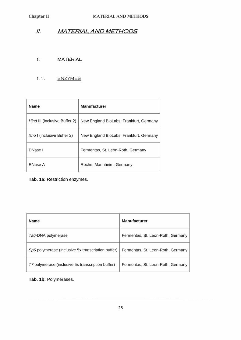
1.1. ENZYMES
Name Manufacturer
Hind III (inclusive Buffer 2) New England BioLabs, Frankfurt, Germany
Xho I (inclusive Buffer 2) New England BioLabs, Frankfurt, Germany
DNase I Fermentas, St. Leon-Roth, Germany
RNase A Roche, Mannheim, Germany
Tab. 1a: Restriction enzymes.
Name Manufacturer
Taq-DNA polymerase Fermentas, St. Leon-Roth, Germany
Sp6 polymerase (inclusive 5x transcription buffer) Fermentas, St. Leon-Roth, Germany
T7 polymerase (inclusive 5x transcription buffer) Fermentas, St. Leon-Roth, Germany
Tab. 1b: Polymerases.

Chapter II MATERIAL AND METHODS
29
1.2. ANTIBODIES
Antibody Name Manufacturer
Secondary Ovine anti-digoxigenin (DIG) Fab-fragments linked to alkaline phosphatase (aP)
Roche, Mannheim, Germany
Tab. 2a: Secondary antibodies.
Name Manufacturer
Normal goat serum (NGS) VectorLaboratories, Burlingame, CA, USA
Bovine serum albumin (BSA) Sigma, Deisenhofen, Germany
Tab. 2b: Further proteins.
Chapter II MATERIAL AND METHODS
30
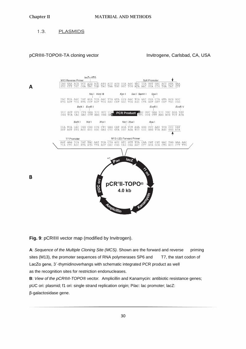
1.3. PLASMIDS
pCR®II-TOPO®-TA cloning vector Invitrogene, Carlsbad, CA, USA
Fig. 9: pCR®II vector map (modified by Invitrogen).
A: Sequence of the Multiple Cloning Site (MCS). Shown are the forward and reverse priming
sites (M13), the promoter sequences of RNA polymerases SP6 and T7, the start codon of
LacZα gene, 3´-thymidinoverhangs with schematic integrated PCR product as well
as the recognition sites for restriction endonucleases.
B: View of the pCR®II-TOPO® vector. Amplicillin and Kanamycin: antibiotic resistance genes;
pUC ori: plasmid; f1 ori: single strand replication origin; Plac: lac promoter; lacZ:
β-galactosidase gene.
B
A

Chapter II MATERIAL AND METHODS
31
1.4. DESOXYRIBONUCLEIDS
Name QuantiTect® Primer Assay (Qiagen)
Latrophilin 3 (LPHN3) Hs_LPHN3_1_SG
Tab. 3a: Human primer for RT-PCR.
Primer Orientation (forw/rev)
Sequence (5´3´) Location Product size [bp]
Melting temperature [Tm; °C]
SA Mlc1 for TK
Neo 340 rev TK
forw
rev
GGACGACAGCAGAGGTAAGC
ATACTTTCTCGGCAGGAGCA
Exon 1
Neo
1546 57
57
Mlc1 integ ex f
Mlc1(Neo) nested SA rev
forw
rev
AGGGTGCCAATGTCTCCA
CTCGTCCTGCAGTTCATTCA
Exon 1
Neo
735 56
57
Mlc1 ex1 nest f
Mlc1 int nest r
forw
rev
CCAATGTCTCCAGGCAAATG
CTGTTGTGCCCAGTCATAGC
Exon 1
Neo
1879 61
59
Tab. 3b: Used primer for searching of the integrated pMlc1-ko plasmid vector.
Forw / f: forward; rev / r: reverse; neo: neomycin-cassette.

Chapter II MATERIAL AND METHODS
32
Name Manufacturer
100bp DNA ladder Fermentas, St. Leon-Roth, Germany
1kb DNA ladder Fermentas, St. Leon-Roth, Germany
Tab. 3c: DNA gene ladders.
1.5. REACTION KITS
Name Manufacturer
DIG RNA Labeling Kit (Sp6/T7) Roche, Mannheim, Germany
iScriptTM
cDNA Synthesis Kit Bio-Rad, Munich, Germany
RNeasy Mini Kit QIAGEN, Hilden, Germany
PeqGOLD RNAPureTM
-System QIAGEN, Hilden, Germany
Tab. 4: Used reaction kits.

Chapter II MATERIAL AND METHODS
33
1.6. BUFFER
All used buffers are in-house productions.
Buffer Contents
Goldstar PCR buffer (10x)
TAE buffer
TE buffer (1x)
Sodium saline citrate (SSC, 20x)
Phosphate buffered saline (PBS, 10x)
750mM Tris-HCl, pH 9.0
200mM ammoniumsulfate
0.1% Tween-20
1mM EDTA, pH 8.0
40mM Tris-acetat
pH 8.0
0.3 sodium citrate, pH 7.0
3M NaCl
1.3 NaCl
70mM Na2HPO4
30mM NaH2PO4
Tab. 5a: General buffers.

Chapter II MATERIAL AND METHODS
34
Buffer Contents
Acetylation buffer
Hybridization buffer (sterile filtered)
RNase buffer
DIG 1 buffer
DIG 3 buffer (detection buffer)
Blocking buffer
0.1M triethanolamine, pH 8.0
0.25% acetic acid anhydride
50% deionisated formamide
4x SSC
10% dextransulfate
1x Denhardt´s solution, RNase free
250µg/ml denatured salmon sperm DNA, RNase free
10mM Tris-HCl, pH 8.0
500mM NaCl
1mM EDTA
100mM Tris-HCl, pH 7.5
150mM NaCl
100mM Tris-HCl, pH 9.5
100mM NaCl
50mM MgCl2
DIG 1 buffer with
0.5% Blocking reagent
Chapter II MATERIAL AND METHODS
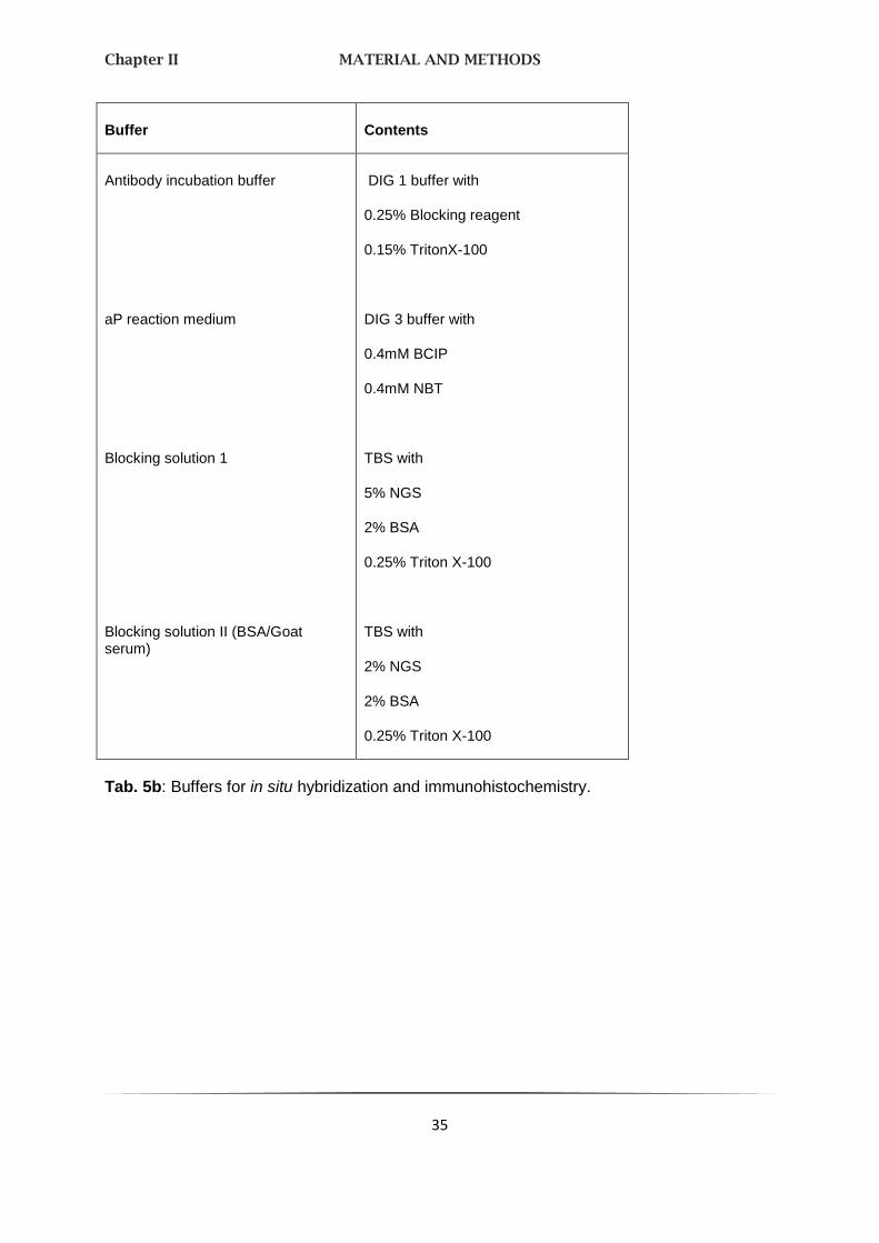
35
Buffer Contents
Antibody incubation buffer
aP reaction medium
Blocking solution 1
Blocking solution II (BSA/Goat serum)
DIG 1 buffer with
0.25% Blocking reagent
0.15% TritonX-100
DIG 3 buffer with
0.4mM BCIP
0.4mM NBT
TBS with
5% NGS
2% BSA
0.25% Triton X-100
TBS with
2% NGS
2% BSA
0.25% Triton X-100
Tab. 5b: Buffers for in situ hybridization and immunohistochemistry.

Chapter II MATERIAL AND METHODS
36
1.7. SOLVENTS AND SOLUTIONS
Name Manufacturer
A. bidest Merck, Darmstadt, Germany
Chloroform Sigma, Deisenhofen, Germany
Ethanol, absolute J.B. Baker, Phillipsburg, NJ, USA
Formamide (deionisated) AppliChem, Darmstadt, Germany
Isopropanol Merck, Darmstadt, Germany
Phenol (waterlogged, stabilized) AppliChem, Darmstadt, Germany
Roti-phenol (TE-buffer logged) Roth, Karlsruhe, Germany
Xylol Merck, Darmstadt, Germany
Tab. 6a: Solvents.
Name Manufacturer
1x Denthardt´s solution, RNase free Sigma, Deisenhofen, Germany
Ethidium bromide solution (10mg/ml) Sigma, Deisenhofen, Germany
Tab. 6b: Solutions.

Chapter II MATERIAL AND METHODS
37
1.8. CHEMICAL COMPOUNDS
Name Manufacturer
Agarose (Seq Kem LE) Biozym, Oldendorf, Germany
Blocking reagent Roche, Mannheim, Germany
BSA (bovine serum albumin) J.B. Baker, Phillipsburg, NJ, USA
Desoxynucleotides (dATP, dCTP, dGTP, dTTP) Promega, Madison, USA
DIG RNA marker mix Roche, Mannheim, Germany
Acetic acid Merck, Darmstadt, Germany
Acetic acid anhydride Sigma, Deisenhofen, Germany
Salmon testis DNA Sigma, Deisenhofen, Germany
NGS (normal goat serum) Sigma, Deisenhofen, Germany
t-RNA Sigma, Deisenhofen, Germany
Tab. 7a: Biochemicals.
Name Manufacturer
BICP (5-bromo-4-chloro-3-indolyl-phosphate) Sigma, Deisenhofen, Germany
DAB (3,3-diaminobenzidine) Roche, Mannheim, Germany
DEPC (diethylpyrocarbonat) Sigma, Deisenhofen, Germany

Chapter II MATERIAL AND METHODS
38
Name Manufacturer
Dextran sulfate Sigma, Deisenhofen, Germany
DTT (dithioreitol) Sigma, Deisenhofen, Germany
EDTA (ethylendiamintetraacetic acid) AppliChem, Darmstadt, Germany
Fluorescin Bio-Rad, Munich, Germany
Hydrochlorid acid (5M) Merck, Darmstadt, Germany
Magnesium chloride (MgCl2) Merck, Darmstadt, Germany
Phosphate buffered saline (PBS) Bio, Whittaker, Charles City, USA
Potassium chloride AppliChem, Darmstadt, Germany
Paraformaldehyde Merck, Darmstadt, Germany
Protease inhibitor cocktail Sigma, Deisenhofen, Germany
RNase inhibitor Fermentas, St. Leon-Roth, Germany
Sodium chloride (NaCl) Merck, Darmstadt, Germany
Triethanolamine (TAE) Merck, Darmstadt, Germany
Tris(hydroxymethyl)aminomethane (Tris) Merck, Darmstadt, Germany
Triton X-100 Sigma, Deisenhofen, Germany
Tween-20 Sigma, Deisenhofen, Germany
Tab. 7b: Further chemical compounds.
Chapter II MATERIAL AND METHODS
39
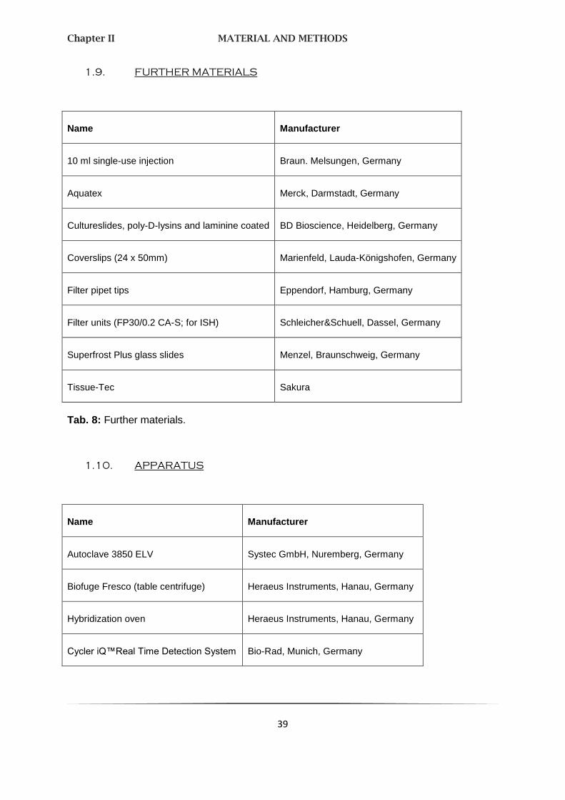
1.9. FURTHER MATERIALS
Name Manufacturer
10 ml single-use injection Braun. Melsungen, Germany
Aquatex Merck, Darmstadt, Germany
Cultureslides, poly-D-lysins and laminine coated BD Bioscience, Heidelberg, Germany
Coverslips (24 x 50mm) Marienfeld, Lauda-Königshofen, Germany
Filter pipet tips Eppendorf, Hamburg, Germany
Filter units (FP30/0.2 CA-S; for ISH) Schleicher&Schuell, Dassel, Germany
Superfrost Plus glass slides Menzel, Braunschweig, Germany
Tissue-Tec Sakura
Tab. 8: Further materials.
1.10. APPARATUS
Name Manufacturer
Autoclave 3850 ELV Systec GmbH, Nuremberg, Germany
Biofuge Fresco (table centrifuge) Heraeus Instruments, Hanau, Germany
Hybridization oven Heraeus Instruments, Hanau, Germany
Cycler iQ™Real Time Detection System Bio-Rad, Munich, Germany

Chapter II MATERIAL AND METHODS
40
Name Manufacturer
Cryostat Microm HM 500 O Microm GmbH, Neuss, Germany
Leica TCS SP2 confocal microscope Leica, Wetzlar, Germany
NanoDrop®ND-1000 fluorospectrometer Peqlab, Erlangen, Germany
PCR gradient thermocycler Biometra, Goettingen, Germany
Chemi-Doc (gel documentation system) Bio-Rad, Munich, Germany
Nanodrop NanoDrop, Wilmington, DE, USA
Axon 4000B scanner Axon instruments, Burlingame, CA, USA
Tab. 9: Apparatus.
1.11. COMPUTER SYSTEMS
Name Manufacturer
iCycler iQ 3.1 Bio-Rad, Munich, Germany
Leica Confocal Software 2.61 Leica, Wetzlar, Germany
Genepix 5.0 Axon Instruments, Union City, Calif., USA
CGHPRO (Chen, Holschneider et al. 2004)
CGH Analytics Agilent, Santa Clara, USA
Tab. 10a: General computer systems.
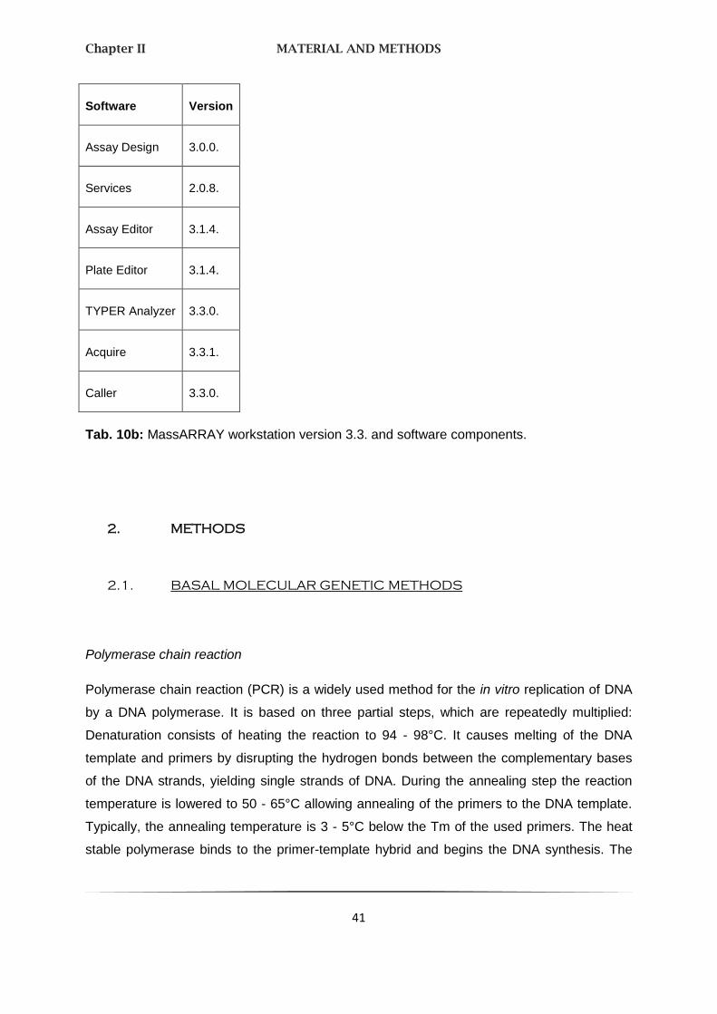
Chapter II MATERIAL AND METHODS
41
Software Version
Assay Design 3.0.0.
Services 2.0.8.
Assay Editor 3.1.4.
Plate Editor 3.1.4.
TYPER Analyzer 3.3.0.
Acquire 3.3.1.
Caller 3.3.0.
Tab. 10b: MassARRAY workstation version 3.3. and software components.
2. METHODS
2.1. BASAL MOLECULAR GENETIC METHODS
Polymerase chain reaction
Polymerase chain reaction (PCR) is a widely used method for the in vitro replication of DNA
by a DNA polymerase. It is based on three partial steps, which are repeatedly multiplied:
Denaturation consists of heating the reaction to 94 - 98°C. It causes melting of the DNA
template and primers by disrupting the hydrogen bonds between the complementary bases
of the DNA strands, yielding single strands of DNA. During the annealing step the reaction
temperature is lowered to 50 - 65°C allowing annealing of the primers to the DNA template.
Typically, the annealing temperature is 3 - 5°C below the Tm of the used primers. The heat
stable polymerase binds to the primer-template hybrid and begins the DNA synthesis. The

Chapter II MATERIAL AND METHODS
42
temperature at the extension step depends on the used DNA polymerase. At this step the
polymerase synthesizes a new DNA strand complementary to the DNA template strand by
adding dNTPs.
Reagent Volume
10x Goldstar buffer 2.5 µl
MgCl2 (25mM) 1.0 µl
dNTPs (2.5 mM each) 2.0 µl
Primer forward (10 pmol/µl) 1.0 µl
Primer reverse (10 pmol/µl) 1.0 µl
Genomic DNA (40 - 60 ng) 2.0 µl
Taq DNA polymerase (5 U/µl) 0.3 µl
a. d. (Merck) 17.2 µl
Final volume 25.0 µl
Tab. 11a: PCR components protocol.
Temperature Time Cycles
95°C (denaturation) 3 min 1x
95°C (denaturation) 45 sec
54-65°C (annealing) 45 sec 35 - 45x
(Primer-specific temperature)
72°C (elongation) 45 sec
72°C (final elongation) 3 min 1x
4°C ∞
Tab. 11b: PCR cycle protocol.
Chapter II MATERIAL AND METHODS
43
For mass spectrometry (see chap. 2.4) composition and cycles of the PCR reactions are
modified.
The used primers are oligonucleotides, allowing the DNA polymerase to extend the
nucleotides and to replicate the complementary strand. Typically, synthesized
oligonucleotides are single-stranded DNA molecules around 17 - 30 bases in length and with
cysteine or guanine at their 3´ end. Whereas polymerases synthesize DNA in 5´ to 3´
direction, chemical DNA synthesis is done backwards in 3´ to 5´ reaction. The G/C content of
the selected DNA sequence averaged 50 - 60%. Diverse software programs (Oligo 4.0;
FastPCR 3.6.97) choose the primers automatically depending on the selected conditions.
Reverse transcriptase polymerase chain reaction
Reverse transcriptase polymerase chain reaction (RT-PCR), established by Powell and
colleagues (Powell, Wallis et al. 1987), is the most sensitive technique for mRNA detection
and quantification, based on the properties of the conventional PCR. After producing a DNA
copy of cDNA of each mRNA molecule, the gene expression levels were further amplified
from the cDNA mixture together with a housekeeping gene as internal control. DNA
amplification was visualized with a fluorescent dye. RT-PCR machines can detect the
amount of fluorescent DNA and thus the amplification progress which is given in a curve with
an initial flat-phase followed by an exponential phase. Here we used the sequence
independent fluorescent dye SYBR-Green I (Qiagen, Hilden, Germany).

Chapter II MATERIAL AND METHODS
44
Reagent Volume
2x QuantiTect SYBR Green master mix 12.50 µl
10x QuantiTect Primer Assay 2.50 µl
(with specific primers)
Fluorescein 0.25 µl
cDNA 1.00 µl
a. d. 8.75 µl
Final volume 25.00 µl
Tab. 12a: RT-PCR components protocol.
The used master mix already contains the required DNA-polymerase as well as free dNTPs.
All required reagents except for the cDNA are components of “QuantiTec TM SYBR Green
PCR Kits” (Qiagen, Hilden, Germany). The analysis was carried out by an “iCycles iQ
Realtime-PCR Detection System” with a corresponding evaluation program “iCycler, Version
3.1” (both Bio-Rad, Munich, Germany).
Temperature Time Cycles
95°C 15 min 1x
95°C 15 sec
55°C 30 sec 35 - 45x
72°C 30 sec
95°C 30 sec 1x
72°C – 94°C (in 0.5°C measures) each 15 sec 50x
15°C ∞
Tab. 12b: RT-PCR cycle protocol.

Chapter II MATERIAL AND METHODS
45
Agarose gel electrophoresis
Agarose gel electrophoresis is used to separate DNA or RNA molecules by size and to
appraise concentration. This is achieved by moving negatively charged nucleic acids through
an agarose matrix (1 - 1.5% in 1x TAE buffer) with an electric field (90 - 120 volt). Shorter
molecules move faster and migrate farther than longer ones. The most common dye used to
make DNA or RNA bands visible is ethidium bromide (EtBr). It fluoresces under UV light
(λ = 302 nm) when intercalated with DNA/RNA. As loading buffer 1x TAE is used; it has a low
buffering capacity but provides a good resolution for large DNA/RNA. Also a DNA ladder
(199 bp or 1 kb) is laid on the gel for valuation of the size of the nucleic acids.
DNA precipitation
Ethanol precipitation is a most facile and rapid method to purify and/or concentrate nucleic
acids and polysaccharides. DNA is precipitated by adding 1/10 volume of sodium acetate
(3 M, pH 5.5). Then, 2.5 volumes of 100% ethanol were admitted and the DNA was stored at
-20°C over night. During incubation DNA and some salts precipitated from the solution, the
precipitate itself was sedimented by centrifugation in a microcentrifuge tube at high speed
(14,000 rpm, 4°C; 30 min). Time and speed of centrifugation have the biggest effect on DNA
recovery rates. During centrifugation the precipitated DNA has moved due to the ethanol
solution to the bottom of the tube, the supernatant solution was removed afterwards, leaving
a pellet of crude DNA. One volume 70% ethanol was added to the pellet, it was gently mixed
to break the pellet loose and to wash it. This step removes some of the salts present in the
leftover supernatant and binds to the DNA pellet making the DNA cleaner. The suspension
was centrifuged once again for 15 min. Finally, the pellet was air-dried and the DNA was
resuspended in a. d. or another desired buffer.
DNA cleaving by restriction endonucleases
The used Type II restriction endonucleases are bacterial enzymes, which recognitions sites
are usually undivided, palindromic sequences. There, they recognize and cleave to the DNA
at this site by hydrolyzation of the phosphodiester bond. For cutting a DNA fragment out of a
plasmid, two different restriction enzymes are necessary, for linearization just one enzyme
with only one cutting site is sufficient. Cleaving is affected by recommendation of the

Chapter II MATERIAL AND METHODS
46
manufacturer. 1 - 5 units of the particular endonuclease were used per µg DNA. Each
approach had the volume of 25 - 50 µl.
2.2. IN SITU HYBRIDIZATION
In situ hybridization (ISH) represents a powerful and sensitive method for examining gene
expression in individual cells and to characterize the phenotype of cells expressing
neurotransmitter or specific neuroreceptors. Via a marked probe both RNA and DNA can be
detected. The basic requirement is that the tissue is native or fixed by paraformaldehyde
(PFA).
In this research we used single stranded, DIG marked cRNA probes for a non-radioactive
ISH.
Establishing of DIG-labeled cDNA
RNA probes have the advantage that RNA-RNA hybrids are very thermostable and are
resistant to digestion by RNases. In vitro transcription of linearized plasmid DNA with RNA
polymerase was used to produce two RNA probes, a “sense” and an “antisense” one. The
first named corresponds with the base sequence of the cellular mRNA and provides a
specificity control. The last named is complementary to the mRNA and shows a specific
signal after hybridization. The used plasmid, the pCR®II vector, contained the polymerase
from the bacteriophages T7 (antisense) and SP6 (sense). The plasmid was linearized with
Hind III, when producing an antisense sensor, and alternatively with Xho I for a sense one.
After DNA precipitation the marked cRNA probes were produced in the following reaction
batch:
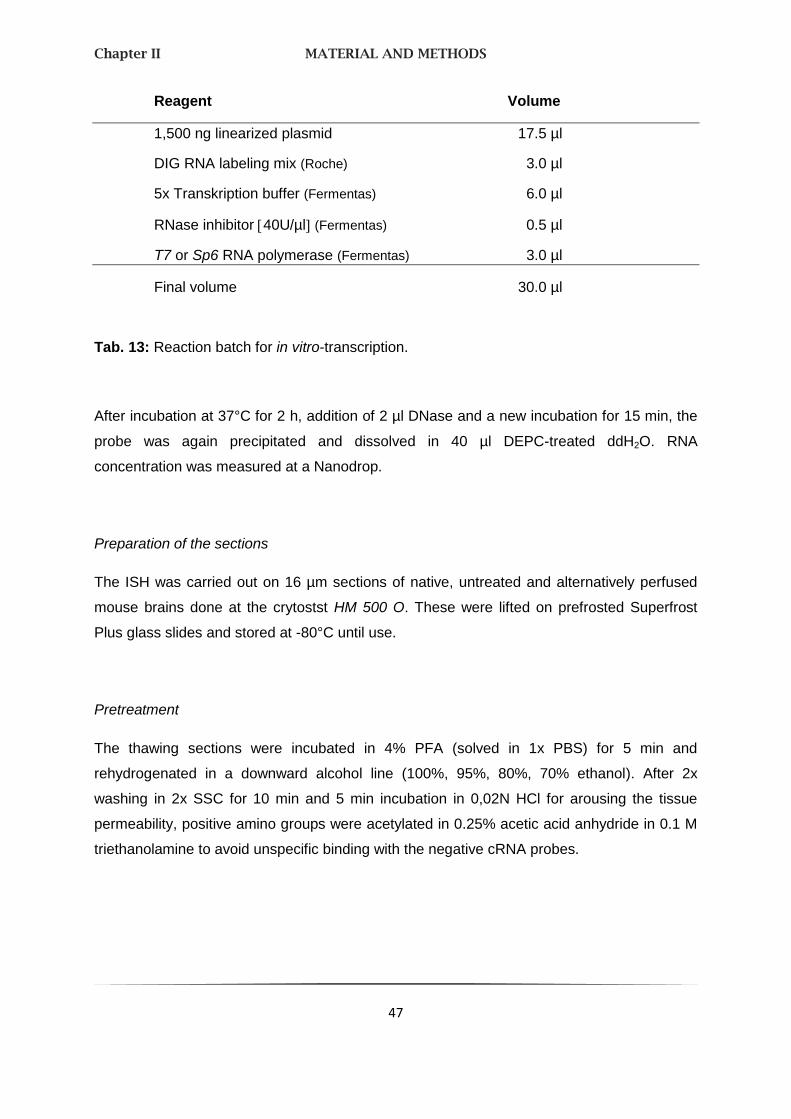
Chapter II MATERIAL AND METHODS
47
Reagent Volume
1,500 ng linearized plasmid 17.5 µl
DIG RNA labeling mix (Roche) 3.0 µl
5x Transkription buffer (Fermentas) 6.0 µl
RNase inhibitor 40U/µl (Fermentas) 0.5 µl
T7 or Sp6 RNA polymerase (Fermentas) 3.0 µl
Final volume 30.0 µl
Tab. 13: Reaction batch for in vitro-transcription.
After incubation at 37°C for 2 h, addition of 2 µl DNase and a new incubation for 15 min, the
probe was again precipitated and dissolved in 40 µl DEPC-treated ddH2O. RNA
concentration was measured at a Nanodrop.
Preparation of the sections
The ISH was carried out on 16 µm sections of native, untreated and alternatively perfused
mouse brains done at the crytostst HM 500 O. These were lifted on prefrosted Superfrost
Plus glass slides and stored at -80°C until use.
Pretreatment
The thawing sections were incubated in 4% PFA (solved in 1x PBS) for 5 min and
rehydrogenated in a downward alcohol line (100%, 95%, 80%, 70% ethanol). After 2x
washing in 2x SSC for 10 min and 5 min incubation in 0,02N HCl for arousing the tissue
permeability, positive amino groups were acetylated in 0.25% acetic acid anhydride in 0.1 M
triethanolamine to avoid unspecific binding with the negative cRNA probes.

Chapter II MATERIAL AND METHODS
48
Hybridization
To avoid unspecific bindings the sections were coated for 1 h with a 100 µl prehybridization
buffer at 58°C. Afterwards each section was overlaid with a 100 µl hybridization buffer
containing 10 – 15 ng of DIG-labeled RNA probe which was prelinearized at 84°C for 5 min.
The samples were covered with a hydrophobic plastic coverslip and incubated overnight at
42°C in a humid chamber. The contained formamide removes hydrogen bonds and therefore
secondary structures.
Posthybridization
After washing two times in 2x SSC for 10 min at room temperature, two times in
2x SSC / 50% formamide buffer for 30 min at 58°C and again in 2x SSC, the sections were
incubated in RNase buffer containing 40 µg/ml RNase A to digest any single-stranded
unbound RNA probes. In a shaking, 58°C water bath the reaction was stopped with RNase
buffer without RNase A.
Using a shaking platform again, the sections were washed 5 min in DIG1 buffer and 30 min
in blocking buffer at room temperature to block unspecific antibody binding sites. They were
covered 1 h with 100 µl buffer containing 0.3% Triton X-100, 1% normal goat serum, and a
1:500 dilution of anti-DIG-alkaline phosphatase (Fab fragments). Afterwards the sections
were again washed two times in DIG1 for 5 min.
The immunological detection resulted from a DIG3-color solution containing 0.4 mM BCIP in
the dark. When the color development was optimal, the reaction was stopped by incubating
the slides in 1x PBS buffer.
2.3. IMMUNOHISTCHEMISTRY
Immunohistochemistry (IHC) refers to processes of localizing proteins in cells of a tissue
section and exploiting the principle of antibody binding specifically to antigens. The indirect,
but specific detection of proteins in tissues by unlabeled prime antibodies (1st layer) and
labeled secondary antibodies (2nd layer) is called the avidin-biotin-complex (ABC-) method.

Chapter II MATERIAL AND METHODS
49
This method is used mainly for double-labeling after ISH with cRNA probes. The glycoprotein
avidin, which is produced by Streptomyces avidinii, is a tetramer and can therefore bind
physically with each subunit of one molecule biotin. A biotinylated secondary antibody, which
is coupled with streptavidin-horseradish peroxidase, is reacted with 3,3´-Diaminobenzidine
(DAB) to produce a brown staining (see Fig. 10).
Fig. 10: Detection of the primary antibody via secondary antibody and
avidin-biotin-peroxidase complex.
(Strept-)Avidin is a basic glycoprotein and has four high affinity binding sites for the
small water soluble vitamin biotin. It forms together with the biotinylated enzyme
peroxidase the avidin-biotin-enzyme complex. The detection of the primary antibody
results from the simultaneous binding of the biotinylated secondary antibody and
the biotinylated peroxidase to avidin.
Directly after ISH the reaction is stopped in 1x TBS-buffer. To remove unspecific protein
bindings and to retrieve antigens, the sections were incubated in 2% BSA / 5% normal goat
serum for 1 h. The LPHN3 primary antibody is produced by a rabbit; 100 µl of a 1:200
antibody-dilution in blocking buffer were applied to each section and incubated over night at
4°C in a humid chamber. The unbounded antibodies were rinsed for 3x 5 min washing in

Chapter II MATERIAL AND METHODS
50
1x TBS, before the sections were incubated in the polyclonal secondary antibody (100 µl
each section; 1:200 dilution in blocking buffer) for 90 min at room temperature. Subsequently
the already compounded AB-complex was applied for 90 min. The visualization of the
peroxidase and therefore the antigen localization resulted from a 5-10 min incubation –
depending on the color intensity – in 1:10 DAB buffer:1x PBS. The reaction was stopped
again with TBS buffer and the glass slides were coversliped.
The documentation of this double staining as well as after ISH occurred via the confocal
microscope Leica TCS SP2.
2.4. ARRAY COMPARATIVE GENOMIC HYBRIDIZATION
Identification of chromosomal imbalances and variations in DNA copy-number is essential to
our understanding of disease mechanisms and pathogenesis, because DNA sequence copy-
number changes have been shown to play an important role in the etiology of many
disorders including trisomy 21 or cancer. Newly developed microarray technologies enable
simultaneous measurements of copy numbers of 1000s of sides in a genome.
In the used Array Comparative Genomic Hybridization (array CGH), differentially labeled total
genomic “test” and “reference” DNAs are cohybridized onto arrays of genomic BAC clones.
An aberration in the genome of the patient is indicated from spots showing aberrant signal
intensity ratios. Fig. 11 shows an overview of array CGH.

Chapter II MATERIAL AND METHODS
51
Fig. 8: Array CGH (http://www.molecular-cancer.com).
Fig. 11: Principle of array CGH.
(A) BAC clones are selected from a physical map of the genome. (B) DNA samples
are extracted from selected BAC clones and their identity is confirmed by DNA
fingerprinting or sequence analysis. (C) A multi-step amplification process generates
sufficient material from each clone for array spotting. (D) Reference and test DNA
are differentially labeled with cyanine 3 and 5 respectively. (E) The two labeled products
are combined and hybridized onto the spotted slide. (F) Images from hybridized slides

Chapter II MATERIAL AND METHODS
52
are obtained by scanning in two channels. Signal intensity ratios from individual spots
can be displayed as a simple plot (G) or by using more complex software which can
display copy number variations throughout the whole genome (H).
(Garnis et al., 2004)
Samples
A cohort of children and adolescents with ADHD (n = 99; 78 male, 21 female) were included
in the CNV scan. Sixty-seven patients were from nuclear families with at least two members
affected with ADHD, eight patients were from extended multigenerational families with high
density of ADHD and 24 patients had sporadic ADHD. Patients and their families were
recruited and phenotypically characterized by a team of experienced psychiatrists in the
outpatient units of the Department of Child and Adolescent Psychiatry, Psychosomatics and
Psychotherapy and the Department of Psychiatry, Psychosomatics and Psychotherapy,
University of Würzburg, Germany, according to DSM-IV criteria (APA 2000). As reference
DNA for the aCGH experiments, we used a sex-matched of unscreened blood donors
(n = 100, 50 females) of European ancestry and originating from the same catchment area
as the patients. All individuals agreed to participate in the study and written informed consent
was obtained from either the participants themselves or the appropriate legal guardian. The
study was approved by the Ethics Committee of the University of Würzburg.
Nuclear families, if they had one or more children affected with ADHD, were recruited to
perform family-based segregation and association studies. The index patient was required to
be older than eight years and to fulfill DSM-IV criteria for ADHD combined subtype, other
affected siblings in a family had to be older than six years. The lower limit was chosen in
order to ensure relative persistence of ADHD symptoms and to exclude children who may
show phenocopies of the disorder during preschool age but lack diagnostic criteria for ADHD
during subsequent developmental stages (Shelton, Barkley et al. 2000; Barkley, Shelton et
al. 2002). Exclusion criteria were: a) general IQ ≤ 80, b) potentially confounding psychiatric
diagnoses such as schizophrenia, any pervasive developmental disorder, Tourette´s
disorder, and primary affective or anxiety disorder, c) neurological disorders such as
epilepsy, d) history of any acquired brain damage or evidence of the fetal alcohol syndrome,
e) premature deliveries, and/or f) maternal reports of severe prenatal, perinatal or postnatal

Chapter II MATERIAL AND METHODS
53
complications. Psychiatric classification was based on the Schedule for Affective Disorders
and Schizophrenia for School-Age Children Present and Lifetime version (K-SADS-PL).
Mothers completed: 1) the unstructured Introductory Interview, 2) the Diagnostic Screening
Interview, and 3) the Supplement Completion Checklist and upon fulfillment of screening
criteria the appropriate Diagnostic Supplements. Children were interviewed with the
screening interview of the K-SADS and in the case of positive screening for affective or
anxiety disorders with the respective supplements of the K-SADS-PL. In addition, we
employed the Child Behavior Checklist and a German Teachers’ Report on ADHD symptoms
according to DSM-IV.
When parents reported individuals with presumable or definite ADHD symptomatology in the
extended family, pedigrees were established to determine family size and structure.
Reported ADHD symptoms in more than two generations resulted in intensified recruitment
of additional family members. Bi-linearity was not an exclusion criterion for recruitment, since
it was presumably present in most recruited families due to assortative mating, intra-familiar
heterogeneity and cannot be completely ruled out in complex traits such as ADHD. All
members of extended pedigrees were assessed by at least two clinicians experienced in
diagnosis of childhood and adult ADHD. Due to a tendency toward severe obesity with
evidence for co-segregation of this trait with ADHD in an extended family, additional data on
body mass index (BMI) and endocrine functions was obtained for further analysis.
Sonification
For a fast cell disruption without detergents or enzymes we used ultrasound with high
amplitude.
10 g test and reference DNA (counterpart) in a total volume of 200 l were sonificated to a
fragment length of 100 bp - 2 kb. Due to the redundancy of heat the probes were
continuously held on ice. For control the sonificated DNAs were applied on a 1% agarose
gel.
Protein contaminations were removed by use of QIAquick PCR Purification Kit (Quiagen,
Hilden, Germany) according to the manufacturer’s recommendations. Finally the DNA was
eluted in 80 l a. d..
Chapter II MATERIAL AND METHODS
54
Labeling and DNA hybridization
Test and reference DNA (1 µg of DNA in a total volume of 21 µl a. d. to each of two tubes)
were labeled using an array CGH Genomic Random Prime Labeling System (Invitrogen,
Carlsbad, Calif., USA). Briefly, 20 µl of 2.5x Random Primer Solution was added to each
tube. After denaturing of the DNA for 5 min at 95°C and cooling down for 5 min on ice, 5 µl
10x dUTP Nucleotide Mix, 3 µl 1 mM Cy3-dUTP (test DNA) or Cy5-dUTP (reference DNA)
(Amersham/ GE Healthcare, Munich, Germany) and 1 µl Klenow Fragment supplied in the kit
were added on ice to produce a final reaction volume of 50 µl. The reaction was incubated at
37°C for 2 h and stopped by adding 5 µl Stop Solution (Kit). Unincorporated nucleotides were
removed by use of the Array CGH Purification Module (Invitrogen, Carlsbad, Calif., USA)
according to the manufacturer’s recommendations. Finally, probes were eluted with 50 µl
a. d. and the same probes were pooled to 100 µl final volume.
Test and reference DNA (100 µl each) were combined, precipitated together with 500 µg
human Cot1 DNA (competitor DNA), 30 µl sodium acetate (3 M, pH 5.5) and 825 l at -20°C
overnight. During the incubation Cot1 DNA binds to the repetitive sequences of the human
DNA and thus diminishes the risk of false positive results.
After ethanol precipitation the DNA pellet was dissolved in 4 l tRNA (100g/l; Invitrogen),
8 l 10% SDS and 30 l FDST (formamide dextran sulfat). The added formamide influences
the denaturizing of nucleic acids, i.e. by unrequested hairpins. Finally, the DNA was
denatured by heating it up to 70°C for 15 min and incubated for 2 h at 42°C for preannealing.
Afterwards, the probes were coated with prehybridzed glass slides (see next subitem) and
hybridized under a coverslip for 20 - 24 h at 42°C using a Slide Booster (Advalytix, Munich,
Germany) (3:7 mixing/pausing).
Prehybridization of the slides
For the array CGH a submegabase resolution tiling path BAC array was used, comprising
the human 32 k Re-Array set (http://bacpac.chori.org/pHumanMinSet.htm; clones and DNA
provided by Pieter de Jong) (Osoegawa, de Jong et al. 2001), the 1 Mb Sander set (clones
provided by Nigel Carter, Wellcome Trust Sanger Centre) (Fiegler, Carr et al. 2003), and a
set of 390 subtelomeric clones (assembled by members of the COST B19 initiative:
Molecular Cytogenetics of Solid Tumors). BAC DNA was amplified using linker-adaption

Chapter II MATERIAL AND METHODS
55
ligation PCR, ethanol precipitated, dissolved in 3x SSC, 1.5 M betaine and spotted on epoxy-
coated slides (NUNC, Wiesbaden, Germany).
Arrays were prehybridized as follows: 0.3 g BSA, 60 ml PreHyb solution (composition see
Material & Methods) and 200 l of herring sperm DNA (10 mg/ml; Sigma) was prepared and
warmed up to 42°C. The used glass slides were incubated in the same solution for 1 h,
washed in a. d. and stored in an opaque box until use.
Washing
Slides were immersed into 2x SSC and the coverslips were carefully removed. Then they
were washed in a prewarmed wash solution for 15 min at 42°C, shortly immersed in PN
buffer (room temperature), again in a second coplin jar with fresh PN buffer and put on a
rocking table for 10 min at room temperature. The slides were washed in PBS for 30 sec at
room temperature and immersed for a few seconds in a.d., before being dried by spinning in
a centrifuge for 5 min at 150 g and stored until scanning.
Data analysis
The high-stringency washed slides were scanned using an Axon 4000B scanner (Axon
instruments, Burlingame, CA, USA) and images were analyzed using Genepix 5.0 (Axon
Instruments, Union City, Calif., USA). For the analysis and visualization of array CGH data
the especially designed software package CGHPRO (Chen, Holschneider et al. 2004) was
employed. No background substraction was applied, and the raw data were normalized by
“Subgrid LOWESS” and manually adjusted where necessary. Fluorescence intensities of all
spots were then calculated after the subtraction of local background. For identifying
potentially disease-related DNA copy number gains and losses, we initially called those
genomic variants that were composed of three or more consecutive clones with log2 signal
intensity ratios beyond 0.3 and -0.3, respectively. In order to increase sensitivity of the read-
out, we then the selection criteria to enable the identification of CNVs in which as few as two
consecutive clones scored above threshold. As this approach entails the risk of an increased
false positive rate, only selected CNVs with highest quality scores (defined by the coefficient
of median average deviation and ratio shift) were added to the list obtained using the
previous, more stringent selection criteria. CNVs were then prioritized and categorized by

Chapter II MATERIAL AND METHODS
56
mirroring them against two CNV datasets derived from individuals not affected by clinically
relevant ADHD. One dataset was composed of CNVs from 700 healthy individuals and
patients suffering from diseases other than ADHD. These samples have been analyzed in
our laboratory using the same BAC array platform and data interpretation parameters as
those for the ADHD samples in this study.
The second dataset, which had also been employed to assess potential disease association
in a recent SNP-based CNV study of ADHD patients (Iafrate, Feuk et al. 2004), was obtained
from the Database of Genomic Variants (DoGV). The DoGV is a public domain depository for
CNVs identified in the healthy population (http://projects.tcag.ca/variation/, release Aug 2009)
(Iafrate, Feuk et al. 2004). It includes all CNVs that were identified in a cohort of 2026
clinically well characterized individuals free of serious medical disorder, including but not
limited to neurodevelopmental disorders (including severe ADHD), cancer, chromosomal
abnormalities, and known metabolic or genetic disorders (Shaikh, Gai et al. 2009). Based on
this comparison, we first identified those CNVs that were not present in either of these two
reference datasets and refer to them as those above the high stringency thresholds. Given
the fact that, compared to SNP data, BAC array data are known to exaggerate the real size
of a CNV, in an inter-platform comparison, CNVs were considered identical if the size
differed no more than 100kb at both ends or, for CNVs smaller than 300kb, if they shared at
least 50% of the genomic sequence. In a separate, less stringent category we have
summarized the CNVs that have been previously reported in the healthy population but are
rare, or where independent evidence exists that genes within these intervals could be
associated with ADHD. All CNVs discussed here were either verified by confirmation of
inheritance using the same method, or by CGH on 244K oligo arrays, performed according to
the protocol provided by the manufacturer and analyzed using the company’s software CGH
Analytics (Agilent, Santa Clara, CA).
Plasma neuropeptide Y
NPY plasma concentrations were determined in 12 individuals of the extended
multigenerational family 3. Plasma was immediately separated from venous blood samples
by centrifugation, kept on dry ice during transportation, and stored at -80°C until processing.
For measurement of plasma NPY a commercial radioimmunoassay (IBL Hamburg,

Chapter II MATERIAL AND METHODS
57
http://www.ibl-hamburg.com) was performed according to protocol provided by the
manufacturer.
Functional magnetic resonance imaging analysis of 7p15 duplication carriers
The impact of the 7p15 duplication and associated increase in NPY plasma concentrations
on brain function was explored by functional magnetic resonance imaging (fMRI). Imaging
was performed during two paradigms: Both were modified versions of the Monetary Incentive
Delay (MID) Task which has been shown to reliably elicit neural responses related to the
anticipation of rewards and losses, respectively (Knutson, Adams et al. 2001). Data were
pre-processed and analyzed using Statistical Parametric Mapping software (SPM5,
Wellcome Department of Cognitive Neurology, UK) as described previously (Hahn, Dresler et
al. 2009). To show potential alterations in reward- and loss-related neural responses, we
compared four ADHD patients carrying the 7p15 duplication (F2-1, F2-4, F2-6, F2-8; Fig. 2A
and B) to an age-matched sample of healthy control subjects (n = 21; mean = 42.0, SD = 6.7;
all within 10 years of the median of the patient group) using a voxel-wise non-parametric
procedure. p values represent the probability of the median neural activation during the
anticipation of rewards/losses of the patient group to be smaller than the median distribution
obtained from all possible sets of four subjects (k = 4) that can be drawn from the control
sample (5985 combinations). Subsequent statistical analyses focused on the ventral striatum
and the posterior insula as defined by voxel masks from a publication-based probabilistic
MNI atlas at a probability threshold of 0.9 35. Correction for multiple comparisons was
realized using AlphaSim (provided with AFNI software) with a single voxel p-value of 0.05.
With this procedure, we assured an overall corrected alpha threshold of p < 0.05.
Statistics
The family-based association test (FBAT; http://biosun1.harvard.edu/~fbat/fbat.htm) (Laird,
Horvath et al. 2000; Rabinowitz and Laird 2000) was used to investigate whether the
7p15.2-15.3 duplication is associated with ADHD, sex, BMI (kg/m2), binge eating (no/yes),
and NPY plasma concentrations (pmol/ml) within a multigenerational pedigree comprising
20 individuals. By means of 10,000 simulations empirical two-sided p values were obtained,
which are more reliable than the respective asymptotic p values in the case of small sample
Chapter II MATERIAL AND METHODS
58
size. The offset parameter was set to null for residuals of BMI and NPY which were adjusted
for sex and age, whereas for binge eating an offset minimizing variance of the test statistic
was chosen. The reported p values are nominal, i. e. not adjusted for multiple testing, at the
significance level of 0.05.
2.5. HIGH THROUPUT SNP GENOTYPING USING MALDI-TOF MASS
SPECTROMETRY
Matrix assisted laser desorption/ionization time of flight mass spectrometry (MALDI-TOF MS)
system is a relatively novel technique in which a co-precipitate of an UV-light ( = 337 nm)
absorbing matrix and a biomolecule is irradiated by a nanosecond laser pulse. The ionized
biomolecules are accelerated in an electric field and enter the flight tube. During the flight in
this tube, different molecules are separated according to their mass to charge ratio and reach
the detector at different times. This method enables the analysis of SNPs directly from
human genomic DNA without the need for initial target amplification by PCR.
SNPs are the most abundant type of variation found in the human genome (10x106),
approximately 10 million are registered in public databases
(http://www.ncbi.nlm.nih.gov/SNP/index.html; dbSNP BUILD 122) and seems to play an
important role in the development of many diseases (depression, anxiety disorders) (Strobel,
Gutknecht et al. 2003; Pearson, Huentelman et al. 2007).
For the whole course we used the iPlexTM Assay protocols and apparatus of
Sequenom®GmbH, Hamburg, Germany as well as reagents of Quiagen, Hilden, Germany.
Samples
All patients were diagnosed with ADHD as described in 2.3. Samples. In total, 437 in- and
outpatients were recruited at the Department of Psychiatry, University of Würzburg. The
control sample consisted of 540 subjects who were either health blood donors of Caucasian
origin, not screened for psychiatric disorders (n = 273) or screened and psychiatrically
healthy un-related individuals from the same ascertainment area as the recruited patients.
The study was approved by the local Ethics Committee of the University of Würzburg. In

Chapter II MATERIAL AND METHODS
59
addition, 453 patients were ascertained from all over Norway, as described by Johansson
(Johansson, Halleland et al. 2008) and Halleland (Halleland, Lundervold et al. 2009). Here
the control group (n = 548) was comprised of 137 university students, 251 randomly selected
people (in the age-range of 18 to 40 years) from the general population and 198 healthy
blood donors (Franke, Neale et al. 2009).
Selection of adequate SNPs
Both, the 5´ and 3´ region of the analyzing genes were determined exactly by HapMap
Genome Browser B35 (http://www.hapmap.org), whereas a putative promoter region (about
10.000 bp upstream exon 1) and an end region (about 3.000bp downstream the last exon)
were included. By use of Haploview version 3.32 (http://www.broad.mit.edu/mpg/haploview)
markers were selected automatically depending on adjustments (p-value cutoff = 0.001;
minimum minor allele frequency = 0.001; r2 threshold = 0.8) and was shown tabular and
figurative (LD plot). Additional synonymous and non-synonymous SNPs with high population
diversity found by genome-wide association studies (GWAS) were also included
(http://www.ncbi.nlm.nih.gov).
PCR amplification
SNPs were investigated by the Sequenom iPlex® method (Sequenom, San Diego, CA). The
principles of PCR were described before (see Material & Methods). Admittedly, both the
configuration of the reaction batch and the cycles were modified in mass spectrometry. The
used primers were created due to the selected SNPs by RealSNPTM Assay Database
(http://www.realsnp.com) (Sequenom®GmbH, Hamburg, Germany). All primer sequences
were available on request and were ordered by Metabion, Martinsried, Germany.
The PCR was performed in a 384 well plate following amplification in a Biometra
thermocycler (Biometra, Goettingen, Germany):
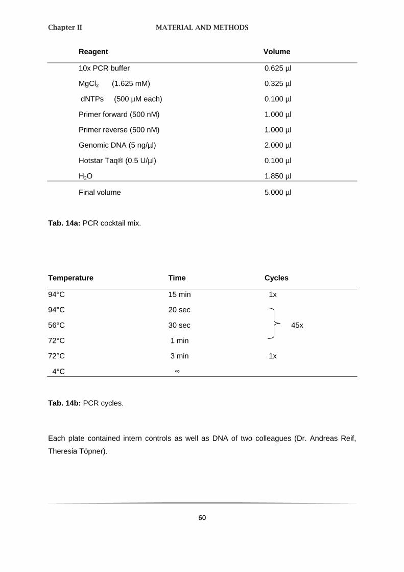
Chapter II MATERIAL AND METHODS
60
Reagent Volume
10x PCR buffer 0.625 µl
MgCl2 (1.625 mM) 0.325 µl
dNTPs (500 µM each) 0.100 µl
Primer forward (500 nM) 1.000 µl
Primer reverse (500 nM) 1.000 µl
Genomic DNA (5 ng/µl) 2.000 µl
Hotstar Taq® (0.5 U/µl) 0.100 µl
H2O 1.850 µl
Final volume 5.000 µl
Tab. 14a: PCR cocktail mix.
Temperature Time Cycles
94°C 15 min 1x
94°C 20 sec
56°C 30 sec 45x
72°C 1 min
72°C 3 min 1x
4°C ∞
Tab. 14b: PCR cycles.
Each plate contained intern controls as well as DNA of two colleagues (Dr. Andreas Reif,
Theresia Töpner).
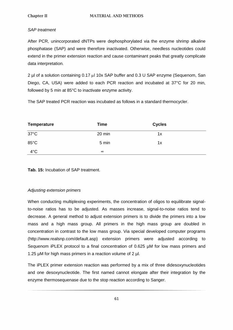
Chapter II MATERIAL AND METHODS
61
SAP treatment
After PCR, unincorporated dNTPs were dephosphorylated via the enzyme shrimp alkaline
phosphatase (SAP) and were therefore inactivated. Otherwise, needless nucleotides could
extend in the primer extension reaction and cause contaminant peaks that greatly complicate
data interpretation.
2 µl of a solution containing 0.17 l 10x SAP buffer and 0.3 U SAP enzyme (Sequenom, San
Diego, CA, USA) were added to each PCR reaction and incubated at 37°C for 20 min,
followed by 5 min at 85°C to inactivate enzyme activity.
The SAP treated PCR reaction was incubated as follows in a standard thermocycler.
Temperature Time Cycles
37°C 20 min 1x
85°C 5 min 1x
4°C ∞
Tab. 15: Incubation of SAP treatment.
Adjusting extension primers
When conducting multiplexing experiments, the concentration of oligos to equilibrate signal-
to-noise ratios has to be adjusted. As masses increase, signal-to-noise ratios tend to
decrease. A general method to adjust extension primers is to divide the primers into a low
mass and a high mass group. All primers in the high mass group are doubled in
concentration in contrast to the low mass group. Via special developed computer programs
(http://www.realsnp.com/default.asp) extension primers were adjusted according to
Sequenom iPLEX protocol to a final concentration of 0.625 µM for low mass primers and
1.25 µM for high mass primers in a reaction volume of 2 µl.
The iPLEX primer extension reaction was performed by a mix of three didesoxynucleotides
and one desoxynucleotide. The first named cannot elongate after their integration by the
enzyme thermosequenase due to the stop reaction according to Sanger.
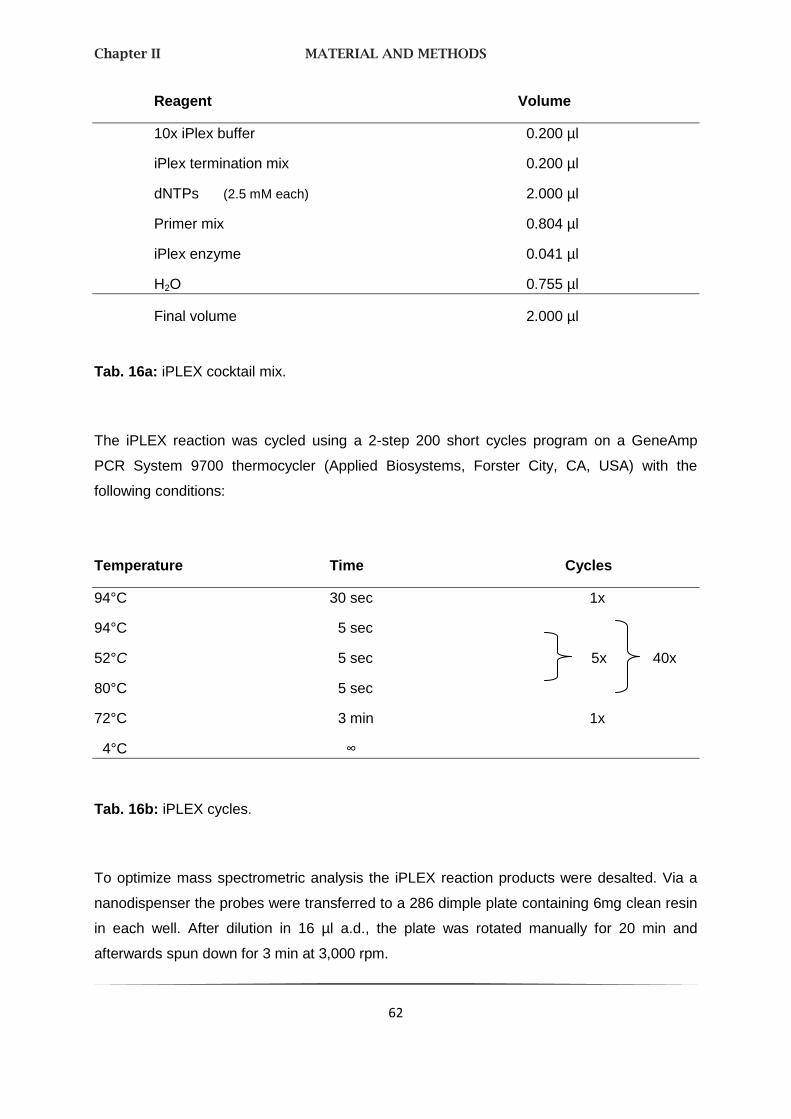
Chapter II MATERIAL AND METHODS
62
Reagent Volume
10x iPlex buffer 0.200 µl
iPlex termination mix 0.200 µl
dNTPs (2.5 mM each) 2.000 µl
Primer mix 0.804 µl
iPlex enzyme 0.041 µl
H2O 0.755 µl
Final volume 2.000 µl
Tab. 16a: iPLEX cocktail mix.
The iPLEX reaction was cycled using a 2-step 200 short cycles program on a GeneAmp
PCR System 9700 thermocycler (Applied Biosystems, Forster City, CA, USA) with the
following conditions:
Temperature Time Cycles
94°C 30 sec 1x
94°C 5 sec
52°C 5 sec 5x 40x
80°C 5 sec
72°C 3 min 1x
4°C ∞
Tab. 16b: iPLEX cycles.
To optimize mass spectrometric analysis the iPLEX reaction products were desalted. Via a
nanodispenser the probes were transferred to a 286 dimple plate containing 6mg clean resin
in each well. After dilution in 16 µl a.d., the plate was rotated manually for 20 min and
afterwards spun down for 3 min at 3,000 rpm.

Chapter II MATERIAL AND METHODS
63
Dispensing to SpectroCHIP® Bioarray
By the use of the Sequenom Mass ARRAY Nanodispenser the reaction products were
dispensed onto a 384-element SpectroCHIP bioarray. Further instructions can be seen in
chapter 4 “High Troughput Dispensing” in the “MassARRAY Nanodispenser User´s Guide”.
MALDI-TOF MS analysis
Mass spectrometric analysis was carried out on a Bruker Autoflex time-of-flight mass
spectrometer (Bruker Daltonics, Billerica, MA, USA). To process bioarrays, the MassARRAY
workstation version 3.3. software was used. Software components and their respective
versions are found in Material & Methods chap. 1.11. Computer systems.
Statistical analysis
Hardy-Weinberg equilibrium (HWE) was assessed for all available samples as well as
chi-square-tests for frequency differences between cases and controls. The reported p-
values are nominal, i. e. not adjusted for multiple testing, at the significance level of 0.05.
The procedure for the analysis of the GLUT3 and GLUT6 SNPs was divided into two steps.
First, the SNPs were studied in pairs in regard to their common genotype distribution, using
Fishers extract tests. An interaction existed if the p-value (FisherPx) was less than the
GLUT3 or GLUT6 value. Relevant for further investigation are mainly these results where
FisherPx is smaller than 0.05 or when FisherPx is relatively small and deviates sharply of the
GLUT3 and GLUT6 value. These results were shown in graphics. The second part is the
logistic regression including the interaction to explain the interaction out of step one.
2.6. TARGETING VECTOR CONSTRUCTION FOR KNOCKOUT MICE
Homologous recombination with exogenous DNA constructs is used to capture two genomic
fragments into a compatible vector and is therefore the most powerful technique available for
analyses and fundamental insights into mammalian gene functions. To circumvent the

Chapter II MATERIAL AND METHODS
64
embryonic lethality problem and to investigate gene function temporally in vivo and spatially,
conditional knockout (cko) approaches have been developed over the past several years.
The current cko strategy takes advantage of the bacteriophage-derived Cre-loxP site specific
recombination system that functions well in mouse cells. In a typical cko allele, the critical
exon(s) of a gene is flanked by two loxP sites that can be deleted by spatial and temporal
Cre expression. So, gene targeting involves the inactivation of a given gene in the genome of
totipotent embryonic stem (ES) cells. Transfer of mutant ES cells into early mouse embryos
allows the transmission of the mutation in question into the mouse germline.
Ligation
Oppositely orientated mutant loxP sites (floxed exons 1 and 2 for the short arm (SA) rather
4 and 5 for the long arm (LA)) were synthesized by cutting out Mlc1 via the restriction
enzymes Not I and Xho I for SA and alternatively Bam I and EcoR I for LA and subcloned via
ligation into the pPNT vector (see Fig. 12) containing a neomycin resistance gene which is
under the control of the mouse phosphoglycerate kinase 1 gene (Pgk-1). We used a 1:2
molar ratio of vector:insert DNA when cloning the fragments consecutively into the plasmid
vector. According to the recommendations of the manufacturer (Promega, Madison, USA)
100 ng vector DNA, 33 ng insert DNA, 5 µl 2x Rapid Ligation Buffer and 3 U T4 DNA Ligase
were filled up with nuclease-free water to a total volume of 10 µl and incubated 5 min for
cohesive-ended ligations.

Chapter II MATERIAL AND METHODS
65
Fig. 12: pPNT vector map.
This gene targeting vector is based on the pUC/Bluescript vector. Elements are:
PGK promoter, neomycin resistence gene (PGKneo cassette), PGK polyA site, hsv-tk
gene and the unique Not I site for linearization. (Tybulewicz, Crawford et al. 1991)
Transformation
DH5α-FT™ competent cells (Invitrogen, Carlsbad, CA, USA) were thawed on wet ice. For
DNA from ligation reaction, 1-10 ng of DNA was added to 100 µl competent cells, tapped to
mix and incubated on ice for 30 min, followed by a heat-shock for 45 sec at 42°C and again
2 min on ice. 900 µl room temperature S.O.C. medium (Invitrogen, Carlsbad, CA, USA) was
added and shaken at 225 rpm (37°C) for 1 h.

Chapter II MATERIAL AND METHODS
66
Selection of positive clones via colony screening
To find positive clones the transformation was spread on IPTG/X-gal LB-plates for blue/white
selection and incubated overnight at 37°C. In the Prime-a-Gene® Labeling System
(Promega, Madison, USA) a mixture of random hexadeoxyribonucleotides was used to prime
the DNA synthesis in vitro from any double-stranded DNA template. The radioactive labeled
DNA probe was produced by following protocol:
Reagent Volume
Labeling 5x Buffer 10 µl
Mixture of unlabeled dNTPs 2 µl
Denatured DNA template 25 ng
Nuclease-free BSA 2 µl
[α-32P]dCTP, 50 µCi, 3,000 Ci/mmol 5 µl
DNA Polymerase I, Klenow Fragment 5 units
Nuclease-free water to achieve a
Final volume 50 µl
Tab. 17: Radioactive DNA labeling protocol.
The tube was incubated at room temperature for 60 min. Then the reaction was terminated
by heating at 95-100°C for 2 min with subsequent chilling in an ice bath. 20 mM EDTA was
added to use it directly for a hybridization reaction or to store at -20°C for later use.
Unincorporated, labeled nucleotides were removed by size exclusion chromatography using
Sephadex® G-50 spin columns following the instructions of the manufacturer (Amersham
Bioscience, Freiburg, Germany).
Colony/Plaque Screen™ are circles of a supported, positively charged nylon membrane.
These dry membrane discs were placed carefully onto the agar plates. After 2 - 3 min the
disc with colony side up was laid two times into a pool of 0.75 ml 0.5 N NaOH on a plastic

Chapter II MATERIAL AND METHODS
67
wrap for 2 min. On a new sheet of plastic wrap 0.75 ml 1.0 M Tris-Hcl, pH 7.5 was pipetted,
the disc was then placed in the same direction as before and repeated.
Before prehybridization the ExpressHyb Hybridization Solution (Clontech Laboratories, Saint-
Germain-en-Laye, France) was warmed up to 60°C. The dried membranes were put in a
heat sealable bag with about 5 ml ExpressHyb solution and heated with continuous shaking
at 60°C for 30 min.
Meanwhile, the radioactive labeled carrier DNA was denatured for 2 - 5 min at 95 - 100°C
and chilled on ice for at least 15 min before adding 5 ml fresh prehybridization buffer to the
bag. The membranes were agitated overnight at 60°C.
The next day the membranes were rinsed repeatedly in wash solution 1 (2x SSC, 0.05%
SDS) for 30 min at room temperature replacing the wash solution several times to remove
non-specifically bound probes. After each wash the blots were monitored for background.
Then the plots were washed again under continuous shaking for 40 min at 50°C with wash
solution 2 (2x SSC, 0.1% SDS). After the final rinse, the damp membranes were wrapped
securely in plastic wrap. Finally the blots were exposed to x-ray film at -80°C with two
intensifying screens.
Electroporation
Positive clones were picked and incubated in LB medium containing 100 µg/ml ampicillin
under permanent shaking at 37°C overnight. Via Wizard® Plus SV Minipreps DNA Purifiction
System using a vacuum (Promega, Madison, USA) the plasmids were isolated and linearized
by the 1-cut endonuclease Not I.
The targeting vector was electroporated into ES cells kindly supported by the Institute for
Clinical Neurobiology, University Würzburg. Finally, the ES clones with correct targeting
events should be identified by PCR (used primers see Material & Methods chap. 1.4.
Desoxyribonuceotides).
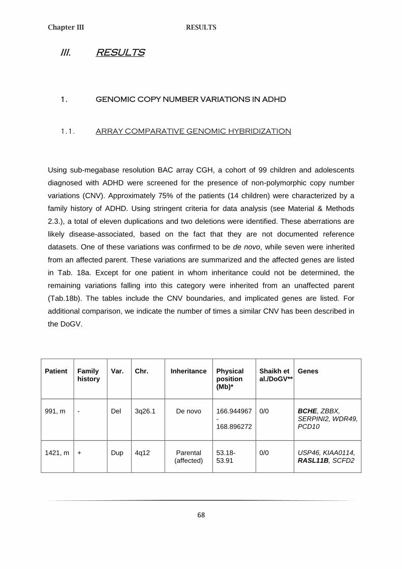
Chapter III RESULTS
68
III. RESULTS
1. GENOMIC COPY NUMBER VARIATIONS IN ADHD
1.1. ARRAY COMPARATIVE GENOMIC HYBRIDIZATION
Using sub-megabase resolution BAC array CGH, a cohort of 99 children and adolescents
diagnosed with ADHD were screened for the presence of non-polymorphic copy number
variations (CNV). Approximately 75% of the patients (14 children) were characterized by a
family history of ADHD. Using stringent criteria for data analysis (see Material & Methods
2.3.), a total of eleven duplications and two deletions were identified. These aberrations are
likely disease-associated, based on the fact that they are not documented reference
datasets. One of these variations was confirmed to be de novo, while seven were inherited
from an affected parent. These variations are summarized and the affected genes are listed
in Tab. 18a. Except for one patient in whom inheritance could not be determined, the
remaining variations falling into this category were inherited from an unaffected parent
(Tab.18b). The tables include the CNV boundaries, and implicated genes are listed. For
additional comparison, we indicate the number of times a similar CNV has been described in
the DoGV.
Patient Family history
Var. Chr. Inheritance Physical position (Mb)*
Shaikh et al./DoGV**
Genes
991, m - Del 3q26.1 De novo 166.944967-168.896272
0/0 BCHE, ZBBX, SERPINI2, WDR49, PCD10
1421, m + Dup 4q12 Parental (affected)
53.18-53.91
0/0 USP46, KIAA0114, RASL11B, SCFD2

Chapter III RESULTS
69
Patient Family history
Var. Chr. Inheritance Physical position (Mb)*
Shaikh et al./DoGV**
Genes
201, m + Dup 5q11.2 Maternal (affected)
58.263673-58.339293
0/0 PDE4D
241, m + Del 5q12.1 Maternal (affected)
60.069539-60.353244
0/0 ELOVL7, ERCC8, NDUFA12L
21, m + Dup 5q13.3 Parental (affected)
75.590060-75.782477
0/0 SV2C, IQGAP2
1671, f
+ Dup 7p15.2-15.3
Paternal (affected)
23.01-26.07
0/0 NUPL2, GPNMB, IGF2BP3, RPS2P32, TRA2A, CLK2P, CCDC126,C7orf46, STK31, NPY, MPP6, DFNA5, OSBPL3, CYCS, C7orf31, NPVF, NFE2L3, HNRPA2B1, CBX3
51, m + Dup 11q13.4 Maternal (affected,
also in affected sibling)
72.90-73.40
0/0 FAM168A, PLEKHB1, RAB6A, MRPL48, CHCHD8, WDR71, DNAJB13, UCP2, UCP3 (none in DoGV)
701, f + Dup 17q25.1 Maternal (affected)
69.25-70.18
0/4 C17orf54, RPL38, TTYH2, DNAI2, KIF19, LOC388419, GPR142, GPRC5C, CD300A, CD300LB, CD300C, C17orf77, CD300E
Tab. 18a: De novo and co-segregating CNVs not present in the reference dataset.
Bold = potential candidate genes.

Chapter III RESULTS
70
Patient Family history
Var. Chr. Inheritance Physical position (Mb)*
Shaikh et al./DoGV**
Genes
1191, m - Dup 2p25.3 Maternal (healthy)
0.66-0.94 0/4 TMEM18, SNTG2
441, f - Dup 4p14 Maternal (healthy)
36.31-37.03
0/0 No genes
461, m - Dup 4q26 Maternal (healthy)
114.833251-115.343086
0/0 CAMK2D, ARSJ
431, m - Dup Xq12 Not determined
65.4-65.6 0/1 EDA2R
471, m - Dup Xq21.33
Maternal (healthy)
93.81-94.64
0/0 No genes
Tab. 18b: Other variations not observed in the reference datasets.
Bold = potential candidate genes.
*Chromosome coordinates according to HG18 (NCBI36) based on BAC clone or oligo hybridisation
results; for BAC clone hybridisation results, coordinates were rounded appropriately in order to reflect
inherent limitations in determining precise CNV boundaries.
**Number of corresponding CNVs in 2026 healthy individuals published by Shaikh et al. (Shaikh, Gai
et al. 2009)/ number of corresponding CNVs in the Database of Genomic Variants (DoGV, including
those from Shaikh et al. (Shaikh, Gai et al. 2009) as of Aug 21, 2009 (Iafrate, Feuk et al. 2004).
Finally, we detected an additional two duplications and two deletions that we consider
potentially syndrome-associated despite the fact that they did not meet the high stringency
threshold scores because they were also observed at low frequency in one or both of the
reference datasets (Tab. 19). All aberrations were definite as all have been verified either
directly by oligo array or indirectly by analysis of parental DNA. None were observed at high
frequencies in other patient cohorts, suggesting that they may indeed be risk factors for

Chapter III RESULTS
71
ADHD. One of these CNVs was de novo, two were inherited from affected parents, and one
was inherited from a health person.
Patient Family history
Var. Chr. Inheritance Physical position (Mb)*
Shaikh et al./DoGV**
Genes
1761, m - Del 6q16.1 De novo 95.447226-95.664033
1/4 No genes
131, m - Dup 8q11.1 Parental (healthy)
47.61-47.98
1/1 BEYLA
1141, f + Del 9p21.3 Parental (affected)
25.217246-25.336971
1/4 No genes
211, m + Dup 12p13.31
Maternal (affected)
7.894681-8.009303
11/37 SLC2A14, SLC2A3
Tab. 19: CNVs present in healthy controls at low frequency or affecting genes with
independent support for disease association.
Bold = potential candidate genes.
*Chromosome coordinates according to HG18 (NCBI36) based on BAC clone or oligo hybridisation
results; for BAC clone hybridisation results, coordinates were rounded appropriately in order to reflect
inherent limitations in determining precise CNV boundaries.
**Number of corresponding CNVs in 2026 healthy individuals published by Shaikh et al. (Shaikh, Gai
et al. 2009) / number of corresponding CNVs in the Database of Genomic Variants (DoGV, including
those from Shaikh et al.(Shaikh, Gai et al. 2009) as of Aug 21, 2009 (Iafrate, Feuk et al. 2004).
Among apparent candidates is the gene encoding neuropeptide Y (NPY) contained in a
duplication on chromosome 7p15.2-15.3 (described in detail in the next section). Further
candidates included genes expressing acetylcholine-metabolising butyrylcholinesterase
(BCHE) involved in a de novo chromosome 3q26.1 deletion in an individual severely affected

Chapter III RESULTS
72
with ADHD, and a brain-specific pleckstrin homology domain-containing protein (PLEKHB1),
with an established function in primary sensory neurons, in two siblings with severe ADHD
carrying a 11q13.4 duplication inherited from their affected mother. Other potentially
disorder-causing genes involved in confirmed aberrations and inherited from affected parents
include the genes for the mitochondrial NADH dehydrogenase 1 alpha subcomplex,
assembly factor 2 (NDUFAF2), the brain-specific phosphodiesterase 4D isoform 6 (PDE4D6)
(Fig. 13), and the neuronal glucose transporter 3 (SLC2A3).

Chapter III RESULTS
73

Chapter III RESULTS
74
Fig. 13: Duplication of 5q11.2 in patient 201.
A, schematic view of chromosome 5, with mapped genomic clones depicted to the right. For
each BAC clone, Cy3/Cy5 signal intensity ratios are plotted alongside the chromosome. Red
and green lines correspond to log2 ratios -0.3 (loss) and 0.3 gain), respectively. The region
encompassing the aberration is highlighted by a red rectangular. B, closer view of the relevant
region. C, ratio plot of the corresponding verification experiment using a 244k oligonucleotide
array. D, UCSC screenshot epicting genomic region chr5:57,791,039-58,811,926 (HG17).
The red bar indicates the location of the duplication identified in patient 201. Grey bars in the
custom track below represent CNVs detected in 2026 control individuals by Shaikh et al. The
specific identifying number is given on the left. Genes and their positions are indicated below
these. Finally, all variations observed in the Database of Genomic Variants (DoGV) are
included at the bottom of each panel for reference. These variations are colour-coded
according to DoGV convention to reflect gain (red), loss (blue), or gain/loss (green).
Noteworthy, the CNV identified in patient 201 includes the complete brain-specific PDE4D6
isoform described by Wang et al., while all other CNVs are located within intronic regions.
1.2. PHENOTYPE OF THE 7q15 DUPLICATION IN A
MULTIGENERATIONAL PEDIGREE
Based on the findings in the initial patient cohort resulting in the identification of a 3 Mb
duplication located on chromosome 7q15.2-15.3 (Fig. 14), we ascertained the extended
multigenerational pedigree (displaying a high density of ADHD) of the index patient to further
investigate the phenotypical consequences of an additional copy of the NPY gene.

Chapter III RESULTS
75
Fig. 14: Array CGH result for patient F3-4 using BAC-Array.
Data analysis and visualization was performed by CGHPRO. Cy3 and Cy5 signal intensity ratios are
given for each BAC clone. Red and green lines correspond to log2 rations -0.3 (loss) and 0.3 (gain).
Insert: closer view of the duplication of 7q15.3.
Using array CGH, the described duplication was detected in several additional family
members throughout three generations (Fig. 15). It is inherited from individual F1-2 of the
first generation and 8 out of 12 affected family members of the F2 and F3 generation are also
carriers. All individuals carrying the duplication are affected by ADHD, whereas in four
affected descendants of the F1 generation no chromosomal rearrangement was detected at
7q15 suggesting a bilineal transmission of the syndrome in this family, as F1/1 also suffered
from ADHD. Assuming that the 7q15 duplication may influence the development of ADHD
and further phenotypes such as BMI, binge eating, and NPY plasma concentration, we
additionally conducted FBAT for these phenotypes.

Chapter III RESULTS
76
Fig. 15: Segregation of the chromosome 7p15.2-15.3 duplication (D) in a multigenerational
family with diagnosed ADHD.
Affected members are symbolized by solid black symbols when the duplication is present, and by solid
grey when absent; unaffected members are identified by open symbols. Unknown clinical status is
indicated by a circle. DNA of individuals F2-2, F2-3a and F2-5 was not available for analyzes.
Tab. 20a displays the clinical phenotype in carriers and non-carriers with respect to ADHD,
food intake and obesity-related parameters as well as NPY plasma concentrations. Tab. 20b
describes these phenotypes in relation to the transmission pattern of the 7p15 duplication.
NPY plasma concentrations were significantly higher in offsprings having inherited the
7p15 duplication than in non-carriers (empirical FBAT, p = 0.023; median NPY level 78.5
versus 46.6 pmol/L; Tab. 20b, Fig. 16). There was a trend towards a preferable transmission
of the 7p15 duplication to affected family members (empirical FBAT, p = 0.138, 8
transmissions versus 3 nontransmissions) and binge eating (empirical FBAT, p = 0.117, 6
transmissions versus 1 non-transmissions). However, these results did not reach an overall
significance level if corrected by Bonferroni’s approach. Finally, the empirical FBAT for BMI
indicated no association with this trait (p = 0.192).
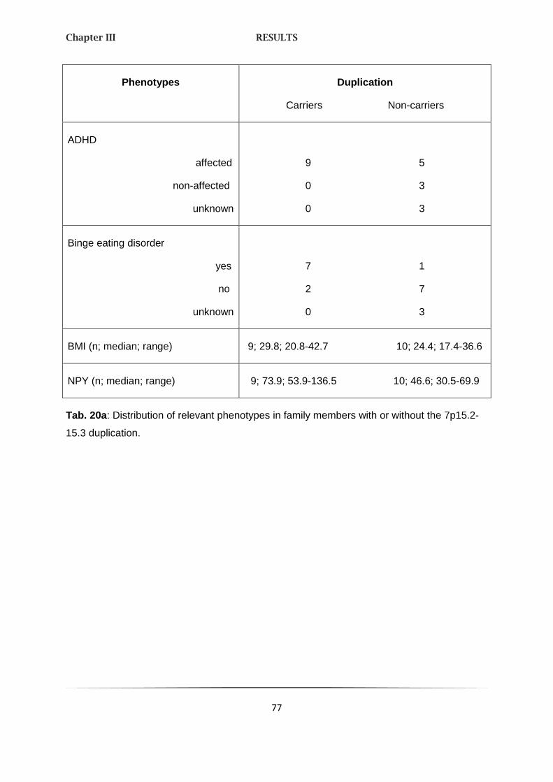
Chapter III RESULTS
77
Phenotypes Duplication
Carriers Non-carriers
ADHD
affected
non-affected
unknown
9 5
0 3
0 3
Binge eating disorder
yes
no
unknown
7 1
2 7
0 3
BMI (n; median; range) 9; 29.8; 20.8-42.7 10; 24.4; 17.4-36.6
NPY (n; median; range) 9; 73.9; 53.9-136.5 10; 46.6; 30.5-69.9
Tab. 20a: Distribution of relevant phenotypes in family members with or without the 7p15.2-
15.3 duplication.

Chapter III RESULTS
78
Phenotypes Duplication
transmitted non-transmitted
Nominal empirical1 FBAT
p-value
ADHD
affected
not affected
8 3
0 1
0.138
Binge eating behavior
yes
no
6 1
2 3
0.117
BMI (n; median; range) 8; 28.4; 8-42.7 4; 21.3; 17.4-36.3 0.192
NPY (n; median; range) 8; 78.5; 53.9-136.5 4; 46.6; 43.3-50.3 0.023
Tab. 20b: Investigation of association between relevant phenotypes and the 7p15.2-15.3
duplication.
1 Test based on 10.000 simulations.

Chapter III RESULTS
79
Fig. 16: Neuropeptide Y (NPY) plasma concentrations blotted against the body
mass index (BMI) in 7p15.2-15.3 duplication carriers with ADHD, non-carriers
with ADHD, and healthy family members.
F numbers allow allocation to the pedigree.
The effect of the 7p15 duplication and gene dose-dependent increase in NPY plasma
concentrations on brain function was explored by fMRI in four carriers with ADHD compared
to healthy controls. Region of interest analyses revealed a significantly lower activation of the
left ventral striatum during the anticipation of large rewards for duplication carriers than for
controls (p < 0.05, corrected; Fig. 17, upper panels). A significantly lower activation of the left
posterior insula during the anticipation of large losses was also observed in carriers
compared to controls (p < 0.05, corrected; Fig. 17, lower panels). In none of the two regions,
a significant difference between carriers and controls was observed for no or small rewards
or losses. Furthermore, activation for the carriers never exceeded the controls’ responses in
those two structures.

Chapter III RESULTS
80
Fig. 17: Neural activation in the ventral striatum during the anticipation of
large rewards (upper panel) and in the posterior insula during the anticipation of
large losses (lower panel) for 7p15.2-15.3 duplication carriers with ADHD
(n = 4) and healthy controls (n = 21).
Brain maps show significant –log10-transformed p values (p < .05, corrected) in the left
ventral striatum (upper right panel) and in the left Posterior insula (lower right panel).
Boxplots show medians, 25th and 75th percentiles and most extreme signal changes
(whiskers extend to the most extreme subject values) corresponding to the brain maps of
the ventral striatum (upper left panel) and the posterior insula (lower left panel).
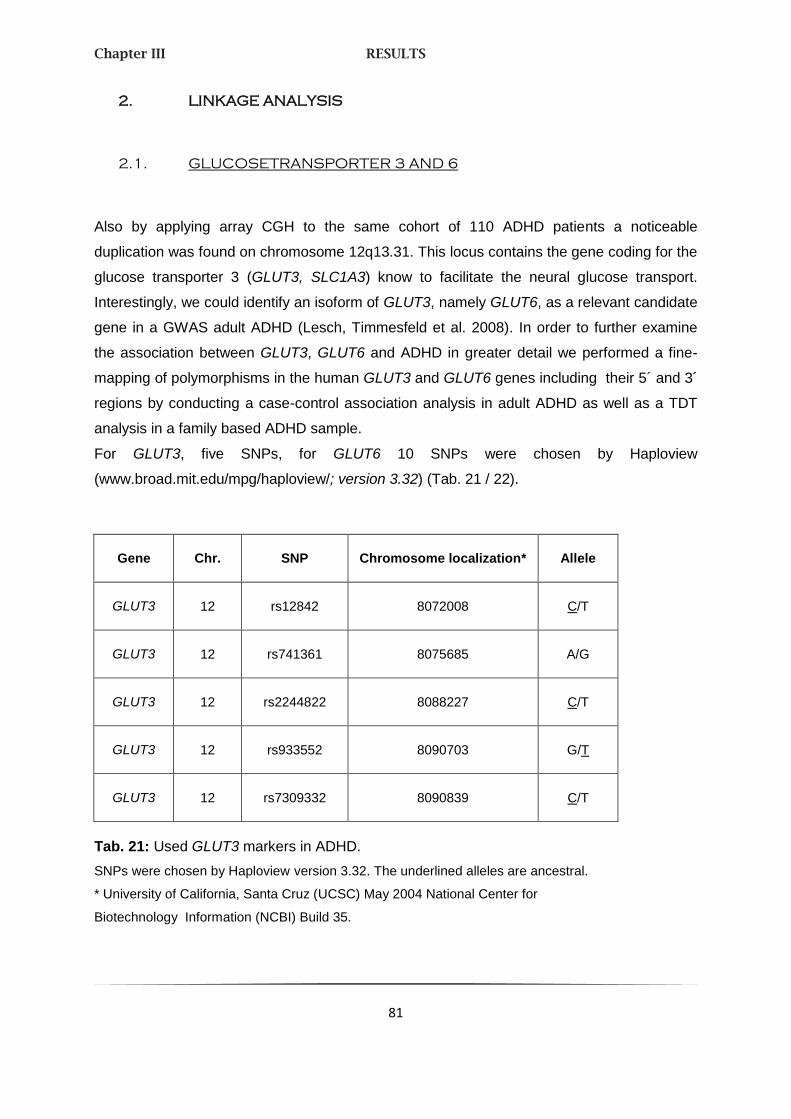
Chapter III RESULTS
81
2. LINKAGE ANALYSIS
2.1. GLUCOSETRANSPORTER 3 AND 6
Also by applying array CGH to the same cohort of 110 ADHD patients a noticeable
duplication was found on chromosome 12q13.31. This locus contains the gene coding for the
glucose transporter 3 (GLUT3, SLC1A3) know to facilitate the neural glucose transport.
Interestingly, we could identify an isoform of GLUT3, namely GLUT6, as a relevant candidate
gene in a GWAS adult ADHD (Lesch, Timmesfeld et al. 2008). In order to further examine
the association between GLUT3, GLUT6 and ADHD in greater detail we performed a fine-
mapping of polymorphisms in the human GLUT3 and GLUT6 genes including their 5´ and 3´
regions by conducting a case-control association analysis in adult ADHD as well as a TDT
analysis in a family based ADHD sample.
For GLUT3, five SNPs, for GLUT6 10 SNPs were chosen by Haploview
(www.broad.mit.edu/mpg/haploview/; version 3.32) (Tab. 21 / 22).
Gene Chr. SNP Chromosome localization* Allele
GLUT3 12 rs12842 8072008 C/T
GLUT3 12 rs741361 8075685 A/G
GLUT3 12 rs2244822 8088227 C/T
GLUT3 12 rs933552 8090703 G/T
GLUT3 12 rs7309332 8090839 C/T
Tab. 21: Used GLUT3 markers in ADHD.
SNPs were chosen by Haploview version 3.32. The underlined alleles are ancestral.
* University of California, Santa Cruz (UCSC) May 2004 National Center for
Biotechnology Information (NCBI) Build 35.
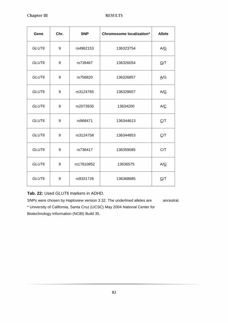
Chapter III RESULTS
82
Gene Chr. SNP Chromosome localization* Allele
GLUT6 9 rs4962153 136323754 A/G
GLUT6 9 rs739467 136326054 G/T
GLUT6 9 rs756820 136326857 A/G
GLUT6 9 rs3124765 136328657 A/G
GLUT6 9 rs2073935 13634200 A/C
GLUT6 9 rs968471 136344613 C/T
GLUT6 9 rs3124758 136344853 C/T
GLUT6 9 rs736417 136359085 C/T
GLUT6 9 rs17810852 13636575 A/G
GLUT6 9 rs9331726 136368685 G/T
Tab. 22: Used GLUT6 markers in ADHD.
SNPs were chosen by Haploview version 3.32. The underlined alleles are ancestral.
* University of California, Santa Cruz (UCSC) May 2004 National Center for
Biotechnology Information (NCBI) Build 35.

Chapter III RESULTS
83
The distribution equilibrium of these two genes was shown in a LD-block. The LD-block of
GLUT3 consists of two blocks; block1 comprises the marker rs663303, rs741361 and
rs2244822, whereas block 2contains rs933552 and 7309332 (Fig.18a).
On the contrary, the LD-block of GLUT6 revealed three blocks. Here, block 1 contains
rs4962153 to rs2073935, block 2 rs9368471 and rs3124758, and block 3 rs736417 to
rs9331726 (Fig. 18b).
All SNPs in one block are transmitted mostly together.
Fig. 18a: Linkage disequilibrium map for GLUT3.
LD map of SNP Markers created using HAPLOVIEW version
3.32. Only markers selected by HAPLOVIEW are shown. The
dark squares represent higher r² values; triangles
surrounding markers represent haplotype blocks under the 4
gamete rule.

Chapter III RESULTS
84
Fig. 18b: Linkage disequilibrium map for GLUT6.
LD map of SNP Markers created using HAPLOVIEW version 3.32. Only markers
selected by HAPLOVIEW are shown. The dark squares represent higher r² values;
triangles surrounding markers represent haplotype blocks under the 4 gamete rule.
2.2. GENOTYPING OF PLEKHB1, RAB6A AND PDE4D
For examination of an association between the candidate genes RAB6A, PLEKHB1 and
alternatively PDE4D, found by array CGH, and ADHD several SNPs (four RAB6ASNPs,
eight PLEKHB1SNPs and ten PDE4D SNPs) were analyzed in a case-control study. We
genotyped these variants in a sample of 450 HKS probands, 200 families with almost one

Chapter III RESULTS
85
child affected by ADHD according to DSM-IV criteria and 90 controls. For all probands all
SNPs were ascertained.
PLEKHB1 and RAB6A, analyzed together due to their localization side by side on
chromosome 11, were located in three haplotypes blocks, whereas PDE4D markers were in
two haplotype blocks (not shown). The degree of LD varied among the SNPs examined. This
most likely is a factor of the wide distribution of the SNPs, the large genomic size of the
analyzed genes and the complex linkage disequilibrium structure. All analyzed marker weres
in the Hardy-Weinberg equilibrium (HWE).
PLEKHB1 and RAB6A
In total, 59 PLEKHB1/RAB6A markers span 134.978 bp (including 10.000 bp upstream and
downstream). 11 SNPs were chosen by Haploview (www.broad.mit.edu/mpg/haploview/;
version 3.32), three others ones were included because of their occurrence in the population
(Tab. 23). The distribution equilibrium of these two genes has shown three LD-blocks
(Fig. 19). Block 1 comprises the marker rs663303 to rs591804, block 2 rs6592527 and
rs940828, block 3 rs10736793 to rs7127066. All SNPs in one block are transmitted mostly
together. Only rs3741147 and rs12274970 are transmitted separately and are not linked to
the others.

Chapter III RESULTS
86
Gene Chr. SNP Localization Chromosome localization* Allele
11 rs663303 5´UTR 73029494 C/T
PLEKHB1 11 rs4944850 5´UTR 73033744 A/C
PLEKHB1 11 rs11538627 Intron 4 73040539 A/T
PLEKHB1 11 rs591804 Intron 4 73040858 A/G
PLEKHB1 11 rs6592527 Intron 5 73042016 C/G
PLEKHB1 11 rs940828 Intron 5 73043807 G/T
PLEKHB1 11 rs3741147 Intron 5 73044399 G/T
11 rs12274970 73055790 C/T
RAB6A 11 rs3182788 3´UTR 73066620 A/C
RAB6A 11 rs10736793 Exon 8 73080224 A/C
RAB6A 11 rs3203705 Intron 6 73107544 C/T
RAB6A 11 rs11235876 Intron 3 73108249 A/G
RAB6A 11 rs11235880 Intron 2 73112944 A/C
RAB6A 11 rs7127066 Intron 1 73136522 C/G
Tab. 23: Used PLEKHB1 and RAB6A markers in ADHD.
SNPs were chosen by Haploview version 3.32, underlined SNPs were added afterwards. The
underlined alleles are ancestral.
* University of California, Santa Cruz (UCSC) May 2004 National Center for Biotechnology Information
(NCBI) Build 35. UTR, untranslated region.

Chapter III RESULTS
87
Fig. 19: Linkage disequilibrium map for PLEKHB1 and RAB6A.
LD map of SNP Markers created using HAPLOVIEW version 3.32.
Only markers selected by HAPLOVIEW are shown. The dark squares
represent higher r² values; triangles surrounding markers represent
haplotype blocks under the 4 gamete rule.
Two markers of PLEKHB1 (rs6592527 and rs940828) within intron 5 and one marker of
RAB6A (rs3182788), located in exon 8, archived a statistical significance in Fisher´s exact
test (p < 0.1) (Tab. 24). Notable, rs3182788 also showed a strong significant HWE (case,
p = 0.0 vs. control, p = 0.0577; data not shown) as well as in the χ2 test (Tab. 24). The
genomic distribution of the other polymorphisms did not deviate significantly from HWE in
both patient and controls (p < 0.05).

Chapter III RESULTS
88
Gene SNP HWE_P HWE_P HWE_P χ2 χ2 Case χ2 total P value P value
Control Case total Control combSubt
rs663303 0.025 0.254 0.242 0.875 0.614 0.623 0.459 0.231
PLEKHB1 rs4944850 0.568 0.343 0.013 0.452 0.558 0.911 0.919 0.572
PLEKHB1 rs11538627 NaN NaN NaN NaN NaN NaN NaN NaN
PLEKHB1 rs591804 0.025 0.077 0.102 0.875 0.781 0.750 0.478 0.385
PLEKHB1 rs6592527 0.923 0.298 0.984 0.334 0.585 0.321 0.075 0.100
PLEKHB1 rs940828 0.788 0.159 0.694 0.375 0.691 0.405 0.078 0.118
PLEKHB1 rs3741147 0.024 0.580 0.446 0.877 0.446 0.504 0.357 0.181
rs12274970 0.232 0.058 0.268 0.670 0.810 0.612 0.784 0.655
RAB6A rs3182788 18.406 3.603 15.364 0.0 0.058 0.0 0.000 0.000
RAB6A rs10736793 0.170 0.001 0.102 0.680 0.970 0.749 0.665 0.705
RAB6A rs3203705 NaN NaN NaN NaN NaN NaN NaN NaN
RAB6A rs11235876 1.667 0.928 2.513 0.197 0.335 0.113 0.975 0.753
RAB6A rs11235880 0.310 0.052 0.053 0.578 0.820 0.817 0.945 0.639
RAB6A rs7127066 0.336 3.123 2.922 0.562 0.073 0.087 0.793 0.729
Tab. 24: Hardy-Weinberg equilibrium, chi-square-tests for frequency differences between
cases and controls and P value of the PLEKHB1 and RAB6A markers in ADHD.
NaN: Not a Number; red: significant results

Chapter III RESULTS
89
PDE4D
12 PDE4D markers out of a calculable possible 66 SNPs (chosen by Haploview,
www.broad.mit.edu/mpg/haploview/; version 3.32), spanning 49.910 bp (including 10.000bp
upstream and downstream) on chromosome 5, were tested (Tab. 25). Rs17291089 to
rs12656462 in block 1 as well as rs1005662 to rs7714708 in block 2 were transmitted mostly
together. The LD-plot of this gene is shown in Fig. 20, the genomic localization is listed in
Tab. 25.
Fig. 20: Linkage disequilibrium map for PDE4D.
LD map of SNP markers created using HAPLOVIEW. Only 10 selected
markers are shown. The dark squares represent higher r² values;
triangles surrounding markers represent haplotype blocks under the
4 gamete rule.

Chapter III RESULTS
90
. Gene Chr. SNP Chromosome localization* Allele
PDE4D 5 rs17291089 58301362 G/T
PDE4D 5 rs829259 58303733 A/T
PDE4D 5 rs1058458 58306373 C/T
PDE4D 5 rs17719378 58309205 C/T
PDE4D 5 rs10055954 58309342 C/G
PDE4D 5 rs10461656 58312107 C/T
PDE4D 5 rs7713345 58314588 C/G
PDE4D 5 rs12656462 58316381 A/T
PDE4D 5 rs17853590 58320100 A/G
PDE4D 5 rs10056492 58327740 A/G
PDE4D 5 rs4700316 58330448 C/G
PDE4D 5 rs7714708 58330771 A/G
Tab. 25: Used PDE4D markers in ADHD.
SNPs were chosen by Haploview version 3.32. The underlined alleles are
ancestral.
* University of California, Santa Cruz (UCSC) May 2004 National Center for
Biotechnology Information (NCBI) Build 35.
As expected, no marker has a statistical significance in HWE. Also no marker shows a
significant P value (p < 0.05) (Tab. 26). In summary, there were no differences in genotype or
allele frequencies between cases and controls.

Chapter III RESULTS
91
Gene SNP HWE_P HWE_P HWE_P χ2 χ2 Case χ2 total P value P value
Control Case total Control combSubt
PDE4D rs17291089 1.700 0.393 0.144 0.192 0.531 0.704 0.554 0.387
PDE4D rs829259 0.817 0.083 0.672 366 0.774 0.412 0.403 0.602
PDE4D rs1058458 NaN NaN NaN NaN NaN NaN NaN NaN
PDE4D rs17719378 0.897 0.025 0.273 0.344 0.874 0.602 0.602 0.414
PDE4D rs10055954 0.246 0.561 0.038 0.620 0.454 0.846 0.077 0.066
PDE4D rs10461656 0.293 0.874 1.038 0.588 0.350 0.308 0.164 0.184
PDE4D rs7713345 0.0518 0.504 0.456 0.820 0.478 0.500 0.956 0.771
PDE4D rs12656462 0.149 0.836 0.859 0.699 0.360 0.354 0.217 0.139
PDE4D rs17853590 NaN NaN NaN NaN NaN NaN NaN NaN
PDE4D rs10056492 0.225 0.087 0.296 0.635 0.768 0.586 0.793 0.815
PDE4D rs4700316 0.309 0.266 0.559 0.579 0.606 0.455 0.411 0.299
PDE4D rs7714708 1.263 0.725 0.022 0.261 0.395 0.882 0.359 0.415
Tab. 26: Hardy-Weinberg equilibrium, chi-square-tests for frequency differences between
cases and controls and P value (p < 0.05) of the PDE4D markers in ADHD.
NaN: Not a Number

Chapter III RESULTS
92
2.3. THE SYNAPTIC VESICLE PROTEIN 2C
Three SNPs (rs1862519, rs30196 and rs30198) in the promoter region of SV2C (Fig. 21)
which may be associated with ADHD were chosen for an association linkage analysis by
Haploview (www.broad.mit.edu/mpg/haploview/; version 3.32).
Fig. 21: Linkage disequilibrium map for the
promoter region of SV2C.
LD plot was created using HAPLOVIEW.
The dark squares represent higher r² values; triangles
surrounding markers represent haplotype blocks under
the 4 gamete rule.
A haplotype analysis using 200 nuclear families, identified through a proband child with
ADHD according to DSM-IV criteria derived from the Department of Child and Adolescent
Psychiatry and Psychotherapy and the Department of Psychiatry, Psychosomatics and
Psychotherapy, University of Würzburg, was tested for associations with ADHD. Allele
frequencies for all markers showed a significant deviation according to Hardy-Weinberg

Chapter III RESULTS
93
equilibrium (HWE) (Tab. 27) in the case of the parents which argue for a genotypical failure.
A revision exhibited the same results again.
p-value
SNP HWE_P HWE_C
rs1862519 0.9884 0.174
rs30196 0.0442* 0.2357
rs31098 0.0025* 0.2691
Tab. 27: Hardy-Weinberg equilibrium
(HWE) in parents (P) and children (C).
* significant; P = permutation based p value
with p < 0.05 considered significant
Transmission disequilibrium test (TDT) analyses of haplotypes were performed on the total
sample (p-value = 0.8764). Three haplotypes were not used for TDT analyses because of
their rare appearance. As shown in Tab. 28, neither haplotype was significantly associated
with the disease.

Chapter III RESULTS
94
Marker
rs1862519 rs30196 rs31098
Frequency
(%) T NT OR
G G G 58.897 79,951 70 1,142
C T A 18.047 37,952 37 1,026
G G A 0.248
G T A 20.049 48,047 53 0,907
C T G 0.248
C G G 0.509
G T G 2.003 6,001 6 1
Tab. 28: Haplotype distribution in SV2C.
T: transmitted; NT: not transmitted; OR: odds ratio.

Chapter III RESULTS
95
The pedigree disequilibrium test (PDT), was applied to search for evidence of allelic
association in general pedigrees, but showed no significant linkage (Tab. 29).
SNP Allele P
rs1862519 G 0.917411
rs1862519 C 0.917411
rs30196 G 0.796253
rs30196 T 0.796253
rs31098 G 0.680051
rs31098 A 0.680051
Tab. 29: Pedigree disequilibrium
test with nominal significance level
0.05 on the basis of 200 nuclear
families.
P = permutation based p value with
p < 0.05 considered significant.

Chapter III RESULTS
96
3. IMMUNOHISTOCHEMICAL ANALYSIS OF LPHN3
3.1. REGIONAL DISTRIBUTION OF LPHN1-mRNA IN THE MURINE BRAIN
USING ISH
The regional distribution of Lphn3 transcripts was accomplished by ISH in wildtype
(C57BL/6J) murine brain sections. To verify the specificity of the used Lphn3-mRNA
antisense probe a DIG-marked sense probe was used.
Lphn3 mRNA was widely distributed in the murine brain. The striatum, cortex, hippocampus
and cerebellum were analyzed in greater detail. Whereas all laminae in the cortex contained
LPHN3-mRNA, but with less expression in laminae IV (Fig. 22a and b), no noticeable
expression pattern was found in the striatum in comparison to the environmental cerebral
regions. Also the corpus callosum showed no Lphn3 expression at all (Fig. 22c and d). The
hippocampus revealed a distinct expression. Mainly the CA1 region showed a strong
Lphn3 expression, but declined abruptly to CA2 and CA3 (Fig. 22e). In the striatum
granulosum of the gyrus dentatus some cells were clearly colored and different in size in
contrast to others (Fig. 22f). An inhomogeneous distribution of Lphn3-mRNA also appeared
in the brain stem. Here, the expression was especially strong in the Erdinger-Westphal
nucleus, the dorsal raphe and the central raphe nucleus (Fig. 22g and h). In the purkinje
layer of the cerebellum a very strong expression has been revealed (Fig. 22i), followed by
the striatum granulosum and the molecular layer. The clear colored cells in the two last
named layers could be basket or stellate cells (Fig. 22k). These results were also found by
Mario Kreutzfeldt (Diploma thesis).

Chapter III RESULTS
97
Fig. 22: Overview of the Lphn3-mRNA distribution in the
murine brain.
Different regions such as the cortex (A,B), striatum (C,D),
Hippocampus (E,F), raphe (G,H) and cerebellum (I,K) are shown.
Right column displays higher power images of boxed inserts from
the left column. (Picture: Mario Kreutzfeldt).
Aq (aqueduct), CA1/3 (cornu ammonis regions), cc (corpus
callosum), Co (cerebral cortex), CPu (caudate putamen), CS (central
raphe nucleus), DG (gyrus dentatus), DRI (inferior dorsal
raphe), DRD (dorsolateral raphe), ML/mo (molecular layer),
PCL (purkinje cell layer), po (polyform layer), sg/GL (striatum
granulosum), VL (lateral ventricle), WM (white matter).
Chapter III RESULTS
98
3.2. CELLULAR AND REGIONAL DISTRIBUTION PATTERN OF LPHN3
PROTEIN IN HUMAN AND MURINE BRAIN SECTIONS
Immunohistochemistry in the human hippocampus revealed Lphn3 protein in both the
striatum granulosum of the gyrus dentatus (Fig. 23a) and the pyramidal cells of CA1 - CA4.
The stronger coloring in region CA3 could be explained by its higher cell density. The stratum
oriens and the stratum radiatum showed very little Lphn3 expression. The individual laminae
in the human cortex were distinguishable due to their Lphn3 distribution pattern. Basically all
laminae were strained, but the strongest one could be found in the pyramidal cells of laminae
V, less straining in contrast in laminae I and VI (Fig. 23b). Whereas the striatum granulosum
and the molecular layer of the cerebellum seemed to be homogenously strained, the purkinje
cells became strata surface (Fig. 23c). The strongest Lphn3 expression was found in their
somas and also involved the dentrites, but not the nuclei. However, the expression intensity
decreased in direction to the soma. Lphn3 immunoreactive regions were rarely found, but
this doesn`t speak against a principal protein localization in axons.
Due to the high interspecific LPHN3 homology between mouse and human and also because
of the missing offers for murine polyclonal LPHN3 antibodies, the human ones were also
used in murine tissue sections.
Hippocampus, cortex and cerebellum were dyed via the ABC-method. Clearly noticeable
were some cells in the gyrus dentatus of the hippocampus, mainly in the CA3 (stratum
lucidium) (Fig. 23b). In the cortex the individual laminae were distinguished clearly, especially
Laminae II and V (Fig. 23d). In contrast, in the cerebellum both stratum granulosum and the
molecular layer showed some homogenous colored cells. Indeed, the strongest staining was
found in the purkinje cell layer; its distribution pattern referred to immunoreactive purkinje
cells (Fig. 23f).
All results were again also found in the Diploma thesis of Mario Kreutzfeldt.

Chapter III RESULTS
99
Fig. 23: Immunohistochemical detection of LPHN3 on human
(A,C,E) and murine (B,D,F) paraffined brain sections.
For staining the ABC-method was used. Indicated are representative
images of the dentate gyrus (hippocampus, A,B), of the laminae VI / V
of the cortex (C,D) and the purkinje cells in the cerebellum (E,F).
(Picture: Mario Kreutzfeldt).
CA1-4 (cornu ammonis regions), DG (dentate gyrus), mo/ml (molecular
layer), PCL (purkinje cell layer), po (polyform layer), sg/gl (stratum
granulosum).

Chapter III RESULTS
100
4. RESEARCHES IN MLC1
4.1. GENOTYPING OF MLC1 POLYMORPHISMS FOR ASSOCIATION
WITH PERIODIC CATATONIA
SNP1 (rs235349) and SNP2 (rs2076137), previously found to be associated with
Schizophrenia (SCZ) (Verma, Mukerji et al. 2005), were chosen for an initial association
screen. They were significant in LD (D´ = 0.95; p ≥ 0.001; Tab. 30).
Marker TCR1 SNP1 SNP2
TCR1 0.091 0.99 0.92
TCR2 0.96 0.95
SNP1 0.95
Tab. 30: 2-Locus Linkage Disequilibria between MLC1 markers.
D´, all p < 0.001. TCR: transcriptional control region; SNP: single nucleotide polymorphism.
First it was tested for an association with Periodic Catatonia (PC); second, in an exploratory
analysis, it was investigated to see if the SNPs were associated with all cases combined,
SZC alone, or Bipolar Affective Disorder (BPD) alone. As shown in Tab. 31, both were
significantly associated with PC. However, no association was found with the combined
patient sample, SCZ, BPD, or type A or type B schizophrenia (Reif, Fritzen et al. 2006) (all
p > 0.05). Thus, both named SNPs were specifically associated with PC. Therefore, we
restricted further analyses incorporating transcriptional control region (TCR) variants to PC
cases. These variants also showed a significant LD (Tab. 30). In Tab. 31, TCR 1 and 2
showed association with PC, which slightly missed the conventional significance level when
comparing carriers of the rare alleles to subjects homozygous for the frequent variant (TCR1:
p = 0.061; TCR2: p = 0.051).
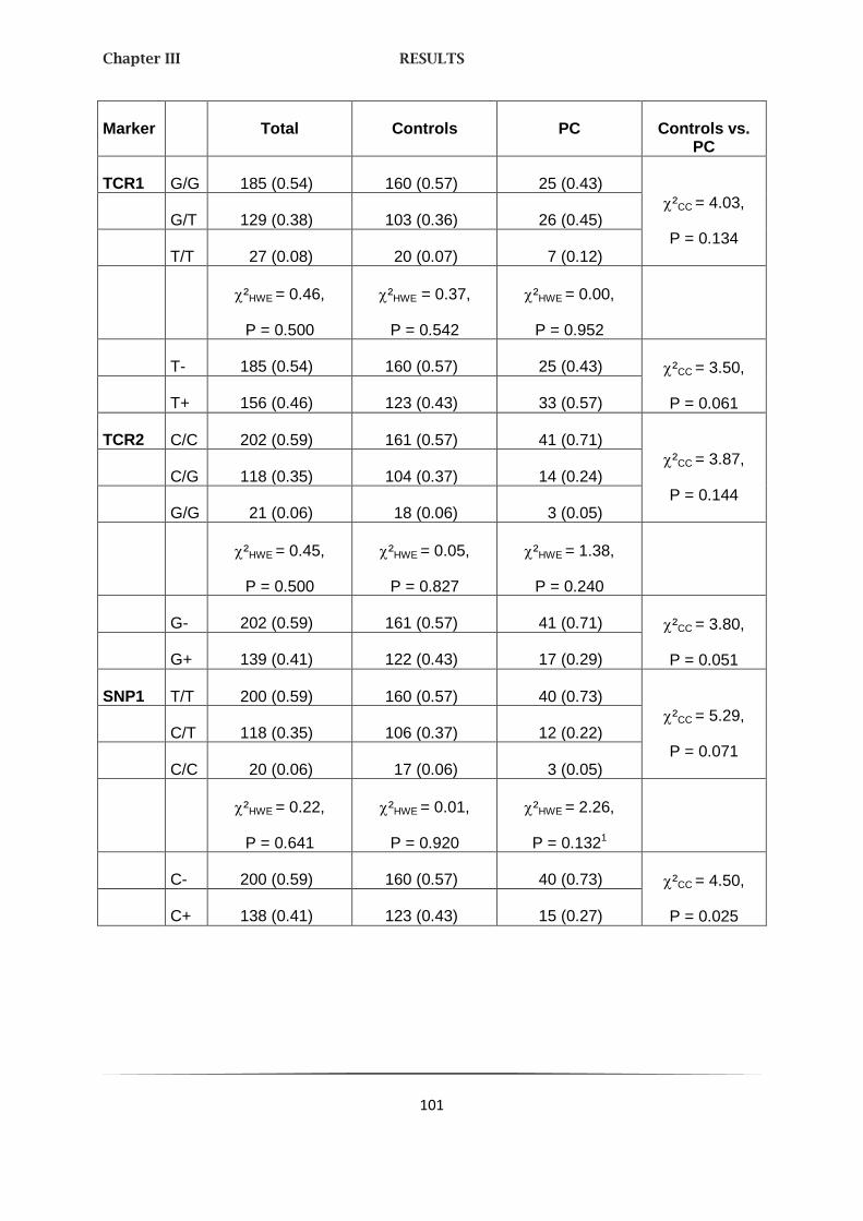
Chapter III RESULTS
101
Marker Total Controls PC Controls vs. PC
TCR1 G/G 185 (0.54) 160 (0.57) 25 (0.43)
²CC = 4.03,
P = 0.134 G/T 129 (0.38) 103 (0.36) 26 (0.45)
T/T 27 (0.08) 20 (0.07) 7 (0.12)
²HWE = 0.46,
P = 0.500
²HWE = 0.37,
P = 0.542
²HWE = 0.00,
P = 0.952
T- 185 (0.54) 160 (0.57) 25 (0.43) ²CC = 3.50,
P = 0.061 T+ 156 (0.46) 123 (0.43) 33 (0.57)
TCR2 C/C 202 (0.59) 161 (0.57) 41 (0.71)
²CC = 3.87,
P = 0.144 C/G 118 (0.35) 104 (0.37) 14 (0.24)
G/G 21 (0.06) 18 (0.06) 3 (0.05)
²HWE = 0.45,
P = 0.500
²HWE = 0.05,
P = 0.827
²HWE = 1.38,
P = 0.240
G- 202 (0.59) 161 (0.57) 41 (0.71) ²CC = 3.80,
P = 0.051 G+ 139 (0.41) 122 (0.43) 17 (0.29)
SNP1 T/T 200 (0.59) 160 (0.57) 40 (0.73)
²CC = 5.29,
P = 0.071 C/T 118 (0.35) 106 (0.37) 12 (0.22)
C/C 20 (0.06) 17 (0.06) 3 (0.05)
²HWE = 0.22,
P = 0.641
²HWE = 0.01,
P = 0.920
²HWE = 2.26,
P = 0.1321
C- 200 (0.59) 160 (0.57) 40 (0.73) ²CC = 4.50,
P = 0.025 C+ 138 (0.41) 123 (0.43) 15 (0.27)

Chapter III RESULTS
102
Marker Total Controls PC Controls vs. PC
SNP2 C/C 241 (0.72) 195 (0.69) 46 (0.84)
²CC = 5.94,
P = 0.051 C/T 87 (0.26) 80 (0.28) 7 (0.13)
T/T 9 (0.03) 7 (0.02) 2 (0.04)
²HWE = 0.12,
P = 0.734
²HWE = 0.13,
P = 0.721
²HWE = 4.72,
P = 0.0302
T- 241 (0.72) 195 (0.69) 46 (0.84) ²CC = 4.74,
P = 0.029 T+ 96 (0.28) 87 (0.31) 9 (0.16)
Tab. 31: Genotype frequencies of MLC1 markers.
²HWE = chi-square-tests for deviation from Hardy-Weinberg-equilibrium, df = 1; deviations observed
for 1 SNP1 C/C genotype slightly overrepresented, and
2 SNP2 T/T genotype overrepresented; ²CC =
chi-square-tests for frequency differences between cases and controls, df = 2 for full genotype tests,
df = 1 for dichotomous genotype tests.
Haplotype analyses included all SNPs. A test for global haplotype association with PC did
not show a significant result (p = 0.35). However, on the level of specific haplotypes, the
T-C-T-C haplotype was significantly more common in PC (p = 0.025, Tab. 32). Similar to the
4-marker test, analyses using 3-marker and 2-marker haplotypes yielded insignificant results
(all p > 0.05).

Chapter III RESULTS
103
TCR1 TCR2 SNP1 SNP2 Controls PC Perm. P
G C C C 0.010 0.00 0.190
G C C T 0.000015 0.00 0.583
G C T C 0.494 0.491 0.686
G G C C 0.077 0.064 0.470
G G C T 0.158 0.100 0.116
G G T T 0.004 0.00 0.111
G G T T 0.002 0.00 0.167
G G C C 0.002 0.00 0.167
T C T C 0.242 0.345 0.025
T G C C 0.006 0.00 0.024
T G C C 0.000062 0.000045 0.819
T G C T 0.000003 0.000020 0.636
G C C C 0.636 0.00 0.183
Tab. 32: Estimated MLC1 haplotype frequency differences between control subjects and
patients suffering from Periodic Catatonia using GENECOUNTING.
Bold = significant and meaningful haplotype association: in the PC patient group, the estimated
T-C-T-C haplotype is overrepresented. PC = periodic catatonia, Perm. P = permutation based
p value with p<0.05 considered significant.
4.2. MLC1 KNOCKOUT PLASMID VECTOR
The Mlc1-targeting vector was constructed by inserting a 1,2 kb Pst I-fragment containing
complete 492 bp exon 1 (untranslated) and 154 bp of the 217 bp exon 2 (“left arm”), which
contains the start codon ATG, into a Not I/Xho I site and additionally a 4,8 kb Sac I-fragment
containing complete exon 4 (54 bp) and 5 (102 bp) (“right arm”) into a BamH I/EcoR I site of
pPXT.

Chapter III RESULTS
104
Both arms were inserted by in vitro integration and are bordered by loxP sites. Fig. 24 shows
a schematic representation of the Mlc1 ko plasmid vector (pMlc1).
Fig. 24 Schematic representation of the linearized pMlc1 ko vector.
Left and right arm are inserted by ligation. Both are generated between two LoxP sites. SA: short arm;
LA: long arm; Neo: Neo cassette; E: exon; →: used forward primers; ←: used reverse primers.
After electroporation the ES cells were analyzed for integration of this pMlc1 ko vector
plasmid by PCR (Fig. 25). The used primers and their localization are summarized in
Material & Methods chapter 1.4. Desoxyribonucleotides.
Not I
SA – 1.2kb
Neo – 1.8kb
LA – 4.8kb
Vector rest
Total size: 13,172kb
E1
E2
E5
E4

Chapter III RESULTS
105
Fig. 25: PCR amplification for integration of the pMlc1 knockout
vector plasmid into human embryonal stem cells (ES) and the
PCR products were analyzed by agarose gel electrophoresis.
1st lane: 1 kb plus DNA marker; lane 1-3: human ES cells: lanes
2/3 show the expected size (1546 bp) of the knockout vector plasmid;
last lane: H2O as negative control.
Chapter IV DISCUSSION
106
IV. DISCUSSION
1. NEW ADHD CANDIDATE GENES BY ARRAY CGH
Sub-megabase resolution array CGH identified a total of 17 potentially disease-associated
CNVs in a cohort of 99 children and adolescents severely affected with ADHD. The
aberrations comprise five deletions and 13 duplications with approx. sizes between 130 kb
and 3 Mb. Two CTVs occurred de novo and eight were inherited from a parent with ADHD,
whereas five were transmitted by an unaffected parent. For one case, inheritance was not
determined. These CNVs showed no overlap between individual patients, i. e. they are not
recurrent, but several of the genes involved may be integrated into behaviorally relevant
functional pathways, including neurodevelopment, neurotransmission, and synaptic plasticity.
1.1. NEUROPEPTIDE Y
Given the remarkable heritability of ADHD, polymorphisms, inherited from an affected parent,
are likely to contain risk genes. Among the most apparent identified candidate genes, an
approx. 3 Mb duplication, occurring in two affected cousins, includes the gene encoding for
NPY on chromosome 7q15.2-15.3. This co-segregates with a unique syndrome comprising
severe ADHD and obesity. The subsequent investigation of the extended multigenerational
family with high density of ADHD patients revealed evidence for an association of this
duplication with ADHD, increased BMI as well as binge eating, suggesting that the aberration
contributes to the syndrome in this family. Admittedly, in four descendants of the
F1 generation affected with ADHD no chromosomal rearrangement was detected. Due to the
often appearing assortative mating which is common in ADHD, bilinear transmission of at
least two causative gene variants, including the NPY-containing duplication, passed by two
affected F1 founders can be assumed.
The additional copy of NPY within the investigated extended family was associated with an
almost 2-fold increase of plasma NPY concentrations in peripheral blood. This provides
indirect evidence for NPY overexpression. Enhanced NPY receptor subtype-dependent
signaling in the brain with consequences on learning/memory, cognition, and emotion

Chapter IV DISCUSSION
107
regulation are likely to be altered in duplication carriers. Although increased plasma NPY
concentrations were previously observed in children with ADHD (Oades, Daniels et al. 1998),
the general role of this neuropeptide in the pathophysiology of ADHD remains to be
determined.
The potential link between ADHD, metabolic dysregulation, and NPY is underscored by
studies revealing that ADHD is highly prevalent among obese patients and highest in those
with extreme obesity. NPY is an orexigenic key regulator in the brain of mammalians and
non-mammalians. The neuropeptide increases food intake especially carbohydrates (Beck
2006). Also elevated levels of NPY cause an increase of food intake and reciprocally
(Williams, McKibbin et al. 1991; Widdowson, Upton et al. 1997; Ishihara, Tanaka et al. 1998).
In the analyzed family both ADHD and obesity are observed. The potential link between
ADHD, food intake, metabolic dysregulation, and NPY is also underscored by studies
revealing that ADHD is highly prevalent among obese patients and highest in those with
extreme obesity (Agranat-Meged, Deitcher et al. 2005; Curtin, Bandini et al. 2005; Fleming,
Levy et al. 2005). Mechanisms for this co-morbidity are unknown, but may involve brain
dopamine function, glucose utilization, and insulin receptor activity (Agranat-Meged, Deitcher
et al. 2005). Alterations in the brain dopamine system affect a wide range of behavioral
phenotypes ranging from ADHD-associated behavior to food intake and from an evolutionary
perspective, gene variations selected to increase cognitive and behavioral flexibility may
presently be associated with attention deficits and increased food consumption in an
obesogenic environment. However, both ADHD and adiposity are of multigenetic origin and
the consideration of a monogenetic cause is obsolete. This is again in line with the relatively
small effects detected by the statistical analysis.
Despite NPY being widely investigated in the context of body weight regulation and energy
balance, it has recently not been implicated in behavioral traits, including aggression and
negative emotionality, but also in several neuropsychiatric disorders like schizophrenia, panic
disorder, bipolar disorder and depression (Karl and Herzog 2007). A recent study revealed
that the functional Leu7Pro polymorphism in the human NPY resulting in increased NPY
released from sympathetic nerves is associated with traits of the metabolic syndrome
(Ruohonen, Pesonen et al. 2008). Moreover, diverse studies suggested that the Pro7 allele
is associated with an increased risk for alcohol dependence (Lappalainen, Kranzler et al.
2002; Zhu, Pollak et al. 2003) a common comorbid disorder in ADHD.
In the rodent model central administration or viral vector-induced overexpression of NPY
produces a profound increase in food intake, whereas a NPY reduction leads to a decrease

Chapter IV DISCUSSION
108
(Primeaux, York et al. 2006; Thorsell, Repunte-Canonigo et al. 2007). Food deprivation
upregulates NPY in the arcuate nucleus of the hypothalamus (Beck 2006), and repeated
administration of NPY induces obesity (Stanley, Anderson et al. 1989; Ruohonen, Pesonen
et al. 2008). Transgenic mice overexpressing NPY in noradrenergic neurons were reported
to display disturbances in glucose and lipid metabolism, key components of the cluster of
abnormalities characterizing the metabolic syndrome (Ruohonen, Pesonen et al. 2008).
Bannon revealed that NPY-deficient mice show reduced food intake in response to fasting
and an anxiety-like phenotype with increased startle response (Bannon, Seda et al. 2000).
Several receptors (Y1, Y2, Y4-Y6) mediate the physiological effects of NPY (Chamorro,
Della-Zuana et al. 2002; Karl and Herzog 2007) and data suggest that the energy balance
effects of NPY are mediated by both the NPY Y1 and the Y5 receptor (Chamorro, Della-
Zuana et al. 2002). NPY Y4 receptor knockout display increased locomotor activity, less
anxiety-like behavior and behavioral despair, whereas behavioral characterization of NPY Y2
knockout mice revealed reduced attention and increased impulsivity (Greco and Carli 2006;
Painsipp, Wultsch et al. 2008).
Finally, it is noteworthy that the NPY level is also related to the DA system, especially to
DRD1 (Sunahara, Guan et al. 1991). There is evidence that the NPY level is regulated
through DRD1. For example, a DRD1 antagonist could block the inhibitory effects of the
psychotomimetic drug methamphetamine on NPY levels, especially in nucleus accumbens
and caudate (Westwood and Hanson 1999). In addition, NPY expression in the PFC
(Caberlotto and Hurd 1999) supports the assumption that the NPY level has a stake in the
etiology of ADHD. The dysfunction of the PFC in this neurodevelopmental disorder is
suggested in several functional as well as morphological studies (Hynd, Semrud-Clikeman et
al. 1990; Filipek, Semrud-Clikeman et al. 1997; Rubia, Overmeyer et al. 2000; Langleben,
Austin et al. 2001; Mostofsky, Cooper et al. 2002). Alike, drugs used for the treatment of
ADHD often interfere with the NE system by inhibiting the reuptake of DA and NE. This
raises the question if the concentration of NPY is also affected by such medication because
of its co-expression with NA (Karl and Herzog 2007) and as well in which way.
Since we observed increased plasma NPY concentrations in the presence of an additional
copy of NPY within the investigated extended family as a peripheral biomarker, receptor
subtype-dependent signaling in the brain with consequences on the regulation of metabolic
homeostasis as well as cognition, learning/memory, and emotion regulation are likely to be
altered in duplication carriers.
Chapter IV DISCUSSION
109
In support of an impact of gene dosage-dependent increases in NPY expression on brain
function, fMRI of reward and emotion processing detected lower activation of the left ventral
striatum and left posterior insula during anticipation of large rewards/losses in duplication
carriers, respectively. As left ventral striatal hyporesponsiveness during reward anticipation
has repeatedly been shown in patients with adult ADHD (Scheres, Milham et al. 2007;
Strohle, Stoy et al. 2008) NPY overexpression may result in deviant rewardrelated neural
processing in duplication carriers. Moreover, the relative hypoactivity within the left posterior
insula during the anticipation of monetary loss in carriers could reflect anxiolytic effects of
NPY (Bannon, Seda et al. 2000; Greco and Carli 2006; Painsipp, Wultsch et al. 2008).
Higher genotype-driven NPY expression has recently been shown to be associated with
reduced pain/stress-induced activations of endogenous opioid neurotransmission and
accounted for 37% variance in left posterior insular cortex activation (Zhou, Zhu et al. 2008).
Hence, our fMRI findings replicate previously reported NPY-related alterations in the
processing of aversive stimuli while extending evidence for an interaction of NPY with reward
circuits. Taken together, our findings provide evidence that increased NPY dosage is not only
reflected by the peripheral biomarker of increased NPY plasma concentration but also by
fMRI elicited alteration in brain function related to reward and emotion processing.
In summary, there is substantial evidence supporting a role for NPY in the ADHD-related
behavioral phenotype and dysregulation of energy balance in carriers of the 7p15.2-15.3
duplication, especially in this specific family, but its role for the general population is
relativized by the interaction and modulation with other genes and environmental factors.
While presumably increased NPY concentrations in the brain are likely to play a causative
role in the ADHD and obesity-related phenotype of NPY duplication carriers, it should be
noted that the duplication is large and also harbors other brain-expressed genes that may
influence behavior. This kind of interaction is suggested especially for complex psychiatric
diseases with a clinical phenotype being an extreme variant of a personality trait.
1.2. GLUCOSETRANSPORTER 3 AND 6
Another duplication, which was bequeathed by an affected mother to her child, was found via
the same method on chromosome 12p13.31 and led to subsequent investigations of GLUT3,

Chapter IV DISCUSSION
110
a glucose transporter mainly expressed in the brain. A whole-genome association
examination using an Affymetrix 500k chip set revealed, amongst others, a promising peak at
chromosome 9q34.2 (data not published). This region includes GLUT6, which could also be
involved in ADHD. This finding needed to be confirmed by SNP genotyping in ADHD patients
and controls using mass spectrometry. In order to examine the possible associations
between GLUT3 and alternatively GLUT6 in greater detail than has been done so far, we
performed a direct genetic analysis of polymorphisms in these genes including regions both
5` and 3´ to the coding sequence to cover flanking regulatory elements.
Glucose is the main energy source for the mammalian brain and plays a central role in
cellular homeostasis and metabolism. A family of facilitative transmembrane glucose
transporter proteins, the GLUT (glucose transporter), also known as SLC2A (solute carrier)
family, allows the transport of glucose across the plasma membrane into or out of cells. The
12 family members encode for integral membrane proteins, which are highly homologous
(Joost and Thorens 2001). The predominant glucose transporters in the brain are GLUT 1, 3
and 6. While the first named is expressed in astrocytes and the blood-tissue barrier (Flier,
Mueckler et al. 1987; Walker, Donovan et al. 1988), GLUT3 (SLC2A3) is responsible for the
glucose uptake in neurons (Walmsley 1988; Duelli and Kuschinsky 2001), whereas GLUT6 is
found in leucocytes as well as in the brain (Joost and Thorens 2001).
Immunochemical analysis revealed that GLUT3, first detected by Kayano and colleagues in
1988 (Kayano, Fukumoto et al. 1988), is highly expressed in tissues which show a high
glucose demand such as the brain or nerves (Shepherd, Gould et al. 1992; Gould and
Holman 1993; Maher, Davies-Hill et al. 1996). Here, GLUT3 can be found in the neuronal cell
bodies of the cerebellar Purkinje cell layer and in neurofilament expressing processes
(Mantych, James et al. 1992). GLUT3 mRNA was also detected in regions such as the
cerebellar cortex and hippocampus (Maher, Vannucci et al. 1994). The cerebellum is of
increasing interest in ADHD because of its involvement in cognitive and emotional
processing and in behavioral control (Schneider, Retz et al. 2006). An additional distracting
effect such as a duplicated glucose transporter could cause further disturbances in this part
of the brain.
GLUT3 maps to chromosome 12p13.31, which interestingly was also identified as a
suggestive locus in one of the first linkage analyses on ADHD producing a peak LOD score
of 2.6 between the two markers D12S352 (chromosome 12p13.33: 431652 – 631971) and
D12S336 (chromosome 12p13.31: 9285296 – 9485634) (Fisher, Francks et al. 2002). This
Chapter IV DISCUSSION
111
linked region also harbors the coding sequence of GLUT3. Furthermore, a linkage scan from
our group utilizing a 50K SNP chip detected a broad linkage peak with a maximal parametric
LOD score of 2.92 on chromosome 12, also containing the GLUT3 gene (Lesch, Timmesfeld
et al. 2008; Zhou, Dempfle et al. 2008)(Romanos M.; data not published). Thus, together with
our CNV analyses, three independent genome wide studies provided converging evidence
for GLUT3 being a risk gene for ADHD, which could be corroborated in this study using a
candidate-gene based approach.
On the other side, not much is known about function and possible interactions of GLUT6. It
appears to be regulated by sub-cellular redistribution, because it is targeted to intra-cellular
compartments by di-leucine motifs in a dynamin dependent manner (Joost and Thorens
2001) and also seems to be involved in the glucose transmembrane transport via its
sugar:hydrogen symporter activity .
Of particular interest affecting both genes is the assumption that sugar influences ADHD
(Schnoll, Burshteyn et al. 2003; Cormier and Elder 2007). Several issues have been
addressed in this context. Mothers and teachers who have witnessed ADHD-children before,
during and after sugar consumption claimed that the kids became more hyperactive
afterwards. Wender and Solanto (Wender and Solanto 1991) concluded that inattention
increased only in the ADHD group following sugar consumption but not after saccharin and
aspartame. According to this data, a high carbohydrate diet exacerbated inattentiveness at
least in some ADHD children. In line with this, another study revealed a relationship between
the consumption of soft-drinks and hyperactivity in adolescents in a cross-sectional
population-based survey (Lien, Lien et al. 2006), although this study raised discussions
about this methodology. Despite technical shortcomings of these epidemiological
investigations, there is still further support for the notion that carbohydrates might negatively
influence ADHD as animal studies demonstrated a cross-sensitivity between sugar and
stimulants (Avena and Hoebel 2003; Avena and Hoebel 2003). In a series of studies
Wolraich and associates reported that there is no effect even of high doses of sugar on
hyperactive children neither after consumption of sugar, aspartame nor saccharine
(Wolraich, Milich et al. 1985), although these children’s parents claimed that sugar triggered
hyperactive behavior. But also, these studies were technically flawed by methodological
issues (Rojas and Chan 2005) and the small sample sizes.

Chapter IV DISCUSSION
112
1.3. CUB AND SUSHI MULTIPLE DOMAINS 1
Also, inherited by the affected mother, a duplication at chromosome 8q23 is related to CUB
and Sushi multiple domains 1 (CSMD1), a gene which may be an important regulator of
complement activation in the developing CNS (Kraus, Elliott et al. 2006). The 3,508 amino
acid protein has 14 alternating CUB and sushi domains, 13 additional tandem sushi domains
and a cytoplasmatic C-terminus, which contains several phosphorylation sites. Rare
alternative transcripts that lack diverse exons are also identified (OMIM).
Duplications of distal 8p with and without clinical phenotypes have already been reported and
seem to be often associated with an unusual degree of structural complexity. Glancy et al.
ascertained a duplication of chromosome 8 in a patient with autism and his mother suffering
from learning problems, which distal breakpoint interrupts CSMD1 in 8p23.2 (Glancy,
Barnicoat et al. 2009). Duplicated repressors at the 3´ end of CSMD1, which is directed on
the minus strand, as well as a doubled phosphorylation site could inhibit the normal
expression of the protein which blocks the developing CNS by a decrease in the nerve
growth cone (Kraus, Elliott et al. 2006). Because learning problems belong to most common
co-morbidities of ADHD an association between a disturbed gene function and the pathology
is not to be dismissed, but needs further investigation.
In a recent study to improve the understanding of human methamphetamine dependence,
Uhl identified several genes by association studies. Variants in these genes were likely to
alter, amongst others, cell adhesion, enzymatic function, transcription, DNA/RNA/protein
handling and modification (Uhl, Drgon et al. 2008). The cell adhesion genes CSMD1 and
CDH13 displayed the largest number of clustered nominally positive SNPs.
1.4. BUTYRYLCHOLINESTERASE
Both de novo CNVs are deletions on chromosome 3q26 and 6q16.1. First named
(patient 991) comprises an interval of 2 Mb and involves at least five genes, the
butyrylcholinesterase (BCHE, OMIM *177400), B-box domain containing zinc finger protein
(ZBBX), WDR49, serpin peptidase inhibitor (SERPINI2), and programmed cell death

Chapter IV DISCUSSION
113
protein 10 (PDCD10, disrupted by deletion of four exons at the 3’ end). Among these, the
BCHE gene is of particular interest, given that variations in BCHE enzyme levels have
recently been associated with specific differences in cognitive functioning (Manoharan,
Kuznetsova et al. 2007). BCHE is a glycoprotein enzyme within the family of serine
esterases, such as acetyl choline. In the brain, BCHE is strongly expressed in cholinergic
neurons of the pedunculopontine tegmentum where it regulates the interaction with
dopaminergic, noradrenergic, and serotonergic networks the sleep-wake behavior and
vigilance (Darvesh, Hopkins et al. 2003) suggesting, it may also directly influence locomotor
activity, attention and reward-related behavior. It seems to be involved in the catalysis of
endogenous choline esters, and is known to deactivate various toxic substances in the
plasma (Raveh, Grunwald et al. 1993). For example, BCHE administration inhibits cocaine-
induced behavioral changes in mice, apparently by catalyzing the breakdown of cocaine into
non-toxic metabolites (Koetzner and Woods 2002). In addition, BCHE is expressed in glia
and neurons in the brain, and in a subset of brain structures (Darvesh, Grantham et al. 1998;
Darvesh, Hopkins et al. 2003) suggesting it may also directly influence behavior disorders.
Variations in BCHE concentration have recently been associated with specific differences in
cognitive function (Manoharan, Kuznetsova et al. 2007).
Haplotype insufficiency with reduced BCHE activity in the patient carrying the deletion in
conjunction with as yet unknown environmental factors during brain development may
possibly moderate the risk for the development of ADHD symptoms including cognitive
dysfunction. It has been shown in mice that BCHE protects against cognitive deficits that
arise from soman administration (Brandeis, Raveh et al. 1993).
The other genes within the 3q26 deletion are not obvious candidate genes for behavioral
disorders, display no (ZBBX, SERPINI2) or moderate (WDR49) to low (ODCD10) brain
expression, but contribution to the phenotype cannot be excluded.
Patient 1761 carries a confirmed de novo deletion on chromosome 6q16.1. There are no
genes in the deleted interval, but it is possible that a critical regulatory region is affected.
There are confirmed examples of disease-causing chromosome aberrations affecting critical
regulatory regions at considerable distance from the disease genes themselves (Kleinjan and
van Heyningen 1998).
Chapter IV DISCUSSION
114
1.5. PLEKHB1, RAB6A AND PDE4D
Patient 51, who is severely affected, carries a ~500 kb deletion located at 11q13.4, inherited
from his affected mother. The aberration, which was also detected in an affected brother,
harbors the brain-expressed gene PLEKHB1, which exerts cellular functions in primary
sensory neurons (Xu, Wang et al. 2004), making it an interesting candidate gene for
disordered attention. PLEKHB1 encodes for an evolutionary conserved protein that is
required for normal synapse development. PLEKHB1 postnatal expression includes regions
associated with long-term changes in synaptic activity, and has been shown to inhibit
adenylyl cyclase activity, suggesting an involvement in learning and memory (Scholich,
Pierre et al. 2001). The expression pattern also proposes a role of PLEKHB1 in the
establishment of nerve terminal morphology and activity for multiple neural cell types in the
developing nervous system (Burgess, Peterson et al. 2004; Young, Stauber et al. 2005).
Next to PLEKHB1 on chromosome 11 maps the mammalian Ras-associated GTP-binding
protein RAB6A which is involved in the regulation of synaptic vesicle function and secretion.
RAB6A, which exists in two isoforms, is expressed ubiquitously but in a large part in the
brain, and interacts with rabkinesin-6. It has been shown that a downregulation of RAB6A
expression i.e. caused by a deletion disturbs the organization of the Golgi apparatus and
delays microtubule-dependent Golgi-to-ER recycling (Young, Stauber et al. 2005). Moreover,
when RAB6A function is altered, cells are unable to progress normally through mitosis
(Miserey-Lenkei, Couedel-Courteille et al. 2006). Since many diseases have been shown to
be caused by kinesin deficits as well as by a disturbed microtubule-dependent recycling
system, this aspect should not be disregarded due to the fact that a destabilization of
microtubles plays a critical role of learning in memory (Yuen, Jiang et al. 2005).
Another candidate gene from this deleted interval is the mitrochondrial uncoupling protein
UCP2. It has a neuroprotective effect in both the developing brain (Sullivan, Dube et al.
2003) and following traumatic brain injury in adults (Mattiasson, Shamloo et al. 2003). This is
compatible with the hypothesis that ADHD is a multifactorial disorder caused by genetic and
environmental factors which, in combination, have direct effects on aspects of the cognitive
development and function. Whether the last two genes contribute to the general risk towards
ADHD in the population remains to be established.

Chapter IV DISCUSSION
115
The gene encoding the supershort brain-specific isoform 6 of the phoshodiesterase 4D
(PDE4D6) (Wang, Deng et al. 2003) including its presumed transcriptional control region is
exclusively duplicated in patient 201. The duplication of PDE4D6 is inherited from the
affected mother and located on chromosome 5q11.2, a region adjacent to the 5q13.1 locus
of genome-wide significance in a high-resolution linkage study to ADHD (~2.5 Mb 5’ of
rs895381, family P1) (Romanos, Freitag et al. 2008). It is noteworthy that the 5q12.1 deletion
in patient 241 (also see preceding section) is only ~250 kb upstream of the transcription start
site for the longest PDE4D isoform. Moreover, the PDE4D region is contained in a linkage
interval flanked by markers D5S1968-D5S629 in an extended pedigree (Lin et al., manuscript
submitted) and nominally significant association of several SNPs (highest ranking SNP
rs17780175, p = 3.41 x 10⁻⁹) in PDE4D was also revealed by a pooling-based genome-wide
association (GWA) study in adult ADHD (Lesch, Timmesfeld et al. 2008). Of related interest,
PDE4D variants that distinguish dependent versus non-dependent individuals abusing
methamphetamine, alcohol, nicotine and other substances has been previously identified in
several GWA studies of addiction vulnerability (Uhl, Drgon et al. 2008). Given the high co-
morbidity of ADHD with substance use disorders, the convergence with genes identified in
GWA studies of addiction vulnerability and related phenotypes provides further confidence in
this data. While previous association to ADHD has not been reported for these genes, those
identified by both the present study and findings from other related reports, appear especially
relevant of further detailed evaluation. Furthermore, the PDE4-specific inhibitor rolipram
shows antidepressant effects on animals and humans (Fleischhacker, Hinterhuber et al.
1992; Zhang, Huang et al. 2002). PDE4D ko-mice also show antidepressant-like behavior
which is further increased by rolipram. Recently, variants in two genes encoding PDEs were
found to be associated with major depression (Wong, Whelan et al. 2006). Together, these
observations indicate that PDE4D may be involved in the susceptibility to diverse neural
diseases.
In summary, often genes which are rarely in population show a larger effect than those found
more frequently. From this follows that also a private mutation only in one special family
could be a susceptibility factor for the multifactorial disease ADHD. The hypothesis, how the
appropriated gene is segregating in the affected family, is further analyzed.

Chapter IV DISCUSSION
116
1.6. SYNAPTIC VESICLE PROTEIN 2C
At 5q13.3 SV2C encoding the synaptic vesicle protein 2C is partially duplicated in patient 21
with preservation of the 3’ segment. SV2C belongs to the sugar transporter family and is only
present in a small subset of neurons in phylogenetically old brain regions like pallidum,
substantia nigra, midbrain, brainstem and olfactory bulb (Janz and Sudhof 1999). Notably,
SV2C mediates the uptake of botulinum neurotoxin A into peripheric nerves (Mahrhold,
Rummel et al. 2006). In addition, the synaptic vesicle protein shows 20 - 22% sequence
identity to the relatively novel vesicle protein called SVOP (SV two-related protein).
Immunocytochemical straining of adjacent rat brain section for both genes demonstrated that
SV2C and SVOP are co-expressed in most neurons (Janz, Hofmann et al. 1998). Synaptic
vesicle-associated proteins are known to be important regulators of neurotransmitter
releases at synaptic terminals. They are also often associated with ADHD as in case of NET
or DAT, both affect presynaptic nerve terminals. Although our analysis indicates that the
three chosen promotor polymorphisms are no susceptibility factors for ADHD, this does not
argue against a role of SV2C in this psychiatric disorder. However, the physiological role of
SV2C is suggestive of being associated with ADHD. Because SV2C is studied for an
association to ADHD only for a short time, there exist no preliminary expression data as for
other genes. Its expression remains unaffected by MPH treatment as far as possible. An
enhancement of the expression is determined by trend only in the hippocampus of
DAT-deficient mice; a significant duplication of expression was detected in the cerebellum of
these animals in comparison to wildtype mice (Kreutzfeldt, 2008, diploma thesis). Due to the
essential relevance of SV2C in Ca2+- dependent secretion (Schivell, Mochida et al. 2005),
these chances could be involved in the increased neurotransmitter release in the cerebellum
of these mice and contribute to their hyperactive phenotype. Furthermore, genetically
modified mice have become important tools to investigate functions of previously unexplored
proteins and to define mechanism of action.
Another common copy number polymorphism, found in patient 211, results in a duplication of
the gene for the neuronal glucose transporter 3 (SLC2A3). Both gene products, SLC2A3 and
SV2C, are associated with synaptic vesicles and participate in the regulation of
neurotransmitter release. Interestingly, reduced SLC2A3 expression resulting from a trans-
regulation effect of a locus on 4q32.1 was recently implicated in dyslexia 62. Given the
remarkable comorbidity of dyslexia and ADHD, and the anecdotal reports of sugar

Chapter IV DISCUSSION
117
intolerance in ADHD associated with an exacerbation of the symptomatology, systematic
investigation of the role of common CNVs in the SLC2A3 region in neuronal glucose
utilization is warranted.
1.7. FURTHER CANDIDATE GENES
Several aberrations inherited from healthy parents, as well as those were observed at low
frequencies in the reference datasets may also represent candidate risk factors for ADHD. Of
particular relevance, the CAMK2D gene is disrupted by the duplication in patient 461 (as well
as in his healthy mother). CAMK2D belongs to the family of calcium- and calmodulin-
dependent protein kinases. Several isoforms have been described, one of these is
expressed exclusively in rodent and human cerebral cortex (OMIM) making it a candidate for
brain disorders.
The 5q12.1 deletion in patient 241 (inherited from his affected mother) affects four genes, of
which the partially deleted NDUFAF2 is a plausible candidate. NDUFAF2 is a Myc-induced
mitochondrial NADH dehydrogenase and complex I assembly factor. Complex I catalyses the
first step in the mitochondrial respiratory chain, and a homozygous mutation in this gene was
found in a child with severe progressive leucencephalopathy (Ogilvie, Kennaway et al. 2005).
Disruption and functional loss or a dominant interfering effect of one copy of NDUFAF2 may
have caused neurometabolic deficiency resulting in an allelic disorder with phenotype
resembling ADHD.
While several ADHD and other neuropsychiatric disorder-relevant and inherited CNVs
involving neurodevelopmental genes, such as A2BP1, CNTNAP2, CNTN6, and DPP6, have
recently been reported by Elia and co-worker (Elia, Gai et al. 2009), only a ~635 kb
duplication displayed overlap with the de novo deletion on chromosomes 3q26 reported here.
However, the latter two of these candidates, CNTN6 and DPP6, also gave nominally
significant and high-ranking signals in our GWA study of adult ADHD (Lesch, Timmesfeld et
al. 2008).
Although our findings implicate rare variants in the pathogenesis of ADHD, GWA studies are
by and large considered to support the common disease/common variants (CDCV)

Chapter IV DISCUSSION
118
hypothesis, whose validity for psychiatric disorders is currently controversial (Lesch,
Timmesfeld et al. 2008; Franke, Neale et al. 2009; Mitchell and Porteous 2009). While
several genes affected by CNVs identified in the present study contain SNPs that yield
significant signals in GWA studies, there is presently no obvious relationship between the
heritability of ADHD and the number or strength of the observed effects. Unlike rare CNVs,
common variants for ADHD may be of very small effect and thus require very large samples
to be reliably detected. This argues for the requirement of meta-analysis of various whole-
genome (including classical or high resolution) linkage, GWA, and CNV scans as well as
larger sample collections. In conclusion, our findings from this first array CGH CNV screen in
ADHD are consistent with the notion that multiple rare and common CNVs involving genes
functioning in shared dosage-sensitive neurobiological pathways contribute to ADHD
pathology.
2. DISTRIBUTION OF LPHN3 mRNA IN CNS
Recent genetic studies have shown a susceptibility haplotype for ADHD in the LPHN3 gene,
which could be found in about 22% of the examined patients (Arcos-Burgos, Jain et al.).
LPHN3 is one of at least three closely related forms of latrophilin expressed in vertebrates.
Latrophilins are G-protein coupled receptors with unusually large extra- and intracellular
sequences. So far, not much is known about the function of LPHN3.
The comparison between the LPHN3 distribution in humans and mice showed an identical
staining of the homologous anatomical structures (Kreutzfeldt, 2008, diploma thesis).
To find out more about the distribution of Lphn3 mRNA in CNS we used ISH and IHC for the
detection of the protein. Unfortunately, besides these evidences of the ISH experiments not
much is known about the LPHN3 expressing cell type, so the specificity of the LPHN3 IHC
staining could not be verified by co-localization with other proteins. However, Arcos-Burgos
detected LPHN3 protein in pyramidal and purkinje cells of human brain sections
(Arcos-Burgos, Jain et al.). Unspecific bindings between human and murine LPHN3 are not
assumed due to the strong homology.
The susceptibility haplotype and a protective one, encompassing the coding sequence of
LPHN3 exons 4 till 19, contain important functional domains. This suggests that the

Chapter IV DISCUSSION
119
regulation of LPHN3 expression could be involved in the etiology of ADHD. So LPHN3 is one
of the first genes recognized for association with a substantially increased risk for
manifesting ADHD (Bobb, Castellanos et al. 2005). This is in line with the LPHN3 function as
a G-protein coupled receptor and argues for a putative role in neuronal transmission and
maintenance of neuron viability. Further the spatial and temporal expression of the protein
supports this thesis. IHC straining indicates that LPHN3, the most brain-specific of the
latrophilin family, is distributed independently from the neurotransmitter systems and
expressed in brain regions most affected by ADHD, i.e. in the amygdala, the caudate and
serotonergic raphe, the glutamatergic hippocampal granule cell layer as well as in the
GABAergic purkinje cells (Krain and Castellanos 2006).
Different data indicates that LPHN3 is mainly implicated in brain development, during which
ADHD is considered to arise (Ichtchenko, Bittner et al. 1999; Krain and Castellanos 2006). In
fact, a tendency to ADHD may represent a selected trait from which humans have further
evolved. Cladistic analysis has suggested the LPHN3 susceptibility haplotype for ADHD
identified by Arcos-Burgos is phylogenetically older than the complementary protective
haplotype (Arcos-Burgos, Jain et al.). Additionally, one of 49 regions in the human genome,
identified as “human accelerated regions” reflecting a rapid evolution of human systems, is
HAR28 on chromosome 4: 62,506,874-62,506,977, the exact locus of the ADHD-
susceptibility haplotype within the LPHN3 gene (Williams and Taylor 2006).
3. NEW FINDINGS OF MLC
3.1. MLC1 POLYMORPHISMS ARE ASSOCIATED WITH PERIODIC
CATATONIA
the aforementioned results replicate the findings of Verma in 2005 (Verma, Mukerji et al.
2005) that both intronic SNPs, rs2235349 and rs2076137, are associated with schizophrenic
psychoses. But as predicted, they were specifically associated only with PC. Also both TCR
SNPs were suggestive of an association with PC. These results underscore the notion that
MLC1 variants influence the susceptibility towards PC (Meyer, Huberth et al. 2001).

Chapter IV DISCUSSION
120
This data argues for a restriction of the MLC1 Leu309Met mutation found in two affected
families. The presence of a Met-encoding variant in an affected member in each family, both
stemming from the same catchment area, supports the thesis of a founder effect, as this
variant could not be found in more than 1800 patients (Meyer, Huberth et al. 2001; Devaney,
Donarum et al. 2002; Ewald and Lundorf 2002; Rubie, Lichtner et al. 2003; Kaganovich,
Peretz et al. 2004; Verma, Mukerji et al. 2005). So the MLC1 Leu309Met mutation seems to
cause PC only in the spoken families. But despite this, other polymorphic genetic variations,
preferential in regulatory regions, could also be associated with PC in other cases. This
provides an example of how mutations with severe functional consequences in a gene aid in
the identification of risk variants which act probabilisticly yet not deterministicly. Despite the
tested mutation not beingt found in replication studies, this does not argue against a role of
MLC1 in SCZ, as Ewald and Lundorf demonstrated in 2002 (Ewald and Lundorf 2002). In this
context it is noteworthy that the two TCR SNPs are also associated with PC. The region that
harbors both SNPs is shown to contain potential binding sites for transcription factors
(http://www.genomatrix.de/cgi-bin/matinspector_prof/mat_fam.pl), which could be altered by
the polymorphisms.
The Leonard system (Leonard 1999), which was applied in the present study, is mirrored by
ICD-10 diagnostic criteria and thus is not equal to “catatonia schizophrenia”. So
discrepancies between different studies might be further explained. On the other side, MLC1
may also be a modifier gene causing psychomotor symptoms specifically in PC rather than
being a susceptibility gene in SCZ. This is in line with concepts on the genetic of SCZ
suggesting susceptibility, modifier, and mixed SCZ genes (Fanous and Kendler 2005).
Finally, another possible explanation could be that both associated SNPs are in linkage
disequilibrium with a potential “true” disease causing variants in a gene nearby. Because
TCR1 lies within an intronic region of the adjacent MOV10-like gene, the marker could be
useful to determine the borders of LD. The marker is not counted as a candidate for PC as it
shows only testis-specific expression. However, together with previous data (Meyer, Huberth
et al. 2001; Verma, Mukerji et al. 2005), these results add further evidence to the view that
MLC1 is implicated in the pathogenesis of at least some forms of SCZ.

Chapter IV DISCUSSION
121
3.2. GENERATION OF A KNOCKOUT MOUSE BY GENE TARGETING
Gene targeting technology in mice by homologous recombination like knockout or
knockdown techniques has become an important method to generate loss-of-function of
genes in a predetermined locus.
Several lines of evidence suggest that white matter tract abnormalities observed in this
disorder may result from a primary astrocytic defect because of the temporal expression
profile of Mlc1 (Schmitt, Gofferje et al. 2003). Also diverse pathogenetic mutations in the
Mlc1 gene (missense, splice site, insertion, and deletions) are responsible for at least one
form of the neurological disorder. The relation of these findings to its pathogenesis is still
uncertain. In the context of suggestive human genetic data and because of the high
conservation throughout evolution in a variety of different vertebrate species, the generation
of a genetic mouse model, whose Mlc1 gene is inoperable, is essential for the diagnosis of
this disease, for genetic counseling and for prenatal diagnosis. The development and
characterization of animal models that express molecular defects found in MLC are one of
the major achievements in the applied research.
The mouse Mlc1 gene at chromosome 15 is expressed throughout the brain, the highest
expression is in the pituitary gland, spinal cords and pineal gland (smd-www.standford.edu).
However, the lack of Mlc1 allows the examination of the role of this gene. By observation of
any differences from normal behavior or condition the still unknown function in the
neurodegenerative disorder MLC can be inferred. For this ko mouse motor and cognition
tests would be fruitful. Motor deficits were defined best by the Rotarod Test (Wang, Xu et al.
2008) as well as the Inverted Screen Test (Guenther, Deacon et al. 2001). Also the Weight
Lift Test is often applied to analyze changed motor skills. Here mice were brought to lift a heft
to measure the resistance till loosing to a force transducer. Cognitive testing is performed by
Morris Water Maze (Morris 1984), the Two-Object Recognition Test (Kowal, Degiorgio et al.
2006) or the Cogitat (Heim, Pardowitz et al. 2000). So, a ko mouse may provide a most
powerful and necessary tool to dissect this psychiatric disorder in much more detail to
understand the complex nervous system and to correct the inherited disorder.

Chapter V APPENDIX
122
V. APPENDIX
1. REFERENCES
Agranat-Meged, A. N., C. Deitcher, et al. (2005). "Childhood obesity and attention
deficit/hyperactivity disorder: a newly described comorbidity in obese hospitalized children."
Int J Eat Disord 37(4): 357-9.
Ahlskog, J. E. (2001). "Parkinson's disease: medical and surgical treatment." Neurol Clin 19(3): 579-
605, vi.
Alexander, G. E., M. R. DeLong, et al. (1986). "Parallel organization of functionally segregated circuits
linking basal ganglia and cortex." Annu Rev Neurosci 9: 357-81.
Arcos-Burgos, M., F. X. Castellanos, et al. (2004). "Attention-deficit/hyperactivity disorder in a
population isolate: linkage to loci at 4q13.2, 5q33.3, 11q22, and 17p11." Am J Hum Genet
75(6): 998-1014.
Arcos-Burgos, M., M. Jain, et al. "A common variant of the latrophilin 3 gene, LPHN3, confers
susceptibility to ADHD and predicts effectiveness of stimulant medication." Mol Psychiatry.
Arnsten, A. F. (2006). "Fundamentals of attention-deficit/hyperactivity disorder: circuits and
pathways." J Clin Psychiatry 67 Suppl 8: 7-12.
Arnsten, A. F., J. C. Steere, et al. (1996). "The contribution of alpha 2-noradrenergic mechanisms of
prefrontal cortical cognitive function. Potential significance for attention-deficit hyperactivity
disorder." Arch Gen Psychiatry 53(5): 448-55.
Avena, N. M. and B. G. Hoebel (2003). "Amphetamine-sensitized rats show sugar-induced
hyperactivity (cross-sensitization) and sugar hyperphagia." Pharmacol Biochem Behav 74(3):
635-9.
Avena, N. M. and B. G. Hoebel (2003). "A diet promoting sugar dependency causes behavioral cross-
sensitization to a low dose of amphetamine." Neuroscience 122(1): 17-20.
Banerjee, T. D., F. Middleton, et al. (2007). "Environmental risk factors for attention-deficit
hyperactivity disorder." Acta Paediatr 96(9): 1269-74.
Bannon, A. W., J. Seda, et al. (2000). "Behavioral characterization of neuropeptide Y knockout mice."
Brain Res 868(1): 79-87.
Barkley, R. A., T. L. Shelton, et al. (2002). "Preschool children with disruptive behavior: three-year
outcome as a function of adaptive disability." Dev Psychopathol 14(1): 45-67.

Chapter V APPENDIX
123
Barr, C. L., Y. Feng, et al. (2000). "Identification of DNA variants in the SNAP-25 gene and linkage
study of these polymorphisms and attention-deficit hyperactivity disorder." Mol Psychiatry
5(4): 405-9.
Barr, C. L., J. Kroft, et al. (2002). "The norepinephrine transporter gene and attention-deficit
hyperactivity disorder." Am J Med Genet 114(3): 255-9.
Beaulieu, J. M., X. Zhang, et al. (2008). "Role of GSK3 beta in behavioral abnormalities induced by
serotonin deficiency." Proc Natl Acad Sci U S A 105(4): 1333-8.
Beck, B. (2006). "Neuropeptide Y in normal eating and in genetic and dietary-induced obesity." Philos
Trans R Soc Lond B Biol Sci 361(1471): 1159-85.
Biederman, J. (2005). "Attention-deficit/hyperactivity disorder: a selective overview." Biol Psychiatry
57(11): 1215-20.
Biederman, J. and T. Spencer (2000). "Non-stimulant treatments for ADHD." Eur Child Adolesc
Psychiatry 9 Suppl 1: I51-9.
Bobb, A. J., A. M. Addington, et al. (2005). "Support for association between ADHD and two
candidate genes: NET1 and DRD1." Am J Med Genet B Neuropsychiatr Genet 134B(1): 67-72.
Bobb, A. J., F. X. Castellanos, et al. (2005). "Molecular genetic studies of ADHD: 1991 to 2004." Am J
Med Genet B Neuropsychiatr Genet 132B(1): 109-25.
Boor, P. K., K. de Groot, et al. (2005). "MLC1: a novel protein in distal astroglial processes." J
Neuropathol Exp Neurol 64(5): 412-9.
Bouwknecht, J. A., T. H. Hijzen, et al. (2001). "Absence of 5-HT(1B) receptors is associated with
impaired impulse control in male 5-HT(1B) knockout mice." Biol Psychiatry 49(7): 557-68.
Brandeis, R., L. Raveh, et al. (1993). "Prevention of soman-induced cognitive deficits by pretreatment
with human butyrylcholinesterase in rats." Pharmacol Biochem Behav 46(4): 889-96.
Brookes, K. J., J. Mill, et al. (2006). "A common haplotype of the dopamine transporter gene
associated with attention-deficit/hyperactivity disorder and interacting with maternal use of
alcohol during pregnancy." Arch Gen Psychiatry 63(1): 74-81.
Brunner, D., M. C. Buhot, et al. (1999). "Anxiety, motor activation, and maternal-infant interactions in
5HT1B knockout mice." Behav Neurosci 113(3): 587-601.
Burgess, R. W., K. A. Peterson, et al. (2004). "Evidence for a conserved function in synapse formation
reveals Phr1 as a candidate gene for respiratory failure in newborn mice." Mol Cell Biol 24(3):
1096-105.
Bush, G., B. A. Vogt, et al. (2002). "Dorsal anterior cingulate cortex: a role in reward-based decision
making." Proc Natl Acad Sci U S A 99(1): 523-8.

Chapter V APPENDIX
124
Caberlotto, L. and Y. L. Hurd (1999). "Reduced neuropeptide Y mRNA expression in the prefrontal
cortex of subjects with bipolar disorder." Neuroreport 10(8): 1747-50.
Casey, B. J., F. X. Castellanos, et al. (1997). "Implication of right frontostriatal circuitry in response
inhibition and attention-deficit/hyperactivity disorder." J Am Acad Child Adolesc Psychiatry
36(3): 374-83.
Castellanos, F. X., P. P. Lee, et al. (2002). "Developmental trajectories of brain volume abnormalities
in children and adolescents with attention-deficit/hyperactivity disorder." Jama 288(14):
1740-8.
Chamorro, S., O. Della-Zuana, et al. (2002). "Appetite suppression based on selective inhibition of
NPY receptors." Int J Obes Relat Metab Disord 26(3): 281-98.
Chen, K., D. P. Holschneider, et al. (2004). "A spontaneous point mutation produces monoamine
oxidase A/B knock-out mice with greatly elevated monoamines and anxiety-like behavior." J
Biol Chem 279(38): 39645-52.
Chronwall, B. M., D. A. DiMaggio, et al. (1985). "The anatomy of neuropeptide-Y-containing neurons
in rat brain." Neuroscience 15(4): 1159-81.
Comings, D. E., R. Gade-Andavolu, et al. (1999). "Additive effect of three noradrenergic genes
(ADRA2a, ADRA2C, DBH) on attention-deficit hyperactivity disorder and learning disabilities
in Tourette syndrome subjects." Clin Genet 55(3): 160-72.
Cormier, E. and J. H. Elder (2007). "Diet and child behavior problems: fact or fiction?" Pediatr Nurs
33(2): 138-43.
Curran, S., S. Purcell, et al. (2005). "The serotonin transporter gene as a QTL for ADHD." Am J Med
Genet B Neuropsychiatr Genet 134B(1): 42-7.
Curtin, C., L. G. Bandini, et al. (2005). "Prevalence of overweight in children and adolescents with
attention deficit hyperactivity disorder and autism spectrum disorders: a chart review." BMC
Pediatr 5: 48.
da Silva, T. L., T. G. Pianca, et al. (2008). "Adrenergic alpha2A receptor gene and response to
methylphenidate in attention-deficit/hyperactivity disorder-predominantly inattentive type."
J Neural Transm 115(2): 341-5.
Daly, G., Z. Hawi, et al. (1999). "Mapping susceptibility loci in attention deficit hyperactivity disorder:
preferential transmission of parental alleles at DAT1, DBH and DRD5 to affected children."
Mol Psychiatry 4(2): 192-6.
Darvesh, S., D. L. Grantham, et al. (1998). "Distribution of butyrylcholinesterase in the human
amygdala and hippocampal formation." J Comp Neurol 393(3): 374-90.
Chapter V APPENDIX
125
Darvesh, S., D. A. Hopkins, et al. (2003). "Neurobiology of butyrylcholinesterase." Nat Rev Neurosci
4(2): 131-8.
Dearry, A., J. A. Gingrich, et al. (1990). "Molecular cloning and expression of the gene for a human D1
dopamine receptor." Nature 347(6288): 72-6.
Devaney, J. M., E. A. Donarum, et al. (2002). "No missense mutation of WKL1 in a subgroup of
probands with schizophrenia." Mol Psychiatry 7(4): 419-23.
Diagnosis, N. I. o. H. C. C. S. (2000). Diagnosis and Treatment of Attention-Deficit/Hyperactivity
Disorder (ADHD). Journal of the American Academy of Child and Adolescent Psychiatry. 39:
182-193.
Ding, Y. C., H. C. Chi, et al. (2002). "Evidence of positive selection acting at the human dopamine
receptor D4 gene locus." Proc Natl Acad Sci U S A 99(1): 309-14.
Dougherty, D. D., A. A. Bonab, et al. (1999). "Dopamine transporter density in patients with attention
deficit hyperactivity disorder." Lancet 354(9196): 2132-3.
Dubois, B., B. Defontaines, et al. (1995). "Cognitive and behavioral changes in patients with focal
lesions of the basal ganglia." Adv Neurol 65: 29-41.
Duelli, R. and W. Kuschinsky (2001). "Brain glucose transporters: relationship to local energy
demand." News Physiol Sci 16: 71-6.
Durston, S., H. E. Hulshoff Pol, et al. (2004). "Magnetic resonance imaging of boys with attention-
deficit/hyperactivity disorder and their unaffected siblings." J Am Acad Child Adolesc
Psychiatry 43(3): 332-40.
Elia, J., X. Gai, et al. (2009). "Rare structural variants found in attention-deficit hyperactivity disorder
are preferentially associated with neurodevelopmental genes." Mol Psychiatry.
Ewald, H. and M. D. Lundorf (2002). "The missense mutation in the WKL1 gene not found in patients
with bipolar affective disorder." Mol Psychiatry 7(4): 340-1.
Fanous, A. H. and K. S. Kendler (2005). "Genetic heterogeneity, modifier genes, and quantitative
phenotypes in psychiatric illness: searching for a framework." Mol Psychiatry 10(1): 6-13.
Faraone, S. V., A. E. Doyle, et al. (2001). "Meta-analysis of the association between the 7-repeat allele
of the dopamine D(4) receptor gene and attention deficit hyperactivity disorder." Am J
Psychiatry 158(7): 1052-7.
Faraone, S. V., R. H. Perlis, et al. (2005). "Molecular genetics of attention-deficit/hyperactivity
disorder." Biol Psychiatry 57(11): 1313-23.
Fiegler, H., P. Carr, et al. (2003). "DNA microarrays for comparative genomic hybridization based on
DOP-PCR amplification of BAC and PAC clones." Genes Chromosomes Cancer 36(4): 361-74.
Chapter V APPENDIX
126
Filipek, P. A., M. Semrud-Clikeman, et al. (1997). "Volumetric MRI analysis comparing subjects having
attention-deficit hyperactivity disorder with normal controls." Neurology 48(3): 589-601.
Fisher, S. E., C. Francks, et al. (2002). "A genomewide scan for loci involved in attention-
deficit/hyperactivity disorder." Am J Hum Genet 70(5): 1183-96.
Fleischhacker, W. W., H. Hinterhuber, et al. (1992). "A multicenter double-blind study of three
different doses of the new cAMP-phosphodiesterase inhibitor rolipram in patients with
major depressive disorder." Neuropsychobiology 26(1-2): 59-64.
Fleming, J. P., L. D. Levy, et al. (2005). "Symptoms of attention deficit hyperactivity disorder in
severely obese women." Eat Weight Disord 10(1): e10-3.
Flier, J. S., M. Mueckler, et al. (1987). "Distribution of glucose transporter messenger RNA transcripts
in tissues of rat and man." J Clin Invest 79(2): 657-61.
Franke, B., B. M. Neale, et al. (2009). "Genome-wide association studies in ADHD." Hum Genet
126(1): 13-50.
Friedel, S., K. Saar, et al. (2007). "Association and linkage of allelic variants of the dopamine
transporter gene in ADHD." Mol Psychiatry 12(10): 923-33.
Glancy, M., A. Barnicoat, et al. (2009). "Transmitted duplication of 8p23.1-8p23.2 associated with
speech delay, autism and learning difficulties." Eur J Hum Genet 17(1): 37-43.
Gorospe, J. R., B. S. Singhal, et al. (2004). "Indian Agarwal megalencephalic leukodystrophy with cysts
is caused by a common MLC1 mutation." Neurology 62(6): 878-82.
Gould, G. W. and G. D. Holman (1993). "The glucose transporter family: structure, function and
tissue-specific expression." Biochem J 295 ( Pt 2): 329-41.
Grady, D. L., H. C. Chi, et al. (2003). "High prevalence of rare dopamine receptor D4 alleles in children
diagnosed with attention-deficit hyperactivity disorder." Mol Psychiatry 8(5): 536-45.
Grandy, D. K., Y. A. Zhang, et al. (1991). "Multiple human D5 dopamine receptor genes: a functional
receptor and two pseudogenes." Proc Natl Acad Sci U S A 88(20): 9175-9.
Greco, B. and M. Carli (2006). "Reduced attention and increased impulsivity in mice lacking NPY Y2
receptors: relation to anxiolytic-like phenotype." Behav Brain Res 169(2): 325-34.
Guenther, K., R. M. Deacon, et al. (2001). "Early behavioural changes in scrapie-affected mice and the
influence of dapsone." Eur J Neurosci 14(2): 401-9.
Hahn, T., T. Dresler, et al. (2009). "Neural response to reward anticipation is modulated by Gray's
impulsivity." Neuroimage 46(4): 1148-53.

Chapter V APPENDIX
127
Halleland, H., A. J. Lundervold, et al. (2009). "Association between catechol O-methyltransferase
(COMT) haplotypes and severity of hyperactivity symptoms in adults." Am J Med Genet B
Neuropsychiatr Genet 150B(3): 403-10.
Halperin, J. M., J. H. Newcorn, et al. (1997). "Noradrenergic mechanisms in ADHD children with and
without reading disabilities: a replication and extension." J Am Acad Child Adolesc Psychiatry
36(12): 1688-97.
Hawi, Z., M. Dring, et al. (2002). "Serotonergic system and attention deficit hyperactivity disorder
(ADHD): a potential susceptibility locus at the 5-HT(1B) receptor gene in 273 nuclear families
from a multi-centre sample." Mol Psychiatry 7(7): 718-25.
Heim, C., I. Pardowitz, et al. (2000). "The analysis system COGITAT for the study of cognitive
deficiencies in rodents." Behav Res Methods Instrum Comput 32(1): 140-56.
Hein, L., J. D. Altman, et al. (1999). "Two functionally distinct alpha2-adrenergic receptors regulate
sympathetic neurotransmission." Nature 402(6758): 181-4.
Hu, Z. T., P. Zhao, et al. (2006). "Alpha-latrotoxin triggers extracellular Ca(2+)-dependent exocytosis
and sensitizes fusion machinery in endocrine cells." Acta Biochim Biophys Sin (Shanghai)
38(1): 8-14.
Hynd, G. W., M. Semrud-Clikeman, et al. (1990). "Brain morphology in developmental dyslexia and
attention deficit disorder/hyperactivity." Arch Neurol 47(8): 919-26.
Iafrate, A. J., L. Feuk, et al. (2004). "Detection of large-scale variation in the human genome." Nat
Genet 36(9): 949-51.
Ichtchenko, K., M. A. Bittner, et al. (1999). "A novel ubiquitously expressed alpha-latrotoxin receptor
is a member of the CIRL family of G-protein-coupled receptors." J Biol Chem 274(9): 5491-8.
Ishihara, A., T. Tanaka, et al. (1998). "A potent neuropeptide Y antagonist, 1229U91, suppressed
spontaneous food intake in Zucker fatty rats." Am J Physiol 274(5 Pt 2): R1500-4.
Jain, M., L. G. Palacio, et al. (2007). "Attention-deficit/hyperactivity disorder and comorbid disruptive
behavior disorders: evidence of pleiotropy and new susceptibility loci." Biol Psychiatry
61(12): 1329-39.
Janz, R., K. Hofmann, et al. (1998). "SVOP, an evolutionarily conserved synaptic vesicle protein,
suggests novel transport functions of synaptic vesicles." J Neurosci 18(22): 9269-81.
Janz, R. and T. C. Sudhof (1999). "SV2C is a synaptic vesicle protein with an unusually restricted
localization: anatomy of a synaptic vesicle protein family." Neuroscience 94(4): 1279-90.
Johansson, S., H. Halleland, et al. (2008). "Genetic analyses of dopamine related genes in adult ADHD
patients suggest an association with the DRD5-microsatellite repeat, but not with DRD4 or
SLC6A3 VNTRs." Am J Med Genet B Neuropsychiatr Genet 147B(8): 1470-5.

Chapter V APPENDIX
128
Joost, H. G. and B. Thorens (2001). "The extended GLUT-family of sugar/polyol transport facilitators:
nomenclature, sequence characteristics, and potential function of its novel members
(review)." Mol Membr Biol 18(4): 247-56.
Kaganovich, M., A. Peretz, et al. (2004). "Is the WKL1 gene associated with schizophrenia?" Am J Med
Genet B Neuropsychiatr Genet 125B(1): 31-7.
Karl, T. and H. Herzog (2007). "Behavioral profiling of NPY in aggression and neuropsychiatric
diseases." Peptides 28(2): 326-33.
Kayano, T., H. Fukumoto, et al. (1988). "Evidence for a family of human glucose transporter-like
proteins. Sequence and gene localization of a protein expressed in fetal skeletal muscle and
other tissues." J Biol Chem 263(30): 15245-8.
Kent, L., U. Doerry, et al. (2002). "Evidence that variation at the serotonin transporter gene
influences susceptibility to attention deficit hyperactivity disorder (ADHD): analysis and
pooled analysis." Mol Psychiatry 7(8): 908-12.
Kim, C. H., M. K. Hahn, et al. (2006). "A polymorphism in the norepinephrine transporter gene alters
promoter activity and is associated with attention-deficit hyperactivity disorder." Proc Natl
Acad Sci U S A 103(50): 19164-9.
Kim, C. H., C. P. Zabetian, et al. (2002). "Mutations in the dopamine beta-hydroxylase gene are
associated with human norepinephrine deficiency." Am J Med Genet 108(2): 140-7.
Kirouac, G. J. and P. K. Ganguly (1993). "Up-regulation of dopamine receptors in the brain of the
spontaneously hypertensive rat: an autoradiographic analysis." Neuroscience 52(1): 135-41.
Kleinjan, D. J. and V. van Heyningen (1998). "Position effect in human genetic disease." Hum Mol
Genet 7(10): 1611-8.
Knutson, B., C. M. Adams, et al. (2001). "Anticipation of increasing monetary reward selectively
recruits nucleus accumbens." J Neurosci 21(16): RC159.
Koetzner, L. and J. H. Woods (2002). "Characterization of butyrylcholinesterase antagonism of
cocaine-induced hyperactivity." Drug Metab Dispos 30(6): 716-23.
Kopeckova, M., I. Paclt, et al. (2006). "Polymorphisms of dopamine-beta-hydroxylase in ADHD
children." Folia Biol (Praha) 52(6): 194-201.
Kowal, C., L. A. Degiorgio, et al. (2006). "Human lupus autoantibodies against NMDA receptors
mediate cognitive impairment." Proc Natl Acad Sci U S A 103(52): 19854-9.
Krain, A. L. and F. X. Castellanos (2006). "Brain development and ADHD." Clin Psychol Rev 26(4): 433-
44.

Chapter V APPENDIX
129
Kraus, D. M., G. S. Elliott, et al. (2006). "CSMD1 is a novel multiple domain complement-regulatory
protein highly expressed in the central nervous system and epithelial tissues." J Immunol
176(7): 4419-30.
Kustanovich, V., B. Merriman, et al. (2003). "Biased paternal transmission of SNAP-25 risk alleles in
attention-deficit hyperactivity disorder." Mol Psychiatry 8(3): 309-15.
Laird, N. M., S. Horvath, et al. (2000). "Implementing a unified approach to family-based tests of
association." Genet Epidemiol 19 Suppl 1: S36-42.
Langleben, D. D., G. Austin, et al. (2001). "Interhemispheric asymmetry of regional cerebral blood
flow in prepubescent boys with attention deficit hyperactivity disorder." Nucl Med Commun
22(12): 1333-40.
Lappalainen, J., H. R. Kranzler, et al. (2002). "A functional neuropeptide Y Leu7Pro polymorphism
associated with alcohol dependence in a large population sample from the United States."
Arch Gen Psychiatry 59(9): 825-31.
Leegwater, P. A., B. Q. Yuan, et al. (2001). "Mutations of MLC1 (KIAA0027), encoding a putative
membrane protein, cause megalencephalic leukoencephalopathy with subcortical cysts." Am
J Hum Genet 68(4): 831-8.
Leonard (1999). Classification of Endogenous Psychoses and Theis Differentiated Ethiology. Wien,
Austria, Springer.
Lesch, K. P. (1997). Molecular biology, pharmacology, and genetics of the serotonin transporter:
psychobiological and clinical implications. Berlin, Heidelberg, New York, Springer Verlag.
Lesch, K. P., D. Bengel, et al. (1996). "Association of anxiety-related traits with a polymorphism in the
serotonin transporter gene regulatory region." Science 274(5292): 1527-31.
Lesch, K. P., N. Timmesfeld, et al. (2008). "Molecular genetics of adult ADHD: converging evidence
from genome-wide association and extended pedigree linkage studies." J Neural Transm
115(11): 1573-85.
Li, D., P. C. Sham, et al. (2006). "Meta-analysis shows significant association between dopamine
system genes and attention deficit hyperactivity disorder (ADHD)." Hum Mol Genet 15(14):
2276-84.
Li, J., C. Kang, et al. (2007). "Monoamine oxidase A gene polymorphism predicts adolescent outcome
of attention-deficit/hyperactivity disorder." Am J Med Genet B Neuropsychiatr Genet
144B(4): 430-3.
Lien, L., N. Lien, et al. (2006). "Consumption of soft drinks and hyperactivity, mental distress, and
conduct problems among adolescents in Oslo, Norway." Am J Public Health 96(10): 1815-20.

Chapter V APPENDIX
130
Lou, H. C. (1996). "Etiology and pathogenesis of attention-deficit hyperactivity disorder (ADHD):
significance of prematurity and perinatal hypoxic-haemodynamic encephalopathy." Acta
Paediatr 85(11): 1266-71.
Lowe, N., A. Kirley, et al. (2004). "Joint analysis of the DRD5 marker concludes association with
attention-deficit/hyperactivity disorder confined to the predominantly inattentive and
combined subtypes." Am J Hum Genet 74(2): 348-56.
Madras, B. K., G. M. Miller, et al. (2005). "The dopamine transporter and attention-
deficit/hyperactivity disorder." Biol Psychiatry 57(11): 1397-409.
Maher, B. S., M. L. Marazita, et al. (2002). "Dopamine system genes and attention deficit
hyperactivity disorder: a meta-analysis." Psychiatr Genet 12(4): 207-15.
Maher, F., T. M. Davies-Hill, et al. (1996). "Substrate specificity and kinetic parameters of GLUT3 in
rat cerebellar granule neurons." Biochem J 315 ( Pt 3): 827-31.
Maher, F., S. J. Vannucci, et al. (1994). "Glucose transporter proteins in brain." Faseb J 8(13): 1003-
11.
Mahrhold, S., A. Rummel, et al. (2006). "The synaptic vesicle protein 2C mediates the uptake of
botulinum neurotoxin A into phrenic nerves." FEBS Lett 580(8): 2011-4.
Manoharan, I., A. Kuznetsova, et al. (2007). "Comparison of cognitive functions between people with
silent and wild-type butyrylcholinesterase." J Neural Transm 114(7): 939-45.
Mantych, G. J., D. E. James, et al. (1992). "Cellular localization and characterization of Glut 3 glucose
transporter isoform in human brain." Endocrinology 131(3): 1270-8.
Matsushita, H., V. G. Lelianova, et al. (1999). "The latrophilin family: multiply spliced G protein-
coupled receptors with differential tissue distribution." FEBS Lett 443(3): 348-52.
Mattiasson, G., M. Shamloo, et al. (2003). "Uncoupling protein-2 prevents neuronal death and
diminishes brain dysfunction after stroke and brain trauma." Nat Med 9(8): 1062-8.
McEvoy, B., Z. Hawi, et al. (2002). "No evidence of linkage or association between the norepinephrine
transporter (NET) gene polymorphisms and ADHD in the Irish population." Am J Med Genet
114(6): 665-6.
Meyer, J., A. Huberth, et al. (2001). "A missense mutation in a novel gene encoding a putative cation
channel is associated with catatonic schizophrenia in a large pedigree." Mol Psychiatry 6(3):
302-6.
Middleton, F. A. and P. L. Strick (2001). "Cerebellar projections to the prefrontal cortex of the
primate." J Neurosci 21(2): 700-12.

Chapter V APPENDIX
131
Mill, J., S. Curran, et al. (2002). "Association study of a SNAP-25 microsatellite and attention deficit
hyperactivity disorder." Am J Med Genet 114(3): 269-71.
Misener, V. L., P. Luca, et al. (2004). "Linkage of the dopamine receptor D1 gene to attention-
deficit/hyperactivity disorder." Mol Psychiatry 9(5): 500-9.
Miserey-Lenkei, S., A. Couedel-Courteille, et al. (2006). "A role for the Rab6A' GTPase in the
inactivation of the Mad2-spindle checkpoint." Embo J 25(2): 278-89.
Missale, C., S. R. Nash, et al. (1998). "Dopamine receptors: from structure to function." Physiol Rev
78(1): 189-225.
Mitchell, K. J. and D. J. Porteous (2009). "GWAS for psychiatric disease: is the framework built on a
solid foundation?" Mol Psychiatry 14(8): 740-1.
Montagna, G., O. Teijido, et al. (2006). "Vacuolating megalencephalic leukoencephalopathy with
subcortical cysts: functional studies of novel variants in MLC1." Hum Mutat 27(3): 292.
Morris, R. (1984). "Developments of a water-maze procedure for studying spatial learning in the rat."
J Neurosci Methods 11(1): 47-60.
Mostofsky, S. H., K. L. Cooper, et al. (2002). "Smaller prefrontal and premotor volumes in boys with
attention-deficit/hyperactivity disorder." Biol Psychiatry 52(8): 785-94.
Oades, R. D., R. Daniels, et al. (1998). "Plasma neuropeptide-Y levels, monoamine metabolism,
electrolyte excretion and drinking behavior in children with attention-deficit hyperactivity
disorder." Psychiatry Res 80(2): 177-86.
Ogilvie, I., N. G. Kennaway, et al. (2005). "A molecular chaperone for mitochondrial complex I
assembly is mutated in a progressive encephalopathy." J Clin Invest 115(10): 2784-92.
Osoegawa, K., P. J. de Jong, et al. (2001). "Construction of bacterial artificial chromosome (BAC/PAC)
libraries." Curr Protoc Hum Genet Chapter 5: Unit 5 15.
Overmeyer, S. and E. Taylor (2000). "Neuroimaging in hyperkinetic children and adults: an overview."
Pediatr Rehabil 4(2): 57-70.
Painsipp, E., T. Wultsch, et al. (2008). "Reduced anxiety-like and depression-related behavior in
neuropeptide Y Y4 receptor knockout mice." Genes Brain Behav 7(5): 532-42.
Pearson, J. V., M. J. Huentelman, et al. (2007). "Identification of the genetic basis for complex
disorders by use of pooling-based genomewide single-nucleotide-polymorphism association
studies." Am J Hum Genet 80(1): 126-39.
Polanczyk, G., C. Zeni, et al. (2007). "Association of the adrenergic alpha2A receptor gene with
methylphenidate improvement of inattentive symptoms in children and adolescents with
attention-deficit/hyperactivity disorder." Arch Gen Psychiatry 64(2): 218-24.

Chapter V APPENDIX
132
Primeaux, S. D., D. A. York, et al. (2006). "Neuropeptide Y administration into the amygdala alters
high fat food intake." Peptides 27(7): 1644-51.
Quist, J. F., C. L. Barr, et al. (2003). "The serotonin 5-HT1B receptor gene and attention deficit
hyperactivity disorder." Mol Psychiatry 8(1): 98-102.
Rabinowitz, D. and N. Laird (2000). "A unified approach to adjusting association tests for population
admixture with arbitrary pedigree structure and arbitrary missing marker information." Hum
Hered 50(4): 211-23.
Raveh, L., J. Grunwald, et al. (1993). "Human butyrylcholinesterase as a general prophylactic antidote
for nerve agent toxicity. In vitro and in vivo quantitative characterization." Biochem
Pharmacol 45(12): 2465-74.
Reif, A., S. Fritzen, et al. (2006). "Neural stem cell proliferation is decreased in schizophrenia, but not
in depression." Mol Psychiatry 11(5): 514-22.
Retz, W., J. Thome, et al. (2002). "Association of attention deficit hyperactivity disorder-related
psychopathology and personality traits with the serotonin transporter promoter region
polymorphism." Neurosci Lett 319(3): 133-6.
Rojas, N. L. and E. Chan (2005). "Old and new controversies in the alternative treatment of attention-
deficit hyperactivity disorder." Ment Retard Dev Disabil Res Rev 11(2): 116-30.
Roman, T., M. Schmitz, et al. (2001). "Attention-deficit hyperactivity disorder: a study of association
with both the dopamine transporter gene and the dopamine D4 receptor gene." Am J Med
Genet 105(5): 471-8.
Romanos, M., C. Freitag, et al. (2008). "Genome-wide linkage analysis of ADHD using high-density
SNP arrays: novel loci at 5q13.1 and 14q12." Mol Psychiatry 13(5): 522-30.
Rubia, K., S. Overmeyer, et al. (2000). "Functional frontalisation with age: mapping
neurodevelopmental trajectories with fMRI." Neurosci Biobehav Rev 24(1): 13-9.
Rubie, C., P. Lichtner, et al. (2003). "Sequence diversity of KIAA0027/MLC1: are megalencephalic
leukoencephalopathy and schizophrenia allelic disorders?" Hum Mutat 21(1): 45-52.
Ruohonen, S. T., U. Pesonen, et al. (2008). "Transgenic mice overexpressing neuropeptide Y in
noradrenergic neurons: a novel model of increased adiposity and impaired glucose
tolerance." Diabetes 57(6): 1517-25.
Russell, V. A. (2002). "Hypodopaminergic and hypernoradrenergic activity in prefrontal cortex slices
of an animal model for attention-deficit hyperactivity disorder--the spontaneously
hypertensive rat." Behav Brain Res 130(1-2): 191-6.
Russell, V. A., T. Sagvolden, et al. (2005). "Animal models of attention-deficit hyperactivity disorder."
Behav Brain Funct 1: 9.

Chapter V APPENDIX
133
Scheres, A., M. P. Milham, et al. (2007). "Ventral striatal hyporesponsiveness during reward
anticipation in attention-deficit/hyperactivity disorder." Biol Psychiatry 61(5): 720-4.
Schivell, A. E., S. Mochida, et al. (2005). "SV2A and SV2C contain a unique synaptotagmin-binding
site." Mol Cell Neurosci 29(1): 56-64.
Schmitt, A., V. Gofferje, et al. (2003). "The brain-specific protein MLC1 implicated in megalencephalic
leukoencephalopathy with subcortical cysts is expressed in glial cells in the murine brain."
Glia 44(3): 283-95.
Schmitz, M., D. Denardin, et al. (2006). "Association between alpha-2a-adrenergic receptor gene and
ADHD inattentive type." Biol Psychiatry 60(10): 1028-33.
Schneider, M., W. Retz, et al. (2006). "Anatomical and functional brain imaging in adult attention-
deficit/hyperactivity disorder (ADHD)--a neurological view." Eur Arch Psychiatry Clin Neurosci
256 Suppl 1: i32-41.
Schnoll, R., D. Burshteyn, et al. (2003). "Nutrition in the treatment of attention-deficit hyperactivity
disorder: a neglected but important aspect." Appl Psychophysiol Biofeedback 28(1): 63-75.
Scholich, K., S. Pierre, et al. (2001). "Protein associated with Myc (PAM) is a potent inhibitor of
adenylyl cyclases." J Biol Chem 276(50): 47583-9.
Seeger, G., P. Schloss, et al. (2001). "Functional polymorphism within the promotor of the serotonin
transporter gene is associated with severe hyperkinetic disorders." Mol Psychiatry 6(2): 235-
8.
Seidman, L. J., E. M. Valera, et al. (2005). "Structural brain imaging of attention-deficit/hyperactivity
disorder." Biol Psychiatry 57(11): 1263-72.
Shaikh, T. H., X. Gai, et al. (2009). "High-resolution mapping and analysis of copy number variations in
the human genome: a data resource for clinical and research applications." Genome Res
19(9): 1682-90.
Sheehan, K., N. Lowe, et al. (2005). "Tryptophan hydroxylase 2 (TPH2) gene variants associated with
ADHD." Mol Psychiatry 10(10): 944-9.
Shelton, T. L., R. A. Barkley, et al. (2000). "Multimethod psychoeducational intervention for preschool
children with disruptive behavior: two-year post-treatment follow-up." J Abnorm Child
Psychol 28(3): 253-66.
Shepherd, P. R., G. W. Gould, et al. (1992). "Distribution of GLUT3 glucose transporter protein in
human tissues." Biochem Biophys Res Commun 188(1): 149-54.
Smoller, J. W., J. Biederman, et al. (2006). "Association between the 5HT1B receptor gene (HTR1B)
and the inattentive subtype of ADHD." Biol Psychiatry 59(5): 460-7.

Chapter V APPENDIX
134
Solanto, M. V. (1998). "Neuropsychopharmacological mechanisms of stimulant drug action in
attention-deficit hyperactivity disorder: a review and integration." Behav Brain Res 94(1):
127-52.
Sowell, E. R., P. M. Thompson, et al. (2003). "Cortical abnormalities in children and adolescents with
attention-deficit hyperactivity disorder." Lancet 362(9397): 1699-707.
Stanley, B. G., K. C. Anderson, et al. (1989). "Repeated hypothalamic stimulation with neuropeptide Y
increases daily carbohydrate and fat intake and body weight gain in female rats." Physiol
Behav 46(2): 173-7.
Strobel, A., L. Gutknecht, et al. (2003). "Allelic variation in 5-HT1A receptor expression is associated
with anxiety- and depression-related personality traits." J Neural Transm 110(12): 1445-53.
Strohle, A., M. Stoy, et al. (2008). "Reward anticipation and outcomes in adult males with attention-
deficit/hyperactivity disorder." Neuroimage 39(3): 966-72.
Sullivan, P. G., C. Dube, et al. (2003). "Mitochondrial uncoupling protein-2 protects the immature
brain from excitotoxic neuronal death." Ann Neurol 53(6): 711-7.
Sunahara, R. K., H. C. Guan, et al. (1991). "Cloning of the gene for a human dopamine D5 receptor
with higher affinity for dopamine than D1." Nature 350(6319): 614-9.
Tatemoto, K. (1982). "Neuropeptide Y: complete amino acid sequence of the brain peptide." Proc
Natl Acad Sci U S A 79(18): 5485-9.
Teijido, O., A. Martinez, et al. (2004). "Localization and functional analyses of the MLC1 protein
involved in megalencephalic leukoencephalopathy with subcortical cysts." Hum Mol Genet
13(21): 2581-94.
Thapar, A., J. Holmes, et al. (1999). "Genetic basis of attention deficit and hyperactivity." Br J
Psychiatry 174: 105-11.
Thapar, A., K. Langley, et al. (2007). "Advances in genetic findings on attention deficit hyperactivity
disorder." Psychol Med 37(12): 1681-92.
Thorsell, A., V. Repunte-Canonigo, et al. (2007). "Viral vector-induced amygdala NPY overexpression
reverses increased alcohol intake caused by repeated deprivations in Wistar rats." Brain
130(Pt 5): 1330-7.
Tiberi, M., K. R. Jarvie, et al. (1991). "Cloning, molecular characterization, and chromosomal
assignment of a gene encoding a second D1 dopamine receptor subtype: differential
expression pattern in rat brain compared with the D1A receptor." Proc Natl Acad Sci U S A
88(17): 7491-5.
Tybulewicz, V. L., C. E. Crawford, et al. (1991). "Neonatal lethality and lymphopenia in mice with a
homozygous disruption of the c-abl proto-oncogene." Cell 65(7): 1153-63.

Chapter V APPENDIX
135
Uhl, G. R., T. Drgon, et al. (2008). ""Higher order" addiction molecular genetics: convergent data from
genome-wide association in humans and mice." Biochem Pharmacol 75(1): 98-111.
Uhl, G. R., T. Drgon, et al. (2008). "Genome-wide association for methamphetamine dependence:
convergent results from 2 samples." Arch Gen Psychiatry 65(3): 345-55.
Verma, R., M. Mukerji, et al. (2005). "MLC1 gene is associated with schizophrenia and bipolar
disorder in Southern India." Biol Psychiatry 58(1): 16-22.
Volkow, N. D., J. S. Fowler, et al. (2002). "Role of dopamine in the therapeutic and reinforcing effects
of methylphenidate in humans: results from imaging studies." Eur Neuropsychopharmacol
12(6): 557-66.
Wahlestedt, C., R. Ekman, et al. (1989). "Neuropeptide Y (NPY) and the central nervous system:
distribution effects and possible relationship to neurological and psychiatric disorders." Prog
Neuropsychopharmacol Biol Psychiatry 13(1-2): 31-54.
Walitza, S., T. J. Renner, et al. (2005). "Transmission disequilibrium of polymorphic variants in the
tryptophan hydroxylase-2 gene in attention-deficit/hyperactivity disorder." Mol Psychiatry
10(12): 1126-32.
Walker, P. S., J. A. Donovan, et al. (1988). "Glucose-dependent regulation of glucose transport
activity, protein, and mRNA in primary cultures of rat brain glial cells." J Biol Chem 263(30):
15594-601.
Walmsley, A. R. (1988). "The dynamics of the glucose transporter." Trends Biochem Sci 13(6): 226-31.
Wang, D., C. Deng, et al. (2003). "Cloning and characterization of novel PDE4D isoforms PDE4D6 and
PDE4D7." Cell Signal 15(9): 883-91.
Wang, F., Z. Xu, et al. (2008). "GABA(A) receptor subtype selectivity underlying selective anxiolytic
effect of baicalin." Neuropharmacology 55(7): 1231-7.
Washbourne, P., P. M. Thompson, et al. (2002). "Genetic ablation of the t-SNARE SNAP-25
distinguishes mechanisms of neuroexocytosis." Nat Neurosci 5(1): 19-26.
Watanabe, Y., M. Fujita, et al. (1997). "Brain dopamine transporter in spontaneously hypertensive
rats." J Nucl Med 38(3): 470-4.
Wender, E. H. and M. V. Solanto (1991). "Effects of sugar on aggressive and inattentive behavior in
children with attention deficit disorder with hyperactivity and normal children." Pediatrics
88(5): 960-6.
Westwood, S. C. and G. R. Hanson (1999). "Effects of stimulants of abuse on extrapyramidal and
limbic neuropeptide Y systems." J Pharmacol Exp Ther 288(3): 1160-6.

Chapter V APPENDIX
136
Widdowson, P. S., R. Upton, et al. (1997). "Reciprocal regional changes in brain NPY receptor density
during dietary restriction and dietary-induced obesity in the rat." Brain Res 774(1-2): 1-10.
Williams, G., P. E. McKibbin, et al. (1991). "Hypothalamic regulatory peptides and the regulation of
food intake and energy balance: signals or noise?" Proc Nutr Soc 50(3): 527-44.
Williams, J. and E. Taylor (2006). "The evolution of hyperactivity, impulsivity and cognitive diversity."
J R Soc Interface 3(8): 399-413.
Wolraich, M., R. Milich, et al. (1985). "Effects of sucrose ingestion on the behavior of hyperactive
boys." J Pediatr 106(4): 675-82.
Wong, M. L., F. Whelan, et al. (2006). "Phosphodiesterase genes are associated with susceptibility to
major depression and antidepressant treatment response." Proc Natl Acad Sci U S A 103(41):
15124-9.
Xu, S., Y. Wang, et al. (2004). "PHR1, a PH domain-containing protein expressed in primary sensory
neurons." Mol Cell Biol 24(20): 9137-51.
Yang, B., R. C. Chan, et al. (2007). "A meta-analysis of association studies between the 10-repeat
allele of a VNTR polymorphism in the 3'-UTR of dopamine transporter gene and attention
deficit hyperactivity disorder." Am J Med Genet B Neuropsychiatr Genet 144B(4): 541-50.
Young, J., T. Stauber, et al. (2005). "Regulation of microtubule-dependent recycling at the trans-Golgi
network by Rab6A and Rab6A'." Mol Biol Cell 16(1): 162-77.
Yuen, E. Y., Q. Jiang, et al. (2005). "Microtubule regulation of N-methyl-D-aspartate receptor
channels in neurons." J Biol Chem 280(33): 29420-7.
Zhang, H. T., Y. Huang, et al. (2002). "Antidepressant-like profile and reduced sensitivity to rolipram
in mice deficient in the PDE4D phosphodiesterase enzyme." Neuropsychopharmacology
27(4): 587-95.
Zhou, K., A. Dempfle, et al. (2008). "Meta-analysis of genome-wide linkage scans of attention deficit
hyperactivity disorder." Am J Med Genet B Neuropsychiatr Genet 147B(8): 1392-8.
Zhou, Z., G. Zhu, et al. (2008). "Genetic variation in human NPY expression affects stress response
and emotion." Nature 452(7190): 997-1001.
Zhu, G., L. Pollak, et al. (2003). "NPY Leu7Pro and alcohol dependence in Finnish and Swedish
populations." Alcohol Clin Exp Res 27(1): 19-24.

Chapter V APPENDIX
137
2. LIST OF FIGURES AND TABLES
FIGURES
I Introduction Page
Fig. 1: Schematic picture of the human brain. -3-
Fig. 2: The dopamine synthesis pathway. -8-
Fig. 3: Dopaminergic synapsis. -9-
Fig. 4: Dopamine degration. -10-
Fig. 5: Noradrenergic system. -15-
Fig. 6: (Nor-) Epinephrine biosynthesis. -16-
Fig. 7: The serotonergic system. -19-
Fig. 8: Pathway for the synthesis of serotonin from -20-
tryptophan.
II Material and Methods
Fig. 9: pCR®II vector map (modified by Invitrogen). -30-
Fig.10: Detection of the primary antibody via secondary -49-
antibody and avidin-biotin-peroxidase complex.
Fig. 11: Principle of array CGH. -51-
Fig. 12: pPNT vector map. -65-
III Results
Fig. 13: Duplication of 5q11.2 in patient 201. -73-
Fig. 14: Array CGH result for patient F3-4 using BAC-Array. -75-
Fig. 15: Segregation of the chromosome 7p15.2-15.3 -76-
duplication in a multigenerational family with
diagnosed ADHD.
Fig. 16: Neuropeptide Y (NPY) plasma concentrations. -79-

Chapter V APPENDIX
138
Fig. 17: Neural activation in the ventral striatum during the -80-
anticipation of large reward or losses.
Fig. 18a: Linkage disequilibrium map for GLUT3. -83-
Fig. 18b: Linkage disequilibrium map for GLUT6. -84-
Fig. 19: Linkage disequilibrium map for PLEKHB1 and -87-
RAB6A.
Fig. 20: Linkage disequilibrium map for PDE4D. -89-
Fig. 21: Linkage disequilibrium map for the promoter -92-
region SV2C.
Fig. 22: Overview of the Lphn3-mRNA distribution in the -97-
murine brain.
Fig. 23: Immunohistochemical detection of LPHN3 on -99-
human and murine paraffined brain section.
Fig. 24: Schematic representation of the linearized -104-
MLC1 ko vector.
Fig. 25: PCR amplification for integration of the -105-
pMlc1 knockout vector plasmid into human
embryonal stem cells (ES) and the PCR products
were analyzed by agarose gel electrophoresis.
TABLES
II Material and Methods
Tab. 1a: Restriction enzymes. -28-
Tab. 1b: Polymerases. -28-
Tab. 2a: Secondary antibodies. -29-
Tab. 2b: Further proteins. -29-
Tab. 3a: Human primer for RT-PCR. -31-
Tab. 3b: Used primer for searching of the integrated -31-
pMlc1-ko plasmid vector.

Chapter V APPENDIX
139
Tab. 3c: DNA gene ladders. -32-
Tab. 4: Used reaction kits. -32-
Tab. 5a: General buffers. -33-
Tab. 5b: Buffers for in situ hybridization and -34-
immunohistochemistry.
Tab. 6a: Solvents. -36-
Tab. 6b: Solutions. -36-
Tab. 7a: Biochemicals. -37-
Tab. 7b: Further chemical compounds. -37-
Tab. 8: Further materials. -39-
Tab. 9: Apparatus. -39-
Tab. 10a: General computer systems. -40-
Tab. 10b: MassARRAY workstation 3.3. and software -41-
components.
Tab. 11a: PCR components protocol. -42-
Tab. 11b: PCR cycle protocol. -42-
Tab. 12a: RT-PCR components protocol. -44-
Tab.12b: RT-PCR cycle protocol. -44-
Tab.13: Reaction batch for in vitro-transcription. -47-
Tab. 14a: PCR cocktail mix. -60-
Tab. 14b: PCR cycles. -60-
Tab. 15: Incubation of SAB treatment. -61-
Tab. 16a: iPLEX cocktail mix. -62-
Tab. 16b: iPLEX cycles. -62-
Tab. 17: Radioactive DNA labeling protocol. -66-

Chapter V APPENDIX
140
III Results
Tab. 18a: De novo and co-segregating CNVs not present in -69-
the reference dataset.
Tab. 18b: Other variations not observed in the reference -70-
datasets.
Tab. 19: CNVs present in healthy controls at low frequency -71-
or affecting genes with independent support
for disease association.
Tab. 20a: Distribution of relevant phenotypes in family -77-
members with or without the 7p15.2-15.3 duplication.
Tab. 20b: Investigation of association between relevant -78-
phenotypes and the 7p15.2-15.3 duplication.
Tab. 21: Used GLUT3 markers in ADHD. -81-
Tab. 22: Used GLUT6 markers in ADHD. -82-
Tab. 23: Used PLEKHB1 and RAB6A markers in ADHD. -86-
Tab. 24: Hardy-Weinberg equilibrium, chi-square tests for -88-
frequency differences between cases and controls
and P value of the PLEKHB and RAB6A markers
in ADHD.
Tab. 25: Used PDE4D markers in ADHD. -90-
Tab. 26: Hardy-Weinberg equilibrium, chi-square-tests for -91-
frequency differences between cases and controls
and P value (p < 0.05) of the PDE4D markers
in ADHD.
Tab. 27: Hardy-Weinberg equilibrium (HWE) in parents -93-
and children.
Tab. 28: Haplotype distribution in SV2C. -94-
Tab. 29: Pedigree disequilibrium test with nominal -95-
significance level 0.05 on the basis of 200 nuclear
families.
Tab. 30: 2-Locus Linkage Disequilibria between MLC1 -100-
markers.
Tab. 31: Genotype frequencies of MLC1 markers. -101-
Chapter V APPENDIX
141
Tab. 32: Estimated MLC1 haplotype frequency differences -102-
between control subjects and patients suffering
from Periodic Catatonia using GENECOUNTING
3. LIST OF ABBREVIATIONS
µ Micro (10-6)
5-HIAA 5-hydroxyindoleacetic acid
5-HT Serotonin
5-HTP 5-hydroxytryptophan
5-HTT Serotonin transporter
A
aa Amino acids
ABC-method Avidin-biotin-complex method
a. d. Aqua destillatra (distilled water)
ADD Attention Deficit Disorder
ADHD Attention-Deficit/Hyperactivity Disorder
ADR Adrenergic receptor
aP Alkaline phosphatase
approx. Approximately
array CGH Array comparative genomic hybridization
B
BAC Bacterial artificial chromosome

Chapter V APPENDIX
142
BCHE Butyrycholinesterase
bp Base pair(s)
BPD Bipolar affective disorder
BMI Body mass index
BSA Bovine serum albumin
C
Ca2+ Calcium
CAM Cell adhesion molecules
CAMK2D Calcium- and calmodulin-dependent protein kinase 2D
CC Corpus callosum
CDCV Common disease/common variants
cDNA Copy DNA
CGH Comparative genomic hybridization
chap. Chapter
cko Conditional knockout
CNS Central nervous system
CNV Copy number variation
COMT Catechol-O-methyl transferase
Cot1 DNA Competitor DNA
cRNA Copy RNA
CSMD1 CUB and Sushi multiple domains 1
Cy3/5 Cyanine 3/5
D
DA Dopamine
DAB 3,3´-Diaminobenzidine

Chapter V APPENDIX
143
dACC Dorsal anterior cingulated cortex
DAT (SLC6A3) Dopamine transporter
DBH Dopamine-beta hydroxylase
DDC 5-HTP decarboxylase
ddH2O Double distilled water
DEPC Diethylpyrocarbonate
DIG Digoxygenin
DNA Dexoxyribonucleic acid
dNTP Desoxynucleotide triphosphate
DRD Dopamine receptor
DSM-IV Diagnostic and Statistical Manual of Mental Disorders
DoGV Database of Genomic Variants
E
ES Embryonal stem cells
EtBr Ethidium bromide
F
FBAT Family-based association test
Fig. Figure
G
GABA -aminobutyric acid
GLUT Glucose transporter
GPATCH1 G patch domain containing 1
GPCR G-protein coupled, Ca2+-independent receptors
GWAS Genome-wide association studies
Chapter V APPENDIX
144
H
HC Hippocampus
HKS Hyperkinetic syndrome
HTR 5-HT receptor
HVA Homovanillic acid
HWE Hardy-Weinberg equilibrium
I
i. e. Id est
IHC Immunohistochemistry
IPTG/X-gal Isopropyl-ß-D-thiogalactopyranosid/bromo-chloro-
indolyl-galactopyranoside (BCIG)
IQ Intelligence quotient
ISH In situ hybridization
K
kb Kilobases
ko Knockout
L
LA Long arm
LB Lysogeny broth
LD Linkage disequilibrium
L-DOPA L-dihydroxyphenylalanine
LOD Logarithm of the odds (to the base 10)
LPHN Latrophilin
LTX -latroxin

Chapter V APPENDIX
145
M
m Mol
MALDI-TOF MS Matrix assisted laser desorption/ionization time of flight
mass spectrometry
MAO Monoamine oxidase
MAO-I Monoamine oxidase inhibitors
Mb Megabases
MID Moneary Incentive Delay
min Minute(s)
MLC Megaloencephalic leukoencephalopathy with Subcortical
cysts
mM Millimolar
MOPEG Methoxy-4-hydroxyphenyethyenglycol
MPH Methylphenidate, “Ritalin”
MRI Magnetic resonance imaging
mRNA Messenger RNA
N
NDUFAF2 NADH dehydrogenase 1 alpha subcomplex, assembly
factor 2
NE Norepinephrine
NET (SLC6A2) Norepinephrine transporter
ng Nanogram
NGS Normal goat serum
NPY Neuropeptide thyrosine
O
OCD Obsessive-compulsive disorder

Chapter V APPENDIX
146
ODD Oppositional defiant disorder
P
PC Periodic Catatonia
PCR Polymerase-chain reaction
PDCD10 Programmed cell death protein 10
PDE4D Phosphodieserase 4D
PFA Paraformaldehyde
PFC Prefrontal cortex
Pgk-1 Phosphoglycerate kinase 1
pH Pondus Hydrogenii
PLEKHB1 Pleckstrin homology domain-containing protein
pMol Picomol
PNMT Phenylamine N-methyltransferase
R
RAB6A Ras-associated protein 6A
RNA Ribonucleic acid
RT-PCR Reverse transcriptase PCR
S
SA Short arm
SAP Shrimp alkaline phosphatase
SCZ Schizophrenia
sec Second(s)
SERT (SLC6A4, 5-HTT) Serotonin transporter

Chapter V APPENDIX
147
SERPINI2 Serpin peptidase inhibitor 2
SHR rat Spontaneous hyperactive rat
SLC Solute carrier
SNAP-25 Synaptosomal associated protein 25
SNP Single nucleotide polymorphism
SSC Sodium saline citrate
SSRI Serotonin reuptake inhibitor
SV2C Synaptic vesicle protein 2C
SVOP SV two-related protein
T
Tab. Table
TAE Tris-acetate-EDTA
TBS Tris buffered saline
TCR Transcriptional control region
Tm Medial temperature
TPH Tryptophan hydroxylase
tRNA Transfer RNA
U
UTP Uridine 5’-triphosphate
UTR Untranslated region
UCP2 Uncoupling protein 2
UV Ultraviolet

Chapter V APPENDIX
148
V
VMAT Vesicular monoamine transporter
VNTR Variable number tandem repeat
vs Versus
VTA Ventral tegmental area
W
WDR49 WD repeat domain 49
WKL1 MLC1 (Megaloencephalic leukoencephalopathy with
subcortical cysts)
Z
ZBBX B-box domain containing zinc finger protein

Chapter V APPENDIX
149
4. ACKNOWLEDGEMENT
First of all I would like to express my greatest appreciation and thanks to my supervisor Prof.
Dr. Klaus-Peter Lesch for giving me the opportunity to work in his group and for this
interesting topic. His constant support, constructive criticism, and interest were of enormous
value for me and the success of this work. Without him none of this could be possible.
Special thanks to PD Dr. Bertram Gerber for the assent to supervise my thesis as a biologist
and second supervisor.
My cordial thanks to all my former colleagues from the Max-Planck-Institute for Human
Molecular Genetics in Berlin, Germany, especially Dr. Reinhard Ullmann for introducing me
into the most interesting field of molecular cytogenetics, and for all his advice in the lab, his
encouragement and interest in my project.
Furthermore I want to thank all the technical assistants from the Department of Psychiatry,
Psychosomatic and Psychotherapy Würzburg for all the help with any kind of technical
problems, at any time, as well as my colleagues for the great atmosphere. Special thanks to
Prof. Dr. Andreas Reif and Dr. Angelika Schmitt for teaching me with unconditional patience.
I am deeply grateful that my beloved parents supported me at all times during my studies
and my work and that they fulfilled all my small and great “I just need…” despite the long
distance between Canada and Germany.
André, what would this work have become without your patience, your understanding, your
endless encouragement and believing that I can achieve everything that I intend to? With all
my heart thank you for sharing all the good and bad times with me – and all the times to
come!
Chapter V APPENDIX
150
5. DECLARATION / ERKLAERUNG
Declaration
I hereby declare that the submitted dissertation „The contribution of common and
rare variants to the complex genetics of psychiatric disorders“ was completed by myself
and no other at the Department of Psychiatry, Psychosomatics and Psychotherapy,
University of Würzburg. I have not used any sources or materials other than those enclosed.
Moreover, I declare that the following dissertation has not been submitted further in
this form or any other form and has not been used for obtaining any other equivalent
qualification in any other organization.
Würzburg, April 2010
Sandra Schulz
Eidesstattliche Erklärung
Hiermit erkläre ich ehrenwörtlich, dass ich die eingereichte Arbeit „The contribution
of common and rare variants to the complex genetics of psychiatric disorders“
selbstständig am Lehrstuhl für Psychiatrie, Psychosomatik und Psychotherapie der
Universität Würzburg angefertigt und nur die angegebenen Quellen und Hilfsmittel
verwendet habe.
Weiterhin versichere ich, dass ich die vorliegende Dissertation weder in gleicher noch
in ähnlicher Form in einem anderen Prüfungsverfahren vorgelegt habe und ich bisher keine
akademischen Grade erworben oder zu erwerben versucht habe.
Würzburg, April 2010
Sandra Schulz
Related Documents