The circadian clock gene BMAL1 is a novel therapeutic target for malignant pleural mesothelioma Momen Elshazley 1 , Mitsuo Sato 1 , Tetsunari Hase 1 , Ryo Yamashita 1 , Kenya Yoshida 1 , Shinya Toyokuni 2 , Futoshi Ishiguro 3,4 , Hirotaka Osada 3 , Yoshitaka Sekido 3 , Kohei Yokoi 4 , Noriyasu Usami 4 , David S. Shames 5 , Masashi Kondo 1 , Adi F. Gazdar 6 , John D. Minna 6 and Yoshinori Hasegawa 1 1 Department of Respiratory Medicine, Nagoya University Graduate School of Medicine, Japan 2 Department of Pathology and Biological responses, Nagoya University Graduate School of Medicine, Japan 3 Division of Molecular Oncology, Aichi Cancer Center Research Institute, Japan 4 Department of Thoracic Surgery, Nagoya University Graduate School of Medicine, Japan 5 Department of Oncology Diagnostics, Genentech Inc., South San Francisco, CA 6 Hamon Center for Therapeutic Oncology Research, University of Texas Southwestern Medical Center at Dallas, 6000 Harry Hines, Dallas, TX Malignant pleural mesothelioma (MPM) is a highly aggressive neoplasm arising from the mesothelial cells lining the parietal pleura and it exhibits poor prognosis. Although there has been significant progress in MPM treatment, development of more efficient therapeutic approaches is needed. BMAL1 is a core component of the circadian clock machinery and its constitutive overexpression in MPM has been reported. Here, we demonstrate that BMAL1 may serve as a molecular target for MPM. The majority of MPM cell lines and a subset of MPM clinical specimens expressed higher levels of BMAL1 compared to a nontumorigenic mesothelial cell line (MeT-5A) and normal parietal pleural specimens, respectively. A serum shock induced a rhythmical BMAL1 expression change in MeT-5A but not in ACC-MESO-1, suggesting that the circadian rhythm pathway is deregulated in MPM cells. BMAL1 knockdown suppressed proliferation and anchorage-dependent and independent clonal growth in two MPM cell lines (ACC-MESO-1 and H290) but not in MeT-5A. Notably, BMAL1 depletion resulted in cell cycle disruption with a substantial increase in apoptotic and polyploidy cell population in association with downregulation of Wee1, cyclin B and p21 WAF1/CIP1 and upregulation of cyclin E expression. BMAL1 knockdown induced mitotic catastrophe as denoted by disruption of cell cycle regulators and induction of drastic morphological changes including micronucleation and multiple nuclei in ACC-MESO-1 cells that expressed the highest level of BMAL1. Taken together, these findings indicate that BMAL1 has a critical role in MPM and could serve as an attractive therapeutic target for MPM. Malignant pleural mesothelioma (MPM) is a highly aggres- sive neoplasm arising from the mesothelial or submesothelial cells lining the parietal pleura. MPM has a particularly poor prognosis with a median survival of approximately 12 months from the onset of diagnosis. 1 Asbestos exposure is considered the most important etiologic factor that has been mentioned in relation to MPM. 2 Although a significant pro- gress in MPM treatment has been achieved, there is an urgent need for developing new therapeutic approaches to improve the clinical outcome of patients with MPM. 3 Many physiological, biological and metabolic processes are controlled by a homeostatic system called the circadian clock. This system is regulated by a circadian pacemaker located in the suprachiasmatic nucleus of the anterior hypothalamus that controls peripheral clocks over the 24 hr. 4 Recent studies have shown that peripheral tissues also have advanced molec- ular mechanisms that regulate circadian events, many of which have also been observed in established cell lines. 5–7 Several circadian clock genes have been reported to con- trol circadian rhythms in peripheral tissues, including three period proteins (PER1, PER2 and PER3), two cryptochromes (CRY1 and CRY2), CLOCK, NPAS2 and BMAL proteins. BMAL1 is an indispensible core component in the circadian Key words: apoptosis, BMAL1, mesothelioma, targeted therapy, mitotic catastrophe Abbreviations: BMAL1: brain and muscle ARNT-like protein-1; STR: short tandem repeat; GAPDH: glyceraldehyde 3-phosphate dehydrogenase; BSS: balanced salt solution; H–E: Hematoxlin and Eosin; HEPES: 4-2-hydroxyethyl-1-piperazineetha-nesulfonic acid; MPM: malignant pleural mesothelioma; NPAS2: neuronal PAS domain protein 2; qRT-PCR: quantitative real-time reverse transcriptase-PCR; siRNA: short interfering RNA; DAPI: 4,6- diamidino-2-phenylindole Additional Supporting Information may be found in the online version of this article. DOI: 10.1002/ijc.27598 History: Received 25 Dec 2011; Accepted 23 Mar 2012; Online 17 Apr 2012 Correspondence to: Mitsuo Sato, Department of Respiratory Medicine, Nagoya University Graduate School of Medicine, 65 Tsurumai-cho, Showaku, Nagoya, Aichi 466-8550, Japan, Tel.: þ81-52-744-2167, Fax: þ81-52-744-2176, E-mail: [email protected] Cancer Cell Biology Int. J. Cancer: 131, 2820–2831 (2012) V C 2012 UICC International Journal of Cancer IJC

Welcome message from author
This document is posted to help you gain knowledge. Please leave a comment to let me know what you think about it! Share it to your friends and learn new things together.
Transcript
The circadian clock gene BMAL1 is a novel therapeutic targetfor malignant pleural mesothelioma
Momen Elshazley1, Mitsuo Sato1, Tetsunari Hase1, Ryo Yamashita1, Kenya Yoshida1, Shinya Toyokuni2, Futoshi Ishiguro3,4,
Hirotaka Osada3, Yoshitaka Sekido3, Kohei Yokoi4, Noriyasu Usami4, David S. Shames5, Masashi Kondo1, Adi F. Gazdar6,
John D. Minna6 and Yoshinori Hasegawa1
1 Department of Respiratory Medicine, Nagoya University Graduate School of Medicine, Japan2 Department of Pathology and Biological responses, Nagoya University Graduate School of Medicine, Japan3 Division of Molecular Oncology, Aichi Cancer Center Research Institute, Japan4 Department of Thoracic Surgery, Nagoya University Graduate School of Medicine, Japan5 Department of Oncology Diagnostics, Genentech Inc., South San Francisco, CA6 Hamon Center for Therapeutic Oncology Research, University of Texas Southwestern Medical Center at Dallas, 6000 Harry Hines, Dallas, TX
Malignant pleural mesothelioma (MPM) is a highly aggressive neoplasm arising from the mesothelial cells lining the parietal
pleura and it exhibits poor prognosis. Although there has been significant progress in MPM treatment, development of more
efficient therapeutic approaches is needed. BMAL1 is a core component of the circadian clock machinery and its constitutive
overexpression in MPM has been reported. Here, we demonstrate that BMAL1 may serve as a molecular target for MPM. The
majority of MPM cell lines and a subset of MPM clinical specimens expressed higher levels of BMAL1 compared to a
nontumorigenic mesothelial cell line (MeT-5A) and normal parietal pleural specimens, respectively. A serum shock induced a
rhythmical BMAL1 expression change in MeT-5A but not in ACC-MESO-1, suggesting that the circadian rhythm pathway is
deregulated in MPM cells. BMAL1 knockdown suppressed proliferation and anchorage-dependent and independent clonal
growth in two MPM cell lines (ACC-MESO-1 and H290) but not in MeT-5A. Notably, BMAL1 depletion resulted in cell cycle
disruption with a substantial increase in apoptotic and polyploidy cell population in association with downregulation of Wee1,
cyclin B and p21WAF1/CIP1 and upregulation of cyclin E expression. BMAL1 knockdown induced mitotic catastrophe as denoted
by disruption of cell cycle regulators and induction of drastic morphological changes including micronucleation and multiple
nuclei in ACC-MESO-1 cells that expressed the highest level of BMAL1. Taken together, these findings indicate that BMAL1
has a critical role in MPM and could serve as an attractive therapeutic target for MPM.
Malignant pleural mesothelioma (MPM) is a highly aggres-sive neoplasm arising from the mesothelial or submesothelialcells lining the parietal pleura. MPM has a particularly poorprognosis with a median survival of approximately 12months from the onset of diagnosis.1 Asbestos exposure isconsidered the most important etiologic factor that has beenmentioned in relation to MPM.2 Although a significant pro-gress in MPM treatment has been achieved, there is anurgent need for developing new therapeutic approaches toimprove the clinical outcome of patients with MPM.3
Many physiological, biological and metabolic processes arecontrolled by a homeostatic system called the circadian clock.This system is regulated by a circadian pacemaker located inthe suprachiasmatic nucleus of the anterior hypothalamusthat controls peripheral clocks over the 24 hr.4 Recent studieshave shown that peripheral tissues also have advanced molec-ular mechanisms that regulate circadian events, many ofwhich have also been observed in established cell lines.5–7
Several circadian clock genes have been reported to con-trol circadian rhythms in peripheral tissues, including threeperiod proteins (PER1, PER2 and PER3), two cryptochromes(CRY1 and CRY2), CLOCK, NPAS2 and BMAL proteins.BMAL1 is an indispensible core component in the circadian
Key words: apoptosis, BMAL1, mesothelioma, targeted therapy,
mitotic catastrophe
Abbreviations: BMAL1: brain and muscle ARNT-like protein-1;
STR: short tandem repeat; GAPDH: glyceraldehyde 3-phosphate
dehydrogenase; BSS: balanced salt solution; H–E: Hematoxlin and
Eosin; HEPES: 4-2-hydroxyethyl-1-piperazineetha-nesulfonic acid;
MPM: malignant pleural mesothelioma; NPAS2: neuronal PAS
domain protein 2; qRT-PCR: quantitative real-time reverse
transcriptase-PCR; siRNA: short interfering RNA; DAPI: 4,6-
diamidino-2-phenylindole
Additional Supporting Information may be found in the online
version of this article.
DOI: 10.1002/ijc.27598
History: Received 25 Dec 2011; Accepted 23 Mar 2012; Online 17
Apr 2012
Correspondence to: Mitsuo Sato, Department of Respiratory
Medicine, Nagoya University Graduate School of Medicine,
65 Tsurumai-cho, Showaku, Nagoya, Aichi 466-8550, Japan,
Tel.: þ81-52-744-2167, Fax: þ81-52-744-2176,
E-mail: [email protected]
Can
cerCellBiology
Int. J. Cancer: 131, 2820–2831 (2012) VC 2012 UICC
International Journal of Cancer
IJC

clock machinery. It can form heterodimer complexes withCLOCK or NPAS2 genes; this complex drives transcriptionfrom E-box elements found in the promoters of circadian-re-sponsive genes.8 Period and cryptochrome proteins negativelyregulate CLOCK/BMAL1 dimer-mediated transcription,thereby forming the feedback loop that regulates the timingof clock gene transcription.9
Disruption of the circadian clock has been associated with awide variety of human disorders including cancer.10 Previousstudies have shown that clock genes are involved in the pathoge-nesis of human cancers. These genes seem to function primarilyas tumor suppressors.11 Several studies have reported theinvolvement of BMAL1 in human cancers. High BMAL1 expres-sion was associated with poor patients’ prognosis and distantmetastasis in colorectal and breast cancer.12,13 In addition, vas-cular endothelial growth factor is transcriptionally upregulatedby BMAL1.14 These reports suggest an oncogenic role forBMAL1. By contrast, Taniguchi et al.4 showed that BMAL1expression is inactivated by promoter methylation in hemato-logic malignancies but not in solid cancers and that exogenouslyoverexpressed BMAL1 suppresses in vitro and in vivo growth ofa lymphoma cell line, indicating a tumor suppressive role ofBMAL1 that may be specific for hematologic malignancies.
Recently, expression microarray analysis of MPM showedoverexpression of several circadian rhythm genes comparedto normal parietal pleural. Specifically, the BMAL1 transcriptwas found to be overexpressed in MPM whereas negativeregulators of BMAL1 were expressed at lower levels. Thesefindings raise the possibility that BMAL1 could contribute tothe aggressive malignant phenotypes of MPM.15 To the bestof our knowledge, no prior studies have analyzed the func-tional roles of BMAL1 in MPM and thus we sought to inves-tigate the role of BMAL1 in the pathogenesis of MPM and itspotential utility as a therapeutic target for MPM.
Material and MethodsCell lines and tissue culture
Thirteen MPM cell lines and a nontumorgenic mesothelial cellline (MeT-5A) were used in this study. We purchased H2452,H2052, MSTO-211H, H28 and MeT-5A cell lines from theAmerican Type Culture Collection and confirmed their au-thenticity by short tandem repeat (STR) analysis. H290 andH2373 were gifts from Dr Adi F. Gazdar (University of TexasSouthwestern Medical Center, Dallas, TX). ACC-MESO-1,Y-MESO-12, Y-MESO-9, ACC-MESO-4, Y-MESO-22 (epithe-lioid) Y-MESO-14 (biphasic), and Y-MESO-8D (sarcomatoid)cell lines are established by ourselves.16 Cells were grown inmonolayer cultures in RPMI 1640 (Sigma-Aldrich Corp., St.Louis, MO, USA) containing 10% fetal bovine serum, 2 mmol/L glutamine and 1 mmol/L sodium pyruvate at 37�C in ahumidified atmosphere of 95% air and 5% CO2. MeT-5A cellswere cultured in Medium 199 with Earle’s balanced salt solu-tion, 0.75 mM L-glutamine and 1.25 g/L sodium bicarbonatesupplemented with 3.3 nM epidermal growth factor, 400 nM
hydrocortisone, 870 nM insulin, 20 mM 4-2-hydroxyethyl-1-piperazineetha-nesulfonic acid and 10% fetal bovine serum.
RNA isolation and quantitative real-time reverse
transcriptase-PCR analysis
For mRNA analysis, 5 lg of total RNA isolated using Trizol(Invitrogen, Carlsbad, CA, USA) were reverse transcribed withSuper script III First-Strand Synthesis System using Randomprimer system (Invitrogen, Carlsbad, CA, USA). Quantitativereal-time reverse transcriptase-PCR (qRT-PCR) analysis ofBMAL1, NPAS2 and CLOCK was performed as described pre-viously.17 GAPDH (Assays-on-Demand; Applied Biosystems,Foster City, CA, USA) was used as an internal control.
Transfection of short interfering RNA
Cells (4.5 � 105) were plated in 10 cm2 dish plate. Next day,cells were transiently transfected with either 10 nM prede-signed short interfering RNA (siRNA) [Stealth Select RNAinterference (RNAi)] targeting BMAL1 or control siRNA pur-chased from Invitrogen using Lipofectamine RNAiMAX(Invitrogen Corp., Carlsbad, CA, USA) according to the man-ufacturer’s protocol. After 48 hr, the transfected cells wereharvested for further analysis or plated for growth assays.
Western blot analysis
Cells were collected and washed twice in 1� phosphate-buf-fered saline (PBS), then lysed in ice-cold lysis buffer (0.5 MTris-HCl with pH 7.4, 1.5 M NaCl, 2.5% deoxycholic acid, 10mM EDTA, 10% NP-40, 0.5 mM DTT, 1 mM phenylmethyl-sulfonyl fluoride, 5 mg/mL leupeptin and 10 mg/mL aprotinin)for 5 min. The lysate was centrifuged at 13,000 rpm for 20 minat 4�C, and protein content of the supernatant was measured.Total cell lysates (30 lg/well) were separated by SDS-PAGEand the gels transferred into nitrocellulose membranes (What-man, Piscataway, NJ). Membranes were blocked with 5% non-fat dry milk in PBS containing Tween-20 (PBST) (1� PBS,0.1% Tween-20) for 1 hr at room temperature and incubatedwith primary antibody at 4�C overnight. Membranes werethen washed three times with PBST and probed with appropri-ate horseradish peroxidase-conjugated secondary antibody for1 hr at room temperature. The membranes were washed threetimes in PBST and bands were visualized using Western blotchemiluminescence reagent (BioRad, Hercules, CA 94547,USA). Antibodies were obtained from Santa Cruz (CA. 95060,USA) and Cell Signaling (Danvers, MA 01923, USA) Biotech-nologies and used at the following dilutions (BMAL1, 1:1,000;Cyclin B, 1:1,000; Cyclin E, 1:1,000; Wee1, 1:1,000; p21WAF1/CIP1,1:1,000; Cleaved caspase 3, 1:1,000; beta-actin, 1:5,000).
Immunofluorescence staining
Forty-eight-hour post-transfection with BMAL1-siRNA orcontrol-siRNA oligos, in vitro growing cells were seeded at 2 �104 cells/chamber in two-well Lab-TekTM Chamber Slide Sys-tem, incubated overnight at 37�C and 5% CO2. Next day, cellswere washed with PBS and fixed with 4% paraformaldehyde
Can
cerCellBiology
Elshazley et al. 2821
Int. J. Cancer: 131, 2820–2831 (2012) VC 2012 UICC
solution, permeabilized with 0.01% Triton X-100 and blocked in1% BSA in PBS for 20 min at room temperature. Subsequentantibody incubations were in PBS containing 1% BSA–0.01% Tri-ton X-100. Antibody reagents were mouse anti-a-tubulin (1:100,Sigma-Aldrich St. Louis, MO, USA) and rabbit anti-BMAL1 H-170 (1:200, Santa Cruz Biotechnology, CA. 95060, USA). Incuba-tion with primary antibody was kept overnight at 4�C, followedby three washes with PBS solution. Secondary reagent with AlexaFlourVR 488-cojugated goat antimouse IgG, goat antirabbit IgG(1:1,000, Molecular probes, Invitrogen, Eugene, OR 97402, USA)and Alexa FlourVR 594-cojugated goat antimouse IgG (1:700, Mo-lecular probes, Invitrogen Eugene, OR 97402, USA). Secondaryantibodies were incubated for 1 hr at room temperature. AfterPBS washing, independent mounting was done with ProlongGold antifade reagent supplemented with 4,6-diamidino-2-phe-nylindole (DAPI) (Invitrogen, Carlsbad, CA, USA). All stainedcells were visualized by confocal Eclipse TE 2000-E microscope(Nikon, Tokyo, Japan) with 20�, 40� and 60� objectives.
Tumor specimens of patients with MPM
Sixteen resected MPM tissue specimens were obtained frompatients diagnosed with MPM who underwent surgery at theDepartment of Thoracic Surgery, Nagoya University Hospitalbetween 2005 and 2011. Eleven patients received neoadjuvantchemotherapy with pemetrexed and cisplatin, while one patientreceived adjuvant chemotherapy and radiotherapy. Fifteenspecimens were obtained through extra-pleural pneumonectomyand one was obtained by pleurectomy. Diagnosis of mesothe-lioma was made based on clinical evaluation, histopathologic ex-amination, and the clinical stage was determined according tothe International Mesothelioma Interest Group.18 Patients’ over-all survival was defined as the length of time from the date ofsurgery to that of death. Four noncancerous (normal parietalpleura) tissue specimens were obtained from patients whounderwent thoracic surgery for different causes (i.e., not includ-ing mesothelioma) and were used as normal controls. The studyprotocol was approved by the Institutional Review Boards ofNagoya University Graduate School of Medicine. Informed con-sent was obtained from the patients following institutionalguidelines. The expression level of the circadian clock protein,BMAL1, was examined in the aforementioned surgically resectedspecimens. Sections from formalin-fixed paraffin-embeddedwere treated for immunostaining with commercially availableBMAL1 antibody (Sanata Cruz Biotechnology, CA.95060, USA)according to the procedures described elsewhere.13
Cell growth assays
Colorimetric proliferation assay was performed using WST-1assay kit (Roche, Basel, Switzerland) according to manufac-turer’s instruction. Liquid and soft agar colony formationassays were done as described previously.19
Cell cycle analysis
Post-transfection with BMAL1-siRNA or control oligos, cellswere synchronized by serum starvation for 12 hr or by double-
thymidine treatment (to block mitosis and induce late G1/earlyS phase arrest; subconfluent cell cultures were incubated incomplete medium containing 2 mM thymidine for 18 hr, andthen, the thymidine medium was removed and replaced withcomplete medium lacking thymidine for 12 hr followed byanother 18-hr incubation in the presence of thymidine).Synchronized populations were harvested and washed in ice-cold PBS. Following centrifugation at 900g for 5 min, cellswere suspended in 150 lL of cold PBS while vortex gently,and cells were fixed by dropwise addition of 350 lL ice-coldethanol. Fixed cells were stored at �20�C for at least 30 min.For staining, pelleted cells were washed twice with cold PBSand resuspended in 0.5 mL PBS containing 200 lg/mL RNase,and stained with propidium iodide 20 lg. Cells were incu-bated at 37�C for 30 min and maintained at 4�C before analy-sis, cells were filtered through 40 lM nylon mesh and ana-lyzed by flow cytometry for cell cycle status [FACS Caliburinstrument (Becton Dickinson), with BD Cell QuestTM ProVer. 5.2.1 (BD) Bioscience, Franklin Lakes, NJ, USA].
Apoptosis analysis
Apoptosis was quantified by detecting surface exposure ofphosphatidylserine in apoptotic cells using a phycoerythrin(PE)—Annexin V Apoptosis Detection Kit I (BD Bioscien-ces). Cells were harvested 5 days after transfection of siRNAoligos, treated according to the manufacturer’s instructionsand measured with PE 7-amino-actinomycin D (7-AAD)staining using flow cytometry [FACS Calibur instrument(Becton Dickinson), with BD Cell QuestTM Pro Ver. 5.2.1(BD) Bioscience, Franklin Lakes, NJ, USA].
Hematoxylin and eosin staining
Post-transfection with BMAL1-siRNA or control oligos, ACC-MESO-1 cells were harvested after 48 hr and 10,000 cells wereplated/chambered in eight-well Lab-TekTM Chamber Slide Sys-tem, and then incubated overnight at 37�C and 5% CO2. Nextday, cells washed with PBS and fixed with 1% glutaraldehydefor 5 min and subsequently stained with Mayer’s Hematoxylinsolution (Muto Puro Chemical LTD, Tokyo, Japan) for 2–3min, rinsed with distilled water and then submerged in Scott’stap water substitute (0.2% NaHCO3 and 2% MgSO4) for blu-ing, finally cells were washed with distilled water once andexamined under the microscope.
Statistics
SPSS Ver. 18 software was used for all statistics analysis inthis study. Mann–Whitney U-test was used for analyzing dif-ference between two groups.
ResultsMPM cell lines express higher levels of BMAL1 than
normal pleural mesothelial cells
Quantitative detection of BMAL1 and NPAS2 mRNA in apanel of MPM and MeT-5A cell lines was performed usingqRT-PCR. Ten out of 13 (77%) MPM cell lines expressed
Can
cerCellBiology
2822 BMAL1 as a therapeutic target for MPM
Int. J. Cancer: 131, 2820–2831 (2012) VC 2012 UICC
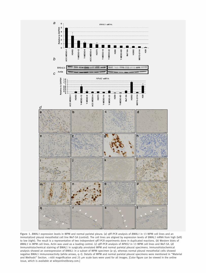
Figure 1. BMAL1 expression levels in MPM and normal parietal pleura. (a) qRT-PCR analysis of BMAL1 in 13 MPM cell lines and an
immortalized pleural mesothelial cell line MeT-5A (control). The cell lines are aligned by expression levels of BMAL1 mRNA from high (left)
to low (right). The result is a representative of two independent qRT-PCR experiments done in duplicated reactions. (b) Western blots of
BMAL1 in MPM cell lines. Actin was used as a loading control. (c) qRT-PCR analysis of NPAS2 in 13 MPM cell lines and MeT-5A. (d)
Immunohistochemical staining of BMAL1 in surgically annotated MPM and normal parietal pleural specimens. Immunohistochemical
analysis showed an overexpression of BMAL1 in a subset of MPM specimen (a–p), whereas normal pleural mesothelial cells showed
negative BMAL1 immunoreactivity (white arrows, q–t). Details of MPM and normal parietal pleural specimens were mentioned in ‘‘Material
and Methods’’ Section. �400 magnification and 25 lm scale bars were used for all images. [Color figure can be viewed in the online
issue, which is available at wileyonlinelibrary.com.]

higher levels of BMAL1 mRNA than MeT-5A, while NPAS2expression profile seems different from BMAL1 profile in MPMcell lines (Figs. 1a and 1c). Western blot analysis for the same setof cell lines clearly demonstrated variable degrees of BMAL1 pro-tein expression (Fig. 1b). Furthermore, we investigated BMAL1expression levels in 16 surgically annotated MPM and four nor-mal parietal pleural specimens (Table 1). Through immunohisto-chemical analysis, BMAL1 was detected in a subset of MPMspecimens with nuclear and/or cytoplasmic localization, whilenone of the four normal parietal pleural samples showed detecta-ble levels of BMAL1 protein (Fig. 1d), suggesting that BMAL1may be important in the development of MPM.
Expression profile of BMAL1 and CLOCK over 24 hr in
ACC-MESO-1 and MeT-5A cells
Based on previous data showing that expression of BMAL1followed a rhythmical pattern in animal models,20 we decidedto examine the expression of BMAL1 and CLOCK by qRT-PCR analysis to evaluate their oscillation over 24 hr in ACC-MESO-1 and MeT-5A cells. We used the mouse fibroblastcell line (NIH3T3) as a positive control as it shows a rhyth-mical expression of BMAL1.20 Serum shock was done asdescribed previously.5 BMAL1 rhythmical expression wasfound in serum-shocked normal nontumorigenic mesothelialcells but not in ACC-MESO-1 cells, whereas CLOCK showedrhythmical expression in both MPM and normal mesothelialcells (Fig. 2). These findings indicate that BMAL1 rhythmicalexpression was intact in normal mesothelial cells but not inMPM cells.
BMAL1 knockdown suppresses proliferation, anchorage-
dependent and independent clonal growth of MPM cells
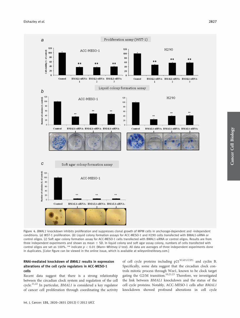
To investigate the role of BMAL1 in MPM cell growth, we per-formed RNAi-mediated gene silencing against BMAL1. ACC-MESO-1 and H290 cell lines were selected as the MPM cellmodels to be used for further investigations (Fig. 1a). MeT-5Awas used as a normal control. To reduce the off target effects, weused low dose (10 nM) stealth selected RNAi (Invitrogen Carls-bad, CA, USA) which includes three siRNA oligos with nono-verlapping sequences targeting BMAL1. Efficient BMAL1 knock-down was confirmed by qRT-PCR, western blotting andimmunostaining (Fig. 3). Next, to evaluate the effect of BMAL1knockdown on cell proliferation in mass culture and clonogenicgrowth in anchorage-dependent and independent conditions,we performed colorimetric growth, liquid and soft agar colonyformation assays. We found that in ACC-MESO-1 and H290cells, BMAL1 knockdown significantly suppressed proliferationand dramatically suppressed colony formation in anchorage-de-pendent (liquid colony formation assay) and anchorage-inde-pendent conditions (soft agar assay) (Fig. 4). By contrast, we didnot see significant suppression of proliferation in MeT-5A (Sup-porting Information, Fig. S1a).
BMAL1 depletion induces massive apoptosis in MPM cells,
with limited consequences in the normal pleural
mesothelial cells
Next, we investigated whether the antiproliferative effect ofBMAL1 depletion is due to cell death. BMAL1 knockdownresulted in massive apoptosis and necrosis in MPM cells (Fig.5a), as evidenced by increases in cleaved caspase-3 activation
Table 1. Clinical features of 16 patients with MPM
Staging
ID Age/Sex HistologyBMAL1 Status(IHC score)
Asbestosexposure OS (M) Clinical Pathological Surgery Therapy
1 50/M Biphasic Negative (1þ) þ 2.5 T2N0M0/II T3N2M0/III Right EPP No
2 54/M Biphasic Negative (1þ) þ 24.0 T2N0M0/II T2N0M0/II Left EPP No
3 56/M Epithelioid Negative (1þ) þ 25.3 T3N2M0/III T4N0M0/IV Left EPP No
4 65/M Epithelioid Negative (0) þ 8.8 T3N1M0/III T3N2M0/III Right EPP Neoadjuvant CT
5 46/F Epithelioid Negative (1þ) � 36.0 T1bN0M0/Ib T3N2M0/III Left EPP Adjuvant CT þ LRT
6 70/F Epithelioid Negative (0) � 27.6 T1bN1M0/III T2N1M0/III Left EPP Neoadjuvant CT
7 65/M Epithelioid Positive (3þ) þ 8.4 T3N0M0/III T4N0M0/IV Right EPP Neoadjuvant CT
8 60/M Biphasic Negative (0) þ 31.3 T3N2M0/III T3N0M0/III Right EPP Neoadjuvant CT þ PHR
9 62/M Epithelioid Positive (2þ) þ 13.0 T3N0M0/III T4NxM0/IV Left pleurectomy Neoadjuvant CT
10 67/M Biphasic Negative (1þ) � 11.6 T2N1M0/III T3N0M0/III Right EPP Neoadjuvant CT þ PHR
11 66/M Biphasic Negative (0) þ 8.3 T2N0M0/II T3N0M0/III Right EPP Neoadjuvant CT
12 67/M Epithelioid Positive (2þ) þ 3.6 T2N0M0/II T3N2M0/III Right EPP No
13 68/M Epithelioid Positive (3þ) � 7.7 T3N0M0/III T3N0M0/III Left EPP Neoadjuvant CT þ PHR
14 63/M Epithelioid Positive (3þ) þ 1.9 T2N0M0/II T3N0M0/III Lt EPP Neoadjuvant CT
15 68/M Sarcomatoid Negative (0) þ 1.0 T3N2M0/III T3N0M0/III Right EPP Neoadjuvant CT
16 64/M Epithelioid Negative (1þ) þ 0.8 T2N0M0/II T3N2M0/III Left EPP Neoadjuvant CT
Abbreviations: Id, patient’s number; IHC, immunohistochemical score for BMAL1; OS, overall survival; M, month; EPP, extra-pleural pneumonectomy;neoadjuvant CT, neoadjuvant chemotherapy (cisplatin þ pemetrexed); adjuvant chemotherapy (cisplatin þ pemetrexed); LRT, local radiationtherapy; PHR, post-operative hemithoracic radiation.
Can
cerCellBiology
2824 BMAL1 as a therapeutic target for MPM
Int. J. Cancer: 131, 2820–2831 (2012) VC 2012 UICC

in ACC-MESO-1 cells (Fig. 5b). By contrast, subtle apoptosiscould be found in MeT-5A cells following BMAL1 knockdown(Fig. 5a). These results indicate that BMAL1 knockdown-induced growth inhibition occurs in part through apoptoticmechanisms and MPM cells are more dependent on BMAL1expression for their survival than normal mesothelial cells.
BMAL1 knockdown leads to cell cycle disruption in
ACC-MESO-1 cells with a clear rise of polyploidy
We investigated whether BMAL1 knockdown-induced growthinhibition also was caused by cell cycle arrest. Consistent withinduction of apoptosis as measured by cleaved caspase-3expression, there was an increase in subG1 DNA content inACC-MESO-1 transfected with BMAL1-siRNA oligos com-pared to cells transfected with control oligos (Fig. 5c). Theproportion of cells in G1 phase decreased whereas a modestincrease in the proportion of cells in G2/M phase. Notably,the profiling also showed an increase in the fraction of cellswhose DNA contents exceed 4 N that correspond to poly-ploidy cell population. With much longer times in culture,high percentages of those cells (in G2/M phase and polyploidyregion) decreased while subG1 population significantlyincreased (Fig. 5c), implying that polyploidy cells underwent
apoptosis. By contrast, we did not see significant changes incell cycle profiling of MeT-5A following BMAL1 knockdown(Supporting Information, Fig. S1b).
Depletion of BMAL1 induces drastic morphological
alterations indicative of mitotic catastrophe in
ACC-MESO-1 cells
We noted that following BMAL1 knockdown, ACC-MESO-1cells underwent drastic morphological changes; the cellsenlarged and elongated by visual examination under phasecontrast microscopy. Next, we performed hematoxlin and eo-sin (H–E) staining for these cells. As shown in Figure 6a,dramatic morphological alterations were seen in ACC-MESO-1 cells after BMAL1 knockdown; cells exhibit muchenlarged flattened shape, micronucleation, multiple nucleiand vacuolization occasionally were found. Next, we exam-ined BMAL1 siRNA-treated ACC-MESO-1 cells by detailedmorphological analysis of DAPI-stained cell nuclei and a-tubulin immunostaining to visualize the cytoskeletal altera-tions associated with changes in nuclear morphology. Multi-ple morphological defects were identified which are consist-ent with the findings observed by H–E staining (Figs. 6b and
Figure 2. Expression profiles of BMAL1 and CLOCK genes over 24 hr in ACC-MESO-1 and MeT-5A cells. qRT-PCR analysis of BMAL1 and
CLOCK mRNA in ACC-MESO-1, MeT-5A at time indicated above. NIH3T3 cells were used as a positive control. Serum shock was performed
as mentioned in the ‘‘Materials and Methods’’ Section. We found that serum shock induces rhythmical expression changes of BMAL1 and
CLOCK mRNA in NIH3T3 as well as MeT-5A cells. Serum shocked ACC-MESO-1 cells showed rhythmical changes of CLOCK mRNA, but not
BMAL1 (GAPDH was used as an internal control). The results are averages of two independent experiments done in duplicate.
Can
cerCellBiology
Elshazley et al. 2825
Int. J. Cancer: 131, 2820–2831 (2012) VC 2012 UICC

6c). Importantly, the existence of micronucleation is a highlyindicative sign of catastrophic mitosis. To examine the possibil-ity of mitotic catastrophe as a sequel of BMAL1 knockdown andto explain its role in BMAL1-induced cell death during the cellcycle, ACC-MESO-1 cells transfected with BMAL1-siRNA orcontrol oligos synchronized using a double thymidine block (toinduce pharmacological block of mitosis in these cells). Interest-ingly, inhibition of mitosis in ACC-MESO-1 transfected withBMAL1 siRNA resulted in a marked decrease of polyploidy andsubG1 population (Fig. 6d), suggesting aberrant mitosis as a
cause of cell death after BMAL1 knockdown. To confirm theoccurrence of mitotic catastrophe following BMAL1 knock-down, we performed time lapse microscopic examination forACC-MESO-1 cells transfected with BMAL1-siRNA or controloligos. Examination showed that cells rounding up as theyentered mitosis, then attempting to undergo cytokinesis. ACC-MESO-1 cells transfected with control siRNA successfully com-pleted mitosis, but BMAL1 siRNA-treated cells failed to divideproperly and exhibited large cell volume and micronucleation(Fig. 6e), indicating mitotic catastrophe as a cell fate followingBMAL1 knockdown in those cells.
Figure 3. BMAL1 knockdown in MPM and MeT-5A cells. Confirmation of BMAL1 knockdown by (a) qRT-PCR, (b) Western blot analysis and
(c) immunofluorescence (IF) assay. IF was used to examine BMAL1 levels in the parental, siRNA control or siRNA-BMAL1-treated ACC-MESO-
1 cells. All data are averages of three independent experiments done in duplicates. [Color figure can be viewed in the online issue, which
is available at wileyonlinelibrary.com.]
Can
cerCellBiology
2826 BMAL1 as a therapeutic target for MPM
Int. J. Cancer: 131, 2820–2831 (2012) VC 2012 UICC
RNAi-mediated knockdown of BMAL1 results in expression
alterations of the cell cycle regulators in ACC-MESO-1
cells
Recent data suggest that there is a strong relationshipbetween the circadian clock system and regulation of the cellcycle.21,22 In particular, BMAL1 is considered a key regulatorof cancer cell proliferation through coordinating the activity
of cell cycle proteins including p21WAF1/CIP1 and cyclin B.Specifically, some data suggest that the circadian clock con-trols mitotic process through Wee1, known to be clock targetgating the G2/M transition.10,21,23 Therefore, we investigatedthe link between BMAL1 knockdown and the status of thecell cycle proteins. Notably, ACC-MESO-1 cells after BMAL1knockdown showed profound alterations in cell cycle
Figure 4. BMAL1 knockdown inhibits proliferation and suppresses clonal growth of MPM cells in anchorage-dependent and -independent
conditions. (a) WST-1 proliferation. (b) Liquid colony formation assays for ACC-MESO-1 and H290 cells transfected with BMAL1-siRNA or
control oligos. (c) Soft agar colony formation assay for ACC-MESO-1 cells transfected with BMAL1-siRNA or control oligos. Results are from
three independent experiments and shown as mean 6 SD. In liquid colony and soft agar assay colony, numbers of cells transfected with
control oligos are set as 100%. ** indicate p < 0.01 (Mann–Whitney U test). All data are averages of three independent experiments done
in duplicates. [Color figure can be viewed in the online issue, which is available at wileyonlinelibrary.com.]
Can
cerCellBiology
Elshazley et al. 2827
Int. J. Cancer: 131, 2820–2831 (2012) VC 2012 UICC
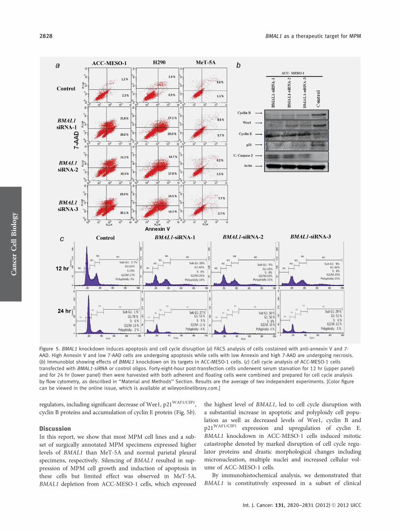
regulators, including significant decrease of Wee1, p21WAF1/CIP1,cyclin B proteins and accumulation of cyclin E protein (Fig. 5b).
DiscussionIn this report, we show that most MPM cell lines and a sub-set of surgically annotated MPM specimens expressed higherlevels of BMAL1 than MeT-5A and normal parietal pleuralspecimens, respectively. Silencing of BMAL1 resulted in sup-pression of MPM cell growth and induction of apoptosis inthese cells but limited effect was observed in MeT-5A.BMAL1 depletion from ACC-MESO-1 cells, which expressed
the highest level of BMAL1, led to cell cycle disruption witha substantial increase in apoptotic and polyploidy cell popu-lation as well as decreased levels of Wee1, cyclin B andp21WAF1/CIP1 expression and upregulation of cyclin E.BMAL1 knockdown in ACC-MESO-1 cells induced mitoticcatastrophe denoted by marked disruption of cell cycle regu-lator proteins and drastic morphological changes includingmicronucleation, multiple nuclei and increased cellular vol-ume of ACC-MESO-1 cells.
By immunohistochemical analysis, we demonstrated thatBMAL1 is constitutively expressed in a subset of clinical
Figure 5. BMAL1 knockdown induces apoptosis and cell cycle disruption (a) FACS analysis of cells costained with anti-annexin V and 7-
AAD. High Annexin V and low 7-AAD cells are undergoing apoptosis while cells with low Annexin and high 7-AAD are undergoing necrosis.
(b) Immunoblot showing effects of BMAL1 knockdown on its targets in ACC-MESO-1 cells. (c) Cell cycle analysis of ACC-MESO-1 cells
transfected with BMAL1-siRNA or control oligos. Forty-eight-hour post-transfection cells underwent serum starvation for 12 hr (upper panel)
and for 24 hr (lower panel) then were harvested with both adherent and floating cells were combined and prepared for cell cycle analysis
by flow cytometry, as described in ‘‘Material and Methods’’ Section. Results are the average of two independent experiments. [Color figure
can be viewed in the online issue, which is available at wileyonlinelibrary.com.]
Can
cerCellBiology
2828 BMAL1 as a therapeutic target for MPM
Int. J. Cancer: 131, 2820–2831 (2012) VC 2012 UICC

MPM samples. Two out of three Stage IV-MPM patientswere positive for BMAL1, whereas three out of 12 Stage III-MPM patients were positive (Table 1). This suggests thatBMAL1 expression may be associated with advanced stage-
MPM, but the small patients’ number makes it difficult todraw a firm conclusion on this possible association (SupportingInformation, Fig. S2). It would be of importance to analyzethe association between BMAL1 expression and
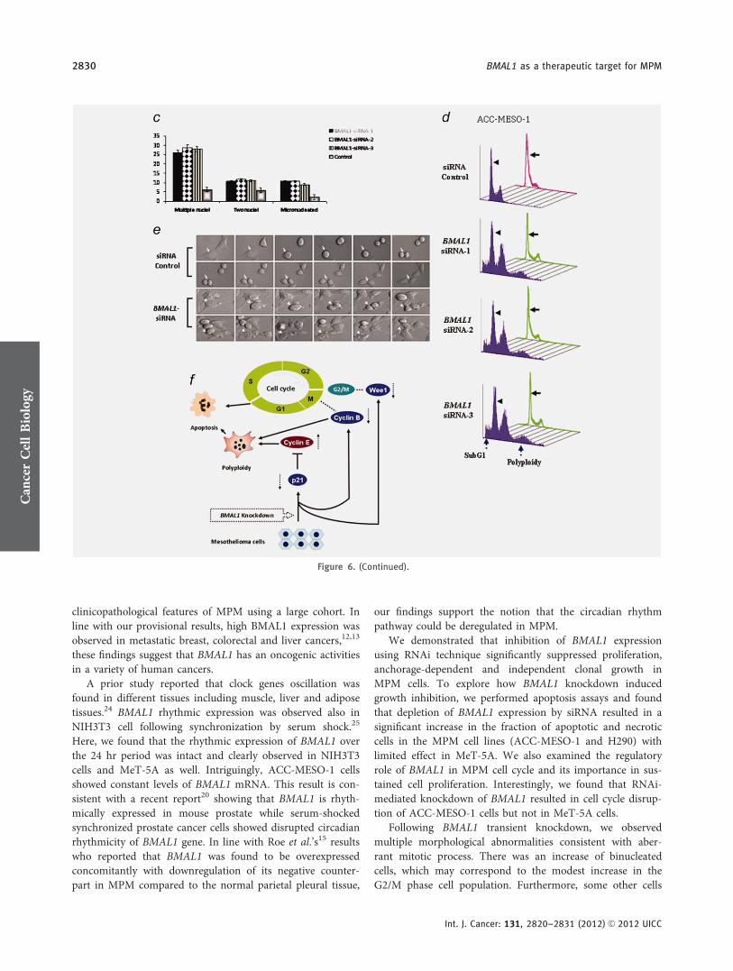
Figure 6. BMAL1 knockdown induces dramatic morphological alterations in ACC-MESO-1 cells. (a) H–E stain showing the nuclear
morphological changes identified in ACC-MESO-1 cells after ablation of BMAL1. (b) IF of a-tubulin and DAPI stains. The upper panels
represent ACC-MESO-1 cells treated with siRNA control. The lower panels represent the most frequent morphological changes (arrow
indicates micronucleation and arrow head indicates multiple nuclei) in single ACC-MESO-1 cell after BMAL1 depletion. The middle panels
represent ACC-MESO-1 cells treated with BMAL1 siRNA. (c) Quantification of the binuclear, multinuclear and micronuclear phenotypes in
ACC-MESO-1 cells after BMAL1 knockdown. (d) Cell cycle profiling of ACC-MESO-1 cells showing marked decrease of BMAL1-induced cell
death and polyploidy formation following mitosis block. ACC-MESO-1 cells transfected with BMAL1 siRNA or control oligos with double-
thymidine (black arrows) and without thymidine treatment (arrow heads) were harvested for analyses of DNA content by flow cytometry.
Synchronized cells at late G1/early S by double-thymidine escaped from BMAL1 knockdown-induced cell death with marked reduction in
subG1 and polyploidy formation. (e) Time lapse microscopic examination showing the aberrant mitosis in ACC-MESO-1 cells transfected
with BMAL1 siRNA (white arrow head) and intact mitosis in cells transfected with control oligos (white arrow). (f) Proposed molecular
mechanism of BMAL1 knockdown-induced mitotic catastrophe in ACC-MESO-1 cells. Decreasing cyclin B below a critical level results in
mitosis skipping and enhances polyploidy formation. Downregulation of p21WAF1/CIP1 results in accumulation of cyclin E which could
lead to increased number of polyploidy cells. Wee1 downregulation could contribute to eventual escape from G2/M arrest, which in turns
participates in impairment of mitotic integrity. [Color figure can be viewed in the online issue, which is available at wileyonlinelibrary.com.]
Can
cerCellBiology
Elshazley et al. 2829
Int. J. Cancer: 131, 2820–2831 (2012) VC 2012 UICC
clinicopathological features of MPM using a large cohort. Inline with our provisional results, high BMAL1 expression wasobserved in metastatic breast, colorectal and liver cancers,12,13
these findings suggest that BMAL1 has an oncogenic activitiesin a variety of human cancers.
A prior study reported that clock genes oscillation wasfound in different tissues including muscle, liver and adiposetissues.24 BMAL1 rhythmic expression was observed also inNIH3T3 cell following synchronization by serum shock.25
Here, we found that the rhythmic expression of BMAL1 overthe 24 hr period was intact and clearly observed in NIH3T3cells and MeT-5A as well. Intriguingly, ACC-MESO-1 cellsshowed constant levels of BMAL1 mRNA. This result is con-sistent with a recent report20 showing that BMAL1 is rhyth-mically expressed in mouse prostate while serum-shockedsynchronized prostate cancer cells showed disrupted circadianrhythmicity of BMAL1 gene. In line with Roe et al.’s15 resultswho reported that BMAL1 was found to be overexpressedconcomitantly with downregulation of its negative counter-part in MPM compared to the normal parietal pleural tissue,
our findings support the notion that the circadian rhythmpathway could be deregulated in MPM.
We demonstrated that inhibition of BMAL1 expressionusing RNAi technique significantly suppressed proliferation,anchorage-dependent and independent clonal growth inMPM cells. To explore how BMAL1 knockdown inducedgrowth inhibition, we performed apoptosis assays and foundthat depletion of BMAL1 expression by siRNA resulted in asignificant increase in the fraction of apoptotic and necroticcells in the MPM cell lines (ACC-MESO-1 and H290) withlimited effect in MeT-5A. We also examined the regulatoryrole of BMAL1 in MPM cell cycle and its importance in sus-tained cell proliferation. Interestingly, we found that RNAi-mediated knockdown of BMAL1 resulted in cell cycle disrup-tion of ACC-MESO-1 cells but not in MeT-5A cells.
Following BMAL1 transient knockdown, we observedmultiple morphological abnormalities consistent with aber-rant mitotic process. There was an increase of binucleatedcells, which may correspond to the modest increase in theG2/M phase cell population. Furthermore, some other cells
Figure 6. (Continued).
Can
cerCellBiology
2830 BMAL1 as a therapeutic target for MPM
Int. J. Cancer: 131, 2820–2831 (2012) VC 2012 UICC
(about 10%) showed micronucleation. Bergman et al.26
reported that the occurrence of cells with double nucleus sug-gests that these cells undergo cell division without segre-gating their DNA and could be explained by the increasein cells with 4 N DNA content seen with flow cytometry.Importantly, micronucleation is highly indicative of mitoticcatastrophe and could be resulted from chromosomal mis-segregation caused by DNA breaks. It is quite possible thatACC-MESO-1 cells underwent mitotic catastrophe due toimpairment of cell cycle regulator proteins, such as, cyclinB, p21WAF1/CIP1 and Wee1 (Fig. 6f). It is demonstratedthat decreasing cyclin B below a critical level results in mi-tosis skipping and enhances polyploidy formation, which isconsidered as one of the characteristics of mitotic catastro-phe.27 Polyploidy cells pass an extra round of cell cycleand finally undergo apoptosis.28 In addition, Wee1 down-regulation could contribute to eventual escape from G2/Marrest, which in turns participates in impairment of mitoticintegrity. In this study, we observed downregulation ofp21WAF1/CIP1 and cyclin E upregulation following BMAL1knockdown, and the latter could also lead to increasednumber of polyploidy cells. Other investigators noted that
BMAL1 modulate the transcriptional activity of p53 towardits target p21 and clearly showed that BMAL1 knockdowncaused a decrease in p21 level.22 Moreover, a previousstudy reported that overexpression of cyclin E led toimpairment of mitosis and polyploidy formation.29 Webelieve that accumulation of polyploidy, large multi-nucleated cells and micronucleation in ACC-MESO-1 afterBMAL1 knockdown were consistent with cell death mecha-nism involving mitotic catastrophe. It has been shown thatmitotic catastrophe could be considered as an importantsafeguard to prevent the proliferation of polyploidy cells30
and apoptosis frequently follows mitotic catastrophe.31
Taken together, these findings show that BMAL1 plays acritical role in the mitotic process of cancer cells. To ourknowledge, this study is the first to shed light on theinvolvement of BMAL1 in polyploidy formation, impair-ment of mitotic events, and also to highlight the role ofcircadian clock genes in MPM. In conclusion, we provideevidence that BMAL1 plays an important role and it maybe a promising therapeutic target for MPM, but carefulconsideration is needed to avoid the counter effect on tis-sues dependant on BMAL1 such as muscles and liver.
References
1. Robinson BW, Musk AW, Lake RA. Malignantmesothelioma. Lancet 2005;366:397–408.
2. Sekido Y. Molecular biology of malignantmesothelioma. Environ Health Prev Med 2008;13:65–70.
3. Tsao AS, Wistuba I, Roth JA, et al. Malignantpleural mesothelioma. J Clin Oncol 2009;27:2081–90.
4. Taniguchi H, Fernandez AF, Setien F, et al.Epigenetic inactivation of the circadian clockgene BMAL1 in hematologic malignancies.Cancer Res 2009;69:8447–54.
5. Balsalobre A, Damiola F, Schibler U. A serumshock induces circadian gene expression inmammalian tissue culture cells. Cell 1998;93:929–37.
6. Straif K, Baan R, Grosse Y, et al. Carcinogenicityof shift-work, painting, and fire-fighting. LancetOncol 2007;8:1065–6.
7. Canaple L, Kakizawa T, Laudet V. The days andnights of cancer cells. Cancer Res 2003;63:7545–52.
8. Bunger MK, Wilsbacher LD, Moran SM, et al.Mop3 is an essential component of the mastercircadian pacemaker in mammals. Cell 2000;103:1009–17.
9. Gauger MA, Sancar A. Cryptochrome, circadiancycle, cell cycle checkpoints, and cancer. CancerRes 2005;65:6828–34.
10. Matsuo T, Yamaguchi S, Mitsui S, et al. Controlmechanism of the circadian clock for timing ofcell division in vivo. Science 2003;302:255–9.
11. Fu L, Lee CC. The circadian clock: pacemakerand tumour suppressor. Nat Rev Cancer 2003;3:350–61.
12. Oshima T, Takenoshita S, Akaike M, et al.Expression of circadian genes correlates with livermetastasis and outcomes in colorectal cancer.Oncol Rep 2011;25:1439–46.
13. Kuo SJ, Chen ST, Yeh KT, et al. Disturbance ofcircadian gene expression in breast cancer.Virchows Arch 2009;454:467–74.
14. Koyanagi S, Kuramoto Y, Nakagawa H, et al. Amolecular mechanism regulating circadianexpression of vascular endothelial growth factorin tumor cells. Cancer Res 2003;63:7277–83.
15. Roe OD, Anderssen E, Helge E, et al. Genome-wide profile of pleural mesothelioma versusparietal and visceral pleura: the emerging geneportrait of the mesothelioma phenotype. PLoSOne 2009;4:e6554.
16. Usami N, Fukui T, Kondo M, et al.Establishment and characterization of fourmalignant pleural mesothelioma cell lines fromJapanese patients. Cancer Sci 2006;97:387–94.
17. Takeyama Y, Sato M, Horio M, et al. Knockdownof ZEB1, a master epithelial-to-mesenchymaltransition (EMT) gene, suppresses anchorage-independent cell growth of lung cancer cells.Cancer Lett 2010;296:216–24.
18. Husain AN, Colby TV, Ordonez NG, et al.Guidelines for pathologic diagnosis of malignantmesothelioma: a consensus statement from theInternational Mesothelioma Interest Group. ArchPathol Lab Med 2009;133:1317–31.
19. Sato M, Vaughan MB, Girard L, et al. Multipleoncogenic changes (K-RAS(V12), p53knockdown, mutant EGFRs, p16 bypass,telomerase) are not sufficient to confer a fullmalignant phenotype on human bronchialepithelial cells. Cancer Res 2006;66:2116–28.
20. Cao Q, Gery S, Dashti A, et al. A role for theclock gene per1 in prostate cancer. Cancer Res2009;69:7619–25.
21. Grechez-Cassiau A, Rayet B, Guillaumond F,et al. The circadian clock component BMAL1 is acritical regulator of p21WAF1/CIP1 expressionand hepatocyte proliferation. J Biol Chem 2008;283:4535–42.
22. Mullenders J, Fabius AW, Madiredjo M, et al. Alarge scale shRNA barcode screen identifies thecircadian clock component ARNTL as putativeregulator of the p53 tumor suppressor pathway.PLoS One 2009;4:e4798.
23. Wood PA, Du-Quiton J, You S, et al. Circadianclock coordinates cancer cell cycle progression,thymidylate synthase, and 5-fluorouraciltherapeutic index. Mol Cancer Ther 2006;5:2023–33.
24. Yang X, Downes M, Yu RT, et al. Nuclearreceptor expression links the circadian clock tometabolism. Cell 2006;126:801–10.
25. Yagita K, Tamanini F, van Der Horst GT, et al.Molecular mechanisms of the biologicalclock in cultured fibroblasts. Science 2001;292:278–81.
26. Bergman LM, Birts CN, Darley M, et al. CtBPspromote cell survival through the maintenance ofmitotic fidelity. Mol Cell Biol 2009;29:4539–51.
27. Galimberti F, Thompson SL, Ravi S, et al.Anaphase catastrophe is a target for cancertherapy. Clin Cancer Res 2011;17:1218–22.
28. Li S, Szymborski A, Miron MJ, et al. Theadenovirus E4orf4 protein induces growth arrestand mitotic catastrophe in H1299 human lungcarcinoma cells. Oncogene 2009;28:390–400.
29. Keck JM, Summers MK, Tedesco D, et al. CyclinE overexpression impairs progression throughmitosis by inhibiting APC(Cdh1). J Cell Biol2007;178:371–85.
30. Marrazzo E, Marchini S, Tavecchio M, et al. Theexpression of the DeltaNp73beta isoform ofp73 leads to tetraploidy. Eur J Cancer 2009;45:443–53.
31. Rivera A, Mavila A, Bayless KJ, et al. Cyclin A1is a p53-induced gene that mediates apoptosis,G2/M arrest, and mitotic catastrophe in renal,ovarian, and lung carcinoma cells. Cell Mol LifeSci 2006;63:1425–39.
Can
cerCellBiology
Elshazley et al. 2831
Int. J. Cancer: 131, 2820–2831 (2012) VC 2012 UICC
Related Documents


![Research Paper Circadian Clock Gene Bmal1 …differentiation and metabolism [4]. Bilirubin is a toxic end-product of heme catabolism in the body [5,6]. High levels of free bilirubin](https://static.cupdf.com/doc/110x72/5f961a1ffab55d152953aefd/research-paper-circadian-clock-gene-bmal1-differentiation-and-metabolism-4-bilirubin.jpg)


