Citation: Ono, M.; Toyoda, N.; Kagami, K.; Hosono, T.; Matsumoto, T.; Horike, S.-i.; Yamazaki, R.; Nakamura, M.; Mizumoto, Y.; Fujiwara, T.; et al. Uterine Deletion of Bmal1 Impairs Placental Vascularization and Induces Intrauterine Fetal Death in Mice. Int. J. Mol. Sci. 2022, 23, 7637. https:// doi.org/10.3390/ijms23147637 Academic Editor: Dariusz Szukiewicz Received: 27 June 2022 Accepted: 9 July 2022 Published: 11 July 2022 Publisher’s Note: MDPI stays neutral with regard to jurisdictional claims in published maps and institutional affil- iations. Copyright: © 2022 by the authors. Licensee MDPI, Basel, Switzerland. This article is an open access article distributed under the terms and conditions of the Creative Commons Attribution (CC BY) license (https:// creativecommons.org/licenses/by/ 4.0/). International Journal of Molecular Sciences Article Uterine Deletion of Bmal1 Impairs Placental Vascularization and Induces Intrauterine Fetal Death in Mice Masanori Ono 1,2,† , Natsumi Toyoda 1,3,† , Kyosuke Kagami 1 , Takashi Hosono 1,4 , Takeo Matsumoto 1 , Shin-ichi Horike 5 , Rena Yamazaki 1 , Mitsuhiro Nakamura 1,6 , Yasunari Mizumoto 1 , Tomoko Fujiwara 7 , Hitoshi Ando 4 , Hiroshi Fujiwara 1, * and Takiko Daikoku 3, * 1 Department of Obstetrics and Gynecology, Graduate School of Medical Science, Kanazawa University, Takaramachi 13-1, Kanazawa 920-8641, Japan; [email protected] (M.O.); [email protected] (N.T.); [email protected] (K.K.); [email protected] (T.H.); [email protected] (T.M.); [email protected] (R.Y.); [email protected] (M.N.); [email protected] (Y.M.) 2 Department of Obstetrics and Gynecology, Tokyo Medical University, Shinjuku, Tokyo 160-0023, Japan 3 Division of Animal Disease Model, Research Center for Experimental Modeling of Human Disease, Kanazawa University, Takaramachi 13-1, Kanazawa 920-8641, Japan 4 Department of Cellular and Molecular Function Analysis, Graduate School of Medical Science, Kanazawa University, Kanazawa 920-8640, Japan; [email protected] 5 Division of Integrated Omics Research, Research Center for Experimental Modeling of Human Disease, Kanazawa University, Kanazawa 920-8640, Japan; [email protected] 6 Department of Obstetrics and Gynecology, Public Central Hospital of Matto Ishikawa, Hakusan 924-8588, Japan 7 Department of Human Life Environments, Kyoto Notre Dame University, Kyoto 606-0847, Japan; [email protected] * Correspondence: [email protected] (H.F.); [email protected] (T.D.); Tel.: +81-76-265-2425 (H.F.); +81-76-265-2460 (T.D.); Fax: +81-76-234-4266 (H.F.); +81-76-234-4245 (T.D.) † These authors contributed equally to this work. Abstract: Recently, it was demonstrated that the expression of BMAL1 was decreased in the en- dometrium of women suffering from recurrent spontaneous abortion. To investigate the pathological roles of uterine clock genes during pregnancy, we produced conditional deletion of uterine Bmal1 (cKO) mice and found that cKO mice could receive embryo implantation but not sustain pregnancy. Gene ontology analysis of microarray suggested that uterine NK (uNK) cell function was suppressed in cKO mice. Histological examination revealed the poor formation of maternal vascular spaces in the placenta. In contrast to WT mice, uNK cells in the spongiotrophoblast layer, where maternal uNK cells are directly in contact with fetal trophoblast, hardly expressed an immunosuppressive NK marker, CD161, in cKO mice. By progesterone supplementation, pregnancy could be sustained until the end of pregnancy in some cKO mice. Although this treatment did not improve the structural abnormalities of the placenta, it recruited CD161-positive NK cells into the spongiotrophoblast layer in cKO mice. These findings indicate that the uterine clock system may be critical for pregnancy maintenance after embryo implantation. Keywords: BMAL1; cKO mouse; miscarriage; NK cell; placental formation; vascularization 1. Introduction The uterus is a unique organ specialized for pregnancy, which achieves embryo implantation and fetal delivery. In women, dysmenorrhea is an important clinical sign to show functional or organic disorders of the uterus. Previously, we found that dysmenorrhea is associated with breakfast skipping [1]. Later, a similar positive relationship between breakfast skipping and dysmenorrhea has been shown elsewhere [2–5]. Recently, we further found that pregnant women who experienced dysmenorrhea at a younger age in the past Int. J. Mol. Sci. 2022, 23, 7637. https://doi.org/10.3390/ijms23147637 https://www.mdpi.com/journal/ijms

Welcome message from author
This document is posted to help you gain knowledge. Please leave a comment to let me know what you think about it! Share it to your friends and learn new things together.
Transcript
Citation: Ono, M.; Toyoda, N.;
Kagami, K.; Hosono, T.; Matsumoto,
T.; Horike, S.-i.; Yamazaki, R.;
Nakamura, M.; Mizumoto, Y.;
Fujiwara, T.; et al. Uterine Deletion of
Bmal1 Impairs Placental
Vascularization and Induces
Intrauterine Fetal Death in Mice. Int.
J. Mol. Sci. 2022, 23, 7637. https://
doi.org/10.3390/ijms23147637
Academic Editor:
Dariusz Szukiewicz
Received: 27 June 2022
Accepted: 9 July 2022
Published: 11 July 2022
Publisher’s Note: MDPI stays neutral
with regard to jurisdictional claims in
published maps and institutional affil-
iations.
Copyright: © 2022 by the authors.
Licensee MDPI, Basel, Switzerland.
This article is an open access article
distributed under the terms and
conditions of the Creative Commons
Attribution (CC BY) license (https://
creativecommons.org/licenses/by/
4.0/).
International Journal of
Molecular Sciences
Article
Uterine Deletion of Bmal1 Impairs Placental Vascularizationand Induces Intrauterine Fetal Death in MiceMasanori Ono 1,2,† , Natsumi Toyoda 1,3,†, Kyosuke Kagami 1, Takashi Hosono 1,4, Takeo Matsumoto 1,Shin-ichi Horike 5, Rena Yamazaki 1, Mitsuhiro Nakamura 1,6, Yasunari Mizumoto 1, Tomoko Fujiwara 7,Hitoshi Ando 4 , Hiroshi Fujiwara 1,* and Takiko Daikoku 3,*
1 Department of Obstetrics and Gynecology, Graduate School of Medical Science, Kanazawa University,Takaramachi 13-1, Kanazawa 920-8641, Japan; [email protected] (M.O.);[email protected] (N.T.); [email protected] (K.K.);[email protected] (T.H.); [email protected] (T.M.);[email protected] (R.Y.); [email protected] (M.N.);[email protected] (Y.M.)
2 Department of Obstetrics and Gynecology, Tokyo Medical University, Shinjuku, Tokyo 160-0023, Japan3 Division of Animal Disease Model, Research Center for Experimental Modeling of Human Disease,
Kanazawa University, Takaramachi 13-1, Kanazawa 920-8641, Japan4 Department of Cellular and Molecular Function Analysis, Graduate School of Medical Science,
Kanazawa University, Kanazawa 920-8640, Japan; [email protected] Division of Integrated Omics Research, Research Center for Experimental Modeling of Human Disease,
Kanazawa University, Kanazawa 920-8640, Japan; [email protected] Department of Obstetrics and Gynecology, Public Central Hospital of Matto Ishikawa,
Hakusan 924-8588, Japan7 Department of Human Life Environments, Kyoto Notre Dame University, Kyoto 606-0847, Japan;
[email protected]* Correspondence: [email protected] (H.F.); [email protected] (T.D.);
Tel.: +81-76-265-2425 (H.F.); +81-76-265-2460 (T.D.); Fax: +81-76-234-4266 (H.F.); +81-76-234-4245 (T.D.)† These authors contributed equally to this work.
Abstract: Recently, it was demonstrated that the expression of BMAL1 was decreased in the en-dometrium of women suffering from recurrent spontaneous abortion. To investigate the pathologicalroles of uterine clock genes during pregnancy, we produced conditional deletion of uterine Bmal1(cKO) mice and found that cKO mice could receive embryo implantation but not sustain pregnancy.Gene ontology analysis of microarray suggested that uterine NK (uNK) cell function was suppressedin cKO mice. Histological examination revealed the poor formation of maternal vascular spaces inthe placenta. In contrast to WT mice, uNK cells in the spongiotrophoblast layer, where maternaluNK cells are directly in contact with fetal trophoblast, hardly expressed an immunosuppressive NKmarker, CD161, in cKO mice. By progesterone supplementation, pregnancy could be sustained untilthe end of pregnancy in some cKO mice. Although this treatment did not improve the structuralabnormalities of the placenta, it recruited CD161-positive NK cells into the spongiotrophoblast layerin cKO mice. These findings indicate that the uterine clock system may be critical for pregnancymaintenance after embryo implantation.
Keywords: BMAL1; cKO mouse; miscarriage; NK cell; placental formation; vascularization
1. Introduction
The uterus is a unique organ specialized for pregnancy, which achieves embryoimplantation and fetal delivery. In women, dysmenorrhea is an important clinical sign toshow functional or organic disorders of the uterus. Previously, we found that dysmenorrheais associated with breakfast skipping [1]. Later, a similar positive relationship betweenbreakfast skipping and dysmenorrhea has been shown elsewhere [2–5]. Recently, we furtherfound that pregnant women who experienced dysmenorrhea at a younger age in the past
Int. J. Mol. Sci. 2022, 23, 7637. https://doi.org/10.3390/ijms23147637 https://www.mdpi.com/journal/ijms

Int. J. Mol. Sci. 2022, 23, 7637 2 of 16
had a higher incidence of developing hypertensive disorders of pregnancy (HDP) [6].From these findings, we hypothesized that poor dietary habits during adolescence andyoung adulthood impair uterine functions and induce latent progression of obstetrics andgynecologic disorders (adolescent dietary habit-induced obstetric and gynecologic disease:ADHOGD) [7].
Since breakfast skipping enhanced starvation stress at the initial stage of the activephase, we proposed that breakfast skipping impairs reproductive functions by disturbinga circadian rhythm via central and/or peripheral clock systems [7,8]. Using a breakfastskipping murine model, we demonstrated that food intake directly regulates the circadianrhythm of the uterine clock [9]. We further found that abnormal starvation stress byirregular uptake of breakfast impaired uterine clock function in mice (in preparation).Consequently, we speculated that the uterine peripheral clock system is influenced bydietary habits and is involved in the mechanism of obstetric and gynecologic diseases.
The circadian oscillation is controlled by clock genes such as Period (Per1-3), Cryptochrome(Cry1-2), Circadian locomotor output cycles kaput (Clock), and Brain and muscle aryl hydrocarbonreceptor nuclear translocator-like protein 1 (Bmal1). Systemic Bmal1 knockout (KO) female micewere reported to have disorders in multiple organs including multifactorial infertility [10].However, although circadian expression of clock genes, such as Per1-3, Cry1-2, Bmal1,and Clock, was demonstrated in the murine and rat uterus [11,12], the precise roles ofuterine clock genes in reproductive functions remain unclear. Recently, the expressionof BMAL1 was demonstrated to be decreased in the endometrium of women sufferingfrom recurrent spontaneous abortion. Researchers also showed that BMAL1-depleteddecidual cells inhibited trophoblast invasion, suggesting that the endometrial clock playsan important role in human pregnancy [13].
Based on this background, to investigate the pathological roles of uterine clock genesduring pregnancy, we created uterine Bmal1 cKO mice by crossing Bmal1-loxP [14] andprogesterone receptor-cre mice [15,16]. In this study, we further examined the effects of uterineBmal1-deletion on uterine functions, especially fertility.
2. Results2.1. Uterine Bmal1 cKO Mice Gave No Live Birth
The present study focused on the roles of uterine clock genes in pregnancy events.Thus, we created Bmal1f/f/PRcre/+ (cKO) mice crossing with Bmal1-flox and PR-cre mice.The loss of BMAL1 in cKO female mice was confirmed by RT-PCR (Figure 1A), Westernblotting (Figure 1B), and immunofluorescence (Figure 1C) using non-pregnant cKO and WT(Bmal1f/f/PR+/+) uteruses. To examine the effect of cKO on pregnancy outcome, we matedcKO and WT female mice with fertile C57BL/6 male mice. While seven out of eight WTvaginal plug-positive female mice produced pups, no cKO (n = 15) female mice producedlive pups (Figure 1D).
2.2. cKO Female Mice Increased Incomplete Miscarriage after Day Six of Pregnancy
To determine the cause of no live birth in cKO female mice, we next examined stage-specific effects of cKO during pregnancy (day one of pregnancy = virginal plug). On dayfive of pregnancy, the embryo implantation sites in cKO showed no difference in numbersand morphological appearance as compared to WT (Table 1 and Figure 2A). On day sixof pregnancy, the number of female mice with implantation and implantation sites wasdecreased in cKO compared to the WT (57.1% vs. 85.7%, 7.1 ± 0.67 vs. 9.4 ± 0.40, Table 1).The size of the decidual bed seemed smaller in cKO than in WT (Figure 2B).

Int. J. Mol. Sci. 2022, 23, 7637 3 of 16Int. J. Mol. Sci. 2022, 23, x FOR PEER REVIEW 3 of 16
Figure 1. Bmal1 cKO uteri showed efficient deletion of BMAL1 and no live birth. (A–C) The uterine
deletion of BMAL1 was observed by (A) RT-PCR, (B) Western blotting, and (C) immunofluores-
cence. (D) Uterine Bmal1 cKO mice showed no live births. The bar shows SE. ACTB, ACTIN; em,
endometrium; mm/i, inner circular layer of myometrium; mm/o, the outer longitudinal layer of my-
ometrium; ND, not determined. The scale bar shows 200 µm.
2.2. cKO Female Mice Increased Incomplete Miscarriage after Day Six of Pregnancy
To determine the cause of no live birth in cKO female mice, we next examined stage-
specific effects of cKO during pregnancy (day one of pregnancy = virginal plug). On day
five of pregnancy, the embryo implantation sites in cKO showed no difference in numbers
and morphological appearance as compared to WT (Table 1 and Figure 2A). On day six
of pregnancy, the number of female mice with implantation and implantation sites was
decreased in cKO compared to the WT (57.1% vs. 85.7%, 7.1 ± 0.67 vs. 9.4 ± 0.40, Table 1).
The size of the decidual bed seemed smaller in cKO than in WT (Figure 2B).
Table 1. cKO mice start to fail pregnancy maintenance after day six of pregnancy.
Day of Preg-
nancy Genotype No. of Mice
No. of Mice with
Normal IS (%)
Mean No. of IS
Sites (No. ± SE) *
Day 5 WT 8 100.0 8.3 ± 0.65
cKO 11 100.0 8.3 ± 0.38
Day 6 WT 14 85.7 9.4 ± 0.40
cKO 21 57.1 7.1 ± 0.67
Day 8 WT 16 81.3 8.8 ± 0.53
cKO 30 ** 40.0 7.3 ± 0.56
Day 12 WT 20 85.0 9.5 ± 0.46
cKO 39 20.5 7.6 ± 1.00
* Mean IS numbers among only female mice with normal IS; ** 7 out of 30 mice had resorption. WT; Bmal1f/f/PR+/+, cKO; Bmal1f/f/PRcre/+, IS; implantation site.
Figure 1. Bmal1 cKO uteri showed efficient deletion of BMAL1 and no live birth. (A–C) The uterinedeletion of BMAL1 was observed by (A) RT-PCR, (B) Western blotting, and (C) immunofluores-cence. (D) Uterine Bmal1 cKO mice showed no live births. The bar shows SE. ACTB, ACTIN; em,endometrium; mm/i, inner circular layer of myometrium; mm/o, the outer longitudinal layer ofmyometrium; ND, not determined. The scale bar shows 200 µm.
Table 1. cKO mice start to fail pregnancy maintenance after day six of pregnancy.
Day ofPregnancy Genotype No. of Mice No. of Mice with
Normal IS (%)Mean No. of IS
Sites (No. ± SE) *
Day 5WT 8 100.0 8.3 ± 0.65
cKO 11 100.0 8.3 ± 0.38
Day 6WT 14 85.7 9.4 ± 0.40
cKO 21 57.1 7.1 ± 0.67
Day 8WT 16 81.3 8.8 ± 0.53
cKO 30 ** 40.0 7.3 ± 0.56
Day 12WT 20 85.0 9.5 ± 0.46
cKO 39 20.5 7.6 ± 1.00
* Mean IS numbers among only female mice with normal IS; ** 7 out of 30 mice had resorption. WT; Bmal1f/f/PR+/+,cKO; Bmal1f/f/PRcre/+, IS; implantation site.
Among cKO mice on day 8 of pregnancy (n = 30), 11 had no implantation sites,7 showed resorption sites, and the remaining 12 had normal implantation sites (Table 1and Figure 2C). There were no differences in the numbers of embryo implantation sitesamong implantation-positive mice between the cKO and WT. At this stage of pregnancy,branches of the maternal uterine vessels in the mesometrial region become dilated tosupport fetoplacental development in WT (Figure 2D, green-dotted ellipse); however,vessel dilatation in this region seemed poor in the cKO mice (Figure 2D, red-dotted ellipse).The calculated pixel areas of the blood vessel cavities in the mesometrial region in thecKO seem decreased compared to those in WT, although the difference is not significant(Figure 2E).

Int. J. Mol. Sci. 2022, 23, 7637 4 of 16Int. J. Mol. Sci. 2022, 23, x FOR PEER REVIEW 4 of 16
Figure 2. The miscarriage rate is increased in cKO mice after implantation. (A,B) Representative of
uteri and HE-stained section of implantation sites from WT and cKO on days 5 (A) and 6 (B) of
pregnancy. (C) Representative of uteri of WT and cKO on day 8. (D) HE-stained section of implan-
tation sites and area of the blood vessel (BV) on day 8. (E) Although the difference is not significant,
the calculated pixel areas of the blood vessel cavities in the mesometrial region in the cKO seem
decreased compared to those in WT. The scale bar shows 100 µm. Arrowheads show embryo. Circles
show uterine vessels in the mesometrial region. le, luminal epithelium; ge, glandular epithelium; st,
endometrial stroma; mm, myometrium.
Among cKO mice on day 8 of pregnancy (n = 30), 11 had no implantation sites, 7
showed resorption sites, and the remaining 12 had normal implantation sites (Table 1 and
Figure 2C). There were no differences in the numbers of embryo implantation sites among
implantation-positive mice between the cKO and WT. At this stage of pregnancy,
branches of the maternal uterine vessels in the mesometrial region become dilated to sup-
port fetoplacental development in WT (Figure 2D, green-dotted ellipse); however, vessel
dilatation in this region seemed poor in the cKO mice (Figure 2D, red-dotted ellipse). The
calculated pixel areas of the blood vessel cavities in the mesometrial region in the cKO
seem decreased compared to those in WT, although the difference is not significant (Fig-
ure 2E).
Figure 2. The miscarriage rate is increased in cKO mice after implantation. (A,B) Representativeof uteri and HE-stained section of implantation sites from WT and cKO on days 5 (A) and 6 (B)of pregnancy. (C) Representative of uteri of WT and cKO on day 8. (D) HE-stained section ofimplantation sites and area of the blood vessel (BV) on day 8. (E) Although the difference is notsignificant, the calculated pixel areas of the blood vessel cavities in the mesometrial region in thecKO seem decreased compared to those in WT. The scale bar shows 100 µm. Arrowheads showembryo. Circles show uterine vessels in the mesometrial region. le, luminal epithelium; ge, glandularepithelium; st, endometrial stroma; mm, myometrium.
2.3. cKO Mice Showed Abnormal Placental Formation on Day Twelve of Pregnancy
On day 12 of pregnancy, normal implantation sites were observed in 17 out of 20(85.0%) WT mice and in 8 (20.5%) out of 39 cKO mice (Table 1 and Figure 3A). Amongimplantation-positive mice, the numbers of implantation sites in cKO mice were lowerthan those in WT mice (7.6 ± 1.00 in the cKO group and 9.5 ± 0.46 in the control group,respectively). At this stage of pregnancy, the placenta has three layers: an outer layer oftrophoblast giant cells, a middle spongiotrophoblast layer (junctional zone), and the innerlabyrinth layer [17]. Maternal vessels contained denucleated red blood cells, whereas fetalvessels still had nucleated erythrocytes. Therefore, we could clearly distinguish maternal

Int. J. Mol. Sci. 2022, 23, 7637 5 of 16
vessels from fetal vessels in the placenta using HE staining and found that the placenta ofcKO mice had abnormalities in vascular structures. The labyrinth formation was impairedand the areas of maternal vessels became narrowed (Figure 3B). The ratio of calculatedareas of maternal vessels (green-dotted areas) among the total areas of both maternal andfetal (red-dotted areas) vessels in cKO mice (Figure 3C) was significantly decreased thanthose in WT mice (Figure 3D).
Int. J. Mol. Sci. 2022, 23, x FOR PEER REVIEW 5 of 16
2.3. cKO Mice Showed Abnormal Placental Formation on Day Twelve of Pregnancy
On day 12 of pregnancy, normal implantation sites were observed in 17 out of 20
(85.0%) WT mice and in 8 (20.5%) out of 39 cKO mice (Table 1 and Figure 3A). Among
implantation-positive mice, the numbers of implantation sites in cKO mice were lower
than those in WT mice (7.6 ± 1.00 in the cKO group and 9.5 ± 0.46 in the control group,
respectively). At this stage of pregnancy, the placenta has three layers: an outer layer of
trophoblast giant cells, a middle spongiotrophoblast layer (junctional zone), and the inner
labyrinth layer [17]. Maternal vessels contained denucleated red blood cells, whereas fetal
vessels still had nucleated erythrocytes. Therefore, we could clearly distinguish maternal
vessels from fetal vessels in the placenta using HE staining and found that the placenta of
cKO mice had abnormalities in vascular structures. The labyrinth formation was impaired
and the areas of maternal vessels became narrowed (Figure 3B). The ratio of calculated
areas of maternal vessels (green-dotted areas) among the total areas of both maternal and
fetal (red-dotted areas) vessels in cKO mice (Figure 3C) was significantly decreased than
those in WT mice (Figure 3D).
Int. J. Mol. Sci. 2022, 23, x FOR PEER REVIEW 6 of 16
Figure 3. cKO mice show abnormal placental formation on day 12 of pregnancy. (A) Representative
of uteri from WT and cKO on day 12 of pregnancy. (B–D) HE-stained section of the placenta. Blue-
dotted areas show maternal blood vessels. Red-dotted areas show fetal blood vessels which contain
nucleated erythrocytes. (E) The percentage of the maternal blood vessels that contains denucleated
red blood cells (RBC). The bar shows SE. Differences in the ratio of calculated areas of maternal
vessels between the wild-type and cKO mice were analyzed by the unpaired t-test. p < 0.05. The
scale bar shows 100 µm.
2.4. cKO Mice Showed Functional Changes in the Uterine Immune Environment
To investigate the key genes related to uterine functional changes in cKO mice, we
performed microarray analysis using uterine tissues of WT and cKO mice. In a total of
20,983 genes, up-regulated genes were defined as those with both up-regulated in ZT0
and ZT12 (fold-change >1; 6147 genes) and were selected by multiplying the absolute
value of each WAD (>0.02; 597 genes), whereas down-regulated genes were those with
both down-regulated (fold-change <1; 5169 genes) and were selected (>0.02; 515 genes) as
described previously [18]. Gene ontology biological process term enrichment analyses of
the identified up- and down-regulated genes detected by WAD were performed using the
Database for Annotation, Visualization, and Integrated Discovery (DAVID) with the
threshold at a p-value <0.05, and the top 11 groups are presented [18] (Figure 4A). Since
the gene ontology terms of immune responses including NK cell activation were domi-
nantly detected in the down-regulated group, we focused on the uterine NK (uNK) cells.
uNK cells are recruited and activated by ovarian hormones, which are dominant im-
mune cells at the maternal-fetal boundary in pregnancy [19]. uNK cells release cytokines
or chemokines that induce trophoblast invasion, tissue remodeling, embryonic develop-
ment, and placentation [20]. We first analyzed the localization of uNK cells using Dolichos
biflorus agglutinin (DBA) and periodic Schiff (PAS) staining methods [21,22]. Murine
uNK cells are classified into two subsets by DBA and PAS reactivity [23]. DBA-positive
(PAS+DBA+) uNK cells produce angiogenic factors [24] and progesterone-induced block-
ing factor [25], and IFN-γ-producing uNK cells contribute to vessel instability and facili-
tate pregnancy-induced remodeling of decidual arteries [26,27]. It was reported that DBA-
negative (PAS+ DBA-) NK cells exist in the spongiotrophoblast layer, which did not con-
tain DBA-positive uNK cells [25].
Figure 3. cKO mice show abnormal placental formation on day 12 of pregnancy. (A) Representative ofuteri from WT and cKO on day 12 of pregnancy. (B–D) HE-stained section of the placenta. Blue-dotted
Int. J. Mol. Sci. 2022, 23, 7637 6 of 16
areas show maternal blood vessels. Red-dotted areas show fetal blood vessels which contain nucleatederythrocytes. (E) The percentage of the maternal blood vessels that contains denucleated red bloodcells (RBC). The bar shows SE. Differences in the ratio of calculated areas of maternal vessels betweenthe wild-type and cKO mice were analyzed by the unpaired t-test. p < 0.05. The scale bar shows100 µm.
2.4. cKO Mice Showed Functional Changes in the Uterine Immune Environment
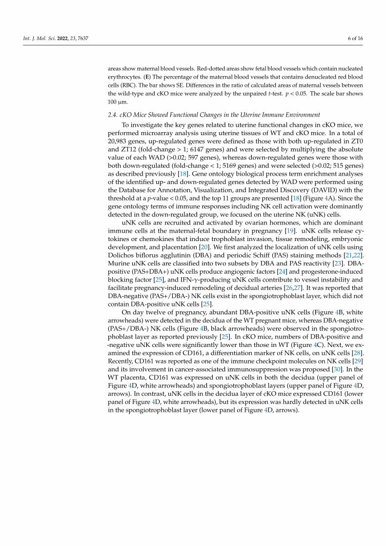
To investigate the key genes related to uterine functional changes in cKO mice, weperformed microarray analysis using uterine tissues of WT and cKO mice. In a total of20,983 genes, up-regulated genes were defined as those with both up-regulated in ZT0and ZT12 (fold-change > 1; 6147 genes) and were selected by multiplying the absolutevalue of each WAD (>0.02; 597 genes), whereas down-regulated genes were those withboth down-regulated (fold-change < 1; 5169 genes) and were selected (>0.02; 515 genes)as described previously [18]. Gene ontology biological process term enrichment analysesof the identified up- and down-regulated genes detected by WAD were performed usingthe Database for Annotation, Visualization, and Integrated Discovery (DAVID) with thethreshold at a p-value < 0.05, and the top 11 groups are presented [18] (Figure 4A). Since thegene ontology terms of immune responses including NK cell activation were dominantlydetected in the down-regulated group, we focused on the uterine NK (uNK) cells.
uNK cells are recruited and activated by ovarian hormones, which are dominantimmune cells at the maternal-fetal boundary in pregnancy [19]. uNK cells release cy-tokines or chemokines that induce trophoblast invasion, tissue remodeling, embryonicdevelopment, and placentation [20]. We first analyzed the localization of uNK cells usingDolichos biflorus agglutinin (DBA) and periodic Schiff (PAS) staining methods [21,22].Murine uNK cells are classified into two subsets by DBA and PAS reactivity [23]. DBA-positive (PAS+DBA+) uNK cells produce angiogenic factors [24] and progesterone-inducedblocking factor [25], and IFN-γ-producing uNK cells contribute to vessel instability andfacilitate pregnancy-induced remodeling of decidual arteries [26,27]. It was reported thatDBA-negative (PAS+/DBA-) NK cells exist in the spongiotrophoblast layer, which did notcontain DBA-positive uNK cells [25].
On day twelve of pregnancy, abundant DBA-positive uNK cells (Figure 4B, whitearrowheads) were detected in the decidua of the WT pregnant mice, whereas DBA-negative(PAS+/DBA-) NK cells (Figure 4B, black arrowheads) were observed in the spongiotro-phoblast layer as reported previously [25]. In cKO mice, numbers of DBA-positive and-negative uNK cells were significantly lower than those in WT (Figure 4C). Next, we ex-amined the expression of CD161, a differentiation marker of NK cells, on uNK cells [28].Recently, CD161 was reported as one of the immune checkpoint molecules on NK cells [29]and its involvement in cancer-associated immunosuppression was proposed [30]. In theWT placenta, CD161 was expressed on uNK cells in both the decidua (upper panel ofFigure 4D, white arrowheads) and spongiotrophoblast layers (upper panel of Figure 4D,arrows). In contrast, uNK cells in the decidua layer of cKO mice expressed CD161 (lowerpanel of Figure 4D, white arrowheads), but its expression was hardly detected in uNK cellsin the spongiotrophoblast layer (lower panel of Figure 4D, arrows).

Int. J. Mol. Sci. 2022, 23, 7637 7 of 16Int. J. Mol. Sci. 2022, 23, x FOR PEER REVIEW 7 of 16
Figure 4. The immune environment in cKO uteri. (A) Gene ontology analysis with microarray data.
The top 11 groups are shown. (B) DBA/PAS double staining with the placenta on day 12 of preg-
nancy. Black arrowheads show PAS-positive and DBA-negative cells. White arrowheads show both
PAS- and DBA-positive cells. (C) In cKO mice, numbers of DBA-positive and -negative uNK cells
were significantly lower than those in WT. (D) CD161 staining with the placenta on day 12 of
Figure 4. The immune environment in cKO uteri. (A) Gene ontology analysis with microarray data.The top 11 groups are shown. (B) DBA/PAS double staining with the placenta on day 12 of pregnancy.Black arrowheads show PAS-positive and DBA-negative cells. White arrowheads show both PAS- and
Int. J. Mol. Sci. 2022, 23, 7637 8 of 16
DBA-positive cells. (C) In cKO mice, numbers of DBA-positive and -negative uNK cells weresignificantly lower than those in WT. (D) CD161 staining with the placenta on day 12 of pregnancy.In the WT placenta, CD161 was expressed on uNK cells in both the decidua (upper panel, whitearrowheads) and spongiotrophoblast layers (upper panel, arrows). In contrast, uNK cells in thedecidua layer of cKO mice expressed CD161 (lower panel, white arrowheads) but its expression washardly detected in uNK cells in the spongiotrophoblast layer (lower panel, arrows). Arrows showCD161 positive cells. The scale bar shows 100 µm.
2.5. Progesterone Supplementation Rescues Perinatal Outcomes in cKO Female Mice
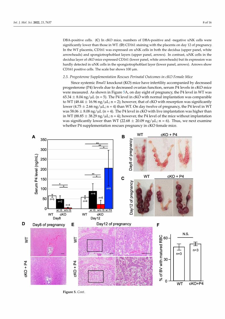
Since systemic Bmal1 knockout (KO) mice have infertility accompanied by decreasedprogesterone (P4) levels due to decreased ovarian function, serum P4 levels in cKO micewere measured. As shown in Figure 5A, on day eight of pregnancy, the P4 level in WT was65.34 ± 8.04 ng/µL (n = 5). The P4 level in cKO with normal implantation was comparableto WT (48.44 ± 16.96 ng/µL; n = 2); however, that of cKO with resorption was significantlylower (4.75 ± 2.66 ng/µL; n = 4) than WT. On day twelve of pregnancy, the P4 level in WTwas 58.06 ± 8.08 ng/µL (n = 4). The P4 level in cKO with live implantation was higher thanin WT (88.85 ± 38.29 ng/µL; n = 4); however, the P4 level of the mice without implantationwas significantly lower than WT (22.68 ± 20.09 ng/µL; n = 6). Thus, we next examinewhether P4 supplementation rescues pregnancy in cKO female mice.
Int. J. Mol. Sci. 2022, 23, x FOR PEER REVIEW 8 of 16
pregnancy. In the WT placenta, CD161 was expressed on uNK cells in both the decidua (upper
panel, white arrowheads) and spongiotrophoblast layers (upper panel, arrows). In contrast, uNK
cells in the decidua layer of cKO mice expressed CD161 (lower panel, white arrowheads) but its
expression was hardly detected in uNK cells in the spongiotrophoblast layer (lower panel, arrows).
Arrows show CD161 positive cells. The scale bar shows 100 µm.
On day twelve of pregnancy, abundant DBA-positive uNK cells (Figure 4B, white
arrowheads) were detected in the decidua of the WT pregnant mice, whereas DBA-nega-
tive (PAS+ /DBA−) NK cells (Figure 4B, black arrowheads) were observed in the spongi-
otrophoblast layer as reported previously [25]. In cKO mice, numbers of DBA-positive
and -negative uNK cells were significantly lower than those in WT (Figure 4C). Next, we
examined the expression of CD161, a differentiation marker of NK cells, on uNK cells [28].
Recently, CD161 was reported as one of the immune checkpoint molecules on NK cells
[29] and its involvement in cancer-associated immunosuppression was proposed [30]. In
the WT placenta, CD161 was expressed on uNK cells in both the decidua (upper panel of
Figure 4D, white arrowheads) and spongiotrophoblast layers (upper panel of Figure 4D,
arrows). In contrast, uNK cells in the decidua layer of cKO mice expressed CD161 (lower
panel of Figure 4D, white arrowheads), but its expression was hardly detected in uNK
cells in the spongiotrophoblast layer (lower panel of Figure 4D, arrows).
2.5. Progesterone Supplementation Rescues Perinatal Outcomes in cKO Female Mice
Since systemic Bmal1 knockout (KO) mice have infertility accompanied by decreased
progesterone (P4) levels due to decreased ovarian function, serum P4 levels in cKO mice
were measured. As shown in Figure 5A, on day eight of pregnancy, the P4 level in WT
was 65.34 ± 8.04 ng/µL (n = 5). The P4 level in cKO with normal implantation was compa-
rable to WT (48.44 ± 16.96 ng/µL; n = 2); however, that of cKO with resorption was signif-
icantly lower (4.75 ± 2.66 ng/µL; n = 4) than WT. On day twelve of pregnancy, the P4 level
in WT was 58.06 ± 8.08 ng/μL (n = 4). The P4 level in cKO with live implantation was
higher than in WT (88.85 ± 38.29 ng/µL; n = 4); however, the P4 level of the mice without
implantation was significantly lower than WT (22.68 ± 20.09 ng/µL; n = 6). Thus, we next
examine whether P4 supplementation rescues pregnancy in cKO female mice.
Int. J. Mol. Sci. 2022, 23, x FOR PEER REVIEW 9 of 16
Figure 5. P4 supplement rescues pregnancy failure in cKO mice. (A) Serum P4 levels in WT and
cKO on days 8 and 12 of pregnancy. The mean P4 levels in the control group (WT, n = 5) and in the
cKO groups with normal implantation (w/IS, n = 2) and with confirmed inhalation (w/o IS, n = 4) on
day 8 of pregnancy, and those in the control group (WT, n = 4), the cKO groups with implantation
(w/IS, n = 4) and without implantation (w/o IS, n = 6), and the progesterone-treated cKO group
(w/P4, n = 5) on day 12 of pregnancy. The bar shows STDEV. Differences between groups were
analyzed by the unpaired t-test. *, p < 0.01; **, p < 0.05. (B,C) Representative of the uterus of WT and
cKO with P4 supplementation on days 8 (B) and 12 (C) of pregnancy. (D,E) Representative of HE-
stained uterus of WT and cKO with P4 supplementation on days 8 (D) and 12 (E). (F) The percentage
of maternal blood vessels, which contain denucleated red blood cells (RBC). The bar shows SE. Dif-
ferences in the ratio of calculated areas of maternal vessels between the WT and cKO mice were
analyzed by the unpaired t-test. (G) DBA/PAS double staining of the placenta from cKO with P4
supplementation on day 12 of pregnancy. Black arrowheads show PAS-positive and DBA-negative
cells. White arrowheads show both PAS- and DBA-positive cells. (H) Numbers of DBA-positive and
-negative uNK cells were not significantly different between cKO and cKO witn P4 supplementa-
tion. (I) CD161 staining of the placenta from cKO with P4 supplementation on day 12 of pregnancy.
Arrows show CD161 positive cells in the spongiotrophoblast layers. The scale bar shows 100 µm.
N.S., not significant.
cKO mice with P4 supplementation (n = 5) increased serum P4 levels by more than
145 ng/μL, which was significantly higher than those in WT (Figure 5A). In this condition,
the ratio of the mice with normal implantation sites was increased in cKO supplemented
with P4 compared to non-treated cKO on both days 8 and 12 of pregnancy (80.0% vs.
40.0% and 57.9% vs. 20.5%, respectively) (Tables 1 and 2). Notably, in some P4-treated
cKO mice (8 out of 12, 66.7%), live pups could be obtained by cesarean section on
Figure 5. Cont.
Int. J. Mol. Sci. 2022, 23, 7637 9 of 16
Int. J. Mol. Sci. 2022, 23, x FOR PEER REVIEW 9 of 16
Figure 5. P4 supplement rescues pregnancy failure in cKO mice. (A) Serum P4 levels in WT and
cKO on days 8 and 12 of pregnancy. The mean P4 levels in the control group (WT, n = 5) and in the
cKO groups with normal implantation (w/IS, n = 2) and with confirmed inhalation (w/o IS, n = 4) on
day 8 of pregnancy, and those in the control group (WT, n = 4), the cKO groups with implantation
(w/IS, n = 4) and without implantation (w/o IS, n = 6), and the progesterone-treated cKO group
(w/P4, n = 5) on day 12 of pregnancy. The bar shows STDEV. Differences between groups were
analyzed by the unpaired t-test. *, p < 0.01; **, p < 0.05. (B,C) Representative of the uterus of WT and
cKO with P4 supplementation on days 8 (B) and 12 (C) of pregnancy. (D,E) Representative of HE-
stained uterus of WT and cKO with P4 supplementation on days 8 (D) and 12 (E). (F) The percentage
of maternal blood vessels, which contain denucleated red blood cells (RBC). The bar shows SE. Dif-
ferences in the ratio of calculated areas of maternal vessels between the WT and cKO mice were
analyzed by the unpaired t-test. (G) DBA/PAS double staining of the placenta from cKO with P4
supplementation on day 12 of pregnancy. Black arrowheads show PAS-positive and DBA-negative
cells. White arrowheads show both PAS- and DBA-positive cells. (H) Numbers of DBA-positive and
-negative uNK cells were not significantly different between cKO and cKO witn P4 supplementa-
tion. (I) CD161 staining of the placenta from cKO with P4 supplementation on day 12 of pregnancy.
Arrows show CD161 positive cells in the spongiotrophoblast layers. The scale bar shows 100 µm.
N.S., not significant.
cKO mice with P4 supplementation (n = 5) increased serum P4 levels by more than
145 ng/μL, which was significantly higher than those in WT (Figure 5A). In this condition,
the ratio of the mice with normal implantation sites was increased in cKO supplemented
with P4 compared to non-treated cKO on both days 8 and 12 of pregnancy (80.0% vs.
40.0% and 57.9% vs. 20.5%, respectively) (Tables 1 and 2). Notably, in some P4-treated
cKO mice (8 out of 12, 66.7%), live pups could be obtained by cesarean section on
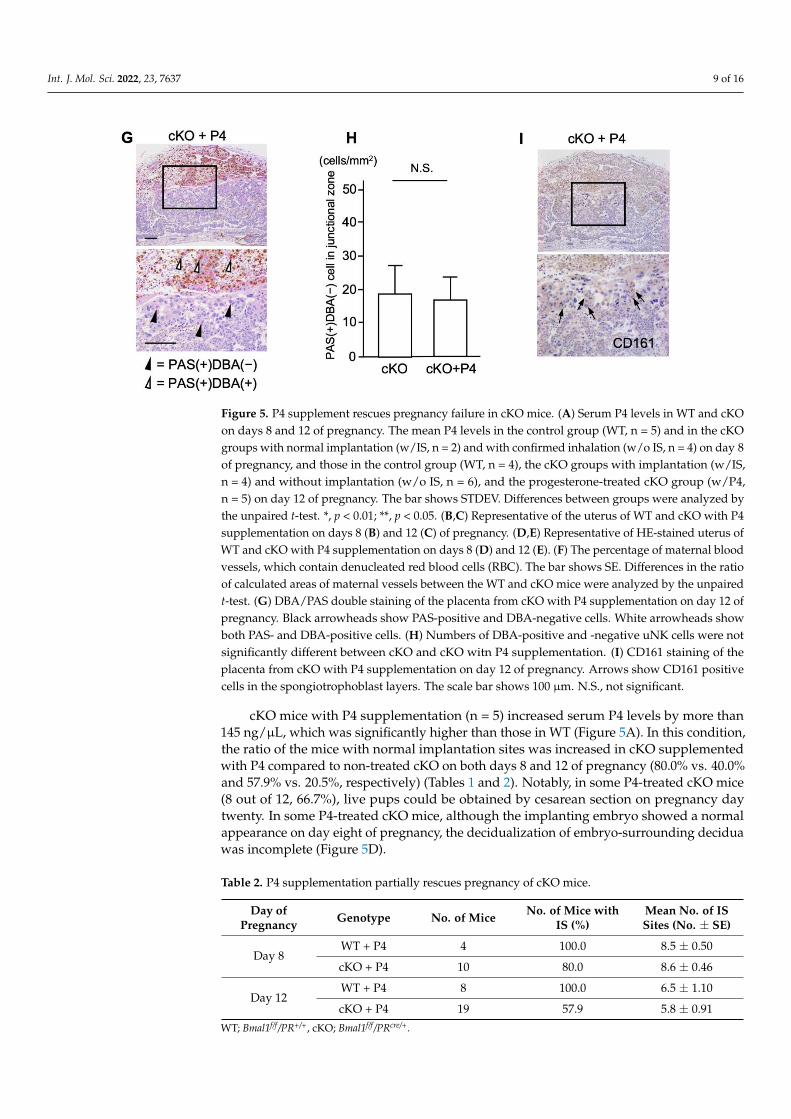
Figure 5. P4 supplement rescues pregnancy failure in cKO mice. (A) Serum P4 levels in WT and cKOon days 8 and 12 of pregnancy. The mean P4 levels in the control group (WT, n = 5) and in the cKOgroups with normal implantation (w/IS, n = 2) and with confirmed inhalation (w/o IS, n = 4) on day 8of pregnancy, and those in the control group (WT, n = 4), the cKO groups with implantation (w/IS,n = 4) and without implantation (w/o IS, n = 6), and the progesterone-treated cKO group (w/P4,n = 5) on day 12 of pregnancy. The bar shows STDEV. Differences between groups were analyzed bythe unpaired t-test. *, p < 0.01; **, p < 0.05. (B,C) Representative of the uterus of WT and cKO with P4supplementation on days 8 (B) and 12 (C) of pregnancy. (D,E) Representative of HE-stained uterus ofWT and cKO with P4 supplementation on days 8 (D) and 12 (E). (F) The percentage of maternal bloodvessels, which contain denucleated red blood cells (RBC). The bar shows SE. Differences in the ratioof calculated areas of maternal vessels between the WT and cKO mice were analyzed by the unpairedt-test. (G) DBA/PAS double staining of the placenta from cKO with P4 supplementation on day 12 ofpregnancy. Black arrowheads show PAS-positive and DBA-negative cells. White arrowheads showboth PAS- and DBA-positive cells. (H) Numbers of DBA-positive and -negative uNK cells were notsignificantly different between cKO and cKO witn P4 supplementation. (I) CD161 staining of theplacenta from cKO with P4 supplementation on day 12 of pregnancy. Arrows show CD161 positivecells in the spongiotrophoblast layers. The scale bar shows 100 µm. N.S., not significant.
cKO mice with P4 supplementation (n = 5) increased serum P4 levels by more than145 ng/µL, which was significantly higher than those in WT (Figure 5A). In this condition,the ratio of the mice with normal implantation sites was increased in cKO supplementedwith P4 compared to non-treated cKO on both days 8 and 12 of pregnancy (80.0% vs. 40.0%and 57.9% vs. 20.5%, respectively) (Tables 1 and 2). Notably, in some P4-treated cKO mice(8 out of 12, 66.7%), live pups could be obtained by cesarean section on pregnancy daytwenty. In some P4-treated cKO mice, although the implanting embryo showed a normalappearance on day eight of pregnancy, the decidualization of embryo-surrounding deciduawas incomplete (Figure 5D).
Table 2. P4 supplementation partially rescues pregnancy of cKO mice.
Day ofPregnancy Genotype No. of Mice No. of Mice with
IS (%)Mean No. of ISSites (No. ± SE)
Day 8WT + P4 4 100.0 8.5 ± 0.50
cKO + P4 10 80.0 8.6 ± 0.46
Day 12WT + P4 8 100.0 6.5 ± 1.10
cKO + P4 19 57.9 5.8 ± 0.91
WT; Bmal1f/f/PR+/+, cKO; Bmal1f/f/PRcre/+.

Int. J. Mol. Sci. 2022, 23, 7637 10 of 16
On day twelve of pregnancy, a severe structural abnormality was observed in thelabyrinth layer of P4-treated cKO, which had not been observed in non-treated cKO mice.Although the structure of the labyrinth seems fragile, the areas of maternal and fetal vesselswere dilated enough in P4-treated cKO (Figure 5E). The fetuses bearing these malformedplacentae would probably have miscarried much earlier if not for P4 supplementation.There were no differences in the ratio of calculated areas of maternal vessels among the totalareas of both maternal and fetal vessels between P4-treated cKO and WT mice (Figure 5F).
In the placenta of P4-treated cKO mice, abundant DBA-positive uNK cells (Figure 5G,white arrowheads) were detected in the decidua as observed in those of WT mice. AlthoughDBA-negative (PAS+/DBA-) NK cells (Figure 5G, black arrowheads) were observed in thespongiotrophoblast layer, the numbers were not recovered as compared with non-treatedcKO mice (Figure 5H). However, in contrast to non-treated cKO mice, CD161-positive uNKcells were detected in the spongiotrophoblast layer (Figure 5I, arrows).
3. Discussion
This study demonstrated that mice lacking BMAL1 expression in the uterus can im-plant embryos but not sustain a pregnancy. The previous studies showed the possibleinvolvement of Bmal1 in regulation of male and female reproductive functions [31–33]. Itwas shown that systemic Bmal1 KO female mice can ovulate but both embryo developmentand ovarian production of progesterone (P4) are not enough to achieve embryo implanta-tion [10,34]. Later, steroidogenic factor-1 (SF-1) expression-dependent Bmal1-deleted femalemice (Bmal1SF1d/d) were demonstrated to fail embryo implantation, which could be rescuedby P4 supplementation or normal ovarian transplantation [35], indicating that poor ovarianproduction of P4 is one of the main causes for infertility of Bmal1 KO female mice. However,the precise roles of uterine clock genes in pregnancy have been unclear. In this regard,the present study is the first report to show that the uterine clock system is critical for asuccessful pregnancy.
Histological examinations revealed that cKO mice developed abnormal placentalstructures. The main feature of placental abnormality in cKO mice was the poor formationof maternal vascular space. Since maternal vascular spaces become narrowed in cKO mice,maternal blood flow into the intervillous spaces in the labyrinth layer is considered to bereduced, suggesting the impaired maternal-fetal nutritional exchanges through this layer.During human placental formation, extravillous trophoblast (EVT) invades the maternaldecidua and reconstructs maternal spiral arteries, which is a process essential to main-taining adequate maternal blood flow into the intervillous spaces, by reducing arterialcontractility [36]. If this vascular reconstruction is interrupted in the early stage of preg-nancy, the subsequent poor blood supply to the intervillous space in the placenta will causeplacental dysfunction and induce various pregnancy complications, such as hypertensivedisorders of pregnancy (HDP), fetal growth restriction, preterm labor, abortions, and still-birth in the late stage of pregnancy [37]. In terms of the impaired vascular reconstructionassociated with abnormal placental formation, the pathogenesis of our cKO mouse modelis corresponding to that of placental dysfunction in human pregnancy complications.
One of the most important properties of the uterus is the acceptance of hemi-allograftduring pregnancy. Gene ontology analysis of microarray showed that the immune responsegenes were down-regulated in the cKO uterus, suggesting the involvement of immune dys-function in the abnormal placental formation (Figure 3A). It was reported that the cellularimmune responses and major cellular subsets, such as regulatory T cells, effector T cells,NK cells, monocytes, macrophages, and neutrophils are involved in the pathogenesis ofHDP [38]. Among them, NK cells were shown to directly interact with EVT and regulatetheir invasion and vascular reconstruction [37]. In accordance with it, GO:0045954 “positiveregulation of natural killer cell-mediated cytotoxicity” was detected as down-regulatedgenes by gene ontology analysis in cKO mice. In the human placenta, it was reported thatuNK cells were decreased in placental bed biopsies from women with HDP comparedto the control third-trimester decidua, suggesting that altered local cytokine balance may

Int. J. Mol. Sci. 2022, 23, 7637 11 of 16
be important in defective trophoblast invasion and spiral artery transformation in thesepathological pregnancies [39]. Therefore, we examined uNK distribution and their sub-types around the embryo implantation sites at early pregnancy and in the placenta atmid-pregnancy. In cKO mice, the numbers of NK cells expressing PAS(+)/DAB(-) in thespongiotrophoblast layer decreased and hardly expressed CD161. These findings indicatethat the subtype of uNK cells in cKO mice is different from those in WT mice. This alsosuggests that functional changes in uNK cells were induced by clock gene deletion, es-pecially in the spongiotrophoblast layer where maternal uNK cells directly interact withfetal trophoblast. Considering the immunosuppressive role of CD161 [29], this functionalalteration of uNK cells may be critical for trophoblast function to sustain maternal-fetalimmune tolerance.
Notably, although this treatment did not completely improve the structural abnor-malities of the placenta, pregnancy could be sustained until the end of pregnancy in somecKO mice. It was reported that, although murine uNK cells do not express the classicalprogesterone receptor, progesterone affects the recruitment and function of uNK cells [25].In accordance with it, this study showed that P4 supplementation could recruit CD161-positive NK cells into the spongiotrophoblast layer even in cKO mice. It was demonstratedthat uNK cells express glucocorticoid receptors and progesterone can regulate NK cellsvia glucocorticoid receptor [40]. Allowing for a higher concentration of serum P4 in P4-supplemented cKO mice compared to WT mice, the possibility that expression of CD161 onuNK cells in the spongiotrophoblast layer was mediated through a glucocorticoid receptorpathway. As clinical evidence, progesterone supplement was demonstrated to preventmiscarriage in women with recurrent miscarriage of unclear etiology [41] and protect theonset of HDP [42]. Recently, it was also reported that progesterone supplement therapyis clinically effective for the functional failure of the placenta with structural abnormali-ties [43]. Our cKO model provides experimental evidence in support of this treatment forpregnancy complications.
This study has several limitations. First, we did not show the direct relationshipbetween meal skipping and uterine clock dysfunction-induced miscarriage. Second, sinceclock genes may have non-clock functions, the deletion of uterine Bmal1 does not necessarilyequal the dysfunction of the uterine clock system. In the future, it is necessary to validatethis issue by experimentation using other clock-gene-deleted mice or by constitutivelyexpressed BMAL1 in the uterus.
In conclusion, uterine deletion of Bmal1 impairs placental vascularization and inducesintrauterine fetal death in mice. The maternal vascular spaces in the placenta were reducedin cKO mice, suggesting the presence of placental dysfunction. Since uNK cells in the spon-giotrophoblast layer of the placenta did not express CD161, dysfunction in uNK cells is oneof the possible mechanisms to induce failure of placental formation in cKO mice. Notably,P4 supplementation recruited CD161-positive NK cells into the spongiotrophoblast layerand rescued pregnant outcomes in some cKO mice. These findings indicate that the uterineclock system is critical for a successful pregnancy and suggest that P4 supplementation canpartially recover clock-induced uterine dysfunction. This study also provides experimentalevidence that the disorder of a uterine clock system can be a candidate to induce uterinedysfunction during pregnancy in ADHOGD.
4. Materials and Methods4.1. Mice
We produced Bmal1f/f/PRcre/+ (cKO) mice by crossing Bmal-loxP mice (strain #007668,The Jackson Laboratory, Sacramento, CA, USA) [14] and PR-cre mice [44]. PR-cre mice werekindly provided by Dr. Francesco Demayo (the National Institutes of Health, Bethesda, MD,USA) and Dr. John P. Lydon (Baylor College of Medicine, Houston, TX, USA). C57BL/6-Jmale mice were purchased from Charles River (Yokohama, Japan) and used for experimentsafter checking fertility. All experimental procedures and housing conditions were approvedby the Kanazawa University Animal Experiment Committee (AP-163729 and AP-214218).

Int. J. Mol. Sci. 2022, 23, 7637 12 of 16
All animals were housed and cared for in accordance with the Institutional Guidelines forExperiments using Animals.
4.2. Analysis of Pregnancy Events
Female cKO and WT (Bmal1f/f/PR+/+) mice were mated with fertile C57BL/6-J malemice to induce pregnancy (Day 1 = virginal plug). To visualize the implantation sites ondays 5 and 6 of pregnancy, 0.1 mL of 1% (w/v) Chicago Sky Blue 6B (Sigma, St. Louis,MO, USA) in saline was intravenously injected into mice [45]. To examine whether P4supplementation maintains pregnancy, a Silastic implant (4 cm length × 0.31 cm diameter,Kaneka medical products, Osaka, Japan) containing P4 (Sigma-Aldrich, Saint Louis, MO,USA) was placed under dorsal skin on day 2 of pregnancy [45].
4.3. RT-PCR
RNA was extracted from the uteruses of WT and cKO mice with TRIzol Reagent(Thermo Fisher, Waltham, MA, USA) according to the manufacturer’s instructions. Af-ter removing contaminating genomic DNA with TURBO DNA-free Kit (Thermo Fisher,Waltham, MA, USA), one µg RNA was reverse-transcribed with Superscript II and oligo(dT)primer to prepare cDNA (Thermo Fisher, Waltham, MA, USA). PCR was performedas previously described [18,46]. The primer sequences for PCR are forward primer(5′-aaagaggcgtcgggacaaaa-3′) and reverse primer (5′-ccatctgctgccctgagaat-3′) for Bmal1, andforward primer (5′-ctctcgctttctggagggtg-3′) and reverse primer (5′-tcagtctccacagacaatgcc-3′)for Rplp0 as an internal control.
4.4. Western Blot Analysis
Tissue samples were prepared as previously described [47]. After measuring the pro-tein concentration, extracts with SDS-PAGE sample buffer were boiled for 5 min. Sampleswere run on 10% SDS-PAGE gel and transferred to a polyvinylidene fluoride membrane.Membranes were blocked with 5% skim milk in Tris-buffered saline Tween 20 and probedwith antibodies to BMAL1 (1:2000; Novus Biologicals, Centennial, CO, USA) or ACTIN(1:5000; Santa Cruz Biotechnology, Santa Cruz, CA, USA). Blots were incubated with donkeyanti-goat or donkey anti-rabbit IgG conjugated to peroxidase (Jackson ImmunoResearchLaboratories, West Grove, PA, USA). All signals were detected using chemiluminescentreagents (GE Healthcare, Chicago, IL, USA). ACTIN was used as a loading control.
4.5. Immunofluorescence
Uteruses harvested from WT and cKO non-pregnant mice were fixed with 4% paraf-ormaldehyde and embedded in OCT after being dehydrated with 30% sucrose. Frozensections (10 µm) were stained with BMAL1 antibody (1:1000; Novus Biologicals, Centennial,CO, USA) and Hoechst.
4.6. Histology and Measurement of Blood Vessel Area
Formalin-fixed paraffin-embedded tissue sections (5 µm) were stained with hema-toxylin and eosin for histological analysis. Blood vessel area in the decidua and the placentawere measured by Image—J software on days 8 and 12 of pregnancy, respectively [48].
4.7. Immunohistochemistry
Formalin-fixed paraffin-embedded tissue sections (5 µm) were subjected to immuno-histochemistry with anti-CD161 rabbit polyclonal antibody (1:2000; Abcam ab197979, SanFrancisco, CA, USA) after antigen retrieval. The signal was visualized with DAB (Fuji-film Wako Pure Chemical Corporation, Osaka, Japan). Hematoxylin staining was used ascounterstaining.

Int. J. Mol. Sci. 2022, 23, 7637 13 of 16
4.8. DBA Lectin and PAS Dual Staining
Formalin-fixed paraffin-embedded tissue sections (5 µm) were first stained with DBAlectin and then subjected to PAS staining. In brief, after antigen retrieval, sections wereincubated with biotinylated DBA lectin (1:5000; Sigma) and visualized positive cells withDAB. Next, sections were incubated with 1% periodic acid (Fujifilm Wako Pure ChemicalCorporation) for 20 min, with Schiff reagent (Fujifilm Wako Pure Chemical Corporation)for 30 min, and 0.5% sodium bisulfite solution (Fujifilm Wako Pure Chemical Corporation,Osaka, Japan) for 5 min. Hematoxylin was used for counter-staining.
4.9. Microarray Analysis
Uteruses were collected from matured WT and cKO mice at 9 am and 9 pm. RNA wasextracted with an RNeasy mini kit (Qiagen, Hilden, Germany) according to the manufac-turer’s instructions. Cyanine-3 (Cy3)-labeled cRNA was prepared from 0.2 µg RNA usinga Low Input Quick Amp Labeling Kit (Agilent) according to the manufacturer’s instruc-tions. A total of 0.6 µg of Cy3-labelled cRNA was hybridized to SurePrint G3 Mouse GEmicroarray 8 × 60 K Ver. 2.0 (G4858A #74809, Agilent Technologies, Santa Clara, CA, USA)for 17 h at 65 ◦C in a rotating Agilent hybridization oven. After hybridization, microarrayswere washed and slides were scanned immediately after washing on an Agilent DNAMicroarray Scanner (G2539A) using the one color scan setting for 1× 60 K array slides. Thescanned images were analyzed with Feature Extraction Software 11.0.1.1 (Agilent) usingdefault parameters to obtain background-subtracted and spatially detrended processedsignal intensities. Data were normalized and filtered with three filters with GeneSpringsoftware 12 (Agilent Technologies). Differentially expressed genes were extracted by theweighted average difference (WAD) ranking method [49].
4.10. P4 Assay
The concentration of P4 in mouse serum was measured by an enzyme-linked im-munosorbent assay kit (Cayman, Ann Arbor, MI, USA). All samples were measured intriplicate.
5. Statistical Methods
Statistical analyses were performed by unpaired t-test (two-sided test). p values < 0.05were accepted as statistically significant.
Author Contributions: Conceptualization, M.O., H.F. and T.D.; Methodology, T.D.; Validation, H.F.and T.D.; Formal analysis, N.T., S.-i.H., H.F. and T.D.; Investigation, M.O., N.T., K.K., T.H., T.M., S.-i.H.and T.D.; Resources, N.T. and T.D.; Data curation, S.-i.H.; Writing—original draft, M.O., H.F. and T.D.;Writing—review editing, M.O., R.Y., M.N., Y.M., T.F., H.A., H.F. and T.D.; Visualization, M.O., H.F.and T.D.; Supervision, H.A., H.F. and T.D.; Project administration, M.O.; Funding acquisition, M.O.,T.F., H.F. and T.D. All authors have read and agreed to the published version of the manuscript.
Funding: This work was supported in part by Grants-in-Aid for Scientific Research [17H04337,18K08470, 19H01617, 19K22681, 20K21651, and 21K18297], by a Japan Science and Technology Agency[19216689], and the Japan Agency for Medical Research and Development [19gk0110024h0002,19gk0210016h0001, 19cm0106610h0002, and 20ck0106549h0001] from the Japanese government.
Institutional Review Board Statement: The animal study protocol was approved by the institutionalEthics Committee of Kanazawa University (AP-163729 (8 April 2016) and AP-214218 (26 March 2021)).
Acknowledgments: We thank Michihiro Mieda (Kanazawa University) and Jumpei Terakawa (AzabuUniversity) for their technical support and valuable advice. We also thank Tae Mieda for her helpwith experiments.
Conflicts of Interest: The authors declare no conflict of interest.

Int. J. Mol. Sci. 2022, 23, 7637 14 of 16
Abbreviations
ADHOGD adolescent dietary habit-induced obstetric and gynecologic diseaseBMAL1 brain and muscle aryl hydrocarbon receptor nuclear translocator-like protein 1CLOCK circadian locomotor output cycles kaputCRY cryptochromeHRP horse-radish peroxidaseOCT optimal cutting temperaturePER PerioduNK uterine natural killerKO knockoutcKO conditional knockoutWT wild type
References1. Fujiwara, T. Skipping breakfast is associated with dysmenorrhea in young women in Japan. Int. J. Food Sci. Nutr. 2003, 54,
505–509. [CrossRef]2. Angelin, P.; Dileep, D.; Manju, T.; Veena, M.; Pradeep, D.; Amreen, K.; Soumitra, S. Effect of Skipping Breakfast on Young Girls’
Menstruation. Ind. J. Youth Adolesc. Health 2017, 4, 17–20.3. Abu Helwa, H.A.; Mitaeb, A.A.; Al-Hamshri, S.; Sweileh, W.M. Prevalence of dysmenorrhea and predictors of its pain intensity
among Palestinian female university students. BMC Womens Health 2018, 18, 18. [CrossRef] [PubMed]4. Hu, Z.; Tang, L.; Chen, L.; Kaminga, A.C.; Xu, H. Prevalence and Risk Factors Associated with Primary Dysmenorrhea among
Chinese Female University Students: A Cross-sectional Study. J. Pediatr. Adolesc. Gynecol. 2020, 33, 15–22. [CrossRef] [PubMed]5. Fujiwara, T.; Ono, M.; Iizuka, T.; Sekizuka-Kagami, N.; Maida, Y.; Adachi, Y.; Fujiwara, H.; Yoshikawa, H. Breakfast Skipping in
Female College Students Is a Potential and Preventable Predictor of Gynecologic Disorders at Health Service Centers. Diagnostics2020, 10, 476. [CrossRef]
6. Nakayama, M.; Ono, M.; Iizuka, T.; Kagami, K.; Fujiwara, T.; Sekizuka-Kagami, N.; Maida, Y.; Obata, T.; Yamazaki, R.; Daikoku,T.; et al. Hypertensive disorders of pregnancy are associated with dysmenorrhea in early adulthood: A cohort study. J. Obstet.Gynaecol. Res. 2020, 46, 2292–2297. [CrossRef] [PubMed]
7. Fujiwara, T.; Ono, M.; Mieda, M.; Yoshikawa, H.; Nakata, R.; Daikoku, T.; Sekizuka-Kagami, N.; Maida, Y.; Ando, H.; Fujiwara, H.Adolescent Dietary Habit-induced Obstetric and Gynecologic Disease (ADHOGD) as a New Hypothesis-Possible Involvement ofClock System. Nutrients 2020, 12, 1294. [CrossRef]
8. Fujiwara, T.; Nakata, R. Skipping breakfast is associated with reproductive dysfunction in post-adolescent female college students.Appetite 2010, 55, 714–717. [CrossRef]
9. Hosono, T.; Ono, M.; Daikoku, T.; Mieda, M.; Nomura, S.; Kagami, K.; Iizuka, T.; Nakata, R.; Fujiwara, T.; Fujiwara, H.; et al.Time-Restricted Feeding Regulates Circadian Rhythm of Murine Uterine Clock. Curr. Dev. Nutr. 2021, 5, nzab064. [CrossRef]
10. Boden, M.J.; Varcoe, T.J.; Voultsios, A.; Kennaway, D.J. Reproductive biology of female Bmal1 null mice. Reproduction 2010, 139,1077–1090. [CrossRef] [PubMed]
11. Johnson, M.H.; Lim, A.; Fernando, D.; Day, M.L. Circadian clockwork genes are expressed in the reproductive tract and conceptusof the early pregnant mouse. Reprod. Biomed. Online 2002, 4, 140–145. [CrossRef]
12. Nakamura, T.J.; Moriya, T.; Inoue, S.; Shimazoe, T.; Watanabe, S.; Ebihara, S.; Shinohara, K. Estrogen differentially regulatesexpression of Per1 and Per2 genes between central and peripheral clocks and between reproductive and nonreproductive tissuesin female rats. J. Neurosci. Res. 2005, 82, 622–630. [CrossRef]
13. Lv, S.; Wang, N.; Ma, J.; Li, W.P.; Chen, Z.J.; Zhang, C. Impaired decidualization caused by downregulation of circadian clockgene BMAL1 contributes to human recurrent miscarriagedagger. Biol. Reprod. 2019, 101, 138–147. [CrossRef]
14. Storch, K.F.; Paz, C.; Signorovitch, J.; Raviola, E.; Pawlyk, B.; Li, T.; Weitz, C.J. Intrinsic circadian clock of the mammalian retina:Importance for retinal processing of visual information. Cell 2007, 130, 730–741. [CrossRef]
15. Daikoku, T.; Dey, S.K. Two faces of PTEN. Nat. Med. 2008, 14, 1192–1193. [CrossRef]16. Daikoku, T.; Hirota, Y.; Tranguch, S.; Joshi, A.R.; DeMayo, F.J.; Lydon, J.P.; Ellenson, L.H.; Dey, S.K. Conditional loss of uterine
Pten unfailingly and rapidly induces endometrial cancer in mice. Cancer Res. 2008, 68, 5619–5627. [CrossRef] [PubMed]17. Cross, J.C.; Hemberger, M.; Lu, Y.; Nozaki, T.; Whiteley, K.; Masutani, M.; Adamson, S.L. Trophoblast functions, angiogenesis and
remodeling of the maternal vasculature in the placenta. Mol. Cell Endocrinol. 2002, 187, 207–212. [CrossRef]18. Iizuka, T.; Wakae, K.; Ono, M.; Suzuki, T.; Mizumoto, Y.; Kitamura, K.; Horike, S.I.; Muramatsu, M.; Fujiwara, H. Activation-
induced cytidine deaminase is a possible regulator of cross-talk between oocytes and granulosa cells through GDF-9 and SCFfeedback system. Sci. Rep. 2021, 11, 3833. [CrossRef] [PubMed]
19. Croy, B.A.; Kiso, Y. Granulated metrial gland cells: A natural killer cell subset of the pregnant murine uterus. Microsc. Res. Tech.1993, 25, 189–200. [CrossRef] [PubMed]
20. Zhang, X.; Wei, H. Role of Decidual Natural Killer Cells in Human Pregnancy and Related Pregnancy Complications. Front.Immunol. 2021, 12, 728291. [CrossRef] [PubMed]

Int. J. Mol. Sci. 2022, 23, 7637 15 of 16
21. Paffaro, V.A., Jr.; Bizinotto, M.C.; Joazeiro, P.P.; Yamada, A.T. Subset classification of mouse uterine natural killer cells by DBAlectin reactivity. Placenta 2003, 24, 479–488. [CrossRef] [PubMed]
22. Peng, J.; Monsivais, D.; You, R.; Zhong, H.; Pangas, S.A.; Matzuk, M.M. Uterine activin receptor-like kinase 5 is crucial forblastocyst implantation and placental development. Proc. Natl. Acad. Sci. USA 2015, 112, E5098–E5107. [CrossRef] [PubMed]
23. Chen, Z.; Zhang, J.; Hatta, K.; Lima, P.D.; Yadi, H.; Colucci, F.; Yamada, A.T.; Croy, B.A. DBA-lectin reactivity defines mouseuterine natural killer cell subsets with biased gene expression. Biol. Reprod. 2012, 87, 81. [CrossRef]
24. Lima, P.D.; Tu, M.M.; Rahim, M.M.; Peng, A.R.; Croy, B.A.; Makrigiannis, A.P. Ly49 receptors activate angiogenic mouse DBA(+)uterine natural killer cells. Cell Mol. Immunol. 2014, 11, 467–476. [CrossRef] [PubMed]
25. Bogdan, A.; Berta, G.; Szekeres-Bartho, J. PIBF positive uterine NK cells in the mouse decidua. J. Reprod. Immunol. 2017, 119,38–43. [CrossRef] [PubMed]
26. Ashkar, A.A.; Di Santo, J.P.; Croy, B.A. Interferon gamma contributes to initiation of uterine vascular modification, decidualintegrity, and uterine natural killer cell maturation during normal murine pregnancy. J. Exp. Med. 2000, 192, 259–270. [CrossRef]
27. Zhang, J.H.; He, H.; Borzychowski, A.M.; Takeda, K.; Akira, S.; Croy, B.A. Analysis of cytokine regulators inducing interferonproduction by mouse uterine natural killer cells. Biol. Reprod. 2003, 69, 404–411. [CrossRef]
28. Kurioka, A.; Cosgrove, C.; Simoni, Y.; van Wilgenburg, B.; Geremia, A.; Bjorkander, S.; Sverremark-Ekstrom, E.; Thurnheer, C.;Gunthard, H.F.; Khanna, N.; et al. CD161 Defines a Functionally Distinct Subset of Pro-Inflammatory Natural Killer Cells. Front.Immunol. 2018, 9, 486. [CrossRef]
29. Buckle, I.; Guillerey, C. Inhibitory Receptors and Immune Checkpoints Regulating Natural Killer Cell Responses to Cancer.Cancers 2021, 13, 4263. [CrossRef]
30. Di, W.; Fan, W.; Wu, F.; Shi, Z.; Wang, Z.; Yu, M.; Zhai, Y.; Chang, Y.; Pan, C.; Li, G.; et al. Clinical characterization andimmunosuppressive regulation of CD161 (KLRB1) in glioma through 916 samples. Cancer Sci. 2021, 113, 756. [CrossRef]
31. Schoeller, E.L.; Clark, D.D.; Dey, S.; Cao, N.V.; Semaan, S.J.; Chao, L.W.; Kauffman, A.S.; Stowers, L.; Mellon, P.L. Bmal1 IsRequired for Normal Reproductive Behaviors in Male Mice. Endocrinology 2016, 157, 4914–4929. [CrossRef] [PubMed]
32. Alvarez, J.D.; Hansen, A.; Ord, T.; Bebas, P.; Chappell, P.E.; Giebultowicz, J.M.; Williams, C.; Moss, S.; Sehgal, A. The circadianclock protein BMAL1 is necessary for fertility and proper testosterone production in mice. J. Biol. Rhythm. 2008, 23, 26–36.[CrossRef] [PubMed]
33. Jiang, Y.; Li, S.; Xu, W.; Ying, J.; Qu, Y.; Jiang, X.; Zhang, A.; Yue, Y.; Zhou, R.; Ruan, T.; et al. Critical Roles of the CircadianTranscription Factor BMAL1 in Reproductive Endocrinology and Fertility. Front. Endocrinol. 2022, 13, 818272. [CrossRef][PubMed]
34. Ratajczak, C.K.; Boehle, K.L.; Muglia, L.J. Impaired steroidogenesis and implantation failure in Bmal1−/− mice. Endocrinology2009, 150, 1879–1885. [CrossRef] [PubMed]
35. Liu, Y.; Johnson, B.P.; Shen, A.L.; Wallisser, J.A.; Krentz, K.J.; Moran, S.M.; Sullivan, R.; Glover, E.; Parlow, A.F.; Drinkwater, N.R.;et al. Loss of BMAL1 in ovarian steroidogenic cells results in implantation failure in female mice. Proc. Natl. Acad. Sci. USA 2014,111, 14295–14300. [CrossRef] [PubMed]
36. Kaufmann, P.; Black, S.; Huppertz, B. Endovascular trophoblast invasion: Implications for the pathogenesis of intrauterine growthretardation and preeclampsia. Biol. Reprod. 2003, 69, 1–7. [CrossRef]
37. Pollheimer, J.; Vondra, S.; Baltayeva, J.; Beristain, A.G.; Knofler, M. Regulation of Placental Extravillous Trophoblasts by theMaternal Uterine Environment. Front. Immunol. 2018, 9, 2597. [CrossRef]
38. Miller, D.; Motomura, K.; Galaz, J.; Gershater, M.; Lee, E.D.; Romero, R.; Gomez-Lopez, N. Cellular immune responses in thepathophysiology of preeclampsia. J. Leukoc. Biol. 2022, 111, 237–260. [CrossRef]
39. Williams, P.J.; Bulmer, J.N.; Searle, R.F.; Innes, B.A.; Robson, S.C. Altered decidual leucocyte populations in the placental bedin pre-eclampsia and foetal growth restriction: A comparison with late normal pregnancy. Reproduction 2009, 138, 177–184.[CrossRef]
40. Guo, W.; Li, P.; Zhao, G.; Fan, H.; Hu, Y.; Hou, Y. Glucocorticoid receptor mediates the effect of progesterone on uterine naturalkiller cells. Am. J. Reprod. Immunol. 2012, 67, 463–473. [CrossRef]
41. Haas, D.M.; Hathaway, T.J.; Ramsey, P.S. Progestogen for preventing miscarriage in women with recurrent miscarriage of unclearetiology. Cochrane Database Syst. Rev. 2019, 2019, CD003511. [CrossRef]
42. Wu, H.; Zhang, S.; Lin, X.; He, J.; Wang, S.; Zhou, P. Pregnancy-related complications and perinatal outcomes followingprogesterone supplementation before 20 weeks of pregnancy in spontaneously achieved singleton pregnancies: A systematicreview and meta-analysis. Reprod. Biol. Endocrinol. 2021, 19, 165. [CrossRef]
43. Barinov, S.V.; Tirskaya, Y.I.; Shamina, I.V.; Ledovskikh, I.O.; Atamanenko, O.J. Placental blood flow and pregnancy outcomesin women with abnormal placental localization and absence of placental “migration”. J. Matern.-Fetal Neonatal Med. 2021, 34,3496–3502. [CrossRef] [PubMed]
44. Soyal, S.M.; Mukherjee, A.; Lee, K.Y.; Li, J.; Li, H.; DeMayo, F.J.; Lydon, J.P. Cre-mediated recombination in cell lineages thatexpress the progesterone receptor. Genesis 2005, 41, 58–66. [CrossRef]
45. Tranguch, S.; Wang, H.; Daikoku, T.; Xie, H.; Smith, D.F.; Dey, S.K. FKBP52 deficiency-conferred uterine progesterone resistance isgenetic background and pregnancy stage specific. J. Clin. Investig. 2007, 117, 1824–1834. [CrossRef] [PubMed]

Int. J. Mol. Sci. 2022, 23, 7637 16 of 16
46. Tranguch, S.; Cheung-Flynn, J.; Daikoku, T.; Prapapanich, V.; Cox, M.B.; Xie, H.; Wang, H.; Das, S.K.; Smith, D.F.; Dey, S.K.Cochaperone immunophilin FKBP52 is critical to uterine receptivity for embryo implantation. Proc. Natl. Acad. Sci. USA 2005,102, 14326–14331. [CrossRef]
47. Daikoku, T.; Tranguch, S.; Trofimova, I.N.; Dinulescu, D.M.; Jacks, T.; Nikitin, A.Y.; Connolly, D.C.; Dey, S.K. Cyclooxygenase-1 isoverexpressed in multiple genetically engineered mouse models of epithelial ovarian cancer. Cancer Res. 2006, 66, 2527–2531.[CrossRef]
48. Iizuka, T.; Wakae, K.; Nakamura, M.; Kitamura, K.; Ono, M.; Fujiwara, H.; Muramatsu, M. APOBEC3G is increasingly expressedon the human uterine cervical intraepithelial neoplasia along with disease progression. Am. J. Reprod. Immunol. 2017, 78, e12703.[CrossRef]
49. Kadota, K.; Nakai, Y.; Shimizu, K. A weighted average difference method for detecting differentially expressed genes frommicroarray data. Algorithms Mol. Biol. 2008, 3, 8. [CrossRef]
Related Documents