The chemokine-binding protein encoded by the poxvirus orf virus inhibits recruitment of dendritic cells to sites of skin inflammation and migration to peripheral lymph nodes Zabeen Lateef, Margaret A. Baird, † Lyn M. Wise, Sarah Young, Andrew A. Mercer and Stephen B. Fleming* † Department of Microbiology and Immunology, University of Otago, PO Box 56, Dunedin, New Zealand. Summary Orf virus (ORFV) is a zoonotic parapoxvirus that induces acute pustular skin lesions in sheep and humans. ORFV can reinfect its host and the dis- covery of several secreted immune modulatory factors that include a chemokine-binding protein (CBP) may explain this phenomenon. Dendritic cells (DC) are professional antigen presenting cells that induce adaptive immunity and their recruit- ment to sites of infection in skin and migration to peripheral lymph nodes is critically dependent on inflammatory and constitutive chemokine gradients respectively. Here we examined whether ORFV-CBP could disable these gradients using mouse models. Previously we established that ORFV-CBP bound murine inflammatory chemo- kines with high affinity and here we show that this binding spectrum extends to constitutive chemo- kines CCL19 and CCL21. Using cell-based chemo- taxis assays, ORFV-CBP inhibited the movement of both immature and mature DC in response to these inflammatory and constitutive chemokines respec- tively. Moreover in C57BL/6 mice, intradermally injected CBP potently inhibited the recruitment of blood-derived DC to lipopolysaccharide-induced sites of skin inflammation and inhibited the migra- tion of ex vivo CpG-activated DC to inguinal lymph nodes. Finally we showed that ORFV-CBP com- pletely inhibited T responsiveness in the inguinal lymph nodes using intradermally injected DC pulsed with ovalbumin peptide and transfused transgenic T cells. Introduction The parapoxvirus, Orf virus (ORFV), is a member of the family Poxviridae (Fauquet et al., 2005). ORFV mainly infects sheep and is readily transmissible to humans (Haig et al., 2002). The pathology of ORFV is confined to the epithelium and oral mucosa. The virus usually infects through cuts and abrasions to the skin, and replicates exclusively within keratinocytes (Haig and Mercer, 1998). The resolution of the virus usually takes 4–6 weeks but there have been cases of persistent infection (Savage and Black, 1972; Tan et al., 1991; Fleming and Mercer, 2007). ORFV has the ability to reinfect its host, although the lesions are not as severe as for a primary infection and resolve sooner (Haig and McInnes, 2002). The mechanisms underlying this apparent escape from immu- nity do not appear to involve impaired memory, as a strong delayed-type hypersensitivity reaction to ORFV antigen occurs in previously exposed animals (Buddle and Pulford, 1984; Haig and McInnes, 2002). Immunity to ORFV infection is characterized by an initial innate inflammatory response that is followed by the development of an acquired immune response (Haig and McInnes, 2002; Haig, 2006). Dendritic cells (DC) that acquire antigen, detect danger signals and subse- quently activate naïve T cells are important in this process (Steinman and Banchereau, 2007). DC are actively recruited to the source of infection from blood vessels by a process of extravasation under the influ- ence of inflammatory chemokine gradients (reviewed in Alvarez et al., 2008). Upon contact with microorganisms and stimulation with inflammatory cytokines, DC ingest antigens and undergo a process of maturation that involves the upregulation of co-stimulatory molecules and MHC-II. Mature DC then traffic through the afferent lymphatics into the T-cell areas of the peripheral draining lymph nodes following constitutive chemokine gradients to initiate immune responses (Martin-fontecha et al., Received 13 August, 2009; revised 28 October, 2009; accepted 9 December, 2009. *For correspondence. E-mail stephen.fleming@ stonebow.otago.ac.nz; Tel. (+64) 3 479 7727; Fax (+64) 3 479 7744. † Senior authors Margaret Baird and Stephen Fleming have contri- buted equally to this study. Cellular Microbiology (2010) 12(5), 665–676 doi:10.1111/j.1462-5822.2009.01425.x First published online 10 February 2010 © 2010 Blackwell Publishing Ltd cellular microbiology

Welcome message from author
This document is posted to help you gain knowledge. Please leave a comment to let me know what you think about it! Share it to your friends and learn new things together.
Transcript
The chemokine-binding protein encoded by thepoxvirus orf virus inhibits recruitment of dendriticcells to sites of skin inflammation and migration toperipheral lymph nodes
Zabeen Lateef, Margaret A. Baird,† Lyn M. Wise,Sarah Young, Andrew A. Mercer andStephen B. Fleming*†
Department of Microbiology and Immunology, Universityof Otago, PO Box 56, Dunedin, New Zealand.
Summary
Orf virus (ORFV) is a zoonotic parapoxvirus thatinduces acute pustular skin lesions in sheep andhumans. ORFV can reinfect its host and the dis-covery of several secreted immune modulatoryfactors that include a chemokine-binding protein(CBP) may explain this phenomenon. Dendriticcells (DC) are professional antigen presenting cellsthat induce adaptive immunity and their recruit-ment to sites of infection in skin and migration toperipheral lymph nodes is critically dependenton inflammatory and constitutive chemokinegradients respectively. Here we examined whetherORFV-CBP could disable these gradients usingmouse models. Previously we established thatORFV-CBP bound murine inflammatory chemo-kines with high affinity and here we show that thisbinding spectrum extends to constitutive chemo-kines CCL19 and CCL21. Using cell-based chemo-taxis assays, ORFV-CBP inhibited the movement ofboth immature and mature DC in response to theseinflammatory and constitutive chemokines respec-tively. Moreover in C57BL/6 mice, intradermallyinjected CBP potently inhibited the recruitment ofblood-derived DC to lipopolysaccharide-inducedsites of skin inflammation and inhibited the migra-tion of ex vivo CpG-activated DC to inguinal lymphnodes. Finally we showed that ORFV-CBP com-pletely inhibited T responsiveness in the inguinal
lymph nodes using intradermally injected DCpulsed with ovalbumin peptide and transfusedtransgenic T cells.
Introduction
The parapoxvirus, Orf virus (ORFV), is a member of thefamily Poxviridae (Fauquet et al., 2005). ORFV mainlyinfects sheep and is readily transmissible to humans(Haig et al., 2002). The pathology of ORFV is confined tothe epithelium and oral mucosa. The virus usually infectsthrough cuts and abrasions to the skin, and replicatesexclusively within keratinocytes (Haig and Mercer, 1998).The resolution of the virus usually takes 4–6 weeks butthere have been cases of persistent infection (Savageand Black, 1972; Tan et al., 1991; Fleming and Mercer,2007). ORFV has the ability to reinfect its host, althoughthe lesions are not as severe as for a primary infectionand resolve sooner (Haig and McInnes, 2002). Themechanisms underlying this apparent escape from immu-nity do not appear to involve impaired memory, as astrong delayed-type hypersensitivity reaction to ORFVantigen occurs in previously exposed animals (Buddleand Pulford, 1984; Haig and McInnes, 2002).
Immunity to ORFV infection is characterized by aninitial innate inflammatory response that is followed bythe development of an acquired immune response (Haigand McInnes, 2002; Haig, 2006). Dendritic cells (DC)that acquire antigen, detect danger signals and subse-quently activate naïve T cells are important in thisprocess (Steinman and Banchereau, 2007). DC areactively recruited to the source of infection from bloodvessels by a process of extravasation under the influ-ence of inflammatory chemokine gradients (reviewed inAlvarez et al., 2008). Upon contact with microorganismsand stimulation with inflammatory cytokines, DC ingestantigens and undergo a process of maturation thatinvolves the upregulation of co-stimulatory moleculesand MHC-II. Mature DC then traffic through the afferentlymphatics into the T-cell areas of the peripheral draininglymph nodes following constitutive chemokine gradientsto initiate immune responses (Martin-fontecha et al.,
Received 13 August, 2009; revised 28 October, 2009; accepted 9December, 2009. *For correspondence. E-mail [email protected]; Tel. (+64) 3 479 7727; Fax (+64) 3 479 7744.†Senior authors Margaret Baird and Stephen Fleming have contri-buted equally to this study.
Cellular Microbiology (2010) 12(5), 665–676 doi:10.1111/j.1462-5822.2009.01425.xFirst published online 10 February 2010
© 2010 Blackwell Publishing Ltd
cellular microbiology
2003; Sozzani, 2005; Alvarez et al., 2008). InflammatoryCC chemokines that play a predominant role in therecruitment of DC to sites of infection include CCL2(MCP-1), CCL3 (MIP-1a) and CCL5 (RANTES) and areproduced by the early influx of neutrophils and residentcells (Caux et al., 2000; Diacovo et al., 2005). ImmatureDC display CCR2 and CCR5 on their surface, whichallow these cells to respond to CCL2 and both CCL3and CCL5 respectively (Alvarez et al., 2008). Constitu-tive chemokines comprise CCL19 (MIP-3b) and CCL21(SLC) and are produced from lymph nodes (Martin-fontecha et al., 2003). When DC mature, the inflamma-tory chemokine receptors are downregulated with aconcomitant increase in the receptor CCR7 that allowsthese mature DC to migrate into lymphoid tissue inresponse to the constitutive chemokines CCL19 andCCL21 (Dieu et al., 1998; Sallusto et al., 1998; Ohlet al., 2004; Zabel et al., 2005). CCL19 and CCL21are pivotal molecules for priming T-cell responses,co-stimulating the expansion of naïve CD4+ and CD8+ Tcells (Flanagan et al., 2004). CCL21 recruits CCR7+ Tcells and DC into the lymph nodes (Ashour et al., 2007)and is involved in the movement of CD4+ T cells withinthe lymph node (Okada and Cyster, 2007; Worbs et al.,2007). Furthermore CCL21 is upregulated during afebrile response, promoting uptake of lymphocytesacross high endothelial venules (Chen et al., 2006).
ORFV has evolved a strategy to subvert the hostresponse by producing secreted factors from virus-infected cells that includes a chemokine-binding protein(CBP) (Seet et al., 2003), a dual-specificity granulocytemacrophage colony-stimulating factor (GM-CSF) andinterleukin-2-binding protein (Deane et al., 2000), vascu-lar endothelial growth factor (Lyttle et al., 1994; Savoryet al., 2000) and a homologue of interleukin-10 (Fleminget al., 1997; Haig, 2006). These factors are thought toact locally within peripheral tissues at the site of infec-tion. ORFV-CBP is unique in that it has no mammalianhomologue and shares low sequence identity (< 17%) toother poxvirus and herpes virus CBPs (Seet et al.,2003). We have previously shown that ORFV strain NZ2CBP binds a wide range of human CC chemokines, andthe C chemokine lymphotactin with high affinity bysurface plasmon resonance (Seet et al., 2003). Morerecently we have shown that it binds a range of murineinflammatory chemokines and inhibits the recruitmentof pro-inflammatory monocytes into the infection site(Lateef et al., 2009). Moreover, based on these obser-vations we predicted that ORFV-CBP impairs the recruit-ment of DC or their precursors from the blood into theskin as well disrupting chemokine gradients that regulatethe migration of activated DC to peripheral lymph nodes.We tested this hypothesis using mouse skin models inwhich purified ORFVNZ2-CBP was injected into the
dermis of inflamed skin and examined the DC infiltrate atthe site of inflammation. In addition, we examined theeffects of CBP on mature DC trafficking to peripherallymph nodes and their ability to stimulate T-cellresponses.
Results
CBP binds constitutive chemokines CCL19 and CCL21with high affinity
Inflammatory chemokines are critical for the recruitmentof inflammatory cells to sites of inflammation whereashomeostatic chemokines CCL19 and CCL21 regulatethe trafficking of mature DC through the peripheral lym-phatics. We have previously reported that ORFVNZ2-CBPbinds with high affinity to both human and murine inflam-matory chemokines CCL2, CCL3 and CCL5, which arethe predominant chemokines involved in recruitment ofmonocyte-derived DC to sites of skin inflammation (Seetet al., 2003; Lateef et al., 2009). Here we investigatedthe binding affinity of ORFVNZ2-CBP-FLAG for murineand human CCL19 and CCL21 using surface plasmonresonance. CBP-FLAG was expressed and purified aspreviously described (Seet et al., 2003). The CBP-FLAGcoupled to the CM5 chip was exposed to murine andhuman chemokines and monitored as described inExperimental procedures. The results are summarizedin Table 1. CBP-FLAG bound to murine CCL19 witha KD value of 0.025 nM whereas it bound to murineCCL21 with a much lower affinity with a KD of 7.13 nM.Furthermore, CBP-FLAG was shown to bind tohuman CCL19 and CCL21 with similar affinities to themurine chemokines (Table 1). The high-affinity inter-actions are the result of very fast association kinetics(Kon > 106 M-1 S-1).
CBP inhibits chemotaxis of DC in response toinflammatory and constitutive chemokines in vitro
The ability of CBP-FLAG to inhibit DC migration inresponse to the inflammatory chemokines CCL2, CCL3and CCL5 and constitutive chemokines CCL19 andCCL21 was tested in a transwell migration assay system.DC were generated from the bone marrow of C57BL/6
Table 1. Binding affinity of ORFVNZ2-CBP to CCL19 and CCL21.
ChemokinesKon (¥107)M-1 s-1
Koff (¥10-3)s-1 KD (nM)
mCCL21 0.0235 � 0.0011 16.7 � 0.99 7.13hCCL21 0.086 � 0.0072 14.7 � 0.87 1.71mCCL19 0.136 � 0.003 0.034 � 0.005 0.025hCCL19 0.363 � 0.004 0.0557 � 0.0014 0.015
666 Z. Lateef et al.
© 2010 Blackwell Publishing Ltd, Cellular Microbiology, 12, 665–676
mice in the presence of GM-CSF. By day 5, bone marrowcells were 70% positive for CD11c+, a marker for DC, anddisplayed an immature phenotype by FACS analysis;MHC-II low, CD80 low, CD86 low, CCR5 high and CCR7low (Fig. S1). To determine maximal migration of imma-ture DC to inflammatory chemokines, the transwell assaywas first optimized for chemokine concentration and timeof incubation. The optimum time of incubation wasapproximately 90 min. DC were most responsive to CCL2with a sevenfold increase in migration but less responsiveto CCL5 and CCL3 with a sixfold and fivefold increase incell migration respectively (Fig. S2A). The optimal con-centration for the chemokines was 25 ng ml-1 and thiswas used in subsequent assays to investigate the effectsof CBP on DC migration.
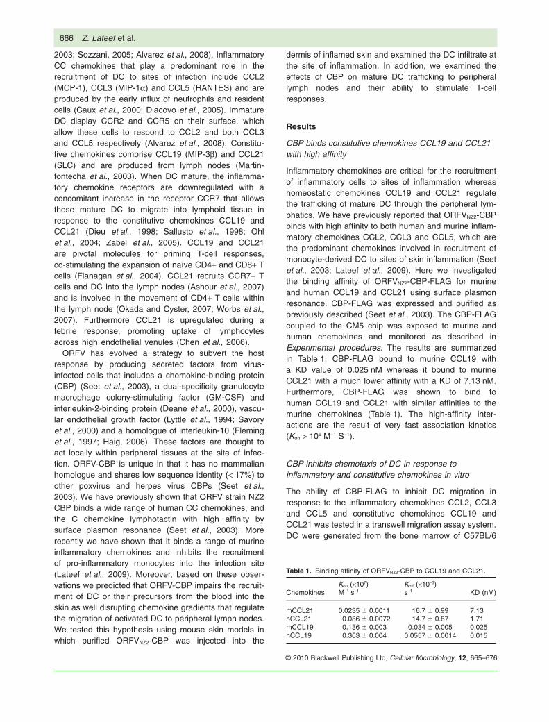
The ability of CBP to block migration of DC inresponse to CCL2, CCL3 and CCL5 was then tested.Various amounts of CBP-FLAG were added to a con-stant amount of chemokine in the bottom chamber ofa transwell assay and 105 DC in a volume of 100 mlwere added to the upper chamber. Following a 90 minincubation, cells collected in the bottom chamberwere counted using flow cytometry. The CBP-FLAG-reduced chemokine induced migration of DC in a dose-dependent manner (Fig. 1A). The CBP-FLAG waseffective at reducing the migration of DC in responseto all chemokines tested, even at a molar ratio ofchemokine:CBP-FLAG of 1:0.5 for CCL2 and CCL5(P < 0.05, ANOVA Tukey’s test) and at molar ratio of 1:1for CCL3 (P < 0.05).
Fig. 1. CBP-FLAG inhibits migration of immature DC in response to inflammatory chemokines and mature DC in response to constitutivechemokines in vitro. Chemotaxis assays were performed with a transwell assay system. The inflammatory chemokines CCL2, CCL3 andCCL5 (A) were used at a concentration of 25 ng ml-1 and the constitutive chemokines CCL19 and CCL21 (B) were used at a concentration of12.5 ng ml-1. CBP-FLAG was added to give the molar ratios shown (CCL:CBP). Fold increase represents the migration of DC above the cellsonly control. The data are shown as mean � SD of three combined experiments, in which each assay was performed in duplicate. Resultssignificantly different to CCL only (P < 0.05; ANOVA, Tukey’s test) are indicated by asterisks.
Orf virus-encoded chemokine-binding protein 667
© 2010 Blackwell Publishing Ltd, Cellular Microbiology, 12, 665–676
The ability of CBP to block the migration of mature DCin response to CCL19 and CCL21 was then tested. DCwere incubated overnight in the presence of CpG andthe degree of expression of activation markers on thecell surface analysed flow cytometrically to confirmmaturity. CpG-treated DC showed increased expressionof MHC-II, CD80 and CD86 (Fig. S1). Importantly thesecells showed upregulation of CCR7 and displayed fewerCCR5 markers, compared with cells not exposed toCpG. Mature DC did not respond to CCL3 nor CCL5(the ligands for CCR5). The optimal concentration ofCCL19 and CCL21 to ensure maximal migration ofmature DC in the transwell assay was determined to beapproximately 12.5 ng ml-1 (Fig. S2B). CBP-FLAG inhib-ited migration in response to both constitutive chemo-kines in a dose-dependant manner but higher levels ofCBP-FLAG were required to produce complete inhibitionusing CCL21 (molar ratio CCL21:CBP-FLAG 1:32) com-pared with CCL19 (molar ratio CCL19:CBP-FLAG 1:2)(Fig. 1B).
CBP inhibits the recruitment of DC to inflamed skin
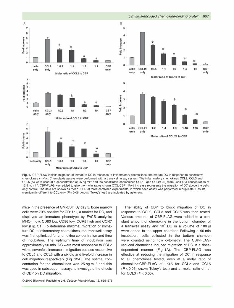
Having established that CBP inhibited cell movement inresponse to inflammatory chemokines in vitro, we nextinvestigated whether CBP could impair the recruitmentof DC to inflamed skin using a murine model. The injec-tion of small amounts of LPS into the dermis has beenshown to result in highly localized inflammation and apredominant increase in the inflammatory chemokinesCCL2, CCL3 and CCL5 (Kopydlowski et al., 1999). Inpreliminary experiments we determined the optimallevels of LPS to inject into the dermal layer of theabdominal region of C57BL/6 mice and the time at whichmaximal inflammatory cell recruitment was evident(Fig. S3). Animals were then co-injected with 1 mg ofLPS with or without various amounts of CBP-FLAG and24 h later cells were isolated from excised skin, stainedand quantified as described in Experimental procedures.Figure 2A shows that 100 ng and 1 ng of CBP-FLAGsignificantly inhibited recruitment of CD11c+/MHC-II+ DC(P < 0.01; paired Student’s t-test) whereas 0.01 ng ofCBP had no affect on recruitment.
We then tested whether CBP could impair recruitmentof blood-derived precursor DC to the skin. Bone marrowcells from eGFP transgenic C57BL/6 donor mice wereadministered to C57BL/6 mice and 24 h later LPS withvarious concentrations of CBP-FLAG were injected intothe dermis of the abdominal region of the recipients. A daylater mice were euthanized and skin around the intrader-mal injection site excised and cells isolated and analysedflow cytometrically. The results show that CBP-FLAGsignificantly inhibited the recruitment of blood-derivedeGFP-DC at 1 ng and 100 ng of CBP-FLAG (P < 0.01,
Student’s t-test for paired samples) with the most potentinhibition seen at 100 ng of CBP-FLAG (Fig. 2B). Thesedata suggest that CBP can block chemokines in vivoimpairing their capacity to attract DC from the blood to thesite of inflammation.
CBP inhibits the migration of activated DC from the skinto draining lymph nodes
When DC mature, they downregulate their responsive-ness to inflammatory chemokines and traffic to the drain-ing lymph node by upregulating the receptor CCR7 that
Fig. 2. CBP-FLAG inhibits LPS-induced recruitment ofCD11c+/MHC-II+ DC into the skin.A. Mice received intradermal injections of LPS (1 mg per site) in theabdomen with or without CBP-FLAG. In addition, each mousereceived injections of CBP-FLAG only and PBS, i.e. a total of fourinjections per mouse (n = 3 animals per group). Twenty-four hourslater mice were euthanized and the skin around the injection siteswas excised, weighed and digested to form single-cell suspensions.In addition, cells were prepared in the same manner from untreatedskin. Cells were stained for CD11c and MHC-II and counted by flowcytometry.B. Bone marrow cells from eGFP donor mice were transfusedintravenously into recipient mice 24 h before intradermal injection ofLPS (1 mg per site) with and without CBP-FLAG as describedabove and 24 h later the recruitment of eGFP+/MHC II+/CD11c+cells into the skin sites were analysed.In both (A) and (B) each bar represents the mean � SD from threecombined experiments. Asterisks indicate results that aresignificantly different (P < 0.01; ANOVA, paired Student’s t-test).
668 Z. Lateef et al.
© 2010 Blackwell Publishing Ltd, Cellular Microbiology, 12, 665–676
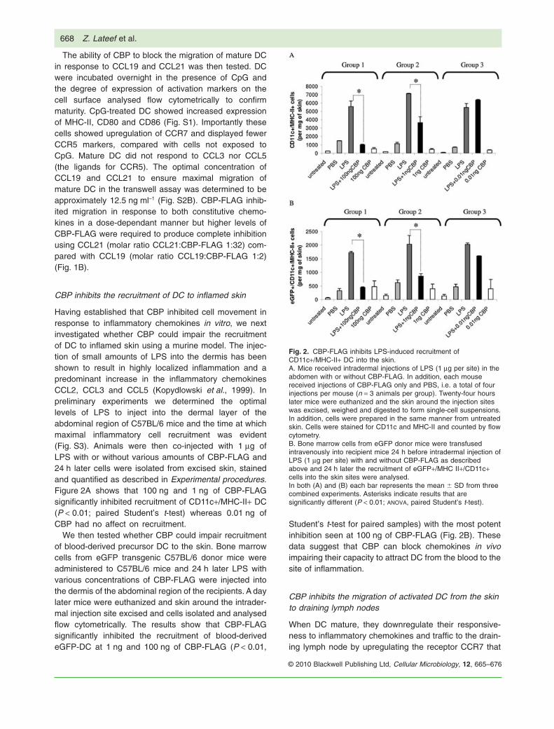
responds to the constitutive chemokines CCL19 andCCL21 (Dieu et al., 1998). The ability of CBP to blockCCL19 and CCL21 and thereby impair the migration ofDC from the skin to draining lymph nodes in vivo wastherefore examined. In preliminary experiments DC gen-erated from eGFP mice and activated with CpG wereinjected into the lower abdomen of mice. Within 24 h alarge proportion of these cells had migrated to the inguinaldraining lymph nodes (Fig. S4). We then investigatedwhether or not CBP could block this migration. Variousamounts of CBP-FLAG were injected with CpG-treatedDC into the lower abdomen. The results show that CBP-FLAG significantly inhibited DC migration at 100 ng(P < 0.01) with the most potent inhibition seen at 1 mg(Fig. 3). The data show that CBP is able to block consti-tutive as well as inflammatory chemokines, thereby
impeding the migration of DC to the draining node toinitiate an acquired immune response.
CBP blocks T-cell proliferation in the draininglymph node
Since CBP impairs the migration of DC to peripherallymph nodes, we wished to investigate whether or not thisresulted in reduced T-cell responses at the draining lymphnode. We used cells from transgenic OT-I and OT-II micefor these experiments.
OT-I mice have CD8+ T cells that respond specifically tothe ovalbumin peptide SIINFEKL whereas OT-II micehave cells that respond to ovalbumin (OVA). In preliminaryexperiments bone marrow-derived DC pulsed with OVA orSIINFEKL were shown to mature (Fig. S5). C57BL/6mice were adoptively transferred with CSFE-labelledCD8+ T cells from OT-I donor mice. DC pulsed with SIIN-FEKL were injected intradermally the next day. OT-I CD8T cells harvested from the inguinal nodes of individualmice that had been injected with DC pulsed with 5 mg ml-1
SIINFEKL induced the greatest proliferation (Fig. S6A), sothis concentration was used against increasing concen-trations of CBP. Co-injection of increasing doses of CBP-FLAG with the pre-pulsed DC resulted in fewer divisionsof CD8+ T cells within draining lymph nodes (Fig. 4A).One microgram of CBP inhibited T-cell proliferation bygreater than 80%, whereas 100 ng of CBP inhibited pro-liferation by approximately 50%.
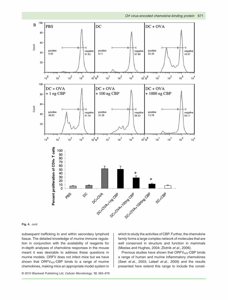
A similar experiment was also carried out with CD4cells isolated from OT-II transgenic mice that recognizeOVA protein (specifically ovalbumin segment 323–339)in the context of MHC-II. In this case the optimal con-centration of OVA with which to pulse DC was deter-mined to be 100 mg (Fig. S6B). Again the co-injection ofincreasing amounts of CBP-FLAG with OVA-treated DCinhibited the proliferation of CD4+ T cells in a dose-dependent manner similar to that described above forCD8+ T cells (Fig. 4B). This shows that CBP introducedinto the skin can inhibit antigen-specific T-cell activationwithin the draining lymph node. This is likely to be attri-butable to impaired migration of antigen-pulsed DCrequired to prime naïve T cells.
Discussion
ORFV has evolved to replicate exclusively within kerati-nocytes of the skin and it encodes a number of virulencefactors that interact with immune cells and cytokineswithin this localized environment that allow it to do this. Inthis study we asked whether the ORFV-CBP has a role inimpairing the development of adaptive immunity bytargeting the chemokine gradients that are critical forthe recruitment of DC to skin during inflammation and
Fig. 3. CBP-FLAG inhibits the migration of mature DC to thedraining lymph nodes. Bone marrow-derived DC from eGFP micewere matured with CpG and co-injected intradermally with variousconcentrations of CBP-FLAG. Each mouse received two injections,one on either side of the abdomen (n = 3 animals per group).Twenty-four hours later the draining inguinal lymph nodes wereharvested and single-cell suspensions stained for CD11c andanalysed flow cytometrically. (A) Total CD11c+ cells; (B)eGFP+/CD11C+ cells in lymph nodes. Each bar represents meanand standard deviation of three combined experiments. Asterisksindicate results that are significantly different (P < 0.05; ANOVA,Student’s unpaired t-test) when compared with the DC + CpGcontrol.
Orf virus-encoded chemokine-binding protein 669
© 2010 Blackwell Publishing Ltd, Cellular Microbiology, 12, 665–676
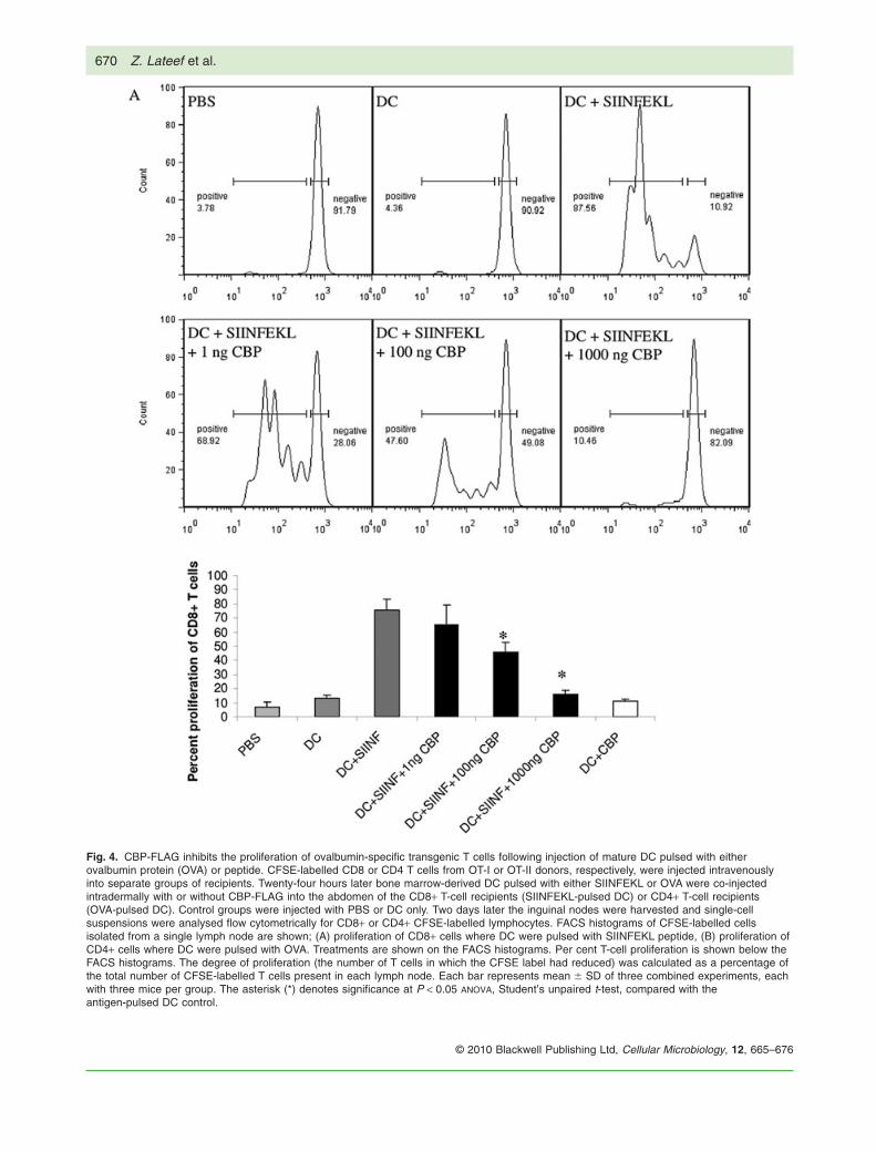
Fig. 4. CBP-FLAG inhibits the proliferation of ovalbumin-specific transgenic T cells following injection of mature DC pulsed with eitherovalbumin protein (OVA) or peptide. CFSE-labelled CD8 or CD4 T cells from OT-I or OT-II donors, respectively, were injected intravenouslyinto separate groups of recipients. Twenty-four hours later bone marrow-derived DC pulsed with either SIINFEKL or OVA were co-injectedintradermally with or without CBP-FLAG into the abdomen of the CD8+ T-cell recipients (SIINFEKL-pulsed DC) or CD4+ T-cell recipients(OVA-pulsed DC). Control groups were injected with PBS or DC only. Two days later the inguinal nodes were harvested and single-cellsuspensions were analysed flow cytometrically for CD8+ or CD4+ CFSE-labelled lymphocytes. FACS histograms of CFSE-labelled cellsisolated from a single lymph node are shown; (A) proliferation of CD8+ cells where DC were pulsed with SIINFEKL peptide, (B) proliferation ofCD4+ cells where DC were pulsed with OVA. Treatments are shown on the FACS histograms. Per cent T-cell proliferation is shown below theFACS histograms. The degree of proliferation (the number of T cells in which the CFSE label had reduced) was calculated as a percentage ofthe total number of CFSE-labelled T cells present in each lymph node. Each bar represents mean � SD of three combined experiments, eachwith three mice per group. The asterisk (*) denotes significance at P < 0.05 ANOVA, Student’s unpaired t-test, compared with theantigen-pulsed DC control.
670 Z. Lateef et al.
© 2010 Blackwell Publishing Ltd, Cellular Microbiology, 12, 665–676
subsequent trafficking to and within secondary lymphoidtissue. The detailed knowledge of murine immune regula-tion in conjunction with the availability of reagents forin-depth analyses of chemokine responses in the mousemeant it was desirable to address these questions inmurine models. ORFV does not infect mice but we haveshown that ORFVNZ2-CBP binds to a range of murinechemokines, making mice an appropriate model system in
which to study the activities of CBP. Further, the chemokinefamily forms a large complex network of molecules that arewell conserved in structure and function in mammals(Mestas and Hughes, 2004; Zlotnik et al., 2006).
Previous studies have shown that ORFVNZ2-CBP bindsa range of human and murine inflammatory chemokines(Seet et al., 2003; Lateef et al., 2009) and the resultspresented here extend this range to include the consti-
Fig. 4. cont.
Orf virus-encoded chemokine-binding protein 671
© 2010 Blackwell Publishing Ltd, Cellular Microbiology, 12, 665–676
tutive chemokines CCL19 and CCL21 for murine andhuman species. The binding affinity of CBP to CCL19was several orders of magnitude higher than its bindingaffinity to CCL21. Chemotaxis assays demonstratedthat low levels of CBP could effectively impair murinechemokine-induced migration of both immature andmature DC. The binding affinity of the CBP for bothinflammatory and constitutive chemokines was generallyreflected in their ability to block migration of DC inresponse to these molecules in transwell assays. Amolar ratio of CBP:chemokine of approximately 4:1 inthe case of CCL2, CCL3, CCL5 and CCL19 was suffi-cient to inhibit approximately all DC migration indicatingthe potential of this factor to disable chemokine gradi-ents that are critical for DC trafficking in vivo. In contrast,we observed that relatively high levels of CBP wererequired to block mature DC migration in response toCCL21 (32:1) and these levels correlated with itsreduced affinity for this chemokine.
The murine models clearly showed that small amountsof CBP (100 ng) were sufficient to inhibit DC recruitmentto inflamed skin, but higher levels were required to impairDC migration to the inguinal lymph node. We have esti-mated that CBP is produced from ORFV-infected cells inculture in the order of 6–120 ng ml-1 over 2–4 days (ourunpublished data). Such levels of CBP are sufficient toeffectively block inflammatory cell recruitment in periph-eral tissues based on our findings in this study and pre-vious studies (Lateef et al., 2009). However, the levels ofCBP required to block DC migration to lymph nodes andT-cell proliferation in the mouse models were substantiallygreater than the predicted physiological levels in virus-infected tissue. An explanation for the high levels of CBPin this case could involve the reduced ability of CBP todisable the chemokine gradients involved in trafficking ofDC to the lymph nodes and to regions within the lymphnode in which DC/T cell interactions take place. CCL19 isproduced exclusively within the lymph node and directsDC to T-cell zones, whereas CCL21 is produced from theafferent lymphatic ducts and T-cell zones within the lymphnode (Martin-fontecha et al., 2003; Alvarez et al., 2008).In addition to CCL21, other chemokines that are involvedin DC migration from peripheral tissues to the lymph nodeinclude CCL1 and CXCL12. These factors are required forthe migration of DC to the afferent lymphatic endotheliumand then a range of factors are required for DC to traversethe endothelium (Alvarez et al., 2008). In this study we didnot determine whether CBP binds CCL1 or CXCL12.CCL21 is essential for the migration of DC to the lymphnode (Ohl et al., 2004) and interactions with this chemo-kine alone would block DC trafficking within the lymphaticendothelium duct. The relatively reduced binding affinityof CBP for CCL21 compared with CCL19 and the inflam-matory chemokines may explain the higher levels of CBP
required to block DC trafficking in lymphatic tissue whichprevented them from reaching the lymph node. Similarly,in transwell assays we observed that it required almost10-fold more CBP to inhibit migration of DC in response toCCL21 compared with CCL19 and the inflammatorychemokines.
Although CBP shows high binding affinity for CCL19 andCCL21 the question remains whether it is able to enter theparenchyma or high endothelial venules of the lymph nodewhere the CCL19 and CCL21 gradients exist. Low-molecular-weight molecules and chemokines have beenshown to enter high endothelial venules from the subcap-sular sinus but were excluded from the cortical lymphocytemicroenvironments (Gretz et al., 2000). The above obser-vations suggest that CBP could potentially disrupt CCL19and CCL21 gradients within the lymph node. In this studywe did not investigate whether CBP was present in thelymph node; however, the fact that eGFP-DC were blockedfrom entering the lymph node suggests that disruption ofCCL19 gradients may be secondary to disruption of CCL21gradients in the afferent lymphatics. In addition, the infiltra-tion of CBP to various compartments within the lymph nodealso suggests that CBP could potentially block the move-ment of blood-derived T cells and B cells that constantlydiapedese across high endothelial venules.
Immune mechanisms exist in which T-cell priming is notdependant on the migration of antigen-loaded DC to thelymph node. Soluble free antigen can enter lymph and isprocessed by lymph node resident DC that initiate earlyT-cell priming (Kissenpfennig et al., 2005; Alvarez et al.,2008) thus providing a mechanism by which the hostimmune system could potentially bypass the effectsof ORFV-CBP. However, antigen-bearing skin-derivedDC are still required for inducing full-fledged effectorresponses (Itano et al., 2003). In our model it is possiblethat resident DC were also involved in T-cell responses ifin fact free antigen (antigen released from ex vivo loadedDC) entered the afferent lymphatics. The finding that CBPat the highest concentration reduced CD8+ and CD4+proliferation to levels that were only slightly higher thanthe controls (Fig. 4) suggests that resident DC played aminimal role in T-cell responsiveness.
Although we showed that CBP blocked T-cell activationand proliferation, the most likely interpretation of our datais that CBP acts by disrupting the CCL21 gradient withinthe afferent lymphatic ducts since mature eGFP-DCinjected into the skin were prevented from entering thelymph node. This model is supported by the observationthat anti-CCL21 antibody has been shown to block DCtrafficking within the afferent lymphatics (Saeki et al.,1999). ORFV-CBP has clearly evolved to bind a broadrange of inflammatory and constitutive chemokines,although it most likely binds the inflammatory chemokinesduring ORFV infection. It remains to be established
672 Z. Lateef et al.
© 2010 Blackwell Publishing Ltd, Cellular Microbiology, 12, 665–676
whether it disrupts CCL21 and CCL19 gradients in ovinespecies. The evolutionary adaptation of ORFV to its naturalhost suggests that ORFV-CBP will have a stronger bindingaffinity for ovine constitutive chemokines than to its murinecounterparts. This view is supported by the observationthat ORFV-CBP binds human CCL19 and CCL21 withhigher affinity than murine CCL19 and CCL21. CCL21 is atarget for many viruses that cause persistent infections.Simian and human immunodeficiency viruses (Choi et al.,2003), hepatitis C virus (Nattermann et al., 2006) andmurine Y-herpesvirus 68 (Jensen et al., 2003) haveevolved different mechanisms to block its activity. Thepattern emerging is that ORFV produces several factorsthat have the ability to target cytokines and cells that arecritical for the development of adaptive immunity.
The effects of ORFV-CBP on DC trafficking and T-cellresponses suggest that it has therapeutic potential fortreatment of inflammatory skin diseases. Chronic skininflammation is a process where DC are constantly sam-pling antigen in the skin and migrating to the lymph nodewhere they induce the activation and proliferation of Tcells. The T cells travel back to the skin where theyrelease cytokines that induce/maintain the inflammatorycondition. The finding that ORFV-CBP can impair both therecruitment of DC to sites of inflammation and the stimu-lation of T-cell responses by antigen-loaded DC suggeststhat CBP could break this cycle.
Experimental procedures
Mice
Specific Pathogen Free C57BL/6 mice, eGFP transgenicC57BL/6 mice, OT-I TCR transgenic mice (recognizing OVAsegment 257–264) and OT-II TCR transgenic mice (recognizingOVA segment 323–339) were obtained from the University ofOtago Animal Facility and used with institutional ethical approval.
Antibodies and recombinant proteins
The following rat anti-murine antibodies and their isotype controlswere obtained from BD Pharmingen: anti-CD86 (phycoerythrinconjugated, clone GL1, isotype IgG2a), anti-I-A/I-E (phycoeryth-rin conjugated, clone M5/114.15.2, isotype IgG2b), anti-CCR5(phycoerythrin conjugated, clone C34-3448, isotype IgG2c). Thehamster anti-mouse CD80 (phycoerythrin conjugated, clone16-10A1, isotype IgG2) and CD11c (allophycocyanin-conjugated,clone HL3, isotype IgG1) were also from BD Pharmingen. The ratanti-mouse CCR7 primary antibody (clone B4-12, isotype IgG2a)and the goat anti-rat secondary antibody (allophycocyanin-conjugated) were both from R&D Systems. The recombinantmurine chemokines GM-CSF, CCL2, CCL3, CCL5, CCL19 andCCL21 were obtained from R&D systems. The ORFV-CBP-FLAGwas purified by affinity chromatography using anti-FLAG beads(Sigma-Aldrich, MO, USA) from the supernatant of 293 EBNAcells transfected with pAPEX expressing ORFVNZ2-CBP-FLAG asdescribed previously (Seet et al., 2003).
Surface plasmon resonance
The surface of a CM-5 chip was immobilized with approximately300–400 response units (RU; pg mm-2) of ORFVNZ2-CBP-FLAGby standard amine coupling using a Biacore 2000 instrument. Allexperiments were performed at 25°C with HBS-EP (10 mMHepes, 150 mM NaCl, 3 mM EDTA, 0.005% polysorbate 20; pH7.4). The coupled CBP-FLAG was exposed to murine chemok-ines at a flow rate of 100 ml per minute, at concentrations rangingfrom 3 nM to 300 nM for the duration of 60 s. Chemokines wereallowed to dissociate for 200 s before the surface was regener-ated using a mixture of ionic and acidic solutions. The data wereanalysed using BIAevaluation software using the 1:1 protocol forbinding with mass transfer (Lateef et al., 2009).
Generation of DC
Bone marrow-derived DC were cultured from C57BL/6 mice aspreviously described (Faulkner et al., 2000). Briefly, the tibias andfemurs of mice were removed and the ends trimmed to exposebone marrow. The bone marrow cells were suspended in Dul-becco’s phosphate-buffered saline (DPBS) with 5% fetal calfserum (FCS; Gibco), and RBC-depleted using pre-warmedammonium chloride. The bone marrow cells (2 ¥ 106 cells ml-1)were cultured in DMEM (Sigma-Aldrich, MO, USA) supplementedwith arginine, aspartate, folic acid, L-glutamine, 0.1 mg ml-1
penicillin-streptomycin (Roche Diagnostics, IN, USA), 5% FCSand 20 ng ml-1 GM-CSF. Cells were incubated at 37°C with 10%CO2. On day 3, cells were fed by replacing 3 ml of the superna-tant with fresh medium. Immature DC were harvested on day 5.To generate mature DC, these cells were pulsed with 5 mg ml-1
CpG overnight and washed three times in PBS before use. Thephenotype of both immature and mature cells was analysedusing flow cytometric analysis.
Transwell migration assays
Chemotaxis assays were performed with 12-well transwells con-taining 5 mm membrane inserts (Costar). Chemokines in 600 ml ofDMEM containing 5% FCS with or without CBP-FLAG wereadded to the bottom chamber of the transwells and 105 DC in100 ml of DMEM and 5% FCS were added to the top chamber.The assay was incubated for 90 min, after which time the cellsthat had collected in the bottom chamber were counted usingflow cytometry.
Flow cytometric analysis
This was performed using a FACSCalibur (Becton Dickinson). Allflow cytometric data were analysed using CellQuest and FlowJosoftwares.
Murine skin inflammation model
C57BL/6 mice received intradermal injections (20 ml total volume)of LPS (Escherichia coli derived, Sigma) in the abdominal region.Evans blue dye (2 ml of 1% solution) was mixed in each sampleto visualize the rate of diffusion of injected material. A day later,animals were euthanized and the skin around the intradermal
Orf virus-encoded chemokine-binding protein 673
© 2010 Blackwell Publishing Ltd, Cellular Microbiology, 12, 665–676
injection site was excised and weighed prior to incubating incollagenase dispase (Roche Diagnostics) for 3 h. The isolatedcells were counted using the haemocytometer and stained forcell surface expression of CD11c/MHC-II, with the respectiveisotype controls. DC recruitment from the blood was alsoassessed using adoptive transfer of eGFP bone marrow trans-genic cells into sex-matched C57BL/6 recipients. Bone marrowwas collected from donor mice as described above, suspended inDPBS (without FCS) and 3 ¥ 107 cells were administered to eachrecipient in a 200 ml volume via the tail vein. Twenty-four hourspost adoptive transfer of the eGFP cells inflammation wasinduced in the skin with LPS as described above. All sampleswere analysed using flow cytometry counts of 10 000 events, andwere normalized to haemocytometer counts and weight of indi-vidual skin samples.
DC migration from skin to lymph nodes
Dendritic cells cultured from eGFP mice were matured overnightwith 5 mg of CpG and washed and re-suspended in PBS. Cells ata concentration of 105 were injected with or without CBP into thelower abdomen of C57BL/6 mice. Twenty-four hours later, thedraining inguinal lymph node was extracted and collagenasedigested. The total cells from each lymph node were determinedusing haemocytometer counts. Cells were also stained for CD11cmarker and analysed using flow cytometry. For each sample,200 000 events were counted and data were normalized tohaemocytometer counts for each lymph node.
T-cell proliferation in lymph nodes
CD8+ or CD4+T cells were isolated from the spleens and inguinallymph nodes of donor OT-1 or OT-II mice, respectively, enrichedimmunomagnetically using antibody-conjugated beads (Milltenyi)with an AUTOMacs System and labelled with carboxyfluorosceinsuccinomide ester (CFSE) before being injected intravenously(200 ml at a concentration of 2.5 ¥ 107 cells ml-1) into sex-matched C57BL/6 mice. A day later, each mouse adoptivelytransferred with OT-I CD8 cells received an intradermal injectionof DC previously pulsed with SIINFEKL peptide for 3 h, whilethose adoptively transferred with OT-II CD4 cells received DCpreviously pulsed with OVA protein overnight. Pulsed DC werewashed in PBS prior to injection in 20 ml of PBS in the skin of thelower abdomen. Some groups of mice also received CBP thatwas co-injected with the pulsed DC. Inguinal lymph nodes wereharvested 48 h post injection and CFSE-labelled T cells analysedusing flow cytometry. For each sample, dot plots were gated forCFSE-positive cells under the FL1 channel. Live gating was usedto acquire 1 000 000 counts of only the CFSE-positive cells.
Statistical analyses
All statistical analyses were performed using the MegaStat suiteunder Excel. The variance between multiple samples was calcu-lated using ANOVA. For comparisons between two samples, eitherthe Student’s paired or unpaired t-tests were used, with P-valuesof 0.05 and 0.01 considered significant. All experiments wereperformed three times and individual data were combined(exceptions are stated in the figure legends).
Acknowledgements
This project was supported by funding from the Health ResearchCouncil of New Zealand. Z.L. was supported by a Careers Devel-opment Award, Health Research Council of New Zealand. Wethank Catherine McCaughan, Michelle Wilson and Nichola Realfrom the Department of Microbiology and Immunology, Universityof Otago for expert technical assistance and Fiona Clow (Depart-ment of Molecular Medicine and Pathology, University of Auck-land) for assistance with Biacore analysis. The OT mice were agift from Sarah Hook, School of Pharmacy, University of Otago.
References
Alvarez, D., Vollmann, E.H., and von Andrian, U.H. (2008)Mechanisms and consequences of dendritic cell migration.Immunity 29: 325–342.
Ashour, A.E., Turnquist, H.R., Singh, R.K., Talmadge, J.E.,and Solheim, J.C. (2007) CCL21-induced immune cell infil-tration. Int Immunopharmacol 7: 272–276.
Buddle, B.M., and Pulford, H.D. (1984) Effect of passively-acquired antibodies and vaccination of the immuneresponse to contagious ecthyma virus. Vet Microbiol 9:515–522.
Caux, C., Ait-Yahia, S., Chemin, K., de Bouteiller, O., Dieu-Nosjean, M.C., Homey, B., et al. (2000) Dendritic cellbiology and regulation of dendritic cell trafficking bychemokines. Springer Semin Immunopathol 22: 345–369.
Chen, Q., Fisher, D.T., Clancy, K.A., Gauguet, J.M., Wang,W.C., Unger, E., et al. (2006) Fever-range thermal stresspromotes lymphocyte trafficking across high endothelialvenules via an interleukin 6 trans-signaling mechanism.Nat Immunol 7: 1299–1308.
Choi, Y.K., Fallert, B.A., Murphey-Corb, M.A., and Reinhart,T.A. (2003) Simian immunodeficiency virus dramaticallyalters expression of homeostatic chemokines and dendriticcell markers during infection in vivo. Blood 101: 1684–1691.
Deane, D., McInnes, C.J., Percival, A., Wood, A., Thomson,J., Lear, A., et al. (2000) Orf virus encodes a novelsecreted protein inhibitor of granulocyte-macrophagecolony-stimulating factor and interleukin-2. J Virol 74:1313–1320.
Diacovo, T.G., Blasius, A.L., Mak, T.W., Cella, M., andColonna, M. (2005) Adhesive mechanisms governinginterferon-producing cell recruitment into lymph nodes.J Exp Med 202: 687–696.
Dieu, M.C., Vanbervliet, B., Vicari, A., Bridon, J.M., Oldham,E., Ait-Yahia, S., et al. (1998) Selective recruitment ofimmature and mature dendritic cells by distinct chemokinesexpressed in different anatomic sites. J Exp Med 188:373–386.
Faulkner, L., Buchan, G., and Baird, M. (2000) Interleukin-10does not affect phagocytosis of particulate antigen by bonemarrow-derived dendritic cells but does impair antigen pre-sentation. Immunology 99: 523–531.
Fauquet, C.M., Mayo, M.A., Maniloff, U., Desselberger, L.A.,and Ball, L.A. (2005) Virus Taxonomy. Eighth Report of theInternational Committee on the Taxonomy of Viruses.London: Acadmic Press, Elservier.
Flanagan, K., Moroziewicz, D., Kwak, H., Horig, H., andKaufman, H.L. (2004) The lymphoid chemokine CCL21
674 Z. Lateef et al.
© 2010 Blackwell Publishing Ltd, Cellular Microbiology, 12, 665–676
costimulates naive T cell expansion and Th1 polarization ofnon-regulatory CD4+ T cells. Cell Immunol 231: 75–84.
Fleming, S.B., McCaughan, C.A., Andrews, A.E., Nash, A.D.,and Mercer, A.A. (1997) A homologue of interleukin-10 isencoded by the poxvirus orf virus. J Virol 71: 4857–4861.
Fleming, S.B., and Mercer, A.A. (2007) Genus Parapoxvirus.In Poxviruses. Mercer, A.A., Schmidt, A., and Weber, O.(eds). Basel: Birkhaeuser Verlag, pp. 127–165.
Gretz, J.E., Norbury, C.C., Anderson, A.O., Proudfoot, A.E.,and Shaw, S. (2000) Lymph-borne chemokines and otherlow molecular weight molecules reach high endothelialvenules via specialized conduits while a functional barrierlimits access to the lymphocyte microenvironments inlymph node cortex. J Exp Med 192: 1425–1440.
Haig, D.M. (2006) Orf virus infection and host immunity. CurrOpin Infect Dis 19: 127–131.
Haig, D.M., and McInnes, C.J. (2002) Immunity and counter-immunity during infection with the parapoxvirus orf virus.Virus Res 88: 3–16.
Haig, D.M., and Mercer, A.A. (1998) Ovine diseases. Orf. VetRes 29: 311–326.
Haig, D.M., Thomson, J., McInnes, C.J., Deane, D.L., Ander-son, I.E., McCaughan, C.A., et al. (2002) A comparison ofthe anti-inflammatory and immunostimulatory activities oforf virus and ovine interleukin-10. Virus Res 90: 303–316.
Itano, A.A., McSorley, S.J., Reinhardt, R.L., Ehst, B.D.,Ingulli, E., Rudensky, A.Y., and Jenkins, M.K. (2003) Dis-tinct dendritic cell populations sequentially present antigento CD4 T cells and stimulate different aspects of cell-mediated immunity. Immunity 19: 47–57.
Jensen, K.K., Chen, S.C., Hipkin, R.W., Wiekowski, M.T.,Schwarz, M.A., Chou, C.C., et al. (2003) Disruption ofCCL21-induced chemotaxis in vitro and in vivo by M3, achemokine-binding protein encoded by murine gammaher-pesvirus 68. J Virol 77: 624–630.
Kissenpfennig, A., Henri, S., Dubois, B., Laplace-Builhe, C.,Perrin, P., Romani, N., et al. (2005) Dynamics and functionof Langerhans cells in vivo: dermal dendritic cells colonizelymph node areas distinct from slower migrating Langer-hans cells. Immunity 22: 643–654.
Kopydlowski, K.M., Salkowski, C.A., Cody, M.J., van Rooijen,N., Major, J., Hamilton, T.A., and Vogel, S.N. (1999) Regu-lation of macrophage chemokine expression by lipopoly-saccharide in vitro and in vivo. J Immunol 163: 1537–1544.
Lateef, Z., Baird, M.A., Wise, L.M., Mercer, A.A., andFleming, S.B. (2009) The orf virus encoded chemokinebinding protein is a potent inhibitor of inflammatory mono-cyte recruitment in a mouse skin model. J Gen Virol 90:1477–1482.
Lyttle, D.J., Fraser, K.M., Fleming, S.B., Mercer, A.A., andRobinson, A.J. (1994) Homologs of vascular endothelialgrowth factor are encoded by the poxvirus orf virus. J Virol68: 84–92.
MartIn-Fontecha, A., Sebastiani, S., Hopken, U.E., Uguc-cioni, M., Lipp, M., Lanzavecchia, A., and Sallusto, F.(2003) Regulation of dendritic cell migration to the draininglymph node: impact on T lymphocyte traffic and priming.J Exp Med 198: 615–621.
Mestas, J., and Hughes, C.C. (2004) Of mice and not men:differences between mouse and human immunology.J Immunol 172: 2731–2738.
Nattermann, J., Zimmermann, H., Iwan, A., von Lilienfeld-Toal, M., Leifeld, L., Nischalke, H.D., et al. (2006) HepatitisC virus E2 and CD81 interaction may be associated withaltered trafficking of dendritic cells in chronic hepatitis C.Hepatology 44: 945–954.
Ohl, L., Mohaupt, M., Czeloth, N., Hintzen, G., Kiafard, Z.,Zwirner, J., et al. (2004) CCR7 governs skin dendritic cellmigration under inflammatory and steady-state conditions.Immunity 21: 279–288.
Okada, T., and Cyster, J.G. (2007) CC chemokine receptor 7contributes to Gi-dependent T cell motility in the lymphnode. J Immunol 178: 2973–2978.
Saeki, H., Moore, A.M., Brown, M.J., and Hwang, S.T. (1999)Cutting edge: secondary lymphoid-tissue chemokine (SLC)and CC chemokine receptor 7 (CCR7) participate in theemigration pathway of mature dendritic cells from the skinto regional lymph nodes. J Immunol 162: 2472–2475.
Sallusto, F., Schaerli, P., Loetscher, P., Schaniel, C., Lenig,D., Mackay, C.R., et al. (1998) Rapid and coordinatedswitch in chemokine receptor expression during dendriticcell maturation. Eur J Immunol 28: 2760–2769.
Savage, J., and Black, M.M. (1972) ‘Giant orf’ of a finger in apatient with lymphoma. Proc R Soc Med 64: 766–768.
Savory, L.J., Stacker, S.A., Fleming, S.B., Niven, B.E., andMercer, A.A. (2000) Viral vascular endothelial growth factorplays a critical role in orf virus infection. J Virol 74: 10699–10706.
Seet, B.T., McCaughan, C.A., Handel, T.M., Mercer, A., Bru-netti, C., McFadden, G., and Fleming, S.B. (2003) Analysisof an orf virus chemokine-binding protein: shifting ligandspecificities among a family of poxvirus viroceptors. ProcNatl Acad Sci USA 100: 15137–15142.
Sozzani, S. (2005) Dendritic cell trafficking: more than justchemokines. Cytokine Growth Factor Rev 16: 581–592.
Steinman, R.M., and Banchereau, J. (2007) Taking dendriticcells into medicine. Nature 449: 419–426.
Tan, S.T., Blake, G.B., and Chambers, S. (1991) Recurrentorf in an immunocompromised host. Br J Plast Surg 44:465–467.
Worbs, T., Mempel, T.R., Bolter, J., von Andrian, U.H., andForster, R. (2007) CCR7 ligands stimulate the intranodalmotility of T lymphocytes in vivo. J Exp Med 204: 489–495.
Zabel, B.A., Silverio, A.M., and Butcher, E.C. (2005)Chemokine-like receptor 1 expression and chemerin-directed chemotaxis distinguish plasmacytoid from myeloiddendritic cells in human blood. J Immunol 174: 244–251.
Zlotnik, A., Yoshie, O., and Nomiyama, H. (2006) Thechemokine and chemokine receptor superfamilies andtheir molecular evolution. Genome Biol 7: 243.
Supporting information
Additional Supporting Information may be found in the onlineversion of this article:
Fig. S1. Flow cytometry analysis of immature and mature DCphenotype. Bone marrow-derived DC (day 6) with or withoutexposure to CpG were phenotyped flow cytometrically usingantibodies to cell surface molecules. Dot plot showing percent-age of cells in the culture that are CD11c-positive. Histogramsshow the expression of MHC-II, CD86, CD80, CCR5, and CCR7
Orf virus-encoded chemokine-binding protein 675
© 2010 Blackwell Publishing Ltd, Cellular Microbiology, 12, 665–676
on CD11c+ gated cells. The percentages of positive cells areshown for immature (DC, blue line) and mature (DC + CpG, redline) in each histogram.Fig. S2. DC migrate in response to chemokines in vitro. (A)Various concentrations of the inflammatory chemokines CCL2,CCL3 and CCL5 or (B) constitutive chemokines CCL19, CCL21were placed in the lower chambers of a transwell plate, andimmature (A) or mature bone marrow-derived DC (B) were addedto the upper chambers and the assay incubated for 90 min. Thenumber of DC that had migrated to the lower chamber wasanalysed flow cytometrically and expressed as the fold increaseover the number of DC migrating to medium only. Each point onthe graph represents the mean � SD of three repeats, eachperformed in duplicate.Fig. S3. DC recruitment in murine skin in response to LPS. DCrecruitment was induced in abdominal skin by intradermal injec-tion of LPS (volume 20 ml) or PBS (control). Mice were eutha-nized post LPS treatment and the skin at the site of injectionexcised. Cells were isolated by digesting skin with collagenaseand dispase, stained for CD11c+ and analysed by flow cytometry(10 000 events were counted per sample).A. Recruitment of DC as a function of time in response to 1 mg ofLPS.B. Dose–response of DC recruitment by LPS at 24 h post injec-tion. Each bar represents mean � SD of samples from two miceand is representative of three separate experiments.Fig. S4. Mature DC migrate to the lymph node in vivo. Bonemarrow-derived DC from eGFP donors with or without exposureto the maturation signal, CpG, were administered intradermallyand the draining inguinal lymph nodes were harvested at 24, 48or 72 h post injection. Lymph node single-cell suspensions were
analysed flow cytometrically for total CD11c+ (A) or eGFP+/CD11c+ (B). Each bar represents the mean � SD of three miceand is representative of three separate experiments.Fig. S5. Bone marrow-derived DC pulsed with OVA or SIINFEKLundergo maturation. DC were pulsed with SIINFEKL (A) or OVAfor 24 h (B) and their phenotype was compared with that ofunpulsed cells using flow cytometric analysis. Cells were stainedwith antibodies to MHC-II, CD80, CD86, CCR5 and CCR7. Eachbar represents the mean fluorescence intensity of one sample,and is representative of five experiments for SIINFEKL-pulsedDC and five experiments for OVA-pulsed DC.Fig. S6. Adoptively transferred, antigen-pulsed DC induceantigen-specific T-cell proliferation in vivo. OTI (A) or OTII (B)TCR transgenic T cells labelled with CFSE were adoptively trans-ferred into recipients intravenously and 24 h later mature bonemarrow-derived DC, pulsed with the indicated concentrations ofSIINFEKL (A) or OVA (B), were injected intravenously. Forty-eighthours later lymph nodes were harvested and a single-cell sus-pension was analysed flow cytometrically for proliferation ofCFSE-labelled T lymphocytes. The dot plots and histograms arefrom individual lymph nodes and indicate comparative cycles ofcell division as a function of the concentration of SIINFEKL orOVA. The histograms are representative of samples collectedfrom three mice per group, and each experiment was repeatedtwice.
Please note: Wiley-Blackwell are not responsible for the contentor functionality of any supporting materials supplied by theauthors. Any queries (other than missing material) should bedirected to the corresponding author for the article.
676 Z. Lateef et al.
© 2010 Blackwell Publishing Ltd, Cellular Microbiology, 12, 665–676
Related Documents