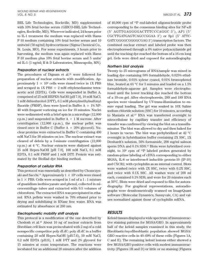
Chemokine and chemokine receptor expression in keloid and normal fibroblasts CHATANYA S. NIRODI, PhD a,b ; RADIKA DEVALARAJA, PhD a,b ; LILLIAN B. NANNEY, PhD a,b,c ; SAUNDRETT ARRINDELL, MD d ; SHIRLEY RUSSELL, PhD e ; JOEL TRUPIN, PhD e ; ANN RICHMOND, PhD a,b Keloids are benign collagenous tumors that occur during dermal wound healing in genetically predisposed individuals. The lesions are characterized by over-proliferation of fibroblasts, some leukocyte infiltration, and prolonged high rates of collagen synthesis. To determine whether leukocyte chemoattractants or chemokines are participating in this disease process, immunohistochemical staining for the CXC chemokine, MGSA/GRO, and its receptor, CXCR2, was performed on tissue from keloids, hypertrophic scars and normal skin. Immunoreactive MGSA/GRO was not observed in hypertro- phic scars or normal dermis, but was present in some myofibroblasts and lymphocytes in nodular areas of the keloid samples. This staining positively correlated with the degree of inflammatory infiltrate in the lesions. Keloids, but not hypertrophic scars or normal dermis, also exhibited intensive immunoreactivity for the CXCR2 receptor in endothelial cells and inflammatory infiltrates with occasional staining of myofibroblasts. In contrast, cultured fibroblasts from either keloids or normal skin did not express detectable amounts of mRNA for MGSA/GRO or CXCR2, although interleukin- 1 strongly induced MGSA/GRO mRNA in both cell types. Interleukin-1 induction of MGSA/GRO was inhibited by glucocorticoid in normal and keloid fibroblasts, and the effect was more pronounced in keloid fibroblasts. This event was not correlated with inhibition of nuclear activation of NF-B, AP-1 or Sp1, and might therefore be mediated by another mechanism such as decreased mRNA stability or transcriptional repression through the glucocorticoid response element in the MGSA/GRO promoter. Data from in vitro wounding experiments with cultured normal and keloid fibroblasts indicate that there were no significant differences in MGSA/GRO or CXCR2 receptor levels between normal and keloid fibroblasts. We also show that cultured keloid fibroblasts exhibit a delayed wound healing response. We postulate that the inflammatory component is important in development of keloid lesions and chemotactic cytokines may participate in this process. (WOUND REP REG 2000;8:371–382) Keloids are benign collagenous tumors that form in the DTT Dithiothreitol reticular layer of the dermis during a prolonged wound EDTA Ethylenediamine tetraacetic acid healing process in persons with a genetic predisposi- EMSA Electrophonetic mobility shift assay GRO Growth-regulated protein tion. 1,2 Keloids occur predominantly in Black and Asian IL Interleukin populations. The altered tissue repair mechanism ap- IP-10 Interferon- inducible protein-10 MGSA Melanoma growth stimulatory activity PBS Phosphate buffered saline solution From the Department of Veterans Affairs a , Departments SSC Saline sodium chloride solution of Cell Biology b and Plastic Surgery c , Vanderbilt University School of Medicine and Departments of Medicine d and Microbiology e , Meharry Medi- cal College, Nashville, Tennessee. Reprint requests: Ann Richmond, PhD, Department of Cell pears to be restricted to dermal wound healing, because Biology, MCN T2212, Vanderbilt University School other growth or connective tissue abnormalities are not of Medicine, Nashville, TN 37232. Fax: (615) 343- frequently reported in keloid patients. The disorder may 4539; Email: [email protected]. be genetically heterogeneous, with both dominant and edu. recessive modes of inheritance having been reported. Copyright 2000 by the Wound Healing Society. ISSN: 1067-1927 $15.00 0 Genetically susceptible individuals form keloids after 371

Welcome message from author
This document is posted to help you gain knowledge. Please leave a comment to let me know what you think about it! Share it to your friends and learn new things together.
Transcript
Chemokine and chemokine receptor expression in keloidand normal fibroblasts
CHATANYA S. NIRODI, PhDa,b; RADIKA DEVALARAJA, PhDa,b; LILLIAN B. NANNEY, PhDa,b,c;SAUNDRETT ARRINDELL, MDd; SHIRLEY RUSSELL, PhDe; JOEL TRUPIN, PhDe; ANN RICHMOND, PhDa,b
Keloids are benign collagenous tumors that occur during dermal wound healing in genetically predisposed individuals.The lesions are characterized by over-proliferation of fibroblasts, some leukocyte infiltration, and prolonged high ratesof collagen synthesis. To determinewhether leukocyte chemoattractants or chemokines are participating in this diseaseprocess, immunohistochemical staining for theCXCchemokine,MGSA/GRO�, and its receptor, CXCR2, was performedon tissue from keloids, hypertrophic scars and normal skin. Immunoreactive MGSA/GRO�was not observed in hypertro-phic scars or normal dermis, but was present in some myofibroblasts and lymphocytes in nodular areas of the keloidsamples. This staining positively correlated with the degree of inflammatory infiltrate in the lesions. Keloids, but nothypertrophic scars or normal dermis, also exhibited intensive immunoreactivity for the CXCR2 receptor in endothelialcells and inflammatory infiltrates with occasional staining of myofibroblasts. In contrast, cultured fibroblasts from eitherkeloids or normal skin did not express detectable amounts of mRNA for MGSA/GRO or CXCR2, although interleukin-1 strongly induced MGSA/GRO mRNA in both cell types. Interleukin-1 induction of MGSA/GRO was inhibited byglucocorticoid in normal and keloid fibroblasts, and the effect was more pronounced in keloid fibroblasts. This eventwas not correlated with inhibition of nuclear activation of NF-�B, AP-1 or Sp1, and might therefore be mediated byanothermechanism suchas decreasedmRNA stability or transcriptional repression through theglucocorticoid responseelement in the MGSA/GRO promoter. Data from in vitro wounding experiments with cultured normal and keloidfibroblasts indicate that there were no significant differences in MGSA/GRO or CXCR2 receptor levels between normaland keloid fibroblasts. We also show that cultured keloid fibroblasts exhibit a delayed wound healing response. Wepostulate that the inflammatory component is important in development of keloid lesions and chemotactic cytokinesmay participate in this process. (WOUND REP REG 2000;8:371–382)
Keloids are benign collagenous tumors that form in the DTT Dithiothreitolreticular layer of the dermis during a prolonged wound EDTA Ethylenediamine tetraacetic acidhealing process in persons with a genetic predisposi- EMSA Electrophonetic mobility shift assay
GRO� Growth-regulated proteintion.1,2 Keloids occur predominantly in Black and AsianIL Interleukinpopulations. The altered tissue repair mechanism ap-IP-10 Interferon-� inducible protein-10MGSA Melanoma growth stimulatory activityPBS Phosphate buffered saline solution
From the Department of Veterans Affairsa, Departments SSC Saline sodium chloride solutionof Cell Biologyb and Plastic Surgeryc, VanderbiltUniversity School of Medicine and Departmentsof Medicined and Microbiologye, Meharry Medi-cal College, Nashville, Tennessee.
Reprint requests: Ann Richmond, PhD, Department of Cell pears to be restricted to dermal wound healing, becauseBiology, MCN T2212, Vanderbilt University School other growth or connective tissue abnormalities are notof Medicine, Nashville, TN 37232. Fax: (615) 343- frequently reported in keloid patients. The disorder may4539; Email: [email protected].
be genetically heterogeneous, with both dominant andedu.recessive modes of inheritance having been reported.Copyright � 2000 by the Wound Healing Society.
ISSN: 1067-1927 $15.00 � 0 Genetically susceptible individuals form keloids after
371

WOUND REPAIR AND REGENERATIONSEPTEMBER–OCTOBER 2000372 NIRODI ET AL.
wounding. Abnormalities in cell migration, proliferation, scar revisions or abdominoplasty procedures. Tissuesamples were obtained in accordance with proceduresinflammation, synthesis and secretion of extracellular
matrix proteins and cytokines, and remodeling of the approved by the Institutional Review Board. None of thekeloids had received corticosteroid injection within awound matrix have all been described in keloids.3,4 Black
patients with keloids often exhibit increased activity of one-year period and all were removed from the truncalregion.fibrogenic cytokines5,6 as well as an altered cytokine pro-
file.7 The exaggerated wound healing process in keloidsappears to be due in part to loss of glucocorticoid sup- Tissue processing
Whenever possible, specimens were divided with a por-pression of collagen and elastin gene expression in cellsderived from these lesions.8,9 tion homogenized for RNA preparations and the other
portion fixed in 4% paraformaldehyde. After 18 hours ofBecause glucocorticoids also suppress the activationof NF-�B, decreased glucocorticoid suppression in ke- fixation, the tissues were embedded in paraffin, sec-
tioned, and sections used for immunohistochemicalloid lesions could potentially lead to enhanced NF-�Bdependent cytokine gene transcription10 and thus signifi- analyzes. Sections were deparaffinized, endogenous per-
oxidase activity was quenched for 20 minutes in a 3%cantly alter the wound healing profile within these le-sions. The chemotactic cytokines melanoma growth H2O2/methanol solution, and preincubated in normal 10%
porcine serum (Dako, Carpinteria, CA) for 20 minutes.stimulatory activity/growth-regulated protein (MGSA/GRO) and interleukin-8 (IL-8), are regulated in part by Sections were incubated overnight in a humidified cham-
ber at 4�C in rabbit antiserum (Dako, Carpinteria, CA) forNF-�B, in cooperation with AP-1, Sp1 or other transcrip-tion factors.11–15 Glucocorticoids have been shown to sup- MGSA/GRO� at a concentration of 2 mg/ml as previously
described.18 For CXCR2 immunostaining, samples under-press the expression of MGSA/GRO homologs in ratfibroblasts.16 Moreover, synovial fibroblasts cultured went antigen retrieval at neutral pH (Biogenix, San
Ramon, CA) by microwave irradiation before overnightfrom patients with rheumatoid arthritis, another fibro-proliferative disease, express receptors for MGSA/GRO.17 incubation with antibody against CXCR2 at a concentra-
tion of 660 ng/ml. For identification of myofibroblasts,We have shown that the expression of MGSA/GRO andits receptor is temporally increased during the wound sections were incubated in goat anti-human �-smooth
muscle actin antisera (C-11; Santa Cruz Technologies,healing process.18 Based upon these findings, we pro-posed that chemokine and chemokine receptor expres- Santa Cruz, CA). Negative controls included sections in-
cubated in identical concentrations of nonspecific rabbitsion might be exaggerated in keloid lesions.To test this hypothesis we examined the expression immunoglobulin (Dako, Carpinteria, CA) G or phosphate
buffered saline solution (PBS) substituted for the pri-of the chemokine, MGSA/GRO, and its receptor, CXCR2,in keloid lesions as compared to hypertrophic scars, and mary antisera. All sections were further reacted with the
reagents in a commercial avidin-biotin staining kit (Dako,normal skin, as well as the endogenous mRNA expres-sion of MGSA/GRO and its receptor, CXCR2, in cultured Carpinteria, CA). Immunoreactive sites were visualized
with 3,3-diaminobenzidine as the chromogen (Biogenex).fibroblasts from normal skin and keloid lesions. The ef-fects of glucocorticoids on the IL-1 activation of nuclear Sections were rinsed in water, counterstained with hema-
toxylin, dehydrated, and cover-slipped. Images were cap-NF-�B and AP-1 complexes in fibroblasts cultured fromkeloid lesions and normal skin using gel shift assay with tured with an Olympus AH Vanox light microscope
(Melville, NY) interfaced to a Kontorn Eliktronik 3008probes for NF-�B and AP-1 as compared to the noninduc-ible transactivator, Sp1, were determined. Lastly, the ex- camera (Eching, Germany). Images were arranged
through the use of Adobe Photoshop software.pression of CXCR2 and MGSA/GRO in cultured keloidand normal fibroblasts that have been subjected to invitro wounding and the in vitro wound closure rates Cell cultures
Tissue sources and methods of isolation, propagationfor these cultured keloid and normal fibroblasts wasassessed. and freezing of fibroblasts have been reported.19 Normal
fibroblast cell strains 21, 116, 130, 131 and 170 and keloidfibroblast cell-lines 33, 50, 124, 125 and 261 were used
MATERIALS AND METHODS in these studies. Cell-lines were routinely tested for My-coplasma infection by the Hoechst staining method andActively growing keloid tissues (N � 10), hypertrophic
scars from traumatic linear scars greater than 2 years of were found to be negative. Normal and keloid fibroblastcell strains, at passage numbers under 6, were grown toage (N � 10) and normal skin (N � 5) were collected
from patients undergoing elective excision of keloids, about 80% confluence in Hams F-10 medium (GIBCO-
WOUND REPAIR AND REGENERATIONVOL. 8, NO. 5 NIRODI ET AL. 373
BRL Life Technologies, Rockville, MD) supplemented of 40,000 cpm of 32P end-labeled oligonucleotide probecorresponding to the consensus binding sites for NF-�Bwith 10% fetal bovine serum (GIBCO-BRL Life Technol-
ogies, Rockville, MD). Wherever indicated, 24 hours prior (5� AGTTGAGGGGACTTTCCCAGGC 3�), AP1 (5�
CGCTTGATGAGTCAGCCGGAA 3�) or Sp1 (5� ATTC-to IL-1 treatment the medium was replaced with HamsF-10 medium containing 10% fetal bovine serum and 10 GATCGGGGCGGGGCGAG 3�) transcription factors. The
combined nuclear extract and labeled probe was thenunits/ml (50 ng/ml) hydrocortisone (Sigma Chemical Co.,St. Louis, MO). For some experiments, 3 hours prior to electrophoresed through a 6% native polyacrylamide gel
until the tracking dye reached the bottom of a 16-cm longharvesting, the medium was again replaced with HamsF-10 medium plus 10% fetal bovine serum and 5 units/ gel. Gels were dried and exposed for autoradiography.ml IL-1 (1 ng/ml; R & D Laboratories, Minneapolis, MN).
Northern blot analysisPreparation of nuclear extracts Twenty to 25 micrograms of RNA/sample was mixed inThe procedures of Dignam et al.20 were followed for loading dye containing 50% formaldehyde, 0.025% ethid-preparation of nuclear extracts with modification. Ap- ium bromide, 0.01% xylene cyanol, 0.01% bromophenolproximately 1 � 106 cells were rinsed twice in 1X PBS blue, heated at 65 �C for 5 minutes and loaded on a 1.5%and scraped in 1X PBS � 2 mM ethylenediamine tetra- formaldehyde-agarose gel. Samples were electropho-acetic acid (EDTA). Cells were suspended in Buffer A, resed until the lower tracking dye reached the bottomcomprised of 25 mM HEPES-NaOH [pH 7.9], 10 mM NaCl, of a 10-cm gel. After electrophoresis, 28S and 18S RNA1 mM dithiothreitol (DTT), 0.1 mM phenylmethylsulfonyl species were visualized by UV-trans-illumination to en-fluoride (PMSF), then were lyzed in Buffer A � 1% NP- sure equal loading. The gel was soaked in 10X Saline40 with frequent vortexing on ice for 10 minutes. Nuclei sodium chloride solution (SSC) for 10 minutes, accordingwere sedimented with a brief spin in a microfuge (12,000 to Maniatis et al.23 RNA was transferred overnight tor.p.m.) and suspended in Buffer A � 1 M sucrose. After nitrocellulose by capillary transfer and efficiency ofcentrifugation (12,000 r.p.m.), the nuclear pellet was transfer was confirmed by viewing the blot on a transillu-rinsed once in Buffer C (Buffer A � 20% glycerol). Nu- minator. The blot was allowed to dry and then baked forclear proteins were extracted in Buffer C containing 400 2 hours in vacuo. The blot was prehybridized at 42 �CmM NaCl for 30 minutes on ice. The nuclear extract was overnight in hybridization buffer containing 2X SSC, 1Xcleared of debris by a 5-minute centrifugation (12,000 Denhardt’s solution, 50% formamide, 200 mg/ml salmonr.p.m.) at 4 �C. Nuclear extracts were dialyzed against sperm DNA and 0.1% SDS.23 Blots were hybridized over-25 mM Hepes-NaOH [pH 7.9], 100 mM NaCl, 0.1 mM night, to 108 cpm of 32P labeled probes generated byEDTA, 0.1 mM PMSF and 1 mM DTT. Protein was esti- random-prime labeling of cDNA corresponding to eithermated by the BioRad dye binding method. MGSA, IL-8 or interferon-8 inducible protein-10 (IP-10)
and CXCR2, with cyclophilin as an internal control. BlotsPreparation of cellular RNA
were washed twice with 2X SSC, twice with 0.2X SSC,This protocol was essentially as described by Chomczyn-
and twice with 0.1X SSC. All washes were of 200 mlski and Sacchi.21 Approximately 1 � 106 cells were rinsed
each, contained 0.1% SDS, and were for 20 minutes eachin 1 � PBS. Cells were scraped in 1 ml of a 1 : 1 mixture
at 50�C. Blots were dried and exposed to film for autora-of guanidium isothiocyanate and phenol, collected in mi-
diography. For graphical representations, autoradio-crocentrifuge tubes and extracted with 0.5 volumes of
graphs were densitometrically scanned on ImageQuantchloroform. The extracted RNA was precipitated in etha-
software (Molecular Dynamics, Sunnyvale, CA) and val-nol. RNA pellets were washed in 70% ethanol prior to
ues normalized against those of cyclophilin mRNA.drying and solubilizing in RNase free water. RNA wasestimated by absorbance at 260 nm.
RESULTSElectrophoretic mobility shift analysisThis protocol is a modification of the one described by Keloid tissues displayed a wide spectrum of immunoreac-
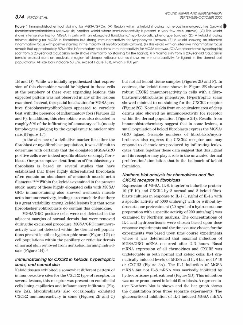
tive staining patterns for MGSA/GRO. In approximatelyChodosh et al.22 About 10 ng of nuclear extracts fromfibroblast cell lines was preincubated with 2 mg of a cold half of the keloid samples examined in this study, the
fibroblastic/myofibroblastic population showed MGSA/nonspecific competitor poly dI.dC: poly dI.dC in a buffercontaining 20 mM Hepes-NaOH (pH:7.6), 50 mM NaCl, GRO reactive cells in 40–60% of these cells (Figures 1A,
C and E). The remaining keloid lesions either showed a0.2 mM EDTA (pH:8), 1 mM DTT and 2% glycerol for15 minutes at room temperature. The reactions were few MGSA/GRO positive cells with modest immunoreac-
tivity (Figures 1B and D) or little or no staining (Figuresincubated for an additional 20 minutes after the addition
WOUND REPAIR AND REGENERATIONSEPTEMBER–OCTOBER 2000374 NIRODI ET AL.
�Figure 1 Immunohistochemical staining for MGSA/GRO�. (A) Region within a keloid showing numerous immunoreactive (brown)fibroblasts/myofibroblasts (arrows); (B) Another keloid where immunoreactivity is present in very few cells (arrows). (C) This keloidshows intense staining for MGSA in cells with an elongated fibroblastic/myofibroblastic phenotype (arrows). (D) A keloid showingminimal staining for MGSA in fibroblasts but strong immunoreactivity for lymphocytes (arrows). (E) A keloid showing an intensiveinflammatory focus with positive staining in the majority of myofibroblasts (arrows). (F) This keloid with an intensive inflammatory focusreveals that approximately 50% of the inflammatory cells show immunoreactivity for MGSA (arrows). (G) A representative hypertrophicscar from a 20-year-old Caucasian male shows minimal to no staining for the ligand). (H) Normal skin from a 20-year-old Caucasianfemale excised from an equivalent region of deeper reticular dermis shows no immunoreactivity for ligand in the dermal cellpopulations). All size bars indicate 50 �m, except Figure 1(H), which is 100 �m.
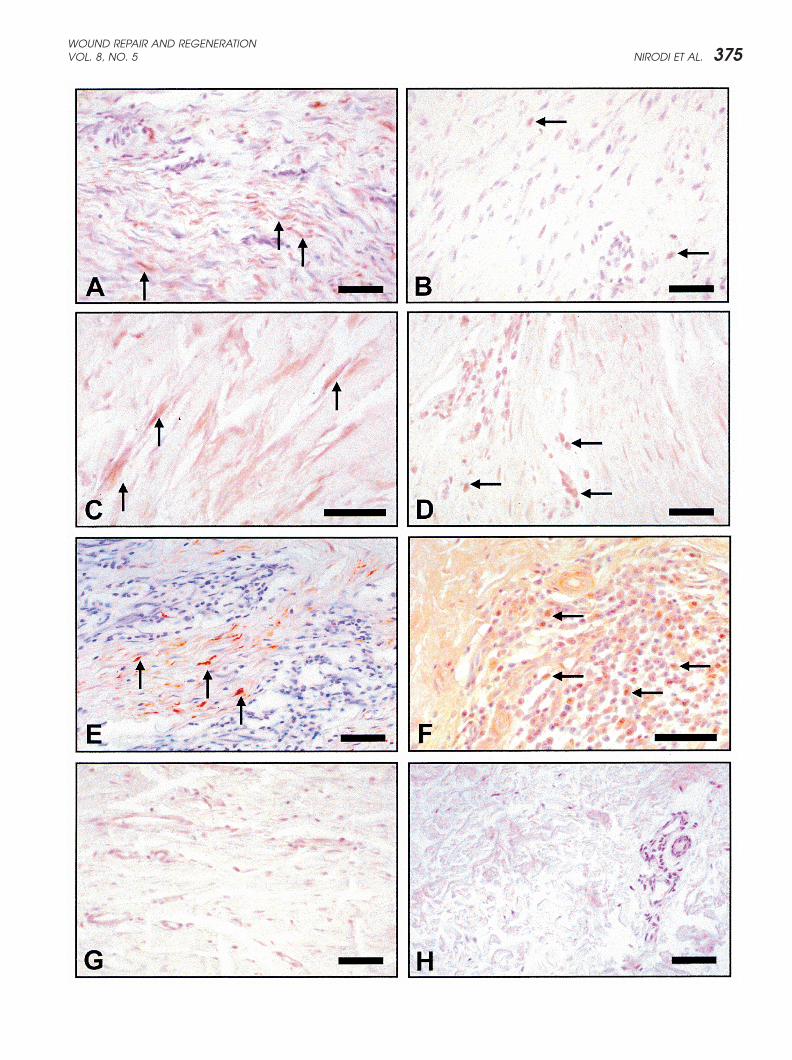
1B and D). While we initially hypothesized that expres- but not all keloid tissue samples (Figures 2D and F). Incontrast, the keloid tissue shown in Figure 2E showedsion of this chemokine would be highest in those cells
at the periphery of these ever expanding lesions, this robust CXCR2 immunoreactivity in cells with a fibro-blastic/myofibroblastic phenotype. Hypertrophic scarsexpected pattern was not observed in any of the lesions
examined. Instead, the spatial localization for MGSA pos- showed minimal to no staining for the CXCR2 receptor(Figure 2G). Normal skin from an equivalent area of deepitive fibroblasts/myofibroblasts appeared to correlate
best with the presence of inflammatory foci (Figures 1E dermis also showed no immunoreactivity for receptorwithin the dermal population (Figure 2H). Results fromand F). In addition, this chemokine was also detected in
roughly 50% of the infiltrating inflammatory cells (mostly immunohistochemistry suggest that in some lesions, asmall population of keloid fibroblasts express the MGSA/lymphocytes, judging by the cytoplasmic to nuclear size
ratio)(Figure 1F). GRO ligand. Sizeable numbers of fibroblasts/myofi-broblasts also express the CXCR2 receptor and mayIn the absence of a definitive marker for either the
fibroblast or myofibroblast population, it was difficult to respond to chemokines produced by infiltrating leuko-cytes. Taken together these data suggest that this liganddetermine with certainty that the elongated MGSA/GRO
positive cells were indeed myofibroblasts or simply fibro- and its receptor may play a role in the unwanted dermalproliferation/stimulation that is the hallmark of keloidblasts. Our presumptive identification of fibroblasts/myo-
fibroblasts is based on several studies that have formation.established that these highly differentiated fibroblasts
Northern blot analysis for chemokines and theoften contain an abundance of �-smooth muscle actinCXCR2 receptor in fibroblastsfilaments.24–26 Within the keloids examined in the presentExpression of MGSA, IL-8, interferon inducible protein-study, many of these highly elongated cells with MGSA/10 (IP-10) and CXCR2 by 2 normal and 2 keloid fibro-GRO immunostaining also showed �-smooth muscleblasts cultures in response to IL-1 (1 ng/ml of IL-1� withactin immunoreactivity, leading us to conclude that therea specific activity of 5000 units/mg) with or without hy-is a great variability among keloid lesions but that somedrocortisone pretreatment (50 ng/ml of a hydrocortisonefibroblasts/myofibroblasts do contain this chemokine.preparation with a specific activity of 200 units/mg)) wasMGSA/GRO positive cells were not detected in theexamined by Northern analysis. The concentrations ofadjacent margins of normal dermis that were removedIL-1 and hydrocortisone were chosen based upon doseduring the excisional procedure. MGSA/GRO immunore-response experiments and the time course chosen for theactivity was not detected within the dermal cell popula-experiments was based upon time course experimentstions present in either hypertrophic scars (Figure 1G) orwhere it was determined that maximal induction ofcell populations within the papillary or reticular dermisMGSA/GRO mRNA occurred after 2–3 hours. Basalof normal skin removed from nonkeloid forming individ-mRNA expression of all chemokines and CXCR2 wasuals (Figure 1H).18
undetectable in both normal and keloid cells. IL-1 dra-matically induced levels of MGSA and IL-8 but not IP-10Immunostaining for CXCR2 in keloids, hypertrophic
scars, and normal skin or CXCR2 (Figure 3A). The IL-1 induction of MGSAmRNA but not IL-8 mRNA was markedly inhibited byKeloid tissues exhibited a somewhat different pattern of
immunoreactive sites for the CXCR2 type of receptor. In hydrocortisone pretreatment (Figure 3B). This inhibitionwas more pronounced in keloid fibroblasts. A representa-several lesions, this receptor was present on endothelial
cells lining capillaries and inflammatory infiltrates (Fig- tive Northern blot is shown and the bar graph showsthe quantitation from three separate experiments. Theure 2A). Myofibroblasts also occasionally exhibited
CXCR2 immunoreactivity in some (Figures 2B and C) glucocorticoid inhibition of IL-1 induced MGSA mRNA
WOUND REPAIR AND REGENERATIONVOL. 8, NO. 5 NIRODI ET AL. 375
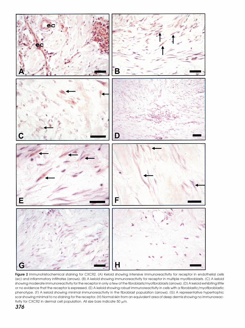
Figure 2 Immunohistochemical staining for CXCR2. (A) Keloid showing intensive immunoreactivity for receptor in endothelial cells(ec) and inflammatory infiltrates (arrows). (B) A keloid showing immunoreactivity for receptor in multiple myofibroblasts. (C) A keloidshowingmoderate immunoreactivity for the receptor in only a fewof the fibroblasts/myofibroblasts (arrows). (D) A keloid exhibiting littleor no evidence that the receptor is expressed. (E) A keloid showing robust immunoreactivity in cells with a fibroblastic/myofibroblasticphenotype. (F) A keloid showing minimal immunoreactivity in the fibroblast population (arrows). (G) A representative hypertrophicscar showingminimal to no staining for the receptor. (H) Normal skin from an equivalent area of deep dermis showing no immunoreac-tivity for CXCR2 in dermal cell population. All size bars indicate 50 �m.
376
WOUND REPAIR AND REGENERATIONVOL. 8, NO. 5 NIRODI ET AL. 377
Figure 3 Northern blot analysis of chemokine gene expression. Total RNA from two normal fibroblast cell-lines, N1 & N2, and twokeloid fibroblast cell-lines, K1 & K2, was resolved on formaldehyde-agarose gels, transferred to nitrocellulose membranes and probedwith 32P labeled probes as indicated on the left. (A) Cells were treated with hydrocortisone (HC) or left untreated. (B) Cells, eitherpretreated with HC or left untreated, received a 3-h IL-1 treatment prior to harvesting. Total RNA from Hs294T malignant melanomacells (H) was used as a positive control. (C) Autoradiograms of MGSA/GRO, cyclophilin B and IL-8 northern blots were densitometricallyscanned using the ImageQuant software (Molecular Dynamics). Values obtained for MGSA/GRO and IL-8 mRNA were normalizedagainst values of cyclophilin B mRNA and the results represented graphically for levels of MGSA/GRO mRNA (top) and IL-8 mRNA(bottom). The Y-axis represents a ratio of MGSA/GRO mRNA levels (top) or IL-8 mRNA levels (bottom) to cyclophilin B mRNA levels.Each graph is based on three experiments. Error bars represent standard deviations.
was significantly impaired in keloid fibroblast (p < 0.05) did not decrease nuclear levels of p65 (RelA) and p50subunits of NF-�B. Based upon EMSA, hydrocortisonebut not in normal fibroblasts (p > 0.7) (Student’s t-test).treatment appeared to have slightly increased the nuclearlevels of these transactivators in K1, K3, K5 keloid fibro-Nuclear levels of AP-1, NF-�B, AND Sp1 in normal
and keloid fibroblasts blasts and in N1, N2 and N5 normal fibroblasts. However,K2, N3 and N4 appeared to show a decrease in nuclearNuclear levels of AP-1, NF-�B and Sp1 in response to IL-
1 (1 ng/ml) with or without glucocorticoid pretreatment p65/p50 binding, and K4 exhibited no change in p65/p50in response to glucocorticoid. Because the slight increase(50 ng/ml which was equivalent to 10 units/ml) were
examined in normal and keloid fibroblasts by electropho- in NF-�B in response to glucocorticoid was variable, theconclusion that can be derived from these experiments isretic mobility shift analysis (EMSA). In these experi-
ments, variability in keloid and control fibroblasts was that hydrocortisone does not significantly inhibit nuclearlocalization or DNA binding of NF-�B p65/p50. Hydrocor-noted in response to glucocorticoid suppression of NF-
�B and AP-1. Results from EMSAs using nuclear extracts tisone pretreatment had no effect on AP1 or Sp1 nuclearlevels (Figures 4C and D, respectively). Immunosuper-from 5 normal and 5 keloid fibroblast cultures indicated
that IL-1 treatment increased binding of the p65 (Rel shift analysis (Figure 4E) indicates that complexes gener-ated with the NF-�B element largely consist of p50/p65A) and p50 subunits of NF-�B (Figure 4A [left and right]
and B). Hydrocortisone pretreatment of these cultures heterodimers. Complexes obtained with the Sp1 probe
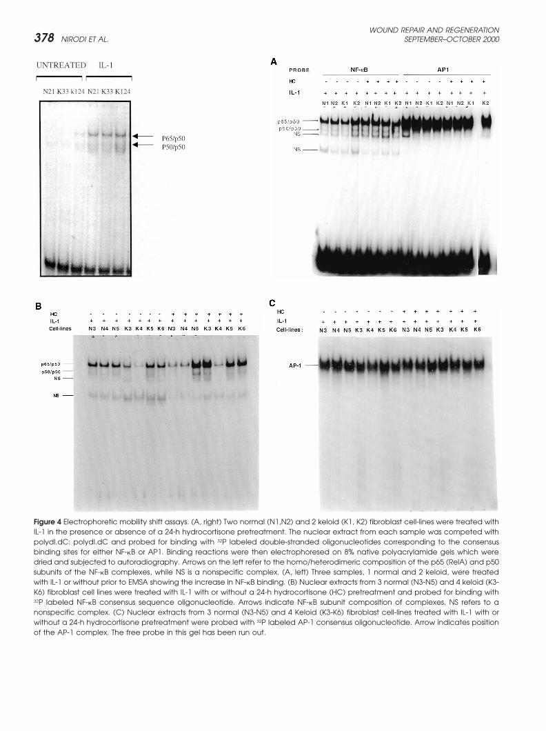
WOUND REPAIR AND REGENERATIONSEPTEMBER–OCTOBER 2000378 NIRODI ET AL.
Figure 4 Electrophoretic mobility shift assays. (A, right) Two normal (N1,N2) and 2 keloid (K1, K2) fibroblast cell-lines were treated withIL-1 in the presence or absence of a 24-h hydrocortisone pretreatment. The nuclear extract from each sample was competed withpolydI.dC: polydI.dC and probed for binding with 32P labeled double-stranded oligonucleotides corresponding to the consensusbinding sites for either NF-�B or AP1. Binding reactions were then electrophoresed on 8% native polyacrylamide gels which weredried and subjected to autoradiography. Arrows on the left refer to the homo/heterodimeric composition of the p65 (RelA) and p50subunits of the NF-�B complexes, while NS is a nonspecific complex. (A, left) Three samples, 1 normal and 2 keloid, were treatedwith IL-1 or without prior to EMSA showing the increase in NF-�B binding. (B) Nuclear extracts from 3 normal (N3-N5) and 4 keloid (K3-K6) fibroblast cell lines were treated with IL-1 with or without a 24-h hydrocortisone (HC) pretreatment and probed for binding with32P labeled NF-�B consensus sequence oligonucleotide. Arrows indicate NF-�B subunit composition of complexes. NS refers to anonspecific complex. (C) Nuclear extracts from 3 normal (N3-N5) and 4 Keloid (K3-K6) fibroblast cell-lines treated with IL-1 with orwithout a 24-h hydrocortisone pretreatment were probed with 32P labeled AP-1 consensus oligonucleotide. Arrow indicates positionof the AP-1 complex. The free probe in this gel has been run out.
WOUND REPAIR AND REGENERATIONVOL. 8, NO. 5 NIRODI ET AL. 379
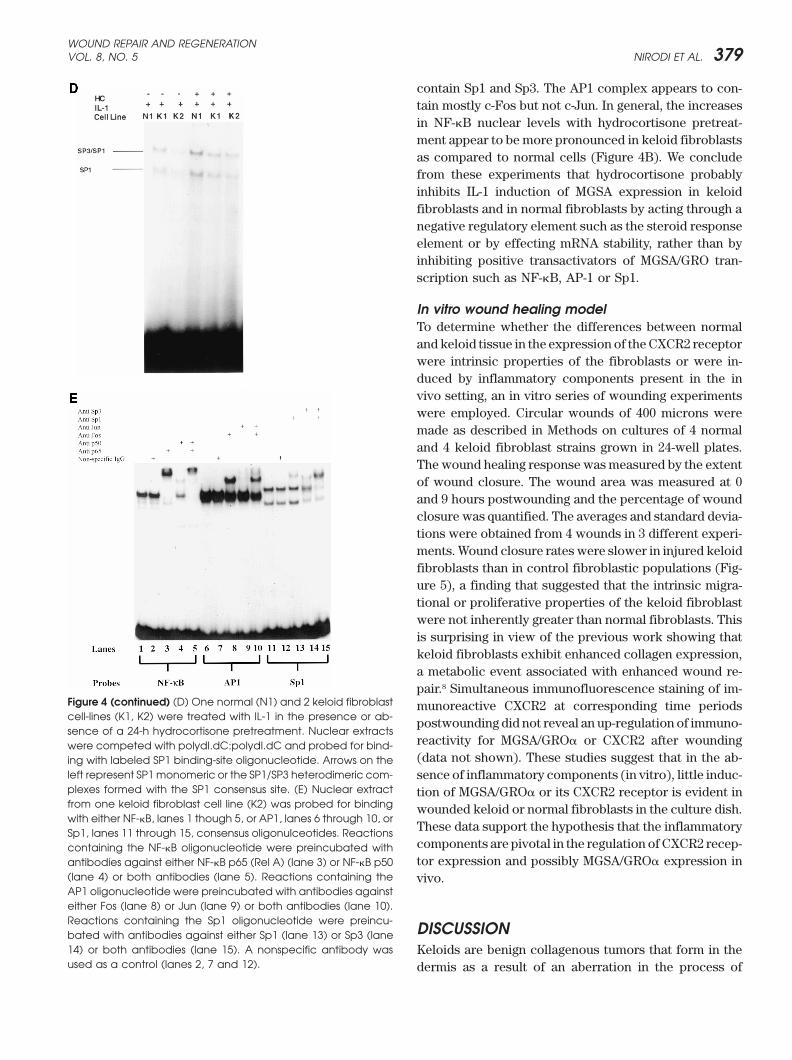
contain Sp1 and Sp3. The AP1 complex appears to con-tain mostly c-Fos but not c-Jun. In general, the increasesin NF-�B nuclear levels with hydrocortisone pretreat-ment appear to be more pronounced in keloid fibroblastsas compared to normal cells (Figure 4B). We concludefrom these experiments that hydrocortisone probablyinhibits IL-1 induction of MGSA expression in keloidfibroblasts and in normal fibroblasts by acting through anegative regulatory element such as the steroid responseelement or by effecting mRNA stability, rather than byinhibiting positive transactivators of MGSA/GRO tran-scription such as NF-�B, AP-1 or Sp1.
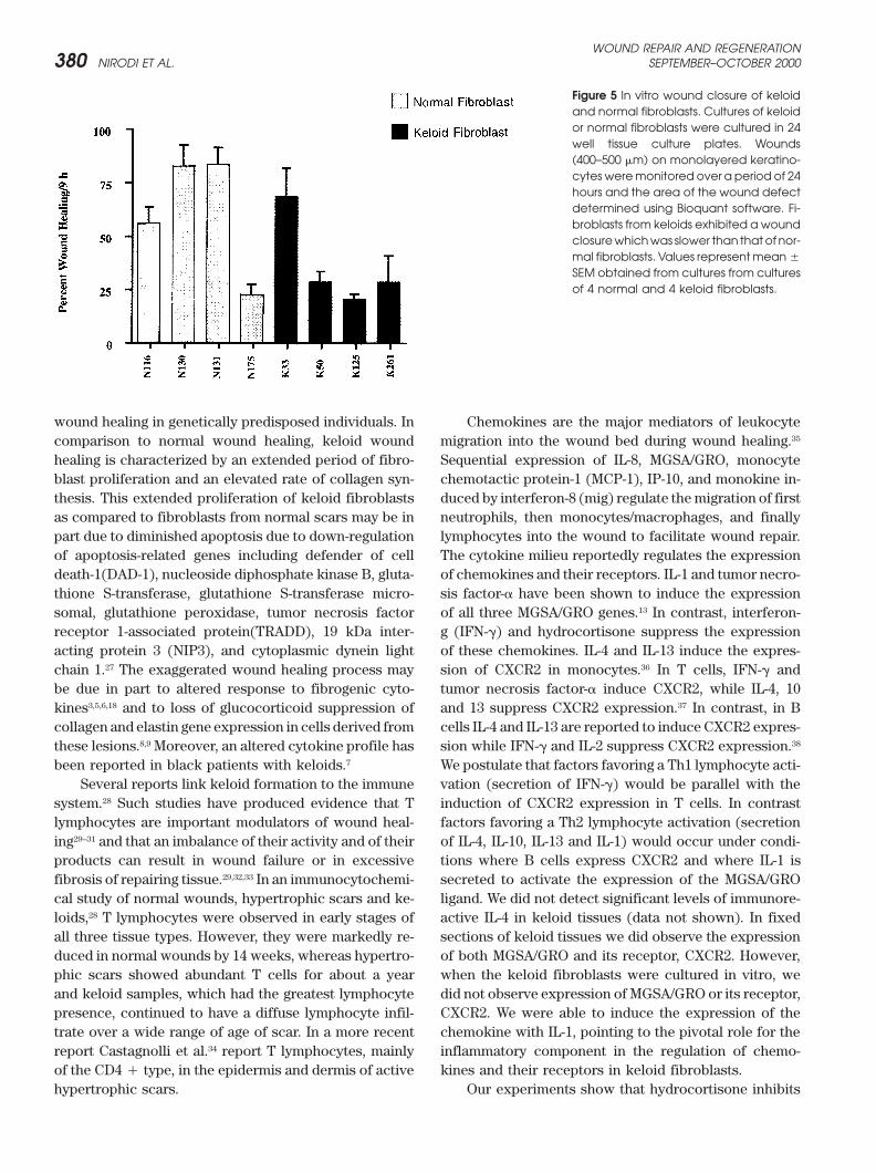
In vitro wound healing modelTo determine whether the differences between normaland keloid tissue in the expression of the CXCR2 receptorwere intrinsic properties of the fibroblasts or were in-duced by inflammatory components present in the invivo setting, an in vitro series of wounding experimentswere employed. Circular wounds of 400 microns weremade as described in Methods on cultures of 4 normaland 4 keloid fibroblast strains grown in 24-well plates.The wound healing response was measured by the extentof wound closure. The wound area was measured at 0and 9 hours postwounding and the percentage of woundclosure was quantified. The averages and standard devia-tions were obtained from 4 wounds in 3 different experi-ments. Wound closure rates were slower in injured keloidfibroblasts than in control fibroblastic populations (Fig-ure 5), a finding that suggested that the intrinsic migra-tional or proliferative properties of the keloid fibroblastwere not inherently greater than normal fibroblasts. Thisis surprising in view of the previous work showing thatkeloid fibroblasts exhibit enhanced collagen expression,a metabolic event associated with enhanced wound re-pair.8 Simultaneous immunofluorescence staining of im-
Figure 4 (continued) (D) One normal (N1) and 2 keloid fibroblast munoreactive CXCR2 at corresponding time periodscell-lines (K1, K2) were treated with IL-1 in the presence or ab- postwounding did not reveal an up-regulation of immuno-sence of a 24-h hydrocortisone pretreatment. Nuclear extracts
reactivity for MGSA/GRO� or CXCR2 after woundingwere competed with polydI.dC:polydI.dC and probed for bind-(data not shown). These studies suggest that in the ab-ing with labeled SP1 binding-site oligonucleotide. Arrows on the
left represent SP1 monomeric or the SP1/SP3 heterodimeric com- sence of inflammatory components (in vitro), little induc-plexes formed with the SP1 consensus site. (E) Nuclear extract tion of MGSA/GRO� or its CXCR2 receptor is evident infrom one keloid fibroblast cell line (K2) was probed for binding wounded keloid or normal fibroblasts in the culture dish.with either NF-�B, lanes 1 though 5, or AP1, lanes 6 through 10, or
These data support the hypothesis that the inflammatorySp1, lanes 11 through 15, consensus oligonulceotides. Reactionscomponents are pivotal in the regulation of CXCR2 recep-containing the NF-�B oligonucleotide were preincubated withtor expression and possibly MGSA/GRO� expression inantibodies against either NF-�B p65 (Rel A) (lane 3) or NF-�B p50
(lane 4) or both antibodies (lane 5). Reactions containing the vivo.AP1 oligonucleotide were preincubated with antibodies againsteither Fos (lane 8) or Jun (lane 9) or both antibodies (lane 10).Reactions containing the Sp1 oligonucleotide were preincu- DISCUSSIONbated with antibodies against either Sp1 (lane 13) or Sp3 (lane14) or both antibodies (lane 15). A nonspecific antibody was Keloids are benign collagenous tumors that form in theused as a control (lanes 2, 7 and 12). dermis as a result of an aberration in the process of
WOUND REPAIR AND REGENERATIONSEPTEMBER–OCTOBER 2000380 NIRODI ET AL.
Figure 5 In vitro wound closure of keloidand normal fibroblasts. Cultures of keloidor normal fibroblasts were cultured in 24well tissue culture plates. Wounds(400–500 �m) on monolayered keratino-cytes weremonitored over a period of 24hours and the area of the wound defectdetermined using Bioquant software. Fi-broblasts from keloids exhibited a woundclosurewhichwas slower than thatof nor-mal fibroblasts. Values representmean�
SEM obtained from cultures from culturesof 4 normal and 4 keloid fibroblasts.
wound healing in genetically predisposed individuals. In Chemokines are the major mediators of leukocytemigration into the wound bed during wound healing.35comparison to normal wound healing, keloid wound
healing is characterized by an extended period of fibro- Sequential expression of IL-8, MGSA/GRO, monocytechemotactic protein-1 (MCP-1), IP-10, and monokine in-blast proliferation and an elevated rate of collagen syn-
thesis. This extended proliferation of keloid fibroblasts duced by interferon-8 (mig) regulate the migration of firstneutrophils, then monocytes/macrophages, and finallyas compared to fibroblasts from normal scars may be in
part due to diminished apoptosis due to down-regulation lymphocytes into the wound to facilitate wound repair.The cytokine milieu reportedly regulates the expressionof apoptosis-related genes including defender of cell
death-1(DAD-1), nucleoside diphosphate kinase B, gluta- of chemokines and their receptors. IL-1 and tumor necro-sis factor-� have been shown to induce the expressionthione S-transferase, glutathione S-transferase micro-
somal, glutathione peroxidase, tumor necrosis factor of all three MGSA/GRO genes.13 In contrast, interferon-g (IFN-�) and hydrocortisone suppress the expressionreceptor 1-associated protein(TRADD), 19 kDa inter-
acting protein 3 (NIP3), and cytoplasmic dynein light of these chemokines. IL-4 and IL-13 induce the expres-sion of CXCR2 in monocytes.36 In T cells, IFN-� andchain 1.27 The exaggerated wound healing process may
be due in part to altered response to fibrogenic cyto- tumor necrosis factor-� induce CXCR2, while IL-4, 10and 13 suppress CXCR2 expression.37 In contrast, in Bkines3,5,6,18 and to loss of glucocorticoid suppression of
collagen and elastin gene expression in cells derived from cells IL-4 and IL-13 are reported to induce CXCR2 expres-sion while IFN-� and IL-2 suppress CXCR2 expression.38these lesions.8,9 Moreover, an altered cytokine profile has
been reported in black patients with keloids.7 We postulate that factors favoring a Th1 lymphocyte acti-vation (secretion of IFN-�) would be parallel with theSeveral reports link keloid formation to the immune
system.28 Such studies have produced evidence that T induction of CXCR2 expression in T cells. In contrastfactors favoring a Th2 lymphocyte activation (secretionlymphocytes are important modulators of wound heal-
ing29–31 and that an imbalance of their activity and of their of IL-4, IL-10, IL-13 and IL-1) would occur under condi-tions where B cells express CXCR2 and where IL-1 isproducts can result in wound failure or in excessive
fibrosis of repairing tissue.29,32,33 In an immunocytochemi- secreted to activate the expression of the MGSA/GROligand. We did not detect significant levels of immunore-cal study of normal wounds, hypertrophic scars and ke-
loids,28 T lymphocytes were observed in early stages of active IL-4 in keloid tissues (data not shown). In fixedsections of keloid tissues we did observe the expressionall three tissue types. However, they were markedly re-
duced in normal wounds by 14 weeks, whereas hypertro- of both MGSA/GRO and its receptor, CXCR2. However,when the keloid fibroblasts were cultured in vitro, wephic scars showed abundant T cells for about a year
and keloid samples, which had the greatest lymphocyte did not observe expression of MGSA/GRO or its receptor,CXCR2. We were able to induce the expression of thepresence, continued to have a diffuse lymphocyte infil-
trate over a wide range of age of scar. In a more recent chemokine with IL-1, pointing to the pivotal role for theinflammatory component in the regulation of chemo-report Castagnolli et al.34 report T lymphocytes, mainly
of the CD4 � type, in the epidermis and dermis of active kines and their receptors in keloid fibroblasts.Our experiments show that hydrocortisone inhibitshypertrophic scars.
WOUND REPAIR AND REGENERATIONVOL. 8, NO. 5 NIRODI ET AL. 381
3. Tredget EF, Nedelec B, Scott PG, Ghahary A. Hypertrophic scars,IL-1 mediated MGSA/GRO expression in keloid fibro-keloids, and contractures: the cellular and molecular basis for
blasts. It has been reported previously that glucocorti- therapy. Surg Clin North Am 1997;77:701–30.coid suppresses expression of the rat homolog of MGSA/ 4. Machesney M, Tidman N, Waseem A, Kirby L, Leigh I. Activated
keratinocytes in the epidermis of hypertrophic scars. Am J PatholGRO by impairing the activation of NF-�B.16 Expression1998;152:1133–41.of other CXC chemokines is also attenuated by glucocor-
5. Babu M, Diegelmann R, Oliver N. Keloid fibroblasts exhibit anticoids.39 Another mechanism for suppression of CC altered response to TGF-beta. J Invest Dermatol 1992;99:650–5.chemokine expression is through glucocorticoid medi- 6. Zhang K, Garner W, Cohen L, Rodriguez J, Phan S. Increased types
I and III collagen and transforming growth factor-beta 1 mRNAated destabilization of chemokine mRNA.40 Based uponand protein in hypertrophic burn scar. J Invest Dermatol 1995;our gel shift analyzes, hydrocortisone inhibition of 104:750–4.
MGSA/GRO mRNA expression does not appear to be 7. McCauley RL, Chopra V, Li YY, Herndon DN, Robson MC. Alteredcytokine production in black patients with keloids. J Clin Immunolmediated by suppression of NF-�B, AP-1 or Sp1, but may1992;12:300–8.be through effects on the putative Steroid Responsive
8. Russell SB, Trupin JS, Myers JC, Broquist AH, Smith JC, MylesElement [AGAACAT] located in the MGSA/GRO� pro- ME, Russell JD. Differential glucocorticoid regulation of collagenmoter at –601 bp to –596 bp relative to the transcription mRNAs in human dermal fibroblasts. Keloid-derived and fetal
fibroblasts are refractory to down-regulation. J Biol Chem 1989;start-site. Effects of hydrocortisone on MGSA/GRO264:13730–5.mRNA stability cannot be ruled out.
9. Russell SB, Trupin JS, Kennedy RZ, Russell JD, Davidson JM.The ability of keloid fibroblasts to respond to hydro- Glucocorticoid regulation of elastin synthesis in human fibro-
blasts: down-regulation in fibroblasts from normal dermis but notcortisone is an essential component of therapy, sincefrom keloids. J Invest Derm 1995;104:241–5.surgery is seldom recommended as a treatment for ke-
10. Auphan N, Didonata JA, Rosette C, Helmberg A, Karin M. Immuno-loids. Instead, hydrocortisone treatment is often success- suppression by glucocorticoids: Inhibition of NF-kappa B activityful for shrinking these benign lesions. The architecture through induction of I-kappa B synthesis. Science 1995;270:
286–90.and histological characteristics of keloids vary greatly11. Anisowicz A, Messineo M, Lee SW, Sager R. An NF-�B-like tran-among samples. Fibroblasts in the central core have
scription factor mediates IL-1/TNF� induction of gro in humanmore of a keloid phenotype as compared to those in fibroblasts. J Immunol 1991;147:520–7.
12. Mukaida N, Mahe Y, Matsushima K. Cooperative interaction ofthe periphery which may be normal. The phenotype ofnuclear factor-�B- and cis-regulatory enhancer binding protein-primary cell cultures derived from keloid fibroblasts willlike factor binding elements in activating the interleukin-8 gene
therefore depend almost entirely on the source, pheno- by pro-inflammatory cytokines. J Biol Chem 1990;265:21128–type and location of the fibroblast within the keloid tis- 33.
13. Shattuck RL, Wood LD, Jaffe GJ, Richmond A. High basal tran-sue. The cytokine milieu in the keloid tissue will alsoscription through the NF-�B element results in accumulation ofcontribute to the keloid nature of the fibroblast. All theseMGSA/GRO� in human melanoma. Mol Cell Biol 1994;14:791–802.
factors may have contributed to the variability of the 14. Wood LD, Richmond A. HMGI (Y) and Sp1 in addition to NF-�Bregulate transcription of the MGSA/GROa gene. Nucleic Acids Resfibroblast cell lines used in this study. Inhibition of the1995;23:4210–9.induction of chemokines which attract the inflammatory
15. Wood LD, Richmond A. Constitutive and cytokine-induced expres-cells is proposed to be a major contributor to the suppres- sion of the melanoma growth stimulatory activity/GROa gene re-sion of keloid growth. By altering the cytokine milieu quires both NF-�B and novel constitutive factors. J Biol Chem
1995;270:30619–26.of the keloid fibroblasts, the growth of these lesions is16. Ohtsuka T, Kubota A, Hirano T, Watanabe K, Yoshida H, Tsurufujipredicted to be reduced.
M, Iizuka Y, Konishi K, Tsurufuji S. Glucocorticoid-mediated genesuppression of rat cytokine-induced neutrophil chemoattractantCINC/gro, a member of the interleukin-8 family, through impair-ment of NF-�B activation. J Biol Chem 1996;271:1651–9.Acknowledgments
17. Unemori EN, Amento EP, Bauer EA, Horuk R. Melanoma growth-We thank Aimee Self, Jesse Britton, and Nancy Cardwell stimulatory activity/GRO decreases collagen expression by humanfor their expert technical assistance. This work was sup- fibroblasts. Regulation by C-X-C but not C-C cytokines. J Biol
Chem 1993;268:1338–42.ported by the Department of Veterans’ Affairs [VA/HBCU18. Nanney LB, Mueller SG, Bueno R, Peiper SC, Richmond A. Distri-grant (AR and SR) and a VA Career Scientist Award (AR)],
butions of MGSA (GRO) and the IL-8 Receptor B in human woundas well as by grants from the National Institutes of Health repair. Am J Pathol 1995;147:1248–60.[GM40439 (LBN), P30 AR 41943]. 19. Russell SB, Trupin KM, Rodriguez-Eaton S, Russell JD, Trupin JS.
Reduced growth-factor requirement of keloid-derived fibroblastsmay account for tumor growth. Proc Natl Acad Sci USA 1988;85:587–91.REFERENCES 20. Dignam JD, Lebovitz RM, Roeder RG. Accurate transcription initia-tion by RNA polymerase II in a soluble extract from isolated1. Murray JC, Pollack SV, Pinnell SR. Keloids: a review. J Am Acadmammalian nuclei. Nucl Acids Res 1983;11:1475–89.Dermatol 1981;4:461–70.
2. Niessen FV, Spauwen PH, Schalkwijk J, Kon M. On the nature of 21. Chomczynski P, Sacchi N. Single-step method of RNA isolation byacid guanididium thiocynate-phenol-chloroform extraction. Analhypertrophic scars and keloids: a review. Plast Reconstr Surg
1999;104:1435–57. Biochem, 1987;162:156–9.
WOUND REPAIR AND REGENERATIONSEPTEMBER–OCTOBER 2000382 NIRODI ET AL.
22. Chodosh LA, Carthew RW, Sharp PA. A single polypeptide pos- 32. Linares HA. Proteoglycan–lymphocyte association in the develop-ment of hypertrophic scars. Burns 1990;16:21–4.sesses the binding and transcription activities of the adenovirus
late transcription factor. Mol Cell Biol 1986;6:4723–33. 33. Kovacs EJ. Fibrogenic cytokines: the role of immune mediatorsin the development of scar tissue. Immunol Today 1991;12:17–23.23. Maniatis T, Fritsch EF, Sambrook J. Molecular cloning: a labora-
tory manual. Cold Spring Harbor, NY: Cold Spring Harbor Labora- 34. Castagnoli C, Trombotto C, Ondei S, Steela M, Calcagni M, Maglia-cani G, Alasia ST. Characterization of T-cell subsets infiltratingtory, 1982.
24. Skalli O, Schurch W, Seemayeer TA, Lagace R, Montandon D, post-burn hypertrophic scar tissues. Burns 1997;23:565–72.35. Engelhardt E, Toksoy A, Goebeler M, Debus S, Brocker E-B, Gil-Pittet B, Gabbiani G. Myofibroblasts from diverse pathological
settings are heterogeneous in their content of actin isoforms and litzer R. Chemokines IL-8, GRO�, MCP-1, IP-10, and Mig are se-quentially and differentially expressed during phase-specificintermediate filament proteins. Lab Invest 1989;60:275–85.
25. Darby I, Skalli O, Gabbiani G. �-Smooth muscle actin is transiently infiltration of leukocyte subsets in human wound healing. Am JPathol 1998;153:1849–60.expressed by myofibroblasts during experimental wound healing.
Lab Invest 1990;63:21–9. 36. Sozzani S, Bonecchi R, Facchetti F, Dusi S, Lissandrini D, LocatiM, Allavena P, Rossi F, Mantovani A. Induction of functional IL-26. Sappino AP, Schurch W, Gabbiani G. Differentiation repertoire of
fibroblastic cells: expression of cytoskeletal proteins as markers 8 receptors by IL-4 and IL-13 in human monocytes. Cytokine 1999;11:936, A98.of phenotypic modulation. Lab Invest 1994;63:144–61.
27. Sayah DN, Soo C, Shaw WW, Watson J, Messadi D, Longaker MT, 37. Tani K, Su SB, Utsunomiya I, Oppenheim JJ, Wang JM. Interferon-gamma maintains the binding and functional capacity of receptorsZhang X, Ting K. Downregulation of apoptosis-related genes in
keloid tissues. J Surg Res 1999;87:209–16. for IL-8 on cultured human T cells. Eur J Immunol 1998;28:502–7.38. Jinquan T, Moller B, Storgaard M, Mukaida N, Bonde J, Grunnet28. Martin CW, Muir IFK. The role of lymphocytes in wound healing.
Br J Plast Surg 1990;43:655–62. N, Black FT, Larsen CG, Matsushima K, Threstrup-Pedersen K.Chemotaxis and IL-8 receptor expression in B cells from normal29. Wahl SM, Allen JB. T-lymphocyte dependent mechanism of fibro-
sis. In: Growth factors and other aspects of wound healing: Biologi- and HIV-infected subjects. J Immunol 1997;158:475–84.39. Smith JB, Herschman HR. Glucocorticoid-attenuated responsecal and clinical implication. Barbul A et al, eds. New York, Alan
R. Liss, 1988. genes encode intercellular mediators, including a new CXC chem-okine. J Biol Chem 1995;270:16756–65.30. Barbul A, Breslin RJ, Woodyard JP, Wasserkrug HL, Efron G. The
effect of in vitro T helper and T suppressor lymphocyte depletion 40. Poon M, Liu B, Taubman MB. Identification of novel dexametha-sone-sensitive RNA-destabilizing region on rat monocyte chemoat-on wound healing. Ann Surg 1989;209:479–83.
31. Barbul A. T cell-dependent immune system in wound healing. In: tractant protein 1 mRNA. Mol Cell Biol 1999;19:6471–8.Barbul A et al., eds. Growth factors and other aspects of woundhealing: biological and clinical implications. New York: Alan R.Liss, 1988: 161–75.
Related Documents