1

Welcome message from author
This document is posted to help you gain knowledge. Please leave a comment to let me know what you think about it! Share it to your friends and learn new things together.
Transcript

1
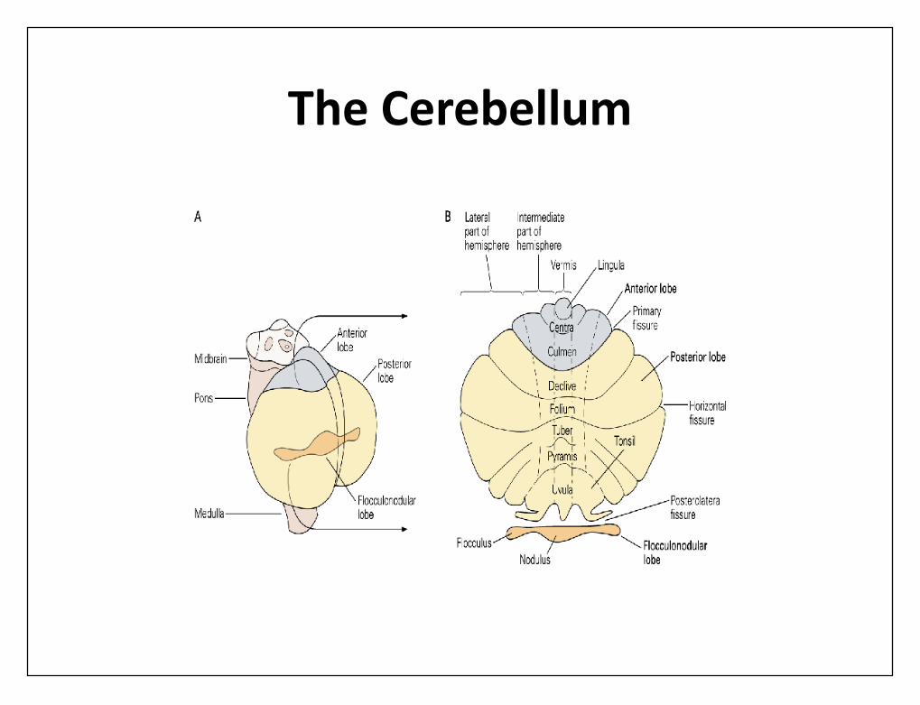
The Cerebellum
cerebellum
• It has no direct ability to cause musclecontraction
• It helps to sequence motor activities• monitors and makes corrective adjustments in
the body's motor activities• they are being executed so that they will
conform to motor signals directed by cerebralmotor cortex and other parts of the brain.
Anatomical Functional of Cerebellum
• Anatomically, the cerebellum is divided intothree lobes by two deep fissures
• (1) the anterior lobe• (2) the posterior lobe• (3) the flocculonodular lobe.• The flocculonodular lobe is oldest of all
portions of cerebellum; it developed alongwith the vestibular system in controlling bodyequilibrium

• Function• the cerebellum• the timing of motor activities• rapid, smooth progression from one muscle movement to the next.• control intensity of muscle contraction when muscle load changes• controlling necessary instantaneous interplay between agonist
and antagonist muscle groups.
• The basal ganglia• plan and control complex patterns of muscle movement• controlling relative intensities of the separate movements,
directions of movements, and sequencing of multiple successiveparallel movements for achieving specific complicated motor goals.
• The cerebellum receives continuously updated informationabout the desired sequence of muscle contractions fromthe brain motor control areas
• it also receives continuous sensory information from theperipheral parts of the body, giving sequential changes inthe status of each part of the body-its position, rate ofmovement, forces acting on it, and so forth.
• The cerebellum then compares the actual movements asdepicted by peripheral sensory feedback information withmovements intended by motor system.
• If the two do not compare favorably, then instantaneoussubconscious corrective signals are transmitted back intomotor system to increase or decrease levels of activation ofspecific muscles.

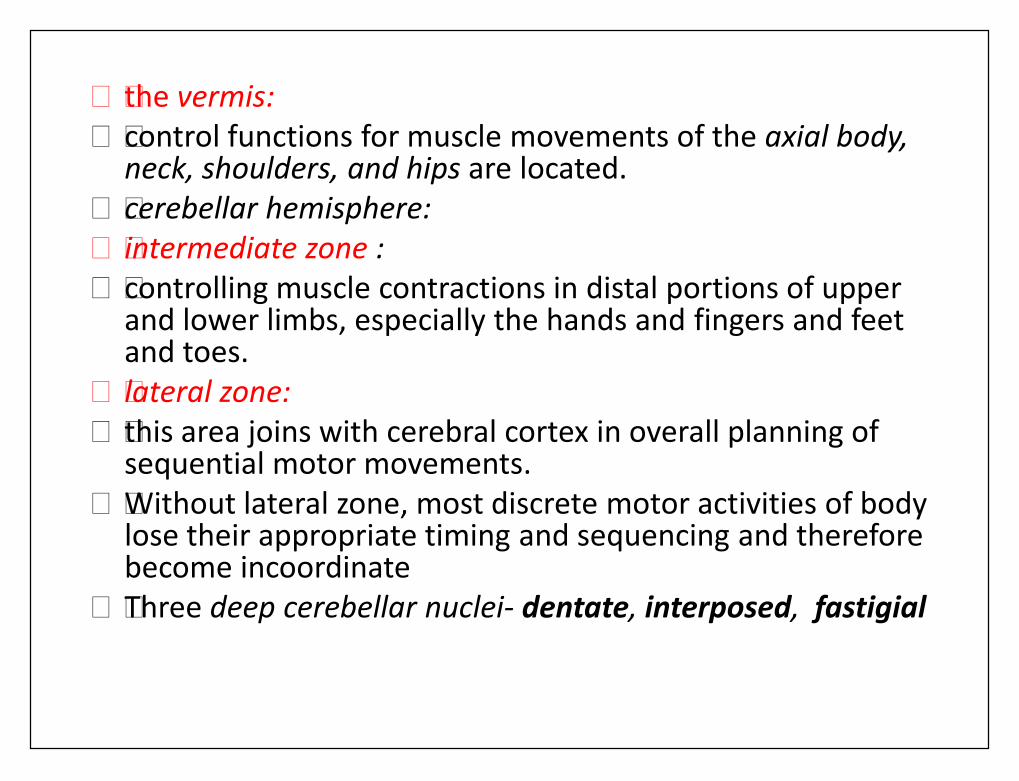
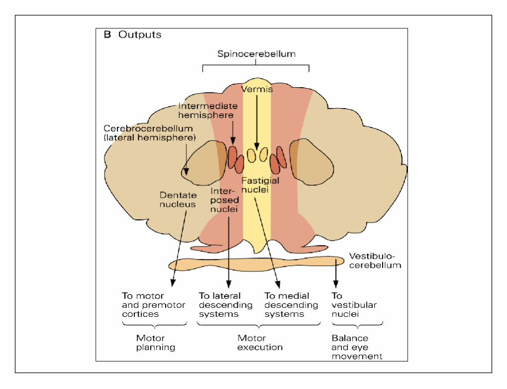
• the vermis:• control functions for muscle movements of the axial body,
neck, shoulders, and hips are located.• cerebellar hemisphere:• intermediate zone :• controlling muscle contractions in distal portions of upper
and lower limbs, especially the hands and fingers and feetand toes.
• lateral zone:• this area joins with cerebral cortex in overall planning of
sequential motor movements.• Without lateral zone, most discrete motor activities of body
lose their appropriate timing and sequencing and thereforebecome incoordinate
• Three deep cerebellar nuclei- dentate, interposed, fastigial

Somatosensory projection areas incerebellar cortex.

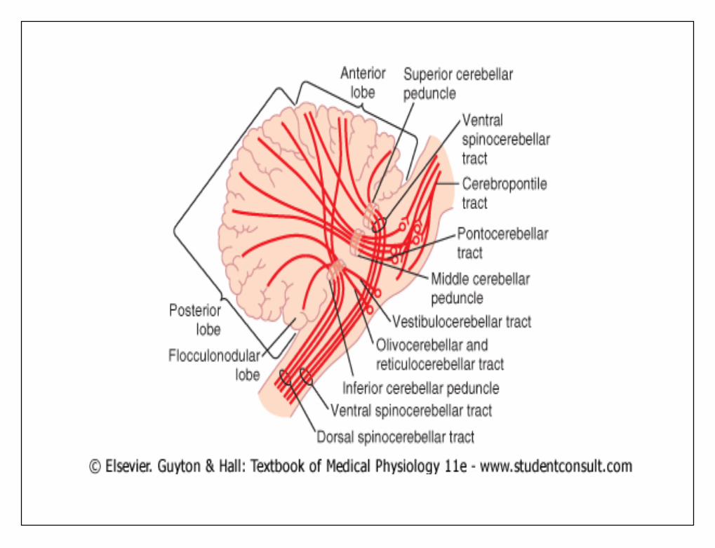
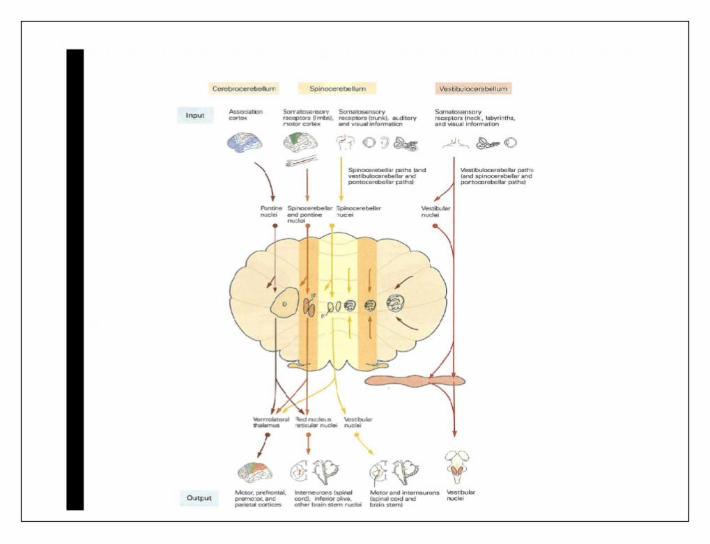
Afferent Pathways
• An extensive and important afferent pathwayis the corticopontocerebellar pathway
• originates in the cerebral motor and premotorcortices and cerebral somatosensory cortex.
• It passes pontile nuclei and pontocerebellartracts to the lateral divisions of the cerebellarhemispheres on the opposite side of the brainfrom the cerebral areas.
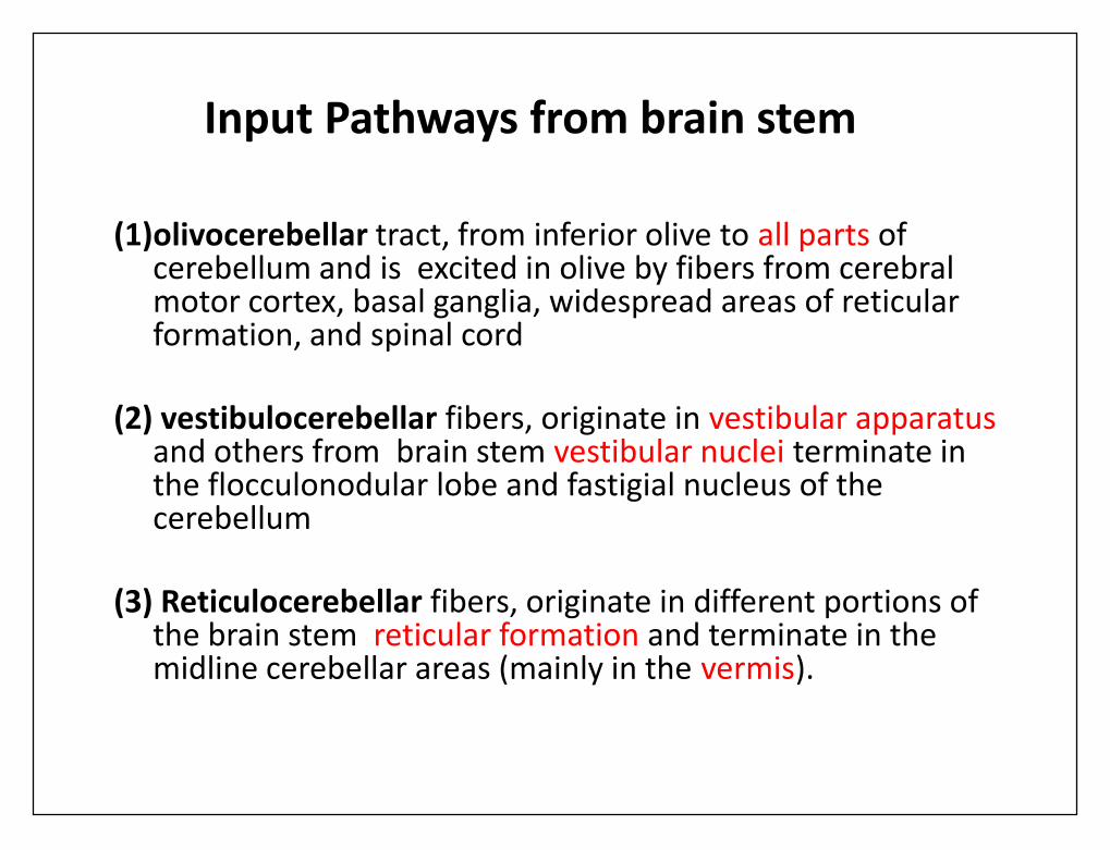
Input Pathways from brain stem
(1)olivocerebellar tract, from inferior olive to all parts ofcerebellum and is excited in olive by fibers from cerebralmotor cortex, basal ganglia, widespread areas of reticularformation, and spinal cord
(2) vestibulocerebellar fibers, originate in vestibular apparatusand others from brain stem vestibular nuclei terminate inthe flocculonodular lobe and fastigial nucleus of thecerebellum
(3) Reticulocerebellar fibers, originate in different portions ofthe brain stem reticular formation and terminate in themidline cerebellar areas (mainly in the vermis).
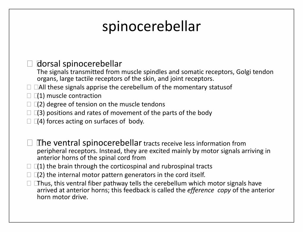
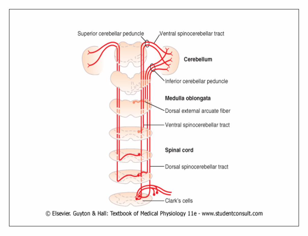
spinocerebellar
• dorsal spinocerebellarThe signals transmitted from muscle spindles and somatic receptors, Golgi tendonorgans, large tactile receptors of the skin, and joint receptors.
• All these signals apprise the cerebellum of the momentary statusof• (1) muscle contraction• (2) degree of tension on the muscle tendons• (3) positions and rates of movement of the parts of the body• (4) forces acting on surfaces of body.
• The ventral spinocerebellar tracts receive less information fromperipheral receptors. Instead, they are excited mainly by motor signals arriving inanterior horns of the spinal cord from
• (1) the brain through the corticospinal and rubrospinal tracts• (2) the internal motor pattern generators in the cord itself.• Thus, this ventral fiber pathway tells the cerebellum which motor signals have
arrived at anterior horns; this feedback is called the efference copy of the anteriorhorn motor drive.
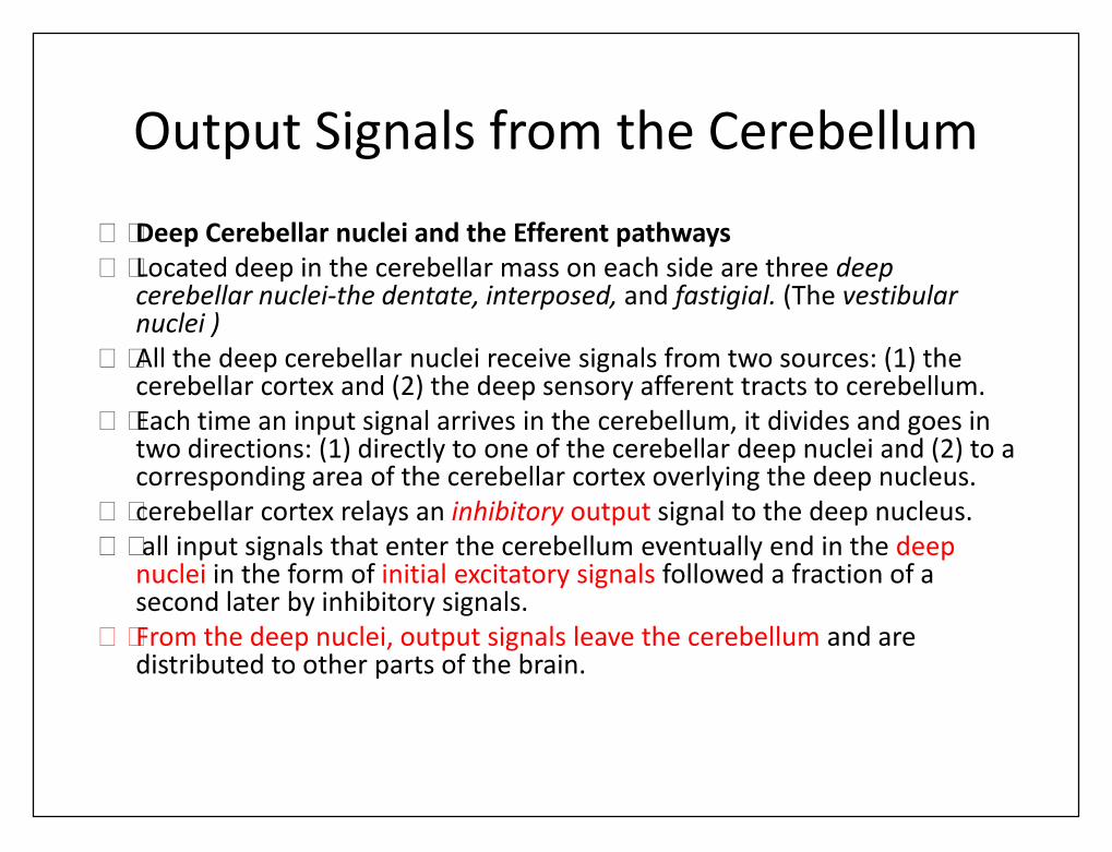
Output Signals from the Cerebellum
• Deep Cerebellar nuclei and the Efferent pathways• Located deep in the cerebellar mass on each side are three deep
cerebellar nuclei-the dentate, interposed, and fastigial. (The vestibularnuclei )
• All the deep cerebellar nuclei receive signals from two sources: (1) thecerebellar cortex and (2) the deep sensory afferent tracts to cerebellum.
• Each time an input signal arrives in the cerebellum, it divides and goes intwo directions: (1) directly to one of the cerebellar deep nuclei and (2) to acorresponding area of the cerebellar cortex overlying the deep nucleus.
• cerebellar cortex relays an inhibitory output signal to the deep nucleus.• all input signals that enter the cerebellum eventually end in the deep
nuclei in the form of initial excitatory signals followed a fraction of asecond later by inhibitory signals.
• From the deep nuclei, output signals leave the cerebellum and aredistributed to other parts of the brain.
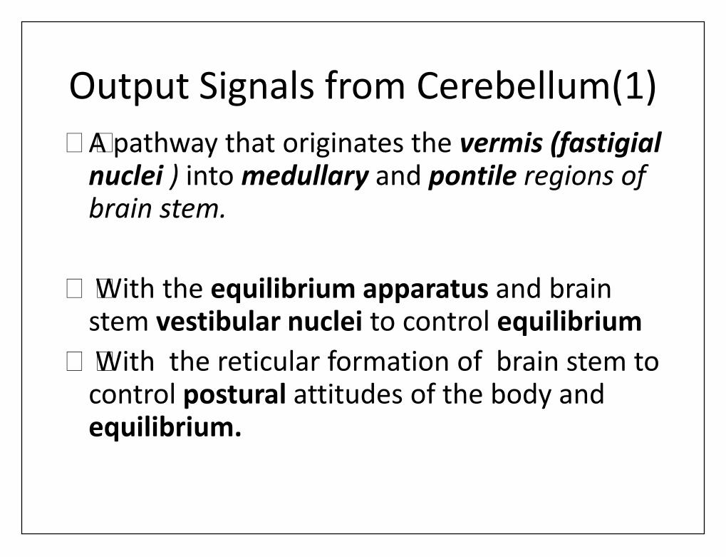
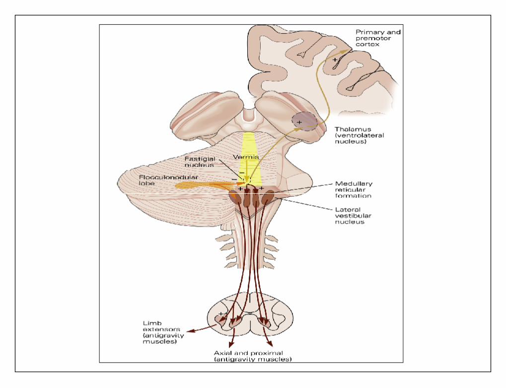
Output Signals from Cerebellum(1)• A pathway that originates the vermis (fastigial
nuclei ) into medullary and pontile regions ofbrain stem.
• With the equilibrium apparatus and brainstem vestibular nuclei to control equilibrium
• With the reticular formation of brain stem tocontrol postural attitudes of the body andequilibrium.
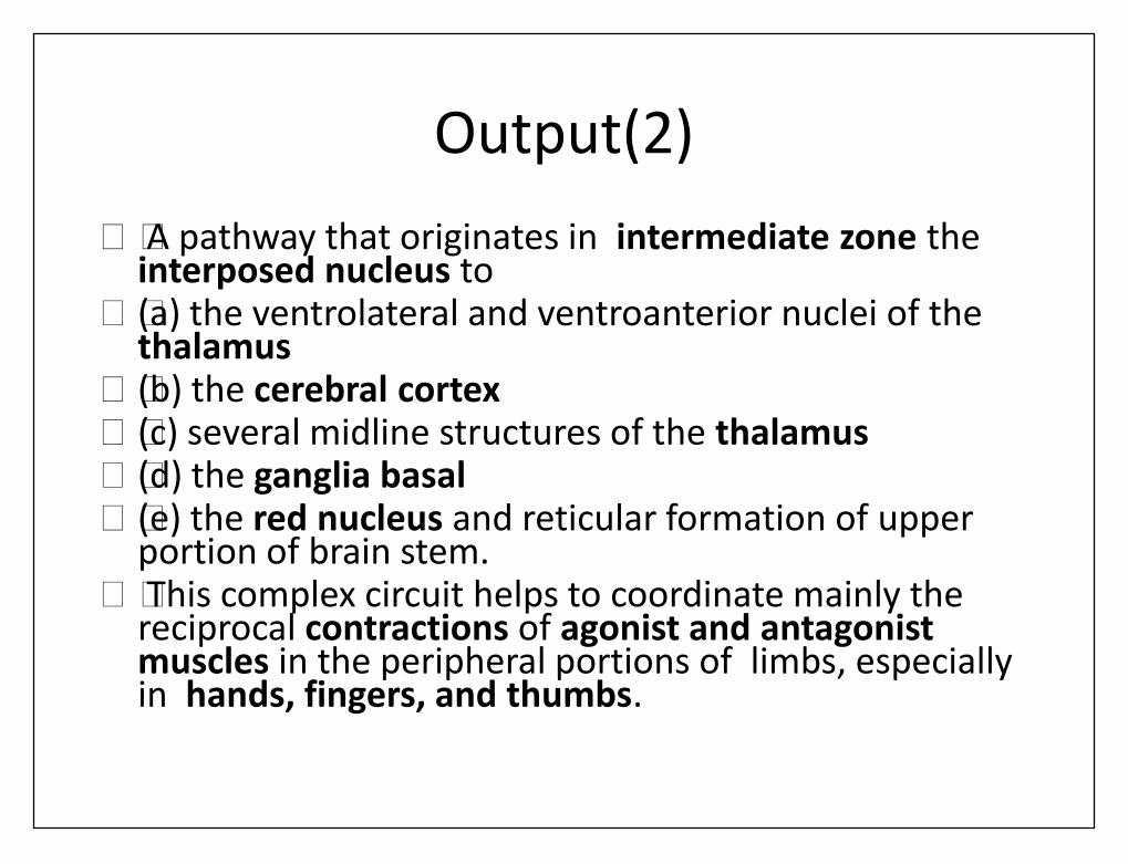
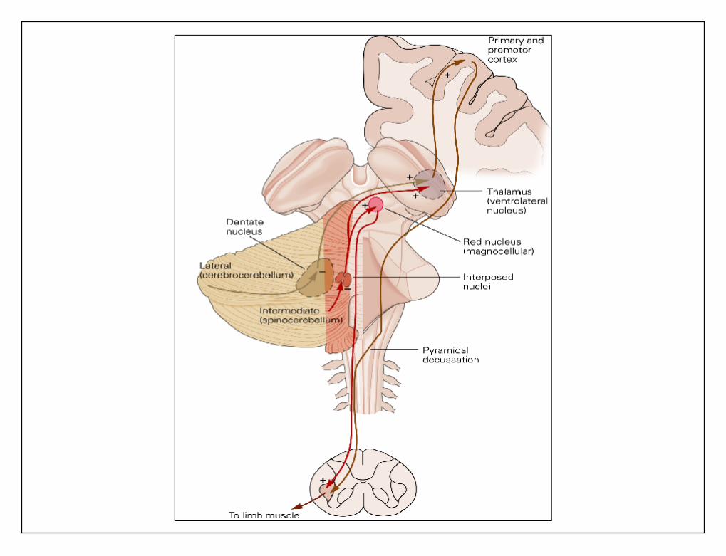
Output(2)• A pathway that originates in intermediate zone the
interposed nucleus to• (a) the ventrolateral and ventroanterior nuclei of the
thalamus• (b) the cerebral cortex• (c) several midline structures of the thalamus• (d) the ganglia basal• (e) the red nucleus and reticular formation of upper
portion of brain stem.• This complex circuit helps to coordinate mainly the
reciprocal contractions of agonist and antagonistmuscles in the peripheral portions of limbs, especiallyin hands, fingers, and thumbs.
Output(3)
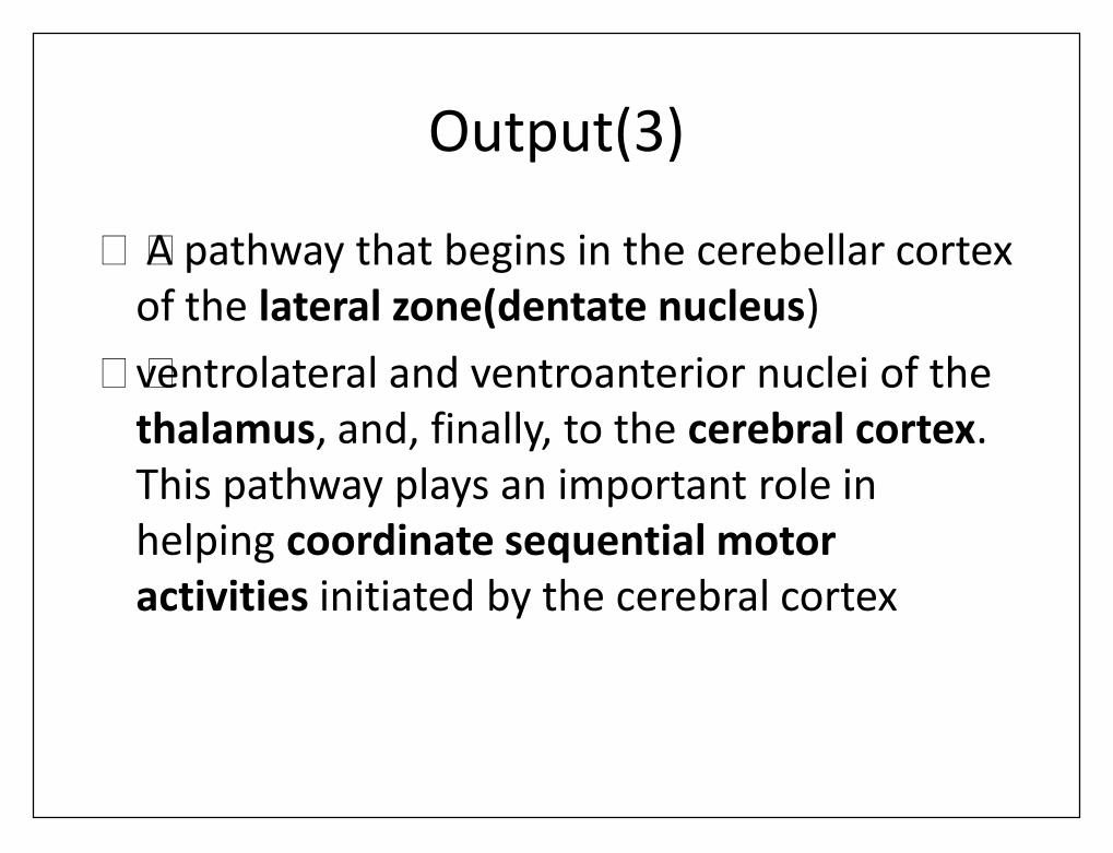
• A pathway that begins in the cerebellar cortexof the lateral zone(dentate nucleus)
• ventrolateral and ventroanterior nuclei of thethalamus, and, finally, to the cerebral cortex.This pathway plays an important role inhelping coordinate sequential motoractivities initiated by the cerebral cortex
Function of Cerebellum inMotor Control
• Vestibulocerebellum equilibrium movements.• Spinocerebellum coordinating mainly movements of the distal
portions of the limbs, especially the hands and fingers.• Cerebrocerebellum It receives its input from cerebral motor cortex
and premotor and somatosensory cortices of cerebrum.• Its output information back to the brain• functioning in a feedback manner with the cerebral cortical
sensorimotor system to plan sequential voluntary body and limbmovements, planning these as much as tenths of a second inadvance of the actual movements.
• This is called development of "motor imagery" of movements to beperformed.
Vestibulocerebellum• Its Function in Association with the Brain Stem and Spinal Cord to
Control Equilibrium and Postural Movements• loss of the flocculonodular lobes and adjacent portions of vermis of
cerebellum,Causes extreme disturbance of equilibrium and posturalmovements.
• equilibrium is far more disturbed during performance of rapidmotions than during stasis, especially so when these movementsinvolve changes in direction of movement and stimulatesemicircular ducts.
• This suggests that vestibulocerebellum is especially important incontrolling balance between agonist and antagonist musclecontractions of the spine, hips, and shoulders during rapid changesin body positions as required by the vestibular apparatus.
Spinocerebellum• Feedback Control of Distal Limb Movements by
Intermediate Cerebellar Cortex and InterposedNucleus
• Intermediate zone receives two types of information• (1) from the cerebral motor cortex and red nucleus,
the intended sequential plan of movement for the next
• (2) feedback information from peripheral parts ofbody, especially from the distal proprioceptors of thelimbs, telling what actual movements result.

• After the intermediate zone has compared the intendedmovements with the actual movements, the deep nuclearcells of interposed nucleus send corrective output signals
• (1) back to the cerebral motor cortex through relay nuclei inthe thalamus
• (2) to the magnocellular portion of red nucleus, Therubrospinal tract in turn joins corticospinal tract ininnervating neurons that control the distal parts of thelimbs, hands and fingers.
• This part of the cerebellar motor control system providessmooth, coordinate movements of agonist and antagonistmuscles of the distal limbs for performing acute purposefulpatterned movements.
Cerebrocerebellum• Function of the Large Lateral Zone of the Cerebellar Hemisphere
to Plan, Sequence, and Time Complex Movements• In human beings, the lateral zones of the two cerebellar
hemispheres are highly developed and greatly enlarged.• human abilities to plan and perform intricate sequential patterns of
movement, especially with the hands and fingers, and to speak.• It have no direct input of information from the peripheral parts of
the body.• Also, almost all communication between these lateral cerebellar
areas and the cerebral cortex is not with the primary cerebralmotor cortex itself but instead with the premotor area and primaryand association somatosensory areas.
• cerebellum are concerned with two other important but indirectaspects of motor control: (1) the planning of sequential movementsand (2) the "timing" of the sequential movements.
Planning of Sequential Movements
• The lateral zones communicate with premotor andsensory portions( two-way communication) withcorresponding basal ganglia.
• The "plan" of sequential movements begins in sensoryand premotor is transmitted to the lateral zonesappropriate motor signals provide transition from onesequence of movements to the next.
• the lateral cerebellar zones appear to be involved notwith what movement is happening at a given momentbut with what will be happening during nextsequential movement a fraction of a second or perhapseven seconds later.
Timing Function• Without this timing capability, the person
becomes unable to determine when the nextsequential movement needs to begin.
• As a result, the succeeding movement may begintoo early or, more likely, too late.
• lesions in the lateral zones of the cerebellumcause complex movements (such as thoserequired for writing, running, or even talking) tobecome incoordinate and lacking ability toprogress in orderly sequence from onemovement to the next.

Extra motor Predictive Functions ofthe Cerebrocerebellum.
• A person can predict from the changing visualscene how rapidly he or she is approaching anobject.
• removing the large lateral portions of thecerebellum in monkeys. Such a monkeyoccasionally charges the wall of a corridor andliterally bashes its brains because it is unableto predict when it will reach the wall.
Functional Unit of Cerebellar Cortex
• The cerebellum has about 30 million nearly identical• functional units.• This functional unit centers on a single, very large
Purkinje cell (30 million of which are in the cerebellarcortex) and a corresponding deep nuclear cell.
• The three major layers of the cerebellar cortex areshown: the molecular layer, Purkinje cell layer, andgranule cell layer.
• Beneath these cortical layers, in the center ofcerebellar mass, are the deep cerebellar nuclei thatsend output signals to other parts of the nervoussystem.
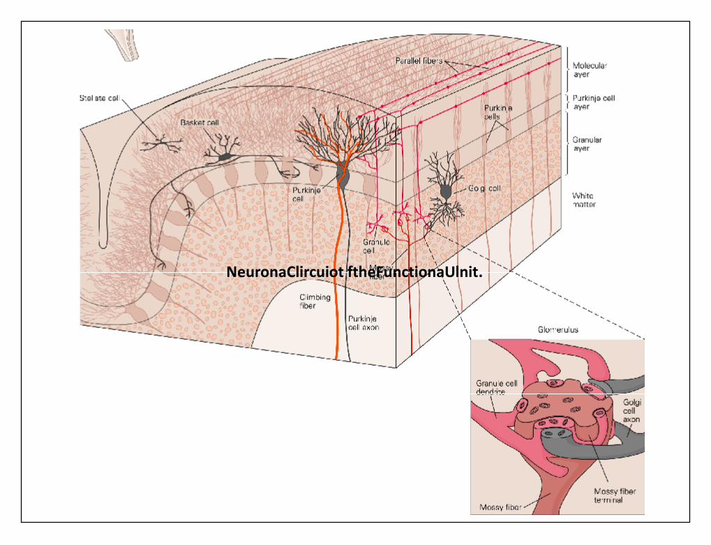
NeuronaClircuiot ftheFunctionaUlnit.
Neuronal Circuit of Functional Unit.
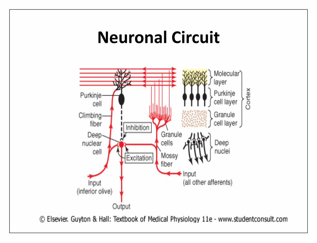
• The output from the functional unit is from a deepnuclear cell.
• This cell is continually under both excitatory andinhibitory influences.
• The excitatory influences arise from direct connectionswith afferent fibers that enter the cerebellum from thebrain or the periphery.
• The inhibitory influence arises entirely from Purkinjecell in the cortex of the cerebellum.
• The afferent inputs to the cerebellum are mainly oftwo types, one called the climbing fiber type and theother called the mossy fiber type.
Neuronal Circuit
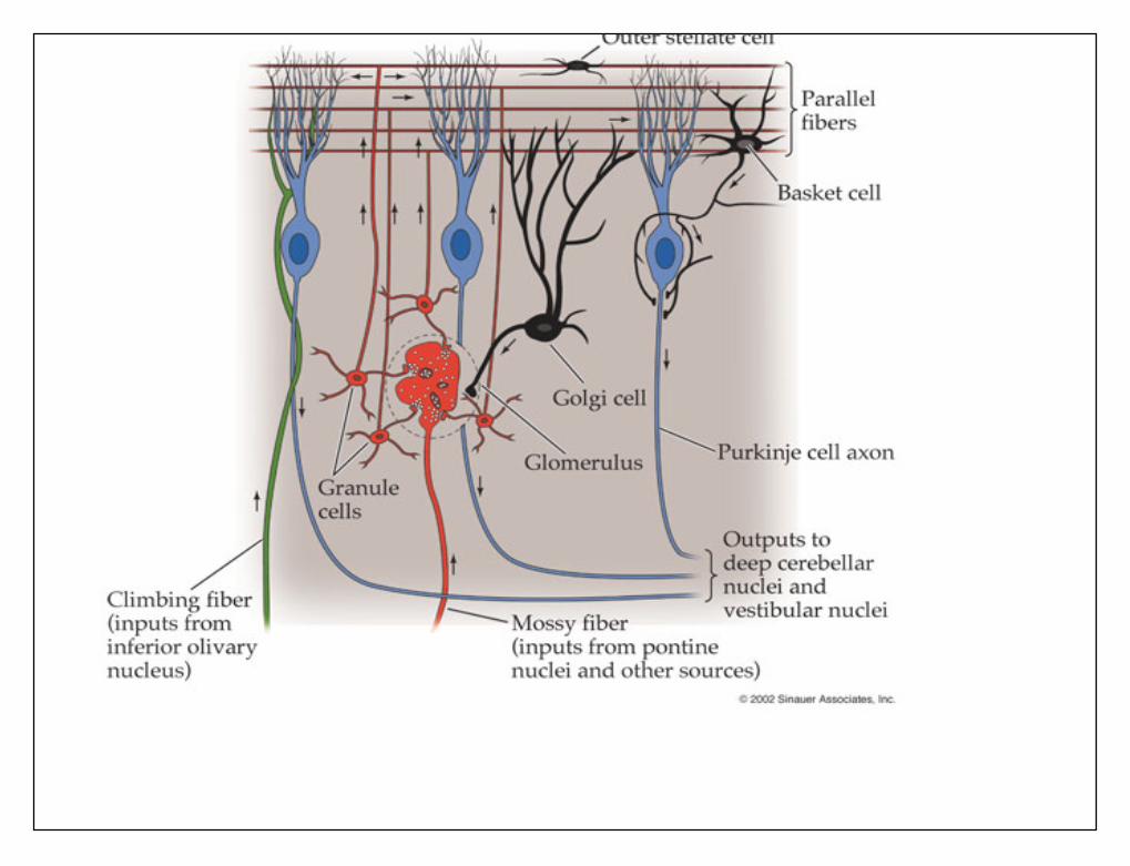
The climbing fibers• The climbing fibers all originate from the inferior olives of the
medulla. There is one climbing fiber for about 5 to 10 Purkinje cells.• After sending branches to several deep nuclear cells, the climbing
fiber continues all the way to outer layers of the cerebellar cortex
• where it makes about 300 synapses with the soma and dendrites ofeach Purkinje cell.
• This climbing fiber is distinguished by the fact that a single impulsein it will always cause a single, prolonged (up to 1second), actionpotential in each Purkinje cell with which it connects, beginningwith a strong spike and followed by a trail of weakening secondaryspikes. This action potential is called the \complex spike.
The mossy fibers• The mossy fibers are all the other fibers that enter cerebellum from
multiple sources: from the higher brain, brain stem, and spinal cord.• These fibers also send collaterals to excite the deep nuclear cells.
Then they proceed to the granule cell layer of the cortex , wherethey too synapse with hundreds to thousands of granule cells. Inturn, the granule cells send very, very small axons, less than 1micrometer in diameter, up to the molecular layer on the outersurface of the cerebellar cortex.
• Here the axons divide into two branches that extend 1 to 2millimeters in each direction parallel to the folia. There are manymillions of these parallel nerve fibers because there are some 500to 1000 granule cells for every 1 Purkinje cell. It is into thismolecular layer that the dendrites of the Purkinje cells project and80,000 to 200,000 of the parallel fibers synapse with each Purkinjecell.
• The mossy fiber input to the Purkinje cell isquite different from the climbing fiber inputbecause their synaptic connections are weak,so that large numbers
• of mossy fibers must be stimulatedsimultaneously to excite the Purkinje cell.Furthermore, activation usually takes the formof a much weaker short duration Purkinje cellaction potential called a simples pike
Purkinj Cells and DeepNuclear cells
• Purkinje cells and deep nuclear cells is thatnormally both of them fire continuously
• the Purkinje cell fires at about 50 to 100action potentials per second
• the deep nuclear cells at much higher rates.• Furthermore , the output activity of both
these cells can be modulated upward ordownward.
Balance Between Excitation andInhibition Deep Cerebellar Nuclei.
• direct stimulation of deep nuclear cells byclimbing and the mossy fibers excites them.
• By contrast, signals arriving from the Purkinjecells inhibit them.
• Normally, the balance between these twoeffects is slightly in favor of excitation,
• under quiet conditions, output from deepnuclear cell remains relatively constant at amoderate level of continuous stimulation.
execution of a rapid motor movement
• initiating signal from the cerebral motor cortex or brain stem at firstgreatly increases deep nuclear cell excitation.
• Then, another few milliseconds later, feedback inhibitory signalsfrom the Purkinje cell circuit arrive.
• there is first a rapid excitatory signal sent by deep nuclear cells intothe motor output pathway to enhance the motor movement, butthis is followed within another small fraction of a second by aninhibitory signal.
• This inhibitory signal resembles a "delay-line" negative feedbacksignal of the type that is effective in providing damping.
• That is, when the motor system is excited, a negative feedbacksignal occurs after a short delay to stop the muscle movement fromovershooting its mark. Otherwise, oscillation of the movementwould occur.
basket cells and stellate cells• These are inhibitory cells with short axons. Both the
basket cells and the stellate cells are located in themolecular layer of the cerebellar cortex, lying amongand stimulated by the small parallel fibers.
• These cells in turn send their axons at right anglesacross the parallel fibers and cause lateral inhibition ofadjacent Purkinje cells, thus sharpening the signal inthe same manner that lateral inhibition sharpenscontrast of signals in many other neuronal circuits ofthe nervous system.
Turn-On/Turn-Off and Turn-Off/Turn-On Output Signals from Cerebellum
• the onset of movement begins with signals from the cerebral cortex.• These signals pass through noncerebellar brain stem and cord pathways
directly to the agonist muscle to begin the initial contraction.• At the same time, parallel signals are sent of mossy fibers into the
cerebellum.• One branch of each mossy fiber goes directly to deep nuclear cells this
instantly sends an excitatory signal back into the cerebral corticospinalmotor system ,return signals through the thalamus to cerebral cortex orby way of neuronal circuitry in the brain stem, to support musclecontraction signal that had already been begun by the cerebral cortex.
• As a consequence, the turn-on signal, after a few milliseconds, becomeseven more powerful than it was at the start because it becomes the sumof both the cortical and the cerebellar signals.
turn-off signal• all mossy fibers have a second branch that transmits signals of the
granule cells to the cerebellar cortex and by way of "parallel“ fibers,to the Purkinje cells.
• The Purkinje cells in turn inhibit the deep nuclear cells. Thispathway passes through some of the smallest, slowest-conductingnerve fibers in the nervous system: that is, the parallel fibers of thecerebellar cortical molecular layer, which have diameters of only afraction of a millimeter
• Also, the signals from these fibers are weak so that they require afinite period of time to build up enough excitation in the dendritesof the Purkinje cell to excite it.
• the Purkinje cell is excited, it in turn sends a strong inhibitory signalto the same deep nuclear cell. this helps to turn off the movementafter a short time
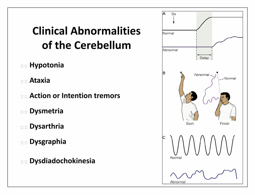
Clinical Abnormalitiesof the Cerebellum
• Hypotonia
• Ataxia
• Action or Intention tremors
• Dysmetria
• Dysarthria
• Dysgraphia
• Dysdiadochokinesia

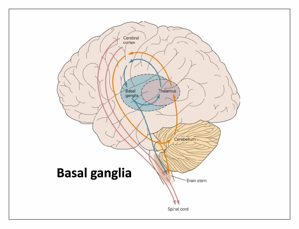
Basal gangliaBasal ganglia
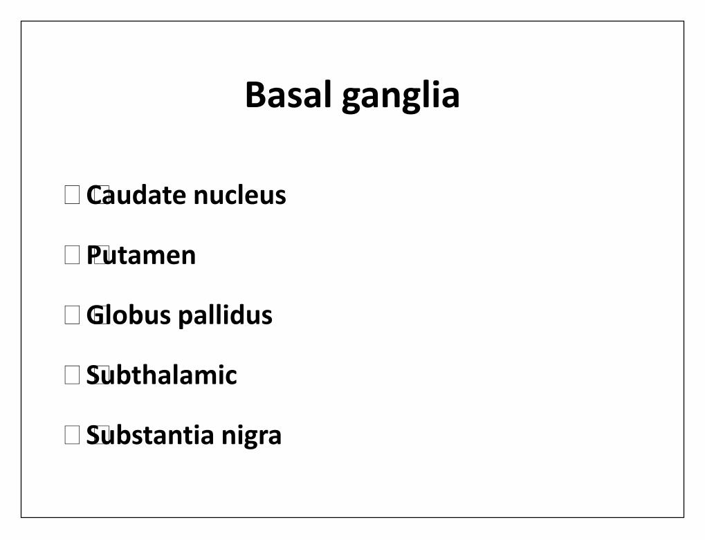
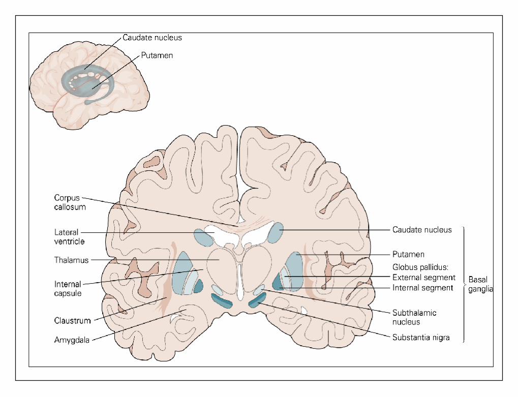
Basal ganglia
• Caudate nucleus
• Putamen
• Globus pallidus
• Subthalamic
• Substantia nigra
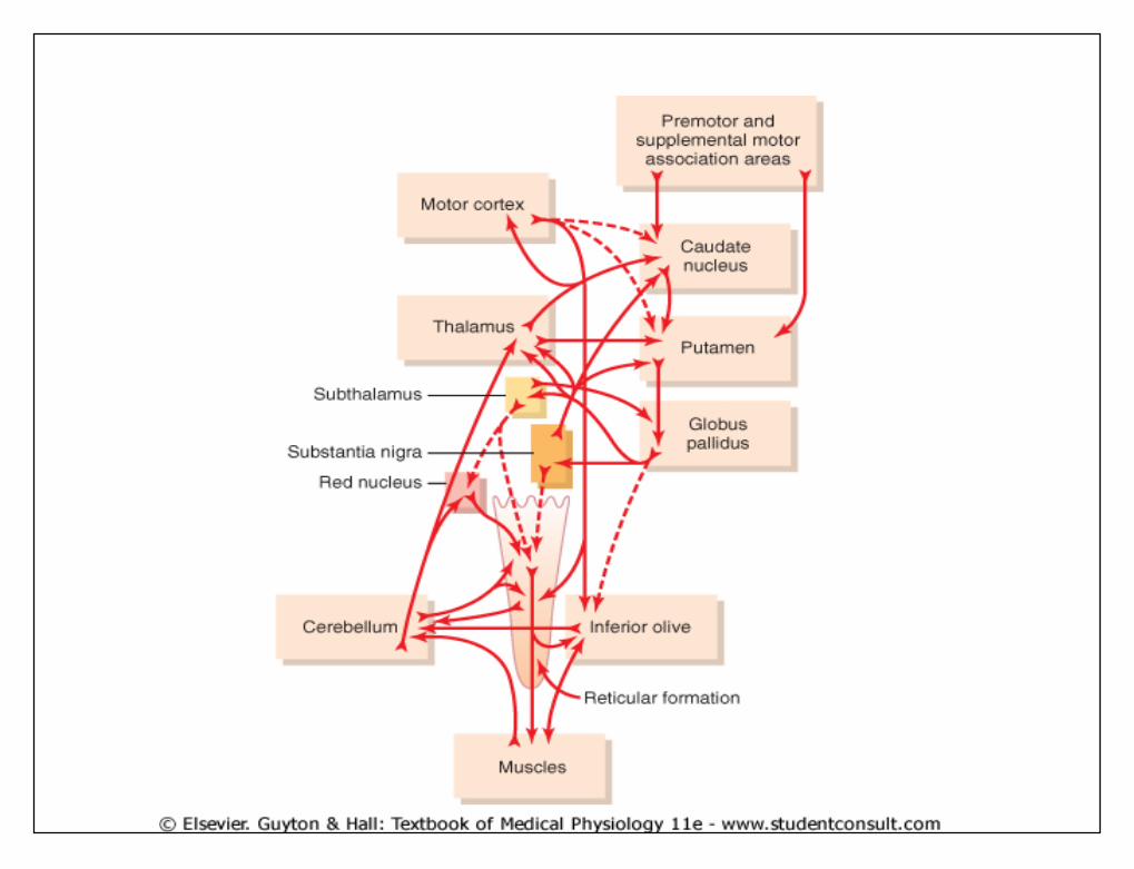
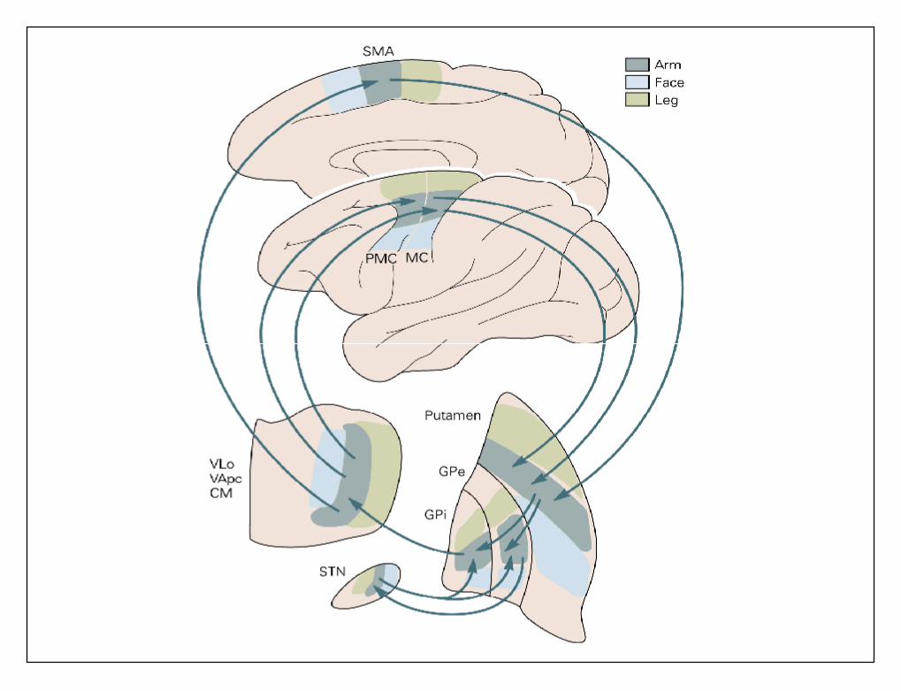
Putamen Circuit• the principal roles of the basal ganglia in motor control
is to function in association with the corticospinalsystem to control complex patterns of motor activity.
• Examples are writing of letters of alphabet. cuttingpaper with scissors, shooting a basketball through ahoop, passing a football, throwing a baseball, themovements of shoveling dirt, most aspects ofvocalization, controlled movements of the eyes, andvirtually any other of our skilled movements, most ofthem performed subconsciously.
•
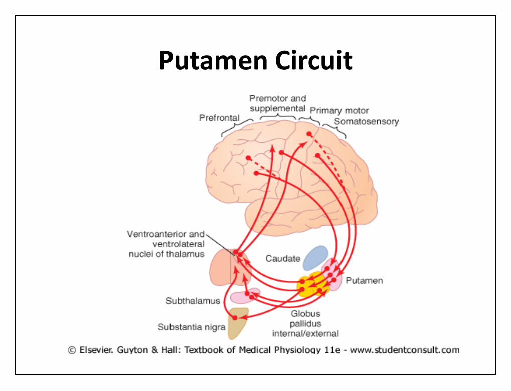
• the principal pathways through the basal gangliafor executing learned patterns of movement.
• They begin mainly in the premotor andsupplementary areas of the motor cortex and inthe somatosensory areas of the sensory cortex.
• Next they pass to the putamen (mainly bypassingthe caudate nucleus), then to the internal portionof the globus pallidus, next to the ventroanteriorand ventrolateral relay nuclei of the thalamus
• finally return to the cerebral primary motorcortex and to portions of the premotor andsupplementary cerebral areas closely associatedwith the primary motor cortex.
Putamen Circuit
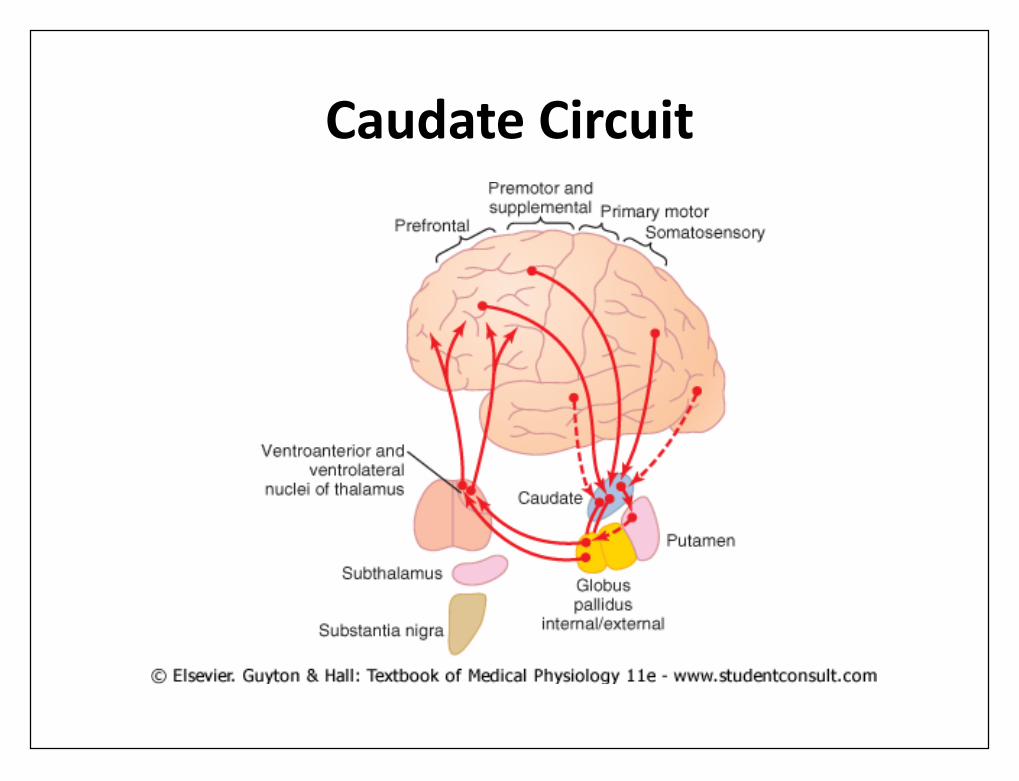
Cognitive Control of Sequences ofMotor Patterns-The Caudate Circuit
• The term cognition means the thinking processesof the brain, using both sensory input to the brainplus information already stored in memory.
• Most of our motor actions occur as aconsequence of thoughts generated in the mind,a process called cognitive control of motoractivity.
• The caudate nucleus plays a major role in thiscognitive control of motor activity.
Caudate Circuit• the caudate nucleus extends into all lobes of the
cerebrum,the caudate nucleus receives large amounts of itsinput from the association areas of the cerebral cortexmainly areas that also integrate different types of sensoryand motor information into usable thought patterns.
• the signals pass from cerebral cortex to caudate nucleus,transmitted to internal globus pallidus, then to nuclei ofventroanterior and ventrolateral thalamus,
• back to prefrontal, premotor, and supplementary motorareas of cerebral cortex, Instead, the returning signals go tothose accessory motor regions that are concerned withputting together sequential patterns of movement lasting5 or more seconds instead of exciting individual musclemovements.
Caudate Circuit
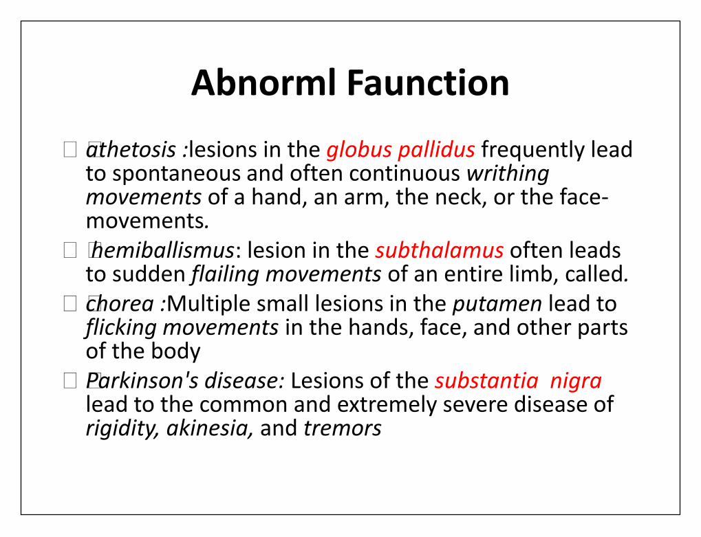
Abnorml Faunction• athetosis :lesions in the globus pallidus frequently lead
to spontaneous and often continuous writhingmovements of a hand, an arm, the neck, or the face-movements.
• hemiballismus: lesion in the subthalamus often leadsto sudden flailing movements of an entire limb, called.
• chorea :Multiple small lesions in the putamen lead toflicking movements in the hands, face, and other partsof the body
• Parkinson's disease: Lesions of the substantia nigralead to the common and extremely severe disease ofrigidity, akinesia, and tremors
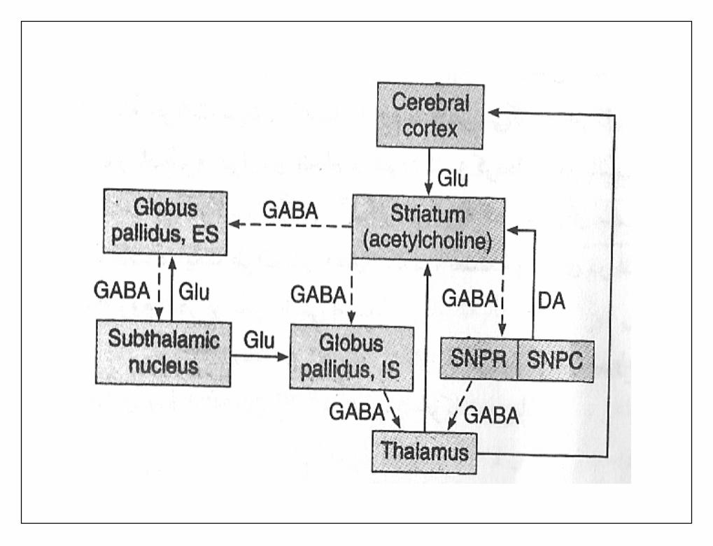
Parkinson'sDiseas• Parkinson's disease, known also as paralysis
agitans,results from widespread destruction ofthat portion of the substantia nigra (the parscompacta) that sends dopamine-secreting nervefibers to the caudate nucleusand putamen.
• The disease is characterized by (1) rigidity ofmuch of the musculature of the body, (2)involuntary tremor of the involved areas evenwhen the person is resting at a fixed rate of 3 to 6cycles per second, and (3) serious difficulty ininitiating movement, called akinesia.
• The causes of these abnormal motor effects areunknown.
• However, the dopamine secreted in the caudatenucleus and putamen is an inhibitory transmitter;
• therefore, destruction of the dopaminergic neurons inthe substantia nigra of the parkinsonian patienttheoretically would allow the caudate nucleus andputamen to become overly active and possibly causecontinuousm output of excitatory signals to thecorticospinal motor control system.
• These signals could overly excite many or all of themuscles of the body, thus leading to rigidity.
Related Documents