The cAMP response element binding protein (CREB) and its role in the nervous system September 2000 - April 2001 Tim Hulsen Supervisor: Prof. Dr. Gerard Martens Department of Molecular Animal Physiology KU Nijmegen

Welcome message from author
This document is posted to help you gain knowledge. Please leave a comment to let me know what you think about it! Share it to your friends and learn new things together.
Transcript
TThhee ccAAMMPP rreessppoonnssee eelleemmeenntt bbiinnddiinngg pprrootteeiinn ((CCRREEBB)) aanndd iittss rroollee iinn tthhee nneerrvvoouuss ssyysstteemm
September 2000 - April 2001
Tim Hulsen
Supervisor: Prof. Dr. Gerard Martens
Department of Molecular Animal Physiology
KU Nijmegen
Index
2
Index
Abstract ............................................................................................................. 3 1. Introduction................................................................................................... 4 1.1. CREB: a transcription factor.............................................................. 4 1.2. The subject of this study ................................................................... 5 2. CREB structure............................................................................................. 6 2.1. CREB structural domains.................................................................. 6 2.2. CREB antagonists............................................................................. 7 3. Activation of CREB....................................................................................... 9 3.1. Multiple activation pathways.............................................................. 9 3.2. Activation by an elevation of the cytosolic cAMP level ...................... 9 3.3. Activation by an elevation of the cytosolic Ca2+ level ...................... 11 4. The role of the CREB binding protein (CBP) ............................................ 14 4.1. CBP is critical for stimulus-induced activation of CREB.................. 14 4.2. KID/KIX domain interaction ............................................................. 14 4.3. CBP mechanism ............................................................................. 15 5. An inducer of long-term memory .............................................................. 17 5.1. Short- and long-term memory ......................................................... 17 5.2. Long-term memory and CREB........................................................ 17 5.3. Mechanism of CREB-induced long-term memory compared to short-term memory.......................................................................... 17 6. The neuroprotective value of CREB.......................................................... 20 6.1. Introduction ..................................................................................... 20 6.2. CREB inhibits apoptosis.................................................................. 20 6.3. Selective vulnerability in the hippocampus...................................... 21 6.4. CREB activation and cell survival.................................................... 22 7. Discussion .................................................................................................. 24 8. References .................................................................................................. 26
Abstract
3
Abstract
The cyclic AMP response element-binding protein (CREB) is a gene regulatory protein: a protein whose presence on the DNA affects the rate of transcription initiation. CREB is able to bind to a short DNA sequence, called the cyclic AMP response element (CRE). The transcription of the gene at the 5� end of the CRE sequence can be induced by activating the CREB protein through an elevation of either cyclic adenosine monophosphate (cAMP) or Ca2+ levels in the cytoplasm. CREB is activated by a phosphorylation of a single serine residue. The binding of CREB and the transcription of the gene can be stimulated by the CREB binding protein (CBP). In this literature study, the role of CREB in the nervous system is discussed, in particular its function in long-term memory (LTM) and its putative neuroprotective value. Its role in long-term memory is rather clear. CREB plays a role in the long-term facilitation, while the short-term facilitation has a pathway without CRE-mediated gene expression and therefore without CREB. CREB induces long-term memory by activating the transcription of ubiquitin hydrolase, leading to persistent phosphorylation of a number of proteins also involved in short-term memory. Additionally, CREB activates proteins which are important for the growth of active zones and the development of new synaptic connections. Its neuroprotective value lies in the fact that selective CREB phosphorylation in the dentate granule cells activates a survival program. CREB acts as a regulator of a general survival program in neurons. This explains the fact that during neuronal cell apoptosis, CREB is cleaved by caspases; the survival program is being destroyed. This neuroprotective function however is not proven yet and still needs a lot of further research. This research should, for example, determine which genes are activated during CREB-mediated neuroprotection. Finally, other functions and effects of CREB in the nervous system, like neuronal compensation, should be researched.
Introduction
4
1. Introduction
1.1. CREB: a transcription factor.
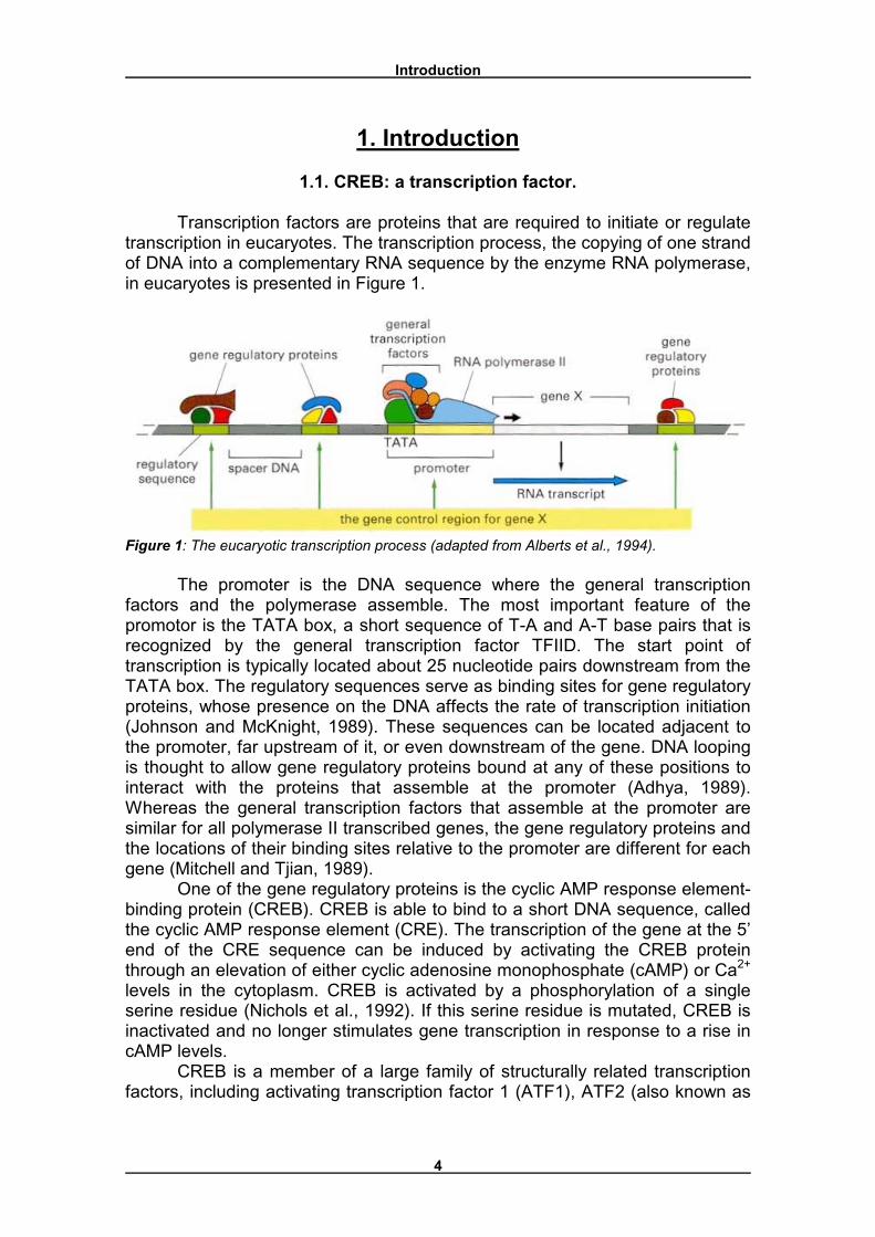
Transcription factors are proteins that are required to initiate or regulate transcription in eucaryotes. The transcription process, the copying of one strand of DNA into a complementary RNA sequence by the enzyme RNA polymerase, in eucaryotes is presented in Figure 1.
Figure 1: The eucaryotic transcription process (adapted from Alberts et al., 1994).
The promoter is the DNA sequence where the general transcription factors and the polymerase assemble. The most important feature of the promotor is the TATA box, a short sequence of T-A and A-T base pairs that is recognized by the general transcription factor TFIID. The start point of transcription is typically located about 25 nucleotide pairs downstream from the TATA box. The regulatory sequences serve as binding sites for gene regulatory proteins, whose presence on the DNA affects the rate of transcription initiation (Johnson and McKnight, 1989). These sequences can be located adjacent to the promoter, far upstream of it, or even downstream of the gene. DNA looping is thought to allow gene regulatory proteins bound at any of these positions to interact with the proteins that assemble at the promoter (Adhya, 1989). Whereas the general transcription factors that assemble at the promoter are similar for all polymerase II transcribed genes, the gene regulatory proteins and the locations of their binding sites relative to the promoter are different for each gene (Mitchell and Tjian, 1989).
One of the gene regulatory proteins is the cyclic AMP response element-binding protein (CREB). CREB is able to bind to a short DNA sequence, called the cyclic AMP response element (CRE). The transcription of the gene at the 5� end of the CRE sequence can be induced by activating the CREB protein through an elevation of either cyclic adenosine monophosphate (cAMP) or Ca2+ levels in the cytoplasm. CREB is activated by a phosphorylation of a single serine residue (Nichols et al., 1992). If this serine residue is mutated, CREB is inactivated and no longer stimulates gene transcription in response to a rise in cAMP levels.
CREB is a member of a large family of structurally related transcription factors, including activating transcription factor 1 (ATF1), ATF2 (also known as
Introduction
5
CREBP1), ATF3 and ATF4, which bind to CRE promoter sites on target genes (Walton and Dragunow, 2000). In common with the inducible transcription factors Fos and Jun, the CREB/ATF proteins consist of three functional domains: a leucine-zipper domain that mediates dimerization, a basic DNA-binding domain and the transcriptional activation domain, which contains important phosphorylation sites. These domains will be discussed in paragraph 2.1.
1.2. The subject of this study
In this study the role of CREB in the nervous system will be discussed, in particular its role in long-term memory (LTM) and its putative neuroprotective value (chapters 5 and 6). Before these functions will be explained, the structure and activation of CREB, by an elevation of either the cytosolic cAMP level or the cytosolic Ca2+ level, and the role of CREB binding protein (CBP) in CREB-induced transcription will be discussed (chapters 2, 3 and 4).
CREB structure
6
2. CREB structure
2.1. CREB structural domains
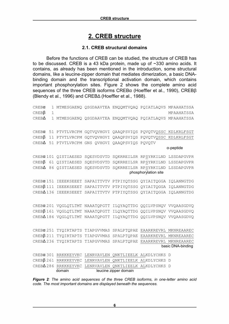
Before the functions of CREB can be studied, the structure of CREB has to be discussed. CREB is a 43 kDa protein, made up of ~330 amino acids. It contains, as already has been mentioned in the introduction, some structural domains, like a leucine-zipper domain that mediates dimerization, a basic DNA-binding domain and the transcriptional activation domain, which contains important phosphorylation sites. Figure 2 shows the complete amino acid sequences of the three CREB isoforms CREBα (Hoeffler et al., 1990), CREBβ (Blendy et al., 1996) and CREB∆ (Hoeffler et al., 1988). CREBα 1 MTMESGAENQ QSGDAAVTEA ENQQMTVQAQ PQIATLAQVS MPAAHATSSACREBβ 1 MPAAHATSSACREB∆ 1 MTMESGAENQ QSGDAAVTEA ENQQMTVQAQ PQIATLAQVS MPAAHATSSA
CREBα 51 PTVTLVRCPM GQTVQVHGVI QAAQPSVIQS PQVQTVQSSC KDLKRLFSGTCREBβ 11 PTVTLVRCPM GQTVQVHGVI QAAQPSVIQS PQVQTVQSSC KDLKRLFSGTCREB∆ 51 PTVTLVRCPM GNS QVHGVI QAAQPSVIQS PQVQTV α-peptide
CREBα 101 QISTIAESED SQESVDSVTD SQKRREILSR RPSYRKILND LSSDAPGVPRCREBβ 61 QISTIAESED SQESVDSVTD SQKRREILSR RPSYRKILND LSSDAPGVPRCREB∆ 86 QISTIAESED SQESVDSVTD SQKRREILSR RPSYRKILND LSSDAPGVPR phosphorylation site
CREBα 151 IEEEKSEEET SAPAITTVTV PTPIYQTSSG QYIAITQGGA IQLANNGTDGCREBβ 111 IEEEKSEEET SAPAITTVTV PTPIYQTSSG QYIAITQGGA IQLANNGTDGCREB∆ 136 IEEEKSEEET SAPAITTVTV PTPIYQTSSG QYIAITQGGA IQLANNGTDG
CREBα 201 VQGLQTLTMT NAAATQPGTT ILQYAQTTDG QQILVPSNQV VVQAASGDVQCREBβ 161 VQGLQTLTMT NAAATQPGTT ILQYAQTTDG QQILVPSNQV VVQAASGDVQCREB∆ 186 VQGLQTLTMT NAAATQPGTT ILQYAQTTDG QQILVPSNQV VVQAASGDVQ
CREBα 251 TYQIRTAPTS TIAPGVVMAS SPALPTQPAE EAARKREVRL MKNREAARECCREBβ 211 TYQIRTAPTS TIAPGVVMAS SPALPTQPAE EAARKREVRL MKNREAARECCREB∆ 236 TYQIRTAPTS TIAPGVVMAS SPALPTQPAE EAARKREVRL MKNREAAREC basic DNA-binding
CREBα 301 RRKKKEYVKC LENRVAVLEN QNKTLIEELK ALKDLYCHKS DCREBβ 261 RRKKKEYVKC LENRVAVLEN QNKTLIEELK ALKDLYCHKS DCREB∆ 286 RRKKKEYVKC LENRVAVLEN QNKTLIEELK ALKDLYCHKS D domain leucine zipper domain Figure 2: The amino acid sequences of the three CREB isoforms, in one-letter amino acid code. The most important domains are displayed beneath the sequences.
CREB structure
7
These different forms of CREB are created by alternative splicing. CREBα and CREB∆ differ only by the presence of a 14-residue insert, termed the α-peptide, in the longer form (amino acids 87 � 100). CREBβ misses the first 40 residues of both CREBα and CREB∆. Through alternative splicing, several other forms of CREB than the three forms mentioned are generated, but these are far less abundant. Serine residue 133 is the phosphorylation site of the CREB protein, a part of the transcriptional activation domain. When this amino acid is phosphorylated, the protein is activated. The amino acids 284 � 309 form the basic DNA-binding domain. This domain mediates binding to the CRE sequence of the DNA. The DNA-binding domain is a lysine- and arginine-rich stretch of amino acids just amino-terminal to the leucine-zipper domain (Dwarki et al., 1990). This leucine-zipper domain is formed by amino acids 311 � 332. This domain contains five leucine residues, which can bind to leucine residues of other CREB proteins and mediate dimerization this way (Yun et al., 1990). The formation of heterodimers, however, is also determined by the presence of intervening polar residues, which form salt bridges to further stabilize heterodimer formation. The basic DNA-binding domain and the leucine-zipper domain together form the bZIP region, which is present in a whole series of transcription factors, like c-Fos, c-Jun and c-Myc. These transcription factors all belong to the bZIP transcription factor family (Shaywitz and Greenberg, 1999).
2.2. CREB antagonists
The three discussed isoforms CREBα, CREBβ and CREB∆, are uniformly expressed in all tissues. However, there are also some other isoforms which are expressed at particularly high levels in the testis (Ruppert et al., 1992, Waeber and Habener, 1992). The biological functions of these isoforms have not been well characterized. However, available data suggest that certain alternatively spliced exons present in these rarer CREB isoforms generate inhibitory forms of CREB that might be important for regulating expression and activity of the active CREB isoforms in germinal cells (Waeber et al., 1991).
Another protein highly related to CREB, is the cAMP response element modulator (CREM, Foulkes et al., 1991). The gene coding for this protein has extensive sequence identity with the CREB gene. By alternative splicing of the CREM gene, either activator or repressor forms of CREM are generated. The α, β and γ isoforms of CREM all bind to CREs, but function as inhibitors of CREB and cAMP-mediated transcription. However, splicing of two additional exons generates a CREM isoform, CREMτ, which is the most similar to CREB with respect to amino acid sequence, that functions as an activator of CRE-mediated transcription (Foulkes et al., 1992, Laiode et al., 1993). Two particularly short forms of CREM are generated through use of either an internal translation initiation site or an internal intronic promoter (Delmas et al., 1992). The resulting products are essentially composed of a DNA-binding and dimerization domain and function as CRE-binding repressors. Unlike CREB, the various isoforms of CREM are not expressed uniformly across different tissues (Laiode et al., 1993). CREMτ mRNA is particularly enriched in testis, with some expression in

CREB structure
8
brain (Foulkes et al., 1992), whereas expression of the truncated CREM isoforms appears to be confined to tissues of the neuroendocrine axis (Delmas et al., 1992).
In this chapter, the structure of CREB has been discussed, which is needed to understand the following chapters: chapter 3 explains the mechanisms of activation, chapter 4 discusses the role of the CREB binding protein (CBP). The subsequent chapters will discuss the functions of CREB.
Activation of CREB
9
3. Activation of CREB
3.1. Multiple activation pathways
As has been mentioned, CREB is activated by phosphorylation of only one residue. This residue is always the same: the serine at position 133 (CREBα numbering). However, this residue can be phosphorylated by many different kinases, including Ca2+/calmodulin-dependent kinases (CaMKs, Sheng et al., 1991), ribosomal protein SG kinases (RSKs, Xing et al., 1996), protein kinase A (PKA), protein kinase C (PKC) and mitogen-activated protein kinase (MAPK, also called extracellular-signal-regulated kinases (ERKs, Nishida and Gotoh, 1993)). The activation of these kinases is induced by a rise in either Ca2+ or cAMP levels in the cytosol. These two induction pathways, in combination with several kinases, will be discussed now.
3.2. Activation by an elevation of the cytosolic cAMP level
Many agents, including neurotransmitters and hormones, act to increase the intracellular level of cAMP. Typically, such a ligand binds to its cognate receptor, most often of the seven-transmembrane domain class of receptors (Strader et al., 1994). Ligand binding of the receptor leads to the activation of a coupled heterotrimeric G-protein, whose activated subunits stimulate one or more of the adenylyl cyclase isoforms, the enzymes catalyzing cAMP production (Gilman, 1987). Production of cAMP is counteracted by the action of phosphodiesterases, the enzymes that cleave cAMP. In most cells, the primary target of cAMP is the cAMP-dependent protein kinase (PKA or cAPK). In the absence of cAMP, PKA consists of an inactive heterotetramer of two catalytic subunits bound to two regulatory subunits (Taylor et al., 1990). Upon binding cAMP, the regulatory subunits dissociate and release the catalytic subunits, which are now free to phosphorylate target proteins. The released catalytic subunits may also translocate to the nucleus (Hagiwara et al., 1993), and the best characterized nuclear substrates of PKA are CREB family members. Pharmacological studies with PKA inhibitors implicated PKA as the mediator of cAMP�s effects on gene expression. In particular, PKA was shown to be required for forskolin induction of transcription of somatostatin promoter-driven reporter genes (Montminy et al., 1986). PKA activation of the somatostatin promoter required an intact CRE, thus implicating CREB as a mediator of the effects of PKA. Subsequent studies have identified a wide range of genes induced by elevated levels of cAMP through a PKA/CRE-dependent mechanism (Montminy, 1997).
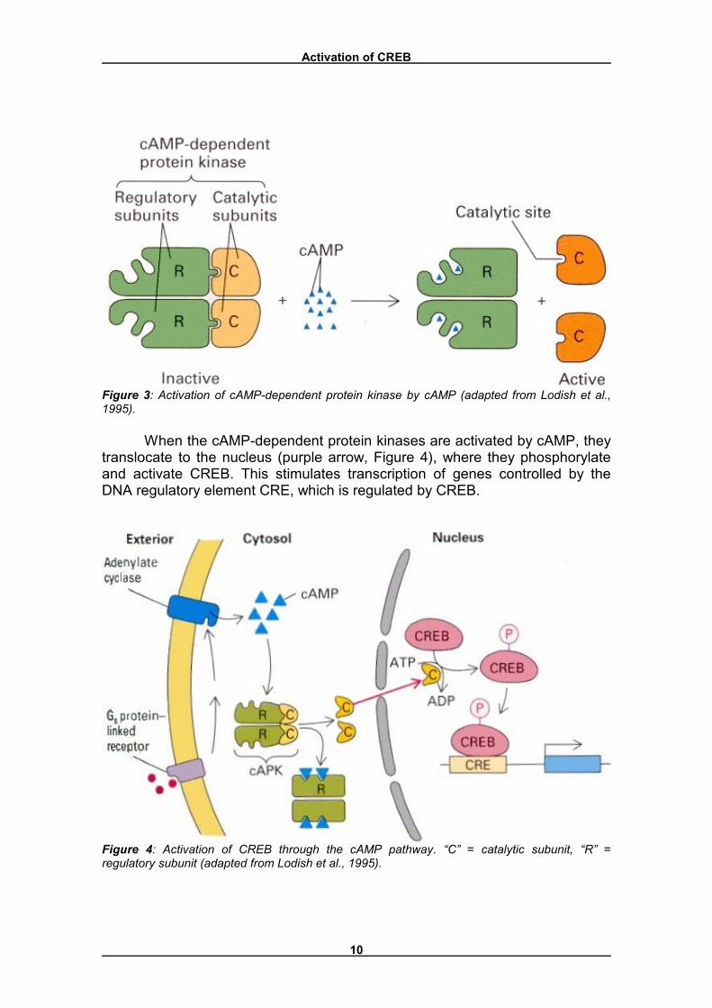
The exact mechanism of the cAMP-dependent CREB activation is shown in Figures 3 and 4. The cAMP-dependent protein kinase contains two regulatory subunits, with binding sites for cAMP, and two catalytic subunits (Figure 3). The tetrameric protein is enzymatically inactive because the regulatory subunits block the catalytic sites on the catalytic subunits. Binding of cAMP, which occurs cooperatively, causes dissociation of the catalytic subunits, which activates the protein kinase (Nichols et al., 1992).
Activation of CREB
10
Figure 3: Activation of cAMP-dependent protein kinase by cAMP (adapted from Lodish et al., 1995).
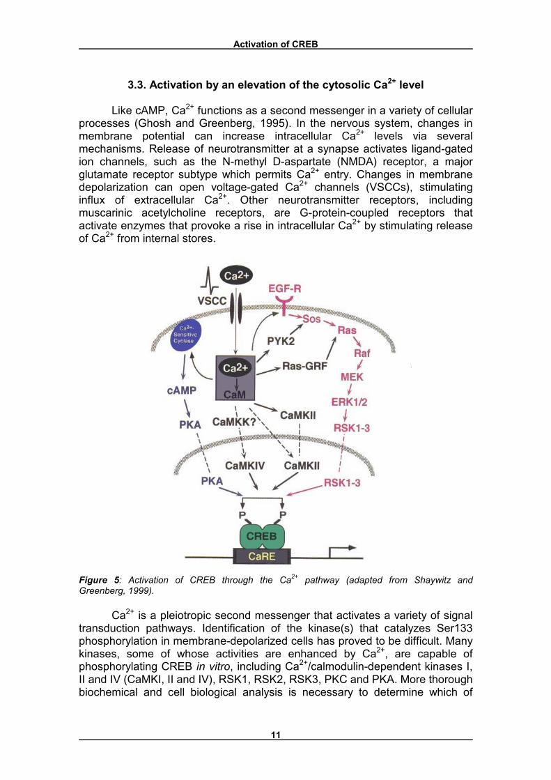
When the cAMP-dependent protein kinases are activated by cAMP, they translocate to the nucleus (purple arrow, Figure 4), where they phosphorylate and activate CREB. This stimulates transcription of genes controlled by the DNA regulatory element CRE, which is regulated by CREB.
Figure 4: Activation of CREB through the cAMP pathway. �C� = catalytic subunit, �R� = regulatory subunit (adapted from Lodish et al., 1995).
Activation of CREB
11
3.3. Activation by an elevation of the cytosolic Ca2+ level
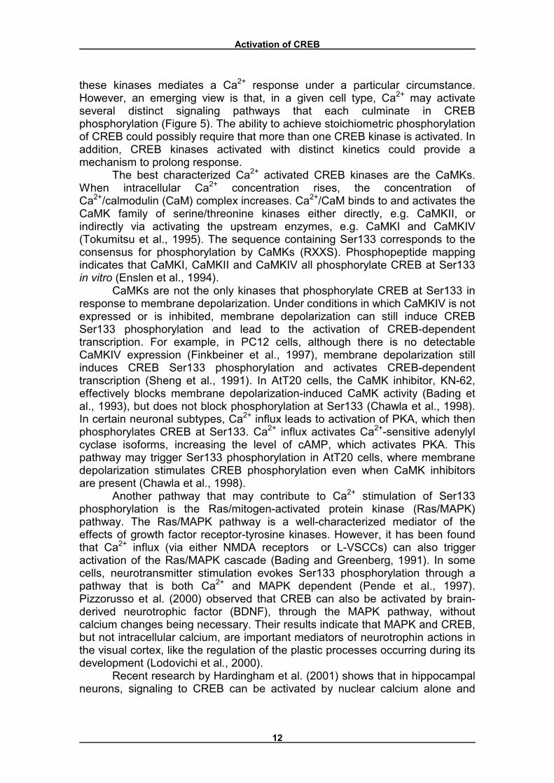
Like cAMP, Ca2+ functions as a second messenger in a variety of cellular processes (Ghosh and Greenberg, 1995). In the nervous system, changes in membrane potential can increase intracellular Ca2+ levels via several mechanisms. Release of neurotransmitter at a synapse activates ligand-gated ion channels, such as the N-methyl D-aspartate (NMDA) receptor, a major glutamate receptor subtype which permits Ca2+ entry. Changes in membrane depolarization can open voltage-gated Ca2+ channels (VSCCs), stimulating influx of extracellular Ca2+. Other neurotransmitter receptors, including muscarinic acetylcholine receptors, are G-protein-coupled receptors that activate enzymes that provoke a rise in intracellular Ca2+ by stimulating release of Ca2+ from internal stores.
Figure 5: Activation of CREB through the Ca2+ pathway (adapted from Shaywitz and Greenberg, 1999).
Ca2+ is a pleiotropic second messenger that activates a variety of signal transduction pathways. Identification of the kinase(s) that catalyzes Ser133 phosphorylation in membrane-depolarized cells has proved to be difficult. Many kinases, some of whose activities are enhanced by Ca2+, are capable of phosphorylating CREB in vitro, including Ca2+/calmodulin-dependent kinases I, II and IV (CaMKI, II and IV), RSK1, RSK2, RSK3, PKC and PKA. More thorough biochemical and cell biological analysis is necessary to determine which of
Activation of CREB
12
these kinases mediates a Ca2+ response under a particular circumstance. However, an emerging view is that, in a given cell type, Ca2+ may activate several distinct signaling pathways that each culminate in CREB phosphorylation (Figure 5). The ability to achieve stoichiometric phosphorylation of CREB could possibly require that more than one CREB kinase is activated. In addition, CREB kinases activated with distinct kinetics could provide a mechanism to prolong response.
The best characterized Ca2+ activated CREB kinases are the CaMKs. When intracellular Ca2+ concentration rises, the concentration of Ca2+/calmodulin (CaM) complex increases. Ca2+/CaM binds to and activates the CaMK family of serine/threonine kinases either directly, e.g. CaMKII, or indirectly via activating the upstream enzymes, e.g. CaMKI and CaMKIV (Tokumitsu et al., 1995). The sequence containing Ser133 corresponds to the consensus for phosphorylation by CaMKs (RXXS). Phosphopeptide mapping indicates that CaMKI, CaMKII and CaMKIV all phosphorylate CREB at Ser133 in vitro (Enslen et al., 1994).
CaMKs are not the only kinases that phosphorylate CREB at Ser133 in response to membrane depolarization. Under conditions in which CaMKIV is not expressed or is inhibited, membrane depolarization can still induce CREB Ser133 phosphorylation and lead to the activation of CREB-dependent transcription. For example, in PC12 cells, although there is no detectable CaMKIV expression (Finkbeiner et al., 1997), membrane depolarization still induces CREB Ser133 phosphorylation and activates CREB-dependent transcription (Sheng et al., 1991). In AtT20 cells, the CaMK inhibitor, KN-62, effectively blocks membrane depolarization-induced CaMK activity (Bading et al., 1993), but does not block phosphorylation at Ser133 (Chawla et al., 1998). In certain neuronal subtypes, Ca2+ influx leads to activation of PKA, which then phosphorylates CREB at Ser133. Ca2+ influx activates Ca2+-sensitive adenylyl cyclase isoforms, increasing the level of cAMP, which activates PKA. This pathway may trigger Ser133 phosphorylation in AtT20 cells, where membrane depolarization stimulates CREB phosphorylation even when CaMK inhibitors are present (Chawla et al., 1998).
Another pathway that may contribute to Ca2+ stimulation of Ser133 phosphorylation is the Ras/mitogen-activated protein kinase (Ras/MAPK) pathway. The Ras/MAPK pathway is a well-characterized mediator of the effects of growth factor receptor-tyrosine kinases. However, it has been found that Ca2+ influx (via either NMDA receptors or L-VSCCs) can also trigger activation of the Ras/MAPK cascade (Bading and Greenberg, 1991). In some cells, neurotransmitter stimulation evokes Ser133 phosphorylation through a pathway that is both Ca2+ and MAPK dependent (Pende et al., 1997). Pizzorusso et al. (2000) observed that CREB can also be activated by brain-derived neurotrophic factor (BDNF), through the MAPK pathway, without calcium changes being necessary. Their results indicate that MAPK and CREB, but not intracellular calcium, are important mediators of neurotrophin actions in the visual cortex, like the regulation of the plastic processes occurring during its development (Lodovichi et al., 2000).
Recent research by Hardingham et al. (2001) shows that in hippocampal neurons, signaling to CREB can be activated by nuclear calcium alone and
Activation of CREB
13
does not require import of cytoplasmic proteins into the nucleus. This suggests that calcium itself, rather than a calcium/calmodulin complex, is the messenger that couples synaptic activity to the nuclear machinery that regulates transcription. However, it cannot be ruled out that signaling through the calcium/calmodulin complex does occur in certain experimental conditions and/or cell types, other than the hippocampal neurons they used. In these cases it would be likely to augment or support nuclear calcium-dependent signaling to CaM kinase IV, the key regulator of neuronal gene expression, or a closely related nuclear CaM kinase.
The role of the CREB-binding protein (CBP)
14
4. The role of the CREB-binding protein (CBP)
4.1. CBP is critical for stimulus-induced activation of CREB
As has been mentioned, CREB induces transcription of a gene through binding to the DNA-regulating element CRE of the particular gene. This mechanism is already shown in Figure 4. The phosphorylated and dimerized CREB molecule recognizes the consensus motif 5�-TGACGTCA-3� of CRE and binds to it, which stimulates the transcription of the gene controlled by the cAMP-response-element. However, phosphorylation and dimerization is not enough for the activation of CREB, which was discovered by Chrivia et al. (1993). To search for factors that associate with CREB in a phosphorylation-dependent manner, a human thyroid cDNA expression library was screened with 32P-labeled CREB and a protein was isolated that specifically bound to Ser133-phosphorylated CREB. This factor, CREB-binding protein (CBP), is a 265-kDa nuclear protein that associates with phosphorylated CREB through a region at the N terminus of CBP known as the KID interaction (KIX) domain. The core KIX domain is a 94-residue sequence (positions 586-679, Parker et al., 1996). The same residues of KID most critical for transcriptional activation by CREB (positions 140 to 160) are also required for interaction of CREB with the KIX domain of CBP, suggesting that CBP binding is important for CREB activity.
Other data support the idea that CBP is critical for stimulus-induced activation of CREB: (a) coexpression of CBP increases stimulus-induced CREB transcription of a CRE reporter gene, an effect that is lost when Ser133 is mutated to an alanine (Kwok et al., 1994); (b) microinjection of cells with neutralizing anti-CBP antibodies inhibits cAMP-induced activation of a CRE reporter gene (Arias et al., 1994); and (c) microinjection of a KIX peptide into cells inhibits stimulus-induced activation of a CRE reporter gene, presumably because the KIX peptide competes with CBP for interaction with CREB (Parker et al., 1996). The KID/KIX interaction may not be sufficient for CREB-mediated transcription because it is possible to mutate a particular residue within the CREB KID and inhibit transcriptional activation without affecting CBP binding (Sun and Maurer, 1995).
4.2. KID/KIX domain interaction
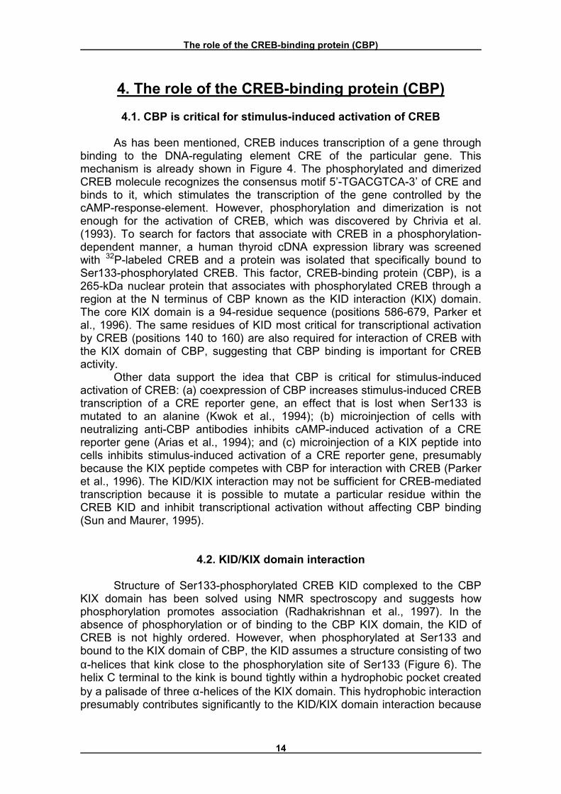
Structure of Ser133-phosphorylated CREB KID complexed to the CBP KIX domain has been solved using NMR spectroscopy and suggests how phosphorylation promotes association (Radhakrishnan et al., 1997). In the absence of phosphorylation or of binding to the CBP KIX domain, the KID of CREB is not highly ordered. However, when phosphorylated at Ser133 and bound to the KIX domain of CBP, the KID assumes a structure consisting of two α-helices that kink close to the phosphorylation site of Ser133 (Figure 6). The helix C terminal to the kink is bound tightly within a hydrophobic pocket created by a palisade of three α-helices of the KIX domain. This hydrophobic interaction presumably contributes significantly to the KID/KIX domain interaction because
The role of the CREB-binding protein (CBP)
15
functional data show that the corresponding segment of CREB (residues 140-144) is critical for transcriptional activation in vivo (Shih et al., 1996).
Figure 6: Ribbon diagram of the interaction between the phosphorylated KID domain of CREB (in blue; Ser-133 in green) and the KIX domain of CBP (in red). PDB ID: 1KDX (Radhakrishnan et al., 1997).
4.3. CBP functions
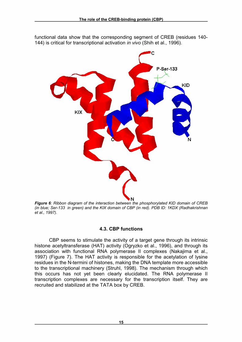
CBP seems to stimulate the activity of a target gene through its intrinsic histone acetyltransferase (HAT) activity (Ogryzko et al., 1996), and through its association with functional RNA polymerase II complexes (Nakajima et al., 1997) (Figure 7). The HAT activity is responsible for the acetylation of lysine residues in the N-termini of histones, making the DNA template more accessible to the transcriptional machinery (Struhl, 1998). The mechanism through which this occurs has not yet been clearly elucidated. The RNA polymerase II transcription complexes are necessary for the transcription itself. They are recruited and stabilized at the TATA box by CREB.
The role of the CREB-binding protein (CBP)
16
Figure 7: The CREB-binding protein as a general mediator of signal-dependent transcription. �NR� = nuclear receptor, �L� = ligand (adapted from Montminy, 1997).
An inducer of long-term memory
17
5. An inducer of long-term memory
5.1. Short- and long-term memory
Memory can be divided into at least two temporally and mechanistically distinct forms: short-term memory, which lasts no longer than several hours, and long-term memory, which lasts days or longer (Goelet et al., 1986). The synthesis of new proteins and mRNA is a universal requirement for the consolidation or formation of long-term memory (Tully et al., 1994). The prevailing model is that learning increases the expression of proteins that generate long-term structural changes in the neuronal circuitry that encodes memories.
5.2. Long-term memory and CREB
Another common feature of long-term memory in vertebrates and invertebrates is a requirement for coincident increases in the second messengers calcium and cAMP (Silva et al., 1992). Indeed, activation of cAMP/PKA signaling may be necessary and sufficient for the induction of neuronal plasticity and learning (Abel et al., 1997) and plasticity-induced gene expression (Impey et al., 1996). Although the role of specific transcriptional cascades in learning and memory has remained elusive, the cAMP/calcium response element (CRE) pathway has several interesting properties that implicate it in long-term memory. Coactivation of calcium and cAMP signaling synergistically increases CRE-mediated gene expression in neurons (Sheng et al., 1990). In addition, CRE-mediated transcription is increased following long-lasting long-term potentiation (L-LTP, Impey et al., 1996), a form of transcription-dependent neuronal plasticity proposed as a model for long-term memory formation (Stevens, 1998). Accordingly, accumulating evidence in both vertebrates and invertebrates suggests that CREB is a key regulator of long-term memory consolidation. For example, inhibition of CREB-mediated transcription attenuates long-term facilitation in Aplysia (Martin et al., 1997). In Drosophila, expression of an inhibitory isoform of CREB blocks the acquisition of long-term memory (Yin et al., 1994), whereas expression of an activating CREB isoform enhances memory formation (Yin et al., 1995). Furthermore, long-term memory formation following massed training is deficient in mice that lack α and ∆CREB (Bourtchuladze et al., 1994). 5.3. Mechanism of CREB-induced long-term memory compared to short-
term memory
Figure 8 shows the differences between short-term facilitation (without CREB) and long-term facilitation (CREB-induced) in Aplysia. In this model, serotonin (5-HT), a transmitter released by facilitatory neurons, acts on a sensory neuron to initiate both the short-term facilitation and the long-term facilitation that contribute to the memory processes.
An inducer of long-term memory
18
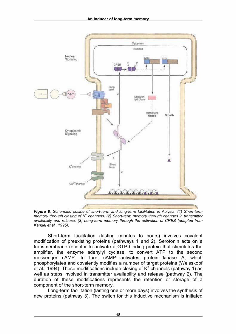
Figure 8: Schematic outline of short-term and long-term facilitation in Aplysia. (1) Short-term memory through closing of K+ channels. (2) Short-term memory through changes in transmitter availability and release. (3) Long-term memory through the activation of CREB (adapted from Kandel et al., 1995).
Short-term facilitation (lasting minutes to hours) involves covalent modification of preexisting proteins (pathways 1 and 2). Serotonin acts on a transmembrane receptor to activate a GTP-binding protein that stimulates the amplifier, the enzyme adenylyl cyclase, to convert ATP to the second messenger cAMP. In turn, cAMP activates protein kinase A, which phosphorylates and covalently modifies a number of target proteins (Weisskopf et al., 1994). These modifications include closing of K+ channels (pathway 1) as well as steps involved in transmitter availability and release (pathway 2). The duration of these modifications represents the retention or storage of a component of the short-term memory.
Long-term facilitation (lasting one or more days) involves the synthesis of new proteins (pathway 3). The switch for this inductive mechanism is initiated
An inducer of long-term memory
19
by the protein kinase A, which translocates to the nucleus where it is thought to phosphorylate one or more transcriptional activators that bind to CREs located in the upstream region of cAMP-inducible genes. The transcriptional activators, thought to belong to the family of CREB proteins (Bourtchuladze et al., 1994, Yin et al., 1994), activate two classes of effector genes that encode two classes of proteins. Inhibiting protein synthesis during learning blocks the expression of these two classes of proteins. These two sets of proteins have distinct functions. One protein, a ubiquitin hydrolase, is a component of a specific protease that leads to downregulation of the regulatory subunit. This results in persistent activity of kinase A, leading to persistent phosphorylation of the substrate proteins of pathways 1 and 2. This persistent phosphorylation may explain why long-term facilitation appears to be a graded extension of the short-term process. The second set of proteins (� in figure 8) is important for the growth of active zones and the development of new synaptic connections. This change was delineated in the sensory and motor cells involved in the gill-withdrawal reflex by examining the synaptic terminals with the electron microscope. The sensory neurons in sensitized animals had twice as many presynaptic terminals as those in untrained animals (Bailey and Chen, 1983). Moreover, long-term sensitization increased the number of active zones from 40% of the synaptic terminals in untrained animals to 65% in trained animals. Finally, in the sensitized animals the dendrites of the motor neurons grew to accommodate the additional synaptic input. Such morphological changes seem to be characteristic only of long-term sensitization; they do not occur with short-term sensitization.
The neuroprotective value of CREB
20
6. The neuroprotective value of CREB
6.1. Introduction
Selective neuronal vulnerability is a feature of a number of neurodegenerative diseases, but the processes that target specific neurons for death while allowing others to remain healthy are unclear. The differential activation of an internal death program in vulnerable neurons has been proposed as a mechanism to explain the selective death of neurons (Schreiber and Baudry, 1995). However, it is equally likely that specific neuronal populations contain an intrinsic survival mechanism. The presence or activity, or both, of such a pathway in different cell types could partly explain their varying sensitivities to detrimental brain insults. Research in this cell-survival area has focused on identifying the key mediators in this survival cascade and has concentrated on endogenous neuroprotective messengers, such as neurotrophic factors and various cytokines (Mattson, 1997). The CREB kinases induced by neurotrophins include ribosomal S-6 kinase-2 (RSK-2), MAPK-activated protein kinase-2 (MAPKAP-2), Ca2+- and calmodulin-dependent kinase IV (CaMKIV) and possibly protein kinase B (PKB/Akt, Finkbeiner, 2000). Recently, several studies implicated CREB not only in the signaling pathway activated by neurotrophines, but also as a possible regulator of a general survival program in neurons. In this chapter, this putative neuroprotective value of CREB will be discussed.
6.2. CREB inhibits apoptosis
Apoptosis, a quick, clean cellular execution, includes shrinkage of neurons, condensation of chromatin, and finally complete cell disintegration (Nijhawan et al., 2000). It is carried out through the activation of caspases, which proteolytically cleave and activate downstream factors that disassemble the cell, such as the actin-severing protein gelsolin and DNase (cleavage of ICAD releases the DNase CAD, Enari et al., 1998). Caspases also inactivate proteins required for survival, such as poly-ADP-ribose polymerase (PARP) and several anti-apoptotic signal transduction molecules, including Ras-GAP, Raf-1, MEKK1 and Akt. Since caspase activity plays a role in the pathogenesis of neurodegenerative disease, and caspase inhibition delays progression of neurodegeneration, for example in mouse models of Huntington�s disease (Ona et al., 1999), it is of biological interest to characterise caspase substrates in neural apoptosis.
CREB, as has been mentioned, regulates many aspects of neuronal function such as nerve cell excitation, CNS development, long-term memory formation (see chapter 5), and circadian rhythms. CREB also plays a key role in regulating neuronal survival and differentiation in response to neurotrophic factors NGF, BDNF, FGF and IGF-1 (Pugazhenthi et al., 1999). Furthermore, CREB appears to be a primary transcriptional activator of the anti-apoptotic gene, bcl-2 (Finkbeiner, 2000, Pugazhenthi et al., 2000). Inhibition of CREB activity induces apoptosis in sympathetic neurones (Riccio et al., 1999), while
The neuroprotective value of CREB
21
CREB overexpression inhibits apoptosis induced by okadaic acid (Walton et al., 1999).
Many metabolic pathways are regulated in both forward and reverse directions, which ensures that futile cycles, reactions in both directions simultaneously, do not occur. The emerging picture is that the regulation of apoptosis follows the same scheme: signaling proteins that turn off the cell death programme are selectively destroyed. François et al. (2000) present evidence that CREB is a target of caspase-3 or -7 during neural cell apoptosis both in vitro and in vivo. CREB appears to be a key target for destruction, being downstream of both Raf-1 and Akt, which are also cleaved by caspases, and upstream of the important survival protein Bcl-2. The inactivation of CREB is thus accomplished by proteolysis rather than by dephosphorylation.
6.3. Selective vulnerability in the hippocampus
Walton and Dragunow (2000) suggest that the selective vulnerability of the hippocampus to brain injury can be explained by the passive involvement of CREB in an active process of neuroprotection. The hippocampal subregions show a distinct pattern of cell death in response to a number of insults, in particular after prolonged seizures and hypoxic-ischemic (HI) episodes (Schreiber and Baudy, 1995). The CA1 pyramidal cells undergo apoptosis after these types of insult, whereas the dentate granule cells remain resistant to damage. A number of years ago it was speculated that this selective vulnerability was due to differential activation of cell-death (in CA1) and cell-survival (in granule cells) programs. In order to identify these programs, the expression of genes for various transcription factors (Fos, Jun, Egr) were examined after a HI insult (Dragunow and Preston, 1995). The CA1 neurons that undergo apoptosis showed a delayed and prolonged expression of Jun, Fos and Nur77 combined with a loss of GER1. Although the resistant dentate granule cells also expressed Jun, as well as Fos, Jund, Junb, Fosb, Nur77 and Egr1, the induction was rapid in onset and more transient. On the basis of these results, the now widely accepted hypothesis, that Jun is involved in neuronal death (Dragunow et al., 1993), was formulated. However, the expression of the genes encoding transcription factors alone does not differentiate between sensitive (CA1) and resistant (dentate granule cells) neurons (although their kinetics of induction are vastly different in the two cell populations, which is likely to have functional consequences). This clearly demonstrates that other factors must be involved in neuronal death, one of which might be CREB activation.
After HI, there is a delayed increase in the phosphorylation of CREB within apoptosis-resistant neurons of the dentate granule-cell layer and neocortex, whereas a dramatic loss of CREB (phosphorylated and unphosphorylated) is found in the dying CA1 pyramidal cells (Walton et al., 1996). The prolonged increase in CREB phosphorylation within the dentate granule cells and its loss in the CA1 neurons, which precedes the onset of cell death, are both consistent with the hypothesis that activated CREB is important for survival of hippocampal neurons. Furthermore, this differential distribution of
The neuroprotective value of CREB
22
CREB protein and its phosphorylation is not confined to this model system; it has been both replicated and demonstrated in another global ischemic paradigm that displays a comparable pattern of cell death (Hu et al., 1999), and after a focal ischemic insult that is characterized by wide-spread neuronal death leading to infarct formation (Tanaka et al., 1999). In the focal-ischemia model, the peri-infarct area shows marked levels of pCREB, while the infarcted core reveals a significant reduction in the number of CREB immunoreactive cells (Tanaka et al., 1999).
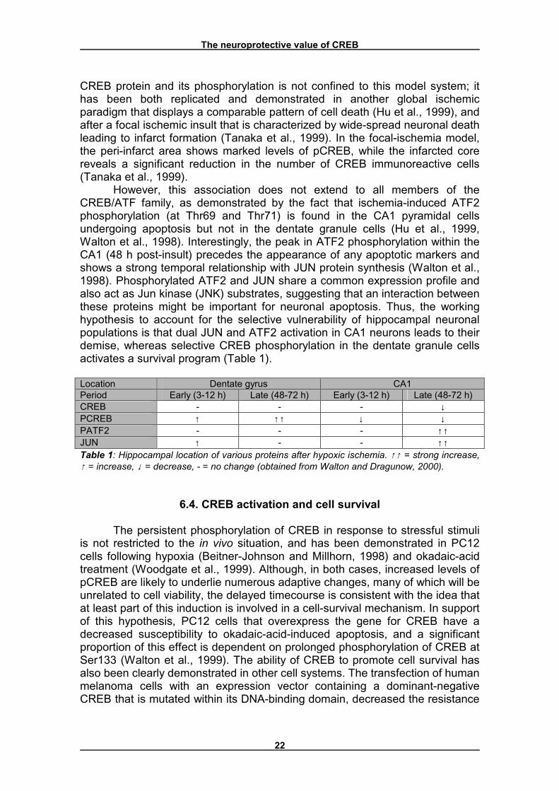
However, this association does not extend to all members of the CREB/ATF family, as demonstrated by the fact that ischemia-induced ATF2 phosphorylation (at Thr69 and Thr71) is found in the CA1 pyramidal cells undergoing apoptosis but not in the dentate granule cells (Hu et al., 1999, Walton et al., 1998). Interestingly, the peak in ATF2 phosphorylation within the CA1 (48 h post-insult) precedes the appearance of any apoptotic markers and shows a strong temporal relationship with JUN protein synthesis (Walton et al., 1998). Phosphorylated ATF2 and JUN share a common expression profile and also act as Jun kinase (JNK) substrates, suggesting that an interaction between these proteins might be important for neuronal apoptosis. Thus, the working hypothesis to account for the selective vulnerability of hippocampal neuronal populations is that dual JUN and ATF2 activation in CA1 neurons leads to their demise, whereas selective CREB phosphorylation in the dentate granule cells activates a survival program (Table 1). Location Dentate gyrus CA1 Period Early (3-12 h) Late (48-72 h) Early (3-12 h) Late (48-72 h) CREB - - - ↓ PCREB ↑ ↑↑ ↓ ↓ PATF2 - - - ↑↑ JUN ↑ - - ↑↑ Table 1: Hippocampal location of various proteins after hypoxic ischemia. ↑↑ = strong increase, ↑ = increase, ↓ = decrease, - = no change (obtained from Walton and Dragunow, 2000).
6.4. CREB activation and cell survival
The persistent phosphorylation of CREB in response to stressful stimuli is not restricted to the in vivo situation, and has been demonstrated in PC12 cells following hypoxia (Beitner-Johnson and Millhorn, 1998) and okadaic-acid treatment (Woodgate et al., 1999). Although, in both cases, increased levels of pCREB are likely to underlie numerous adaptive changes, many of which will be unrelated to cell viability, the delayed timecourse is consistent with the idea that at least part of this induction is involved in a cell-survival mechanism. In support of this hypothesis, PC12 cells that overexpress the gene for CREB have a decreased susceptibility to okadaic-acid-induced apoptosis, and a significant proportion of this effect is dependent on prolonged phosphorylation of CREB at Ser133 (Walton et al., 1999). The ability of CREB to promote cell survival has also been clearly demonstrated in other cell systems. The transfection of human melanoma cells with an expression vector containing a dominant-negative CREB that is mutated within its DNA-binding domain, decreased the resistance
The neuroprotective value of CREB
23
of these cells towards UV-radiation-induced (Yang et al., 1996) and thapsigargin-induced apoptosis (Jean et al., 1998). Transgenic mice that synthesize this dominant-negative form of CREB have thymocytes and T cells with a profound proliferative defect, and show increased sensitivity to apoptosis (Barton et al., 1996). Moreover, suppressed production of CREB has been associated with influenza-A-virus-induced apoptosis of human monocytes (Bussfeld et al., 1997).
The anti-apoptotic effects of CREB might also extend to the neuroprotection conveyed by environmental enrichment. Recently, it was shown that rats exposed to an enriched environment had reduced spontaneous apoptotic cell death in the hippocampus and were protected against kainate-induced seizures and excitotoxic injury (Young et al., 1999). Some of the resistant cell populations in the hippocampus showed increased CREB phosphorylation, which might account for their increased resistance to damage after environmental stimulation. An interaction between CREB activation and the polysialylated form of the neural cell-adhesion molecule (PSA-NCAM) in this system is another possibility, as double immunolabeling showed their co-localization in the apoptotic-resistant precursor cells that align along the hilar-granule-cell border (Young et al., 1999). PSA-NCAM is a cell-to-cell adhesion molecule that is associated with dentate granule-cell neurogenesis, precursor differentiation and migration (Seki and Arai, 1993). In addition, this glycoprotein might activate cell-survival pathways by stimulating CREB phosphorylation in neurons through the RAS-mitogen-activated protein kinase (MAPK) pathway (Schmid et al., 1999). Indeed, disruption of the NCAM receptor by crosslinking induces programmed cell death in neurons (Azizeh et al., 1998).
As has been made clear, many articles suggest that CREB has a neuroprotective value. However, there are also studies that don�t support this suggestion. Hata et al. (1998), for example, shows that after middle cerebral artery occlusion, CREB knockout mice do not show altered neurological scores after three hours, which would be expected if CREB indeed has a protective value in the brain. The fact that CREB is involved in therapies for several neurodegenerative diseases, like depressions and Huntington�s disease (Beal and Hantraye, 2001, Tanaka et al., 2000), however, strengthens the view that CREB is involved in the protection of neurons. Thome et al. (2000) demonstrated that antidepressant treatment increases CREB phosphorylation and CRE-mediated gene expression in limbic brain regions. Steffan et al. (2001) showed that CBP, in combination with CREB, interacts with the Huntington�s disease (HD) protein, as well as with other polyglutamine containing proteins. Beal and Hantraye (2001) suggested that CBP is sequestered in poly-glutamine diseases, resulting in a diminished response to trophic factors, which are essential for neuronal survival. Tanaka et al. (2000), finally, showed that CBP-deficient embryos exhibit defective neural tube closure and defective blood vessel formation in the central nervous system.
Discussion
24
7. Discussion
The structure of CREB, the different activation pathways of CREB and the role of CBP have been made clear by several researches. This knowledge provides a foundation for the research on the functions of CREB in the nervous system. Of the two (possible) functions of CREB, inducing long-term memory and protecting neurons, the first has already been studied intensively. The overall conclusion of these studies is that CREB induces long-term memory by activating the transcription of ubiquitin hydrolase, a component of a specific protease that leads to downregulation of the regulatory subunit. This results in persistent activity of kinase A, leading to persistent phosphorylation of a number of proteins also involved in short-term memory, and the activation of a second set of proteins, important for the growth of active zones and the development of new synaptic connections. This function of CREB has been widely accepted now and does not need an extensive further investigation. The second function however, the protection of neurons, still needs a lot of further research.
It still has to be proven that CREB phosphorylation is neuroprotective in the brain, and that various neuroprotective agents (for example, IGF1) produce neuroprotection by the activation of CREB. Studies on neurons derived from CREB-knockout mice are now required to resolve these issues. In this regard, one recent study has shown that after middle cerebral artery occlusion, CREB knockout mice do not show altered neurological scores (Hata et al., 1998); however, these mice were monitored for only 3 h after the insult, which is before CREB-mediated neuroprotection would be activated. In addition, these mice had a targeted disruption of the α and ∆ isoforms of CREB with compensatory increases in other forms. Furthermore, Duman et al. (2000) suggests that there are other convergent factors, beside CREB, that mediate the actions of neural plasticity and cell death, like BDNF. This could also explain the absence of CREB in the research by Hata et al.. Perhaps the best way of assessing the importance of endogenous CREB phosphorylation to neuronal survival would be to use viral transfection of hippocampal neurons with a dominant-negative CREB construct. If the hypothesis is correct, then this treatment should worsen HI- and seizure-induced hippocampal injury, and block the neuroprotective effects of compounds such as IGF1 and BDNF. Conversely, overexpressing the gene for CREB could protect neurons from various insults, as demonstrated recently in primary cerebellar granule cells (Bonni et al., 1999).
An important issue in the identification of CREB as a neuroprotective protein, could be the discovery made by François et al. (2000), that CREB is cleaved by caspases during neural cell apoptosis both in vitro and in vivo. This corresponds with other processes in the body, like many metabolic pathways, in which the inhibitors of the process are inactivated or even destroyed. The observation that CREB is inactivated by proteolysis rather than by dephosphorylation also needs an explanation. Caspase-mediated cleavage of key proteins involved in survival signaling makes their inactivation irreversible and eliminates possible conflicting signals. That this occurs at a late stage of apoptosis enforces no possibility of escape from execution, and avoids a possibly messy �undead� state somewhere between life and death. In addition, CREB may function without activation by phosphorylation. It has been reported
Discussion
25
that non-phosphorylated CREB has anti-apoptotic activity and can suppress AP-1 (c-Jun/c-Fos heterodimer) transcriptional activity by competing for the AP-1 binding site on target genes (Masquilier and Sassone-Corsi, 1992).
In addition to its neuroprotective value, CREB might also have a function in neuronal compensation. Kim et al. (2000) suggested that the activation of CREB plays a role in the initial events of vestibular compensation in rats. Because of this observation, and because of CREB having a function in both memory formation (through both persistent activation of kinase A and the development of new synaptic connections) and neuronal survival, CREB-protein targeting might be an ideal way to achieve neuroprotective cognitive enhancement. This strategy will have major implications for treating Alzheimer�s disease, although genes activated by CREB after learning may not be the same as those activated during neuronal stress. For example, differences in the degree and kinetics of CREB phosphorylation might lead to different functional outcomes. The challenge for the future is to determine which genes are activated during CREB-mediated neuroprotection (and plasticity), and to develop novel approaches that either mimic the actions of CREB in long-term memory and survival, or enhance its endogenous activity within the brain. In addition, other effects of CREB should be taken into account when developing these new approaches, for its role in the nervous system might not be restricted to the functions mentioned here.
References
26
8. References
Abel, T., Nguyen, P.V., Barad, M., Deuel, T.A.S. and Kandel, E.R. (1997). Genetic demonstration of a role for PKA in the late phase of LTP and in hippocampus-based long-term memory. Cell 88, 615-626. Adhya, S. (1989). Multipartite genetic control elements: communication by DNA looping. Annu. Rev. Genet. 23, 227-250. Alberts, B., Bray, D., Lewis, J., Raff, M., Roberts, K. and Watson, J.D. (1994). Molecular biology of the cell. Third edition. Arias, J., Alberts, A.S., Brindle, P., Claret, F.X., Smeal, T., Karin, M., Feramisco, J. and Montminy, M. (1994). Activation of cAMP and mitogen responsive genes relies on a common nuclear factor. Nature 370, 226-229. Azizeh, B.Y., Cribbs, D.H., Kreng, V.M. and Cotman, C.W. (1998). Cross-linking of NCAM receptors on neurons induces programmed cell death. Brain Res. 796, 20-26. Bading, H. and Greenberg, M.E. (1991). Stimulation of protein tyrosine phosphorylation by NMDA receptor activation. Science 253, 912-914. Bading, H., Ginty, D.D. and Greenberg, M.E. (1993). Regulation of gene expression in hippocampal neurons by distinct calcium signaling pathways. Science 260, 181-186. Bailey, C.H. and Chen, M. (1983). Morphological basis of long-term habituation ans sensitization in Aplysia. Science 220, 91-93. Barton, K., Muthusamy, N., Chanyangam, M., Fischer, C., Clendenin, C. and Leiden, J.M. (1996). Defective thymocyte proliferation and IL-2 production in transgenic mice expressing a dominant-negative form of CREB. Nature 379, 81-85. Beal, M.F. and Hantraye, P. (2001). Novel therapies in the search for a cure for Huntington�s disease. Proc. Natl. Acad. Sci. USA 98, 3-4. Beitner-Johnson, D. and Millhorn, D.E. (1998). Hypoxia induces phosphorylation of the cyclic AMP response element-binding protein by a novel signaling mechanism. J. Biol. Chem. 273, 19834-19839. Blendy, J.A., Kästner, K.H., Schmid, W., Gass, P. and Schutz, G. (1996). Targeting of the CREB gene leads to up-regulation of a novel CREB mRNA isoform. EMBO J. 15, 1098-1106. Bonni, A., Brunet, A., West, A.E., Datta, S.R., Takasu, M.A. and Greenberg, M.E. (1999). Cell survival promoted by the Ras-MAPK signaling pathway by transcription-dependent and -independent mechanisms. Science 286, 1358-1362. Bourtchuladze, R., Frenguelli, B., Blendy, J., Cioffi, D., Schutz, G. and Silva, A.J. (1994). Deficient long-term memory in mice with a targeted mutation of the cAMP-responsive element-binding protein. Cell 79, 59-68. Bussfeld, D., Bacher, M., Moritz, A., Gemsa, D. and Sprenger, H. (1997). Expression of transcription factor genes after influenza A virus infection. Immunobiology 198, 291-298. Chawla, S., Hardingham, G.E., Quinn, D.R. and Bading, H. (1998). CBP: a signal-regulated transcriptional coactivator controlled by nuclear calcium and CaM kinase IV. Science 281, 1505-1509.
References
27
Chrivia, J.C., Kwok, R.P., Lamb, N., Hagiwara, M., Montminy, M.R. and Goodman, R.H. (1993). Phosphorylated CREB binds specifically to the nuclear protein CBP. Nature 365, 855-859. Delmas, V., Laoide, B.M., Masquilier, D., De Groot, R.P., Foulkes, N.S., Sassone-Corsi, P. (1992). Alternative usage of initiation codons in mRNA encoding the cAMP-responsive-element modulator generates regulators with opposite functions. Proc. Natl. Acad. Sci. USA 89, 4226-4230. Dragunow, M., Young, D., Hughes, P., MacGibbon, G., Lawlor, P., Singleton, K., Sirimanne, E., Beilharz, E. and Gluckman, P. (1993). Is c-Jun involved in nerve cell death following status epilepticus and hypoxic-ischaemic brain injury? Mol. Brain Res. 18, 347-352. Dragunow, M. and Preston, K. (1995). The role of inducible transcription factors in apoptotic nerve cell death. Brain Res. Rev. 21, 1-28. Duman, R.S., Malberg, J., Nakagawa, S. and D�Sa, C. (2000). Neuronal plasticity and survival in mood disorders. Biol. Psychiatry 48, 732-739. Dwarki, V.J., Montminy, M. and Verma, I.M. (1990). Both the basic region and the �leucine zipper� domain of the cyclic AMP response element binding (CREB) protein are essential for transcriptional activation. EMBO J. 9, 225-232. Enari, M., Sakahira, H., Yokoyama, H., Okawa, K., Iwamitsu, A. and Nagata, S. (1998). A caspase-activated DNase that degrades DNA during apoptosis, and its inhibitor ICAD. Nature 391, 43-50. Enslen, H., Sun, P.Q., Brickey, D., Soderling, S.H., Klamo, E. and Soderling, T.R. (1994). Characterization of Ca2+/calmodulin-dependent protein kinase IV. Role in transcriptional regulation. J. Biol. Chem. 269, 15520-15527. Finkbeiner, S., Tavazoie, S.F., Maloratsky, A., Jacobs, K.M., Harris, K.M. and Greenberg, M.E. (1997). CREB: a major mediator of neuronal neurotrophin responses. Neuron 19, 1031-1047. Finkbeiner, S. (2000). CREB couples neurotrophin signals to survival messages. Neuron 25, 11-14. Foulkes, N.S., Borrelli, E. and Sassone-Corsi, P. (1991). CREM gene: use of alternative DNA-binding domains generates multiple antagonists of cAMP-induced transcription. Cell 64, 739-749. Foulkes, N.S., Mellstrom, B., Benusiglio, E. and Sassone-Corsi, P. (1992). Developmental switch of CREM function during spermatogenesis: from antagonist to activator. Nature 355, 80-84. François, F., Godinho, M.J. and Grimes, M.L. (2000). CREB is cleaved by caspases during neural cell apoptosis. FEBS Lett. 486, 281-284. Freeman, T.B., Cicchetti, F., Hauser, R.A., Deacon, T.W., Li, X.-J., Hersch, S.M., Nauert, G.M., Sanberg, P.R., Kordower, J.F. and Isacson, O. (2000). Transplanted fetal striatum in Huntington's disease: phenotypic development and lack of pathology. Proc. Natl. Acad. Sci. USA 97, 13877-13882. Ghosh, A. and Greenberg, M.E. (1995). Calcium signaling in neurons: molecular mechanisms and cellular consequences. Science 268, 239-247. Gilman, A.G. (1987). G proteins: transducers of receptor-generated signals. Annu. Rev. Biochem. 56, 615-649.
References
28
Goelet, P., Castellucci, V.F., Schacher, S. and Kandel, E.R. (1986). The long and short of long-term memory � a molecular framework. Nature 322, 419-422. Hagiwara, M., Brindle, P., Harootunian, A., Armstrong, R., Rivier, J., Vale, W, Tsien, R. and Montminy, M.R. (1993). Coupling of hormonal stimulation and transcription via the cyclic AMP-responsive factor CREB is rate limited by nuclear entry of protein kinase A. Mol. Cell. Biol. 13, 4852-4859. Hardingham, G.E., Arnold, F.J.L. and Bading, H. (2001). Nuclear calcium signaling controls CREB-mediated gene expression triggered by synaptic activity. Nature Neurosci. 4, 261-267. Hata, T., Gass, P., Mies, G., Wiessner, C. and Hossmann, K.A. (1998). Attenuated c-fos mRNA induction after middle cerebral artery occlusion in CREB knockout mice does not modulate focal ischemic injury. J. Cereb. Blood Flow Metab. 18, 1325-1335. Hoeffler, J.P., Meyer, T.E., Yun, Y., Jameson, J.L. and Habener, J.F. (1988). Cyclic AMP-responsive DNA-binding protein: structure based on a cloned placental cDNA. Science 242, 1430-1433. Hoeffler, J.P., Meyer, T.E., Waeber, G., Habener, J.F. (1990). Multiple adenosine 3�,5�-cyclic monophosphate response element DNA-binding proteins generated by gene diversification and alternative exon splicing. Mol. Endrocinol. 4, 920-930. Hu, B.R., Fux, C.M., Martone, M.E., Zivin, J.A. and Ellisman, M.H. (1999). Persistent phosphorylation of cyclic AMP responsive element-binding protein and activating transcription factor-2 transcription factors following transient cerebral ischemia in rat brain. Neuroscience 89, 437-452. Impey, S., Mark, M., Villacres, E.C., Poser, S., Chavkin, C. and Storm, D.R. (1996). Induction of CRE-mediated gene expression by stimuli that generate long-lasting LTP in area CA1 of the hippocampus. Neuron 16, 973-982. Jean, D., Harbison, M., McConkey, D.J., Ronai, Z. and Bar-Eli, M. (1998). CREB and its associated proteins act as survival factors for human melanoma cells. J. Biol. Chem. 273, 24884-24890. Johnson, P.F. and McKnight, S.L. (1989). Eukaryotic transcriptional regulatory proteins. Annu. Rev. Biochem. 58, 799-839. Kandel, E.R., Schwartz, J.H. and Jessell, T.M. (1995). Essentials of neural science and behavior. Kim, M.S., Kim, J.H., Lee, M.Y., Chun, S.W., Lee, S.H. and Park, B.R. (2000). Identification of phosphorylated form of cAMP/calcium response element binding protein expression in the brain stem nuclei at early stage of vestibular compensation in rats. Neurosci. Lett. 290, 173-176. Kwok, R.P., Lundblad, J.R., Chrivia, J.C., Richards, J.P., Bächinger, J.P., Brennan, R.G., Roberts, S.G.E., Green, M.R. and Goodman, R.H. (1994). Nuclear protein CBP is a coactivator for the transcription factor CREB. Nature 370, 223-226. Laiode, B.M., Foulkes, N.S., Schlotter, F. and Sassone-Corsi, P. (1993). The functional versatility of CREM is determined by its modular structure. EMBO J. 12, 1179-1191. Landschulz, W.H., Johnson, P.F. and McKnight, S.L. (1988). The leucine zipper: a hypothetical structure common to a new class of DNA binding proteins. Science 240, 1759-1764.
References
29
Lodish, H., Baltimore, D., Berk, A., Zipursky, S.L., Matsudaira, P. and Darnell, M. (1996). Molecular cell biology. Third edition. Lodovichi, C., Berardi, N., Pizzorusso, T. and Maffei, L. (2000). Effects of neurotrophins on cortical plasticity: same or different? J. Neurosci. 20, 2155-2165. Martin, K.C., Casadio, A., Zhu, H., E, Y., Rose, J.C., Chen, M., bailey, C.H. and Kandel, E.R. (1997). Synapse-specific, long-form facilitation of Aplysia sensory to motor synapses: a function for local protein synthesis in memory storage. Cell 91, 927-938. Masquilier, D. and Sassone-Corsi, P. (1992). Transcriptional cross-talk: nuclear factors CREM and CREB bind to AP-1 sites and inhibit activation by Jun. J. Biol. Chem. 267, 22460-22466. Mattson, M.P. (1997). Neuroprotective signal transduction: relevance to stroke. Neurosci. Biobehav. Rev. 21, 193-206. Mitchell, P.J. and Tjian, R. (1989). Transcriptional regulation in mammalian cells by sequence-specific DNA-binding proteins. Science 245, 371-378. Montminy, M.R., Sevarino, K.A., Wagner, J.A., Mandel, G. and Goodman, R.H. (1986). Identification of a cyclic-AMP-responsive element within the rat somatostatin gene. Proc. Natl. Acad. Sci. USA 83, 6682-6686. Montminy, M. (1997). Transcriptional regulation by cyclic AMP. Annu. Rev. Biochem. 66, 807-822. Montminy, M. (1997). Something new to hang your HAT on. Nature 387, 654-655. Nakajima, T., Uchida, C., Anderson, S.F., Parvin, J.D. and Montminy, M. (1997). Analysis of a cAMP-responsive activator reveals a two-component mechanism for transcriptional induction via signal-dependent factors. Genes Dev. 11, 738-747. Nichols, M., Weih, F., Schmid, W., DeVack, C., Kowenz-Leutz, E., Luckow, B., Boshart, M. and Schütz, G. (1992). Phosphorylation of CREB affects its binding to high and low affinity sites: implications for cAMP induced gene transcription. EMBO J. 11, 3337-3346. Nijhawan, D., Honarpour, N. and Wang, X. (2000). Apoptosis in neural development and disease. Annu. Rev. Neurosci. 23, 73-87. Nishida, E. and Gotoh, Y. (1993). The MAP kinase cascade is essential for diverse signal transduction pathways. Trends Biochem. Sci. 18, 128-130. Ogryzko, V.V., Shiltz, R.L., Russanova, V., Howard, B.H. and Nakatani, Y. (1996). The transcriptional coactivators p300 and CBP are histone acetyltransferases. Cell 87, 953-960. Ona, V.O., Li, M., Vonsattel, J.P., Andrews, L.J., Khan, S.Q., Chung, W.M., Frey, A.S., Menon, A.S., Li, X.J., Stieg, P.E., Yuan, J., Penney, J.B., Young, A.B., Cha, J.H. and Friedlander, R.M. (1999). Inhibition of caspase-1 slows disease progression in a mouse model of Huntington's disease. Nature 399, 263-267. Parker, D., Ferreri, K., Nakajima, T., LaMorte, V.J., Evans, R., Koerber, S.C., Hoeger, C. and Montminy, M.R. (1996). Phosphorylation of CREB at Ser-133 induces complex formation with CREB-binding protein via a direct mechanism. Mol. Cell. Biol. 16, 694-703. Pende, M., Fisher, T.L., Simpson, P.B., Russell, J.T., Blenis, J. and Gallo, V. (1997). Neurotransmitter- and growth factor-induced cAMP response element binding protein phosphorylation in glial cell progenitors: role of calcium ions, protein kinase C, and mitogen-activated protein kinase/ribosomal S6 kinase pathway. J. Neurosci. 17, 1291-1301.
References
30
Pizzorusso, T., Ratto, G.M., Putignano, E. and Maffei, L. (2000). Brain-derived neurotrophic factor causes cAMP response element-binding protein phosphorylation in absence of calcium increases in slices and cultured neurons from rat visual cortex. J. Neurosci. 20, 2809-2816. Pugazhenthi, S., Boras, T. O�connor, D., Meintzer, M.K., Heidenreich, K.A. and Reusch, J.E. (1999). Insulin-like growth factor I-mediated activation of the transcription factor cAMP response element-binding protein in PC12 cells. Involvement of p38 mitogen-activated protein kinase-mediated pathway. J. Biol. Chem. 274, 2829-2837. Pugazhenthi, S., Nesterova, A., Sable, C., Heidenreich, K.A., Boxer, L.M., Heasley, L.E. and Reusch, J.E. (2000). Akt/protein kinase B up-regulates Bcl-2 expression through cAMP-response element-binding protein. J. Biol. Chem. 275, 10761-10766. Radhakrishnan, I., Perez-Alvarado, G.C., Parker, D., Dyson, H.J., Montminy, M.R. and Wright, P.E. (1997). Solution structure of the KIX domain of CBP bound to the transactivation domain of CREB: a model for activator:coactivator interactions. Cell 91, 741-752. Riccio, A., Ahn, S., Davenport, C.M., Blendy, J.A. and Ginty, D.D. (1999). Mediation by a CREB family transcription factor of NGF-dependent survival of sympathetic neurons. Science 286, 2358-2361. Ruppert, S., Cole, T.J., Boshart, M., Schmid, E. and Schutz, G. (1992). Multiple mRNA isoforms of the transcription activator protein CREB: generation by alternative splicing and specific expression in primary spermatocytes. EMBO J. 11, 1503-1512. Schmid, R.S., Graff, R.D., Schaller, M.D., Chen, S., Schachner, M., Hemperly, J.J. and Maness, P.F. (1999). NCAM stimulates the Ras-MAPK pathway and CREB phosphorylation in neuronal cells. J. Neurobiol. 38, 542-558. Schreiber, S.S. and Baudry, M. (1995). Selective neuronal vulnerability in the hippocampus: a role for gene expression? Trends Neurosci. 18, 446-451. Seki, T. and Arai, Y. (1993). Highly polysialyted neural cell adhesion molecule (NCAM-H) is expressed by newly generated granule cells in the dentate gyrus of the adult rat. J. Neurosci. 13, 2351-2358. Shaywitz, A.J. and Greenberg, M.E. (1999). CREB: a stimulus-induced transcription factor activated by a diverse array of extracellular signals. Annu. Rev. Biochem. 68, 821-861. Sheng, M., McFadden, G. and Greenberg, M.E. (1990). Membrane depolarization and calcium induce c-fos transcription via phosphorylation of transcription factor CREB. Neuron 4, 571-582. Sheng, M., Thompson, M.A. and Greenberg, M.E. (1991). CREB: a Ca2+-regulated transcription factor phosphorylated by calmodulin-dependent kinases. Science 252, 1427-1430. Shih, H.M., Goldman, P.S., DeMaggio, A.J., Hollenberg, S.M., Goodman, R.H. and Hoekstra, M.F. (1996). A positive genetic selection for disrupting protein-protein interactions: identification of CREB mutations that prevent association with the coactivator CBP. Proc. Natl. Acad. Sci. USA 93, 13896-13901. Silva, A.J., Paylor, R., Wehner, J.M. and Tonegawa, S. (1992). Impaired spatial learning in alpha calcium calmodulin kinase II mutant mice. Science 257, 206-210. Steffan, J.S., Kazantsev, A., Spasic-Boskovic, O., Greenwald, M., Zhu, Y.Z., Gohler, H., Wankner, E.E., Bates, G.P., Housman, D.E. and Thompson, L.M. (2000). The Huntington's disease protein interacts with p53 and CREB-binding protein and represses transcription. Proc. Natl. Acad. Sci. USA 97, 6763-6768.
References
31
Stevens, C.F. (1998). A million dollar question: does LTP = memory? Neuron 20, 1-2. Strader, C.D., Fong, T.M., Tota, M.R., Underwood, D. and Dixon, R.A.F. (1994). Structure and function of G protein-coupled receptors. Annu. Rev. Biochem. 63, 101-132. Struhl, K. (1998). Histone acetylation and transcriptional regulatory mechanisms. Genes Dev. 12, 599-606. Sun, P. and Maurer, R.A. (1995). An inactivating point mutation demonstrates that interaction of cAMP response element binding protein (CREB) with the CREB binding protein is not sufficient for transcriptional activation. J. Biol. Chem. 270, 7041-7044. Tanaka, K., Nagata, E., Suzuki, S., Dembo, T., Nogawa, S. and Fukuuchi, Y. (1999). Immunohistochemical analysis of cyclic AMP response element binding protein phosphorylation in focal cerebral ischemia in rats. Brain Res. 818, 520-526. Tanaka, Y., Naruse, I., Hongo, T., Xu, M.-J., Nakahata, T., Maekawa, T. and Ishii, S. (2000). Extensive brain hemorrhage and embryonic lethality in a mouse null mutant of CREB-binding protein. Mech. Dev. 95, 133-145. Taylor, S.S., Buechler, J.A. and Yonemoto, W. (1990). cAMP-dependent protein kinase: framework for a diverse family of regulatory enzymes. Annu. Rev. Biochem. 59, 971-1005. Thome, J., Sakai, N., Shin, K.H., Steffen, C., Zhang, Y.-J., Impey, S., Storm D. and Duman R.S. (2000). cAMP-response-element mediated gene transcription is up-regulated by chronic antidepressant. J. Neurosci. 20, 4030-4036. Tokomitsu, H., Enslen, H. and Soderling, T.R. (1995). Characterization of a Ca2+/calmodulin-dependent protein kinase cascade. Molecular cloning and expression of calcium/calmodulin-dependent protein kinase kinase. J. Biol. Chem. 270, 19320-19324. Tully, T., Preat, T., Boynton, S.C. and Del Vecchio, M. (1994). Genetic disection of consolidated memory in Drosophila. Cell 79, 35-47. Waeber, G., Meyer, T.E., LeSieur, M., Hermann, H.L., Gerard, N. and Habener, J.F. (1991). Developmental stage-specific expression of cyclic adenosine 3�,5�-monophosphate response element-binding protein CREB during spermatogenesis involves alternative exon splicing. Mol. Endocrinol. 5, 1418-1430. Waeber, G. and Habener, J.F. (1992). Novel testis germ cell-specific transcript of the CREB gene contains an alternatively spliced exon with multiple in-frame stop codons. Endocrinology 131, 2010-2015. Walton, M., Sirimanne, E., Williams, C., Gluckman, P. and Dragunow, M. (1996). The role of the cyclic AMP-responsive element binding protein (CREB) in hypoxic-ischemic brain damage and repair. Mol. Brain Res. 43, 21-29. Walton, M., Woodgate, A.M., Sirimanne, E., Gluckman, P. and Dragunow, M. (1998). ATF-2 phosphorylation in apoptotic neuronal death. Mol. Brain Res. 63, 198-204. Walton, M., Woodgate, A.M., Muravlev, A., Xu, R., During, M.J. and Dragunow, M. (1999). CREB phosphorylation promotes nerve cell survival. J. Neurochem. 73, 1836-1842. Walton, M.R. and Dragunow, M. (2000). Is CREB a key to neuronal survival? Trends Neurosci. 23, 48-53.
References
32
Weisskopf, M.G., Castillo, P.E., Zalutsky, R.A. and Nicoll, R.A. (1994). Mediation of hippocampal long-term potentiation by cyclic AMP. Science 265, 1878-1882. Woodgate, A., Walton, M., MacGibbon, G.A. and Dragunow, M. (1999). Inducible transcription factor expression in a cell culture model of apoptosis. Mol. Brain Res. 66, 211-216. Xing, J., Ginty, D.D. and Greenberg, M.E. (1996). Coupling of the RAS-MAPK pathway to gene activation by RSK2, a growth factor-regulated CREB kinase. Science 273, 959-963. Yang, Y.M., Dolan, L.R. and Ronai, Z. (1996). Expression of dominant negative CREB reduces resistance to radiation of human melanoma cells. Oncogene 12, 2223-2233. Yin, J.C., Wallach, J.S., Del Vecchio, M., Wilder, E.L., Zhou, H., Quinn, W.G. and Tully, T. (1994). Induction of a dominant negative CREB transgene specifically blocks long-term memory in Drosophila. Cell 79, 59-68. Yin, J.C., Del Vecchio, M., Zhou. H. and Tully, T. (1995). CREB as a memory modulator: induced expression of a dCREB2 activator isoform enhances long-term memory in Drosophila. Cell 81, 107-115. Young, D., Lawlor, P.A., Leone, P., Dragunow, M. and During, M.J. (1999). Environmental enrichment inhibits spontaneous apoptosis, prevents seizures and is neuroprotective. Nat. Med. 5, 448-453. Yun, Y.D., Dumoulin, M. and Habener, J.F. (1990). DNA-binding and dimerization domains of adenosine 3�,5�-cyclic monophosphate-responsiv protein CREB reside in the carboxyl-terminal 66 amino acids. Mol. Endocrinol. 4, 931-939.
Related Documents