The Biofilm Life-Cycle and Virulence of Pseudomonas aeruginosa are Dependent on a Filamentous Prophage Scott A. Rice 1,2 , Chuan Hao Tan 1,2,* , Per Jensen Mikkelsen 1,2,† , Vanderlene Kung 3 , Jerry Woo 1,2 , Martin Tay 1,2 , Alan Hauser 3 , Diane McDougald 1,2 , Jeremy S. Webb 1,2,‡ , and Staffan Kjelleberg 1,2,** 1 The School of Biotechnology and Biomolecular Sciences, The University of New South Wales, Sydney NSW 2052 Australia 2 The Centre for Marine Bio-Innovation, The University of New South Wales, Sydney NSW 2052 Australia 3 Departments of Microbiology/Immunology and Medicine, Northwestern University Chicago, IL 60611 USA Abstract Mature Pseudomonas aeruginosa biofilms undergo specific developmental events. Using a bacteriophage mutant, generated by deletion of the entire filamentous Pf4 prophage, we show that the phage is essential for several stages of the biofilm life-cycle and that it significantly contributes to the virulence of P. aeruginosa in vivo. Here, we show for the first time that biofilms of the Pf4 phage deficient mutant did not develop hollow centres or undergo cell death, typical of the differentiation process of wild-type P. aeruginosa PAO1 biofilms. Furthermore, microcolonies of the Pf4 mutant were significantly smaller in size and less stable compared to the wild-type biofilm. Small colony variants (SCVs) were detectable in the dispersal population of the wild-type biofilm at the time of dispersal and cell death, whilst no SCVs were detected in the effluent of the Pf4 biofilm. This study shows that at the time when cell death occurs in biofilms of the wild-type, the Pf4 phage converts into a superinfective form, which correlates with the appearance of variants in the dispersal population. Unexpectedly, mice infected with the Pf4 mutant survived significantly longer than those infected with its isogenic wild-type strain, demonstrating that Pf4 contributes to the virulence of P. aeruginosa. Hence, a filamentous prophage is a major contributor to the life cycle and adaptive behaviour of P. aeruginosa and offers an explanation for the prevalence of phage in this organism. Keywords biofilm; filamentous phage; prophage; Pseudomonas aeruginosa; small colony variant; virulence Introduction Bacteriophage have been suggested to be one of the most abundant biological agents on the planet, estimated at a total of 1 ×10 31 (Rohwer and Edwards, 2002), compared to 2–6 ×10 30 bacterial cells (Whitman et al, 1998), outnumbering prokaryotes approximately 10:1. Given **Corresponding: Author Address: 1 Centre for Marine Bio-Innovation, 2 the School of Biotechnology and Biomolecular Science, The University of New South Wales, Sydney, NSW, 2052, Australia, Telephone: +612 9385 2102, Fax: +612 9385 1779, E-mail: [email protected]. * Present address: Expression Engineering Group, Bioprocessing Technology Institute, 20 Biopolis Way, #06-01 Centros, Singapore 138668, Singapore † Present address: School of Biological Sciences, University of Southampton, Southampton SO16 7PX, United Kingdom ‡ Present address: Budde Schou A/S, Vester Soegade 10, DK-1901 Copenhagen V, Denmark NIH Public Access Author Manuscript ISME J. Author manuscript; available in PMC 2010 March 1. Published in final edited form as: ISME J. 2009 March ; 3(3): 271–282. doi:10.1038/ismej.2008.109. NIH-PA Author Manuscript NIH-PA Author Manuscript NIH-PA Author Manuscript

Welcome message from author
This document is posted to help you gain knowledge. Please leave a comment to let me know what you think about it! Share it to your friends and learn new things together.
Transcript

The Biofilm Life-Cycle and Virulence of Pseudomonasaeruginosa are Dependent on a Filamentous Prophage
Scott A. Rice1,2, Chuan Hao Tan1,2,*, Per Jensen Mikkelsen1,2,†, Vanderlene Kung3, JerryWoo1,2, Martin Tay1,2, Alan Hauser3, Diane McDougald1,2, Jeremy S. Webb1,2,‡, and StaffanKjelleberg1,2,**
1 The School of Biotechnology and Biomolecular Sciences, The University of New South Wales, Sydney NSW2052 Australia
2 The Centre for Marine Bio-Innovation, The University of New South Wales, Sydney NSW 2052 Australia
3 Departments of Microbiology/Immunology and Medicine, Northwestern University Chicago, IL 60611 USA
AbstractMature Pseudomonas aeruginosa biofilms undergo specific developmental events. Using abacteriophage mutant, generated by deletion of the entire filamentous Pf4 prophage, we show thatthe phage is essential for several stages of the biofilm life-cycle and that it significantly contributesto the virulence of P. aeruginosa in vivo. Here, we show for the first time that biofilms of the Pf4phage deficient mutant did not develop hollow centres or undergo cell death, typical of thedifferentiation process of wild-type P. aeruginosa PAO1 biofilms. Furthermore, microcolonies ofthe Pf4 mutant were significantly smaller in size and less stable compared to the wild-type biofilm.Small colony variants (SCVs) were detectable in the dispersal population of the wild-type biofilmat the time of dispersal and cell death, whilst no SCVs were detected in the effluent of the Pf4 biofilm.This study shows that at the time when cell death occurs in biofilms of the wild-type, the Pf4 phageconverts into a superinfective form, which correlates with the appearance of variants in the dispersalpopulation. Unexpectedly, mice infected with the Pf4 mutant survived significantly longer than thoseinfected with its isogenic wild-type strain, demonstrating that Pf4 contributes to the virulence of P.aeruginosa. Hence, a filamentous prophage is a major contributor to the life cycle and adaptivebehaviour of P. aeruginosa and offers an explanation for the prevalence of phage in this organism.
Keywordsbiofilm; filamentous phage; prophage; Pseudomonas aeruginosa; small colony variant; virulence
IntroductionBacteriophage have been suggested to be one of the most abundant biological agents on theplanet, estimated at a total of 1 ×1031 (Rohwer and Edwards, 2002), compared to 2–6 ×1030
bacterial cells (Whitman et al, 1998), outnumbering prokaryotes approximately 10:1. Given
**Corresponding: Author Address: 1Centre for Marine Bio-Innovation, 2the School of Biotechnology and Biomolecular Science, TheUniversity of New South Wales, Sydney, NSW, 2052, Australia, Telephone: +612 9385 2102, Fax: +612 9385 1779, E-mail:[email protected].*Present address: Expression Engineering Group, Bioprocessing Technology Institute, 20 Biopolis Way, #06-01 Centros, Singapore138668, Singapore†Present address: School of Biological Sciences, University of Southampton, Southampton SO16 7PX, United Kingdom‡Present address: Budde Schou A/S, Vester Soegade 10, DK-1901 Copenhagen V, Denmark
NIH Public AccessAuthor ManuscriptISME J. Author manuscript; available in PMC 2010 March 1.
Published in final edited form as:ISME J. 2009 March ; 3(3): 271–282. doi:10.1038/ismej.2008.109.
NIH
-PA Author Manuscript
NIH
-PA Author Manuscript
NIH
-PA Author Manuscript

this numerical dominance, it is not surprising that phage play important roles in the biospheredespite being unable to replicate independently. For example, it has been suggested that phageare the major cause of bacterial death in the environment and thus, bacteriophage are thoughtto have a significant impact on nutrient cycling due to bacterial lysis (Wilhelm and Suttle,1999). Bacteriophage also play important roles in the transfer of genetic material betweenbacteria and clearly represent an important factor in bacterial evolution. Furthermore, the strongselective pressure of phage-mediated lysis can drive evolution of the target population, so thatinfection resistant mutants may become dominant in the population. For example, Brockhurstet al. (Brockhurst et al, 2005) have shown that phage mediate changes in the bacterialpopulation, controlling the emergence of different variants. The selective pressure of lyticbacteriophage has also been shown to drive the appearance of mutator phenotypes in the host,which have the potential to escape infection (Pal et al, 2007).
Selective pressures are also likely to operate on lysogenic phage, which integrate themselvesinto the host genome. This relationship is not neutral, where the integrated phage can representa genetic, eg. replicative, burden to the host. Thus the prophage must either confer a selectiveadvantage on the host or it risks accumulating mutations that render it defective. For example,the pyocins of P. aeruginosa appear to be remnants of phage tail spike genes, and thus havebeen maintained because they provide a competitive advantage by killing off sensitive bacteria(Nakayama et al, 2000). It has also been shown that the lambda encoded gene products Borand Lom respectively are important as virulence factors for attachment or immune systemevasion during infection (Tinsley et al, 2006). Thus, based on their abundance and effects onthe prokaryotic community, it is clear that viruses play a significant role in bacterial survival,activity and evolution.
Bacteria predominantly live in high-density communities, called biofilms, where there arelikely to be significant interactions between bacteriophage and their host. This may beparticularly important if bacteriophage play active roles in the biofilm life-cycle or if theymediate bacterial diversity during biofilm development. For example, it has recently beenshown that filamentous phage can be isolated from biofilms of P. aeruginosa during thedispersal phase of the biofilm life-cycle and that the appearance of the phage correlated withthe appearance of small colony variants (SVCs) (Webb et al, 2004; Webb et al, 2003). Theappearance of dispersal variants has been linked to increased adaptability of the dispersalpopulation (Boles et al, 2004; Koh et al, 2007; Mai-Prochnow et al, 2004; Purevdorj-Gage etal, 2005). SVCs are particularly important during chronic infection of the lungs of cysticfibrosis patients, where the appearance of SCVs correlates with poor lung function andincreased resistance to antibiotic therapies (Haussler et al, 1999). It was further shown that theaddition of purified phage to biofilms of P. aeruginosa resulted in the induction of cell deathwithin microcolonies and the emergence of SVCs linking the phage to both phenomena (Webbet al, 2004; Webb et al, 2003). This is supported by the observation that the Pf4 genes are someof the most strongly induced genes in biofilms of P. aeruginosa (Whiteley et al, 2001). In ourprevious attempt to make a Pf4 deletion strain, it was observed that the replicative form (RF)was retained and that infective Pf4 phage could be recovered from biofilm supernatants (Webbet al, 2003). Thus, it is possible that the RF form of the phage could rescue the defectivechromosomal prophage. In the present study, we have generated a defined Pf4 a chromosomaldeletion mutant of the entire Pf4 prophage genome. Deletion of the chromosomal Pf4 prophageresulted in loss of the RF form of the phage, and we confirmed that the Pf4 mutant did notproduce Pf4 phage. Using this mutant, we showed that the phage plays an important role inbiofilm development of P. aeruginosa PAO1, the formation of small colony variants as wellas the structural integrity of the biofilm. These effects are manifested at the time when thephage becomes capable of superinfection, and we propose that the formation of thesuperinfective form is essential for key biofilm stages including autolysis, microcolonymaturation and stability, and dispersal as well as for the formation of morphotypic variants.
Rice et al. Page 2
ISME J. Author manuscript; available in PMC 2010 March 1.
NIH
-PA Author Manuscript
NIH
-PA Author Manuscript
NIH
-PA Author Manuscript
Finally, we show that the phage mutant was less virulent than the wild-type (wt), highlightingthe overall significance of the phage in the biofilm developmental life-cycle and the ability ofP. aeruginosa to infect a host.
Materials and MethodsBacterial strains and culture conditions
The bacterial strains and plasmids used in this study are listed in Table 1. E. coli and P.aeruginosa strains were maintained on Luria-Bertani (LB) medium (Bertani, 1951), either inthe broth or on the LB plates supplemented with 1.5 % w/v agar when necessary. For plasmidmaintenance in E. coli, the medium was supplemented with 100 μg/ml ampicillin, 100 μg/mlcarbenicillin or 30 μg/ml gentamicin. The P. aeruginosa Pf4 knockout mutant (PAO1ΔPf4)was grown in LB medium supplemented with 30 μg/ml gentamicin. Cultures were incubatedovernight at 37°C at 200 rpm.
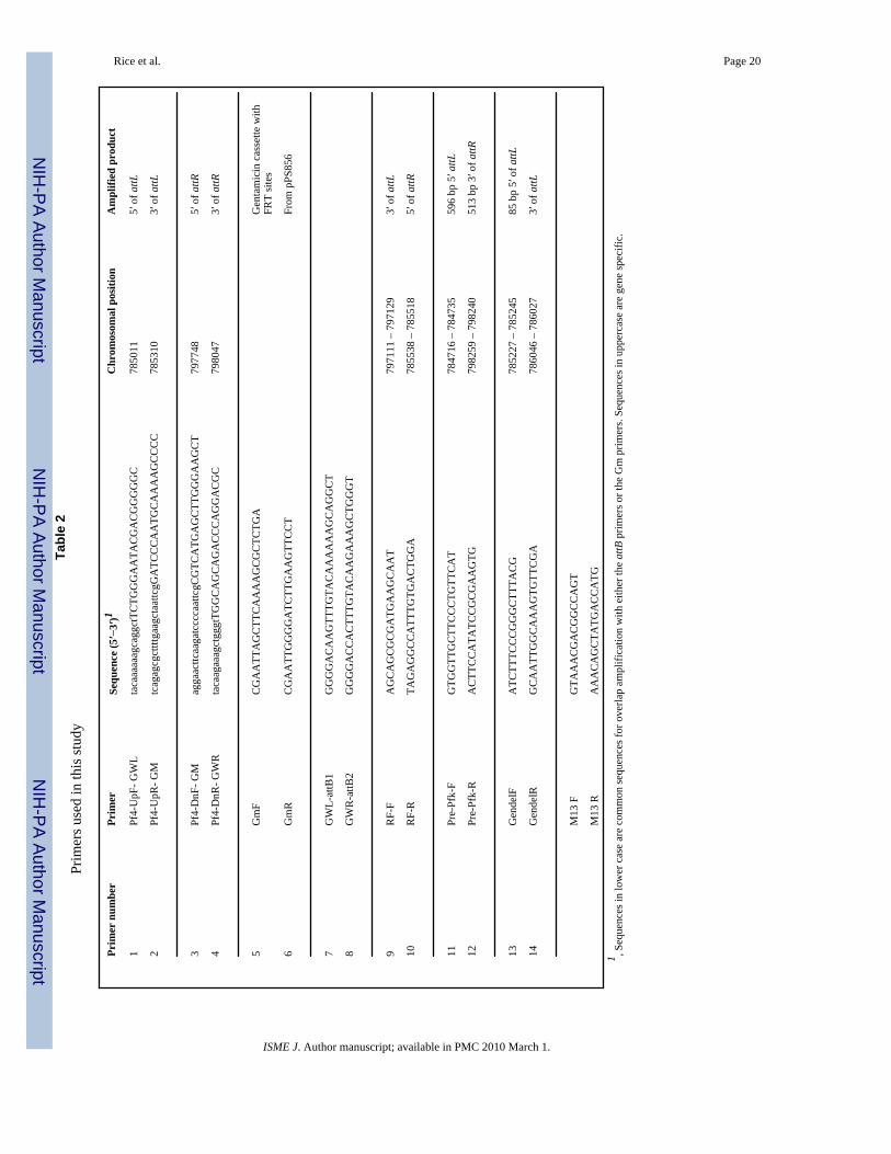
Construction of a Pf4 deletion vectorA genomic knockout of the entire Pf4 prophage was generated by cloning the genomic regionsflanking the Pf4 prophage into the suicide vector pEX18ApGW (Choi and Schweizer, 2005).The ends of the Pf4 prophage were amplified using two sets of primers Pf4-UpF-GWL andPf4-UpR-GM (Table 2), which amplify a 300 bp region from position 785011 to 785310 andprimers Pf4-DnF-GM and Pf4-DnR-GWR which amplify a 300 bp region corresponding topositions 797748–798047 of the published PAO1 genome (Stover et al, 2000)(www.Pseudomonas.com). Fig. 1 shows a schematic representation of primer binding sitesand the final deletion construction. At the same time, a 1053 bp PCR fragment was generatedusing primers GmF and GmR (Table 2), which amplify the gentamicin cassette plus theflanking FRT region from plasmid pPS856 as described (Hoang et al, 1998). These primershave incorporated sequences that allow for the amplified fragments to be joined by SplicingOverlap Extension PCR (SOE PCR) (Horton et al, 1990). Three PCR cycles were performedwithout the addition of primers to join the two Pf4 fragments to the FRT-gentamicin-FRTcassette, after which time, primers GWL-attB1 and GWR-attB2 (Table 2) were added and theentire region was amplified. Note that the latter two primers also incorporate att sites at theends of the PCR fragment. The reaction mixture consisted of 1 × Pwo buffer, 5% DMSO, 200uM dNTP’s and 0.05 U/ul Pwo polymerase (Roche). Cycle conditions were: 94°C hot start for2 min, after which the polymerase was added, followed by 3 cycles of 94°C for 30 s, 55°C for30 s, 68°C for 1 min. After the 3 cycles, primers GWL-attB1 and GWR-attB2 were added forthe second PCR stage which consisted of: 94°C for 30 s, 56°C for 30 s, 68°C for 5 min for atotal of 25 cycles followed by a final 10 min step at 68°C. The gel purified PCR fragment wasthen cloned into plasmid pDONR221 using the clonase method (Invitrogen) and subsequentlytransferred into pEX18ApGW as described (Choi and Schweizer, 2005) to generate plasmidpEX2.5. The construct and correct insertion into pEX18ApGW was confirmed by sequencingfrom the M13 primer sites that are located outside the cloning site.
Generation of a Pf4 chromosomal deletion in P. aeruginosa PAO1The pEX2.5 suicide vector was purified using the Wizard® Plus Minipreps DNA purificationsystems (Promega Inc., Australia) and electroporated into P. aeruginosa PAO1 as described(Choi and Schweizer, 2005). Six ml of an overnight culture grown in LB medium was harvestedin four 2 ml tubes by centrifugation at 16 000 g for 2 min at room temperature and washedtwice with 1 ml of 300 mM sucrose. Pellets from the four tubes were combined together andresuspended to a final volume of 100 ul of 300 mM sucrose. Five hundred ng of the pEX2.5suicide vector DNA was mixed with 100 ul of electrocompetent cells in an electroporationcuvette. Electroporation was performed using a GenePulserXcell™ electroporator (Bio-Rad,USA) at 25 μF, 200 Ohm and 2.5 kV. One ml of LB medium was added immediately after
Rice et al. Page 3
ISME J. Author manuscript; available in PMC 2010 March 1.
NIH
-PA Author Manuscript
NIH
-PA Author Manuscript
NIH
-PA Author Manuscript

electroporation, the cells were transferred to a 2 ml tube and incubated for 2 hours at 37 °Cwith constant shaking at 200 rpm. The culture was concentrated to 100 ul and plated onto LBagar supplemented with 30 μg/ml of gentamicin (LB+Gm30) and incubated at 37 °C for 24 h.Transformant colonies were patched onto LB+Gm30 and LB+Cb200 (LB agar supplementedwith 200 μg/ml of carbenicillin) plates to differentiate single cross-over mutants from doublecross-over mutants. Transformants which were gentamicin resistant and carbenicillin sensitivewere considered to be putative double cross-over deletion mutants.
Confirmation of the deletion strainColonies of putative double crossover mutants were resuspended in 50 μl of sterile water andincubated at 100 °C for 7 min. Cell debris was removed by centrifugation at 16 000 g for 2min and the supernatant was transferred to an ice-cold 1.5 ml tube. Five ul of the supernatantwas used as the template DNA for PCR: 2 mM MgCl2, 1× AmpliTaq PCR buffer, 600 uM ofeach primer (Sigma Genosys Pty Ltd, NSW, Australia) (Table 2), 0.4 μM dNTPs and 0.05 U/ul of AmpliTaq Polymerase (Applied Biosystems, USA). PCR cycling conditions were 95 °Cfor 3 min, followed by 35 cycles of 95 °C for 30 s, 55 °C to 60 °C for 30 s depending on theprimer set used (Table 2) and 72 °C for 2 min; with a final extension at 72 °C for 10 min. PCRproducts were visualized via gel electrophoresis on a 2 % (w/v) agarose gel.
Biofilm formation and assessmentBiofilms were cultivated in flow cells (channel dimensions, 1 mm × 4 mm × 40 mm), asdescribed by Moller et al. (Moller et al, 1998) with some modifications. The flow cell wassterilized in 10% (v/v) bleach for 4 h, thoroughly rinsed in sterile milliQ water, and connectedto sterile silicon tubing (Silastic® laboratory tubings). The flow cell system was flushed withsterile medium (M9 minimal salts medium with glucose: Na2HPO4, 47.8 mM; KH2PO4, 22mM; NH4Cl, 6.8 mM; NaCl, 18.7 mM; CaCl2, 100 uM; MgSO4, 2 mM or 5.6 mM glucose)for 4 h at a flow rate of 6 ml/h prior to the inoculation of bacterial culture. One ml of an overnightculture of PAO1 (WT) or PAOΔPf4 mutant was injected into the flow cell, and incubated for1 h without flow at room temperature in an inverted position. The medium flow was resumedat a flow rate of 6 ml/h. Three independent experiments in triplicate were conducted for 7 days.Biofilms were stained with the LIVE/DEAD BacLight® viability probes (Molecular ProbesInc., Eugene, OR) and visualized using an Olympus FV1000 confocal scanning lasermicroscope (Olympus Optical Co. Ltd., Tokyo, Japan) with argon-ion laser excitation at 488nm or 594 nm and emission filters at 522–535 nm or 605–632 nm to visualize the live, greenfluorescent cells and the dead, red fluorescent cells, respectively. The CSLM images wereanalyzed using ImageJ version 1.36b (http://rsb.info.nih.gov/ij/) and statistical analysis wasperformed using Prism 4.03 for Windows (GraphPad Software Inc., USA).
Phage add-back to biofilmsBecause the integration site for the Pf4 is removed during mutant construction, it was notpossible to perform genetic complementation. Therefore, functional complementation wasperformed by adding superinfective Pf4 phage to biofilms of the wt and the Pf4 mutant.Superinfective Pf4 phage were collected from the effluent of 5–6 day old biofilms of the wild-type P. aeruginosa. Phage particles were concentrated by the addition an equal volume ofphage precipitation buffer (2 M NaCl, 4% Poly Ethylene Glycerol (m.w. 8000)) and incubatedat 4 °C for 8 h. Precipitated phage was then concentrated via centrifugation at 15 000 g for 20min. The pellet containing phage particles was resuspended in 3 ml of SM buffer (100 mMNaCl, 10 mM MgSO4, 50 mM Tris HCl (pH 7.5)). Superinfection and phage titere weredetermined by quantification of pfu on lawns of both the wild-type and Pf4 mutant. Biofilmsof the wt or the ΔPf4 strains were allowed to develop in the flow cell for 3 days, after whichtime, the medium was changed from M9 medium to M9 medium supplemented with filter
Rice et al. Page 4
ISME J. Author manuscript; available in PMC 2010 March 1.
NIH
-PA Author Manuscript
NIH
-PA Author Manuscript
NIH
-PA Author Manuscript

sterilized Pf4 (1×10⁶ pfu/ml) for 24 h. Viability of the phage treated biofilms along with thenegative controls was determined by LIVE/DEAD staining. Biofilms were stained with theLIVE/DEAD BacLight viability stain (Invitrogen, USA) for 1 h before visualizing with aconfocal scanning laser microscope, Olympus LSMGB200 (Olympus Optical Co., Japan),viable cells would be stained green and non-viable cells would be stained red.
Quantification of phagePhage plaque assays were performed using a modified version of top-layer agar (TLA) methodas previously described by Webb et al. (Webb et al, 2003). One ml of culture was collectedand centrifuged at 16 000 g for 5 min and filter sterilised using a 0.2 um filter (Acro-disk, PallCo.). The cell-free supernatant was serially diluted and spotted onto LB medium top layercontaining 0.8% (w/v) agar seeded with 1 × 10⁸ cells/ml of P. aeruginosa PAO1 (WT) or thePAO1ΔPf4 mutant. The number of plaques formed after 18 h of incubation at 37 °C, wasdetermined using a Leica ZOOM 2000™ dissecting microscope (Bufferlab, USA) and plaqueforming units (pfu) per ml for each sample were calculated.
SDS treatment of biofilmsIn order to assess the stability of wild type and P. aeruginosa PAO1 and the PAO1ΔPf4 mutantbiofilms to SDS treatment, 4-day-old biofilms were fed with M9 medium containing 0.01%SDS for 2 hours. CSLM images were acquired before and after the SDS treatment. As a furthercontrol, the P. aeruginosa lasR/rhlR quorum sensing deficient double mutant (Beatson et al,2002), previously shown to be less stable during SDS treatment than the wild type (Davies etal, 1998; Hentzer et al, 2003; Allesen-Holm et al, 2006), was also included. The CSLM imageswere analysed using ImageJ version 1.36b.
Infection by the wild-type and Pf4 mutantSurvival experiments comparing wt and the Pf4 mutant were performed using the nasalaspiration mouse model of acute pneumonia (Comolli et al, 1999). Briefly, bacteria were grownfor 17 h in MINS medium (Nicas and Iglewski, 1984) at 37°C with shaking, diluted 1:100 inMINS medium 3 h prior to infection, concentrated by centrifugation, and then re-suspendedin phosphate-buffered saline (PBS). Bacterial concentrations were determined by measuringoptical densities and verified by viable counts on LB plates. Using a pipette tip, approximately7×107 cfu of bacteria in 50 μL PBS were instilled into the nares of 6- to 8-week-old BALB/cmice anesthetized by intraperitoneal injection of a mixture of ketamine (100 mg/ml) andxylazine (20 mg/ml). For each survival experiment, 5 mice each were infected with mutant orwild type bacteria and then monitored for viability over 7 days. Severely ill mice were sacrificedand scored as dead. Results from 3 separate experiments were pooled. Statistical significancewas assigned by the log-rank test. All experiments were approved by the NorthwesternUniversity Animal Care and Use Committee.
ResultsIsolation of a Pf4 knockout
Transformants of P. aeruginosa that had been electroporated with the suicide Pf4 deletionvector pEX2.5 were screened on LB plates for gentamicin resistance and carbenicillinsensitivity to identify putative double-crossover mutants. One clone was identified and selectedfor further verification by colony PCR. Primers Pre-Pfk-F and Pre-Pfk-R bind to positions784716–784735 and 798259-798240 (Table 2), which lie outside of the Pf4 genomic regionand are not expected to generate a product from either a wild-type (wt) or a single-crossover,but would generate a 2.2 kb product in a double-crossover mutant (also see Fig. 1). PrimersGendelF and GendelR bind to positions 785227–785245 and 786046-786027 (Table 2) and
Rice et al. Page 5
ISME J. Author manuscript; available in PMC 2010 March 1.
NIH
-PA Author Manuscript
NIH
-PA Author Manuscript
NIH
-PA Author Manuscript
will only generate a PCR product in the wt or in a single-crossover but not in the double-crossover mutant. One set of primers, GWL-attB1 and Pre-Pfk-R, was chosen that wouldamplify an internal fragment of the suicide vector in the event of a single-crossover insertion.Lastly, primers RF-F and RF-R (Table 2) were designed to specifically amplify an 839 bpregion of the extra chromosomal replicative form (RF) of the Pf4 filamentous phage. Analysisof the PCR products from the wt as a control and the putative Pf4 mutant demonstrated theexpected banding patterns for the wt and a double-crossover respectively (data not shown).Interestingly, while the RF of the Pf4 filamentous phage, which is an extragenic, circular-copyof the phage genome, could be detected in the wt, no product was observed in the putativemutant, suggesting the putative mutant had been spontaneously cured of the RF form (data notshown). To confirm that the pEX2.5 suicide vector had not integrated into the PAO1 genomeand to confirm a double cross-over knockout insertion, the PCR fragment generated usingprimers Pre-Pfk-F and Pre-Pfk-R was purified. Sequence analysis identified the expectedflanking sequences disrupted by the gentamicin cassette (data not shown) and no vectorsequences were detected, indicating the clone was a true Pf4 deletion mutant.
The Pf4 mutant does not produce infective phageOvernight cultures of both P. aeruginosa wt and the Pf4 mutant were grown in LB at 37°C.Ten μl of the cell free supernatants were then spotted onto soft-agar lawns of either the PAO1wt or the mutant to detect plaque formation, indicative of phage infection. Supernatant fromthe wt produced a zone of clearing on the Pf4 mutant lawn, but not when plated onto the wtlawn, suggesting that the wt supernatant contains active Pf4 phage (Fig. 2). This demonstratesthat the wt is resistant to re-infection by the Pf4 phage. In contrast, supernatant from the mutantdid not produce clearing zones on either lawn (Fig. 2). Hence the mutant, designated P.aeruginosa PAO1ΔPf4, was confirmed to be deficient for the presence of Pf4 sequences andphage.
Maturation, cell death and fitness of ΔPf4 mutant biofilmsBiofilm development by the ΔPf4 mutant was compared to the developmental process of theparental wt in flow cell biofilms over a 7 day period. By day 3, biofilms of both the wt and theΔPf4 mutant had begun to form, where the wt had formed microcolonies with a diameter of41.75 ± 2 um while microcolonies of the mutant were 17 ± 1.7 um in diameter (Fig. 3 A–B).By day 5 (Fig. 3 C–D), microcolonies of the wt had expanded to 74.5 ± 4.6 um while the ΔPf4mutant formed microcolonies that were 51.9 ± 2.7 um. The most striking observation was thaton day 5 the microcolonies of the wt had begun to show areas of hollowing (Fig. 3 C), andsome microcolonies of the wt had regions of dead cells in their centres (data not shown) as hasbeen commonly reported for PAO1 (Webb et al, 2004; Webb et al, 2003). In contrast, theΔPf4 mutant biofilm did not undergo cell death or hollowing of the microcolonies (Fig. 3 D).By day 7, microcolonies of the wt biofilm had expanded to an average size of 478 ± 18 um indiameter (Fig. 3 E). These large microcolonies were not entirely empty, where a number ofviable cells could be observed in the centres along with dead cells (Fig. 3 E), while the mutantmicrocolonies were not significantly larger, 52.2 ± 5.5 um, than those observed on day 5 (Fig.3 F) and no centralised cell death or hollowing was observed. The growth rate and final yieldof the wt and the Pf4 mutant were identical when grown planktonically in the biofilm medium(M9 + glucose) at room temperature indicating that deletion of the Pf4 region did not altergrowth under the conditions tested (data not shown) and therefore, it is unlikely that thedifferences in microcolony size is the result of growth defects. Because the phage integrationsite was deleted in the generation of the Pf4 mutant it was not possible to complement the Pf4mutant. Therefore, we opted to exogenously add back the phage to confirm its effects on thebiofilm. Reinfection of the Pf4 mutant biofilm with purified, superinfective phage restoredbiofilm killing (data not shown), demonstrating that the lack of cell death in the Pf4 mutantbiofilm is not due to resistance to Pf4 infection but rather can be ascribed to the absence of Pf4
Rice et al. Page 6
ISME J. Author manuscript; available in PMC 2010 March 1.
NIH
-PA Author Manuscript
NIH
-PA Author Manuscript
NIH
-PA Author Manuscript
activity in the mutant. These data therefore suggest that the Pf4 phage plays an essential rolein the formation of hollow colonies and the process of cell death within the microcolonies.Moreover, the data suggest that microcolony expansion in the wt requires the presence of thephage in order for the microcolonies to expand beyond 50 um in diameter. Because the ΔPf4mutant biofilm was composed of smaller microcolonies that did not appear to undergo celldeath, we compared the stability of the wt and the mutant biofilms. As shown in Fig. 4, theΔPf4 mutant biofilm was less resistant than the wt strain when treated for 2 hours with 0.01%SDS. Analysis of the percentage coverage of the biofilms remaining after treatment revealedthat the wt biofilm was unaffected by treatment (106% remained) while the ΔPf4 mutantbiofilm was reduced by 61%, which was similar to the control strain ΔLasR/RhlR (59%reduction). The ΔLasR/RhlR strain was included here as a positive control since it waspreviously shown that QS deficient mutants were sensitive to SDS treatment (Allesen-Holmet al, 2006).
Appearance of phage and colony variants during biofilm developmentA hallmark of the differentiation process typical of P. aeruginosa biofilms is the generation ofphage and the simultaneous appearance of variants, such as small colony variants (SCVs), inthe dispersal population. Biofilms were formed for the wt and the ΔPf4 mutant, and effluentwas collected to determine phage titre and to identify and enumerate SCV formation todetermine if the ΔPf4 mutant was affected in the shedding of SCVs during dispersal. In theseexperiments, cell-free biofilm effluent was serially diluted and 10 ul drops were plated ontosoft agar lawns of both the wt and the ΔPf4 mutant to quantify phage titre over a 5 day period.At the same time, unfiltered biofilm effluent was serially diluted and plated onto LB agar todetermine the percentage of SCVs formed. As expected, when cell free effluent from the wtwas serially diluted and spotted onto the wt lawn, no plaque formation was observed for first3 days of biofilm development and plaques were observed from day 4 onward (Fig. 5). Plaqueforming units (pfu’s) increased from 1 ×10⁷/ml on day 4 to 1 × 10⁸/ml by day 5. However,when the same effluent samples were spotted onto the ΔPf4 mutant lawns, plaques weredetected from day 1 and increased from 1 ×10⁶/ml on day 1 to 1×10⁷/ml on day 4, after whichtime, the pfu/ml counts were not significantly different on either lawn (Fig. 5). Based ondetection of plaques on the Pf4 mutant lawn, it is clear that the Pf4 phage is present from thevery initial phase of biofilm formation, but that it can not cause plaque formation on the wtlawn until the time at which cell death and dispersal are observed in the microcolonies (seebelow). The observation that the phage can only infect the wt from day 4 onwards, suggeststhat the Pf4 phage has converted into a superinfective form and that the wt cells are no longerprotected from Pf4 mediated infection and lysis.
When cell free biofilm effluent from the ΔPf4 mutant was spotted onto either the wt or themutant lawn, no plaques were observed for days 1–5 (data not shown). SCVs were alsoquantified in the biofilm effluents for both strains. SCVs appeared in the wt biofilm at day 5,which correlated with the timing of conversion of the Pf4 into the superinfective form (Fig. 5)and autolysis within the microcolonies and constituted approximately 10% of the total cfu’sobserved. In contrast, no SCVs were detected in the effluent of the ΔPf4 mutant at the timewhen the phage converted into the superinfective form.
The Pf4 mutant is less virulent than the wild-type strainTo determine if deletion of the Pf4 phage affected the virulence of P. aeruginosa, a mousemodel of acute pneumonia was used to assess the in vivo virulence of the ΔPf4 mutant strain.Mice were inoculated with approximately 7 ×10⁷ cfu of bacteria by nasal aspiration andmonitored for survival over the subsequent seven days. None of the 15 mice infected with wtbacteria survived to day 7. In contrast, 5 of the 15 mice infected with the ΔPf4 mutant survivedfor the duration of the experiment (Fig. 6). In terms of mean survival times, mice infected with
Rice et al. Page 7
ISME J. Author manuscript; available in PMC 2010 March 1.
NIH
-PA Author Manuscript
NIH
-PA Author Manuscript
NIH
-PA Author Manuscript

wt bacteria survived 42.7 h (95% Confidence Interval 32.9 h to 52.4 h), whereas mice infectedwith the ΔPf4 mutant survived 100.8 h (95% Confidence Interval 74.0 h to 127.6 h), indicatingthat the ΔPf4 mutant was attenuated in its ability to cause acute pneumonia (P <0.001, log-rank test).
DiscussionThis study demonstrates that the filamentous phage Pf4 plays an essential role in the biofilmlife-cycle, mediating mutations (variant formation), and for the first time, shows that the Pf4plays an important role in the structural integrity of the biofilm (autolysis, microcolony sizeand stability), and virulence. Filamentous phage have been found associated with 73 % (8 of11) of isolates from cystic fibrosis (CF) patients (Webb et al, 2003) and filamentous phageactivity was present in 100 % (5 of 5) of CF isolates (Kirov et al, 2007). Thus, the phagemediated effects observed are likely to be conserved across P. aeruginosa strains.
Phage are numerically dominant compared to prokaryotes (Rohwer and Edwards, 2002;Whitman et al, 1998), contribute to the control of bacterial numbers by lysis and mediate genetransfer, which impacts on bacterial evolution. It is known that phage can carry foreign geneswhich may expand the host’s phenotypic capacity. In addition to phage mediated gene transfer,i.e. transduction, bacteriophage can also affect bacterial evolution directly by influencingendogenous mutation rates, resulting in single nucleotide changes. For example, bacteriophageP1 can affect mutation rates through the expression of the hot gene, which can either stabilizeor destabilize the proofreading subunit of DNA polymerase III (Chikova and Schaaper,2006). More generally, Pal et al. (Pal et al, 2007) reported that growth of Pseudomonasfluorescens in the presence of lytic phage gave rise to mutator strains at a high frequency.Mutator strains are now recognised as making significant contribution to the infectivity of arange of human pathogenic bacteria, including Escherichia coli, Haemophilus influenza,Helicobacter pylori, Neiserria meningitidis, Staphylococcus aureus, and Streptococcuspneumoniae (Bucci et al, 1999) reviewed in (Hall and Henderson-Begg, 2006). Both clinicaland environmental isolates of P. aeruginosa demonstrate mutator phenotypes (Kenna et al,2007). As a result of these effects on the host genome, bacteriophage can influence bacterialphenotypes. Phage mediated variation has significant medical implications, where it has beenshown that phage can mediate conversion of P. aeruginosa into a mucoid phenotype, whichhas been associated with poor outcomes for CF patients (Hoiby et al, 2001; Miller and Rubero,1984). Brockhurst et al. (Brockhurst et al, 2005; Webb et al, 2004) have shown that the presenceof bacteriophage drives the selection for smooth, phage resistant morphotypes of P.aerugionsa and that the addition of phage to planktonic cultures of P. aeruginosa leads to theformation of colony variants.
We recently reported that CF clinical strains of P. aeruginosa produce superinfective phageand that biofilms of these isolates also generate SCVs as previously shown for P. aeruginosaPAO1 (Kirov et al, 2007; Webb et al, 2003). This suggests that phage generally may play asignificant role in biofilm development and the generation of phenotypic variants in clinicallyrelevant strains. For example, Kirov et al (2007) demonstrated that high phage titres in biofilmsof clinical strains correlate with biofilm cell death and an increase in the types and overallpercentage of variants (Kirov et al, 2007). Like the mucoid variants, other variants, such as theSCVs are clinically relevant as they commonly show increased antibiotic resistance and biofilmformation (Haussler, 2004). Mooij et al. (2007) observed no correlation between the productionof the Pf5 filamentous phage and SCV formation in P. aeruginosa PA14 (Mooij et al, 2007).However, these authors did not observe superinfective phage. In contrast, we have repeatedlyobserved that conversion of the Pf4 into the superinfective form at the time when SCVs becomedetectable in the biofilm supernatant. This suggests that superinfection may be a key processin the formation of SCVs by phage activity. One key difference between the Pf5 and Pf4
Rice et al. Page 8
ISME J. Author manuscript; available in PMC 2010 March 1.
NIH
-PA Author Manuscript
NIH
-PA Author Manuscript
NIH
-PA Author Manuscript

bacteriophage is that the former does not appear to contain a toxin-antitoxin gene pair; it hasbeen proposed that such addiction molecules are involved in the formation of persister cells(Lewis, 2005) and we have observed that over expression of the parE gene (putative toxin)from a plasmid introduced into P. aeruginosa leads to an increased formation of SCVs (Lauand Kjelleberg, unpublished data). Because such variants are generally slow growing andmetabolically inactive, they exhibit enhanced resistance to drug therapy. However, thephenotype is revertable and hence, they represent a population that is capable of initiatinginfection once the antibiotic pressure is relieved. Thus, their contribution to chronic infectionis potentially very important. In conjunction with the observation that bacteriophage have beenfound in the sputum of patients suffering from chronic lung infections (Hoiby et al, 2001),these data suggest that phage mediated variation plays a significant role in chronic infectionsin situ.
This is the first report to demonstrate that a filamentous phage plays a role in the virulence ofP. aeruginosa. One mechanism by which phage may contribute to virulence, is through theprovision of virulence determinants. For example, virulence can be transmitted between strainsof V. cholerae by the CTX phage which carries the genes coding for cholerae toxin (CT) andthe RTX toxin (Faruque et al, 2003; McLeod et al, 2005). Within the genome of the Pf4bacteriophage is a pair of genes that encode a toxin-antitoxin (T-A) system (Webb et al,2004), however, such genes have not been associated with virulence or toxicity to mammaliancells and therefore it is unlikely the T-A genes of Pf4 are directly toxic in the mouse assay usedhere. Furthermore, T-A genes have not been detected in the genomes of other filamentousphage in P. aeruginosa (Hill et al, 1991; Mooij et al, 2007). With the exception of the Pf4encoded gene PAO726, no other genes were identified as having homology with knownvirulence factors (unpublished observation). PA0726, which is conserved in Pf1 (Webb et al,2004) and Pf5 (Mooij et al, 2007), shares significant homology to the zonula occludens toxin(Zot) encoded by the filamentous CTX phage of V. cholerae (Fasano et al, 1991). Zot disruptsthe intercellular tight junctions between cells in the small intestine and has been suggested tosynergise the effects of CT in causing diarrhoea (Di Pierro et al, 2001; Fasano et al, 1991).However, zot mutants of V. cholerae were not affected in a pulmonary infection model andZot was shown to disrupt tight junctions in the small intestine, but not in the colon (Di Pierroet al, 2001; Fullner et al, 2002). Hence, Zot toxin activity appears be tissue specific and suggeststhat the Pf4 encoded zot homologue is unlikely to act as a virulence factor here. It has beensuggested that Zot has an additional function, where the C terminal region harbours the toxinactivity and the N terminal domonain plays an essential role in phage assembly (Di Pierro etal, 2001).
It has been estimated that 60 % of all infections are biofilm related (Costerton et al, 1999;Davies, 2003), and there is substantial evidence that biofilms play an important role in thepersistent infections of CF lungs by P. aeruginosa. Biofilm formation contributes to infectionby protecting the bacteria from the host immune defence as well as mediating antibioticresistance. Some of the reduced susceptibility of biofilm cells has been attributed to theexopolysaccharide matrix, which may act as a physical barrier to protect the encased cells(reviewed in (Hall-Stoodley et al, 2004)). Therefore, it is possible that the reduced virulenceof the ΔPf4 mutant may be linked to effects on biofilm formation or maintenance. Very clearly,biofilms of the ΔPf4 mutant develop abnormally compared to the wt, where is fails to undergothe process of autolysis and makes smaller microcolonies. Centralised cell death has beenlinked to the production of variants, which as noted above, are important in the infectionprocess. Furthermore, this process of cell death, dispersal and variant formation has beenobserved in many other strains of P. aeruginosa, arguing that this is a stage of developmentthat is conserved amongst P. aeruginosa strains generally (Kirov et al, 2007).
Rice et al. Page 9
ISME J. Author manuscript; available in PMC 2010 March 1.
NIH
-PA Author Manuscript
NIH
-PA Author Manuscript
NIH
-PA Author Manuscript
Alternatively, it is possible that the ΔPf4 biofilms are less stable than the wt. Indeed, we haveshown that biofilms of the ΔPf4 mutant were less stable than the wt when challenged withsurfactant stress (Fig. 4). The ΔPf4 mutant was as sensitive to SDS as a quorum sensing lasR-rhlR mutant. It has been shown that QS controls the secretion of extracellular DNA (eDNA),and that eDNA makes a significant contribution to the EPS of P. aeruginosa as well ascontributing to the matrix of biofilms (Bockelmann et al, 2007;Whitchurch et al, 2002).Biofilms of QS mutants are more susceptible to disruption by surfactants than the wt strain(Davies et al, 1998;Hentzer et al, 2003). It has been proposed that biofilm stability is relatedto the QS controlled secretion of extracellular DNA (eDNA) into the biofilm matrix (Allesen-Holm et al, 2006). Thus, it is possible that the Pf4 phage, either as free DNA released into thebiofilm matrix, or as viral particles, also enhance biofilm stability. Webb et al. (Webb et al,2004) observed that SCVs, which were isolated from biofilms at the time when superinfectivephage could be detected, have high densities of a surface associated material that reactsspecifically with Pf4 directed antibodies. These surface associated phage may serve to cross-link cells within the biofilm.
We submit that bacteriophage make a significant contribution to the biofilm life-cycle andvirulence of P. aeruginosa. These effects may be mediated by multiple mechanisms, wherethe prophage encodes potential virulence factors, phage may contribute to microbial evolutionthrough the formation of phenotypic variants, eg. SCVs, and phage particles may physicallyenhance the stability of biofilms. These data demonstrate for the first time that thebacteriophage Pf4 plays a key role in structural features of the biofilm and in particular, thisstudy is the first to clearly link phage production with centralised cell death and subsequenthollow colony formation. Furthermore, this is the first demonstration that a prophage in P.aeruginosa contributes to infection. Bacteriophage are widely disseminated and numericallydominant in the biosphere. Moreover, they display rapid evolution and the capacity to carryintroduction new genes into their hosts. Therefore it is highly likely that phage may play similarroles in mediating ecological adaptation and virulence in other bacterial hosts and thus playsignificant roles in the evolution of bacterial species.
AcknowledgementsThis work was funded by the Australia Research Council (SK), and the National Institute of Health, USA (AH, grantsAI053674 and AI065615). Special thanks to H. Schweizer for providing the Gateway vectors and advice on theirapplication.
ReferencesAllesen-Holm M, Barken KB, Yang L, Klausen M, Webb JS, Kjelleberg S, et al. A characterization of
DNA release in Pseudomonas aeruginosa cultures and biofilms. Mol Microbiol 2006;59:1114–1128.[PubMed: 16430688]
Beatson SA, Whitchurch CB, Semmler ABT, Mattick JS. Quorum sensing is not required for twitchingmotility in Pseudomonas aeruginosa. J Bacteriol 2002;184:3598–3604. [PubMed: 12057954]
Bertani G. Studies on lysogenesis. J Bacteriol 1951;62:293–300. [PubMed: 14888646]Bockelmann U, Lunsdorf H, Szewzyk U. Ultrastructural and electron energy-loss spectroscopic analysis
of an extracellular filamentous matrix of an environmental bacterial isolate. Environ Microbiol2007;9:2137–2144. [PubMed: 17686013]
Boles BR, Thoendel M, Singh PK. Self-generated diversity produces “Insurance effects” In biofilmcommunities. Proc Natl Acad Sci USA 2004;101:16630–16635. [PubMed: 15546998]
Brockhurst MA, Buckling A, Rainey PB. The effect of a bacteriophage on diversification of theopportunistic bacterial pathogen, Pseudomonas aeruginosa. Proc Royal Soc B-Biol Sci2005;272:1385–1391.
Rice et al. Page 10
ISME J. Author manuscript; available in PMC 2010 March 1.
NIH
-PA Author Manuscript
NIH
-PA Author Manuscript
NIH
-PA Author Manuscript
Bucci C, Lavitola A, Salvatore P, Del Giudice L, Massardo DR, Bruni CB, et al. Hypermutation inpathogenic bacteria: Frequent phase variation in meningococci is a phenotypic trait of a specializedmutator biotype. Molecular Cell 1999;3:435–445. [PubMed: 10230396]
Chikova AK, Schaaper RM. Mutator and antimutator effects of the bacteriophage P1 hot gene product.J Bacteriol 2006;188:5831–5838. [PubMed: 16885451]
Choi K-H, Schweizer H. An improved method for rapid generation of unmarked Pseudomonasaeruginosa deletion mutants. BMC Microbiol 2005;5:30–40. [PubMed: 15907219]
Comolli JC, Hauser AR, Waite L, Whitchurch CB, Mattick JS, Engel JN. Pseudomonas aeruginosa geneproducts Pilt and pilU are required for cytotoxicity in vitro and virulence in a mouse model of acutepneumonia. Infect Immun 1999;67:3625–3630. [PubMed: 10377148]
Costerton JW, Stewart PS, Greenberg EP. Bacterial biofilms: A common cause of peristent infections.Science 1999;284:1318–1322. [PubMed: 10334980]
Davies D. Understanding biofilm resistance to antibacterial agents. Nat Rev Drug Discov 2003;2:114–122. [PubMed: 12563302]
Davies DG, Parsek MR, Pearson JP, Iglewski BH, Costerton JW, Greenberg EP. The involvement ofcell-cell signals in the development of a bacterial biofilm. Science 1998;280:295–298. [PubMed:9535661]
Di Pierro M, Lu R, Uzzau S, Wang W, Margaretten K, Pazzani C, et al. Zonula occludens toxin structure-function analysis. Identification of the fragment biologically active on tight junctions and of thezonulin receptor binding domain. J Biol Chem 2001;276:19160–19165. [PubMed: 11278543]
Faruque SM, Zhu J, Asadulghani Kamruzzaman M, Mekalanos JJ. Examination of diverse toxin-coregulated pilus-positive Vibrio cholerae strains fails to demonstrate evidence for vibriopathogenicity island phage. Infect Immun 2003;71:2993–2999. [PubMed: 12761075]
Fasano A, Baudry B, Pumplin DW, Wasserman SS, Tall BD, Ketley JM. Vibrio cholerae produces asecond enterotoxin, which affects intestinal tight junctions. Proc Natl Acad Sci USA 1991;88:5242–5246. [PubMed: 2052603]
Fullner KJ, Boucher JC, Hanes MA, Haines GK III, Meehan BM, Walchle C, et al. The contribution ofaccessory toxins of Vibrio cholerae O1 El tor to the proinflammatory response in a murine pulmonarycholera model. J Exp Med 2002;195:1455–1462. [PubMed: 12045243]
Hall LMC, Henderson-Begg SK. Hypermutable bacteria isolated from humans - a critical analysis.Microbiology 2006;152:2505–2514. [PubMed: 16946246]
Hall-Stoodley L, Costerton JW, Stoodley P. Bacterial biofilms: From the natural environment toinfectious diseases. Nature Microbiol Rev 2004;2:95–108.
Haussler S. Biofilm formation by the small colony variant phenotype of Pseudomonas aeruginosa.Environ Microbiol 2004;6:546–551. [PubMed: 15142242]
Haussler S, Tummler B, Weissbrodt H, Rohde M, Steinmetz I. Small-colony variants of Pseudomonasaeruginosa in cystic fibrosis. Clin Infect Dis 1999;29:621–625. [PubMed: 10530458]
Hentzer M, Wu H, Andersen JB, Riedel K, Rasmussen TB, Bagge N, et al. Attenuation of Pseudomonasaeruginosa virulence by quorum sensing inhibitors. EMBO J 2003;22:3803–3815. [PubMed:12881415]
Hill DF, Short NJ, Perham RN, Petersen GB. DNA sequence of the filamentous bacteriophage Pf1. J MolBiol 1991;218:349–364. [PubMed: 2010913]
Hoang TT, Karkhoff-Schweizer RR, Kutchma AJ, Schweizer HP. A broad-host-range flp-frtrecombination system for site-specific excision of chromosomally-located DNA sequences:Application for isolation of unmarked Pseudomonas aeruginosa mutants. Gene 1998;212:77–86.[PubMed: 9661666]
Hoiby N, Johansen HK, Moser C, Song ZJ, Ciofu O, Kharazmi A. Pseudomonas aeruginosa and the invitro and in vivo biofilm mode of growth. Microb Infect 2001;3:23–35.
Horton RM, Cai Z, Ho SN, Pease LR. Gene splicing by overlap extension: Tailor made genes using thepolymerase chain reaction. BioTechniques 1990;8:528–535. [PubMed: 2357375]
Kenna DT, Doherty CJ, Foweraker J, Macaskill L, Barcus VA, Govan JRW. Hypermutability inenvironmental Pseudomonas aeruginosa and in populations causing pulmonary infection inindividuals with cystic fibrosis. Microbiology 2007;153:1852–1859. [PubMed: 17526842]
Rice et al. Page 11
ISME J. Author manuscript; available in PMC 2010 March 1.
NIH
-PA Author Manuscript
NIH
-PA Author Manuscript
NIH
-PA Author Manuscript
Kirov SM, Webb JS, O’May CY, Reid DW, Woo JKK, Rice SA, et al. Biofilm differentiation anddispersal in mucoid Pseudomonas aeruginosa isolates from patients with cystic fibrosis.Microbiology 2007;153:3264–3274. [PubMed: 17906126]
Koh KS, Lam KW, Alhede M, Queck SY, Labbate M, Kjelleberg S, et al. Phenotypic diversification andadaptation of Serratia marcescens MG1 biofilm-derived morphotypes. J Bacteriol 2007;189:119–130. [PubMed: 17071749]
Lewis K. Persister cells and the riddle of biofilm survival. Biochemistry (Moscow) 2005;V70:267–274.[PubMed: 15807669]
Mai-Prochnow A, Evans F, Dalisay-Saludes D, Stelzer S, Egan S, James S, et al. Biofilm developmentand cell death in the marine bacterium Pseudoalteromonas tunicata. Appl Environ Microbiol2004;70:3232. [PubMed: 15184116]
McLeod SM, Kimsey HH, Davis BM, Waldor MK. CTX and Vibrio cholerae: Exploring a newlyrecognized type of phage-host cell relationship. Mol Microbiol 2005;57:347–356. [PubMed:15978069]
Miller RV, Rubero VJ. Mucoid conversion by phages of Pseudomonas aeruginosa strains from patientswith cystic fibrosis. J Clin Microbiol 1984;19:717–719. [PubMed: 6429192]
Moller S, Sternberg C, Andersen JB, Christensen BB, Ramos JL, Givskov M, et al. In situ gene expressionin mixed-culture biofilms - evidence of metabolic interactions between community members. ApplEnviron Microbiol 1998;64:721–732. [PubMed: 9464414]
Mooij MJ, Drenkard E, Llamas MA, Vandenbroucke-Grauls CMJE, Savelkoul PHM, Ausubel FM, et al.Characterization of the integrated filamentous phage Pf5 and its involvement in small-colonyformation. Microbiology 2007;153:1790–1798. [PubMed: 17526836]
Nakayama K, Takashima K, Ishihara H, Shinomiya T, Kageyama M, Kanaya S, et al. The R-type pyocinof Pseudomonas aeruginosa is related to P2 phage, and the F-type is related to lambda phage. MolMicrobiol 2000;38:213–231. [PubMed: 11069649]
Nicas T, Iglewski B. Isolation and characterisation of transposon induced mutants of Pseudomonasaeruginosa deficient in production of exoenzyme S. Infect Immun 1984;45:470–474. [PubMed:6086529]
Pal C, Macia MD, Oliver A, Schachar I, Buckling A. Coevolution with viruses drives the evolution ofbacterial mutation rates. Nature 2007;450:1079–1081. [PubMed: 18059461]
Purevdorj-Gage B, Costerton WJ, Stoodley P. Phenotypic differentiation and seeding dispersal in non-mucoid and mucoid Pseudomonas aeruginosa biofilms. Microbiology 2005;151:1569–1576.[PubMed: 15870466]
Rohwer F, Edwards R. The phage proteomic tree: A genome-based taxonomy for phage. J Bacteriol2002;184:4529–4535. [PubMed: 12142423]
Stover CK, Pham XQ, Erwin AL, Mizoguchi SD, Warrener P, Hickey MJ, et al. Complete genomesequence of Pseudomonas aeruginosa PAO1, an opportunistic pathogen. Nature 2000;406:959–964.[PubMed: 10984043]
Tinsley CR, Bille E, Nassif X. Bacteriophages and pathogenicity: More than just providing a toxin?Microb Infect 2006;8:1365–1371.
Webb JS, Lau M, Kjelleberg S. Bacteriophage and phenotypic variation in Pseudomonas aeruginosabiofilm development. J Bacteriol 2004;186:8066–8073. [PubMed: 15547279]
Webb JS, Thompson LS, James S, Charlton T, Tolker-Nielsen T, Koch B, et al. Cell death in Pseudomonasaeruginosa biofilm development. J Bacteriol 2003;185:4585–4592. [PubMed: 12867469]
Whitchurch CB, Tolker-Nielsen T, Ragas PC, Mattick JS. Extracellular DNA required for bacterialbiofilm formation. Science 2002;295:1487. [PubMed: 11859186]
Whiteley M, Bangera MG, Bumgarner RE, Parsek MR, Teitzel GM, Lory S, et al. Gene expression inPseudomonas aeruginosa biofilms. Nature 2001;413:860–864. [PubMed: 11677611]
Whitman WB, Coleman DC, Wiebe WJ. Prokaryotes: The unseen majority. Proc Natl Acad Sci USA1998;95:6578–6583. [PubMed: 9618454]
Wilhelm SW, Suttle CA. Viruses and nutrient cycles in the sea. Bioscience 1999;49:781–783.
Rice et al. Page 12
ISME J. Author manuscript; available in PMC 2010 March 1.
NIH
-PA Author Manuscript
NIH
-PA Author Manuscript
NIH
-PA Author Manuscript

Fig. 1.Construction of the Pf4 chromosomal deletion and location of primers. A) The genomicorganization of the Pf4 prophage in P. aeruginosa PAO1 shown. The integration sites, attLand attR are shown, along with open reading frames, shown as thick arrows. Numbers abovethe arrows indicate gene numbers (adapted from (Webb et al, 2004)). Solid lines indicateamplification products and numbers indicate primers, as listed in Table 2. Small arrows indicateindividual primers and are also numbered with reference to Table 2. B) Organisation of theΔPf4 genomic region subsequent to deletion of the prophage. Black lines indicate the amplified3′ and 5′ regions which are ligated to the FRT sites (hatched circles), recognised by the Flprecombinase, and the gentamycin resistance cassette (grey line, Gmr). The figures are not drawnto scale.
Rice et al. Page 13
ISME J. Author manuscript; available in PMC 2010 March 1.
NIH
-PA Author Manuscript
NIH
-PA Author Manuscript
NIH
-PA Author Manuscript
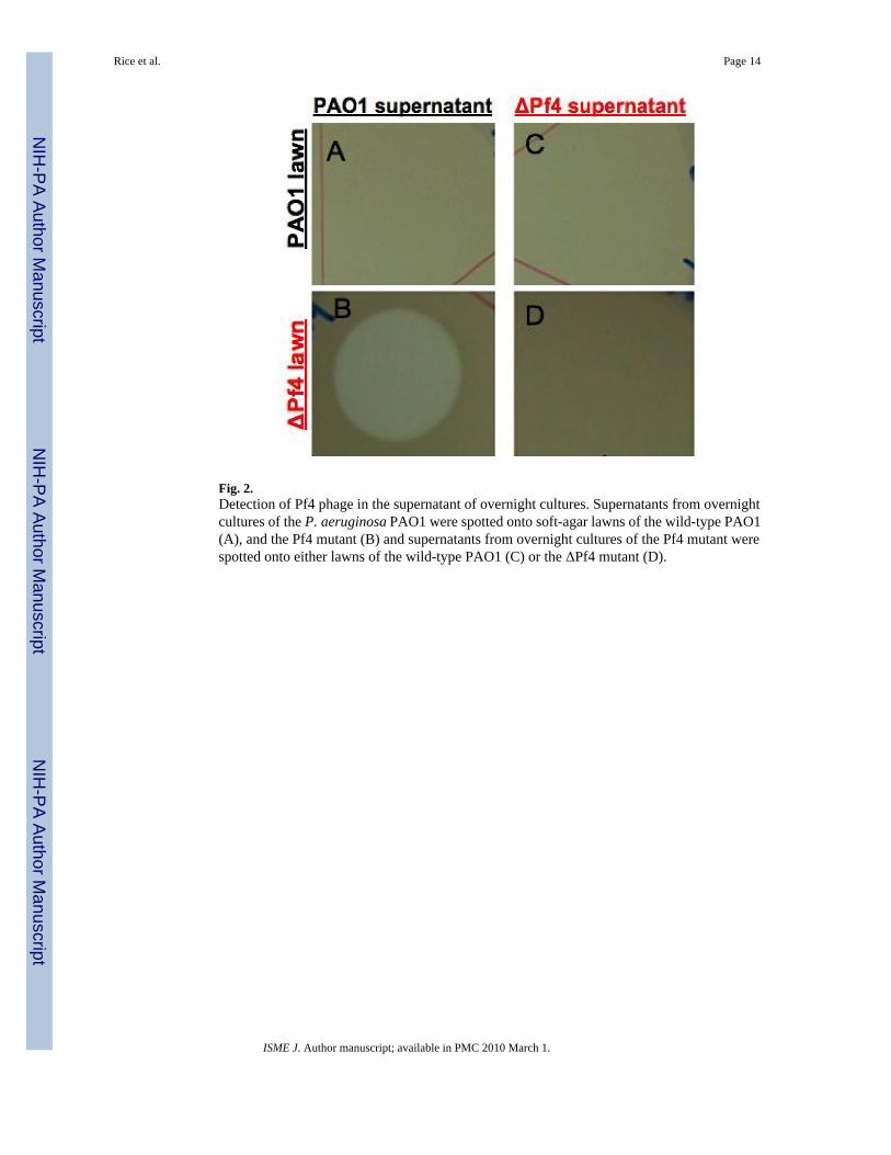
Fig. 2.Detection of Pf4 phage in the supernatant of overnight cultures. Supernatants from overnightcultures of the P. aeruginosa PAO1 were spotted onto soft-agar lawns of the wild-type PAO1(A), and the Pf4 mutant (B) and supernatants from overnight cultures of the Pf4 mutant werespotted onto either lawns of the wild-type PAO1 (C) or the ΔPf4 mutant (D).
Rice et al. Page 14
ISME J. Author manuscript; available in PMC 2010 March 1.
NIH
-PA Author Manuscript
NIH
-PA Author Manuscript
NIH
-PA Author Manuscript
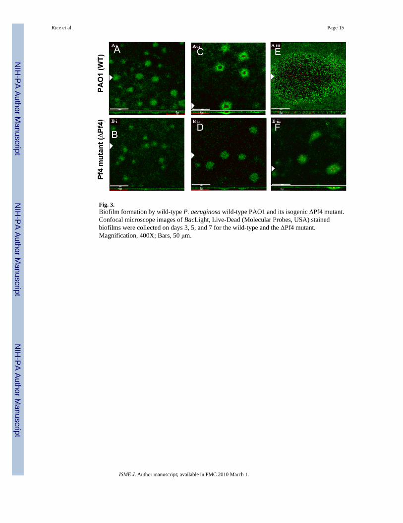
Fig. 3.Biofilm formation by wild-type P. aeruginosa wild-type PAO1 and its isogenic ΔPf4 mutant.Confocal microscope images of BacLight, Live-Dead (Molecular Probes, USA) stainedbiofilms were collected on days 3, 5, and 7 for the wild-type and the ΔPf4 mutant.Magnification, 400X; Bars, 50 μm.
Rice et al. Page 15
ISME J. Author manuscript; available in PMC 2010 March 1.
NIH
-PA Author Manuscript
NIH
-PA Author Manuscript
NIH
-PA Author Manuscript
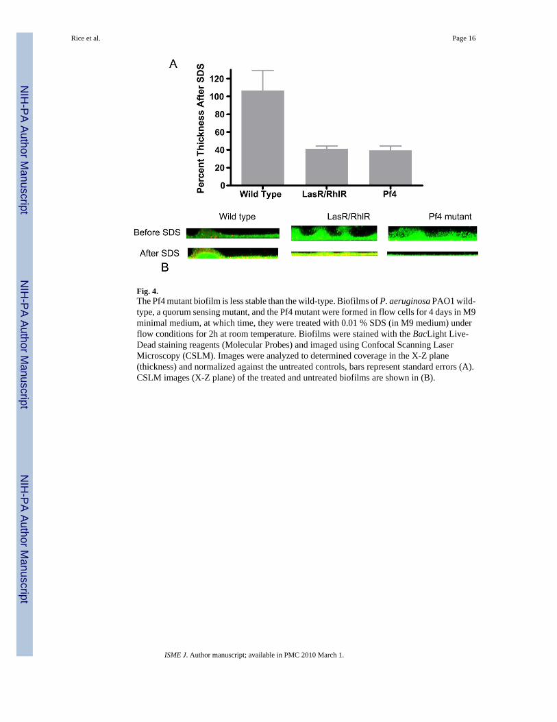
Fig. 4.The Pf4 mutant biofilm is less stable than the wild-type. Biofilms of P. aeruginosa PAO1 wild-type, a quorum sensing mutant, and the Pf4 mutant were formed in flow cells for 4 days in M9minimal medium, at which time, they were treated with 0.01 % SDS (in M9 medium) underflow conditions for 2h at room temperature. Biofilms were stained with the BacLight Live-Dead staining reagents (Molecular Probes) and imaged using Confocal Scanning LaserMicroscopy (CSLM). Images were analyzed to determined coverage in the X-Z plane(thickness) and normalized against the untreated controls, bars represent standard errors (A).CSLM images (X-Z plane) of the treated and untreated biofilms are shown in (B).
Rice et al. Page 16
ISME J. Author manuscript; available in PMC 2010 March 1.
NIH
-PA Author Manuscript
NIH
-PA Author Manuscript
NIH
-PA Author Manuscript
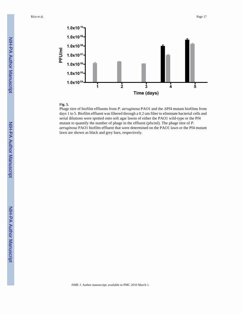
Fig. 5.Phage titre of biofilm effluents from P. aeruginosa PAO1 and the ΔPf4 mutant biofilms fromdays 1 to 5. Biofilm effluent was filtered through a 0.2 um filter to eliminate bacterial cells andserial dilutions were spotted onto soft agar lawns of either the PAO1 wild-type or the Pf4mutant to quantify the number of phage in the effluent (pfu/ml). The phage titre of P.aeruginosa PAO1 biofilm effluent that were determined on the PAO1 lawn or the Pf4 mutantlawn are shown as black and grey bars, respectively.
Rice et al. Page 17
ISME J. Author manuscript; available in PMC 2010 March 1.
NIH
-PA Author Manuscript
NIH
-PA Author Manuscript
NIH
-PA Author Manuscript
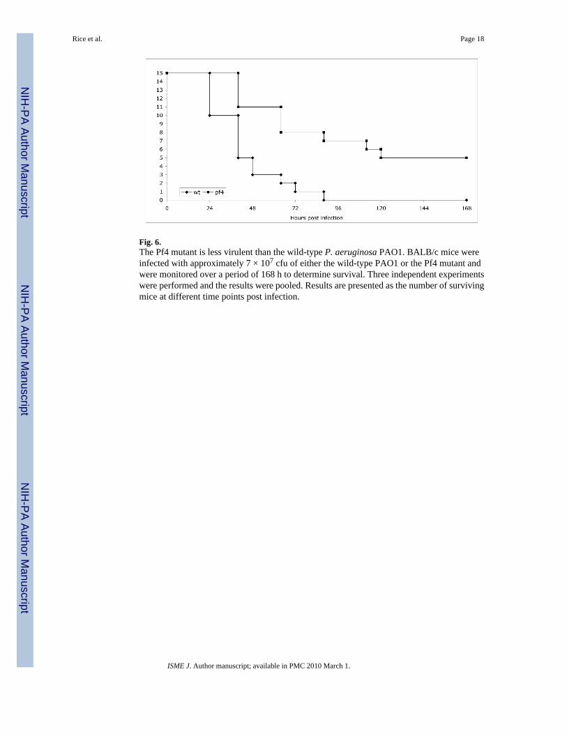
Fig. 6.The Pf4 mutant is less virulent than the wild-type P. aeruginosa PAO1. BALB/c mice wereinfected with approximately 7 × 10⁷ cfu of either the wild-type PAO1 or the Pf4 mutant andwere monitored over a period of 168 h to determine survival. Three independent experimentswere performed and the results were pooled. Results are presented as the number of survivingmice at different time points post infection.
Rice et al. Page 18
ISME J. Author manuscript; available in PMC 2010 March 1.
NIH
-PA Author Manuscript
NIH
-PA Author Manuscript
NIH
-PA Author Manuscript

NIH
-PA Author Manuscript
NIH
-PA Author Manuscript
NIH
-PA Author Manuscript
Rice et al. Page 19
Table 1Bacterial strains and plasmids used in this study.
Strain or plasmid Genotype and/or phenotypea Reference or origin
Strains
E. coli
DH5α F− 80lacZΔM15 Δ(lacZYA-argF) U169 recA1 endA1 hsdR17(rk−, mk
+)phoA supE44 thi-1 gyrA96 relA1 tonA
(48)
P. aeruginosa
PAO1 Wild-type (WT) Lab stock
ΔlasR-rhlR PAO1, double deletion mutation of lasR-rhlR (45)
PAO1ΔPf4 PAO1, Δ(Pf4)::Gmr This study
Plasmids
pPS856 ApR, GmR; 0.83-kb blunt-ended SacI fragment from pUCGM ligated intothe EcoRV site of pPS854
(42)
pDONR221 KmR; Gateway donor vector (40)
pEX18ApGW ApR; Gateway destination vector (40)
pEX2.5 pEX18ApGW gene replacement vector carrying a Gmr cassette flanked bya 300 bp Pf4 upstream fragment and a 300 bp Pf4 downstream fragment(attB1-‘Pf4-FRT-Gm-FRT-Pf4′-attB2); ColE1 replicon; sacB Gmr Apr
Cbr
This study
aAbbreviations: SmR, streptomycin resistance; GmR, gentamicin resistance; ApR, ampicillin resistance; CbR, carbenicillin resistance; KnR, kanamycin
resistance att, attachment site; ‘Pf4, upstream of the Pf4 phage gene cluster; Pf4’, downstream of the Pf4 phage gene cluster (refer to Table 2 for details);FRT, Flp recombinase target; GFP, green fluorescent protein.
ISME J. Author manuscript; available in PMC 2010 March 1.
NIH
-PA Author Manuscript
NIH
-PA Author Manuscript
NIH
-PA Author Manuscript
Rice et al. Page 20Ta
ble
2Pr
imer
s use
d in
this
stud
y
Prim
er n
umbe
rPr
imer
Sequ
ence
(5′−
3′)1
Chr
omos
omal
pos
ition
Am
plifi
ed p
rodu
ct
1Pf
4-U
pF- G
WL
taca
aaaa
agca
ggct
TCTG
GG
AA
TAC
GA
CG
GG
GG
C78
5011
5′ o
f attL
2Pf
4-U
pR- G
Mtc
agag
cgct
tttga
agct
aattc
gGA
TCC
CA
ATG
CA
AA
AG
CC
CC
7853
103′
of a
ttL
3Pf
4-D
nF- G
Mag
gaac
ttcaa
gatc
ccca
attc
gCG
TCA
TGA
GC
TTG
GG
AA
GC
T79
7748
5′ o
f attR
4Pf
4-D
nR- G
WR
taca
agaa
agct
gggt
TGG
CA
GC
AG
AC
CC
AG
GA
CG
C79
8047
3′ o
f attR
5G
mF
CG
AA
TTA
GC
TTC
AA
AA
GC
GC
TCTG
AG
enta
mic
in c
asse
tte w
ithFR
T si
tes
6G
mR
CG
AA
TTG
GG
GA
TCTT
GA
AG
TTC
CT
From
pPS
856
7G
WL-
attB
1G
GG
GA
CA
AG
TTTG
TAC
AA
AA
AA
GC
AG
GC
T
8G
WR
-attB
2G
GG
GA
CC
AC
TTTG
TAC
AA
GA
AA
GC
TGG
GT
9R
F-F
AG
CA
GC
GC
GA
TGA
AG
CA
AT
7971
11 –
797
129
3′ o
f attL
10R
F-R
TAG
AG
GC
CA
TTTG
TGA
CTG
GA
7855
38 –
785
518
5′ o
f attR
11Pr
e-Pf
k-F
GTG
GTT
GC
TTC
CC
TGTT
CA
T78
4716
– 7
8473
559
6 bp
5′ a
ttL
12Pr
e-Pf
k-R
AC
TTC
CA
TATC
CG
CG
AA
GTG
7982
59 –
798
240
513
bp 3′ o
f attR
13G
ende
lFA
TCTT
TCC
CG
GG
CTT
TAC
G78
5227
– 7
8524
585
bp
5′ o
f attL
14G
ende
lRG
CA
ATT
GG
CA
AA
GTG
TTC
GA
7860
46 –
786
027
3′ o
f attL
M13
FG
TAA
AC
GA
CG
GC
CA
GT
M13
RA
AA
CA
GC
TATG
AC
CA
TG1 , S
eque
nces
in lo
wer
cas
e ar
e co
mm
on se
quen
ces f
or o
verla
p am
plifi
catio
n w
ith e
ither
the
attB
prim
ers o
r the
Gm
prim
ers.
Sequ
ence
s in
uppe
rcas
e ar
e ge
ne sp
ecifi
c.
ISME J. Author manuscript; available in PMC 2010 March 1.
Related Documents