Tetrahydrohyperforin and Octahydrohyperforin Are Two New Potent Inhibitors of Angiogenesis Beatriz Martı´nez-Poveda 1 , Luisella Verotta 2 *, Ezio Bombardelli 3 , Ana R. Quesada 1,4 , Miguel A ´ ngel Medina 1,4 * 1 Departamento de Biologı ´a Molecular y Bioquı ´mica, Facultad de Ciencias, Universidad de Ma ´laga, Ma ´laga, Spain, 2 Dipartimento di Chimica Organica e Industriale, University of Milan, Milan, Italy, 3 Indena S.p.A., Milan, Italy, 4 Unidad 741 de CIBER ‘‘de Enfermedades Raras’’, Ma ´laga, Spain Abstract Background: We have previously shown that hyperforin, a phloroglucinol derivative found in St. John’s wort, behaves as a potent anti-angiogenic compound. To identify the reactive group(s) mainly involved in this anti-angiogenic effect, we have investigated the anti-angiogenic properties of a series of stable derivatives obtained by oxidative modification of the natural product. In addition, in the present work we have studied the role of the four carbonyl groups present in hyperforin by investigating the potential of some other chemically stable derivatives. Methodology/Principal Findings: The experimental procedures included the analysis of the effects of treatment of endothelial cells with these compounds in cell growth, cell viability, cell migration and zymographic assays, as well as the tube formation assay on Matrigel. Our study with hyperforin and eight derivatives shows that the enolized b-dicarbonyl system contained in the structure of hyperforin has a dominant role in its antiangiogenic activity. On the other hand, two of the tested hyperforin derivatives, namely, tetrahydrohyperforin and octahydrohyperforin, behave as potent inhibitors of angiogenesis. Additional characterization of these compounds included a cell specificity study of their effects on cell growth, as well as the in vivo Matrigel plug assay. Conclusions/Significance: These observations could be useful for the rational design and chemical synthesis of more effective hyperforin derivatives as anti-angiogenic drugs. Altogether, the results indicate that octahydrohyperforin is a more specific and slightly more potent antiangiogenic compound than hyperforin. Citation: Martı ´nez-Poveda B, Verotta L, Bombardelli E, Quesada AR, Medina MA ´ (2010) Tetrahydrohyperforin and Octahydrohyperforin Are Two New Potent Inhibitors of Angiogenesis. PLoS ONE 5(3): e9558. doi:10.1371/journal.pone.0009558 Editor: Joseph Alan Bauer, Bauer Research Foundation, United States of America Received September 9, 2009; Accepted February 8, 2010; Published March 9, 2010 Copyright: ß 2010 Martı ´nez-Poveda et al. This is an open-access article distributed under the terms of the Creative Commons Attribution License, which permits unrestricted use, distribution, and reproduction in any medium, provided the original author and source are credited. Funding: Work supported in part by EEC (Contract 018834: Antimal), as well as grants CTQ2006-15279-C03-03/BQU, PS09/02216 and TRACE PT2008-0145 (Spanish Ministry of Science and Innovation), Fundacio ´n Ramo ´ n Areces and PIE CTS-3759, P07-CVI-02999 and funds from group BIO-267 (Andalusian Government). The ‘‘CIBER de Enfermedades Raras’’ is an initiative from the ISCIII (Spain). The funders had no role in study design, data collection and analysis, decision to publish, or preparation of the manuscript. Competing Interests: EB is affilitated to Indena S.p.A. (Milan, Italy). Dicyclohexylammonium hyperforinate (hyperforin-DCHA), a stable form of hyperforin (compound 1), was provided by Indena S.p.A. (Milan, Italy). However, Indena S.p.A. was not a funder for this study and had no role in study design, data collection and analysis, decision to publish, or preparation of the manuscript. * E-mail: [email protected] (MAM); [email protected] (LV) Introduction St. John’s wort (Hypericum perforatum L.) is an herbaceous plant that has been known for centuries and used for a variety of medicinal purposes, including the fight against infections and the treatment of respiratory and inflammatory diseases, pectic ulcers and skin wounds [1]. St. John’s wort preparations are increasingly popular in the treatment of mild to moderate depression [2,3]. The main bioactive compound responsible for the antidepressant effects of St. John’s wort extracts is its major lipophilic compound, hyperforin (Figure 1, compound 1). The biomedical relevance of hyperforin is reinforced by the accumulation of scientific evidence pointing to other different effects of hyperforin with potential pharmacological interest. They include effects on Alzheimer disease and as an antibiotic, antiinflammatory, antitumoral and antimetastatic compound [4,5,6,7,8]. Furthermore, the anti angiogenic potential of hyperforin has been recently unveiled [7,9,10,11]. Angiogenesis, the generation of new blood vessels from the existing vascular bed, has been described as one of the hallmarks of cancer, playing essential roles in tumor growth, invasion and metastasis [12]. In contrast to the highly unstable tumor cells, endothelial cells are genetically stable. On the other hand, tumor blood vessels are different to normal vessels. Therefore, tumor blood vessels are potential targets in therapy for all types of cancer [13,14]. When resting endothelial cells are activated by an angiogenic signal, they are stimulated to release degrading enzymes allowing endothelial cells to migrate, proliferate and finally differentiate to form new vessels. Any of the steps involved in angiogenesis may be a potential target for pharmacological intervention of angiogenesis-dependent diseases. This is the main reason why angiogenesis has attracted recent attention in the field of pharmacological research [15]. We have previously shown that hyperforin is able to inhibit angiogenesis in an in vivo model and behaves as a multi-target antiangiogenic drug by inhibiting several key steps of the angiogenic PLoS ONE | www.plosone.org 1 March 2010 | Volume 5 | Issue 3 | e9558

Welcome message from author
This document is posted to help you gain knowledge. Please leave a comment to let me know what you think about it! Share it to your friends and learn new things together.
Transcript
Tetrahydrohyperforin and Octahydrohyperforin Are TwoNew Potent Inhibitors of AngiogenesisBeatriz Martınez-Poveda1, Luisella Verotta2*, Ezio Bombardelli3, Ana R. Quesada1,4, Miguel Angel
Medina1,4*
1 Departamento de Biologıa Molecular y Bioquımica, Facultad de Ciencias, Universidad de Malaga, Malaga, Spain, 2 Dipartimento di Chimica Organica e Industriale,
University of Milan, Milan, Italy, 3 Indena S.p.A., Milan, Italy, 4 Unidad 741 de CIBER ‘‘de Enfermedades Raras’’, Malaga, Spain
Abstract
Background: We have previously shown that hyperforin, a phloroglucinol derivative found in St. John’s wort, behaves as apotent anti-angiogenic compound. To identify the reactive group(s) mainly involved in this anti-angiogenic effect, we haveinvestigated the anti-angiogenic properties of a series of stable derivatives obtained by oxidative modification of the naturalproduct. In addition, in the present work we have studied the role of the four carbonyl groups present in hyperforin byinvestigating the potential of some other chemically stable derivatives.
Methodology/Principal Findings: The experimental procedures included the analysis of the effects of treatment ofendothelial cells with these compounds in cell growth, cell viability, cell migration and zymographic assays, as well as thetube formation assay on Matrigel. Our study with hyperforin and eight derivatives shows that the enolized b-dicarbonylsystem contained in the structure of hyperforin has a dominant role in its antiangiogenic activity. On the other hand, two ofthe tested hyperforin derivatives, namely, tetrahydrohyperforin and octahydrohyperforin, behave as potent inhibitors ofangiogenesis. Additional characterization of these compounds included a cell specificity study of their effects on cellgrowth, as well as the in vivo Matrigel plug assay.
Conclusions/Significance: These observations could be useful for the rational design and chemical synthesis of moreeffective hyperforin derivatives as anti-angiogenic drugs. Altogether, the results indicate that octahydrohyperforin is a morespecific and slightly more potent antiangiogenic compound than hyperforin.
Citation: Martınez-Poveda B, Verotta L, Bombardelli E, Quesada AR, Medina MA (2010) Tetrahydrohyperforin and Octahydrohyperforin Are Two New PotentInhibitors of Angiogenesis. PLoS ONE 5(3): e9558. doi:10.1371/journal.pone.0009558
Editor: Joseph Alan Bauer, Bauer Research Foundation, United States of America
Received September 9, 2009; Accepted February 8, 2010; Published March 9, 2010
Copyright: � 2010 Martınez-Poveda et al. This is an open-access article distributed under the terms of the Creative Commons Attribution License, which permitsunrestricted use, distribution, and reproduction in any medium, provided the original author and source are credited.
Funding: Work supported in part by EEC (Contract 018834: Antimal), as well as grants CTQ2006-15279-C03-03/BQU, PS09/02216 and TRACE PT2008-0145(Spanish Ministry of Science and Innovation), Fundacion Ramon Areces and PIE CTS-3759, P07-CVI-02999 and funds from group BIO-267 (AndalusianGovernment). The ‘‘CIBER de Enfermedades Raras’’ is an initiative from the ISCIII (Spain). The funders had no role in study design, data collection and analysis,decision to publish, or preparation of the manuscript.
Competing Interests: EB is affilitated to Indena S.p.A. (Milan, Italy). Dicyclohexylammonium hyperforinate (hyperforin-DCHA), a stable form of hyperforin(compound 1), was provided by Indena S.p.A. (Milan, Italy). However, Indena S.p.A. was not a funder for this study and had no role in study design, data collectionand analysis, decision to publish, or preparation of the manuscript.
* E-mail: [email protected] (MAM); [email protected] (LV)
Introduction
St. John’s wort (Hypericum perforatum L.) is an herbaceous plant
that has been known for centuries and used for a variety of
medicinal purposes, including the fight against infections and the
treatment of respiratory and inflammatory diseases, pectic ulcers
and skin wounds [1]. St. John’s wort preparations are increasingly
popular in the treatment of mild to moderate depression [2,3].
The main bioactive compound responsible for the antidepressant
effects of St. John’s wort extracts is its major lipophilic compound,
hyperforin (Figure 1, compound 1). The biomedical relevance of
hyperforin is reinforced by the accumulation of scientific evidence
pointing to other different effects of hyperforin with potential
pharmacological interest. They include effects on Alzheimer
disease and as an antibiotic, antiinflammatory, antitumoral
and antimetastatic compound [4,5,6,7,8]. Furthermore, the anti
angiogenic potential of hyperforin has been recently unveiled
[7,9,10,11].
Angiogenesis, the generation of new blood vessels from the
existing vascular bed, has been described as one of the hallmarks of
cancer, playing essential roles in tumor growth, invasion and
metastasis [12]. In contrast to the highly unstable tumor cells,
endothelial cells are genetically stable. On the other hand, tumor
blood vessels are different to normal vessels. Therefore, tumor
blood vessels are potential targets in therapy for all types of cancer
[13,14]. When resting endothelial cells are activated by an
angiogenic signal, they are stimulated to release degrading
enzymes allowing endothelial cells to migrate, proliferate and
finally differentiate to form new vessels. Any of the steps involved
in angiogenesis may be a potential target for pharmacological
intervention of angiogenesis-dependent diseases. This is the main
reason why angiogenesis has attracted recent attention in the field
of pharmacological research [15].
We have previously shown that hyperforin is able to inhibit
angiogenesis in an in vivo model and behaves as a multi-target
antiangiogenic drug by inhibiting several key steps of the angiogenic
PLoS ONE | www.plosone.org 1 March 2010 | Volume 5 | Issue 3 | e9558
process. They include inhibition of endothelial cell growth, capillary
tube formation on a layer of Matrigel, secretion and production of
extracellular matrix degrading enzymes, as well as inhibitory effects
on both migrating and invasive potentials of endothelial cells [9]. In
another recent work, hyperforin has been shown to block microvessel
formation by human dermal microvascular endothelial cells. This
research concludes that hyperforin significantly inhibits tumor
growth, induces apotosis of tumor cells and reduces tumor
vascularisation at concentrations below the toxic effect [10]. It has
also been demonstrated that hyperforin restrains polymorphonucle-
ar cell chemotaxis and chemoinvasion and protects against
inflammatory events taking place in animal models of angiogenesis
[11]. No clear molecular target could, however, be identified. Very
recently, hyperforin has been shown to behave also as a potent
inhibitor of lymphangiogenesis [16].
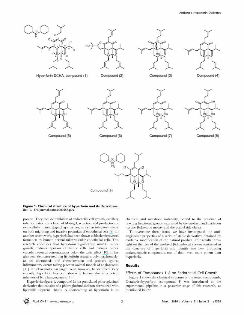
Hyperforin (figure 1, compound 1) is a prenylated phloroglucinol
derivative that consists of a phloroglucinol skeleton derivatized with
lipophilic isoprene chains. A shortcoming of hyperforin is its
chemical and metabolic instability, bound to the presence of
reacting functional groups, expressed by the enolized and oxidation
–prone b-diketone moiety and the prenyl side chains.
To overcome these issues, we have investigated the anti-
angiogenic properties of a series of stable derivatives obtained by
oxidative modification of the natural product. Our results throw
light on the role of the enolized b-dicarbonyl system contained in
the structure of hyperforin and identify two new promising
antiangiogenic compounds, one of them even more potent than
hyperforin.
Results
Effects of Compounds 1–8 on Endothelial Cell GrowthFigure 1 shows the chemical structure of the tested compounds.
Octahydrohyperforin (compound 9) was introduced in the
experimental pipeline in a posterior stage of this research, as
mentioned below.
Figure 1. Chemical structure of hyperforin and its derivatives.doi:10.1371/journal.pone.0009558.g001
Antiangio Hyperforin Derivates
PLoS ONE | www.plosone.org 2 March 2010 | Volume 5 | Issue 3 | e9558
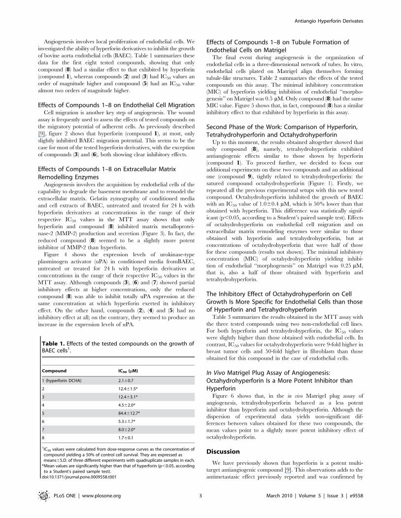
Angiogenesis involves local proliferation of endothelial cells. We
investigated the ability of hyperforin derivatives to inhibit the growth
of bovine aorta endothelial cells (BAEC). Table 1 summarizes these
data for the first eight tested compounds, showing that only
compound (8) had a similar effect to that exhibited by hyperforin
(compound 1), whereas compounds (2) and (3) had IC50 values an
order of magnitude higher and compound (5) had an IC50 value
almost two orders of magnitude higher.
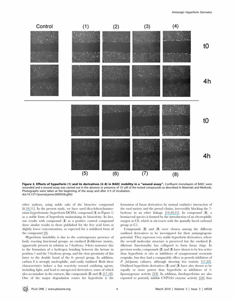
Effects of Compounds 1–8 on Endothelial Cell MigrationCell migration is another key step of angiogenesis. The wound
assay is frequently used to assess the effects of tested compounds on
the migratory potential of adherent cells. As previously described
[9], figure 2 shows that hyperforin (compound 1), at most, only
slightly inhibited BAEC migration potential. This seems to be the
case for most of the tested hyperforin derivatives, with the exception
of compounds (3) and (6), both showing clear inhibitory effects.
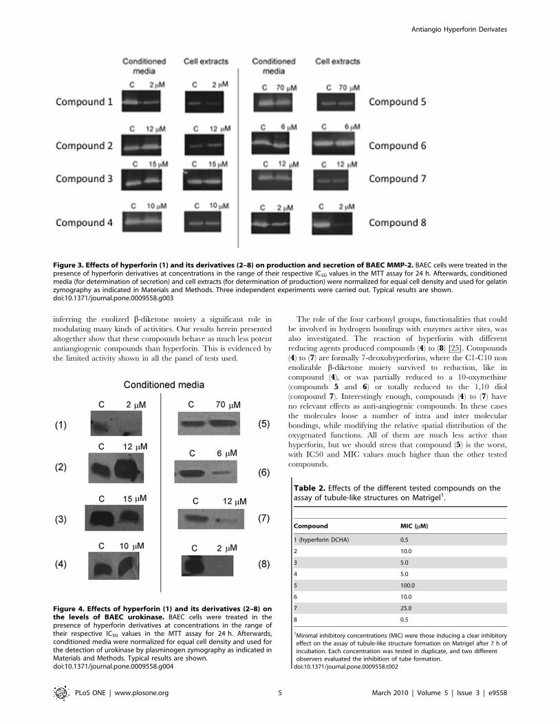
Effects of Compounds 1–8 on Extracellular MatrixRemodelling Enzymes
Angiogenesis involves the acquisition by endothelial cells of the
capability to degrade the basement membrane and to remodel the
extracellular matrix. Gelatin zymography of conditioned media
and cell extracts of BAEC, untreated and treated for 24 h with
hyperforin derivatives at concentrations in the range of their
respective IC50 values in the MTT assay shows that only
hyperforin and compound (8) inhibited matrix metalloprotei-
nase-2 (MMP-2) production and secretion (Figure 3). In fact, the
reduced compound (8) seemed to be a slightly more potent
inhibitor of MMP-2 than hyperforin.
Figure 4 shows the expression levels of urokinase-type
plasminogen activator (uPA) in conditioned media fromBAEC,
untreated or treated for 24 h with hyperforin derivatives at
concentrations in the range of their respective IC50 values in the
MTT assay. Although compounds (3), (6) and (7) showed partial
inhibitory effects at higher concentrations, only the reduced
compound (8) was able to inhibit totally uPA expression at the
same concentration at which hyperforin exerted its inhibitory
effect. On the other hand, compounds (2), (4) and (5) had no
inhibitory effect at all; on the contrary, they seemed to produce an
increase in the expression levels of uPA.
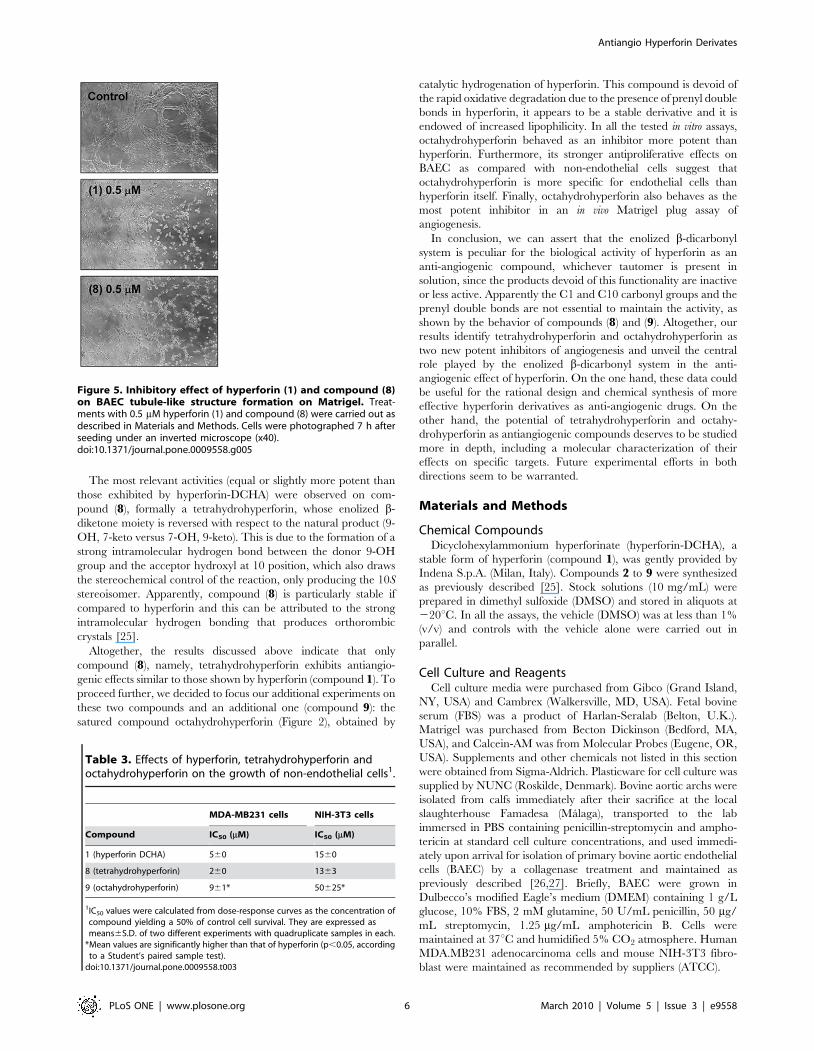
Effects of Compounds 1–8 on Tubule Formation ofEndothelial Cells on Matrigel
The final event during angiogenesis is the organization of
endothelial cells in a three-dimensional network of tubes. In vitro,
endothelial cells plated on Matrigel align themselves forming
tubule-like structures. Table 2 summarizes the effects of the tested
compounds on this assay. The minimal inhibitory concentration
(MIC) of hyperforin yielding inhibition of endothelial ‘‘morpho-
genesis’’ on Matrigel was 0.5 mM. Only compound (8) had the same
MIC value. Figure 5 shows that, in fact, compound (8) has a similar
inhibitory effect to that exhibited by hyperforin in this assay.
Second Phase of the Work: Comparison of Hyperforin,Tetrahydrohyperforin and Octahydrohyperforin
Up to this moment, the results obtained altogether showed that
only compound (8), namely, tetrahydrohyperforin exhibited
antiangiogenic effects similar to those shown by hyperforin
(compound 1). To proceed further, we decided to focus our
additional experiments on these two compounds and an additional
one (compound 9), tightly related to tetrahydrohyperforin: the
satured compound octahydrohyperforin (Figure 1). Firstly, we
repeated all the previous experimental setups with this new tested
compound. Octahydrohyperforin inhibited the growth of BAEC
with an IC50 value of 1.060.4 mM, which is 50% lower than that
obtained with hyperforin. This difference was statistically signif-
icant (p,0.05, according to a Student’s paired sample test). Effects
of octahydrohyperforin on endothelial cell migration and on
extracellular matrix remodeling enzymes were similar to those
obtained with hyperforin and tetrahydrohyperforin, but at
concentrations of octahydrohyperforin that were half of those
for these compounds (results not shown). The minimal inhibitory
concentration (MIC) of octahydrohyperforin yielding inhibi-
tion of endothelial ‘‘morphogenesis’’ on Matrigel was 0.25 mM,
that is, also a half of those obtained with hyperforin and
tetrahydrohyperforin.
The Inhibitory Effect of Octahydrohyperforin on CellGrowth Is More Specific for Endothelial Cells than thoseof Hyperforin and Tetrahydrohyperforin
Table 3 summarizes the results obtained in the MTT assay with
the three tested compounds using two non-endothelial cell lines.
For both hyperforin and tetrahydrohyperforin, the IC50 values
were slightly higher than those obtained with endothelial cells. In
contrast, IC50 values for octahydrohyperforin were 9-fold higher in
breast tumor cells and 50-fold higher in fibroblasts than those
obtained for this compound in the case of endothelial cells.
In Vivo Matrigel Plug Assay of Angiogenesis:Octahydrohyperforin Is a More Potent Inhibitor thanHyperforin
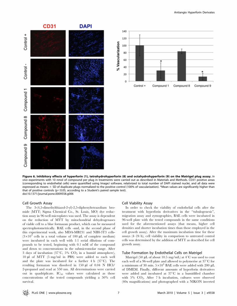
Figure 6 shows that, in the in vivo Matrigel plug assay of
angiogenesis, tetrahydrohyperforin behaved as a less potent
inhibitor than hyperforin and octahydrohyperforin. Although the
dispersion of experimental data yields non-significant dif-
ferences between values obtained for these two compounds, the
mean values point to a slightly more potent inhibitory effect of
octahydrohyperforin.
Discussion
We have previously shown that hyperforin is a potent multi-
target antiangiogenic compound [9]. This observations adds to the
antimetastasic effect previously reported and was confirmed by
Table 1. Effects of the tested compounds on the growth ofBAEC cells1.
Compound IC50 (mM)
1 (hyperforin DCHA) 2.160.7
2 12.461.5*
3 12.463.1*
4 4.562.0*
5 84.4612.7*
6 5.361.7*
7 8.062.0*
8 1.760.1
1IC50 values were calculated from dose-response curves as the concentration ofcompound yielding a 50% of control cell survival. They are expressed asmeans6S.D. of three different experiments with quadruplicate samples in each.
*Mean values are significantly higher than that of hyperforin (p,0.05, accordingto a Student’s paired sample test).
doi:10.1371/journal.pone.0009558.t001
Antiangio Hyperforin Derivates
PLoS ONE | www.plosone.org 3 March 2010 | Volume 5 | Issue 3 | e9558
other authors, using stable salts of the bioactive compound
[6,10,11]. In the present study, we have used dicyclohexylammo-
nium hyperforinate (hyperforin-DCHA, compound (1) in Figure 1)
as a stable form of hyperforin maintaining its bioactivity. In fact,
our results with compound (1) as a positive control compound
show similar results to those published for the free acid form at
slightly lower concentrations, as expected for a stabilized form of
the compound [9].
Hyperforin instability is due to the contemporary presence of
fastly reacting functional groups: an enolized b-diketone moiety,
apparently present in solution as 7-hydroxy, 9-keto tautomer due
to the formation of a hydrogen bonding between the ketone in
position 1 and the 7-hydroxy group, and the close proximity of this
latter to the double bond of the 6- prenyl group. In addition,
carbon 8 is strongly nucleophilic, and easily oxidized. Both these
characteristics induce a fast reactivity toward oxidizing agents,
including light, and lead to unexpected derivatives, some of which
also accumulate in the extracts, like compounds (2) and (3) [17,18].
One of the major degradation routes for hyperforin is the
formation of furan derivatives by mutual oxidative interaction of
the enol moiety and the prenyl chains, irreversibly blocking the 7-
hydroxy in an ether linkage [19,20,21]. In compound (3), a
hemiacetal species is formed by the introduction of an electrophilic
oxygen at C8, which in situ reacts with the spatially faced carbonyl
group at C1.
Compounds (2) and (3) were chosen among the different
oxidized derivatives to be investigated for their antiangiogenic
potential. They represent very stable hyperforin derivatives, where
the overall molecular structure is preserved but the enolized b-
diketone functionality has collapsed to form furan rings. In
previous works, compounds (2) and (3) have shown to be less active
than hyperforin in vitro as inhibitors of synaptosomal serotonin
reuptake, but they had a comparable effect as growth inhibitors of
P. falciparum cultures, although showing less toxicity [17,22].
Oxidized hyperforin derivatives (2) and (3) have also shown to be
equally or more potent than hyperforin as inhibitors of 5-
lipooxygenase activity [23]. In addition, furohyperforins are also
reported to potently inhibit CYP3A4 enzyme activity [24] thus
Figure 2. Effects of hyperforin (1) and its derivatives (2–8) in BAEC mobility in a ‘‘wound assay’’. Confluent monolayers of BAEC werewounded and a wound assay was carried out in the absence or presence of 10 mM of the tested compounds as described in Materials and Methods.Photographs were taken at the beginning of the assay and after 4 h of incubation.doi:10.1371/journal.pone.0009558.g002
Antiangio Hyperforin Derivates
PLoS ONE | www.plosone.org 4 March 2010 | Volume 5 | Issue 3 | e9558
inferring the enolized b-diketone moiety a significant role in
modulating many kinds of activities. Our results herein presented
altogether show that these compounds behave as much less potent
antiangiogenic compounds than hyperforin. This is evidenced by
the limited activity shown in all the panel of tests used.
The role of the four carbonyl groups, functionalities that could
be involved in hydrogen bondings with enzymes active sites, was
also investigated. The reaction of hyperforin with different
reducing agents produced compounds (4) to (8) [25]. Compounds
(4) to (7) are formally 7-deoxohyperforins, where the C1-C10 non
enolizable b-diketone moiety survived to reduction, like in
compound (4), or was partially reduced to a 10-oxymethine
(compounds 5 and 6) or totally reduced to the 1,10 diol
(compound 7). Interestingly enough, compounds (4) to (7) have
no relevant effects as anti-angiogenic compounds. In these cases
the molecules loose a number of intra and inter molecular
bondings, while modifying the relative spatial distribution of the
oxygenated functions. All of them are much less active than
hyperforin, but we should stress that compound (5) is the worst,
with IC50 and MIC values much higher than the other tested
compounds.
Figure 3. Effects of hyperforin (1) and its derivatives (2–8) on production and secretion of BAEC MMP-2. BAEC cells were treated in thepresence of hyperforin derivatives at concentrations in the range of their respective IC50 values in the MTT assay for 24 h. Afterwards, conditionedmedia (for determination of secretion) and cell extracts (for determination of production) were normalized for equal cell density and used for gelatinzymography as indicated in Materials and Methods. Three independent experiments were carried out. Typical results are shown.doi:10.1371/journal.pone.0009558.g003
Figure 4. Effects of hyperforin (1) and its derivatives (2–8) onthe levels of BAEC urokinase. BAEC cells were treated in thepresence of hyperforin derivatives at concentrations in the range oftheir respective IC50 values in the MTT assay for 24 h. Afterwards,conditioned media were normalized for equal cell density and used forthe detection of urokinase by plasminogen zymography as indicated inMaterials and Methods. Typical results are shown.doi:10.1371/journal.pone.0009558.g004
Table 2. Effects of the different tested compounds on theassay of tubule-like structures on Matrigel1.
Compound MIC (mM)
1 (hyperforin DCHA) 0.5
2 10.0
3 5.0
4 5.0
5 100.0
6 10.0
7 25.0
8 0.5
1Minimal inhibitory concentrations (MIC) were those inducing a clear inhibitoryeffect on the assay of tubule-like structure formation on Matrigel after 7 h ofincubation. Each concentration was tested in duplicate, and two differentobservers evaluated the inhibition of tube formation.
doi:10.1371/journal.pone.0009558.t002
Antiangio Hyperforin Derivates
PLoS ONE | www.plosone.org 5 March 2010 | Volume 5 | Issue 3 | e9558
The most relevant activities (equal or slightly more potent than
those exhibited by hyperforin-DCHA) were observed on com-
pound (8), formally a tetrahydrohyperforin, whose enolized b-
diketone moiety is reversed with respect to the natural product (9-
OH, 7-keto versus 7-OH, 9-keto). This is due to the formation of a
strong intramolecular hydrogen bond between the donor 9-OH
group and the acceptor hydroxyl at 10 position, which also draws
the stereochemical control of the reaction, only producing the 10S
stereoisomer. Apparently, compound (8) is particularly stable if
compared to hyperforin and this can be attributed to the strong
intramolecular hydrogen bonding that produces orthorombic
crystals [25].
Altogether, the results discussed above indicate that only
compound (8), namely, tetrahydrohyperforin exhibits antiangio-
genic effects similar to those shown by hyperforin (compound 1). To
proceed further, we decided to focus our additional experiments on
these two compounds and an additional one (compound 9): the
satured compound octahydrohyperforin (Figure 2), obtained by
catalytic hydrogenation of hyperforin. This compound is devoid of
the rapid oxidative degradation due to the presence of prenyl double
bonds in hyperforin, it appears to be a stable derivative and it is
endowed of increased lipophilicity. In all the tested in vitro assays,
octahydrohyperforin behaved as an inhibitor more potent than
hyperforin. Furthermore, its stronger antiproliferative effects on
BAEC as compared with non-endothelial cells suggest that
octahydrohyperforin is more specific for endothelial cells than
hyperforin itself. Finally, octahydrohyperforin also behaves as the
most potent inhibitor in an in vivo Matrigel plug assay of
angiogenesis.
In conclusion, we can assert that the enolized b-dicarbonyl
system is peculiar for the biological activity of hyperforin as an
anti-angiogenic compound, whichever tautomer is present in
solution, since the products devoid of this functionality are inactive
or less active. Apparently the C1 and C10 carbonyl groups and the
prenyl double bonds are not essential to maintain the activity, as
shown by the behavior of compounds (8) and (9). Altogether, our
results identify tetrahydrohyperforin and octahydrohyperforin as
two new potent inhibitors of angiogenesis and unveil the central
role played by the enolized b-dicarbonyl system in the anti-
angiogenic effect of hyperforin. On the one hand, these data could
be useful for the rational design and chemical synthesis of more
effective hyperforin derivatives as anti-angiogenic drugs. On the
other hand, the potential of tetrahydrohyperforin and octahy-
drohyperforin as antiangiogenic compounds deserves to be studied
more in depth, including a molecular characterization of their
effects on specific targets. Future experimental efforts in both
directions seem to be warranted.
Materials and Methods
Chemical CompoundsDicyclohexylammonium hyperforinate (hyperforin-DCHA), a
stable form of hyperforin (compound 1), was gently provided by
Indena S.p.A. (Milan, Italy). Compounds 2 to 9 were synthesized
as previously described [25]. Stock solutions (10 mg/mL) were
prepared in dimethyl sulfoxide (DMSO) and stored in aliquots at
220uC. In all the assays, the vehicle (DMSO) was at less than 1%
(v/v) and controls with the vehicle alone were carried out in
parallel.
Cell Culture and ReagentsCell culture media were purchased from Gibco (Grand Island,
NY, USA) and Cambrex (Walkersville, MD, USA). Fetal bovine
serum (FBS) was a product of Harlan-Seralab (Belton, U.K.).
Matrigel was purchased from Becton Dickinson (Bedford, MA,
USA), and Calcein-AM was from Molecular Probes (Eugene, OR,
USA). Supplements and other chemicals not listed in this section
were obtained from Sigma-Aldrich. Plasticware for cell culture was
supplied by NUNC (Roskilde, Denmark). Bovine aortic archs were
isolated from calfs immediately after their sacrifice at the local
slaughterhouse Famadesa (Malaga), transported to the lab
immersed in PBS containing penicillin-streptomycin and ampho-
tericin at standard cell culture concentrations, and used immedi-
ately upon arrival for isolation of primary bovine aortic endothelial
cells (BAEC) by a collagenase treatment and maintained as
previously described [26,27]. Briefly, BAEC were grown in
Dulbecco’s modified Eagle’s medium (DMEM) containing 1 g/L
glucose, 10% FBS, 2 mM glutamine, 50 U/mL penicillin, 50 mg/
mL streptomycin, 1.25 mg/mL amphotericin B. Cells were
maintained at 37uC and humidified 5% CO2 atmosphere. Human
MDA.MB231 adenocarcinoma cells and mouse NIH-3T3 fibro-
blast were maintained as recommended by suppliers (ATCC).
Table 3. Effects of hyperforin, tetrahydrohyperforin andoctahydrohyperforin on the growth of non-endothelial cells1.
MDA-MB231 cells NIH-3T3 cells
Compound IC50 (mM) IC50 (mM)
1 (hyperforin DCHA) 560 1560
8 (tetrahydrohyperforin) 260 1363
9 (octahydrohyperforin) 961* 50625*
1IC50 values were calculated from dose-response curves as the concentration ofcompound yielding a 50% of control cell survival. They are expressed asmeans6S.D. of two different experiments with quadruplicate samples in each.
*Mean values are significantly higher than that of hyperforin (p,0.05, accordingto a Student’s paired sample test).
doi:10.1371/journal.pone.0009558.t003
Figure 5. Inhibitory effect of hyperforin (1) and compound (8)on BAEC tubule-like structure formation on Matrigel. Treat-ments with 0.5 mM hyperforin (1) and compound (8) were carried out asdescribed in Materials and Methods. Cells were photographed 7 h afterseeding under an inverted microscope (x40).doi:10.1371/journal.pone.0009558.g005
Antiangio Hyperforin Derivates
PLoS ONE | www.plosone.org 6 March 2010 | Volume 5 | Issue 3 | e9558
Cell Growth AssayThe 3-(4,5-dimethylthiazol-2-yl)-2,5-diphenyltetrazolium bro-
mide (MTT; Sigma Chemical Co., St. Louis, MO) dye reduc-
tion assay in 96-well microplates was used. The assay is dependent
on the reduction of MTT by mitochondrial dehydrogenases
of viable cell to a blue formazan product, which can be measured
spectrophotometrically. BAE cells -and, in the second phase of
this experimental work, also MDA-MB231 and NIH-3T3 cells-
(36103 cells in a total volume of 100 mL of complete medium)
were incubated in each well with 1:1 serial dilutions of com-
pounds to be tested, beginning with 0.1 mM of the compound
and down to concentrations in the submicromolar range. After
3 days of incubation (37uC, 5% CO2 in a humid atmosphere),
10 ml of MTT (5 mg/ml in PBS) were added to each well
and the plate was incubated for a further 4 h (37uC). The
resulting formazan was dissolved in 150 ml of 0.04 N HCl/
2-propanol and read at 550 nm. All determinations were carried
out in quadriplicate. IC50 values were calculated as those
concentrations of the tested compounds yielding a 50% cell
survival.
Cell Viability AssayIn order to check the viability of endothelial cells after the
treatment with hyperforin derivatives in the ‘‘tubulogenesis’’,
migration assay and zymographies, BAE cells were incubated in
96-well plate with the tested compounds in the same conditions
used for the aforementioned assays (that means, higher cell
densities and shorter incubation times than those employed in the
cell growth assay). After the maximum incubation time for these
assays (4–24 h), cell viability in comparison to untreated control
cells was determined by the addition of MTT as described for cell
growth assay.
Tube Formation by Endothelial Cells on MatrigelMatrigel (50 mL of about 10.5 mg/mL) at 4uC was used to coat
each well of a 96-well plate and allowed to polymerise at 37uC for
a minimum of 30 min. 56104 BAE cells were added with 200 mL
of DMEM. Finally, different amounts of hyperforin derivatives
were added and incubated at 37uC in a humidified chamber
with 5% CO2. After 7 h incubation, cultures were observed
(40x magnifications) and photographed with a NIKON inverted
Figure 6. Inhibitory effects of hyperforin (1), tetrahydrohyperforin (8) and octahydrohyperforin (9) on the Matrigel plug assay. Invivo experiments with 10 nmol of compound per plug in treatments were carried out as described in Materials and Methods. CD31 positive areas(corresponding to endothelial cells) were quantified using ImageJ software, relativized to total number of DAPI stained nuclei, and all data wereexpressed as means 6 SD of duplicate plugs normalized to the positive control (100% of vascularization). *Mean values are significantly higher thanthat of positive controls (p,0.05, according to a Student’s paired sample test).doi:10.1371/journal.pone.0009558.g006
Antiangio Hyperforin Derivates
PLoS ONE | www.plosone.org 7 March 2010 | Volume 5 | Issue 3 | e9558
microscope DIAPHOT-TMD (NIKON Corp., Tokyo, Japan).
Each concentration was tested in duplicate, and two different
observers evaluated the inhibition of tube formation. Only those
assays where no tubular structure could be observed were
evaluated as positive in the inhibition of morphogenesis of
endothelial cells on Matrigel.
Endothelial Cell Migration AssayThe migratory activity of BAEC was assessed using a wounded
migration assay. Confluent monolayers in 6-well plates were
wounded with pipette tips following two perpendicular diameters,
giving rise to two acellular 1 mm-wide lanes per well. After
washing, cells were supplied with 1.5 mL complete medium in the
absence (controls) or presence of 10 mM hyperforin derivatives.
Wounded areas were photographed. After additional 4 h of
incubation, plates were observed under microscope and photos
were taken from the same areas as those recorded at zero time.
Acellular surface was determined by image analysis in both
controls and treated wells and normalized respect to their
respective values at zero time.
Conditioned Media and Cell LysatesTo prepare conditioned media and cell lysates, BAE cells were
grown in 6-well plates. When the cells were at 75% confluency,
medium was aspirated, cells were washed twice with phosphate-
buffered saline (PBS) and each well received 1.5 mL of DMEM/
0.1% BSA containing 200 KIU of aprotinin/mL. Additionally,
some wells received hyperforin derivatives at the concentrations
mentioned in Results. After 24 h of incubation, conditioned media
were collected. The cells were washed twice with PBS and
harvested by scrapping into 0.5 mL of 0.2% Triton X-100 in 0.1
M Tris/HCl containing 200 KIU of Trasylol/mL. Media and cell
lysates were centrifuged at 1000xg and 4uC for 20 min.
Afterwards, the supernatants were collected and used for
zymography. Duplicates were used to determine cell number with
a Coulter counter.
ZymographiesAssays of urokinase-type plasminogen activator (uPA) activity in
gel were carried out as follows. Aliquots of cell lysates normalized for
equal cell numbers were subjected to sodium dodecylsulfate-
polyacrylamide gel electrophoresis (SDS-PAGE) at 4uC under
non-reducing conditions, with 5% stacking gel and 10% resolving
gel. Gels were washed for 10 min twice with 50 mM Tris/HCl,
pH 7.4, supplemented with 2% Triton X-100 and twice with
50 mM Tris/HCl, pH 7.4 and laid over a substrate gel prepared
with agar (0.8%), plasminogen (40 mg/mL) and skimmed milk
(1.5% in PBS). Gels were incubated under a moist atmosphere
overnight at 4uC and then incubated at 37uC. After 4–8 h, bands of
proteolysis due to uPA activity were photographed under dark field.
The gelatinolytic activity of matrix metalloproteinase-2 (MMP-
2) delivered to the conditioned media or present in cell lysates was
detected in gelatinograms. Aliquots of conditioned media and cell
lysates normalized for equal cell numbers were subjected to non-
reducing SDS/PAGE as above but with gelatin (1 mg/mL) added
to the 10% resolving gel. After electrophoresis, gels were washed
twice with 50 mM Tris/HCl, pH 7.4, supplemented with 2%
Triton X-100, and twice with 50 mM Tris/HCl, pH 7.4. Each
wash with continuous shaking lasted 10 min. After the washes, the
gels were incubated overnight at 37uC and immersed in a
substrate buffer (50 mM Tris/HCl, pH 7.4, supplemented with
1% Triton X-100, 5 mM CaCl2, and 0.02% Na3N). In some
experiments, hyperforin derivatives at the concentrations men-
tioned in results were added to the substrate buffer. Finally, the
gels were stained with Commassie blue R-250 and the bands of
gelatinase activity could be detected as non-stained bands in a
dark, stained background.
In Vivo Mouse Matrigel Plug AssayC57BL/6 female mice were injected s.c. near the abdominal
midline, via a 23-gauge needle with 300 mL of Matrigel (Beckton-
Dickinson) containing basic fibroblast growth factor (bFGF;
0.5 mg/mL) and 10 nmol of the corresponding compound.
Positive control mice received the same volume of Matrigel with
bFGF mixed with the same amount of vehicle (DMSO). Negative
control mice were injected with Matrigel containing the
corresponding dose of PBS and DMSO. After injection, the
Matrigel rapidly formed a single, solid gel plug. After 8 days, mice
were sacrificed and plugs were removed. Plugs were processed for
cryoprotection with increased concentrations of sucrose, embed-
ded in OCT and frozen in liquid nitrogen. Sections of 10 mm
thickness were collected on poly-L-lysinated slides and fixed in
three stepts of acetone, acetone-chloroform (1:1) and acetone,
keeping the samples at 220u while fixing. Immunodetection of
CD31 and DAPI staining were performed and random fields of
the sections were photographed under fluorescence microscope.
CD31 positive areas were quantified using ImageJ software and all
data was expressed as means 6 SD of duplicate plugs normalized
to the positive control (100% vascularization).
Statistical AnalysisStatistical significance was determined by the Student’s paired
sample test. Values of p ,0.05 were considered to be significant.
Author Contributions
Conceived and designed the experiments: ARQ MAM. Performed the
experiments: BMP. Analyzed the data: LV EB ARQ MAM. Contributed
reagents/materials/analysis tools: LV EB. Wrote the paper: LV MAM.
References
1. Di Carlo G, Borrelli F, Ernst E, Izzo AA (2001) St John’s wort: Prozac from the
plant kingdom. Trends Pharmacol Sci 22: 292–297.
2. Bilia AR, Gallori S, Vincieri FF (2002) St. John’s wort and depression: efficacy,safety and tolerability-an update. Life Sci 70: 3077–3096.
3. Mennini T, Gobbi M (2004) The antidepressant mechanism of Hypericumperforatum. Life Sci 75: 1021–1027.
4. Verotta L (2003) Hypericum perforatum, a source of neuroactive lead structures.Curr Top Med Chem 3: 187–201.
5. Verotta L (2003) Are acylphloroglucinols lead structures for the treatment of
neurodegenerative diseases? Phytochem Rev 1: 389–407.6. Dona M, Dell’Aica I, Pezzato E, Sartor L, Calabrese F, et al. (2004) Hyperforin
inhibits cancer invasion and metastasis. Cancer Res 64: 6225–6232.7. Medina MA, Martınez-Poveda B, Amores-Sanchez MI, Quesada AR (2006)
Hyperforin: more than an antidepressant bioactive compound? Life Sci 79:
105–111.
8. Cabrelle A, Dell’Aica I, Melchiori L, Carraro S, Brunetta E, et al. (2008)
Hyperforin down-regulates effector function of activated T lymphocytes and
shows efficacy against Th1-triggered CNS inflammatory-demyelinating disease.J Leukoc Biol 83: 212–219.
9. Martınez-Poveda B, Quesada AR, Medina MA (2005) Hyperforin, a bio-activecompound of St. John’s wort, is a new inhibitor of angiogenesis targeting several
key steps of the process. International Journal of Cancer 117.10. Schempp CM, Kiss J, Kirkin V, Averbeck M, Simon-Haarhaus B, et al. (2005)
Hyperforin acts as an angiogenesis inhibitor. Planta Med 71: 999–1004.
11. Dell’Aica I, Niero R, Piazza F, Cabrelle A, Sartor L, et al. (2007) Hyperforinblocks neutrophil activation of matrix metalloproteinase-9, motility and
recruitment, and restrains inflammation-triggered angiogenesis and lung fibrosis.J Pharmacol Exp Ther 321: 492–500.
12. Hanahan D, Weinberg RA (2000) The hallmarks of cancer. Cell 100: 57–
70.
Antiangio Hyperforin Derivates
PLoS ONE | www.plosone.org 8 March 2010 | Volume 5 | Issue 3 | e9558
13. Folkman J (1995) Angiogenesis in cancer, vascular, rheumatoid and other
disease. Nat Med 1: 27–31.
14. Carmeliet P (2005) Angiogenesis in life, disease and medicine. Nature 438:
932–936.
15. Quesada AR, Munoz-Chapuli R, Medina MA (2006) Anti-angiogenic drugs:
from bench to clinical trials. Med Res Rev 26: 483–530.
16. Rothley M, Schmid A, Thiele W, Schacht V, Plaumann D, et al. (2009)
Hyperforin and aristoforin inhibit lymphatic endothelial cell proliferation in
vitro and suppress tumor-induced lymphangiogenesis in vivo. Int J Cancer 125:
34–42.
17. Verotta L, Appendino G, Jakupovic J, Bombardelli E (2000) Hyperforin
analogues from St. John’s wort (Hypericum perforatum). J Nat Prod 63:
412–415.
18. Verotta L, Lovaglio E, Sterner O, Appendino G, Bombardelli E (2004)
Modulation of chemoselectivity by protein additives. Remarkable effects in the
oxidation of hyperforin. J Org Chem 69: 7869–7874.
19. Verotta L, Appendino G, Belloro E, Jakupovic J, Bombardelli E (1999)
Furohyperforin, a prenylated phloroglucinol from st. John’s wort (Hypericum-
perforatum). J Nat Prod 62: 770–772.
20. Verotta L, Appendino G, Belloro E, Bianchi F, Sterner O, et al. (2002) Synthesis
and biological evaluation of hyperforin analogues. Part I. Modification of theenolized cyclohexanedione moiety. J Nat Prod 65: 433–438.
21. Wolfender JL, Verotta L, Belvisi L, Fuzzati N, Hostettmann K (2003) Structural
investigations of isomeric oxidised forms of hyperforin by HPLC-NMR andHPLC-MSn. Phytochem Anal 14: 290–297.
22. Verotta L, Appendino G, Bombardelli E, Brun R (2007) In vitro antimalarialactivity of hyperforin, a prenylated acylphloroglucinol. A structure-activity study.
Bioorg Med Chem Lett 17: 1544–1548.
23. Feisst C, Albert D, Verotta L, Werz O (2005) Evaluation of hyperforin analoguesfor inhibition of 5-lipoxygenase. Med Chem 1: 287–291.
24. Lee JY, Duke RK, Tran VH, Hook JM, Duke CC (2006) Hyperforin and itsanalogues inhibit CYP3A4 enzyme activity. Phytochemistry 67: 2550–2560.
25. Verotta L, Sterner O, Appendino G, Bombardelli E, Pilati T (2006) Thereaction of hyperforin with hydride reducing agents. Eur J Org Chem 2006. pp
5479–5484.
26. Rodrıguez-Nieto S, Gonzalez-Iriarte M, Carmona R, Munoz-Chapuli R,Medina MA, et al. (2002) Antiangiogenic activity of aeroplysinin-1, a
brominated compound isolated from a marine sponge. Faseb J 16: 261–263.27. Cardenas C, Quesada AR, Medina MA (2006) Evaluation of the anti-angiogenic
effect of aloe-emodin. Cell Mol Life Sci 63: 3083–3089.
Antiangio Hyperforin Derivates
PLoS ONE | www.plosone.org 9 March 2010 | Volume 5 | Issue 3 | e9558
Related Documents