Aptamer-derived peptides as potent inhibitors of the oncogenic RhoGEF Tgat. Nathalie Bouquier, Sylvie Fromont, Jean-Christophe Zeeh, Camille Auziol, Pauline Larrousse, Bruno Robert, Mahel Zeghouf, Jacqueline Cherfils, Anne Debant, Susanne Schmidt To cite this version: Nathalie Bouquier, Sylvie Fromont, Jean-Christophe Zeeh, Camille Auziol, Pauline Larrousse, et al.. Aptamer-derived peptides as potent inhibitors of the oncogenic RhoGEF Tgat.. Chem Biol, 2009, 16 (4), pp.391-400. <10.1016/j.chembiol.2009.02.006>. <hal-00392842> HAL Id: hal-00392842 https://hal.archives-ouvertes.fr/hal-00392842 Submitted on 9 Jun 2009 HAL is a multi-disciplinary open access archive for the deposit and dissemination of sci- entific research documents, whether they are pub- lished or not. The documents may come from teaching and research institutions in France or abroad, or from public or private research centers. L’archive ouverte pluridisciplinaire HAL, est destin´ ee au d´ epˆ ot et ` a la diffusion de documents scientifiques de niveau recherche, publi´ es ou non, ´ emanant des ´ etablissements d’enseignement et de recherche fran¸cais ou ´ etrangers, des laboratoires publics ou priv´ es.

Welcome message from author
This document is posted to help you gain knowledge. Please leave a comment to let me know what you think about it! Share it to your friends and learn new things together.
Transcript

Aptamer-derived peptides as potent inhibitors of the
oncogenic RhoGEF Tgat.
Nathalie Bouquier, Sylvie Fromont, Jean-Christophe Zeeh, Camille Auziol,
Pauline Larrousse, Bruno Robert, Mahel Zeghouf, Jacqueline Cherfils, Anne
Debant, Susanne Schmidt
To cite this version:
Nathalie Bouquier, Sylvie Fromont, Jean-Christophe Zeeh, Camille Auziol, Pauline Larrousse,et al.. Aptamer-derived peptides as potent inhibitors of the oncogenic RhoGEF Tgat.. ChemBiol, 2009, 16 (4), pp.391-400. <10.1016/j.chembiol.2009.02.006>. <hal-00392842>
HAL Id: hal-00392842
https://hal.archives-ouvertes.fr/hal-00392842
Submitted on 9 Jun 2009
HAL is a multi-disciplinary open accessarchive for the deposit and dissemination of sci-entific research documents, whether they are pub-lished or not. The documents may come fromteaching and research institutions in France orabroad, or from public or private research centers.
L’archive ouverte pluridisciplinaire HAL, estdestinee au depot et a la diffusion de documentsscientifiques de niveau recherche, publies ou non,emanant des etablissements d’enseignement et derecherche francais ou etrangers, des laboratoirespublics ou prives.
1
APTAMER-DERIVED PEPTIDES AS POTENT INHIBITORS
OF THE ONCOGENIC RHOGEF TGAT
Nathalie Bouquier1,2
, Sylvie Fromont1,2
, Jean-Christophe Zeeh3, Camille Auziol
1,2, Pauline
Larrousse1,2, Bruno Robert
4, Mahel Zeghouf
3, Jacqueline Cherfils
3, Anne Debant
1,2# and
Susanne Schmidt1,2#
1 Université Montpellier 2 et 1, Centre de Recherche en Biochimie Macromoléculaire, IFR 122
2CNRS UMR 5237, 1919 Route de Mende, 34293 Montpellier, France
3Laboratoire d'Enzymologie et Biochimie Structurales, CNRS, UPR 3082, 91198 Gif-sur-
Yvette, France.
4IRCM, Institut de Recherche en Cancérologie de Montpellier; INSERM, U896; Université
Montpellier1; CRLC Val d'Aurelle Paul Lamarque, Montpellier, F-34298, France.
#Corresponding authors: Anne Debant : [email protected], Susanne Schmidt :
[email protected] Tel: (33) 4 67 61 33 57 Fax: (33) 4 67 52 15 59
Running title : Targeting the Tgat oncogene with peptidic inhibitors
2
SUMMARY
Guanine nucleotide exchange factors (GEFs) activate the Rho GTPases by accelerating their
GDP/GTP exchange rate. Some RhoGEFs have been isolated based on their oncogenic
potency, and strategies to inhibit their activity are therefore actively being sought. In this
study we devise a peptide inhibitor screening strategy to target the GEF activity of Tgat, an
oncogenic isoform of the RhoGEF Trio, based on random mutations of the Trio inhibitor
TRIP , which we previously isolated using a peptide aptamer screen. This identifies one
peptide, TRIPE32G
, which specifically inhibits Tgat GEF activity in vitro and significantly
reduces Tgat-induced RhoA activation and foci formation. Furthermore, subcutaneous
injection of cells expressing Tgat and TRIPE32G
into nude mice reduces the formation of Tgat-
induced tumors. Our approach thus demonstrates that peptide aptamers are potent inhibitors
that can be used to interfere with RhoGEF functions in vivo.

3
INTRODUCTION
By remodeling the actin cytoskeleton, Rho GTPases regulate various cellular processes, such
as proliferation, migration, cell adhesion and cell shape (Etienne-Manneville and Hall, 2002).
They are activated by the Dbl family of Rho Guanine Nucleotide Exchange Factors
(RhoGEFs), which accelerates their GDP/GTP exchange rate (Rossman et al., 2005).
RhoGEFs represent a large family (over seventy members in mammals) of complex proteins
with numerous signaling domains, but they almost invariably contain a functional tandem,
including a Dbl Homology (DH) domain responsible for guanine nucleotide exchange,
followed by a Pleckstrin Homology (PH) domain, which targets the GEF to the plasma
membrane and/or regulates nucleotide exchange (Chhatriwala et al., 2007; Lutz et al., 2007;
Rojas et al., 2007; Rossman et al., 2003; Rossman et al., 2005). Deregulation of Rho GTPase
function has been associated with various human disorders, including mostly cancer and
metastasis, but also cardiovascular and hepatic disease, bacterial and viral pathogenesis, and
developmental disorders, including neurodegenerative diseases (Sahai and Marshall, 2002;
Toksoz and Merdek, 2002). Consistently, many Dbl family RhoGEFs have been isolated
based on their oncogenic potency, which often results from a truncation of the protein, leading
to uncontrolled GEF activity and subsequent aberrant Rho GTPase activation (Eva and
Aaronson, 1985; Katzav et al., 1989; Miki et al., 1993; Whitehead et al., 1995; Whitehead et
al., 1996).
Rho GTPases and their GEFs therefore represent challenging targets for inhibition, not only to
understand their function but also in pathology, and strategies to inhibit their function are
actively being sought (Bos et al., 2007). The main issue when trying to inhibit RhoGEFs is to
achieve a high degree of specificity within such a complex and large family of related
proteins, and to target protein-protein interactions which are not yet well characterized. To
date only few strategies have been devised successfully, allowing the discovery of chemical
4
and peptidic RhoGEF inhibitors, that block the activation of Rho GTPases by their cognate
GEFs (Blangy et al., 2006; Gao et al., 2004; Schmidt et al., 2002). We described previously
peptide aptamer screening as such a strategy, which enabled us to discover the first RhoGEF
inhibitor (Schmidt et al., 2002). Peptide aptamers are short peptides constrained by a bacterial
Thioredoxin (TrxA) scaffold, which bind to their protein targets with high affinity (Baines
and Colas, 2006; Hoppe-Seyler et al., 2004). This technology has been applied initially to the
discovery of inhibitors against various intracellular targets, involved mainly in cell-cycle
control or cell survival (Butz et al., 2000; Colas et al., 1996; Crnkovic-Mertens et al., 2003;
Fabbrizio et al., 1999; Martel et al., 2006; Nouvion et al., 2007). Peptide aptamers present
interesting advantages over other classes of inhibitory molecules, mainly because of their
simple design and their high degree of binding specificity, which enables them to discriminate
between closely related proteins within a functional family. But most remarkably, these
highly combinatorial proteins are screened and designed to function inside living cells and
allow the study of protein function within complex regulatory networks (Bickle et al., 2006).
The RhoGEF inhibitor we have isolated using this aptamer screening strategy, called TRIP
(Trio Inhibitory Peptide ), targets specifically the DH2-PH2 tandem of the RhoGEF Trio and
inhibits its activation of RhoA both in vitro and in intact cells, reverting the neurite retraction
phenotype induced by Trio DH2-PH2 in PC12 cells (Schmidt et al., 2002). Most interestingly,
although TRIP was initially selected with the TrxA scaffold, it remained equally active as a
linear peptide (Schmidt et al., 2002).
The recently identified oncogenic RhoGEF Tgat is an interesting novel candidate target for
such peptidic inhibitors. Indeed, Tgat has been identified from Adult T-Cell Leukemia (ATL)
patient cells as a gene with oncogenic potency and originates from an alternate splicing of the
trio gene (hence the name Tgat, for Trio-related transforming Gene in ATL Tumor cells)
(Yoshizuka et al., 2004). Tgat retains only the RhoA-specific DH2 domain of Trio and,
5
instead of the associated PH2 domain, carries a unique C-terminal sequence of 15 amino
acids. It induces cell transformation and tumor formation in nude mice (Yoshizuka et al.,
2004) and has been proposed to enhance tumor invasion by stimulating Matrix
MetalloProteinases (MMPs) via the RECK protein (Mori et al., 2007) and by activating the
transcription factor NF- B, which plays a crucial role in tumorigenesis, including ATL
(Yamada et al., 2007).
In this context, designing peptide inhibitors against the RhoGEF Tgat is very challenging, not
only from a pathological point of view, but also from a conceptual perspective, addressing the
important issue of specificity when targeting proteins which, like RhoGEFs, belong to
families with high homologies.
In this study we devised an optimization screen based on the TRIP peptide, which allowed
us to identify a novel peptide that is active as a Tgat inhibitor, targeting its GEF activity in
vitro in a highly specific manner. Moreover, it strongly reduces its oncogenic properties in
vivo, most remarkably by decreasing foci formation and tumor development in nude mice.
Our peptide optimization strategy identifies the first inhibitor of the Tgat oncogene, and
demonstrates that aptamers can be used to interfere with RhoGEF functions in vivo with
exquisite specificity.

6
RESULTS
The GEF activity of the DH domain is required for Tgat-induced transformation.
In order to design inhibitors that would target Tgat oncogenic activity, we first established
whether the GEF activity of Tgat is involved in transformation. To do so, we designed a Tgat
mutant, called TgatL190E
, which harbors a point mutation in its DH domain, the equivalent
mutation in Trio DH2-PH2 being known to abolish its exchange activity on RhoA (Figure
1A) (Bellanger et al., 2003). We established NIH3T3 cell lines stably expressing similar
levels of GFP or GFP-tagged Tgat or TgatL190E
(Figure 1B, lower panel), and analyzed the
ability of these constructs to activate RhoA and to induce transformation. We measured RhoA
activation in intact cells by pull-down of RhoA-GTP, using the RhoA-binding domain (RBD)
of its effector Rhotekin fused to GST (Figure 1B-C). Tgat strongly stimulated RhoA
activation (8-fold over control), while the GEF-impairing mutation completely abolished the
formation RhoA-GTP in cells. We then tested the oncogenic properties of the different Tgat
constructs, by scoring the formation of foci in the different cell lines (Figure 1D-E). While
Tgat-expressing cells formed numerous foci, TgatL190E
-expressing cells presented no foci after
3 weeks in culture, showing that the GEF activity of the DH domain is required for the
transforming potential of Tgat. In addition, NIH3T3 cells stably expressing full length Trio
did not exhibit any foci, showing that the transforming potential is not inherent to Trio but
only to its oncogenic isoform Tgat (Figure 1D-E).
Strategy to identify a Tgat inhibitor.
Since the GEF activity of Tgat is necessary for transformation, molecules that block this
biochemical activity could also inhibit its transforming potential. We previously identified a
peptide aptamer, TRIP , which targets the RhoA-specific DH2-PH2 tandem of Trio (Schmidt
et al., 2002). As Tgat harbors the DH2 domain of Trio, we tested whether Tgat activity was
7
also inhibited by TRIP . However, to our surprise, when tested in a [3H]-GDP dissociation
inhibition assay in vitro, TRIP was only a weak inhibitor of Tgat (Kiapp= 89 ± 33 M; see
below).
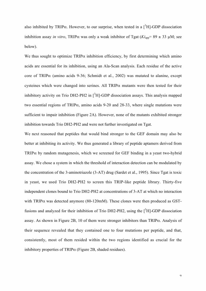
We thus sought to optimize TRIP inhibition efficiency, by first determining which amino
acids are essential for its inhibition, using an Ala-Scan analysis. Each residue of the active
core of TRIP (amino acids 9-36; Schmidt et al., 2002) was mutated to alanine, except
cysteines which were changed into serines. All TRIP mutants were then tested for their
inhibitory activity on Trio DH2-PH2 in [3H]-GDP dissociation assays. This analysis mapped
two essential regions of TRIP , amino acids 9-20 and 28-33, where single mutations were
sufficient to impair inhibition (Figure 2A). However, none of the mutants exhibited stronger
inhibition towards Trio DH2-PH2 and were not further investigated on Tgat.
We next reasoned that peptides that would bind stronger to the GEF domain may also be
better at inhibiting its activity. We thus generated a library of peptide aptamers derived from
TRIP by random mutagenesis, which we screened for GEF binding in a yeast two-hybrid
assay. We chose a system in which the threshold of interaction detection can be modulated by
the concentration of the 3-aminotriazole (3-AT) drug (Sardet et al., 1995). Since Tgat is toxic
in yeast, we used Trio DH2-PH2 to screen this TRIP-like peptide library. Thirty-five
independent clones bound to Trio DH2-PH2 at concentrations of 3-AT at which no interaction
with TRIP was detected anymore (80-120mM). These clones were then produced as GST-
fusions and analyzed for their inhibition of Trio DH2-PH2, using the [3H]-GDP dissociation
assay. As shown in Figure 2B, 10 of them were stronger inhibitors than TRIP . Analysis of
their sequence revealed that they contained one to four mutations per peptide, and that,
consistently, most of them resided within the two regions identified as crucial for the
inhibitory properties of TRIP (Figure 2B, shaded residues).
8
We then analyzed the inhibitory activity towards Tgat of TRIPE32G
and TRIPT16M/L17S
, the two
TRIP-like peptides that displayed the highest inhibition on Trio DH2-PH2 (Figure 2B). Both
peptides inhibited Tgat GEF activity in a dose-dependent manner in a kinetics fluorescence
assay, while GST alone (not shown) or GST-TRIP , at the same concentrations, had no effect
(Figure 2C). Accordingly, the apparent inhibition constant (Kiapp) of TRIP towards Tgat was
89 ± 33 M, and decreased to 7.4 ± 5 M for TRIPE32G
and 5.1 ± 4 M for TRIPT16M/L17S
(Figure 2D). These data show that TRIPE32G
and TRIPT16M/L17S
are both about 15 times more
efficient than TRIP at inhibiting the exchange activity of Tgat.
Interestingly, the optimized peptides were equally efficient on Tgat and on Trio DH2, as
shown by their similar Kiapp values (Figure 2C and 2D). This suggests that the unique C-
terminal extension of Tgat is not involved in the inhibitory mechanism of the optimized
peptides, and is consistent with the fact that this sequence does not interfere with the GEF
activity in vitro (data not shown).
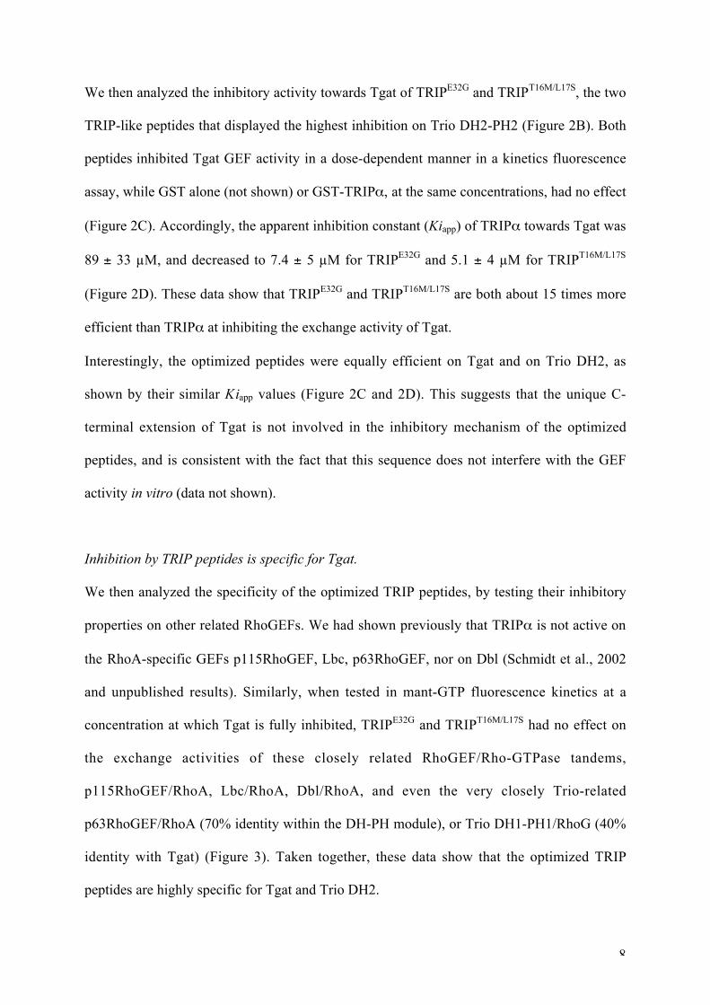
Inhibition by TRIP peptides is specific for Tgat.
We then analyzed the specificity of the optimized TRIP peptides, by testing their inhibitory
properties on other related RhoGEFs. We had shown previously that TRIP is not active on
the RhoA-specific GEFs p115RhoGEF, Lbc, p63RhoGEF, nor on Dbl (Schmidt et al., 2002
and unpublished results). Similarly, when tested in mant-GTP fluorescence kinetics at a
concentration at which Tgat is fully inhibited, TRIPE32G
and TRIPT16M/L17S
had no effect on
the exchange activities of these closely related RhoGEF/Rho-GTPase tandems,
p115RhoGEF/RhoA, Lbc/RhoA, Dbl/RhoA, and even the very closely Trio-related
p63RhoGEF/RhoA (70% identity within the DH-PH module), or Trio DH1-PH1/RhoG (40%
identity with Tgat) (Figure 3). Taken together, these data show that the optimized TRIP
peptides are highly specific for Tgat and Trio DH2.
9
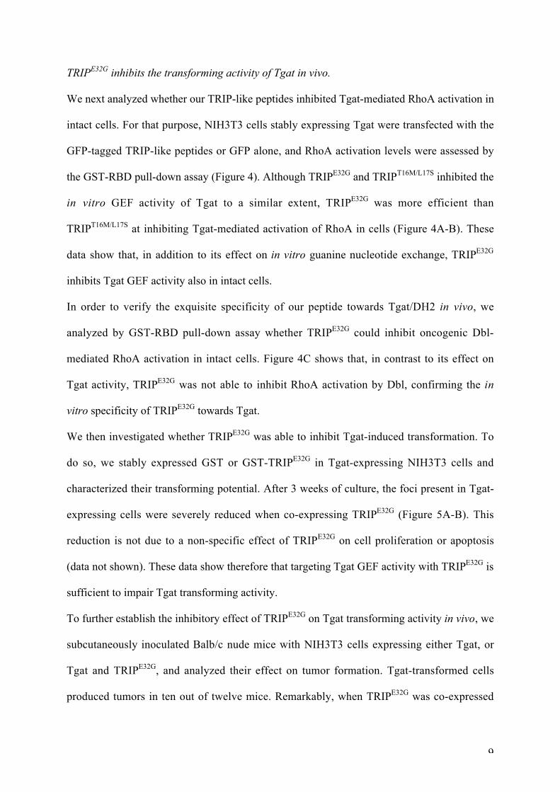
TRIPE32G
inhibits the transforming activity of Tgat in vivo.
We next analyzed whether our TRIP-like peptides inhibited Tgat-mediated RhoA activation in
intact cells. For that purpose, NIH3T3 cells stably expressing Tgat were transfected with the
GFP-tagged TRIP-like peptides or GFP alone, and RhoA activation levels were assessed by
the GST-RBD pull-down assay (Figure 4). Although TRIPE32G
and TRIPT16M/L17S
inhibited the
in vitro GEF activity of Tgat to a similar extent, TRIPE32G
was more efficient than
TRIPT16M/L17S
at inhibiting Tgat-mediated activation of RhoA in cells (Figure 4A-B). These
data show that, in addition to its effect on in vitro guanine nucleotide exchange, TRIPE32G
inhibits Tgat GEF activity also in intact cells.
In order to verify the exquisite specificity of our peptide towards Tgat/DH2 in vivo, we
analyzed by GST-RBD pull-down assay whether TRIPE32G
could inhibit oncogenic Dbl-
mediated RhoA activation in intact cells. Figure 4C shows that, in contrast to its effect on
Tgat activity, TRIPE32G
was not able to inhibit RhoA activation by Dbl, confirming the in
vitro specificity of TRIPE32G
towards Tgat.
We then investigated whether TRIPE32G
was able to inhibit Tgat-induced transformation. To
do so, we stably expressed GST or GST-TRIPE32G
in Tgat-expressing NIH3T3 cells and
characterized their transforming potential. After 3 weeks of culture, the foci present in Tgat-
expressing cells were severely reduced when co-expressing TRIPE32G
(Figure 5A-B). This
reduction is not due to a non-specific effect of TRIPE32G
on cell proliferation or apoptosis
(data not shown). These data show therefore that targeting Tgat GEF activity with TRIPE32G
is
sufficient to impair Tgat transforming activity.
To further establish the inhibitory effect of TRIPE32G
on Tgat transforming activity in vivo, we
subcutaneously inoculated Balb/c nude mice with NIH3T3 cells expressing either Tgat, or
Tgat and TRIPE32G
, and analyzed their effect on tumor formation. Tgat-transformed cells
produced tumors in ten out of twelve mice. Remarkably, when TRIPE32G
was co-expressed

10
with Tgat, only seven mice had tumors and we observed a delay of about three weeks in the
formation of tumors (Figure 5C). In addition, even though tumor formation was not abolished,
the weight of the tumors was significantly reduced when TRIPE32G
was expressed (Figure
5D). Altogether, these data show that expression of TRIPE32G
strongly reduces Tgat
transformation activity in cells and affects tumor formation in nude mice, most likely by
inhibiting Tgat-mediated GTP loading of RhoA.

11
DISCUSSION
Peptide aptamers as new inhibitors of RhoGEFs.
Because of their deregulation in many human disorders including cancer, Rho GTPases and
their activating GEFs represent challenging targets for inhibition. In humans there are only 20
Rho GTPases but more than 70 RhoGEFs, and it appears that signaling specificity is mostly
determined by the GEFs, which activate the GTPases at defined timing and location. RhoGEF
inhibitors therefore represent an emerging field of investigation.
Here we developed a peptide aptamer screening strategy to inhibit the RhoGEF Tgat, a
potential target in the ATL disease. Since Tgat is an isoform of the RhoGEF Trio, which
includes the RhoA-specific DH2 domain plus a unique C-terminal sequence, we based our
screen on our previously identified Trio inhibitor TRIP , the first peptidic RhoGEF inhibitor
described, which targets the DH2-PH2 domain of Trio (Schmidt et al., 2002). Intriguingly,
despite the fact that Tgat harbors the Trio DH2 domain, our original TRIP inhibitor was
rather ineffective at inhibiting Tgat. This suggests that the PH2 domain of Trio is involved in
the mechanism of action of TRIP , and that its replacement by the C-terminal extension
decreases TRIP 's ability to inhibit the GEF activity of Tgat.
We show here that GEF inhibitors selected with the peptide aptamer screening approach are
readily amenable to structure-activity relationship analysis and optimization. Of the 28
residues located in the active core of TRIP , alanine scanning mapped 9 residues in two
regions (residues 9-20 and 28-33) that were critical for the catalytic activity, while 6 had a
moderate effect and the others had no effect. We also show that peptide aptamer optimization
can be achieved by random mutagenesis combined with a selection screen based on
interaction strength. At least one third of the isolated clones yielded stronger inhibition, thus
validating the rationale of the screen. The two selected peptides, TRIPE32G
and TRIPT16M/L17S
,
were 15-fold more efficient than TRIP and inhibited Tgat GEF activity at concentrations
12
lying in the low micromolar range. Interestingly, mutations found in these clones also fell
within the two important regions identified by the Ala-scan. Furthermore, this approach
allowed us to turn TRIP into a Tgat inhibitor, which could be achieved with as few as one
mutation, E32G. It remains to be determined whether these different amino acids are
important for binding to the GEF and/or for inhibition of the exchange reaction.
It should be emphasized that our screening and optimization method is effective, irrespective
of the inhibitory mechanism, which is of big advantage for the discovery of inhibitors of
protein-protein interactions. The way the original screen was performed, i.e. two-hybrid
screening with the GEF as bait in the absence of GTPase, strongly suggests that the target of
the peptides is the GEF itself, rather than the GTPase. This is reinforced by the fact that the
peptides do not inhibit spontaneous GDP release from RhoA using [3H]-GDP-loaded RhoA
(data not shown), and by our specificity data in vitro and in intact cells, which show that other
GEF activities towards RhoA are not inhibited (Figure 3 and 4). At this stage we cannot,
however, distinguish between competitive and allosteric inhibition, or even a less likely
uncompetitive mechanism.
Remarkably, the characterization of our optimized TRIP peptides clearly shows that gain of
efficiency is not associated with loss of specificity. Indeed, none of the RhoA-activating GEFs
we tested, in particular the very closely related p63RhoGEF, were affected by either TRIPE32G
or TRIPT16M/L17S
. In addition, the TRIP peptides did not affect the activity of GEFs with
different specificity such as the RhoG/Rac1-specific TrioDH1-PH1.
Peptide aptamers are functional in vivo
Our screening method demonstrates that TRIPE32G
is not only effective and specific at
inhibiting Tgat GEF activity in vitro, but that it also blocks Tgat-induced cell transformation
and tumor formation in vivo. This is the first example of a peptidic RhoGEF inhibitor that is

13
functional in vivo, and demonstrates that aptamers can be used as active peptides to perturb
the function of GEFs in vivo. In this context, efficient in vivo delivery is a critical issue when
working with peptides. To circumvent this problem, the use of recently developed cell
penetrating peptides represents a good means of delivery for TRIPE32G
, and could be an
attractive strategy to investigate the contribution of Tgat in leukemogenesis. Indeed, to date,
the incidence of Tgat in ATL leukemogenesis is unknown, but given the strong effect of Tgat
on RhoA activation and transformation, we can hypothesize that Tgat is involved in the
progression of ATL by contributing to RhoA-mediated proliferation and/or metastasis. Our
series of TRIP peptides should now prove useful tools to decipher the cellular role of Tgat.
Peptide aptamers versus other GEF inhibitor screening strategies.
Besides our peptide aptamer screening approach, other strategies have recently been devised
to discover chemical inhibitors of Rho GTPase/GEF tandems, and also other classes of small
G proteins, such as the Arf family and their activating GEFs (Blangy et al., 2006; Desire et
al., 2005; Gao et al., 2004; Mayer et al., 2001; Shutes et al., 2007; Viaud et al., 2007).
Computer-assisted virtual screening, for example, identified the NSC23766 compound, based
on structure-function information of the Rac1/Tiam1 complex. This powerful molecule
inhibits specifically Rac1-induced events in vitro and in vivo, however the targeted associated
RhoGEFs include at least Tiam1 and Trio DH1-PH1 (Gao et al., 2004). In silico screening
also yielded the LM11 compound, which inhibits specifically the ARNO/Arf1 interface in
vitro and is active in cells (Viaud et al., 2007). Given their membrane permeability, both
NSC23766 and LM11 have the advantage of being easily applied in vivo.
The Yeast Exchange Assay is another screening method that allowed the identification of the
TrioDH1-PH1 specific NPPD compound and its analogues (Blangy et al., 2006). Like peptide
14
aptamer screening, this strategy has the advantage over virtual screening of identifying
inhibitors directly in cells, and without any bias as to the targeted interaction site.
Finally, in vitro RNA-aptamer screening selected the RNA aptamer M69 as an inhibitor of the
Cytohesin/Arf1 tandem (Mayer et al., 2001). Like peptide-aptamers, these RNA aptamers are
highly combinatorial and easily screened, but their application as potential drugs remains
limited, due to difficult in vivo delivery. To circumvent this problem, RNA-aptamer-
displacement represents an elegant method, in which a small-molecule library is screened for
compounds that displace the RNA-aptamer from its target and reproduce its inhibitory activity
(Hafner et al., 2006).
Our study shows that peptide aptamer screening represents a valid strategy for inhibitor
identification that can be applied to a variety of different proteins, because of the in vivo
screening method and the highly combinatorial libraries available, yielding strong affinity
inhibitors. This is illustrated here by the identification of a highly specific peptidic RhoGEF
inhibitor targeting the Tgat oncogene in vitro and in vivo.
15
SIGNIFICANCE
When trying to inhibit signaling pathways controlled by small G proteins and their activating
GEFs, the challenge is that these are not mere enzymes with a well-defined active site that can
be blocked. Rather, protein-protein interactions have to be targeted and the lack of reactive
pockets to which inhibitors could bind is a challenging issue. This might in part explain why,
although oncogenic Ras has been discovered more than 20 years ago, no inhibitor with
clinical validation has been identified. Therefore, research has focused on trying to inhibit the
guanine nucleotide exchange factors instead, and recent studies report the successful
identification of such inhibitors.
The power of the strategy we used here to identify the TRIP peptides relies on the screening
of a highly combinatorial aptamer library, generating immense possibilities of random
peptides. This variety makes peptide aptamers very suitable molecules to inhibit complex
protein-protein interactions such as the tandem RhoGEF/GTPase, and to discriminate between
closely related proteins. One major advantage of this kind of approach is that the screening is
cell-based, which gives a direct readout for toxicity and is more stringent. Moreover, peptide
aptamers do not mimic cellular targets, which could have non-desired effects in cells. In
addition, to circumvent the problem of in vivo delivery when using peptide aptamers,
aptamer-displacement screens can be performed, in order to convert an aptamer into a small
compound inhibitor (Baines and Colas, 2006). The advantage is that the corresponding
compound targets the same site and shares the same properties as the already characterized
peptide.
In conclusion, peptide aptamers represent a promising alternative for the discovery of leads
for new therapeutic drugs.
16
EXPERIMENTAL PROCEDURES
DNA constructs - Tgat (aa 1-255) was designed by ligating dimerized oligonucleotides coding
for the specific C-terminus of Tgat (15 aa) to the Trio DH2 domain (residues 1862-2101,
corresponding to aa 1-240). The oligonucleotide sequences are available upon request. The
TgatL190E
mutant was obtained using the Quick Change Site Directed Mutagenesis Kit
(Stratagene Inc.), according to the manufacturer’s instructions.
To create stable NIH3T3 cell lines, GFP-tagged Tgat, TgatL190E
and full length Trio were
cloned into the puromycin-resistant retroviral vector pBabePuro. GST-tagged TRIP peptides
were cloned into the G418-resistant retroviral vector pLXSN. For transient transfections, both
Tgat and TRIP peptides were cloned into the pEGFP vector (Clontech Inc.). Myc-Dbl was a
kind gift of Michael Olson (Beatson Institute for Cancer Research, Glasgow). For in vitro
GEF assays, Tgat (aa 1-255) was fused to maltose-binding protein (MBP) by cloning into a
modified pMAL C2X vector (New England Biolabs Inc.). The TRIP peptides were fused to
GST by cloning into the pGEX-5X2 vector (GE Healthcare Inc.). All constructs were checked
by sequencing.
Expression and purification of recombinant proteins - Tgat. MBP-Tgat and MBP-DH2
expression in E. coli was induced for 24h at 16°C with 0.1 mM isopropylthio-
galactopyranoside (IPTG). After cell lysis (in 50 mM Tris pH 7.5, 1 mM EDTA, 2 mM
MgCl2, 1 mM DTT), the suspension was centrifuged at 10,000g for 20 min, then at 400,000g
for 1h30. The supernatant was applied to a Q-Sepharose column fast flow (GE Healthcare)
equilibrated with lysis buffer. The protein was eluted with a linear gradient of 0-250 mM
NaCl in 50 mM Tris pH 7.5. Fractions containing the protein were adjusted to a concentration
of 2 M NaCl and loaded on a Phenyl sepharose Fast flow High Sub (GE Healthcare Inc.). The
17
protein was eluted with a linear gradient of 2-0 M NaCl in 50 mM Tris pH 7.5. The purified
proteins were concentrated on a Vivaspin concentrator (Vivascience AG Inc.) at 18 mg/mL.
Other proteins. Recombinant GST-Trio DH2-PH2, GST-Trio DH1-PH1, GST-Dbl (DH-PH
domain), GST-Lbc (DH-PH), GST-p63RhoGEF (DH domain) and GST-RhoG were purified
as described previously (Schmidt et al., 2002; Souchet et al., 2002). Expression and
purification of GST-p115RhoGEF using the baculo virus system will be described elsewhere.
GST-Peptides. GST-TRIP peptides were purified as described (Schmidt et al., 2002), except
that the cell lysate was centrifuged as above, before loading on a GSTrap Fast Flow column
(GE Healthcare Inc.) equilibrated with lysis buffer. Peptides were eluted with reduced
glutathione (10 mM) in Tris 50 mM pH 7.5 and concentrated on Vivaspin concentrator at
about 5-10 mg/mL.
Optimization of TRIP - Alanine-scanning of TRIP . Every amino acid of the active core of
TRIP (amino acids 9-36) was mutated to alanine (or serine for cysteine residues) by site
directed mutagenesis of GST-TRIP . Each TRIP mutant was tested for its inhibitory activity
on DH2-PH2 in [3H]-GDP dissociation assays.
Two-hybrid screening of TRIP -like peptides. An aptamer library derived from TRIP was
created by PCR-based random mutagenesis of TRIP inserted into the yeast two-hybrid
vector pPC86. Sequencing of a statistically representative number of clones yielded a
mutation rate of ~3 mutations/clone. 6x10⁵ independent clones were screened for interactors,
using Trio DH2-PH2 (in the pPC97 vector) as a bait, in the MAV103 yeast strain, on high
concentrations of 3-AT (3-amino-triazol, Sigma) (80-120mM). Selected peptides were then
produced as GST-fusions and analyzed for their inhibition of Trio DH2-PH2 using the [3H]-
GDP dissociation assay.

18
Nucleotide exchange kinetics assay - Specific exchange rates of Tgat were measured with a
fluorescence-based kinetics assay, using a 6His-RhoA construct (gift of Dr Derewenda,
Charlottesville University, Virginia) purified as described (Oleksy et al., 2004). Exchange
activities were followed by fluorescence resonance energy transfer (FRET) between the
GTPase tryptophanes ( ex=292nm) and the methylanthranyloil group of mant-GTP
( em=440nm) as described (Zeeh et al., 2006). All fluorescence measurements were
performed with a CARY Eclipse fluorimeter (Varian). For each kobs determination, RhoA
(1 M) and Tgat (or Trio DH2) were preincubated 3 min at 25°C in 700 L reaction buffer (50
mM Tris pH 7.5, 50 mM NaCl, 2 mM MgCl2, 1 mM DTT). The exchange reaction was
initiated by 10 M mant-GTP and measured for 10 min until the plateau was reached.
kobs were calculated by fitting the FRET fluorescence changes to a single exponential, using
the Kaleidagraph software. Specific exchange activities were calculated by linear regression
of kobs values determined for a range of GEF concentrations (0, 0.2, 0.3, 0.4, 0.5 and 1 M).
Nucleotide exchange inhibition assays - Radioactive [3H]-GDP dissociation assays were
performed as described (Schmidt et al., 2002). Briefly, 0.15 M GST-Trio DH2-PH2 was pre-
incubated for 15 min with 3 M of GST-TRIP inhibitors. The reaction was started by addition
of 0.4 M [3H]-GDP-loaded RhoA and 1mM GTP, and the reaction mix was filtered after 0
min and 15 min incubation at 25°C. Inhibition efficiency is expressed as the ratio between
[3H]-GDP-bound RhoA at 15 and 0 min.
Apparent inhibition constants (Kiapp) of TRIP-like peptides were determined from kobs values
obtained at increasing peptide concentrations using the above fluorescence nucleotide
exchange assay. Kiapp was calculated from the hyperbolic fit of kobs values as a function of the
inhibitor concentration as described (Zeeh et al., 2006).
19
TRIP-like peptide specificity was assayed using mant-GTP fluorescence kinetics ( ex=360nm,
em=460nm) in a FLX800 Microplate Fluorescence Reader (BioTek Instruments). 0.5 M
Tgat, p63RhoGEF, Lbc, and p115RhoGEF, or 0.1 M Dbl and Trio DH1-PH1 were
preincubated 5 min at 25°C in the presence of 20 M GST, GST-TRIPE32G
or TRIPT16M/L17S
and 1 M mant-GTP. The exchange reaction was initiated by addition of 1 M RhoA or RhoG
and monitored for 10 min.
Cell lines, transfection and focus formation assay - NIH3T3 cells were maintained as
described previously (Sirvent et al., 2007).
Transient transfection experiments were performed using the Jet PEI reagent, according to the
manufacturer’s protocol (QBiogene Inc.). NIH3T3 cell lines stably expressing GFP-Tgat,
GFP-TgatL190E
, or GFP-Trio, with or without the GST-TRIP peptides, were generated as
follows : the indicated retroviral constructs were transfected into BOSC packaging cells, using
the Lipofectamine reagent (Invitrogen Inc.). Forty-eight hours after transfection, virus-
containing supernatants were collected and used to infect NIH3T3 cells. Infected cells were
selected with 6 g/mL puromycin and/or 1mg/mL G418 and stable transfectants were pooled
after selection. Tgat or TRIP mRNA levels in the different cell lines were monitored by RT-
PCR, and protein expression levels by Western blot analysis using a polyclonal anti-GFP
antibody (Torrey Pines Laboratories).
Focus formation assays were performed using stable NIH3T3 cell lines as indicated, seeded at
5x10⁴ cells in 6-well plates and maintained for 15 to 21 days in 10% FBS. Medium was
renewed every two days. After staining with Crystal Violet (1%), plates were photographed
and foci were scored using the Metamorph software. All experiments were done in triplicate.
RhoA activation assay in cells - The level of GTP-bound RhoA was measured by a GST pull-
down assay as described (Schmidt et al., 2002). Briefly, cell lysates were incubated with
20
glutathione beads coated with the recombinant Rho-binding domain (RBD) of the RhoA-
specific effector Rhotekin (Cytoskeleton Inc.). Total or GTP-bound RhoA in the samples was
revealed by Western blot analysis, using a monoclonal anti-RhoA antibody (Santa-Cruz
Biotechnology Inc.).
Mice and xenografting - Female Balb/c nu/nu mice were purchased from Charles River
France and used at 6-8 weeks of age. 12 Balb/c nu/nu mice were subcutaneously grafted with
2x10⁶ cells of each cell line on both sides (Tgat on the left and Tgat+TRIP
E32G on the right
flank of the leg). The appearance of tumors was scored visually every week. 10 weeks post
graft, mice were euthanized and tumors excised and weighed. mRNA and protein levels in the
tumors were verified by RT-PCR and Western blot (data not shown). All experiments on mice
have been approved by the internal ethical committee of the IRCM and have been performed
by B.R. under the authorization number N° 34.156, in the animal facility with agreement N°
B34-172-27.

21
REFERENCES
Baines, I. C., and Colas, P. (2006). Peptide aptamers as guides for small-molecule drug
discovery. Drug Discov Today 11, 334-341.
Bellanger, J. M., Estrach, S., Schmidt, S., Briancon-Marjollet, A., Zugasti, O., Fromont, S.,
and Debant, A. (2003). Different regulation of the Trio Dbl-Homology domains by their
associated PH domains. Biol Cell 95, 625-634.
Bickle, M. B., Dusserre, E., Moncorge, O., Bottin, H., and Colas, P. (2006). Selection and
characterization of large collections of peptide aptamers through optimized yeast two-hybrid
procedures. Nat Protoc 1, 1066-1091.
Blangy, A., Bouquier, N., Gauthier-Rouviere, C., Schmidt, S., Debant, A., Leonetti, J. P., and
Fort, P. (2006). Identification of TRIO-GEFD1 chemical inhibitors using the yeast exchange
assay. Biol Cell 98, 511-522.
Bos, J. L., Rehmann, H., and Wittinghofer, A. (2007). GEFs and GAPs: critical elements in
the control of small G proteins. Cell 129, 865-877.
Butz, K., Denk, C., Ullmann, A., Scheffner, M., and Hoppe-Seyler, F. (2000). Induction of
apoptosis in human papillomaviruspositive cancer cells by peptide aptamers targeting the viral
E6 oncoprotein. Proc Natl Acad Sci U S A 97, 6693-6697.
Chhatriwala, M. K., Betts, L., Worthylake, D. K., and Sondek, J. (2007). The DH and PH
domains of Trio coordinately engage Rho GTPases for their efficient activation. J Mol Biol
368, 1307-1320.
Colas, P., Cohen, B., Jessen, T., Grishina, I., McCoy, J., and Brent, R. (1996). Genetic
selection of peptide aptamers that recognize and inhibit cyclin-dependent kinase 2. Nature
380, 548-550.

22
Crnkovic-Mertens, I., Hoppe-Seyler, F., and Butz, K. (2003). Induction of apoptosis in tumor
cells by siRNA-mediated silencing of the livin/ML-IAP/KIAP gene. Oncogene 22, 8330-
8336.
Desire, L., Bourdin, J., Loiseau, N., Peillon, H., Picard, V., De Oliveira, C., Bachelot, F.,
Leblond, B., Taverne, T., Beausoleil, E., et al. (2005). RAC1 inhibition targets amyloid
precursor protein processing by gamma-secretase and decreases Abeta production in vitro and
in vivo. J Biol Chem 280, 37516-37525.
Etienne-Manneville, S., and Hall, A. (2002). Rho GTPases in cell biology. Nature 420, 629-
635.
Eva, A., and Aaronson, S. A. (1985). Isolation of a new human oncogene from a diffuse B-
cell lymphoma. Nature 316, 273-275.
Fabbrizio, E., Le Cam, L., Polanowska, J., Kaczorek, M., Lamb, N., Brent, R., and Sardet, C.
(1999). Inhibition of mammalian cell proliferation by genetically selected peptide aptamers
that functionally antagonize E2F activity. Oncogene 18, 4357-4363.
Gao, Y., Dickerson, J. B., Guo, F., Zheng, J., and Zheng, Y. (2004). Rational design and
characterization of a Rac GTPase-specific small molecule inhibitor. Proc Natl Acad Sci U S A
101, 7618-7623.
Hafner, M., Schmitz, A., Grune, I., Srivatsan, S. G., Paul, B., Kolanus, W., Quast, T.,
Kremmer, E., Bauer, I., and Famulok, M. (2006). Inhibition of cytohesins by SecinH3 leads to
hepatic insulin resistance. Nature 444, 941-944.
Hoppe-Seyler, F., Crnkovic-Mertens, I., Tomai, E., and Butz, K. (2004). Peptide aptamers:
specific inhibitors of protein function. Curr Mol Med 4, 529-538.
Katzav, S., Martin-Zanca, D., and Barbacid, M. (1989). vav, a novel human oncogene derived
from a locus ubiquitously expressed in hematopoietic cells. Embo J 8, 2283-2290.

23
Lutz, S., Shankaranarayanan, A., Coco, C., Ridilla, M., Nance, M. R., Vettel, C., Baltus, D.,
Evelyn, C. R., Neubig, R. R., Wieland, T., and Tesmer, J. J. (2007). Structure of Galphaq-
p63RhoGEF-RhoA complex reveals a pathway for the activation of RhoA by GPCRs. Science
318, 1923-1927.
Martel, V., Filhol, O., Colas, P., and Cochet, C. (2006). p53-dependent inhibition of
mammalian cell survival by a genetically selected peptide aptamer that targets the regulatory
subunit of protein kinase CK2. Oncogene 25, 7343-7353.
Mayer, G., Blind, M., Nagel, W., Bohm, T., Knorr, T., Jackson, C. L., Kolanus, W., and
Famulok, M. (2001). Controlling small guanine-nucleotide-exchange factor function through
cytoplasmic RNA intramers. Proc Natl Acad Sci U S A 98, 4961-4965.
Miki, T., Smith, C. L., Long, J. E., Eva, A., and Fleming, T. P. (1993). Oncogene ect2 is
related to regulators of small GTP-binding proteins. Nature 362, 462-465.
Mori, T., Moriuchi, R., Okazaki, E., Yamada, K., and Katamine, S. (2007). Tgat oncoprotein
functions as a inhibitor of RECK by association of the unique C-terminal region. Biochem
Biophys Res Commun 355, 937-943.
Nouvion, A. L., Thibaut, J., Lohez, O. D., Venet, S., Colas, P., Gillet, G., and Lalle, P. (2007).
Modulation of Nr-13 antideath activity by peptide aptamers. Oncogene 26, 701-710.
Oleksy, A., Barton, H., Devedjiev, Y., Purdy, M., Derewenda, U., Otlewski, J., and
Derewenda, Z. S. (2004). Preliminary crystallographic analysis of the complex of the human
GTPase RhoA with the DH/PH tandem of PDZ-RhoGEF. Acta Crystallogr D Biol Crystallogr
60, 740-742.
Rojas, R. J., Yohe, M. E., Gershburg, S., Kawano, T., Kozasa, T., and Sondek, J. (2007).
Galphaq directly activates p63RhoGEF and Trio via a conserved extension of the Dbl
homology-associated pleckstrin homology domain. J Biol Chem 282, 29201-29210.
24
Rossman, K. L., Cheng, L., Mahon, G. M., Rojas, R. J., Snyder, J. T., Whitehead, I. P., and
Sondek, J. (2003). Multifunctional roles for the PH domain of Dbs in regulating Rho GTPase
activation. J Biol Chem 278, 18393-18400.
Rossman, K. L., Der, C. J., and Sondek, J. (2005). GEF means go: turning on RHO GTPases
with guanine nucleotide-exchange factors. Nat Rev Mol Cell Biol 6, 167-180.
Sahai, E., and Marshall, C. J. (2002). RHO-GTPases and cancer. Nat Rev Cancer 2, 133-142.
Sardet, C., Vidal, M., Cobrinik, D., Geng, Y., Onufryk, C., Chen, A., and Weinberg, R. A.
(1995). E2F-4 and E2F-5, two members of the E2F family, are expressed in the early phases
of the cell cycle. Proc Natl Acad Sci U S A 92, 2403-2407.
Schmidt, S., Diriong, S., Mery, J., Fabbrizio, E., and Debant, A. (2002). Identification of the
first Rho-GEF inhibitor, TRIPalpha, which targets the RhoA-specific GEF domain of Trio.
FEBS Lett 523, 35-42.
Shutes, A., Onesto, C., Picard, V., Leblond, B., Schweighoffer, F., and Der, C. J. (2007).
Specificity and mechanism of action of EHT 1864, a novel small molecule inhibitor of Rac
family small GTPases. J Biol Chem 282, 35666-35678.
Sirvent, A., Boureux, A., Simon, V., Leroy, C., and Roche, S. (2007). The tyrosine kinase Abl
is required for Src-transforming activity in mouse fibroblasts and human breast cancer cells.
Oncogene 26, 7313-7323.
Souchet, M., Portales-Casamar, E., Mazurais, D., Schmidt, S., Leger, I., Javre, J. L., Robert,
P., Berrebi-Bertrand, I., Bril, A., Gout, B., et al. (2002). Human p63RhoGEF, a novel RhoA-
specific guanine nucleotide exchange factor, is localized in cardiac sarcomere. J Cell Sci 115,
629-640.
Toksoz, D., and Merdek, K. D. (2002). The Rho small GTPase: functions in health and
disease. Histol Histopathol 17, 915-927.
25
Viaud, J., Zeghouf, M., Barelli, H., Zeeh, J. C., Padilla, A., Guibert, B., Chardin, P., Royer, C.
A., Cherfils, J., and Chavanieu, A. (2007). Structure-based discovery of an inhibitor of Arf
activation by Sec7 domains through targeting of protein-protein complexes. Proc Natl Acad
Sci U S A 104, 10370-10375.
Whitehead, I., Kirk, H., Tognon, C., Trigo-Gonzalez, G., and Kay, R. (1995). Expression
cloning of lfc, a novel oncogene with structural similarities to guanine nucleotide exchange
factors and to the regulatory region of protein kinase C. J Biol Chem 270, 18388-18395.
Whitehead, I. P., Khosravi-Far, R., Kirk, H., Trigo-Gonzalez, G., Der, C. J., and Kay, R.
(1996). Expression cloning of lsc, a novel oncogene with structural similarities to the Dbl
family of guanine nucleotide exchange factors. J Biol Chem 271, 18643-18650.
Yamada, K., Moriuchi, R., Mori, T., Okazaki, E., Kohno, T., Nagayasu, T., Matsuyama, T.,
and Katamine, S. (2007). Tgat, a Rho-specific guanine nucleotide exchange factor, activates
NF-kappaB via physical association with IkappaB kinase complexes. Biochem Biophys Res
Commun 355, 269-274.
Yoshizuka, N., Moriuchi, R., Mori, T., Yamada, K., Hasegawa, S., Maeda, T., Shimada, T.,
Yamada, Y., Kamihira, S., Tomonaga, M., and Katamine, S. (2004). An alternative transcript
derived from the trio locus encodes a guanosine nucleotide exchange factor with mouse cell-
transforming potential. J Biol Chem 279, 43998-44004.
Zeeh, J. C., Zeghouf, M., Grauffel, C., Guibert, B., Martin, E., Dejaegere, A., and Cherfils, J.
(2006). Dual specificity of the interfacial inhibitor brefeldin a for arf proteins and sec7
domains. J Biol Chem 281, 11805-11814.
26
ACKNOWLEDGEMENTS
We thank Michel Brissac and Imad Aït Arsa for technical assistance in manipulating the nude
mice in the animal facility of CRLC Val d’Aurelle in Montpellier, Claire Macari and Sandra
Pierredon for technical help, Alexandra Joubert (LEBS, CNRS, Gif-sur-Yvette) for the gift of
purified p115RhoGEF, Pierre Travo, Julien Cau and Sylvain De Rossi from Montpellier Rio
Imaging for invaluable help and constant interest. Jean-Michel Bellanger, Philippe Pasero,
Claude Sardet and Gilles Divita are acknowledged for helpful discussion and critical reading
of the manuscript.
This work was supported by grants from the CNRS, the Agence Nationale de la Recherche
ANR-PCV (Physique et Chimie du Vivant) (A.D. and J.C.), the Association pour la
Recherche contre le Cancer (grant #2736 to A.D., PhD grants to C.A. and J.C.Z). All authors
except Bruno Robert are members of the CNRS consortium GDR2823.

27
FIGURE LEGENDS
Figure 1: The transforming activity of Tgat requires its GEF activity towards RhoA
A. Schematic representation of Trio and its splice variant Tgat, and the Tgat mutant used in
this study. B. RhoA activation assay. Lysates of NIH3T3 cells stably expressing GFP, GFP-
Tgat, or GFP-TgatL190E
were subjected to GST-pulldown using recombinant RBD (RhoA-
binding domain of Rhotekin). The levels of GTP-bound RhoA (top panel) and total RhoA
protein (middle panel) were assessed by Western Blot with a monoclonal anti-RhoA antibody.
All GEF constructs were expressed at a similar level as shown by Western blotting using an
anti-GFP antibody (lower panel). C. Quantification of the RhoA activation assay from at least
three independent experiments. “Fold RhoA activation” means the amount of RhoA-GTP in
the sample, as compared to the amount in the GFP control, which was set to 1. D. Focus
formation assay of NIH3T3 cells stably expressing GFP, GFP-Tgat, GFP-TgatL190E
or GFP-
Trio. E. Quantification of three independent focus formation assays. The number of foci
induced by Tgat was set to 100%. Error bars represent standard deviation in all graphs.
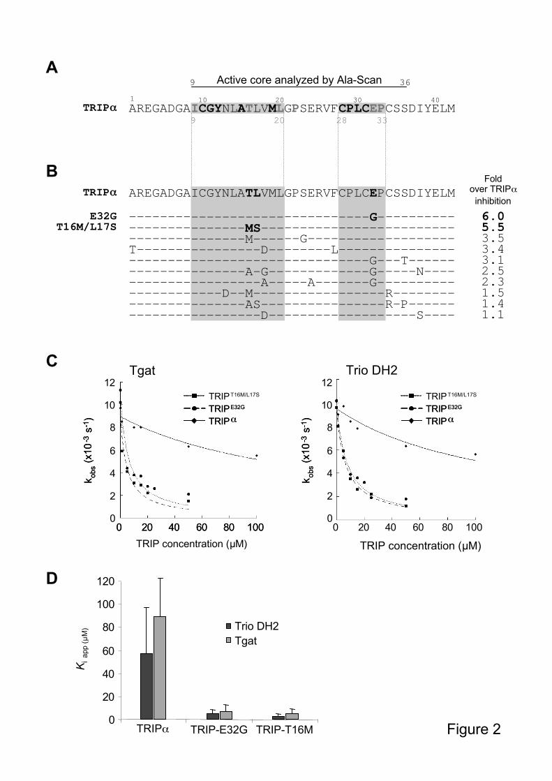
Figure 2: Identification and characterization of optimized inhibitory TRIP-like peptides
A. Alanine-scanning of the active core of TRIP . Inhibition efficiency of the mutated
peptides was measured by [3H]-GDP dissociation assays using Trio DH2-PH2, and compared
to the original TRIP peptide. Black bold letters: residues strictly required for inhibition; grey
bold letters: residues retaining a weak inhibitory potential. All the other residues are non
essential. Shaded residues: regions (aa 9-20 and 28-33) that emerge as being essential for
TRIP activity. B. Amino acid sequence of the optimized TRIP-like peptides, obtained by
random mutagenesis of the original TRIP peptide. Inhibition efficiency was measured on
Trio DH2-PH2 and compared to TRIP , as described in A. “Fold over TRIP inhibition”
means stronger inhibition at the same inhibitor concentration (inhibition by TRIP was set to

28
1). C. Inhibition of Tgat GEF activity by TRIPT16M/L17S
and TRIPE32G
in vitro. FRET
fluorescence exchange assays were performed using constant concentrations of RhoA (1 M),
equal amounts (0.5 M) of Tgat (left panel) or Trio DH2 (right panel), and increasing
concentrations of GST-TRIP peptides, up to 100 M. Results were expressed as kobs values
plotted as a function of the indicated TRIP inhibitor concentration. D. Apparent inhibition
constants (Kiapp) of the TRIP peptides for Tgat and Trio DH2, as indicated. The values and
error bars are calculated from at least three independent experiments. Error bars represent
standard deviation.
Figure 3: Specificity of the optimized inhibitory TRIP peptides
Comparison of TRIPE32G
and TRIPT16M/L17S
inhibition efficiency on different
GTPase/RhoGEF systems, using 1 M GTPase and 0.5 M GEF: A. RhoA/Tgat ; B.
RhoA/p63RhoGEF; C. RhoA/p115RhoGEF; D. RhoA/Lbc; E. RhoA/Dbl; F. RhoG/Trio
DH1PH1. In each assay, the peptides were used at a concentration of 20 M, corresponding to
a 40 fold molar excess of inhibitor versus GEF. All fluorescence kinetics assays were
performed using 1 M mant-GTP. Results are expressed as Relative Fluorescence Units
(RFU) versus time. The reaction performed in the absence of GEF reflects the spontaneous
exchange activity of the GTPase.
Figure 4: TRIPE32G
inhibits Tgat GEF activity in cells
RhoA activation in NIH3T3-Tgat cells stably transfected with GFP, GFP-TRIPE32G
or GFP-
TRIPT16M/L17S
was assayed by the GST-RBD-pulldown assay as described in Figure 1B. A.
The levels of GTP-bound and total RhoA protein are shown in the upper two panels.
Expression levels of all GFP-tagged proteins are shown in the lower panel. B. Quantification
of the RhoA activity assay from at least three independent experiments. Error bars represent
29
standard deviation. C. Effect of GFP-TRIPE32G
on RhoA activation induced by Dbl (left
panel) or Tgat (right panel) in NIH3T3 cells, assayed by GST-RBD pulldown. The levels of
GTP-bound and total RhoA protein are shown in the upper two panels. Expression levels of
Myc-Dbl and of all GFP-tagged proteins are shown in the lower two panels.
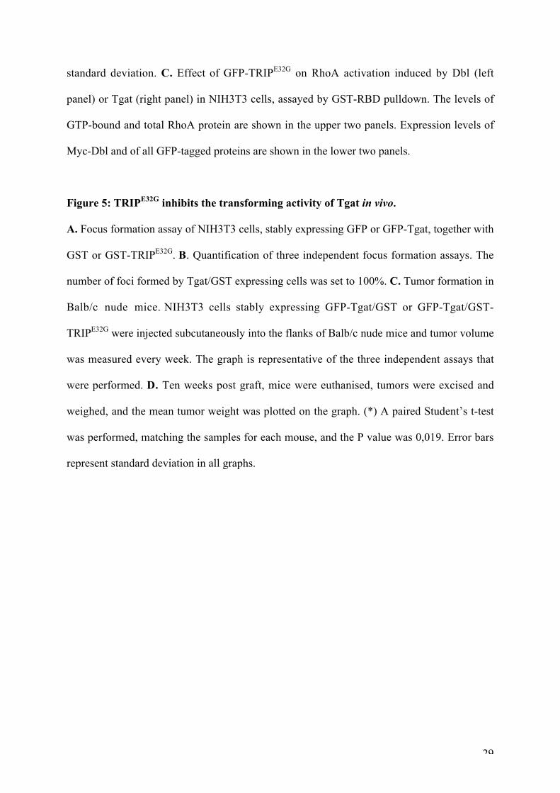
Figure 5: TRIPE32G
inhibits the transforming activity of Tgat in vivo.
A. Focus formation assay of NIH3T3 cells, stably expressing GFP or GFP-Tgat, together with
GST or GST-TRIPE32G
. B. Quantification of three independent focus formation assays. The
number of foci formed by Tgat/GST expressing cells was set to 100%. C. Tumor formation in
Balb/c nude mice. NIH3T3 cells stably expressing GFP-Tgat/GST or GFP-Tgat/GST-
TRIPE32G
were injected subcutaneously into the flanks of Balb/c nude mice and tumor volume
was measured every week. The graph is representative of the three independent assays that
were performed. D. Ten weeks post graft, mice were euthanised, tumors were excised and
weighed, and the mean tumor weight was plotted on the graph. (*) A paired Student’s t-test
was performed, matching the samples for each mouse, and the P value was 0,019. Error bars
represent standard deviation in all graphs.

A
B
DHTgat
TgatL190E L190E
D
C
Figure 1
EGFP Tgat
TgatL190E
Total RhoA
RhoA-GTP
Tgat
GFP0
2
4
6
8
10
12
Fold
RhoA
activation
GFP Tgat TgatL190E
0
20
40
60
80
100
GFP Tgat
Foci fo
rmation (
%)
LCHRFKETFREICWF
TgatL190E
GFP Tgat TgatL190E
Trio DH1 DH2PH1 PH2 Kinase
SH
3
SH
3
Ig
Trio
S P E C T R I N
Trio
B
AREGADGAICGYNLATLVMLGPSERVFCPLCEPCSSDIYELM
-------------------------------G----------
---------------MS-------------------------
---------------M------G-------------------
T----------------D--------L---------------
-------------------------------G---T------
---------------A-G-------------G-----N----
-----------------A-----A-------G----------
------------D--M-----------------R--------
---------------AS----------------R-P------
-----------------D-------------------S----
Foldover TRIP
inhibition
6.05.53.5
3.4
3.1
2.5
2.3
1.5
1.4
1.1
TRIP
E32GT16M/L17S
AREGADGAICGYNLATLVMLGPSERVFCPLCEPCSSDIYELM10 301 20 40
A
9
Active core analyzed by Ala-Scan9 36
TRIP
20 3328
------------
ICGYNLATLVML
-------MS---
-------M----
---------D--
------------
-------A-G--
---------A--
----D--M----
-------AS---
---------D--
----G-
CPLCEP
------
------
------
----G-
----G-
----G-
------
------
------
Figure 2
C
12
2
4
6
8
10
00 20 40 60 80 100
TRIP concentration ( M)
12
2
4
6
8
10
0
kobs
(x10
-3s
- 1)
kobs
(x10
- 3s
- 1)
TRIPT16M/L17S
TRIPE32G
TRIP
TRIPE32G
TRIP
0 20 40 60 80 100
D
0
20
40
60
80
100
120
TRIP TRIP-E32G TRIP-T16M
Trio DH2
Tgat
Ki a
pp (
M)
Trio DH2
TRIP concentration ( M)
Tgat
TRIPT16M/L17S
TRIPE32G
TRIP
TRIPE32G
TRIP
0 20 40 60 80 100
kobs
(x10
-3s
-1)
kobs
(x10
-3s
-1)
12
2
4
6
8
10
0
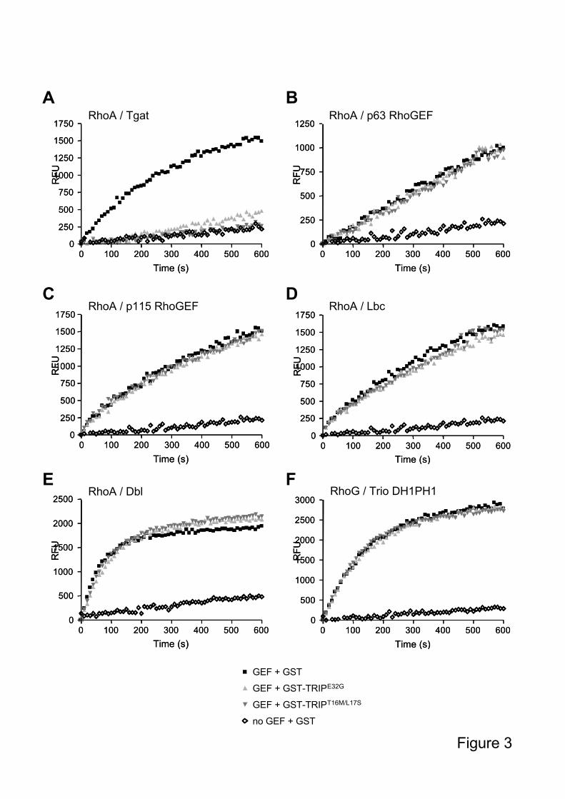
Figure 3
RhoA / Tgat
0 100 200 300 400 500 6000
250
500
750
1000
1250
1500
1750
Time (s)
RF
U
RhoA / Tgat
0 100 200 300 400 500 6000
250
500
750
1000
1250
1500
1750
Time (s)
RF
U
RhoA / p115 RhoGEF
0 100 200 300 400 500 6000
250
500
750
1000
1250
1500
1750
Time (s)
RF
U
RhoA / p115 RhoGEF
0 100 200 300 400 500 6000
250
500
750
1000
1250
1500
1750
Time (s)
RF
U
RhoA / Lbc
0 100 200 300 400 500 6000
250
500
750
1000
1250
1500
1750
Time (s)
RF
URhoA / Lbc
0 100 200 300 400 500 6000
250
500
750
1000
1250
1500
1750
Time (s)
RF
U
RhoA / p63
0 100 200 300 400 500 6000
250
500
750
1000
1250
Time (s)
RF
U
RhoA / p63
0 100 200 300 400 500 6000
250
500
750
1000
1250
Time (s)
RF
U
RhoA / Dbl
0 100 200 300 400 500 6000
500
1000
1500
2000
2500
Time (s)
RF
U
RhoA / Dbl
0 100 200 300 400 500 6000
500
1000
1500
2000
2500
Time (s)
RF
U
RhoG / Trio DH1PH1
0 100 200 300 400 500 6000
500
1000
1500
2000
2500
3000
Time (s)
RF
U
RhoG / Trio DH1PH1
0 100 200 300 400 500 6000
500
1000
1500
2000
2500
3000
Time (s)
RF
U
no GEF + GST
GEF + GST
GEF + GST-TRIPE32G
GEF + GST-TRIPT16M/L17S
RhoA / Tgat RhoA / p63 RhoGEF
RhoA / p115 RhoGEF RhoA / Lbc
RhoA / Dbl RhoG / Trio DH1PH1
A B
C D
E F

Figure 4
Total RhoA
Active RhoA
GFP
GFP
TRIPE32G
GFP
TRIPT16M/L17Sctrl
GFP-Tgat +
GFP-Tgat
GFPGFP-TRIPs 0
20
40
60
80
100
GFP
GFP-Tgat +%
RhoA
activation
BA
ctrl GFP
TRIPE32G
GFP
TRIPT16M/L17S
C
GFP
GFP
TRIPE32GGFP
Myc-Dbl +
Total RhoA
Active RhoA
GFPGFP-TRIPs
Myc-Dbl
GFP
GFP
TRIPE32GGFP
GFP-Tgat +
GFP-Tgat
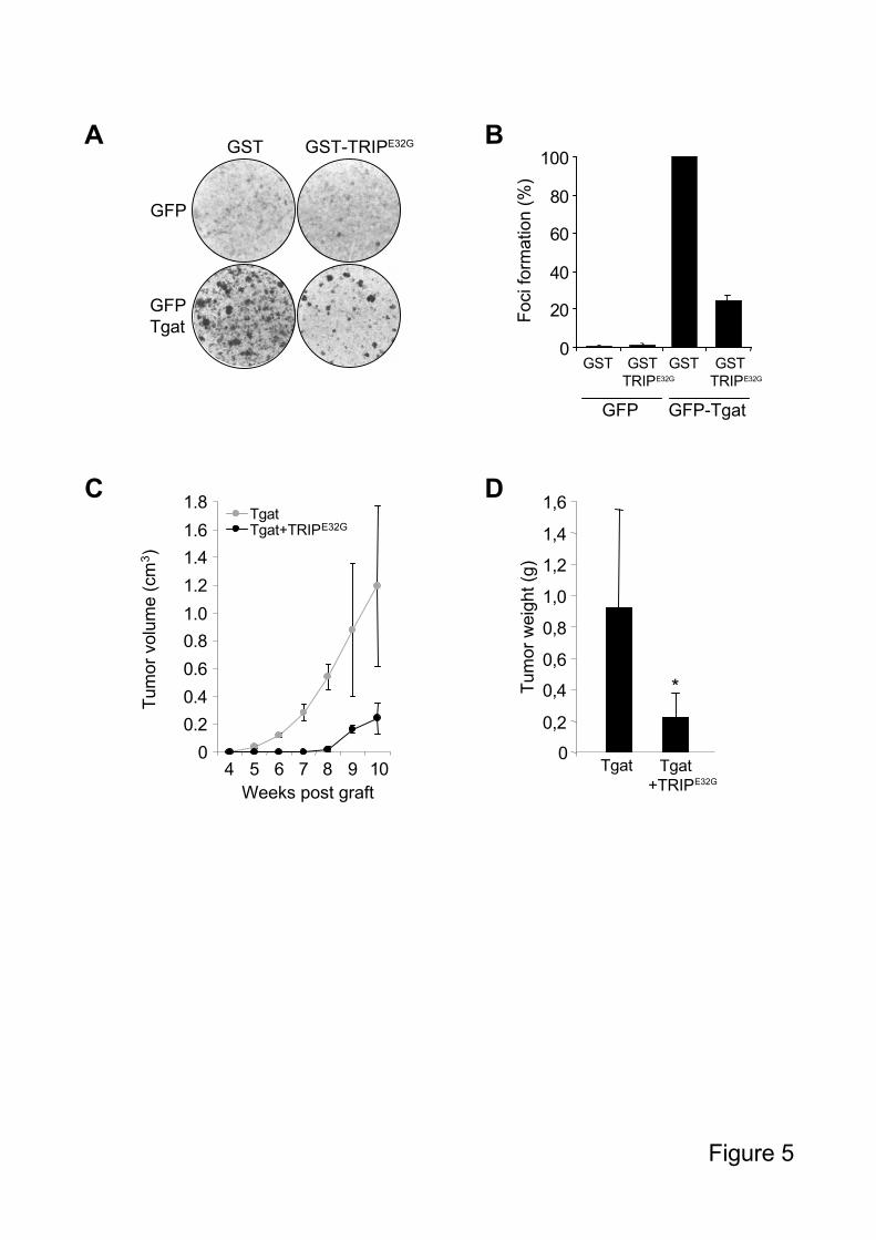
Figure 5
GFP
GFP
Tgat
GST GST-TRIPE32GA
C
B
Foci fo
rmation (
%)
GST GST
TRIPE32G
GST
GFP
0
20
40
60
80
100
GFP-Tgat
GST
TRIPE32G
Tum
or
weig
ht (g
)
Tgat Tgat
+TRIPE32G
0
0,2
0,4
0,6
0,8
1,0
1,2
1,4
1,6
*
0
0.2
0.4
0.6
0.8
1.0
1.2
1.4
1.6
1.8
4 5 6 7 8 9 10
Weeks post graft
Tum
or
volu
me (
cm
3)
TgatTgat+TRIPE32G
D
Related Documents