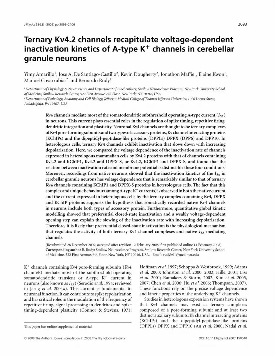
J Physiol 586.8 (2008) pp 2093–2106 2093 Ternary Kv4.2 channels recapitulate voltage-dependent inactivation kinetics of A-type K + channels in cerebellar granule neurons Yimy Amarillo 1 , Jose A. De Santiago-Castillo 2 , Kevin Dougherty 2 , Jonathon Maffie 1 , Elaine Kwon 1 , Manuel Covarrubias 2 and Bernardo Rudy 1 1 Department of Physiology & Neuroscience and Department of Biochemistry, Smilow Neuroscience Program, New York University School of Medicine, Smilow Research Center, 522 First Avenue, 6th Floor, New York, NY 10016, USA 2 Department of Pathology, Anatomy and Cell Biology, Jefferson Medical College of Thomas Jefferson University, 1020 Locust Street, Philadelphia, PA 19107, USA Kv4 channels mediate most of the somatodendritic subthreshold operating A-type current (I SA ) in neurons. This current plays essential roles in the regulation of spike timing, repetitive firing, dendritic integration and plasticity. Neuronal Kv4 channels are thought to be ternary complexes of Kv4 pore-forming subunits and two types of accessory proteins, Kv channel interacting proteins (KChIPs) and the dipeptidyl-peptidase-like proteins (DPPLs) DPPX (DPP6) and DPP10. In heterologous cells, ternary Kv4 channels exhibit inactivation that slows down with increasing depolarization. Here, we compared the voltage dependence of the inactivation rate of channels expressed in heterologous mammalian cells by Kv4.2 proteins with that of channels containing Kv4.2 and KChIP1, Kv4.2 and DPPX-S, or Kv4.2, KChIP1 and DPPX-S, and found that the relation between inactivation rate and membrane potential is distinct for these four conditions. Moreover, recordings from native neurons showed that the inactivation kinetics of the I SA in cerebellar granule neurons has voltage dependence that is remarkably similar to that of ternary Kv4 channels containing KChIP1 and DPPX-S proteins in heterologous cells. The fact that this complex and unique behaviour (among A-type K + currents) is observed in both the native current and the current expressed in heterologous cells by the ternary complex containing Kv4, DPPX and KChIP proteins supports the hypothesis that somatically recorded native Kv4 channels in neurons include both types of accessory protein. Furthermore, quantitative global kinetic modelling showed that preferential closed-state inactivation and a weakly voltage-dependent opening step can explain the slowing of the inactivation rate with increasing depolarization. Therefore, it is likely that preferential closed-state inactivation is the physiological mechanism that regulates the activity of both ternary Kv4 channel complexes and native I SA -mediating channels. (Resubmitted 26 December 2007; accepted after revision 12 February 2008; first published online 14 February 2008) Corresponding author B. Rudy: Smilow Neuroscience Program, Smilow Research Center, New York University School of Medicine, 522 First Avenue, 6th Floor, New York, NY 10016, USA. Email: [email protected] K + channels containing Kv4 pore-forming subunits (Kv4 channels) mediate most of the subthreshold-operating somatodendritic transient or A-type K + current in neurons (also known as I SA ) (Serodio et al. 1994; reviewed in Jerng et al. 2004a). This current is fundamental to neuronal function. It can contribute to spike repolarization and has critical roles in the modulation of the frequency of repetitive firing, signal processing in dendrites and spike timing-dependent plasticity (Connor & Stevens, 1971; This paper has online supplemental material. Hoffman et al. 1997; Schoppa & Westbrook, 1999; Adams et al. 2000; Johnston et al. 2000, 2003; Hille, 2001; Liss et al. 2001; Ramakers & Storm, 2002; Kim et al. 2005, 2007; Chen et al. 2006; Hu et al. 2006; Thompson, 2007). These functions rely on the precise voltage dependence and kinetic properties of the underlying K + channels. Studies in heterologous expression systems have shown that Kv4 channels may exist as ternary complexes composed of a pore-forming subunit and at least two distinct auxiliary subunits: Kv channel interacting proteins (KChIPs) and the dipeptidyl-peptidase-like proteins (DPPLs) DPPX and DPP10 (An et al. 2000; Nadal et al. C 2008 The Authors. Journal compilation C 2008 The Physiological Society DOI: 10.1113/jphysiol.2007.150540

Welcome message from author
This document is posted to help you gain knowledge. Please leave a comment to let me know what you think about it! Share it to your friends and learn new things together.
Transcript
J Physiol 586.8 (2008) pp 2093–2106 2093
Ternary Kv4.2 channels recapitulate voltage-dependentinactivation kinetics of A-type K+ channels in cerebellargranule neurons
Yimy Amarillo1, Jose A. De Santiago-Castillo2, Kevin Dougherty2, Jonathon Maffie1, Elaine Kwon1,
Manuel Covarrubias2 and Bernardo Rudy1
1Department of Physiology & Neuroscience and Department of Biochemistry, Smilow Neuroscience Program, New York University School
of Medicine, Smilow Research Center, 522 First Avenue, 6th Floor, New York, NY 10016, USA2Department of Pathology, Anatomy and Cell Biology, Jefferson Medical College of Thomas Jefferson University, 1020 Locust Street,
Philadelphia, PA 19107, USA
Kv4 channels mediate most of the somatodendritic subthreshold operating A-type current (I SA)
in neurons. This current plays essential roles in the regulation of spike timing, repetitive firing,
dendritic integration and plasticity. Neuronal Kv4 channels are thought to be ternary complexes
of Kv4 pore-forming subunits and two types of accessory proteins, Kv channel interacting proteins
(KChIPs) and the dipeptidyl-peptidase-like proteins (DPPLs) DPPX (DPP6) and DPP10. In
heterologous cells, ternary Kv4 channels exhibit inactivation that slows down with increasing
depolarization. Here, we compared the voltage dependence of the inactivation rate of channels
expressed in heterologous mammalian cells by Kv4.2 proteins with that of channels containing
Kv4.2 and KChIP1, Kv4.2 and DPPX-S, or Kv4.2, KChIP1 and DPPX-S, and found that the
relation between inactivation rate and membrane potential is distinct for these four conditions.
Moreover, recordings from native neurons showed that the inactivation kinetics of the I SA in
cerebellar granule neurons has voltage dependence that is remarkably similar to that of ternary
Kv4 channels containing KChIP1 and DPPX-S proteins in heterologous cells. The fact that this
complex and unique behaviour (among A-type K+ currents) is observed in both the native current
and the current expressed in heterologous cells by the ternary complex containing Kv4, DPPX
and KChIP proteins supports the hypothesis that somatically recorded native Kv4 channels
in neurons include both types of accessory protein. Furthermore, quantitative global kinetic
modelling showed that preferential closed-state inactivation and a weakly voltage-dependent
opening step can explain the slowing of the inactivation rate with increasing depolarization.
Therefore, it is likely that preferential closed-state inactivation is the physiological mechanism
that regulates the activity of both ternary Kv4 channel complexes and native I SA-mediating
channels.
(Resubmitted 26 December 2007; accepted after revision 12 February 2008; first published online 14 February 2008)
Corresponding author B. Rudy: Smilow Neuroscience Program, Smilow Research Center, New York University School
of Medicine, 522 First Avenue, 6th Floor, New York, NY 10016, USA. Email: [email protected]
K+ channels containing Kv4 pore-forming subunits (Kv4channels) mediate most of the subthreshold-operatingsomatodendritic transient or A-type K+ current inneurons (also known as ISA) (Serodio et al. 1994; reviewedin Jerng et al. 2004a). This current is fundamental toneuronal function. It can contribute to spike repolarizationand has critical roles in the modulation of the frequency ofrepetitive firing, signal processing in dendrites and spiketiming-dependent plasticity (Connor & Stevens, 1971;
This paper has online supplemental material.
Hoffman et al. 1997; Schoppa & Westbrook, 1999; Adamset al. 2000; Johnston et al. 2000, 2003; Hille, 2001; Lisset al. 2001; Ramakers & Storm, 2002; Kim et al. 2005,2007; Chen et al. 2006; Hu et al. 2006; Thompson, 2007).These functions rely on the precise voltage dependenceand kinetic properties of the underlying K+ channels.
Studies in heterologous expression systems have shownthat Kv4 channels may exist as ternary complexescomposed of a pore-forming subunit and at least twodistinct auxiliary subunits: Kv channel interacting proteins(KChIPs) and the dipeptidyl-peptidase-like proteins(DPPLs) DPPX and DPP10 (An et al. 2000; Nadal et al.
C© 2008 The Authors. Journal compilation C© 2008 The Physiological Society DOI: 10.1113/jphysiol.2007.150540
2094 Y. Amarillo and others J Physiol 586.8
2001, 2003; Jerng et al. 2004b, 2005; Ren et al. 2005; Zaghaet al. 2005). These associations control the trafficking ofchannel complexes to the plasma membrane and havemajor effects on the voltage dependence and kinetics ofthe channels. However, the significance of these auxiliaryproteins in governing the patterns of expression andfunctional properties of the native ISA channels in neuronshas yet to be demonstrated.
The kinetics of inactivation of ternary Kv4 channelsexpressed in heterologous expression systems has unusualvoltage dependence (G. Wang et al. 2005; Jerng et al.2007). With increasing step depolarizations, the rate ofinactivation slows down. This trend is apparent over a widerange of membrane potentials (−20 to +100 mV; G. Wanget al. 2005) and is also observed with binary combinationsof Kv4.3 and KChIP1 (Kaulin et al. 2008). In contrast, Kv4channels expressed in the absence of the auxiliary proteins(Serodio et al. 1996; Jerng & Covarrubias, 1997; Franquezaet al. 1999; Bahring et al. 2001; Beck & Covarrubias,2001; G. Wang et al. 2005; S. Wang et al. 2005) aswell as inactivating channels from other Kv subfamilies(Tseng-Crank et al. 1990; Rudy et al. 1991; Schroter et al.1991; Vega-Saenz de Miera et al. 1992; Riazanski et al. 2001)do not exhibit such a voltage dependence.
This unusual behaviour is potentially significantbecause it has been observed to one degree or anotherin recordings of A-type K+ currents from hippocampalneurons (Klee et al. 1995; Hoffman et al. 1997; Martinaet al. 1998; Lien et al. 2002); however, it has not receivedmuch attention and its mechanism is not well under-stood. Moreover, the ISA is usually only one of severalcomponents of the total current observed in whole cellneuronal recordings. Therefore, the ISA has to be properlyisolated from other current components to determineits properties accurately. For example, the inactivatingcurrent in CA1 hippocampal pyramidal neurons appearsto include a contribution from channels that are notmembers of the Kv4 subfamily (and are possibly mediatedby Kv1 subunits) (Chen et al. 2006). This is a minorcomponent of the total inactivating current. However,since it has a different voltage dependence and kinetics thanthe Kv4 component, it would influence the properties of thetotal inactivating current differently at different membranepotentials.
Previously, we proposed that the unusual voltagedependence of inactivation kinetics is a hallmark ofpreferential closed-state inactivation in Kv4 channelcomplexes, and is determined by auxiliary subunits(Kaulin et al. 2008). Therefore, this behaviour in neuronalKv4 channels would suggest that they include a specificcombination of Kv4 auxiliary subunits, which conferpreferential closed-state inactivation. To test thishypothesis systematically and under stringent conditions,we recorded the ISA from cerebellar granule cells in acuteslices and compared the voltage dependence of inactivation
kinetics with that of Kv4 channels heterologously expressedin mammalian cells. The latter experiments included theKv4.2 subunit alone and in binary or ternary complexeswith the accessory proteins KChIP1 and DPPX-S. Ourresults show that the kinetics of inactivation of the ISA
in cerebellar granule neurons has a voltage dependencethat is remarkably similar to that observed for ternaryKv4 channels containing KChIP and DPPX proteins inheterologous cells. These results support the view thatKChIP and DPPX auxiliary proteins are components of thenative ISA channels. Furthermore, using kinetic modellingwe show that preferential closed-state inactivation anda weakly voltage-dependent opening step can explainthe slowing of the inactivation rate with increasingdepolarization.
Methods
Electrophysiological recording from heterologousmammalian cells
Electrophysiological recording from heterologousmammalian cells was carried out as previously described(Dougherty & Covarrubias, 2006). Briefly, tsA-201 cells(provided by Dr R. Horn, Thomas Jefferson University,Philadelphia, PA, USA) were transfected with theappropriate plasmids (Kv4.2, KChIP1 and/or DPPX-S) ata 1 : 1 mass ratio using the calcium phosphate method. Aplasmid containing a CD8 cDNA (5 μg) was included inthe cotransfection to allow the identification of individualtransfected cells by decorating them with beads bearinganti-CD8 antibody (Dynal Biotech, Brown Deer, WI, USA)(Kaulin et al. 2008). Kv4.2 currents were recorded usingthe tight-seal whole-cell configuration of the patch-clampmethod with a pipette (intracellular) solution containing(mm): 120 KF, 1 CaCl2, 2 MgCl2, 11 EGTA and 10 Hepes,pH 7.2, adjusted with KOH; and the external bath solutioncontaining (mm): 130 NaCl, 2 KCl, 1.5 CaCl2, 1 MgCl2,20 TEA (tetraethylammonium chloride) and 10 Hepes,pH 7.4, adjusted with NaOH. The calculated free [Ca2+]and [Mg2+] in the intracellular solution were ∼40 nm
and ∼1.4 mm, respectively (MaxChelator, WEBMAXCv2.10; http://www.stanford.edu/∼cpatton/maxc.html).Extracellular TEA was necessary to eliminate a small butsignificant endogenous delayed-rectifier K+ current. Theexperiment began once current kinetics and amplitudebecame stable. Series resistance (Rs = 2–5 M�) wascompensated to yield a total voltage error of < 3 mV. AP/−4 leak subtraction protocol consisting of four sub-pulses from a subsweep holding potential of −110 mV wasused to subtract passive components of the total current.Currents were filtered at 2 kHz and sampled at 10 kHz.Analyses and graphical displays were produced withpCLAMP 9.0 (Axon Instruments, Inc., Union City, CA,USA) and Origin 7.5 (OriginLab Corp., Northhampton,
C© 2008 The Authors. Journal compilation C© 2008 The Physiological Society
J Physiol 586.8 Kv4.2 channel inactivation 2095
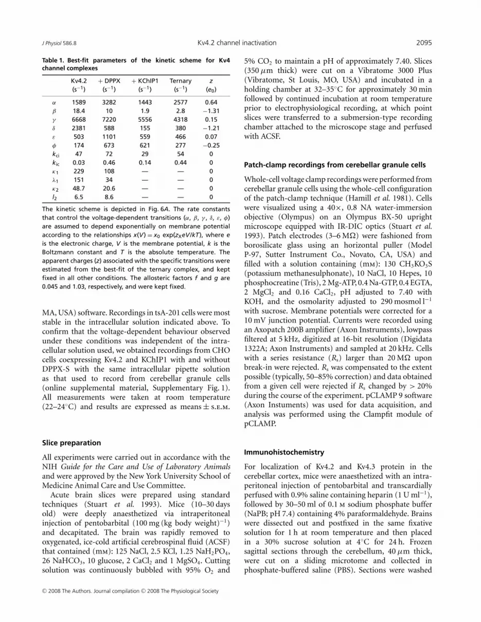
Table 1. Best-fit parameters of the kinetic scheme for Kv4channel complexes
Kv4.2 + DPPX + KChIP1 Ternary z(s−1) (s−1) (s−1) (s−1) (e0)
α 1589 3282 1443 2577 0.64β 18.4 10 1.9 2.8 −1.31γ 6668 7220 5556 4318 0.15δ 2381 588 155 380 −1.21ε 503 1101 559 466 0.07φ 174 673 621 277 −0.25kci 47 72 29 54 0kic 0.03 0.46 0.14 0.44 0κ1 229 108 — — 0λ1 151 34 — — 0κ2 48.7 20.6 — — 0l2 6.5 8.6 — — 0
The kinetic scheme is depicted in Fig. 6A. The rate constantsthat control the voltage-dependent transitions (α, β, γ , δ, ε, φ)are assumed to depend exponentially on membrane potentialaccording to the relationships x(V) = x0 exp(zXeV /kT), where eis the electronic charge, V is the membrane potential, k is theBoltzmann constant and T is the absolute temperature. Theapparent charges (z) associated with the specific transitions wereestimated from the best-fit of the ternary complex, and keptfixed in all other conditions. The allosteric factors f and g are0.045 and 1.03, respectively, and were kept fixed.
MA, USA) software. Recordings in tsA-201 cells were moststable in the intracellular solution indicated above. Toconfirm that the voltage-dependent behaviour observedunder these conditions was independent of the intra-cellular solution used, we obtained recordings from CHOcells coexpressing Kv4.2 and KChIP1 with and withoutDPPX-S with the same intracellular pipette solutionas that used to record from cerebellar granule cells(online supplemental material, Supplementary Fig. 1).All measurements were taken at room temperature(22–24◦C) and results are expressed as means ± s.e.m.
Slice preparation
All experiments were carried out in accordance with theNIH Guide for the Care and Use of Laboratory Animalsand were approved by the New York University School ofMedicine Animal Care and Use Committee.
Acute brain slices were prepared using standardtechniques (Stuart et al. 1993). Mice (10–30 daysold) were deeply anaesthetized via intraperitonealinjection of pentobarbital (100 mg (kg body weight)−1)and decapitated. The brain was rapidly removed tooxygenated, ice-cold artificial cerebrospinal fluid (ACSF)that contained (mm): 125 NaCl, 2.5 KCl, 1.25 NaH2PO4,26 NaHCO3, 10 glucose, 2 CaCl2 and 1 MgSO4. Cuttingsolution was continuously bubbled with 95% O2 and
5% CO2 to maintain a pH of approximately 7.40. Slices(350 μm thick) were cut on a Vibratome 3000 Plus(Vibratome, St Louis, MO, USA) and incubated in aholding chamber at 32–35◦C for approximately 30 minfollowed by continued incubation at room temperatureprior to electrophysiological recording, at which pointslices were transferred to a submersion-type recordingchamber attached to the microscope stage and perfusedwith ACSF.
Patch-clamp recordings from cerebellar granule cells
Whole-cell voltage clamp recordings were performed fromcerebellar granule cells using the whole-cell configurationof the patch-clamp technique (Hamill et al. 1981). Cellswere visualized using a 40×, 0.8 NA water-immersionobjective (Olympus) on an Olympus BX-50 uprightmicroscope equipped with IR-DIC optics (Stuart et al.1993). Patch electrodes (3–6 M�) were fashioned fromborosilicate glass using an horizontal puller (ModelP-97, Sutter Instrument Co., Novato, CA, USA) andfilled with a solution containing (mm): 130 CH3KO3S(potassium methanesulphonate), 10 NaCl, 10 Hepes, 10phosphocreatine (Tris), 2 Mg-ATP, 0.4 Na-GTP, 0.4 EGTA,2 MgCl2 and 0.16 CaCl2, pH adjusted to 7.40 withKOH, and the osmolarity adjusted to 290 mosmol l−1
with sucrose. Membrane potentials were corrected for a10 mV junction potential. Currents were recorded usingan Axopatch 200B amplifier (Axon Instruments), lowpassfiltered at 5 kHz, digitized at 16-bit resolution (Digidata1322A; Axon Instruments) and sampled at 20 kHz. Cellswith a series resistance (Rs) larger than 20 M� uponbreak-in were rejected. Rs was compensated to the extentpossible (typically, 50–85% correction) and data obtainedfrom a given cell were rejected if Rs changed by > 20%during the course of the experiment. pCLAMP 9 software(Axon Instuments) was used for data acquisition, andanalysis was performed using the Clampfit module ofpCLAMP.
Immunohistochemistry
For localization of Kv4.2 and Kv4.3 protein in thecerebellar cortex, mice were anaesthetized with an intra-peritoneal injection of pentobarbital and transcardiallyperfused with 0.9% saline containing heparin (1 U ml−1),followed by 30–50 ml of 0.1 m sodium phosphate buffer(NaPB; pH 7.4) containing 4% paraformaldehyde. Brainswere dissected out and postfixed in the same fixativesolution for 1 h at room temperature and then placedin a 30% sucrose solution at 4◦C for 24 h. Frozensagittal sections through the cerebellum, 40 μm thick,were cut on a sliding microtome and collected inphosphate-buffered saline (PBS). Sections were washed
C© 2008 The Authors. Journal compilation C© 2008 The Physiological Society
2096 Y. Amarillo and others J Physiol 586.8
in PBS and then incubated in a blocking solution(10% normal goat serum, 1% BSA, 0.2% cold fishgelatin, and 0.2% Triton X-100 in PBS) for 1 h at roomtemperature to minimize non-specific binding. Sectionswere then incubated with antibodies directed againstKv4.2 (10 μg ml−1; mouse monoclonal anti-Kv4.2; K57/1;Neuromab; www.neuromab.org; or rabbit polyclonaldirected against Kv4.2; Nadal et al. 2003; at 1 : 1000), orKv4.3 (1 μg ml−1; mouse monoclonal anti-Kv4.3; K75/41;Neuromab) in diluted (1 : 10) blocking solution overnightat 4◦C. Following incubation with primary antibodies,sections were washed with PBS and incubated for1 h in fluorescent Cy3-conjugated secondary antibody(goat anti-mouse or goat anti-rabbit, Jackson Immuno-Research Laboratories, Inc., West Grove, PA, USA) atroom temperature. Sections were then rinsed and finallymounted using Vectashield (Vector Laboratories, Inc.,Burlingame, CA, USA). Immunofluorescence images wereacquired with an Olympus MVX10 microscope equippedwith a Leica DFC-340FX digital camera and Leica Firecam3.0 software. Images were transferred to a graphicsprogram (Adobe Photoshop CS2), in which brightness andcontrast were adjusted. The monoclonal and polyconalantibodies for Kv4.2 both produced the same pattern ofstaining in the cerebellum. Figure 2 depicts staining withthe monoclonal antibody against Kv4.2.
Quantitative global kinetic modelling
Previously, we applied global kinetic modellingand a model discrimination method to explaingating of Kv4 channels quantitatively (Kaulin et al.2008). Using a Chi-squared minimization function,appropriately weighted macroscopic measurements(activation, inactivation and deactivation) were evaluatedsimultaneously to constrain the analysis and estimatethe best global-fit parameters for specific kinetic models.Here, we implemented this strategy in the new programIChMASCOT (Ion-Channel-Markov-Scheme-Optimizer;www.ichmascot.org) and assumed the kinetic schemeshown in Fig. 6A. This scheme is similar to those modelsthat successfully described Kv4 gating in the studyof Kaulin et al. (2008). In particular, these modelsexplained the slowing of inactivation with progressivedepolarization. For the sake of simplification, however,we did not include an unstable closed state connectedto the open state, which reduces the open probability.Also, the scope of the kinetic analysis was limited toexplain the voltage dependence of the rate of inactivationsemiquantitatively. For all subunit combinations, weevaluated the following measurements simultaneously toconstrain global kinetic modelling: (1) complete familiesof currents evoked by step depolarizations (Fig. 1); (2) thesteady-state inactivation curve (not shown); and (3) therecovery from inactivation (Fig. 1F). For display, model
simulations were performed in the program IChSimlab(www.ichmascot.org).
Results
Voltage dependence of inactivation of Kv4.2 channelsin heterologous expression systems
Previous studies of the influence of KChIP and DPPLproteins on the properties of Kv4 channels have primarilyused two-microelectrode voltage-clamp in the Xenopusoocyte expression system (Nadal et al. 2003, 2006;Jerng et al. 2005). To minimize the variability thatmethodological differences may introduce and facilitatecomparison with the properties of native neuronalchannels, we chose to express the Kv4 channel complexes ina mammalian heterologous expression system and obtainthe currents with the same recording method (whole-cellpatch-clamp).
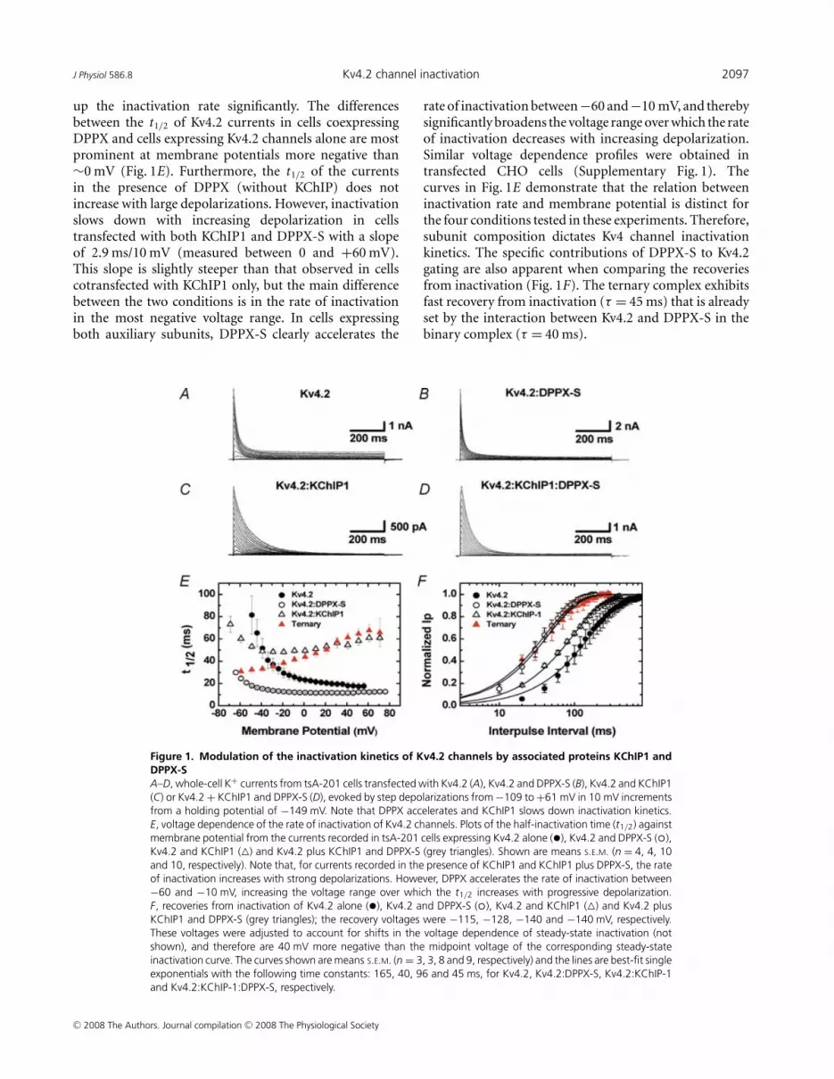
The currents produced by expression of Kv4.2 proteinsalone, were compared to those resulting from coexpressionof Kv4.2 with the accessory subunits KChIP1 and DPPX-S(Fig. 1). Records of the currents recorded during stepdepolarizations from tsA-201 cells transfected with Kv4.2alone, Kv4.2 plus KChIP1, Kv4.2 plus DPPX-S and Kv4.2with both KChIP1 and DPPX-S are shown in Fig. 1A–D.As previously reported, KChIP1 slows down currentinactivation and induces more complete inactivation bythe end of the voltage pulse, and DPPX accelerates theoverall channel kinetics (An et al. 2000; Beck et al. 2002;Nadal et al. 2003; reviewed in Jerng et al. 2004a). InFig. 1E we compare the rate of inactivation as a functionof voltage for the four conditions from a number ofcells. As reported before, the currents mediated by Kv4channels decay with a complex multiexponential timecourse (Jerng & Covarrubias, 1997; Bahring et al. 2001;Jerng et al. 2004b). Since the number of exponentialsthat may describe the decay varies depending on subunitcomposition, we used the time at which half of the peakcurrent is inactivated (half-inactivation time or t1/2) tocompare inactivation kinetics under different conditions.This model-independent strategy has been previously usedby us and others to compare the inactivation kinetics ofKv4 currents (Nadal et al. 2001, 2003; Jerng et al. 2007).
In cells transfected with Kv4.2 alone, the rateof inactivation does not slow down during largedepolarizations (Fig. 1E). In sharp contrast, the voltagedependence of the rate of inactivation of Kv4.2 channelscotransfected with KChIP1 exhibits a transition froman accelerating rate of inactivation at negative voltagesto a decelerating rate at positive membrane potentials(Fig. 1E). In the presence of KChIP1, the t1/2 reaches aminimum between −40 and −10 mV; then, it increaseswith a slope of approximately 2.3 ms/10 mV (0 to +60 mV;Fig. 1E). As previously reported, DPPX-S alone speeds
C© 2008 The Authors. Journal compilation C© 2008 The Physiological Society
J Physiol 586.8 Kv4.2 channel inactivation 2097
up the inactivation rate significantly. The differencesbetween the t1/2 of Kv4.2 currents in cells coexpressingDPPX and cells expressing Kv4.2 channels alone are mostprominent at membrane potentials more negative than∼0 mV (Fig. 1E). Furthermore, the t1/2 of the currentsin the presence of DPPX (without KChIP) does notincrease with large depolarizations. However, inactivationslows down with increasing depolarization in cellstransfected with both KChIP1 and DPPX-S with a slopeof 2.9 ms/10 mV (measured between 0 and +60 mV).This slope is slightly steeper than that observed in cellscotransfected with KChIP1 only, but the main differencebetween the two conditions is in the rate of inactivationin the most negative voltage range. In cells expressingboth auxiliary subunits, DPPX-S clearly accelerates the
Figure 1. Modulation of the inactivation kinetics of Kv4.2 channels by associated proteins KChIP1 andDPPX-SA–D, whole-cell K+ currents from tsA-201 cells transfected with Kv4.2 (A), Kv4.2 and DPPX-S (B), Kv4.2 and KChIP1(C) or Kv4.2 + KChIP1 and DPPX-S (D), evoked by step depolarizations from −109 to +61 mV in 10 mV incrementsfrom a holding potential of −149 mV. Note that DPPX accelerates and KChIP1 slows down inactivation kinetics.E, voltage dependence of the rate of inactivation of Kv4.2 channels. Plots of the half-inactivation time (t1/2) againstmembrane potential from the currents recorded in tsA-201 cells expressing Kv4.2 alone (•), Kv4.2 and DPPX-S ( �),Kv4.2 and KChIP1 (�) and Kv4.2 plus KChIP1 and DPPX-S (grey triangles). Shown are means S.E.M. (n = 4, 4, 10and 10, respectively). Note that, for currents recorded in the presence of KChIP1 and KChIP1 plus DPPX-S, the rateof inactivation increases with strong depolarizations. However, DPPX accelerates the rate of inactivation between−60 and −10 mV, increasing the voltage range over which the t1/2 increases with progressive depolarization.F, recoveries from inactivation of Kv4.2 alone (•), Kv4.2 and DPPX-S ( �), Kv4.2 and KChIP1 (�) and Kv4.2 plusKChIP1 and DPPX-S (grey triangles); the recovery voltages were −115, −128, −140 and −140 mV, respectively.These voltages were adjusted to account for shifts in the voltage dependence of steady-state inactivation (notshown), and therefore are 40 mV more negative than the midpoint voltage of the corresponding steady-stateinactivation curve. The curves shown are means S.E.M. (n = 3, 3, 8 and 9, respectively) and the lines are best-fit singleexponentials with the following time constants: 165, 40, 96 and 45 ms, for Kv4.2, Kv4.2:DPPX-S, Kv4.2:KChIP-1and Kv4.2:KChIP-1:DPPX-S, respectively.
rate of inactivation between −60 and −10 mV, and therebysignificantly broadens the voltage range over which the rateof inactivation decreases with increasing depolarization.Similar voltage dependence profiles were obtained intransfected CHO cells (Supplementary Fig. 1). Thecurves in Fig. 1E demonstrate that the relation betweeninactivation rate and membrane potential is distinct forthe four conditions tested in these experiments. Therefore,subunit composition dictates Kv4 channel inactivationkinetics. The specific contributions of DPPX-S to Kv4.2gating are also apparent when comparing the recoveriesfrom inactivation (Fig. 1F). The ternary complex exhibitsfast recovery from inactivation (τ = 45 ms) that is alreadyset by the interaction between Kv4.2 and DPPX-S in thebinary complex (τ = 40 ms).
C© 2008 The Authors. Journal compilation C© 2008 The Physiological Society
2098 Y. Amarillo and others J Physiol 586.8
Kv4.2 and Kv4.3 proteins in mouse cerebellargranule cells
Cerebellar granule neurons have a large ISA, consistentwith prominent expression of Kv4.2 and Kv4.3 mRNAsand proteins (Serodio & Rudy, 1998; Rhodes et al. 2004;Strassle et al. 2005). They are also among the few neuronalpopulations where the patterns of expression of the variousmolecular components of Kv4 channels have been analysedwith good cellular resolution (Nadal et al. 2003; Rhodeset al. 2004; Strassle et al. 2005; Zagha et al. 2005; Jerng et al.2007). Moreover, compared to other neurons, cerebellargranule cells are elecrotonically compact due to the smallsize of these cells (∼5 μm) and the short length of dendriticprocesses, allowing good voltage control for whole cellrecording (Cull-Candy et al. 1989). We therefore selectedthese neurons to investigate the properties of nativeneuronal ISA for comparison with the currentsheterologously expressed in mammalian cells.
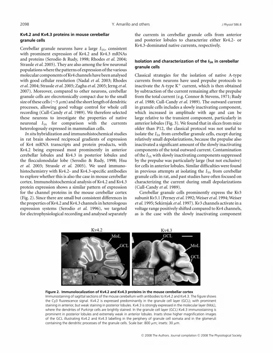
In situ hybridization and immunohistochemical studiesin rat brain showed reciprocal gradients of expressionof Kv4 mRNA transcripts and protein products, withKv4.2 being expressed most prominently in anteriorcerebellar lobules and Kv4.3 in posterior lobules andthe flocculonodular lobe (Serodio & Rudy, 1998; Hsuet al. 2003; Strassle et al. 2005). We used immuno-histochemistry with Kv4.2- and Kv4.3-specific antibodiesto explore whether this is also the case in mouse cerebellarcortex. Immunohistochemical analysis of Kv4.2 and Kv4.3protein expression shows a similar pattern of expressionfor the channel proteins in the mouse cerebellar cortex(Fig. 2). Since there are small but consistent differences inthe properties of Kv4.2 and Kv4.3 channels in heterologousexpression systems (Serodio et al. 1996), we targetedfor electrophysiological recording and analysed separately
Figure 2. Immunolocalization of Kv4.2 and Kv4.3 proteins in the mouse cerebellar cortexImmunostaining of sagittal sections of the mouse cerebellum with antibodies to Kv4.2 and Kv4.3. The figure showsthe Cy3 fluorescence signal. Kv4.2 is expressed predominantly in the granule cell layer (GCL); with prominentstaining in anterior, but weak staining in posterior lobules. Kv4.3 is strongly expressed in the molecular layer (MoL),where the dendrites of Purkinje cells are brightly stained. In the granule cell layer (GCL) Kv4.3 immunostaining isprominent in posterior lobules and extremely weak in anterior lobules. Insets show higher magnification imagesof the GCL illustrating Kv4.2 and Kv4.3 labelling in the periphery of granule cell somata and in the glomerulicontaining the dendritic processes of the granule cells. Scale bar: 800 μm; insets: 30 μm.
the currents in cerebellar granule cells from anteriorand posterior lobules to characterize either Kv4.2- orKv4.3-dominated native currents, respectively.
Isolation and characterization of the ISA in cerebellargranule cells
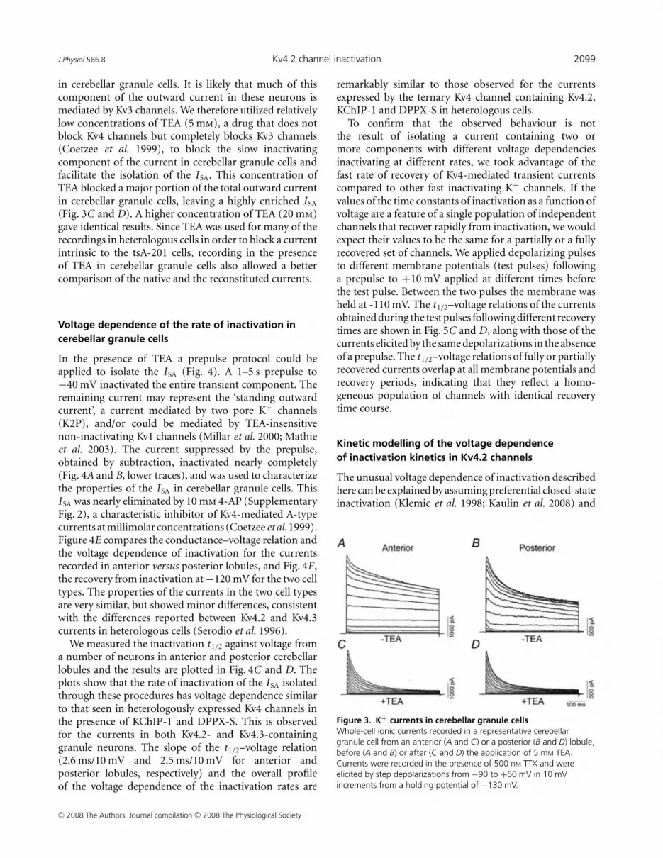
Classical strategies for the isolation of native A-typecurrents from neurons have used prepulse protocols toinactivate the A-type K+ current, which is then obtainedby subtraction of the current remaining after the prepulsefrom the total current (e.g. Connor & Stevens, 1971; Rudyet al. 1988; Cull-Candy et al. 1989). The outward currentin granule cells includes a slowly inactivating component,which increased in amplitude with age and can belarge relative to the transient component, particularly inanterior lobules (Fig. 3). We found that in slices from miceolder than P12, the classical protocol was not useful toisolate the ISA from cerebellar granule cells, except duringrelatively small depolarizations, because the prepulse alsoinactivated a significant amount of the slowly inactivatingcomponents of the total outward current. Contaminationof the ISA with slowly inactivating components suppressedby the prepulse was particularly large (but not exclusive)for cells in anterior lobules. Similar difficulties were foundin previous attempts at isolating the ISA from cerebellargranule cells in rat, and past studies have often focused oncharacterizing the current during small depolarizations(Cull-Candy et al. 1989).
Cerebellar granule cells prominently express the Kv3subunit Kv3.1 (Perney et al. 1992; Weiser et al. 1994; Weiseret al. 1995; Sekirnjak et al. 1997). Kv3 channels activate in avoltage range positively shifted compared to Kv4 channels,as is the case with the slowly inactivating component
C© 2008 The Authors. Journal compilation C© 2008 The Physiological Society
J Physiol 586.8 Kv4.2 channel inactivation 2099
in cerebellar granule cells. It is likely that much of thiscomponent of the outward current in these neurons ismediated by Kv3 channels. We therefore utilized relativelylow concentrations of TEA (5 mm), a drug that does notblock Kv4 channels but completely blocks Kv3 channels(Coetzee et al. 1999), to block the slow inactivatingcomponent of the current in cerebellar granule cells andfacilitate the isolation of the ISA. This concentration ofTEA blocked a major portion of the total outward currentin cerebellar granule cells, leaving a highly enriched ISA
(Fig. 3C and D). A higher concentration of TEA (20 mm)gave identical results. Since TEA was used for many of therecordings in heterologous cells in order to block a currentintrinsic to the tsA-201 cells, recording in the presenceof TEA in cerebellar granule cells also allowed a bettercomparison of the native and the reconstituted currents.
Voltage dependence of the rate of inactivation incerebellar granule cells
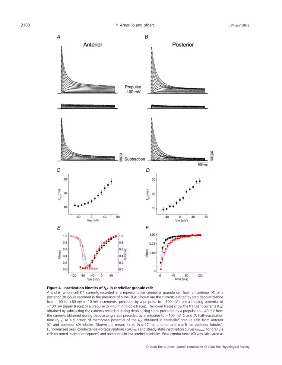
In the presence of TEA a prepulse protocol could beapplied to isolate the ISA (Fig. 4). A 1–5 s prepulse to−40 mV inactivated the entire transient component. Theremaining current may represent the ‘standing outwardcurrent’, a current mediated by two pore K+ channels(K2P), and/or could be mediated by TEA-insensitivenon-inactivating Kv1 channels (Millar et al. 2000; Mathieet al. 2003). The current suppressed by the prepulse,obtained by subtraction, inactivated nearly completely(Fig. 4A and B, lower traces), and was used to characterizethe properties of the ISA in cerebellar granule cells. ThisISA was nearly eliminated by 10 mm 4-AP (SupplementaryFig. 2), a characteristic inhibitor of Kv4-mediated A-typecurrents at millimolar concentrations (Coetzee et al. 1999).Figure 4E compares the conductance–voltage relation andthe voltage dependence of inactivation for the currentsrecorded in anterior versus posterior lobules, and Fig. 4F ,the recovery from inactivation at −120 mV for the two celltypes. The properties of the currents in the two cell typesare very similar, but showed minor differences, consistentwith the differences reported between Kv4.2 and Kv4.3currents in heterologous cells (Serodio et al. 1996).
We measured the inactivation t1/2 against voltage froma number of neurons in anterior and posterior cerebellarlobules and the results are plotted in Fig. 4C and D. Theplots show that the rate of inactivation of the ISA isolatedthrough these procedures has voltage dependence similarto that seen in heterologously expressed Kv4 channels inthe presence of KChIP-1 and DPPX-S. This is observedfor the currents in both Kv4.2- and Kv4.3-containinggranule neurons. The slope of the t1/2–voltage relation(2.6 ms/10 mV and 2.5 ms/10 mV for anterior andposterior lobules, respectively) and the overall profileof the voltage dependence of the inactivation rates are
remarkably similar to those observed for the currentsexpressed by the ternary Kv4 channel containing Kv4.2,KChIP-1 and DPPX-S in heterologous cells.
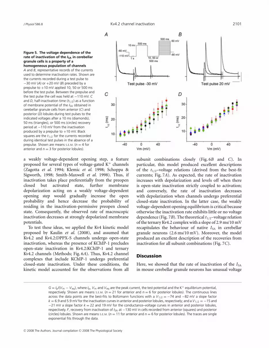
To confirm that the observed behaviour is notthe result of isolating a current containing two ormore components with different voltage dependenciesinactivating at different rates, we took advantage of thefast rate of recovery of Kv4-mediated transient currentscompared to other fast inactivating K+ channels. If thevalues of the time constants of inactivation as a function ofvoltage are a feature of a single population of independentchannels that recover rapidly from inactivation, we wouldexpect their values to be the same for a partially or a fullyrecovered set of channels. We applied depolarizing pulsesto different membrane potentials (test pulses) followinga prepulse to +10 mV applied at different times beforethe test pulse. Between the two pulses the membrane washeld at -110 mV. The t1/2–voltage relations of the currentsobtained during the test pulses following different recoverytimes are shown in Fig. 5C and D, along with those of thecurrents elicited by the same depolarizations in the absenceof a prepulse. The t1/2–voltage relations of fully or partiallyrecovered currents overlap at all membrane potentials andrecovery periods, indicating that they reflect a homo-geneous population of channels with identical recoverytime course.
Kinetic modelling of the voltage dependenceof inactivation kinetics in Kv4.2 channels
The unusual voltage dependence of inactivation describedhere can be explained by assuming preferential closed-stateinactivation (Klemic et al. 1998; Kaulin et al. 2008) and
Figure 3. K+ currents in cerebellar granule cellsWhole-cell ionic currents recorded in a representative cerebellargranule cell from an anterior (A and C) or a posterior (B and D) lobule,before (A and B) or after (C and D) the application of 5 mM TEA.Currents were recorded in the presence of 500 nM TTX and wereelicited by step depolarizations from −90 to +60 mV in 10 mVincrements from a holding potential of −130 mV.
C© 2008 The Authors. Journal compilation C© 2008 The Physiological Society
2100 Y. Amarillo and others J Physiol 586.8
Figure 4. Inactivation kinetics of ISA in cerebellar granule cellsA and B, whole-cell K+ currents recorded in a representative cerebellar granule cell from an anterior (A) or aposterior (B) lobule recorded in the presence of 5 mM TEA. Shown are the currents elicited by step depolarizationsfrom −90 to +60 mV in 10 mV increments, preceded by a prepulse to −100 mV from a holding potential of−130 mV (upper traces) or a prepulse to −40 mV (middle traces). The lower traces show the transient currents (ISA)obtained by subtracting the currents recorded during depolarizing steps preceded by a prepulse to −40 mV fromthe currents obtained during depolarizing steps preceded by a prepulse to −100 mV. C and D, half-inactivationtime (t1/2) as a function of membrane potential of the ISA obtained in cerebellar granule cells from anterior(C) and posterior (D) lobules. Shown are means S.E.M. (n = 17 for anterior and n = 6 for posterior lobules).E, normalized peak conductance–voltage relations (G/Gmax) and steady-state inactivation curves (I/Imax) for granulecells recorded in anterior (squares) and posterior (circles) cerebellar lobules. Peak conductance (G) was calculated as
C© 2008 The Authors. Journal compilation C© 2008 The Physiological Society
J Physiol 586.8 Kv4.2 channel inactivation 2101
Figure 5. The voltage dependence of therate of inactivation of the ISA in cerebellargranule cells is a property of ahomogeneous population of channelsA and B, representative records of the currentsused to determine inactivation rates. Shown arethe currents recorded during a test pulse to−30 mV (A) or +20 mV (B) preceded by aprepulse to +10 mV applied 10, 50 or 500 msbefore the test pulse. Between the prepulse andthe test pulse the cell was held at −110 mV. Cand D, half-inactivation time (t1/2) as a functionof membrane potential of the ISA obtained incerebellar granule cells from anterior (C) andposterior (D) lobules during test pulses to theindicated voltages after a 10 ms (diamonds),50 ms (triangles), or 500 ms (circles) recoveryperiod at −110 mV from the inactivationproduced by a prepulse to +10 mV. Blacksquares are the t1/2 for the currents recordedduring identical test pulses in the absence of aprepulse. Shown are means S.E.M. (n = 4 foranterior and n = 3 for posterior lobules).
a weakly voltage-dependent opening step, a featureproposed for several types of voltage-gated K+ channels(Zagotta et al. 1994; Klemic et al. 1998; Schoppa &Sigworth, 1998; Smith-Maxwell et al. 1998). Thus, ifinactivation takes place preferentially from the preopenclosed but activated state, further membranedepolarization acting on a weakly voltage-dependentopening step would gradually increase the openprobability and hence decrease the probability ofresiding in the inactivation-permissive preopen closedstate. Consequently, the observed rate of macroscopicinactivation decreases at strongly depolarized membranepotentials.
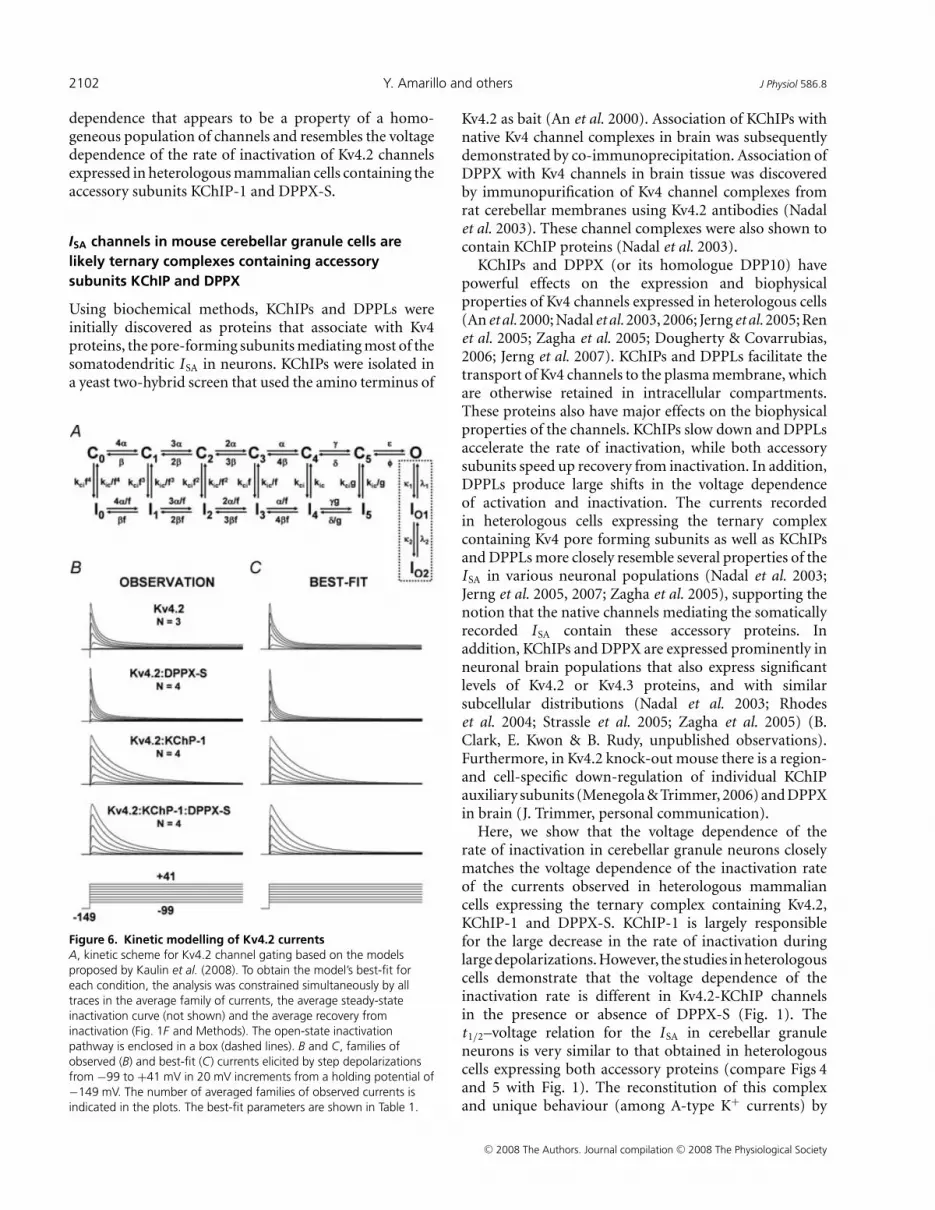
To test these ideas, we applied the Kv4 kinetic modelproposed by Kaulin et al. (2008), and assumed thatKv4.2 and Kv4.2:DPPX-S channels undergo open-stateinactivation, whereas the presence of KChIP-1 precludesopen-state inactivation in Kv4.2:KChIP-1 and ternaryKv4.2 channels (Methods; Fig. 6A). Thus, Kv4.2 channelcomplexes that include KChIP-1 undergo preferentialclosed-state inactivation. Under these conditions, thekinetic model accounted for the observations from all
G = Ip/(Vm − Veq), where Ip, Vm and Veq are the peak current, the test potential and the K+ equilibrium potential,respectively. Shown are means S.E.M. (n = 21 for anterior and n = 6 for posterior lobules). The continuous linesacross the data points are the best-fits to Boltzmann functions with a V1/2 = −74 and −82 mV a slope factork = 6.9 and 5.9 mV for the inactivation curves in anterior and posterior lobules, respectively; and a V1/2 = −15 and−21 mV a slope factor k = 22 and 19 mV for the conductance–voltage curves in anterior and posterior lobules,respectively. F, recovery from inactivation of ISA at −130 mV in cells recorded from anterior (squares) and posterior(circles) lobules. Shown are means S.E.M. (n = 11 for anterior and n = 6 for posterior lobules). The traces are singleexponential fits through the data.
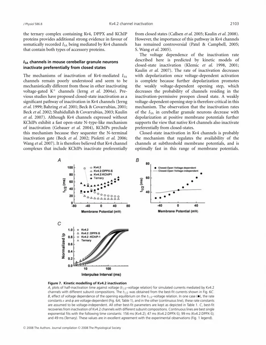
subunit combinations closely (Fig. 6B and C). Inparticular, this model produced excellent descriptionsof the t1/2–voltage relations (derived from the best-fitcurrents; Fig. 7A). As expected, the rate of inactivationincreases with depolarization and levels off when thereis open-state inactivation strictly coupled to activation;and conversely, the rate of inactivation decreaseswith depolarization when channels undergo preferentialclosed-state inactivation. In the latter case, the weaklyvoltage-dependent opening equilibrium is critical becauseotherwise the inactivation rate exhibits little or no voltagedependence (Fig. 7B). The theoretical t1/2–voltage relationof the ternary Kv4.2 complex with a slope of 2.9 ms/10 mVrecapitulates the behaviour of native ISA in cerebellargranule neurons (2.6 ms/10 mV). Moreover, the modelproduced an excellent description of the recoveries frominactivation for all subunit combinations (Fig. 7C).
Discussion
Here, we showed that the rate of inactivation of the ISA
in mouse cerebellar granule neurons has unusual voltage
C© 2008 The Authors. Journal compilation C© 2008 The Physiological Society
2102 Y. Amarillo and others J Physiol 586.8
dependence that appears to be a property of a homo-geneous population of channels and resembles the voltagedependence of the rate of inactivation of Kv4.2 channelsexpressed in heterologous mammalian cells containing theaccessory subunits KChIP-1 and DPPX-S.
ISA channels in mouse cerebellar granule cells arelikely ternary complexes containing accessorysubunits KChIP and DPPX
Using biochemical methods, KChIPs and DPPLs wereinitially discovered as proteins that associate with Kv4proteins, the pore-forming subunits mediating most of thesomatodendritic ISA in neurons. KChIPs were isolated ina yeast two-hybrid screen that used the amino terminus of
Figure 6. Kinetic modelling of Kv4.2 currentsA, kinetic scheme for Kv4.2 channel gating based on the modelsproposed by Kaulin et al. (2008). To obtain the model’s best-fit foreach condition, the analysis was constrained simultaneously by alltraces in the average family of currents, the average steady-stateinactivation curve (not shown) and the average recovery frominactivation (Fig. 1F and Methods). The open-state inactivationpathway is enclosed in a box (dashed lines). B and C, families ofobserved (B) and best-fit (C) currents elicited by step depolarizationsfrom −99 to +41 mV in 20 mV increments from a holding potential of−149 mV. The number of averaged families of observed currents isindicated in the plots. The best-fit parameters are shown in Table 1.
Kv4.2 as bait (An et al. 2000). Association of KChIPs withnative Kv4 channel complexes in brain was subsequentlydemonstrated by co-immunoprecipitation. Association ofDPPX with Kv4 channels in brain tissue was discoveredby immunopurification of Kv4 channel complexes fromrat cerebellar membranes using Kv4.2 antibodies (Nadalet al. 2003). These channel complexes were also shown tocontain KChIP proteins (Nadal et al. 2003).
KChIPs and DPPX (or its homologue DPP10) havepowerful effects on the expression and biophysicalproperties of Kv4 channels expressed in heterologous cells(An et al. 2000; Nadal et al. 2003, 2006; Jerng et al. 2005; Renet al. 2005; Zagha et al. 2005; Dougherty & Covarrubias,2006; Jerng et al. 2007). KChIPs and DPPLs facilitate thetransport of Kv4 channels to the plasma membrane, whichare otherwise retained in intracellular compartments.These proteins also have major effects on the biophysicalproperties of the channels. KChIPs slow down and DPPLsaccelerate the rate of inactivation, while both accessorysubunits speed up recovery from inactivation. In addition,DPPLs produce large shifts in the voltage dependenceof activation and inactivation. The currents recordedin heterologous cells expressing the ternary complexcontaining Kv4 pore forming subunits as well as KChIPsand DPPLs more closely resemble several properties of theISA in various neuronal populations (Nadal et al. 2003;Jerng et al. 2005, 2007; Zagha et al. 2005), supporting thenotion that the native channels mediating the somaticallyrecorded ISA contain these accessory proteins. Inaddition, KChIPs and DPPX are expressed prominently inneuronal brain populations that also express significantlevels of Kv4.2 or Kv4.3 proteins, and with similarsubcellular distributions (Nadal et al. 2003; Rhodeset al. 2004; Strassle et al. 2005; Zagha et al. 2005) (B.Clark, E. Kwon & B. Rudy, unpublished observations).Furthermore, in Kv4.2 knock-out mouse there is a region-and cell-specific down-regulation of individual KChIPauxiliary subunits (Menegola & Trimmer, 2006) and DPPXin brain (J. Trimmer, personal communication).
Here, we show that the voltage dependence of therate of inactivation in cerebellar granule neurons closelymatches the voltage dependence of the inactivation rateof the currents observed in heterologous mammaliancells expressing the ternary complex containing Kv4.2,KChIP-1 and DPPX-S. KChIP-1 is largely responsiblefor the large decrease in the rate of inactivation duringlarge depolarizations. However, the studies in heterologouscells demonstrate that the voltage dependence of theinactivation rate is different in Kv4.2-KChIP channelsin the presence or absence of DPPX-S (Fig. 1). Thet1/2–voltage relation for the ISA in cerebellar granuleneurons is very similar to that obtained in heterologouscells expressing both accessory proteins (compare Figs 4and 5 with Fig. 1). The reconstitution of this complexand unique behaviour (among A-type K+ currents) by
C© 2008 The Authors. Journal compilation C© 2008 The Physiological Society
J Physiol 586.8 Kv4.2 channel inactivation 2103
the ternary complex containing Kv4, DPPX and KChIPproteins provides additional strong evidence in favour ofsomatically recorded ISA being mediated by Kv4 channelsthat contain both types of accessory proteins.
ISA channels in mouse cerebellar granule neuronsinactivate preferentially from closed states
The mechanisms of inactivation of Kv4-mediated ISA
channels remain poorly understood and seem to bemechanistically different from those in other inactivatingvoltage-gated K+ channels (Jerng et al. 2004a). Pre-vious studies have proposed closed-state inactivation as asignificant pathway of inactivation in Kv4 channels (Jernget al. 1999; Bahring et al. 2001; Beck & Covarrubias, 2001;Beck et al. 2002; Shahidullah & Covarrubias, 2003; Kaulinet al. 2007). Although Kv4 channels expressed withoutKChIPs exhibit a fast open-state N-type-like mechanismof inactivation (Gebauer et al. 2004), KChIPs precludethis mechanism because they sequester the N-terminalinactivation gate (Beck et al. 2002; Pioletti et al. 2006;Wang et al. 2007). It is therefore believed that Kv4 channelcomplexes that include KChIPs inactivate preferentially
Figure 7. Kinetic modelling of Kv4.2 inactivationA, plots of half-inactivation time against voltage (t1/2–voltage relation) for simulated currents mediated by Kv4.2channels with different subunit compositions. The t1/2 was obtained from the best-fit currents shown in Fig. 6C.B, effect of voltage dependence of the opening equilibrium on the t1/2–voltage relation. In one case (•), the rateconstants ε and φ are voltage-dependent (Fig. 6A; Table 1), and in the other (continuous line), these rate constantsare assumed to be voltage-independent. All other best-fit parameters are kept as depicted in Table 1. C, best-fitrecoveries from inactivation of Kv4.2 channels with different subunit compositions. Continuous lines are best singleexponential fits with the following time constants: 156 ms (Kv4.2); 47 ms (Kv4.2:DPPX-S); 99 ms (Kv4.2:DPPX-S);and 49 ms (Ternary). These values are in excellent agreement with the experimental observations (Fig. 1 legend).
from closed states (Callsen et al. 2005; Kaulin et al. 2008).However, the importance of this pathway in Kv4 channelshas remained controversial (Patel & Campbell, 2005;S. Wang et al. 2005).
The voltage dependence of the inactivation ratedescribed here is predicted by kinetic models ofclosed-state inactivation (Klemic et al. 1998, 2001;Kaulin et al. 2007). The rate of inactivation decreaseswith depolarization once voltage-dependent activationis complete because further depolarization promotesthe weakly voltage-dependent opening step, whichdecreases the probability of channels residing in theinactivation-permissive preopen closed state. A weaklyvoltage-dependent opening step is therefore critical in thismechanism. The observation that the inactivation ratesof the ISA in cerebellar granule neurons decrease withdepolarization at positive membrane potentials furthersupports the view that native Kv4 channels also inactivatepreferentially from closed-states.
Closed-state inactivation in Kv4 channels is probablythe mechanism that regulates the availability of thechannels at subthreshold membrane potentials, and isoptimally fast in this range of membrane potentials.
C© 2008 The Authors. Journal compilation C© 2008 The Physiological Society
2104 Y. Amarillo and others J Physiol 586.8
Although at typical neuronal resting membrane potentials(−60 mV) these channels are mostly inactivated atsteady state, a brief conditioning hyperpolarization willrecruit them because their recovery from inactivation isfast. Then, activation of Kv4 channels by a subsequentdepolarization will delay firing of the action potential. Thecharacteristically fast voltage-dependent recovery frominactivation of native Kv4 channel complexes distinguishesthem from other Kv channels that inactivate primarilyvia classical N-type and P/C-type mechanisms. Thishallmark of Kv4 channel complexes might be a directconsequence of the mechanism responsible for closed-stateinactivation, which remains unknown. The slowing ofinactivation with depolarization may confer another role.By inactivating more slowly at depolarized membranepotentials, Kv4 channels could have a greater impact onspike repolarization.
References
Adams JP, Anderson AE, Varga AW, Dineley KT, Cook RG,Pfaffinger PJ & Sweatt JD (2000). The A-type potassiumchannel Kv4.2 is a substrate for the mitogen-activatedprotein kinase ERK. J Neurochem 75, 2277–2287.
An WF, Bowlby MR, Betty M, Cao J, Ling HP, Mendoza G,Hinson JW, Mattsson KI, Strassle BW, Trimmer JS & RhodesKJ (2000). Modulation of A-type potassium channels by afamily of calcium sensors. Nature 403, 553–556.
Bahring R, Boland LM, Varghese A, Gebauer M & Pongs O(2001). Kinetic analysis of open- and closed-stateinactivation transitions in human Kv4.2 A-type potassiumchannels. J Physiol 535, 65–81.
Beck EJ, Bowlby M, An WF, Rhodes KJ & Covarrubias M(2002). Remodelling inactivation gating of Kv4 channels byKChIP1, a small-molecular-weight calcium-binding protein.J Physiol 538, 691–706.
Beck EJ & Covarrubias M (2001). Kv4 channels exhibitmodulation of closed-state inactivation in inside-outpatches. Biophys J 81, 867–883.
Callsen B, Isbrandt D, Sauter K, Hartmann LS, Pongs O &Bahring R (2005). Contribution of N- and C-terminal Kv4.2channel domains to KChIP interaction. J Physiol 568,397–412.
Chen X, Yuan LL, Zhao C, Birnbaum SG, Frick A, Jung WE,Schwarz TL, Sweatt JD & Johnston D (2006). Deletion ofKv4.2 gene eliminates dendritic A-type K+ current andenhances induction of long-term potentiation inhippocampal CA1 pyramidal neurons. J Neurosci 26,12143–12151.
Coetzee WA, Amarillo Y, Chiu J, Chow A, Lau D, McCormackT, Moreno H, Nadal MS, Ozaita A, Pountney D, Saganich M,Vega-Saenz de Miera E & Rudy B (1999). Molecular diversityof K+ channels. Ann N Y Acad Sci 868, 233–285.
Connor JA & Stevens CF (1971). Prediction of repetitive firingbehaviour from voltage clamp data on an isolated neuronesoma. J Physiol 213, 31–53.
Cull-Candy SG, Marshall CG & Ogden D (1989). Voltage-activated membrane currents in rat cerebellar granuleneurones. J Physiol 414, 179–199.
Dougherty K & Covarrubias M (2006). A dipeptidylaminopeptidase-like protein remodels gating chargedynamics in Kv4.2 channels. J Gen Physiol 128, 745–753.
Franqueza L, Valenzuela C, Eck J, Tamkun MM, Tamargo J &Snyders DJ (1999). Functional expression of an inactivatingpotassium channel (Kv4.3) in a mammalian cell line.Cardiovasc Res 41, 212–219.
Gebauer M, Isbrandt D, Sauter K, Callsen B, Nolting A, PongsO & Bahring R (2004). N-type inactivation features of Kv4.2channel gating. Biophys J 86, 210–223.
Hamill OP, Marty A, Neher E, Sakmann B & Sigworth FJ(1981). Improved patch-clamp techniques for high-resolution current recording from cells and cell-freemembrane patches. Pflugers Arch 391, 85–100.
Hille B (2001). Ion Channels of Excitable Membranes, 3rd edn.Sinauer Associates, Inc., Sunderland, MA, USA.
Hoffman DA, Magee JC, Colbert CM & Johnston D (1997). K+channel regulation of signal propagation in dendrites ofhippocampal pyramidal neurons. Nature 387, 869–875.
Hsu YH, Huang HY & Tsaur ML (2003). Contrastingexpression of Kv4.3, an A-type K+ channel, in migratingPurkinje cells and other post-migratory cerebellar neurons.Eur J Neurosci 18, 601–612.
Hu HJ, Carrasquillo Y, Karim F, Jung WE, Nerbonne JM,Schwarz TL & Gereau RW 4th (2006). The Kv4.2 potassiumchannel subunit is required for pain plasticity. Neuron 50,89–100.
Jerng HH & Covarrubias M (1997). K+ channel inactivationmediated by the concerted action of the cytoplasmic N- andC-terminal domains. Biophys J 72, 163–174.
Jerng HH, Kunjilwar K & Pfaffinger PJ (2005). Multiproteinassembly of Kv4.2, KChIP3 and DPP10 produces ternarychannel complexes with ISA-like properties. J Physiol 568,767–788.
Jerng HH, Lauver AD & Pfaffinger PJ (2007). DPP10 splicevariants are localized in distinct neuronal populations andact to differentially regulate the inactivation properties ofKv4-based ion channels. Mol Cell Neurosci 35,604–624.
Jerng HH, Pfaffinger PJ & Covarrubias M (2004a). Molecularphysiology and modulation of somatodendritic A-typepotassium channels. Mol Cell Neurosci 27, 343–369.
Jerng HH, Qian Y & Pfaffinger PJ (2004b). Modulation ofKv4.2 channel expression and gating by dipeptidyl peptidase10 (DPP10). Biophys J 87, 2380–2396.
Jerng HH, Shahidullah M & Covarrubias M (1999).Inactivation gating of Kv4 potassium channels: molecularinteractions involving the inner vestibule of the pore. J GenPhysiol 113, 641–660.
Johnston D, Christie BR, Frick A, Gray R, Hoffman DA,Schexnayder LK, Watanabe S & Yuan LL (2003). Activedendrites, potassium channels and synaptic plasticity. PhilosTrans R Soc Lond B Biol Sci 358, 667–674.
Johnston D, Hoffman DA, Magee JC, Poolos NP, Watanabe S,Colbert CM & Migliore M (2000). Dendritic potassiumchannels in hippocampal pyramidal neurons. J Physiol 525,75–81.
Kaulin Y, De Santiago-Castillo JA, Rocha C & Covarrubias M(2008). Mechanism of the modulation of Kv4:KChIP-1channels by elevated external K+. Biophys J 94, 1241–1251.
C© 2008 The Authors. Journal compilation C© 2008 The Physiological Society
J Physiol 586.8 Kv4.2 channel inactivation 2105
Kim J, Jung SC, Clemens AM, Petralia RS & Hoffman DA(2007). Regulation of dendritic excitability by activity-dependent trafficking of the A-type K+ channel subunitKv4.2 in hippocampal neurons. Neuron 54, 933–947.
Kim J, Wei DS & Hoffman DA (2005). Kv4 potassium channelsubunits control action potential repolarization andfrequency-dependent broadening in rat hippocampal CA1pyramidal neurones. J Physiol 569, 41–57.
Klee R, Ficker E & Heinemann U (1995). Comparison ofvoltage-dependent potassium currents in rat pyramidalneurons acutely isolated from hippocampal regions CA1 andCA3. J Neurophysiol 74, 1982–1995.
Klemic KG, Kirsch GE & Jones SW (2001). U-type inactivationof Kv3.1 and Shaker potassium channels. Biophys J 81,814–826.
Klemic KG, Shieh CC, Kirsch GE & Jones SW (1998).Inactivation of Kv2.1 potassium channels. Biophys J 74,1779–1789.
Lien CC, Martina M, Schultz JH, Ehmke H & Jonas P (2002).Gating, modulation and subunit composition of voltage-gated K+ channels in dendritic inhibitory interneurones ofrat hippocampus. J Physiol 538, 405–419.
Liss B, Franz O, Sewing S, Bruns R, Neuhoff H & Roeper J(2001). Tuning pacemaker frequency of individualdopaminergic neurons by Kv4.3L and KChip3.1transcription. EMBO J 20, 5715–5724.
Martina M, Schultz JH, Ehmke H, Monyer H & Jonas P (1998).Functional and molecular differences between voltage-gatedK+ channels of fast-spiking interneurons and pyramidalneurons of rat hippocampus. J Neurosci 18, 8111–8125.
Mathie A, Clarke CE, Ranatunga KM & Veale EL (2003). Whatare the roles of the many different types of potassium channelexpressed in cerebellar granule cells? Cerebellum 2, 11–25.
Menegola M & Trimmer JS (2006). Unanticipated region- andcell-specific downregulation of individual KChIP auxiliarysubunit isotypes in Kv4.2 knock-out mouse brain. J Neurosci26, 12137–12142.
Millar JA, Barratt L, Southan AP, Page KM, Fyffe RE,Robertson B & Mathie A (2000). A functional role for thetwo-pore domain potassium channel TASK-1 in cerebellargranule neurons. Proc Natl Acad Sci U S A 97, 3614–3618.
Nadal MS, Amarillo Y, Vega-Saenz de Miera E & Rudy B(2001). Evidence for the presence of a novel Kv4-mediatedA-type K+ channel-modifying factor. J Physiol 537,801–809.
Nadal MS, Amarillo Y, Vega-Saenz de Miera E & Rudy B(2006). Differential characterization of three alternativespliced isoforms of DPPX. Brain Res 1094, 1–12.
Nadal MS, Ozaita A, Amarillo Y, Vega-Saenz de Miera E, Ma Y,Mo W, Goldberg EM, Misumi Y, Ikehara Y, Neubert TA &Rudy B (2003). The CD26-related dipeptidylaminopeptidase-like protein DPPX is a critical componentof neuronal A-type K+ channels. Neuron 37, 449–461.
Patel SP & Campbell DL (2005). Transient outward potassiumcurrent, ‘Ito’, phenotypes in the mammalian left ventricle:underlying molecular, cellular and biophysical mechanisms.J Physiol 569, 7–39.
Perney TM, Marshall J, Martin KA, Hockfield S & KaczmarekLK (1992). Expression of the mRNAs for the Kv3.1potassium channel gene in the adult and developing ratbrain. J Neurophysiol 68, 756–766.
Pioletti M, Findeisen F, Hura GL & Minor DL Jr (2006).Three-dimensional structure of the KChIP1-Kv4.3 T1complex reveals a cross-shaped octamer. Nat Struct Mol Biol13, 987–995.
Ramakers GM & Storm JF (2002). A postsynaptic transient K+current modulated by arachidonic acid regulates synapticintegration and threshold for LTP induction in hippocampalpyramidal cells. Proc Natl Acad Sci U S A 99, 10144–10149.
Ren X, Hayashi Y, Yoshimura N & Takimoto K (2005).Transmembrane interaction mediates complex formationbetween peptidase homologues and Kv4 channels. Mol CellNeurosci 29, 320–332.
Rhodes KJ, Carroll KI, Sung MA, Doliveira LC, MonaghanMM, Burke SL, Strassle BW, Buchwalder L, Menegola M,Cao J, An WF & Trimmer JS (2004). KChIPs and Kv4 α
subunits as integral components of A-type potassiumchannels in mammalian brain. J Neurosci 24, 7903–7915.
Riazanski V, Becker A, Chen J, Sochivko D, Lie A, Wiestler OD,Elger CE & Beck H (2001). Functional and molecularanalysis of transient voltage-dependent K+ currents in rathippocampal granule cells. J Physiol 537, 391–406.
Rudy B, Hoger JH, Lester HA & Davidson N (1988). At leasttwo mRNA species contribute to the properties of rat brainA-type potassium channels expresed in Xenopus oocytes.Neuron 1, 649–658.
Rudy B, Sen K, Vega-Saenz de Miera E, Lau D, Ried T & WardDC (1991). Cloning of a human cDNA expressing a highvoltage-activating, TEA-sensitive, type-A K+ channel whichmaps to chromosome 1 band p21. J Neurosci Res 29, 401–412.
Schoppa NE & Sigworth FJ (1998). Activation of Shakerpotassium channels. III. An activation gating model forwild-type and V2 mutant channels. J Gen Physiol 111,313–342.
Schoppa NE & Westbrook GL (1999). Regulation of synaptictiming in the olfactory bulb by an A-type potassium current.Nat Neurosci 2, 1106–1113.
Schroter KH, Ruppersberg JP, Wunder F, Rettig J, Stocker M &Pongs O (1991). Cloning and functional expression of aTEA-sensitive A-type potassium channel from rat brain.FEBS Lett 278, 211–216.
Sekirnjak C, Martone ME, Weiser M, Deerinck T, Bueno E,Rudy B & Ellisman M (1997). Subcellular localization of theK+ channel subunit Kv3.1b in selected rat CNS neurons.Brain Res 766, 173–187.
Serodio P, Kentros C & Rudy B (1994). Identification ofmolecular components of A-type channels activating atsubthreshold potentials. J Neurophysiol 72, 1516–1529.
Serodio P & Rudy B (1998). Differential expression of Kv4 K+channel subunits mediating subthreshold transient K+(A-type) currents in rat brain. J Neurophysiol 79, 1081–1091.
Serodio P, Vega-Saenz de Miera E & Rudy B (1996). Cloning ofa novel component of A-type K+ channels operating atsubthreshold potentials with unique expression in heart andbrain. J Neurophysiol 75, 2174–2179.
Shahidullah M & Covarrubias M (2003). The link between ionpermeation and inactivation gating of Kv4 potassiumchannels. Biophys J 84, 928–941.
Smith-Maxwell CJ, Ledwell JL & Aldrich RW (1998).Uncharged S4 residues and cooperativity in voltage-dependent potassium channel activation. J Gen Physiol 111,421–439.
C© 2008 The Authors. Journal compilation C© 2008 The Physiological Society
2106 Y. Amarillo and others J Physiol 586.8
Strassle BW, Menegola M, Rhodes KJ & Trimmer JS (2005).Light and electron microscopic analysis of KChIP and Kv4localization in rat cerebellar granule cells. J Comp Neurol484, 144–155.
Stuart GJ, Dodt HU & Sakmann B (1993). Patch-clamprecordings from the soma and dendrites of neurons in brainslices using infrared video microscopy. Pflugers Arch 423,511–518.
Thompson SM (2007). IA in play. Neuron 54, 850–852.Tseng-Crank JC, Tseng GN, Schwartz A & Tanouye MA (1990).
Molecular cloning and functional expression of a potassiumchannel cDNA isolated from a rat cardiac library. FEBS Lett268, 63–68.
Vega-Saenz de Miera E, Moreno H, Fruhling D, Kentros C &Rudy B (1992). Cloning of ShIII (Shaw-like) cDNAsencoding a novel high-voltage-activating, TEA-sensitive,type-A K+ channel. Proc R Soc Lond B Biol Sci 248, 9–18.
Wang S, Bondarenko VE, Qu YJ, Bett GC, Morales MJ,Rasmusson RL & Strauss HC (2005). Time- andvoltage-dependent components of Kv4.3 inactivation.Biophys J 89, 3026–3041.
Wang G, Shahidullah M, Rocha CA, Strang C, Pfaffinger PJ &Covarrubias M (2005). Functionally active t1–t1 interfacesrevealed by the accessibility of intracellular thiolate groups inKv4 channels. J Gen Physiol 126, 55–69.
Wang H, Yan Y, Liu Q, Huang Y, Shen Y, Chen L, Chen Y, YangQ, Hao Q, Wang K & Chai J (2007). Structural basis formodulation of Kv4 K+ channels by auxiliary KChIPsubunits. Nat Neurosci 10, 32–39.
Weiser M, Bueno E, Sekirnjak C, Martone ME, Baker H,Hillman D, Chen S, Thornhill W, Ellisman M & Rudy B(1995). The potassium channel subunit KV3.1b is localizedto somatic and axonal membranes of specific populations ofCNS neurons. J Neurosci 15, 4298–4314.
Weiser M, Vega-Saenz de Miera E, Kentros C, Moreno H,Franzen L, Hillman D, Baker H & Rudy B (1994).Differential expression of Shaw-related K+ channels in therat central nervous system. J Neurosci 14, 949–972.
Zagha E, Ozaita A, Chang SY, Nadal MS, Lin U, Saganich MJ,McCormack T, Akinsanya KO, Qi SY & Rudy B (2005).DPP10 modulates Kv4-mediated A-type potassium channels.J Biol Chem 280, 18853–18861.
Zagotta WN, Hoshi T & Aldrich RW (1994). Shaker potassiumchannel gating. III: Evaluation of kinetic models foractivation. J Gen Physiol 103, 321–362.
Acknowledgements
We wish to thank Brian Clark and Eddie Zagha for their
critical review of this manuscript. This work was supported by
National Institutes of Health grants NS045217 and NS30989 and
NSF grant IBN-0314645 to B.R., National Institutes of Health
grant NS032337 and Research Enhancement Award (REA) from
Thomas Jefferson University to M.C.; and NIH training grant
T32 AA007463 to K.D.
Supplemental material
Online supplemental material for this paper can be accessed at:
http://jp.physoc.org/cgi/content/full/jphysiol.2007.150540/DC1
and
http://www.blackwell-synergy.com/doi/suppl/10.1113/jphysiol.
2007.150540
C© 2008 The Authors. Journal compilation C© 2008 The Physiological Society
Related Documents


