Note: Within nine months of the publication of the mention of the grant of the European patent in the European Patent Bulletin, any person may give notice to the European Patent Office of opposition to that patent, in accordance with the Implementing Regulations. Notice of opposition shall not be deemed to have been filed until the opposition fee has been paid. (Art. 99(1) European Patent Convention). Printed by Jouve, 75001 PARIS (FR) (19) EP 2 101 748 B1 (Cont. next page) TEPZZ _Z_748B_T (11) EP 2 101 748 B1 (12) EUROPEAN PATENT SPECIFICATION (45) Date of publication and mention of the grant of the patent: 30.07.2014 Bulletin 2014/31 (21) Application number: 07827450.3 (22) Date of filing: 29.11.2007 (51) Int Cl.: A61K 31/343 (2006.01) A61P 29/00 (2006.01) A61P 25/00 (2006.01) A61P 25/28 (2006.01) A61P 25/22 (2006.01) A61P 25/24 (2006.01) A61P 43/00 (2006.01) (86) International application number: PCT/IL2007/001477 (87) International publication number: WO 2008/065666 (05.06.2008 Gazette 2008/23) (54) USE OF INCENSOLE AND DERIVATIVES THEREOF FOR NEUROPROTECTION AND FOR THE TREATMENT OF DEPRESSION AND ANXIETY VERWENDUNG VON INCENSOL UND SEINEN DERIVATEN ZUR NEUROPROTEKTION UND BEHANDLUNG VON DEPRESSION UND ANGSTZUSTÄNDEN UTILISATION D’INCENSOLE ET LEURS DÉRIVÉS POUR LA NEUROPROTECTION ET LE TRAITEMENT DE LA DÉPRESSION ET DE L’ANXIETÉ (84) Designated Contracting States: AT BE BG CH CY CZ DE DK EE ES FI FR GB GR HU IE IS IT LI LT LU LV MC MT NL PL PT RO SE SI SK TR (30) Priority: 29.11.2006 US 861441 P 16.08.2007 US 956276 P (43) Date of publication of application: 23.09.2009 Bulletin 2009/39 (73) Proprietors: • Yissum Research Development Company of the Hebrew University of Jerusalem, Ltd. Jerusalem 91390 (IL) • Ariel-University Research and Development Company Ltd. 40700 Ariel (IL) (72) Inventors: • MOUSSAIEFF, Arik St Jerusalem (IL) • MECHOULAM, Raphael 92581 Jerusalem (IL) • FRIDE, Esther 44837 Ariel (IL) • SHOHAMI, Esther 90805 Mevasseret Zion (IL) • BEN NERIAH, Yinon 90805 Mevasseret Zion (IL) • GALLILY, Ruth 93706 Jerusalem (IL) (74) Representative: Fleuchaus, Michael A. et al Fleuchaus & Gallo Partnerschaft mbB Patent- und Rechtsanwälte Steinerstrasse 15/A 81369 München (DE) (56) References cited: WO-A-02/053138 • BANNO ET AL: "Anti-inflammatory activities of the triterpene acids from the resin of Boswellia carteri" JOURNAL OF ETHNOPHARMACOLOGY, ELSEVIER SCIENTIFIC PUBLISHERS LTD, IE, vol. 107, no. 2, 19 September 2006 (2006-09-19), pages 249-253, XP005613499 ISSN: 0378-8741 • MOUSSAIEFF ARIEH ET AL: "Incensole acetate, a novel anti-inflammatory compound isolated from Boswellia resin, inhibits nuclear factor- kappa B activation" MOLECULAR PHARMACOLOGY, vol. 72, no. 6, December 2007 (2007-12), pages 1657-1664 URL, XP009098362 ISSN: 0026-895X

Welcome message from author
This document is posted to help you gain knowledge. Please leave a comment to let me know what you think about it! Share it to your friends and learn new things together.
Transcript
Note: Within nine months of the publication of the mention of the grant of the European patent in the European PatentBulletin, any person may give notice to the European Patent Office of opposition to that patent, in accordance with theImplementing Regulations. Notice of opposition shall not be deemed to have been filed until the opposition fee has beenpaid. (Art. 99(1) European Patent Convention).
Printed by Jouve, 75001 PARIS (FR)
(19)E
P2
101
748
B1
(Cont. next page)
TEPZZ _Z_748B_T(11) EP 2 101 748 B1
(12) EUROPEAN PATENT SPECIFICATION
(45) Date of publication and mention of the grant of the patent: 30.07.2014 Bulletin 2014/31
(21) Application number: 07827450.3
(22) Date of filing: 29.11.2007
(51) Int Cl.:A61K 31/343 (2006.01) A61P 29/00 (2006.01)
A61P 25/00 (2006.01) A61P 25/28 (2006.01)
A61P 25/22 (2006.01) A61P 25/24 (2006.01)
A61P 43/00 (2006.01)
(86) International application number: PCT/IL2007/001477
(87) International publication number: WO 2008/065666 (05.06.2008 Gazette 2008/23)
(54) USE OF INCENSOLE AND DERIVATIVES THEREOF FOR NEUROPROTECTION AND FOR THE TREATMENT OF DEPRESSION AND ANXIETY
VERWENDUNG VON INCENSOL UND SEINEN DERIVATEN ZUR NEUROPROTEKTION UND BEHANDLUNG VON DEPRESSION UND ANGSTZUSTÄNDEN
UTILISATION D’INCENSOLE ET LEURS DÉRIVÉS POUR LA NEUROPROTECTION ET LE TRAITEMENT DE LA DÉPRESSION ET DE L’ANXIETÉ
(84) Designated Contracting States: AT BE BG CH CY CZ DE DK EE ES FI FR GB GR HU IE IS IT LI LT LU LV MC MT NL PL PT RO SE SI SK TR
(30) Priority: 29.11.2006 US 861441 P16.08.2007 US 956276 P
(43) Date of publication of application: 23.09.2009 Bulletin 2009/39
(73) Proprietors: • Yissum Research Development Company of the
Hebrew University of Jerusalem, Ltd.Jerusalem 91390 (IL)
• Ariel-University Research and Development Company Ltd.40700 Ariel (IL)
(72) Inventors: • MOUSSAIEFF, Arik
St Jerusalem (IL)• MECHOULAM, Raphael
92581 Jerusalem (IL)• FRIDE, Esther
44837 Ariel (IL)• SHOHAMI, Esther
90805 Mevasseret Zion (IL)• BEN NERIAH, Yinon
90805 Mevasseret Zion (IL)
• GALLILY, Ruth93706 Jerusalem (IL)
(74) Representative: Fleuchaus, Michael A. et alFleuchaus & Gallo Partnerschaft mbB Patent- und Rechtsanwälte Steinerstrasse 15/A81369 München (DE)
(56) References cited: WO-A-02/053138
• BANNO ET AL: "Anti-inflammatory activities of the triterpene acids from the resin of Boswellia carteri" JOURNAL OF ETHNOPHARMACOLOGY, ELSEVIER SCIENTIFIC PUBLISHERS LTD, IE, vol. 107, no. 2, 19 September 2006 (2006-09-19), pages 249-253, XP005613499 ISSN: 0378-8741
• MOUSSAIEFF ARIEH ET AL: "Incensole acetate, a novel anti-inflammatory compound isolated from Boswellia resin, inhibits nuclear factor-kappa B activation" MOLECULAR PHARMACOLOGY, vol. 72, no. 6, December 2007 (2007-12), pages 1657-1664 URL, XP009098362 ISSN: 0026-895X

2
EP 2 101 748 B1
• MOUSSAIEFF A. ET AL.: "Incensole acetate, an incense component, elicits psychoactivity by activating TRPV3 channels"[Online] 20 September 2007 (2007-09-20), XP002475422 Retrieved from the Internet: URL:http://www.ispp.org.il/Program2007.pdf > [retrieved on 2008-04-07]
• A. MOUSSAIEFF ET AL.: "Is incense a pharmacological tool for spiritual exaltation?" NEURAL PLASTICITY, [Online] 25 November 2007 (2007-11-25), - 27 November 2007 (2007-11-27) XP002475423 Retrieved from the Internet: URL:www.hindawi.com/GetPDF.aspx?doi=10.115 5/2007/30585> [retrieved on 2008-04-07]
EP 2 101 748 B1
3
5
10
15
20
25
30
35
40
45
50
55
Description
FIELD OF THE INVENTION
[0001] This invention relates to uses of incensole, incensole acetate, their derivatives, and pharmaceutical composi-tions comprising them, for treating various diseases or conditions.
BACKGROUND OF THE INVENTION
[0002] Boswellia species (Burseraceae) are native of Eastern Africa, where their resin ("frankincense" "olibanum")has been widely used as incense and for various medical purposes. For example these species are known as diureticagents, for the treatment of vasious diseases such as Bilharzia, stomachache syphilis and Rheumatism (Watt, 1962).Boswellia resin was found to be useful for the treatment of inflammations (Singh & Atal, 1986), as well as several diseasesassociated with inflammatory conditions such as for example active Crohn’s disease and Asthma (Gerhardt et al., 2001;Gupta, 1998). It was previously reported that the anti-inflammatory properties of Boswellia resin may be attributed tothe Boswellic acid and its derivatives (Ammon et al., 1993).[0003] The use of Boswellia resin for its psychoactive properties extends beyond the Near East and Europe. In Ay-urveda, the Indian medical tradition, Boswellia resin is reported to have a ’strong action on the nervous system’. InEthiopia, where Boswellia trees are indigenous, it is believed to have a tranquilizing effect.The isolation of IA (incensole acetate) and its structural elucidation was first described by Corsano and Nicoletti (Corsano& Nicoletti, 1967). In 2006 the isolation and investigation of several constituents including incensole of Boswellia resinas well as their anti-inflammatory potential were published (Banno et. al, 2006, Journal of Ethnopharmacology 107,249-253). US Patent No. 5064823 discloses pentacyclic triterpenoid compounds such as α boswelic acid and its acetate,which have an inhibitory effect on topoisomerase I and topoisomerase II.[0004] WO 02/053138 discloses the use of incensole and/or furanogermacrens, derivatives, metabolites and analo-geous thereof for selective inhibition of neoplastic cells, for example for the treatment, inhibition or prevention of pre-cancerous lesions, tumors, cancer growth or other neoplasias in mammals.[0005] NF-κB (nuclear factor-κB) is a collective name for a group of inducible dimeric transcription factors. NF-κB isfound in essentially all cell types and is involved in activation of a large number of genes in response to various stressfulsituations, e.g. infection and inflammation. The subcellular localization of NF-κB is controlled by a family of inhibitoryproteins, IκBs, which bind NF-κB and mask its nuclear localization signal, thus preventing nuclear translocation. Exposureof cells to a variety of extracellular stimuli leads to the rapid phosphorylation, ubiquitination, and ultimately proteolyticdegradation of IκB, which frees NF-κB to translocate to the nucleus where it regulates gene transcription (Karin andBen-Neriah, 2000). IκB phosphorylation, followed by its degradation is considered to be the major step in NF-κB regulation(Ghosh & Karin, 2002).[0006] Traumatic brain injury (TBI) is often associated with permanent cognitive disorders, learning disabilities andvarious behavioral and emotional problems. Despite promising pre-clinical data, most of the clinical trials conducted sofar have failed to demonstrate any significant improvement in outcomes, mainly because of ineffective therapies orbecause of the selection of inappropriate target mechanisms (Marmarou et al, 2005, Narayan et al, 2002). Secondarybrain damage, triggered by the initial impact, develops over hours, weeks and even months following injury. Secondarybrain damage can increase mortality and worsen disability but, unlike the primary lesion, may potentially be attenuatedby appropriate treatment. TBI induces early phase neuronal activation of NF-κB, followed by its remarkably prolongedactivation (Beni et al, 2004) even up to 1 year (Nonaka, 1999). Studies on the role of NF-κB in the brain following closedhead injury in (CHI) mice have revealed that inhibition of acute NF-κB activation is associated with enhanced functionalrecovery (Beni et al, 2004).
SUMMARY OF THE INVENTION
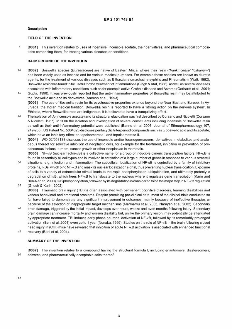
[0007] The invention relates to a compound having the structural formula I, including enantiomers, diastereomers,solvates, and pharmaceutically acceptable salts thereof:
EP 2 101 748 B1
4
5
10
15
20
25
30
35
40
45
50
55
wherein,
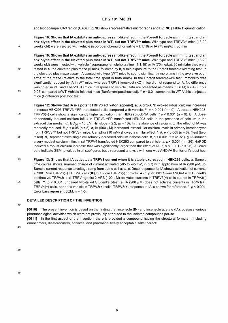
R is selected from H, -C(=O)R’, and -C(=O)OR", wherein R’ is C1-25alkyl and R" is H or C1-25alkyl;R1, R2, R5, and R6 are independently selected from H, OH and CH3;R3, R4, R7, and R8 are independently selected H and OH;R9 is H or CH3; orone of R1 and R2 and one of R3 and R4 taken together form (i) a second bond between C12 and C13 or (ii) an epoxidering, along with the carbon to which they are bonded; and/orone of R5 and R6 and one of R7 and R8 taken together form (iii) a second bond between C8 and C9 or (iv) an epoxidering, along with the carbon to which they are bonded; and/orone of R5 and R6 together with R form a single bond, thereby forming an epoxide ring along with the carbon to whichthey are bonded,
for use in neuroprotection. Moreover, the invention relates to a compound having the structural formula I for use in thetreatment, prevention or amelioration of a disease or condition selected from depression, anxiety, obsessive compulsivebehaviors, deterioration in cognitive function, and deterioration in neurobehavioral function.[0008] The invention further relates to a TRPV3 agonist having the structural formule I for use in treating a disease orcondition selected from mood-disorders, anxiety, and a combination thereof.
BRIEF DESCRIPTION OF THE DRAWINGS
[0009] In order to understand the invention and to see how it may be carried out in practice, embodiments will nowbe described, by way of non-limiting example only, with reference to the accompanying drawings, in which:
Figure 1: Shows that IA (incensole acetate) and IN (incensole) inhibit IκBα degradation in a dose dependantmanner. HeLa cells were pre-incubated with IA (Fig. 1A) or IN (Fig. 1B) at the indicated concentrations for 2 hrsprior to 20 minutes exposure to TNFα (20 ng/ml). At least three more experiments were repeated with highestindicated dose, resulting similarly.
Figure 2: Shows that IA impairs IKK phosphorylation upstream of IKK, thus inhibiting IκBα degradation andNF-κB accumulation in cell nuclei. (A) IA inhibits IKK phosphorylation. HeLa cells were stimulated with TNFα (20ng/ml for 20 minutes) in the absence or presence of IA (140 mM) as shown. Whole cell extracts were prepared andanalyzed for the phosphoryation of IKKα and IKKβ by Western blotting (WB). (B) HeLa cells were stimulated withTNFα (20 ng/ml for 20 minutes) in the absence or presence of IA (140 mM). Cells were fixed and then stained withrabbit anti-p65 followed by anti-rabbit Rhodamine Red-labeled secondary Ab, and with DAPI for nuclei location (notshown). (B1) HeLa cells, no treatment; (B2) HeLa cells + IA; (B3) HeLa cells +TNF; (B4) HeLa cells +IA+TNF. Thecells were examined under an Axioscope Zeiss microscope with a plan-Neofluor * 60 lens. Results of one of three
EP 2 101 748 B1
5
5
10
15
20
25
30
35
40
45
50
55
independent experiments are shown.
Figure 3: Shows IA activity on inflammatory mediators levels. (A(1)) Representative Western Blot bands ofCox2 are shown with tubulin as reference. (A(2)) COX-2 levels were measured in RAW 264.7 cells incubated for24 hrs,. with LPS in the presence or in the absence of IA (60 mM/ml); **, p<0.001. (B) Murine peritoneal macrophageswere activated by LPS (1 mg/ml for 24 hrs.) in absence of IA or in the presence of IA at indicated concentrations.NO generation was determined by measuring the nitrite accumulated in the supernatants; **, p<0.001. (C) Generationof ROS by RAW 264.7 macrophages (5 x 10⁵ cells in 0.5 ml Hanks’ balanced salt solution) was measured bychemiluminescence. Cells were pre-incubated with various doses of IA for 24 hrs before luminol (10 ml) and zymosan(30 ml) were added to the tubes; *, p<0.05; **, p<0.01; ***, p<0.001
Figure 4: Shows that IA inhibited inflammation in the inflamed paw model after injection of carrageenin. IA(50 mg/kg) or vehicle were injected i.p. to Sabra female mice (5 per group) 30 min before induction of the inflammatorystimulus. Hind paws were then injected with 50 ml of saline or λ-carrageenin (4%). Ensuing inflammatory swellingwas measured by increase in foot volume in a plethysmometer. IA also reduced paw redness (as a measure oferythema) and licking (as a measure of pain) (data not shown). There were highly significant effects of treatment(F=11.7, df=3,64, P<0.001). *, different from IA + saline, P<0.05; **, ***, different from vehicle + saline at P<0.01,P<0.001 respectively; #, different from vehicle + carrageenin, P< 0.05.
Figure 5: Shows the beneficial effect of IA (50 mg/kg) on neurobehavioural recovery and cognitive functionfollowing closed head injury (CHI). (A) Motor function was assessed at 1h after CHI and up to 21 days and isexpressed as ΔNSS (Example 5). ΔNSS values were significantly higher in IA-treated (filled bars) as compared tovehicle treated (empty bars) mice. This effect was sustained from 24h to 21 days following injury as determined bythe Mann-Whitney test (n =9-10 per group; * p<0.01; **pH0.001, as compared to vehicle treated, at the same day).(B) Mice were subjected to the object recognition test (Example 6) 3, 7, 14 and 21 days after CHI. The absolutetime spent exploring each of the two objects was recorded and the % time calculated. At baseline (bl), when presentedwith two identical objects, exploration time of each object was about 50% in both groups. In the test (T) situation,after one of the objects was replaced by a novel one the % time spent exploring the new object was calculated. IAmice spent a significantly higher percentage of their exploration time near the novel object (*p≤ 0.01; **p<0.001 ascompared to baseline measurement at the same day) whereas the vehicle-treated mice demonstrated a severedeficit on this test and could not distinguish between the two objects (n=3-5/group).
Figure 6: Shows that IA (50 mg/kg) inhibits IL-1β and TNFα mRNA expression following closed head injury.IL-1β and TNFα mRNA levels were quantified 3 hours post-injury by real time polymarerase chain reaction. β-actinwas used as endogenous control. *p<0.05 vs. vehicle, as determined by student’s t-tests.
Figure 7: Shows that IA exerts a potent and dose dependent effect in the plus-maze test, indicating ananxiolytic effect. Mice (female Sabra strain, aged 3.5-4.5 months old) were injected intraperitoneally with 10, 30or 50 mg/kg of incensole acetate or with vehicle. Each dose was administered to 5 mice. Fourty five min after injectionthe mice were tested in the plus-maze for ’anti-anxiety’ effects. Diazepam (5 mg/kg) was injected to a separataegroup of mice as a positive control. One-way Anova indicated significant effects (F = 4.2, df=4,32, P<0.01). Dataare presented as means 6 SEM. *, P<0.05; **, P<0.01 compared to vehicle.
Figure 8: Shows that IA exerts a potent and dose dependent anti-depressive effect in the Porsolt forcedswimming test, indicating an anti-depressant effect. Mice (female Sabra strain, aged 3.5-4.5 months old) wereinjected intraperitoneally with 10, 30 or 50 mg/kg of incensole acetate or with vehicle. Each dose was administeredto 5 mice. Fifty min after injection the mice were tested in the Porsolt forced swimming test for ’anti-depressant’effects. Desipramine (5 mg/kg) was injected to a separate group of mice as a positive control. One-way Anovaindicated significant effects (F = 8.9, df=4,27, P<0.01). Data are presented as means 6 SEM. DMI=desipramine.*, P<0.05; **, P<0.01; ***, P<0.001 compared to vehicle.
Figure 9: Shows that IA modulates c-Fos expression in several brain areas. The diagram (Fig. 9A) illustratesbrain areas of female Sabra mice (15-20 weeks; n = 4-5) where IA (50 mg/kg) significantly changed the number ofc-Fos-immunoreactive cells, 60 min after i.p. injection of IA or vehicle. The drawings were modified from plates 30,38, 45, 89 respectively from Paxinos and Franklin (2001). The atlas sections are arranged from anterior a to posteriord. The number under each section indicates its distance (mm) from the bregma. "A" is anterior to bregma and "P"is posterior to bregma. IA significantly increased c-Fos in the lateral septum (LS), central nucleus of the amygdala(CEA) and solitary complex (Sol). IA significantly reduced c-Fos in the motor cortex (MCtx), medial striatum (MSt)
EP 2 101 748 B1
6
5
10
15
20
25
30
35
40
45
50
55
and hippocampal CA3 region (CA3). Fig. 9B shows representative micrographs and Fig. 9C (Table 1) quantification.
Figure 10: Shows that IA exhibits an anti-depressant-like effect in the Porsolt forced-swimming test and ananxiolytic effect in the elevated plus maze in WT, but not TRPV3-/- mice. Wild type and TRPV3-/- mice (18-20weeks old) were injected with vehicle (isopropanol:emulphor:saline =1:1:18) or IA (75 mg/kg). 30 min
Figure 10: Shows that IA exhibits an anti-depressant-like effect in the Porsolt forced-swimming test and ananxiolytic effect in the elevated plus maze in WT, but not TRPV3-/- mice. Wild type and TRPV3-/- mice (18-20weeks old) were injected with vehicle (isopropanol:emulphor:saline =1:1:18) or IA (75 mg/kg). 30 min later they weretested in a, the elevated plus maze (5 min), followed by b, 9 min exposure to the Porsolt forced-swimming test. Inthe elevated plus maze assay, IA caused wild type (WT) mice to spend significantly more time in the aversive openarms of the maze (relative to the total time spent in both arms). In the Porsolt forced-swim test, immobility wassignificantly reduced by IA in WT mice, whereas TRPV3 knockout (KO) mice did not respond to IA. No differencewas noted in WT and TRPV3 KO mice in response to vehicle. Data are presented as means 6 SEM; n = 4-5. * p <0.05, compared to WT-Vehicle-injected mice (Bonferroni post hoc test). ** p < 0.01, compared to WT-Vehicle-injectedmice (Bonferroni post hoc test).
Figure 12: Shows that IA is a potent TRPV3 activator (agonist). a, IA or 2-APB evoked robust calcium increasesin mouse HEK293 TRPV3-YFP transfected cells compared with vehicle, #, p < 0.001 (n = 9). IA treated HEK293-TRPV3(+) cells show a significantly higher activation than HEK293-pcDNA cells, * p < 0.001 (n = 9). b, IA dose-dependently induced calcium influx in TRPV3-YFP transfected HEK293 cells in the presence of calcium in theextracellular media , s, EC50 = 16 mM, Hill slope = 2.2, (n = 10). In the absence of calcium, h, the effect of IA wasmarkedly reduced, #, p < 0.05 (n = 5). c, IA (500 mM) increased intracellular calcium levels in primary keratinocytesfrom TRPV3+/+ but not TRPV3-/- mice. Camphor (10 mM) showed a similar effect. *; #, p < 0.005 (n = 6), t test (two-tailed). d, Representative single cell robustly increased calcium in these cells. #, p < 0.001 (n = 41-51). g, IA induceda very modest calcium influx in rat TRPV4 transfected HEK293 compared to vehicle, #, p < 0.001 (n = 26). 4αPDDinduced a robust calcium increase that was significantly larger than the effect of IA, *, p < 0.001 (n = 26). All errorbars indicate SEM; p values in all subfigures but c represent analysis with one-way ANOVA Bonferroni’s post hoc.
Figure 13: Shows that IA activates a TRPV3 current when it is stably expressed in HEK293 cells. a, Sampletime course shows summed charge of current activated (-85 to -45 mV, in pC) with application of IA (200 mM). b,Sample current response to voltage ramp from same cell as a. c, Dose response for IA shows activation of currentsat 200 mM in TRPV3(+) HEK293 cells (j), but not in TRPV3(-) controls (m); *, p < 0.001 1-way ANOVA with Dunnett’sposthoc vs. TRPV3(-). d, TRPV agonist 2-APB (100 mM) activates currents in TRPV3(+) cells but not in TRPV3(-)cells; **, p < 0.001, unpaired two-tailed Student’s t-test. e, IA (200 mM) does not activate currents in TRPV1(+),TRPV4(+) cells, nor does vehicle in TRPV3(+) cells. TRPV3(+) response to IA is shown for reference. *, p < 0:001.Error bars represent SEM, n = 4-5.
DETAILED DESCRIPTION OF THE INVENTION
[0010] The present invention is based on the finding that incensole (IN) and incensole acetate (IA), possess variouspharmacological activities which were not previously attributed to the isolated compounds per-se.[0011] In the first aspect of the invention, there is provided a compound having the structural formula I, includingenantiomers, diastereomers, solvates, and pharmaceutically acceptable salts thereof:
EP 2 101 748 B1
7
5
10
15
20
25
30
35
40
45
50
55
wherein,
R is selected from H, -C(=O)R’, and -C(=O)OR", wherein R’ is C1-25alkyl and R" is H or C1-25alkyl;R1, R2, R5, and R6 are independently selected from H, OH and CH3;R3, R4, R7, and R8 are independently selected H and OH;R9 is H or CH3; orone of R1 and R2 and one of R3 and R4 taken together form (i) a second bond between C12 and C13 or (ii) an epoxidering, along with the carbon to which they are bonded; and/orone of R5 and R6 and one of R7 and R8 taken together form (iii) a second bond between C8 and C9 or (iv) an epoxidering, along with the carbon to which they are bonded; and/orone of R5 and R6 together with R form a single bond, thereby forming an epoxide ring along with the carbon towhich they are bonded,for use in neuroprotection.
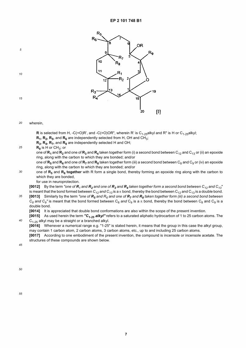
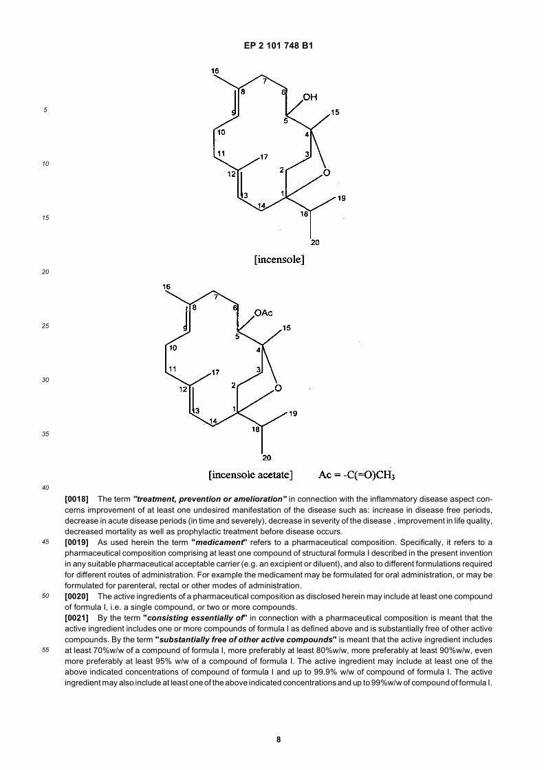
[0012] By the term "one of R1 and R2 and one of R3 and R4 taken together form a second bond between C12 and C13"is meant that the bond formed between C12 and C13 is a π bond, thereby the bond between C12 and C13 is a double bond.[0013] Similarly by the term "one of R5 and R6 and one of R7 and R8 taken together form (iii) a second bond betweenC8 and C9" is meant that the bond formed between C8 and C9 is a π bond, thereby the bond between C8 and C9 is adouble bond.[0014] It is appreciated that double bond conformations are also within the scope of the present invention.[0015] As used herein the term "C1-25 alkyl" refers to a saturated aliphatic hydrocarbon of 1 to 25 carbon atoms. TheC1-25 alkyl may be a straight or a branched alkyl.[0016] Whenever a numerical range e.g. "1-25" is stated herein, it means that the group in this case the alkyl group,may contain 1 carbon atom, 2 carbon atoms, 3 carbon atoms, etc., up to and including 25 carbon atoms.[0017] According to one embodiment of the present invention, the compound is incensole or incensole acetate. Thestructures of these compounds are shown below.
EP 2 101 748 B1
8
5
10
15
20
25
30
35
40
45
50
55
[0018] The term "treatment, prevention or amelioration" in connection with the inflammatory disease aspect con-cerns improvement of at least one undesired manifestation of the disease such as: increase in disease free periods,decrease in acute disease periods (in time and severely), decrease in severity of the disease , improvement in life quality,decreased mortality as well as prophylactic treatment before disease occurs.[0019] As used herein the term "medicament" refers to a pharmaceutical composition. Specifically, it refers to apharmaceutical composition comprising at least one compound of structural formula I described in the present inventionin any suitable pharmaceutical acceptable carrier (e.g. an excipient or diluent), and also to different formulations requiredfor different routes of administration. For example the medicament may be formulated for oral administration, or may beformulated for parenteral, rectal or other modes of administration.[0020] The active ingredients of a pharmaceutical composition as disclosed herein may include at least one compoundof formula I, i.e. a single compound, or two or more compounds.[0021] By the term "consisting essentially of" in connection with a pharmaceutical composition is meant that theactive ingredient includes one or more compounds of formula I as defined above and is substantially free of other activecompounds. By the term "substantially free of other active compounds" is meant that the active ingredient includesat least 70%w/w of a compound of formula I, more preferably at least 80%w/w, more preferably at least 90%w/w, evenmore preferably at least 95% w/w of a compound of formula I. The active ingredient may include at least one of theabove indicated concentrations of compound of formula I and up to 99.9% w/w of compound of formula I. The activeingredient may also include at least one of the above indicated concentrations and up to 99%w/w of compound of formula I.
EP 2 101 748 B1
9
5
10
15
20
25
30
35
40
45
50
55
[0022] By yet a further aspect of the invention there is provided a compound having the structural formula I as here-inabove defined for use in neuroprotection.[0023] In one embodiment said neuroprotection is for treatment, prevention or amelioration of a disease or conditionresulting from injury, trauma, or CNS neurodegenerative diseases.[0024] The term "treatment, prevention or amelioration" in connection with neuroprotection as used herein, meanstreating, preventing, or reversing cognitive decline associated with concentration loss, memory-acquisition loss, andinformation-storage or retrieval loss including, but not limited to, neuronal disorders, such as cognitive decline associatedwith aging, cognitive impairment and neurodegenerative disorders, such as Alzheimer’s disease, Parkinson’s disease,ALS, Huntington Chorea, HIV associated dementia, Lewy body dementia, multiple sclerosis, and prion disease. Theterm also includes treating, preventing, or reversing neuronal dysfunction associated with loss of motor skills (ataxia),such as Parkinson’s disease and amyotrophic lateral sclerosis as well as neuronal dysfunction resulting from CNS injury,such as head trauma, stroke, spinal-cord injury, and peripheral-nerve injury.[0025] As used herein the term "neurodegenerative disease" refers broadly to disorders or diseases that affect thenervous system and are characterized by gradual neuronal loss and/or gradual loss of neuronal function, including butare not limited to age-associated memory impairment, Parkinson’s disease, Alzheimer’s disease, Huntington’s choreadisease, multiple sclerosis and amyotrophic lateral sclerosis (ALS), HIV associated dementia, Lewy body dementia, andprion disease.[0026] In another one of its aspects the present invention provides a compound having the structural formula I for usein the treatment, prevention or amelioration of a disease or condition selected from depression, anxiety, obsessivecompulsive behaviors, deterioration in cognitive function, deterioration in neurobehavioral function, and combination ofany of the above.[0027] The term "deterioration of cognitive and/or neurobehavioral function" refers to decrease in learning andmemory capacitates, to decrease in orientation in time and space and decrease in coordination, and movement capacitiesdue to CNS function. The deterioration may be a natural result of aging but may also be as a result of injury, trauma(caused by accidents, stroke, surgery or diseases) or of disease in the CNS notably neurodegenerative diseases.[0028] The terms "injury" and "trauma" includes physical injury to the CNS (or head) as a result of physical insult,injury or damage due to stroke, ischemia, hypoxia, surgery or a disease such as an infectious disease in the CNS (suchas AIDS -associated dementia) as well as a neurodegenerative disease, for example Alzheimers, Parkinson, HungtintonChorea or old age dementia.[0029] The term "treatment, prevention or amelioration of depression, anxiety or obsessive compulsive be-havior" refers to decrease or elimination of the severity of the condition, decrease in the duration of the episode as wellas preventive treatment in individuals prone for such conditions to avoid or minimize the entry to these undesired episodes.The term "treatment" in connection with depression concerns improvement of at least one undesired manifestation ofthe disease such as anorexia and bulimia as well as the manifestation of clinical depression.[0030] It is demonstrated in the present invention that incensole acetate (IA), a Boswellia resin constituent, is a potentTRPV3 agonist that causes anxiolytic-like and antidepressive-like behavioral effects in wild type (WT) mice with con-comitant changes in c-Fos activation in the brain. These behavioral effects were not noted in TRPV3-/- mice, suggestingthat they are mediated via TRPV3 channels. IA robustly activated TRPV3 channels stably expressed in HEK293 cellsand in keratinocytes from TRPV3+/+ mice. It had no effect on keratinocytes from TRPV3-/- mice and showed modest orno effects on TRPV1, TRPV2 and TRPV4. The results shown below (see Example 15) imply that TRPV3 channels inthe brain play a role in emotional regulation.[0031] In a further aspect of the invention, there is provided a TRPV3 agonist having the structural formula I for usein treating a disease or condition selected from mood-disorders, anxiety, and a combination thereof.[0032] As used herein the term "mood disorders" refers to an emotional and/or behavioral disturbance characterizedby persistent and pervasive bouts of euphoria and/or depression. Exemplary mood disorders include depression andbipolar disorders (also known as manic depressive illness). Anxiety is frequently associated with mood disorders, suchas depression.[0033] By a specific embodiment the mood-disorder is depression.[0034] Said TRPV3 agonist is a compound having the structural formula I as defined hereinabove. In yet a furtherembodiment said compound is incensole or incensole acetate.[0035] The invention relates to a compound having structural formula I for use in the treatment, prevention, or amel-ioration of a disease or condition selected mood disorders, anxiety, and a combination thereof.
Compounds of the Invention:
[0036] In one embodiment R’ and/or R" of structural formula I are each independently C1-20alkyl; in a further embod-iment C1-15alkyl; in yet a further embodiment C1-10alkyl; in a further embodiment C1-6alkyl; in an additional embodimentC1-5alkyl.
EP 2 101 748 B1
10
5
10
15
20
25
30
35
40
45
50
55
[0037] In another embodiment, the bond between carbons 8,9 and\or 12,13 is a single bond. In a further embodimentcarbons 8,9 and\or 12,13 form an epoxide ring, along with the carbon to which they are bonded. In yet a further embod-iment, the substituents on carbons 8,9 and\or 12,13 are substituted as to form a diol. In another embodiment one ormore of R1, R2, R5, R6, and R9 is H.[0038] In one embodiment of the present invention, the compound is incensole or incensole acetate.[0039] Compounds used by the methods and uses of the invention may be synthesized by the synthetic routes describedand detailed in G. STRAPPAGHETTI, G. PROIETTI, S. CORSANO, AND I. GRGURINA. Synthesis of incensole. BIOOR-GANIC CHEMISTRY 11, 1-3 (1982) and T. Kato, C.C. Yen, T. Kobayashi, Y. Kitahara. Cyclization of polyenes XXI.Synthesis of DL-incensole. Chemistry letters 1191-1192 (1976). The derivatives of structural formula I may be synthesizedby procedures as described in FessendenR.J. & Fessenden J.S.; Organic chemistry, 1990, Brooks/Cole Publishingcompany, California (pp. 257-301 (alcohols), 301-323 (ethers and epoxides), 529-591 (aldehydes and ketones), 591-627(Derivatives of carboxylic acids), 391-448 (double bonds)). Synthesis procedures can be also found in additional generaltextbooks, for example, Morrison R.T. & Boyd R.N.; Organic chemistry, 1992, Pramount communication company,California.
Pharmaceutical compositions, dosages, and routes of administration
[0040] As used herein a "pharmaceutical composition" refers to a preparation of one or more compounds describedherein, with other inert chemical components such as suitable pharmaceutically acceptable carriers. The purpose of apharmaceutical composition is to facilitate administration of a compound to a mammal.As used herein the term "pharmaceutically acceptable carrier" refers to an inert nontoxic carrier or diluent that doesnot cause significant irritation to a subject (mammal) and does not abrogate the biological activity and properties of theadministered compound.[0041] Examples without limitation of carriers are lactose, sucrose, water, organic solvents, and polyethyleneglycol.[0042] The carriers may include additional excipients such as binders, disintegrants, lubricants, surface active agents,preservatives and favoring agents.According to one embodiment of the present invention the route of administration of the composition is selected fromoral, parenteral, inhalation, topical, transdermal, nasal, trnsmucosal (e.g. intranasal), intestinal, and rectal.[0043] Additionally according to a preferred embodiment of the present invention the parenteral route of administrationis selected from intravenous, intramuscular, intraperitoneal and subcutaneous administration.[0044] Additional suitable routes may be for example intramedullary, intrathecal, direct intraventicular, and intraocularinjections.[0045] A specific embodiment is the oral route of administration.[0046] The pharmaceutical composition may be formulated as to provide immediate release or sustained release ofthe active ingredient from the dosage form after administration to a patient by employing procedures well known in the art.[0047] The final form of the composition includes but not limited to a liquid, a syrup, an elixir, an emulsion, a suspension,drops, a spray, a cream, an ointment, a lotion, a gel, a paste, a powder, a granule, a tablet, a caplet, a pill, a capsule,a suppository, a transdermal patch or an injection.[0048] The pharmaceutically acceptable carrier selected for preparing the pharmaceutical compositions of the presentinvention depends on the final form of the composition.[0049] Typically, such carriers include additional excipients such as binders, disintegrants, adsorbents, lubricants,wetting agents, buffering agents and surface active agents.[0050] The pharmaceutical compositions are preferably present in a unit dosage form. Unit dosage form as usedherein refers to physically discrete units suited as unitary dosages for the mammalian subject to be treated, such as atablet, a capsule, or powders in vials or ampoules, each unit containing a predetermined quantity of the active ingredientcalculated to produce the desired therapeutic effect.[0051] Preferably the pharmaceutical composition in a unit dosage form comprises a therapeutically effective amountof the active ingredient in an amount from 0.1 mg to 1000 mg, more preferably 1 to 500 mg.[0052] Oral dosage forms suitable for oral administration may be presented as discrete pharmaceutical unit dosageforms, such as capsules, cachets, soft elastic gelatin capsules, tablets, caplets, or aerosols sprays, each containing apredetermined amount of the active ingredients, as a powder or granules, or as a solution or a suspension in an aqueousliquid, a non-aqueous liquid, an oil-in-water emulsion, or a water-in-oil liquid emulsion. Dosage forms such as oil-in-water emulsions typically comprise surfactants such as an anionic surfactant, for example anionic phosphate ester orlauryl sulfates, but other types of surfactants such as cationic or nonionic surfactants may be used in the compositionsof the present invention. See generally, Remington’s Pharmaceutical Sciences, Mack Publishing, Easton Pa., latestedition.[0053] For the purpose of preparing a tablet dosage form, various pharmaceutical carriers which are well-known inthis field can be widely used. As to the examples of carriers, excipients such as lactose, sodium chloride, glucose, starch,
EP 2 101 748 B1
11
5
10
15
20
25
30
35
40
45
50
55
calcium carbonate, kaolin, cellulose, aluminum silicate and the like may be used; the binders may be for example water,ethanol, propanol, glucose solution, starch solution, gelatin solution, carboxymethyl cellulose, shellac, methyl cellulose,polyvinylpyrrolidone and the like; the disintegrants may be for example starch, sodium alginate, sodium laurylsulfate,sodium starch glycolate and the like; the wetting agents may be for example glycerin, surfactants and the like; theadsorbents may be for example starch, lactose, kaolin, bentonite, colloidal silicic acid and the like; lubricants such astalc, strearates, polyethylene glycols and the like can be used. The tablets preparations can be further shaped into tabletscoated with usual tablet coating, for example sugar coated tablets, gelatin film coated tablets, tablets coated with entericcoating, tablets coated with film coating, or double layer tablets and multiple layer tablets.[0054] For the purpose of preparing a capsule dosage form, the compounds of formula [I] as the active ingredientsare mixed with the above-mentioned various carriers and the mixture or granules prepared from the mixtures are placedinto rigid gelatin capsules or soft capsules.[0055] For the purpose of preparing a suppository dosage form, various carriers which are well-known in this field canbe widely used. As to the examples of carries, polyethylene glycols, cacao butter, higher alcohols, esters of higheralcohols, gelatin, semi-synthesized glycerides and the like can be mentioned.[0056] For the purpose of preparing an injection dosage form, liquid preparations, emulsion preparations and suspen-sion preparations are sterilized, further these preparations are preferably isotonic to the blood, and all the diluents whichare conventionally used in this field can also be used for example, water, ethyl alcohol, macrogols, propylene glycol,ethyoxylated isostearyl alcohol, polyoxylated isostearyl alcohol and polyoxyethylenesorbitan fatty acid esters.[0057] Additionally, for the purpose of preparing an isotonic injection solutions, an adequate amount of sodium chloride,glucose or glycerin may be added to the injection preparations, further, usual dissolving additives, buffering agents,preservatives and the like may be added.[0058] An example of a pharmaceutical carrier for preparing an injection emulsion preparation is triglyceride emulsion.An example of an acceptable triglyceride emulsion useful in the intravenous and intraperitoneal administration of thecompounds of the present invention is the triglyceride emulsion commercially distributed under the tradename Intralip-id.RTM.[0059] Moreover, if necessary, coloring agents, preservatives, spices, flavors, sweetening agents and others may beadded to the pharmaceutical preparations of the present invention.[0060] Topical preparations such as creams, ointments, pastes, gels, lotions, transdermal patches, inhalants, sprays,aerosols and the like are formulated by using carriers and exipients which are well known in the field.[0061] Methods of preparing the compositions include the step of bringing into association a compound of the presentinvention with the pharmaceutical carrier. In general, the compositions are prepared by uniformly and intimately bringinginto association a compound of the present invention with liquid, semi-solid or solid carriers, and then, if necessary,shaping the product.[0062] The pharmaceutical compositions may be prepared by methods of pharmacy well known to those skilled in theart, e.g. by means of conventional mixing, dissolving, pulverizing, granulating, compressing, emulsifying, levigating, orlyophilizing processes. Techniques for formulation and administration of drugs may be found in "Remington’s Pharma-ceutical Sciences," Mack Publishing Co., Easton, PA, latest edition.[0063] Pharmaceutical compositions for use in accordance with the present invention may thus be formulated inconventional manner using one or more pharmaceutically acceptable carriers comprising excipients and auxiliaries,which facilitate processing of the active compounds into preparations which, can be used pharmaceutically. The properformulation is dependent upon the route of administration chosen.[0064] The amount of the active ingredient that may be combined with the pharmaceutical carrier to produce a singledosage form will vary depending upon the mammal treated and the particular mode of administration. For example, acomposition intended for oral administration to humans may vary from about 5% to about 95% w/w of the total composition.[0065] Dosage unit forms will generally contain between 0.1 to 1000 mg of the active ingredient, more preferably 1 to500 mg.[0066] The therapeutically or prophylactically effective amount of an active ingredient administered orally may rangefrom 0.1 to 1000 mg daily, more preferably from 1 to 500 mg daily, either singly or in multiple dosage over 24-hour period.For oral administration, the therapeutically effective amount of the active ingredient may be several times greater thanthat for parenteral administration.[0067] The above dosages refer to humans.[0068] The desired dose is suitably administered once daily, or several sub-doses, e.g. 2 to 4 sub-doses, are admin-istered at appropriate intervals through the day, or other appropriate schedule.[0069] In the practice of the invention the amount of the compound incorporated in the pharmaceutical compositionmay vary widely. Factors considered when determining the precise amount are well known to those skilled in the art.Examples of such factors include, but are not limited to, age, sex and weight of the subject being treated, intendedmedical use of the compounds, severity of the disease, the dosage form, route of administration being employed andthe frequency with which the composition is to be administered.
EP 2 101 748 B1
12
5
10
15
20
25
30
35
40
45
50
55
[0070] The exact dose may be determined, in accordance with the standard practice in the medical arts of "dosetitrating" the recipient; that is, initially administering a low dose of the compound, and gradually increasing the dose untilthe desired therapeutic effect is observed.[0071] The ratio between toxicity and therapeutic effect for a particular compound is its therapeutic index and can beexpressed as the ratio between LD50 (the amount of compound lethal in 50% of the population) and ED50 (the amountof compound effective in 50% of the population). Therapeutic index data obtained from animal studies can be used informulating a range of dosages for use in humans. The dosage of such compounds preferably lies within a range ofplasma concentrations that include the ED50 with little or no toxicity. The dosage may vary within this range dependingupon the dosage form employed and the route of administration utilized. The exact formulation, route of administrationand dosage can be chosen by the individual physician in view of the patient’s condition.
EXAMPLES
Materials and Methods
[0072] Extraction and Isolation of IA. Boswellia carterii resin (20 gr., Pamir, Tel Aviv, Israel) was extracted with PE(PE) (3 times with 150 ml). Petroleum ether (PE) extract was washed with NaOH 5% solution (3 times with 200 ml). Theresulting aqueous acid-containing fraction was then acidified with HCl 1M, washed with saturated NaCl and re-extractedwith PE. It was then dried over MgSO4. The non acid containing PE fraction was acidified with HCl (1M) and then washedwith a saturated NaCl solution and dried over MgSO4. After evaporation the residue was chromatographed on a silicacolumn. Fractions were assayed for their activity on IκB degradation as described below. A fraction eluted with 3%diethyl-ether in PE, which contained IA, showed activity. Pure IA was obtained by chromatography on a semi preparativeHPLC column (Spectra-physics applied bio systems 783 absorbance detector with a vydac C18 semi-preparative HPLCcolumn - Valco). Acetonitrile (ACN) and water were used as mobile phase for HPLC and the gradient consisted of 90-99%ACN for 30 min. A Waters HPLC instrument: pump 600, PDA 996 detector 600 with an analytical C18 Symmetry column(4.6/250 mm) were used to analyze the purification process. Several NMR methods (H-NMR, C-NMR, DEPT, COSY,HSQC, HMBC, TOCSY and NOESY) as well as a GC-MS analysis were used for the structure elucidation of the isolatedactive compounds.[0073] NMR spectra were recorded both in CDC13 and in C6D6 solutions using a Bruker avance spectrometer 400MHz and repeated using a Varian Unity Spectrometer Varian Unity Inova spectrometer 500 MHz.[0074] GC-MS Analysis was performed using a Hewlett-Packard G1800A GCD system with a HP5971 gas chomato-graph with an electron ionization detector. An SPB-5 (30 m x 0.25 mm x 0.25mm film thickness) column was used. Thefollowing method was used for analysis: The column was held at 70°C for 4 mins, after which, a temperature gradientwas applied from 70°C to 280°C, at a rate of 50 degree/min. (Inlet temperature: 280°C; Detector temperature: 280°C;Splitless injection; gas - Helium, 1 mL/min).[0075] Cell Cultures. HeLa cells were grown in Dulbecco’s modified Eagle medium supplemented with 10% foetalcalf serum and 1% (v/v) penicillin/streptomycin (all from Biological Industries, Kibbutz Beit Haemek, Israel), in a humidifiedincubator at 37° C.[0076] RAW 264.7 macrophage cell line derived from BALB/c mice was obtained from American Type Culture collection(Rockville, MD, USA). The cells were cultured in Dulbecco’s modified Eagle medium (DMEM) supplemented with 10%fetal calf serum (Hyclone, Logan, UT), 1% (v/v) penicillin/streptomycin (Beit Haemek, Israel), nonessential amino acid(Sigma, St. Louis, USA), glutamine 1% (Beit Haemek, Israel) and pyruvate 1% (Beit Haemek, Israel). Cells were grownin a humidified incubator at 37°C.[0077] Peritoneal macrophages were harvested from C57BL/6 female mice four days after intraperitoneal injection of1.5 ml of a 3% thioglycollate medium (Difco, NJ, USA). The cells were re-suspended in Dulbecco’s modified Eaglemedium (DMEM) supplemented with 5% foetal calf serum (FCS), and plated (1.2x10⁵ cells per well) in 96-microwellplates flat-bottom (Nunc, Roskide, Denmark).[0078] IκBα Degradation. HeLa cells were pre-incubated with IA (50 mg/ml, dissolved in ethanol) for 2 hrs, and thenstimulated for 20 minutes with TNFα (20 ng/ml, Emeryville, CA, USA). After removing the slides from plates for immon-ostaining (see below), proteins were extracted from remaining cells in the plates. Proteins were extracted from cells inNP-40 lysis buffer. Total protein concentration was determined using the Bradford method. Lysates were then analyzedeither by Western blotting (WB).[0079] Western Blot (WB). Following separation by SDS-PAGE, proteins were blotted to a polyvinylidene difluoride(PVDF) membrane (Millipore). The membrane was blocked in 5% (w/v) milk powder and then incubated in TBST con-taining the primary antibody and 2% (w/v) milk powder. All phospho-specific antibodies were purchased from Cellsignaling Inc. αIκBα, αp65 and αCOX2 antibodies from Santa Cruz Inc. (California, USA). After binding of an appropriatesecondary antibody coupled to horseradish peroxidase, proteins were visualized by enhanced chemiluminiscence ac-cording to the instructions of the manufacturer (Amersham Lifescience).
EP 2 101 748 B1
13
5
10
15
20
25
30
35
40
45
50
55
[0080] p65 Subunit Immunostaining. HeLa Cells were preincubated with IA and then stimulated with TNFα asdescribed in the IκBα degradation assay above. Cells were then fixed with formaldehyde 1%, permeabilized with 0.25%Triton X-100, stained with rabbit anti-p65 (Santa Cruz, California, USA) and visualized with anti-rabbit Rhodamine Red-labeled secondary antibody (Jackson ImmunoResearch, Baltimore, USA). Cells were also stained with DAPI (blue) fornuclei location (data not shown). The cells were examined under an Axioscope Zeiss microscope with a plan-Neofluor* 60 lens.[0081] COX-2 Production. RAW 264.7 cells were treated with subtoxic concentrations (confirmed by MTT colorimetricassay) of incensole acetate (10-20 mg/ml, dissolved in ethanol and further diluted in medium) and incubated with lipopol-ysaccharide (LPS, E. coli 1 mg/ml for 24 hs, Sigma, Israel) for 16-24 hrs. Cells treated with vehicle served as control group.[0082] Proteins were extracted from cells in NP-40 lysis buffer (50 mM Tris/HCl pH 7.5, 150 mM NaCl, 0.1% SDS,1% NP-40, 10 mM EDTA, 1 mM phenylmethylsulfonylfluoride (PMSF), and 10 mM DTT). Total protein concentrationwas determined using the Bradford method and the lysates were analyzed by Western blotting.[0083] Nitric Oxide (NO) Levels. Following 2-3 h of incubation of murine peritoneal macrophages at 37°C, the non-adherent cells were removed by intensive rinsing. About 95% of the adherent cells were macrophages. IA was firstdissolved in absolute ethanol, and the solutions were further diluted with Dulbecco’s Modified Eagle’s Medium (DMEMmedium). Various nontoxic concentrations were added to the macrophages, followed by addition of 1 mg/ml of LPS foractivation. The macrophages were then cultivated in a humid atmosphere with 5% CO2 for 24 hrs. The supernatantfluids were harvested and kept at -20°C until assayed. NO generation was determined by measuring the nitrite accu-mulated in the supernatants (100 ml) of the IA-treated macrophages as follows. The cells were then treated with IA invarious doses. An equal volume (100 ml) of Griess reagent (1% sulphanilamide, 0.1% naphthalene diamine HCl, 2%H3PO4) was added to each supernatant. Following 10 min of incubation at room temperature, the color production wasmeasured at 550 nm with an ELISA reader. The concentration of the nitrite was calculated according to a standard curve.[0084] ROS (Reactive Oxygen Species) Production by RAW 264.7 Macrophages. Raw 264.7 cells were scrapped,washed and resuspended in Hanks’ balanced salt solution (without phenol red). For measurement of chemiluminescence,0.5 ml of cell suspension (5 x 10⁵ cells) was added to each luminometer tube, together with various doses of IA tested(dissolved in ethanol and further diluted with Hanks). The cells were incubated for 24 hrs. 10 ml of luminol (Sigma, St.Louis, USA) and 30 ml of zymosan (Sigma, St. Louis, USA) were added to the tubes, and the chemiluminescence wasmeasured immediately in a luminometer (Biolumate LB 95, Berhold, Wilbad, Germany).[0085] Inflamed Paw Model. Sabra female mice were used to assess the response to IA or vehicle in an in vivo modelof inflammation. Drug or vehicle was administered 30 min before induction of the inflammatory stimulus. Mice (5 pergroup) were injected i.p. with vehicle (isopropanol:Emulphor:saline = 1:1:18) or with vehicle containing IA (50 mg/kg,i.p). Emulphor (a polyethoxylated vegetable oil) is a commercial emulsifier. Hind paws were injected with 50 ml of saline(left or right alternatively) or λ-carrageenin (4%, right or left alternatively), using 26G needles. Ensuing inflammatoryswelling was measured by increase in foot volume in a plethysmometer (Ugo-Basile, Italy). Paw volume as well asredness (as a measure of erythema) and licking (as a measure of pain) were assayed before carrageenin applicationand every 60 min until 4 hrs.[0086] Statistical Analysis. Student’s t test was used to assess the differences between the control and IA-treatedgroups. For a dose response effect, analysis of the data was performed using a one way ANOVA followed by Bonferronipost-hoc comparisons. The paw model results were analyzed by ANOVA followed by Bonferroni post-hoc comparisonsat every time point.[0087] For analysis of c-Fos immunoreactivity, positive nuclei were identified based on their round form and opticaldensity at least twice that of background. The numbers of c-Fos immunoreactive nuclei from the right and left hemisphereswere averaged to obtain a representative number for the given region from each mouse. Student t tests were performedcomparing the control (vehicle) with the IA group.[0088] Responses to IA in WT versus TRPV3-/-mice were assessed using two-way analysis-of-variance (ANOVA) withBonferroni post-hoc comparisons (Graphpad Prism 4 software).
Animals and Procedures
[0089] Female Sabra mice (Harlan, Israel, 2.5-3.5 months old) were used for the paw inflammatory model.. FemaleSabra mice (Harlan, Israel, 15-20 weeks old) and wild type C57BL/6 or TRPV3 KO female mice (18-20 weeks old) wereused for behavioral assessments. Ten mice were housed in each cage. For the chronic studies, mice were housed ingroups of eight. Temperature in the animal room was maintained between 20-22°C, the light cycle was 12 h lights on(8-20h); 12 h lights off (20-8.00h). Female mice were used for all behavioural assessments, in order to prevent confoundingdue to potential wound infliction induced by inter-male fighting (See also below "Animals and Procedures" Section relatingto Example 15.[0090] Mice were consecutively tested in the elevated plus maze and the forced swimming test. The animal care andthe protocols met the guidelines of the U.S. National Institutes of Health, detailed in the Guide for the Care and Use of
EP 2 101 748 B1
14
5
10
15
20
25
30
35
40
45
50
55
Laboratory Animals, and were applied in conformity with the Institutional Ethics Committee.
Drugs and Injections for Behavioral Assays
[0091] IA, IN and the extract were dissolved in a mixture of isopropanol: cremophor:saline=1:1:18. Injection volumewas 10ml/g body weight. Injections were performed by the intraperitoneal (i.p.) route.
Behavioral Assays
Elevated Plus Maze
[0092] Mice were placed in the central platform (10 X 10 cm) between the open (10 X 45 cm) and enclosed (10 X 45X 40 cm) arms of a plus maze. The number of entries and the time spent in each of the arms was recorded. As describedby others (Crawley, 2000; Treit and Menard, 1998), an ’anti-anxiety’ effect was calculated both as the ratio of entriesonto the open arms to total arm entries, and as the % time on the open arms proportional to the time in the closed arms.Mice (female Sabra strain, aged 3.5-4.5 months old) were injected intraperitoneally with 10, 30 or 50 mg/kg of incensoleacetate or with vehicle. Each dose was administered to 5 mice. Fifty min after injection the mice were tested in the plus-maze for ’anti-depressant’ effects. Desipramine (5 mg/kg) was injected to a separate group of mice as a positive control.One-way Anova indicated significant effects (F = 8.9, df=4,27, P<0.01). All doses had ’anti-depressant’ effects, but onlythose of 0 and 30 mg/kg were significant. Data are presented as means 6 SEM. DMI=desipramine*) P<0.05, **) P<0.01, ***) P<0.001 compared to vehicle
Posrsolt’s Forced swimming test (FST)
[0093] Mice were placed in a 2 liter glass beaker (11 cm diameter) filled with water (2461°C) up to 30 cm from thebottom (so that the mouse could not touch the bottom and 8 cm from the rim (so that the mouse cannot escape). Immobilitytime (when the animal does not move except for small movements required to float) was recorded by 3 experimentersafter 2, 6 and 9 min.
For Example 15, the following experiments were conducted:
[0094] Drug. IA was isolated as described above under Materials and Methods. It was then dissolved in ethanol forin vitro assays or in isopropanol for in vivo assays. A stock solution of 20 mg/ml for in-vitro assays and 50 mg/ml for invivo assay was prepared.
Cell culture
[0095] Human HEK 293 cells stably expressing TRPV1 were a kind gift from Merck Research Laboratories (WhitehouseStation, NJ). Cells were cultured in minimal essential medium, Eagle, modified with non-essential amino acids, 1mMsodium pyruvate, 2 mM L-glutamine and 1.5 g/L sodium bicarbonate (ATCC, Mabassas, VA), containing 1% Penicillin-streptomycin, and 10% foetal bovine serum. Cells were passaged three times a week using Trypsin- EDTA 1 x (Invitrogen,Carlsbad, CA) and grown under 5% CO2 at 37°C.[0096] TRPV3-YFP [O’dell, D.K., Rimmerman, N., Pickens, S.R. & Walker J.M. Fatty acyl amides of endogenoustetrahydroisoquinolines are active at the recombinant human TRPV1 receptor. Bioorg. Med. Chem. 15, 6164-6169(2007)], TRPV4 and mock-transfected cell lines were cultured in DMEM 1x with L-glutamine (Mediatech, Inc. Herndon,VA), containing 1% penicillin-streptomycin (Invitrogen, Carlsbad, CA) and 10% foetal bovine serum.[0097] HEK293 cells were transiently transfected with a rat TRPV2 plasmid using lipofectamine reagent (Invitrogen,Carlsbad, California) according to manufacturer’s protocol. They were then maintained in Dulbecco’s modified EagleMedium / 10% fetal calf serum ,supplemented with Penicillin, Streptomycin, and L-glutamine. Primary keratinocytes fromTRPV3-deficient and TRPV3+/+ mouse pups (day 1-4) were harvested and cultured as described previously [Chung,M.K., Lee, H., Mizuno, A., Suzuki, M. & Caterina, M.J. TRPV3 and TRPV4 mediate warmth-evoked currents in primarymouse keratinocytes. J. Biol. Chem. 279, 21569-21575 (2004)].[0098] Calcium imaging of HEK 293 cells. TRPV1, TRPV3, and TRPV4 expressing HEK293 cells were plated 24-48h before imaging in 96 well plates, loaded with 3 mM Fura-2 AM and imaged as previously described [O’dell, D.K.,Rimmerman, N., Pickens, S.R. & Walker J.M. Fatty acyl amides of endogenous tetrahydroisoquinolines are active atthe recombinant human TRPV1 receptor. Bioorg. Med. Chem. 15, 6164-6169 (2007)].[0099] For single cell calcium imaging, HEK293-rat TRPV2 and HEK293-mouse TRPV3-YFP expressing cells wereplated on collagen-coated glass cover slips. Cells were loaded for 60 min with 3 mM Fura-2 AM.
EP 2 101 748 B1
15
5
10
15
20
25
30
35
40
45
50
55
[0100] Calcium imaging of TRPV3 +/+ and TRPV3 keratinocytes. Primary keratinocytes from TRPV3-deficient andWT mouse pups (day 1-4) were harvested and cultured as described [Chung, M.K., Lee, H., Mizuno, A., Suzuki, M. &Caterina, M.J. TRPV3 and TRPV4 mediate warmth-evoked currents in primary mouse keratinocytes. J. Biol Chem. 279,21569-21575 (2004)].[0101] Cells were plated on glass coverslips (105/cm2) and incubated for 48-60 h, then loaded with fura-2 AM (20 mM,0.04% pleuronic acid, 32°C for 1h) in imaging buffer containing (in mM): 130 NaCl, 2.5 CaCl2, 0.6 MgCl2, 10 HEPES,1.2 NaHCO3, 10 glucose, pH 7.45. Ratiometric Ca2+ imaging was performed as previously described [O’dell, D.K.,Rimmerman, N., Pickens, S.R. & Walker J.M. Fatty acyl amides of endogenous tetrahydroisoquinolines are active atthe recombinant human TRPV1 receptor. Bioorg. Med. Chem. 15, 6164-6169 (2007)]. Drug was added to the bathfollowing a period of baseline recording. Calcium measurements were made from 30 randomly selected cells per coverslip.[0102] Electrophysiological recording. Currents were recorded using whole-cell voltage-clamp. Pipettes were pulledfrom microcapillary glass (A-M Systems). A coverslip containing cells was transferred to a 300 mL chamber that wasconstantly perfused (1-2 mL/min) with external solution. Voltage protocols were generated and data were digitized andrecorded using Pulse (HEKA Elektronik) software in conjunction with an Axopatch 200A amplifier (Axon Instruments),and the data analyzed using an in-house Visual Basic (Microsoft) analysis program.[0103] The pipette solution contained (in mM): 121.5 Kgluconate, 10 HEPES, 17 KCl, 9 NaCl, 1 MgCl2, 0.2 EGTA, 2MgATP, and 0.5 NaATP, pH 7.2. The external solution contained (in mM): 120 NaCl, 5 KCl, 1 MgCl2, 2 CaCl2, 10 Glucoseand 20 HEPES, pH 7.4 with NaOH. The measured charge (pC) was defined as the charge elicited between-85 and -45mV by a ramping voltage stimulus (-85 mV to +35 mV, 0.54 mV/msec; holding potential -55 mV). Currents were sampledat 5 kHz. Experimental and control cells were alternated whenever possible. Control values were obtained from adjacentcells with no detectable YFP fluorescence, presumed to be non-TRPV3-expressing.[0104] Data analysis of calcium imaging data. Analysis of calcium imaging data was done using a non-linear re-gression curve fit (Graphpad 4 Prism, San Diego, CA). t tests and one way ANOVA were calculated using SPSS (Chicago,IL). In the keratinocyte experiments, drug-induced response for each cell was taken as the maximal post-drug meas-urements over time minus the average of the last 5 pre-drug measurements. Averaged drug responses over 30 randomlyselected cells per coverslip were analyzed with two-tailed unpaired t tests.[0105] For analysis of c-Fos immunoreactivity, positive nuclei were identified based on their round form and opticaldensity at least twice that of background. The numbers of c-Fos immunoreactive nuclei from the right and left hemisphereswere averaged to obtain a representative number for the given region from each mouse. Student t tests were performedcomparing the control (vehicle) with the IA group.[0106] Responses to IA in WT versus TRPV3-/- mice were assessed using two-way analysis-of-variance (ANOVA)with Bonferroni post-hoc comparisons (Graphpad Prism 4 software).[0107] Animals and procedures. Female Sabra mice (Harlan, Israel, 15-20 weeks old) and wild type C57BL/6 orTRPV3 KO female mice (18-20 weeks old) [Chung, M.K., Lee, H., Mizuno, A., Suzuki, M. & Caterina, M.J. TRPV3 andTRPV4 mediate warmth-evoked currents in primary mouse keratinocytes. J. Biol. Chem. 279, 21569-21575 (2004)] wereused for behavioral assessments. 10 mice were housed in each cage. The animal care and protocols met the guidelinesof the U.S. National Institutes of Health, detailed in the Guide for the Care and Use of Laboratory Animals, and wereapplied in conformity with the Institutional Ethics Committees. For the c-Fos immunostaining, female Sabra mice (seeabove) were used. Temperature in the animal room was maintained between 20-22°C, the light cycle was 12 h lightson (8:00-20:00h); 12 h lights off (20:00-8:00h). Mice were injected with intraperitoneal (i.p.) incensole acetate in a mixtureof isopropanol:cremophor:saline (1:1:18) at a volume of 10 ml/g body weight
Example 1: IA and IN Inhibit IκBα Degradation.
[0108] IA and IN were assayed at different concentrations for their activity on IκBα degradation in TNFα-stimulatedHeLa cells. Both compounds inhibited IκBα degradation in a dose dependent manner (Fig. 1A, 1B).
Example 2: IA Inhibits IκBα by Impairment of IKK Activity.
[0109] In order to demonstrate that IA inhibits the NF-κB pathway upstream from the IKKs experimentally, the effectsof IA on TNFα-induced phosphorylation of the IKKs were tested. These experiments showed inhibition of IKKα/IKKβphosphorylation by IA (Fig. 2A). Following IκBα degradation, NF-κB is free to accumulate in the nucleus. Immunostainingof the p65 sub-unit of NF-κB showed that IA inhibited the nuclear accumulation of NF-κB following TNFα stimulation inHeLa cells (Fig. 2B).[0110] Example 3: IA Blocks NF-κB-mediated Inflammatory Response in vitro and in vivo. To investigate whetherthe NF-κB inhibitory effect of IA confers an anti-inflammatory activity, it was determined, as detailed herein above inMaterials and Methods, the levels of COX-2, nitric oxide production and ROS with and without IA in different cell lines.The in vivo anti-inflammatory activity of IA was examined in inflamed paw model in mice. COX-2 production in LPS-
EP 2 101 748 B1
16
5
10
15
20
25
30
35
40
45
50
55
stimulated RAW 264.7 cells was inhibited by IA at a dose of 60 mM (P<0.001) (Fig. 3A). NO production by murineperitoneal macrophages was determined by measuring the nitrite accumulated in the supernatants in an ELISA reader.IA inhibited NO generation in a dose dependent manner (ANOVA P<0.0001), reaching about 45% of NO production at80 mM (p=0.0022) (Fig. 3B). ROS are known to be important in various biological and pathological processes and areinvolved in inflammation. We therefore tested the effects of IA on ROS generation by Zymozan activated Raw 264.7cells at three concentrations. A significant dose-dependent inhibitory effect was found (ANOVA P<0.0001), reachingabout 45% inhibition at 60 mM (p=0.0021) (Fig. 3C).[0111] Having established that IA inhibits the expression of several key inflammatory mediators in vitro, the anti-inflammatory properties of IA in vivo were studies. It was thereupon found that IA significantly reduced inflammation inthe inflamed paw model in mice during a 4 hrs period. The decreased inflamed paw volume in the treated mice reflectsa decrease in edema, which is a component of the inflammatory response. There were highly significant effects oftreatment (F=11.7, df=3,64, P<0.001), time (F=10.6, do-4,64, P<0.0001) and interaction (F=3.9, df=12,64, P<0.001)(Fig. 4). IA also significantly reduced other inflammatory parameters, such as redness and pain (data not shown).
Example 5: Effect of IA on Post-CHI functional Outcome
[0112] To examine the effect of IA on functional recovery after CHI, the parameters of injured mice, treated with IAwere compared with those of injured mice treated with vehicle.[0113] At 1 h after CHI, the functional status of the mice was evaluated according to a set of 10 neurobehavioral tasks(neurological severity score, NSS) that tests reflexes, alertness coordination, and motor abilities. One point was awardedfor absence of reflex or failure to perform a particular task. Hence, a score of 10 reflects maximal neurological impairment.Mice were equally divided to vehicle \ IA groups according to their NSS scores. Only mice with NSS > 4 at 1 h afterinjury were included in the study. Immediately after NSS1h assessment, mice were randomly assigned to intraperitoneal(i.p.) injection with vehicle (isopropanol: Emulphor - a commercial emulsifier: saline = 1:1:18) or with vehicle containingIA (50 mg/kg, n=9-10 mice/group). Recovery (ΔNSSt) was defined as the difference between NSS1h and NSS measuredat any later time point and was determined at several time points up to 21 days following CHI.[0114] NSS at 1h were similar in both groups, (7.03 6 0.19 and 7.03 6 0.19, respectively) indicating no difference inthe initial severity of injury. Markedly greater recovery of motor ability was observed in the IA group 24 h after injury ascompared with vehicle (ΔNSS = 1.00 6 0.12 vs 0.41 6 0.09 , respectively, P=0.002) as depicted in Fig. 5A. ΔNSSvalues increased with time in both IA and vehicle mice as a result of spontaneous recovery, but continued to be significantlyhigher in IA mice at all subsequent time points, up to 3 weeks post injury.
Example 6: Effect of IA on memory function
[0115] Memory function was assessed by ORT (Object Recognition Test) and the results are depicted in Fig. 5B.Whereas naive, non-injured mice were not affected by IA (data not shown), it had a robust effect on the injured animals.Both groups spent equal time at the two objects (~50% of total exploration time) at the baseline measurements, at alltimes post CHI. However, at the test performed 4 h later, when a novel object replaced one of the familiar ones, IA-treated mice spent most of their exploration time at the new object, in contrast to the vehicle-treated animals, that didnot memorize the "old" object. At 3 days post injury IA treated mice spent significantly longer times exploring the newobject (P=0.01), similar to the time spent by a naive animal. This effect of IA was sustained for 7 and 14 days. At 21days, it appears that the vehicle-treated mice regained their ability, and exploration time reached a similar level to thatof the IA-treated mice.
Example 7: Effect on tissue edema formation
[0116] A pronounced increase in tissue water content was observed in the left (ipsilateral) hemisphere of all injuredmice at 24 h after injury, indicating the effect of injury in both groups. Although water accumulation tended to be smallerin IA mice (81.47 6 0.35 % in IA vs 82.16 6 0.30 % in vehicle) the difference did not reach statistical significance (P=0.15).
Example 8: Effect of IA on cytokines expression profile after CHI
[0117] Since it was shown hereinabove that the pro-inflammatory cytokines TNF-α and IL-1β are upregulated within1-4 h post-CHI, and that their inhibition is associated with better recovery. The mRNA levels of these cytokines werequantified at 3 hours after CHI using real-time PCR. Their amounts are expressed relative to β-actin, and it is apparentfrom Fig. 6 that IA significantly inhibited mRNA expression of both TNF-α and IL-1β (P < 0.05, n=5/group).
EP 2 101 748 B1
17
5
10
15
20
25
30
35
40
45
50
55
Example 9: Effect of IA on body temperature
[0118] Thirty minutes after treatment with IA (namely, 90 min post CHI), a mild (~1°C) and short-term (~30-60 min)duration of hypothermia was noted in IA- as compared to vehicle- treated mice (data is not shown).
Example 10: The anxiolytic effect of IA:
[0119] When placed in an elevated plus-maze for the first time, a mouse’s behavior is largely based on its anxietylevel. Normal mice that have not received any anti-anxiety drugs will become moderately anxious in this new environment.Thus, they tend to prefer the closed arms over the less secure open arms. Meanwhile, mice treated with anti-anxietydrugs (e.g., diazepam, commonly known as valium) tend to be less anxious, so they spend more time in the 5 openarms compared to normal mice and they are generally less active. Forty five min after injection the mice were tested inthe plus-maze for ’anti-anxiety’ effects of IA (Fig. 7). Diazepam (5 mg/kg) was injected to a separate group of mice asa positive control. One-way Anova indicated significant effects (F = 4.2, df=4,32, P<0.01). Data are presented as means6 SEM.*, P<0.05; **, P<0.01 compared to vehicle.[0120] Example 11: The c-Fos transcription factor is a product of an immediate early gene and its increase servesas a marker of enhanced neuronal activity. It is thus used in histological sections to map out brain regions that areactivated or attenuated after treatment with psychoactive drugs. IA significantly increased c-Fos in the lateral septum,central nucleus of the amygdala and solitary nucleus, while significantly reducing c-Fos in the motor cortex, medialstriatum and hippocampal CA3 region (Fig. 9A-C). The central nucleus of the amygdala and the lateral septum playmajor roles in the expression of emotions; it is assumed that c-Fos expression in the central nucleus of the amygdalais due to circuits that are engaged by both anxiolytic and anxiogenic drugs.[0121] The data from the behavioral assays together with the c-Fos immunostaining establish the anxiolytic and anti-depressive effects of IA.[0122] Example 12: IA (75 mg/kg) exerted a potent anxiolytic-like effect in WT mice, while TRPV3-/- mice spent identicaltime on the open arms, regardless of whether they were injected IA or only vehicle (Fig. 10a; Fstrain = 6.3, df 1, 14, p <0.05; Finteraction = 5.0, df 1,14, p < 0.05). In the Porsolt forced swim test, IA significantly reduced the immobility time inWT, but not in TRPV3-/- mice (FIA = 5.5, df 1,16, p < 0.04; Finteraction = 5.9, df 1,16, p < 0.03) (Fig. 10b). No significantdifferences were recorded between vehicle-treated WT mice and vehicle-treated TRPV3-/- mice in the forced swim andelevated plus maze assays.[0123] These results indicate that the effects of IA in preclinical models for antidepressants and anxiolytics are mediatedvia TRPV3 channels.
Example 13: The anti-depressant effect of IA:
[0124] We used the Porsolt forced swimming test to examine the anti-depressant effect of IA. The method is basedon the observation that a mouse, when forced to swim in a situation from which there is no escape, will, after an initialperiod of vigorous activity, eventually cease to move altogether making only those movements necessary to keep itshead above water. This characteristic and readily identifiable behavioral immobility indicates a state of despair in whichthe rat has learned that escape is impossible and resigns itself to the experimental conditions. Fifty min after injectionthe mice were tested in the Porsolt forced swimming test for ’anti-depressant’ effects (Fig. 8). Desipramine (5 mg/kg)was injected to a separate group of mice as a positive control. One-way Anova indicated significant effects (F = 8.9,df=4,27, P<0.01). Data are presented as means 6 SEM. DMI=desipramine*, P<0.05; * *, P<0.01; ***, P<0.001 compared to vehicle
Example 15: IA effects on behavioral parameters
[0125] To study the functional effects of IA on the CNS, IA was assayed in a panel of standard behavioral assays inmice (female Sabra strain, 15-20 weeks old), namely: the elevated plus maze [Crawley, J.N. What’s Wrong with myMouse? Behavioral Phenotyping of Transgenic and Knockout Mice (Wiley-Liss, New York, 2000)], the Porsolt forced-swimming test [Petit-Demouliere, B., Chenu, F. & Bourin, M. Forced swimming test in mice: a review of antidepressantactivity. Psychopharmacology 177, 245-255 (2005)], locomotion in the open field test and cataleptic response in a ringtest [Fride, E. & Mechoulam, R. Pharmacological activity of the cannabinoid receptor agonist, anandamide, a brainconstituent. Eur. J. Pharmacol. 231, 313-314 (1993). The elevated plus maze assay is based on the preference of micefor the closed arms of a maze, apparently due to fear of open spaces. At 50 mg/kg IA exerted a potent anxiolytic-likeeffect, causing mice to spend significantly more time in the aversive open arms of the maze. In the Porsolt forced-swimtest, a standard assay for the evaluation of anti-depressant effects, IA significantly reduced the immobility recorded over
EP 2 101 748 B1
18
5
10
15
20
25
30
35
40
45
50
55
9 minutes, thus indicating a reversal of an avolition response. A significant ring. Dose dependency was noted in allassays (10-100 mg/kg, data not shown), and the findings were replicated in 7 independent experiments.[0126] IA (100 mM) significantly increased calcium influx (EC50= 16 mM; Hill slope = 2.2; Fig. 12a,b,d) in HEK293 cellsstably expressing mouce TRPV3-YFP. When calcium was removed from the extracellular medium, the calcium increasein response to IA was significantly reduced (Fig. 12b), providing further evidence for the influx of calcium through TRPV3channels. The effect of IA on TRPV3 resembles the effect of the broad-spectrum agonist 2-aminoethyl diphenylborinate(2-APB), which served as a positive control (Fig. 12a,d). IA (500 mM) also induced a calcium influx in primary keratinocytesfrom WT mice, but not from TRPV3-/- mice [Moqrich A., Hwang S.W., Earley T.J., Petrus M.J., Murray A.N., SpencerK.S., Andahazy M., Story G.M. & Patapoutian A. Impaired thermosensation in mice lacking TRPV3, a heat and camphorsensor in the skin. Science. 307, 1468-72 (2005)] (Fig. 12c). The effect of IA (500 mM) resembles the one of camphor(10 mM), a known agonist of TRPV3. IA, at a concentration (100 mM) that was maximally effective in TRPV3 expressingcells did not induce calcium influx in HEK293 cells transiently transfected with rat-TRPV2 (Fig. 12f), and caused onlyminimal calcium influx in HEK293 cells expressing rat TRPV and human TRPV4 (Fig. 12e,g).[0127] IA also activated a cation current in moue TRPV3-YFP expressing HEK293 cells (Fig. 13a-c) with propertiesconsistent with TRPV3 activation [Smith G.D. et al. TRPV3 is a temperature-sensitive vanilloid receptor-like protein.Nature 418, 186-190 (2002)] and similar to the current activated by 2-APB, which served as a positive control (Fig. 13d).This current was not activated in HEK293 cells not expressing TRPV3 and was also absent from TRPV 1 and TRPV4expressing cells (Fig. 13e).[0128] The effect of IA on different brain regions were studied by looking at the effect of IA on c-Fos immunoreactivityin mice brains 60 min after administration of IA (50 mg/kg; i.p.). The c-Fos transcription factor is a product of an immediateearly gene and its increase serves as a marker of enhanced neuronal activity. It is thus used in histological sections tomap out brain regions that are activated or attenuated after treatment with psychoactive drugs [Werme, M., Ringholm,A., Olson, L. & Brene S. Differential patterns of induction of NGFI-B, Norl and c-fos mRNAs in striatal subregions byhaloperidol and clozapine. Brain Res. 863, 112-119 (2000); and Dragunow, M., Robertson, G.S., Faull, R.L., Robertson,H.A. & Jansen, K. D2 dopamine receptor antagonists induce fos and related proteins in rat striatal neurons. (1990)Neuroscience 37, 287-294]. IA significantly increased c-Fos in the lateral septum, central nucleus of the amygdala andsolitary nucleus, while significantly reducing c-Fos in the motor cortex, medial striatum and hippocampal CA3 region(Fig. 9; Table 1). The central nucleus of the amygdala and the lateral septum play major roles in the expression ofemotions [Thompson, B.L. & Rosen, J.B. Immediate-early gene expression in the central nucleus of the amygdala isnot specific for anxiolytic or anxiogenic drugs. Neuropharmacology 50, 57-68 (2006); and Henry, B., Vale, W. & Markou,A. The effect of lateral septum corticotropin-releasing factor receptor 2 activation on anxiety is modulated by stress. J.Neurosci. 26, 9142-9152 (2006)]; it is assumed that c-Fos expression in the central nucleus of the amygdala is due tocircuits that are engaged by both anxiolytic and anxiogenic drugs [Thompson, B.L. & Rosen, J.B. Immediate-early geneexpression in the central nucleus of the amygdala is not specific for anxiolytic or anxiogenic drugs. Neuropharmacology50, 57-68 (2006).][0129] The data from the behavioral assays together with the c-Fos immunostaining establish the anxiolytic and anti-depressive effects of IA. Given the robust effect of IA on TRPV3 channels and the observation that IA does not interactwith a long list of receptors known to be involved in psychoactivity, the possibility that its behavioral effects are mediatedthrough CNS TRPV3 channels was investigated. Thus, the panel of behavioral assays with WT and TRPV3-/- mice,which were administered either IA or vehicle was repeated.[0130] IA (75 mg/kg) exerted a potent anxiolytic-like effect in WT mice, while TRPV3-/-mice spent identical time on theopen arms, regardless of whether they were injected IA or only vehicle (Fig. 10a; Fstrain= 6.3, df 1, 14, p < 0.05; Finteraction= 5.0, df 1,14, p < 0.05). In the Porsolt forced swim test, IA significantly reduced the immobility time in WT, but not inTRPV3-/- mice (FIA = 5.5, df 1,16, p < 0.04; Finteraction = 5.9, df 1,16, p < 0.03) (Fig. 10b). No significant differences wererecorded between vehicle-treated WT mice and vehicle-treated TRPV3-/- mice in the forced swim and elevated plusmaze assays.[0131] These results indicate that the effects of IA in preclinical models for antidepressants and anxiolytics are mediatedvia TRPV3 channels.[0132] Collectively, the data presented here, along with the expression of TRPV3 mRNA in the brain, indicate thatTRPV3 channels affects emotional and behavioral processes in the CNS, in addition to its known effects on thermosen-sation.
References:
[0133] The Complete Parallel Bible, Proverbs, 27, 9, (Oxford University Press, 1989).
Ammon, H.P., Safayhi H., Mack T. and Sabieraj J. (1993) J Ethnopharmacol 38,113-9.
EP 2 101 748 B1
19
5
10
15
20
25
30
35
40
45
50
55
Barnes P.J., Karin M. (1997) N. Engl J Med.336, 1066-71.
Beni SM, Kohen R, Reiter RJ, Tan DX, Shohami E (2004) Melatonin-induced neuroprotection after closed headinjury is associated with increased brain antioxidants and attenuated late-phase activation of NF-kappaB and AP-1. FASEB J 18, 149-51.
Chung, M.K., Lee, H., Mizuno, A., Suzuki, M. & Caterina, M.J. TRPV3 and TRPV4 mediate warmth-evoked currentsin primary mouse keratinocytes. J. Biol. Chem. 279, 21569-21575 (2004).
Crawley, J.N. What’s Wrong with my Mouse? Behavioral Phenotyping, of Transgenic and Knockout Mice (Wiley-Liss, New York, 2000).
Corsano, S. & Nicoletti, R. The structure of incensole. Tetrahedron 23, 1977-1984 (1967).
Dragunow, M., Robertson, G.S., Faull, R.L., Robertson, H.A. & Jansen, K. D2 dopamine receptor antagonists inducefos and related proteins in rat striatal neurons. (1990) Neuroscience 37, 287-294.
Foo S.Y., Nolan G.P. Trends Genet. (1999) 15 229-35.
Fride, E. & Mechoulam, R. Pharmacological activity of the cannabinoid receptor agonist, anandamide, a brainconstituent. Eur. J. Pharmacol. 231, 313-314 (1993).
Gacs-Baitz E., Radics L. Fardella G. And Corsano S. (1978) Journal of Chemical Research (S), 1701-1709.
Gerhardt, H, Seifert, F, Buvari, P, Vogelsang, H, Repges, R. (2001) Zeitschrift-fur-Gastroenterologie 39, 7-11.
Ghosh S. and Karin M. (2002) Cell 109, S81-S96.
Gupta, I., Gupta, V., Parihar, A., Gupta, S., Ludtke, R., Safayhi, H., Ammon, H.P. (1998) Eur J Med Res 3, 511-4.
Hansson G.K. (2001) Ann N Y Acad Sci. 947 157-65; discussion 165-6.
Haoxing Xu et al. Nature Neuroscience (2006) 9, 628-635.
Henry, B., Vale, W. & Markou, A. The effect of lateral septum corticotropin-releasing factor receptor 2 activation onanxiety is modulated by stress. J. Neurosci. 26, 9142-9152 (2006).
H. Z. Hu et al., J Biol Chem 279, 35741 (Aug 20, 2004).
Karin M. and Ben-Neriah Y. (2000) Annual Review of Immunology 18, 621-663.
T. Kato, C.C. Yen, T. Kobayashi, Y. Kitahara. Cyclization of polyenes XXI. Synthesis of DL-incensole. Chemistryletters 1191-1192 (1976),
H. Lee, M. J. Caterina, Pflugers Arch 451, 160 (Oct, 2005).
Marmarou A, Guy M, Murphey L, Roy F, Layani L, Combal JP, Marquer C; American Brain Injury Consortium (2005)A single dose, three-arm, placebo-controlled, phase I study of the bradykinin B2 receptor antagonist Anatibant(LF16-0687Ms) in patients with severe traumatic brain injury. J Neurotrauma 22, 1444-55.
Moqrich A., Hwang S.W., Earley T.J., Petrus M.J., Murray A.N., Spencer K.S., Andahazy M., Story G.M. & PatapoutianA. Impaired thermosensation in mice lacking TRPV3, a heat and camphor sensor in the skin. Science. 307, 1468-72(2005).
Narayan RK, Michel ME, Ansell B, et al (2002) Clinical trials in head injury. J Neurotrauma 19, 503-57.
Nonaka M, Chen XH, Pierce JE, Leoni MJ, McIntosh TK, Wolf JA, Smith DH (1999) Prolonged activation of NF-kappaB following traumatic brain injury in rats. J Neurotrauma 16, 1023-34.
EP 2 101 748 B1
20
5
10
15
20
25
30
35
40
45
50
55
O’dell, D.K., Rimmerman, N., Pickens, S.R. & Walker J.M. Fatty acyl amides of endogenous tetrahydroisoquinolinesare active at the recombinant human TRPV1 receptor. Bioorg. Med. Chem. 15, 6164-6169 (2007).
Paxinos G. & Franklin K.B.J. The Mouse Brain in Stereotaxic Coordinates (Academic Press, Oxford, 2001).
Petit-Demouliere, B., Chenu, F. & Bourin, M. Forced swimming test in mice: a review of antidepressant activity.Psychopharmacology 177, 245-255 (2005).
Singh G.B, Atal C.K. (1986) Agents and Actions 18, 407-412.
Smith G.D. et al., Nature 418, 186 (Jul 11, 2002).
Syrovets, T., Buchele, B., Krauss, C., Laumonnier, Y., and Simmet, T. (2005), J Immunol. 174, 498-506.
Strappaghetti, G., Proietti G., Proietti, S. Corsano, S. and I. Grgurina, I. Synthesis of incensole. Bioorganic Chemistry11, 1-3 (1982).
Thompson, B.L. & Rosen, J.B. Immediate-early gene expression in the central nucleus of the amygdala is not specificfor anxiolytic or anxiogenic drugs. Neuropharmacology 50, 57-68 (2006).
Treit, D. & Menard, J. in In vivo Neuromethods eds Boulton, A., Baker, G. & Bateson, A. (Humana Press, Totowa,1998), p 89.
Vogt-Eisele, AK et al., Monoterpenoid agonists of TRPV3, British Journal of Pharmacology (2007) 151, 530-540.Watt, J.M. (1962) The Medicinal and Poisonous Plants of Southern and Eastern Africa, 2nd Ed., Livingstone LTD.,Edinburgh and London.
Werme, M., Ringholm, A., Olson, L. & Brene S. Differential patterns of induction of NGFI-B, Nor1 and c-fos mRNAsin striatal subregions by haloperidol and clozapine. Brain Res. 863, 112-119 (2000).
US Patent No. 5064823
WO 02/053138
Claims
1. A compound having the structural formula I, including enantiomers, diastereomers, solvates, and pharmaceuticallyacceptable salts thereof:
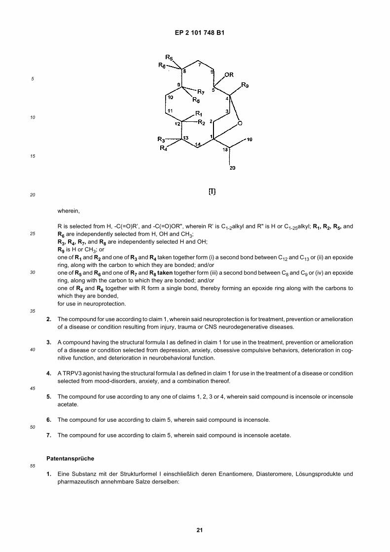
EP 2 101 748 B1
21
5
10
15
20
25
30
35
40
45
50
55
wherein,
R is selected from H, -C(=O)R’, and -C(=O)OR", wherein R’ is C1-2alkyl and R" is H or C1-25alkyl; R1, R2, R5, andR6 are independently selected from H, OH and CH3;R3, R4, R7, and R8 are independently selected H and OH;R9 is H or CH3; orone of R1 and R2 and one of R3 and R4 taken together form (i) a second bond between C12 and C13 or (ii) an epoxidering, along with the carbon to which they are bonded; and/orone of R5 and R6 and one of R7 and R8 taken together form (iii) a second bond between C8 and C9 or (iv) an epoxidering, along with the carbon to which they are bonded; and/orone of R5 and R6 together with R form a single bond, thereby forming an epoxide ring along with the carbons towhich they are bonded,for use in neuroprotection.
2. The compound for use according to claim 1, wherein said neuroprotection is for treatment, prevention or ameliorationof a disease or condition resulting from injury, trauma or CNS neurodegenerative diseases.
3. A compound having the structural formula I as defined in claim 1 for use in the treatment, prevention or ameliorationof a disease or condition selected from depression, anxiety, obsessive compulsive behaviors, deterioration in cog-nitive function, and deterioration in neurobehavioral function.
4. A TRPV3 agonist having the structural formula I as defined in claim 1 for use in the treatment of a disease or conditionselected from mood-disorders, anxiety, and a combination thereof.
5. The compound for use according to any one of claims 1, 2, 3 or 4, wherein said compound is incensole or incensoleacetate.
6. The compound for use according to claim 5, wherein said compound is incensole.
7. The compound for use according to claim 5, wherein said compound is incensole acetate.
Patentansprüche
1. Eine Substanz mit der Strukturformel I einschließlich deren Enantiomere, Diasteromere, Lösungsprodukte undpharmazeutisch annehmbare Salze derselben:
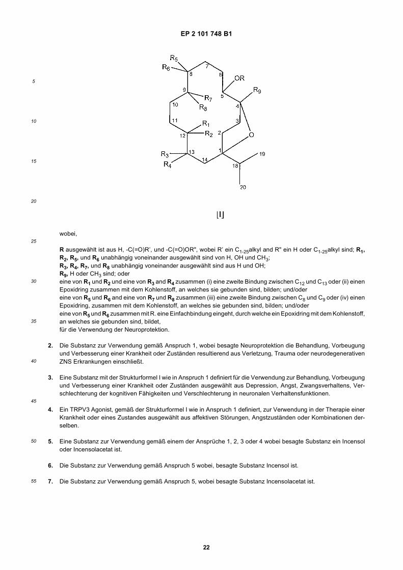
EP 2 101 748 B1
22
5
10
15
20
25
30
35
40
45
50
55
wobei,
R ausgewählt ist aus H, -C(=O)R’, und -C(=O)OR", wobei R’ ein C1-25alkyl and R" ein H oder C1-25alkyl sind; R1,R2, R5, und R6 unabhängig voneinander ausgewählt sind von H, OH und CH3;R3, R4, R7, und R8 unabhängig voneinander ausgewählt sind aus H und OH;R9, H oder CH3 sind; odereine von R1 und R2 und eine von R3 and R4 zusammen (i) eine zweite Bindung zwischen C12 und C13 oder (ii) einenEpoxidring zusammen mit dem Kohlenstoff, an welches sie gebunden sind, bilden; und/odereine von R5 und R6 and eine von R7 und R8 zusammen (iii) eine zweite Bindung zwischen C8 und C9 oder (iv) einenEpoxidring, zusammen mit dem Kohlenstoff, an welches sie gebunden sind, bilden; und/odereine von R5 und R6 zusammen mit R. eine Einfachbindung eingeht, durch welche ein Epoxidring mit dem Kohlenstoff,an welches sie gebunden sind, bildet,für die Verwendung der Neuroprotektion.
2. Die Substanz zur Verwendung gemäß Anspruch 1, wobei besagte Neuroprotektion die Behandlung, Vorbeugungund Verbesserung einer Krankheit oder Zuständen resultierend aus Verletzung, Trauma oder neurodegenerativenZNS Erkrankungen einschließt.
3. Eine Substanz mit der Strukturformel I wie in Anspruch 1 definiert für die Verwendung zur Behandlung, Vorbeugungund Verbesserung einer Krankheit oder Zuständen ausgewählt aus Depression, Angst, Zwangsverhaltens, Ver-schlechterung der kognitiven Fähigkeiten und Verschlechterung in neuronalen Verhaltensfunktionen.
4. Ein TRPV3 Agonist, gemäß der Strukturformel I wie in Anspruch 1 definiert, zur Verwendung in der Therapie einerKrankheit oder eines Zustandes ausgewählt aus affektiven Störungen, Angstzuständen oder Kombinationen der-selben.
5. Eine Substanz zur Verwendung gemäß einem der Ansprüche 1, 2, 3 oder 4 wobei besagte Substanz ein Incensoloder Incensolacetat ist.
6. Die Substanz zur Verwendung gemäß Anspruch 5 wobei, besagte Substanz Incensol ist.
7. Die Substanz zur Verwendung gemäß Anspruch 5, wobei besagte Substanz Incensolacetat ist.
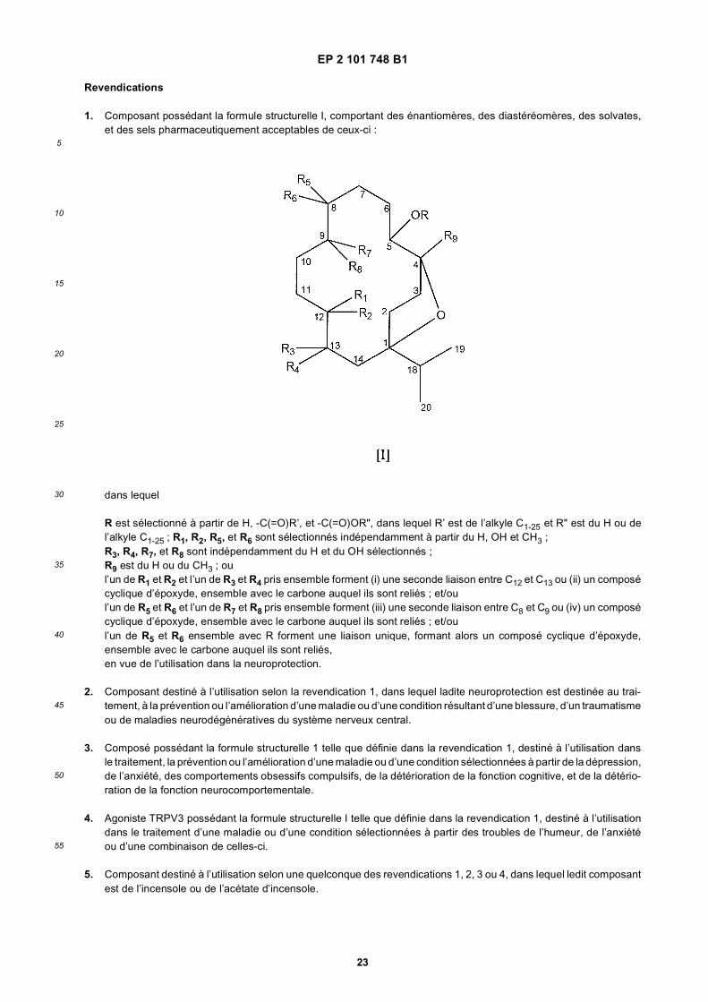
EP 2 101 748 B1
23
5
10
15
20
25
30
35
40
45
50
55
Revendications
1. Composant possédant la formule structurelle I, comportant des énantiomères, des diastéréomères, des solvates,et des sels pharmaceutiquement acceptables de ceux-ci :
dans lequel
R est sélectionné à partir de H, -C(=O)R’, et -C(=O)OR", dans lequel R’ est de l’alkyle C1-25 et R" est du H ou del’alkyle C1-25 ; R1, R2, R5, et R6 sont sélectionnés indépendamment à partir du H, OH et CH3 ;R3, R4, R7, et R8 sont indépendamment du H et du OH sélectionnés ;R9 est du H ou du CH3 ; oul’un de R1 et R2 et l’un de R3 et R4 pris ensemble forment (i) une seconde liaison entre C12 et C13 ou (ii) un composécyclique d’époxyde, ensemble avec le carbone auquel ils sont reliés ; et/oul’un de R5 et R6 et l’un de R7 et R8 pris ensemble forment (iii) une seconde liaison entre C8 et C9 ou (iv) un composécyclique d’époxyde, ensemble avec le carbone auquel ils sont reliés ; et/oul’un de R5 et R6 ensemble avec R forment une liaison unique, formant alors un composé cyclique d’époxyde,ensemble avec le carbone auquel ils sont reliés,en vue de l’utilisation dans la neuroprotection.
2. Composant destiné à l’utilisation selon la revendication 1, dans lequel ladite neuroprotection est destinée au trai-tement, à la prévention ou l’amélioration d’une maladie ou d’une condition résultant d’une blessure, d’un traumatismeou de maladies neurodégénératives du système nerveux central.
3. Composé possédant la formule structurelle 1 telle que définie dans la revendication 1, destiné à l’utilisation dansle traitement, la prévention ou l’amélioration d’une maladie ou d’une condition sélectionnées à partir de la dépression,de l’anxiété, des comportements obsessifs compulsifs, de la détérioration de la fonction cognitive, et de la détério-ration de la fonction neurocomportementale.
4. Agoniste TRPV3 possédant la formule structurelle I telle que définie dans la revendication 1, destiné à l’utilisationdans le traitement d’une maladie ou d’une condition sélectionnées à partir des troubles de l’humeur, de l’anxiétéou d’une combinaison de celles-ci.
5. Composant destiné à l’utilisation selon une quelconque des revendications 1, 2, 3 ou 4, dans lequel ledit composantest de l’incensole ou de l’acétate d’incensole.

EP 2 101 748 B1
24
5
10
15
20
25
30
35
40
45
50
55
6. Composant destiné à l’utilisation selon la revendication 5, dans laquelle ledit composant est de l’incensole.
7. Composant destiné à l’utilisation selon la revendication 5, dans laquelle ledit composant est de l’acétate d’incensole.
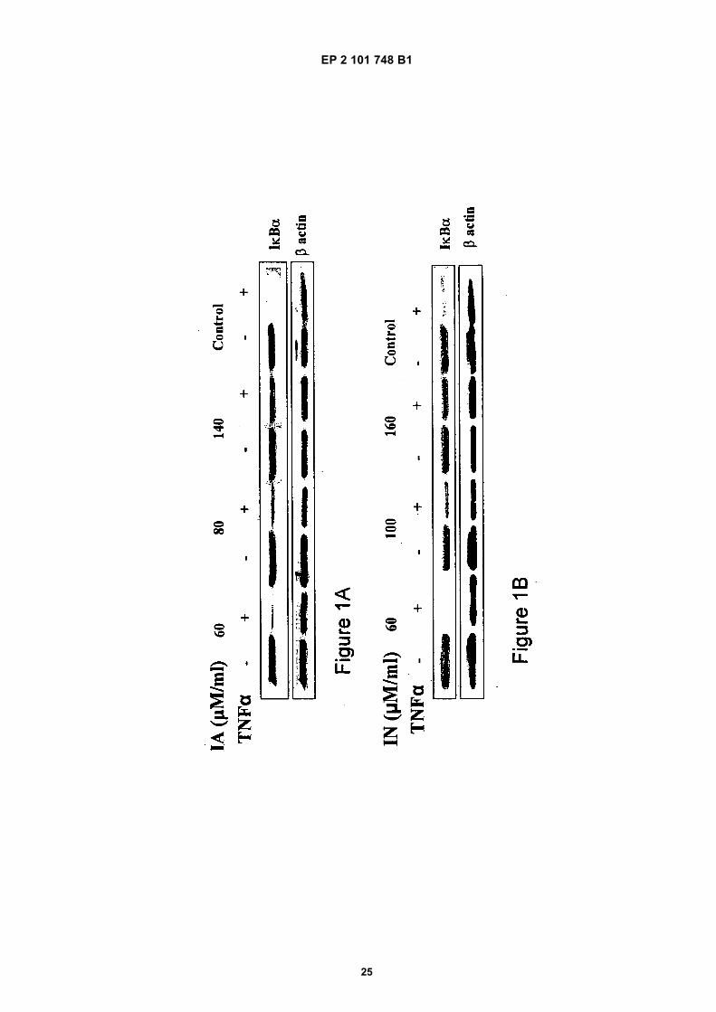
EP 2 101 748 B1
25

EP 2 101 748 B1
26
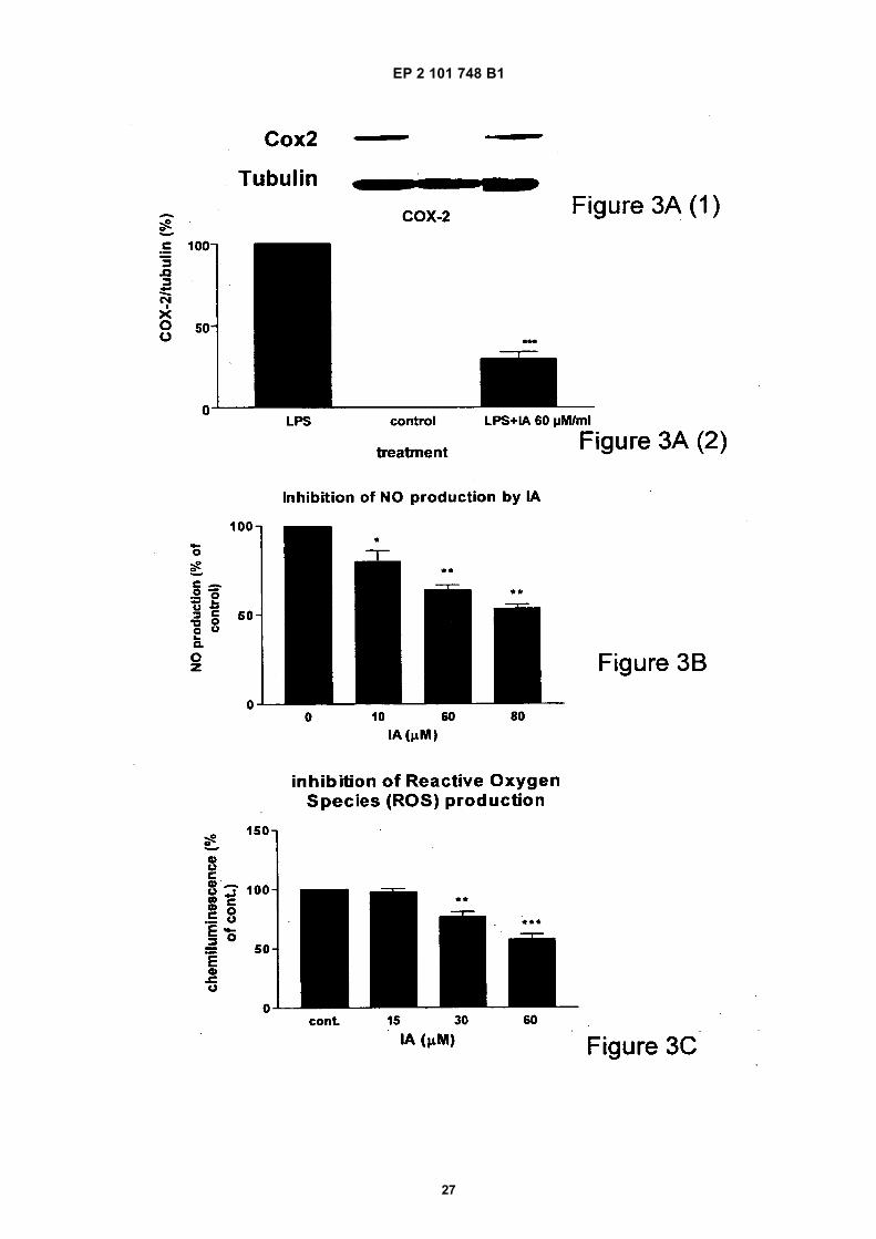
EP 2 101 748 B1
27
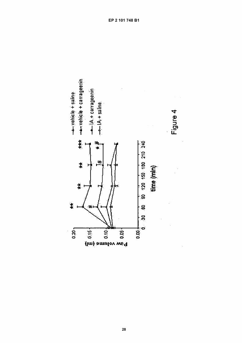
EP 2 101 748 B1
28
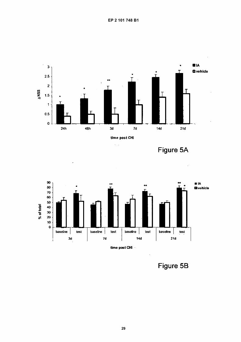
EP 2 101 748 B1
29
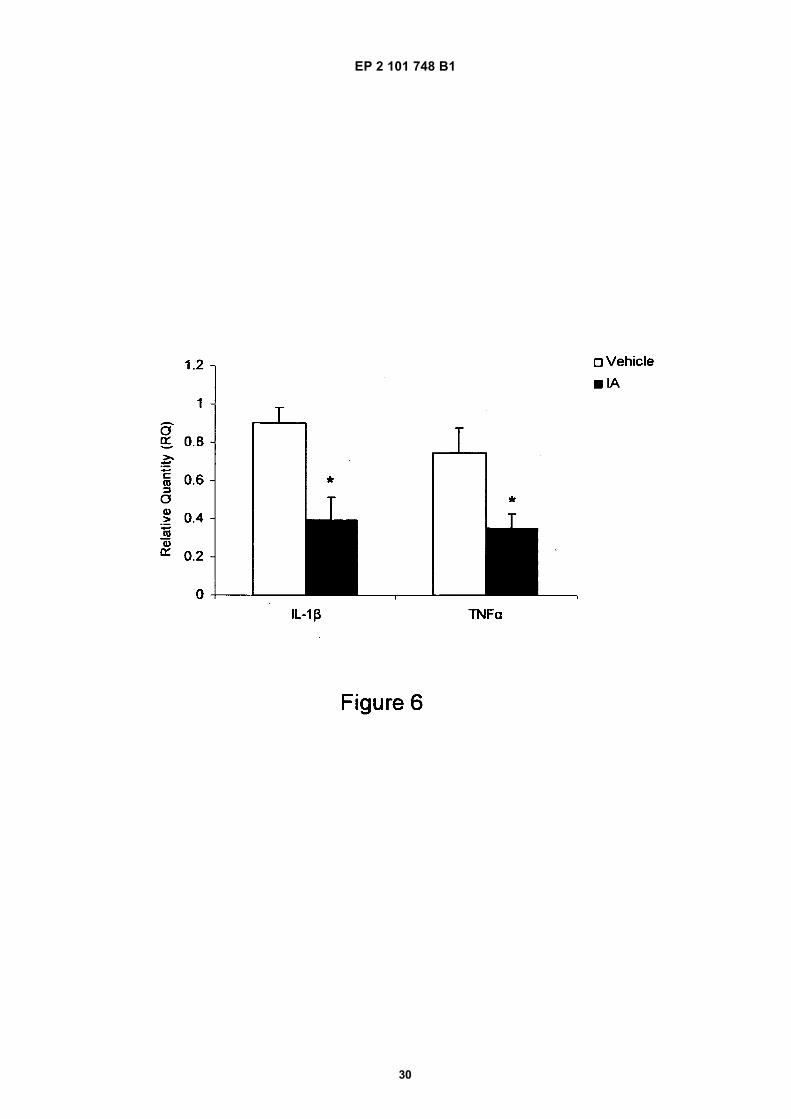
EP 2 101 748 B1
30
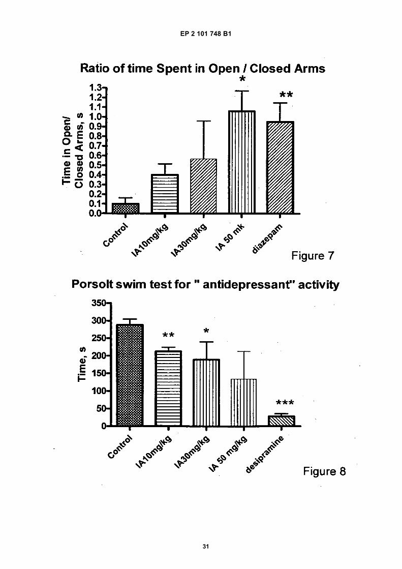
EP 2 101 748 B1
31
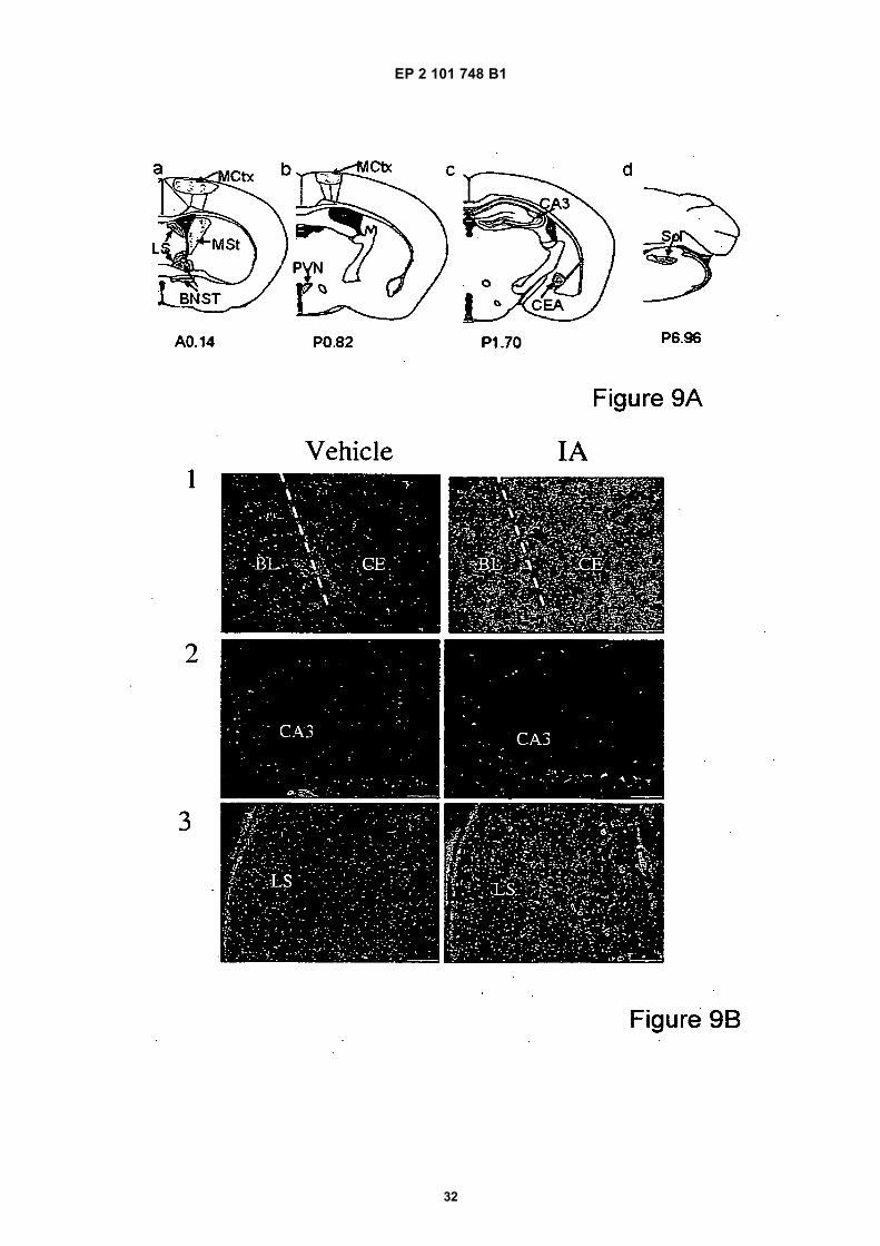
EP 2 101 748 B1
32
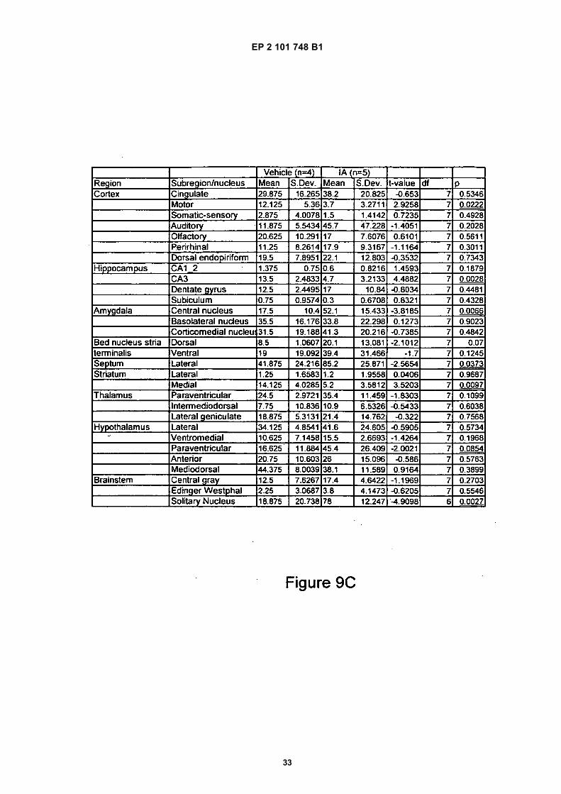
EP 2 101 748 B1
33
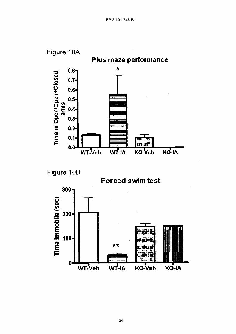
EP 2 101 748 B1
34
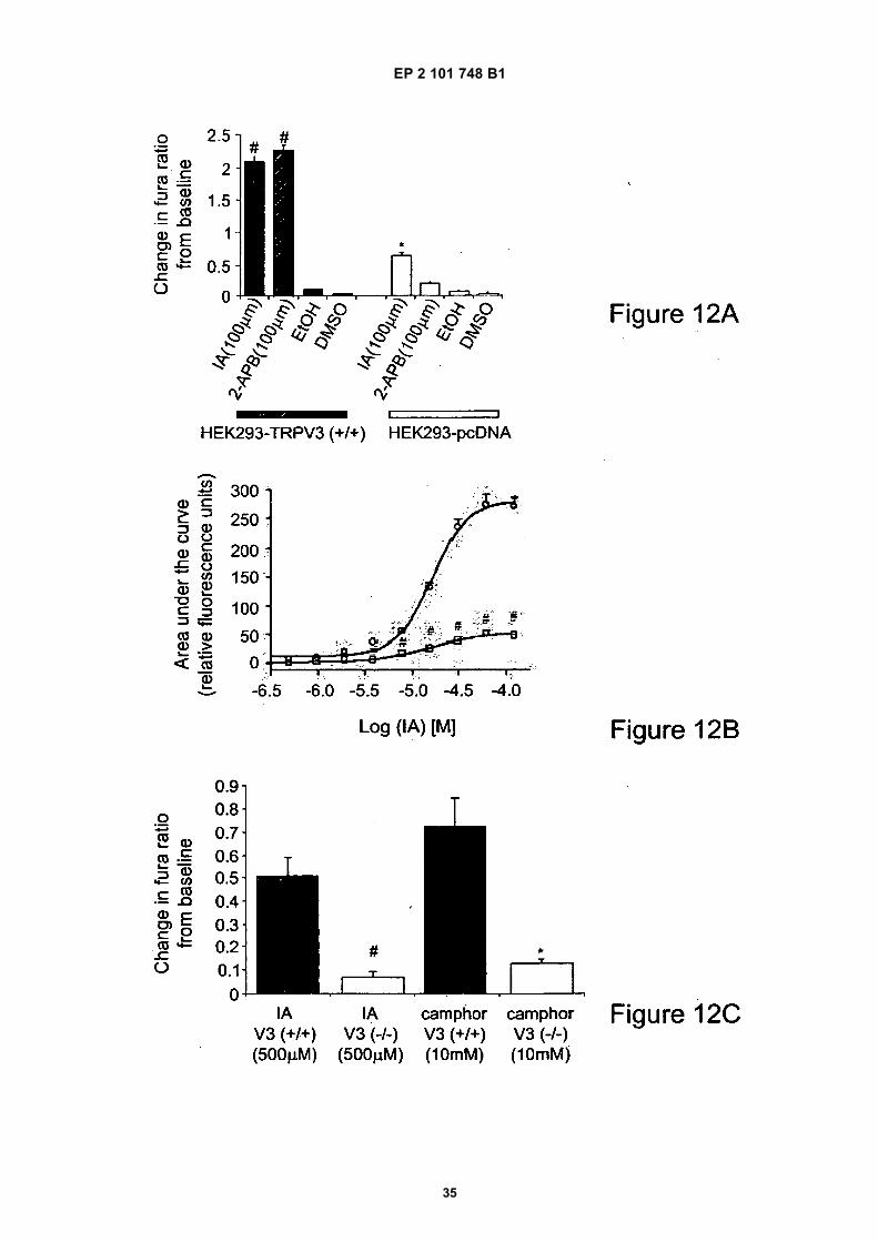
EP 2 101 748 B1
35
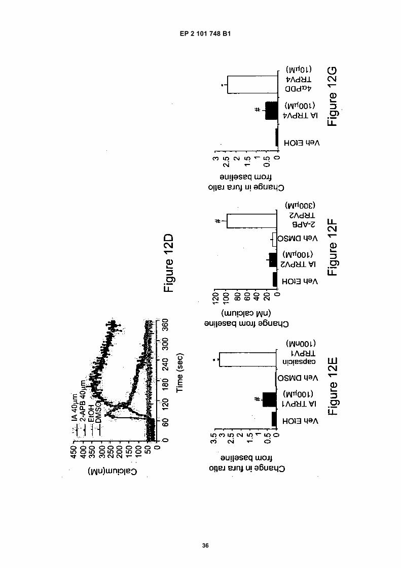
EP 2 101 748 B1
36
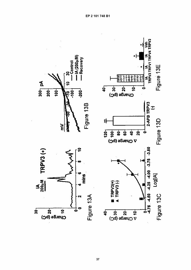
EP 2 101 748 B1
37
EP 2 101 748 B1
38
REFERENCES CITED IN THE DESCRIPTION
This list of references cited by the applicant is for the reader’s convenience only. It does not form part of the Europeanpatent document. Even though great care has been taken in compiling the references, errors or omissions cannot beexcluded and the EPO disclaims all liability in this regard.
Patent documents cited in the description
• US 5064823 A [0003] [0133] • WO 02053138 A [0004] [0133]
Non-patent literature cited in the description
• BANNO. Journal of Ethnopharmacology, 2006, vol.107, 249-253 [0003]
• G. STRAPPAGHETTI ; G. PROIETTI ; S.CORSANO ; I. GRGURINA. Synthesis of incensole.BIOORGANIC CHEMISTRY, 1982, vol. 11, 1-3[0039]
• T. KATO ; C.C. YEN ; T. KOBAYASHI ; Y. KITAHA-RA. Cyclization of polyenes XXI. Synthesis of DL-in-censole. Chemistry letters, 1976, 1191-1192 [0039][0133]
• FESSENDENR.J. ; FESSENDEN J.S. Organicchemistry. Brooks/Cole Publishing company, 1990[0039]
• Remington’s Pharmaceutical Sciences. Mack Pub-lishing [0052]
• Remington’s Pharmaceutical Sciences. Mack Pub-lishing Co, [0062]
• O’DELL, D.K. ; RIMMERMAN, N. ; PICKENS, S.R. ;WALKER J.M. Fatty acyl amides of endogenous tet-rahydroisoquinolines are active at the recombinanthuman TRPV1 receptor. Bioorg. Med. Chem., 2007,vol. 15, 6164-6169 [0096] [0098] [0101] [0133]
• CHUNG, M.K. ; LEE, H. ; MIZUNO, A. ; SUZUKI,M. ; CATERINA, M.J. TRPV3 and TRPV4 mediatewarmth-evoked currents in primary mouse keratino-cytes. J. Biol. Chem., 2004, vol. 279, 21569-21575[0097] [0133]
• CHUNG, M.K. ; LEE, H. ; MIZUNO, A. ; SUZUKI,M. ; CATERINA, M.J. TRPV3 and TRPV4 mediatewarmth-evoked currents in primary mouse keratino-cytes. J. Biol Chem., 2004, vol. 279, 21569-21575[0100]
• CHUNG, M.K. ; LEE, H. ; MIZUNO, A. ; SUZUKI,M. ; CATERINA, M.J. TRPV3 and TRPV4 mediatewarmth-evoked currents in primary mouse keratino-cytes. J. Biol. Chem., 2004, vol. 279, 21569-21575[0107]
• What’s Wrong with my Mouse?. CRAWLEY, J.N. Be-havioral Phenotyping of Transgenic and KnockoutMice. Wiley-Liss, 2000 [0125]
• PETIT-DEMOULIERE, B. ; CHENU, F. ; BOURIN,M. Forced swimming test in mice: a review of antide-pressant activity. Psychopharmacology, 2005, vol.177, 245-255 [0125] [0133]
• FRIDE, E. ; MECHOULAM, R. Pharmacological ac-tivity of the cannabinoid receptor agonist, anandam-ide, a brain constituent. Eur. J. Pharmacol., 1993, vol.231, 313-314 [0125] [0133]
• MOQRICH A. ; HWANG S.W. ; EARLEY T.J. ;PETRUS M.J. ; MURRAY A.N. ; SPENCER K.S. ;ANDAHAZY M. ; STORY G.M. ; PATAPOUTIAN A.Impaired thermosensation in mice lacking TRPV3, aheat and camphor sensor in the skin. Science, 2005,vol. 307, 1468-72 [0126] [0133]
• SMITH G.D. et al. TRPV3 is a temperature-sensitivevanilloid receptor-like protein. Nature, 2002, vol. 418,186-190 [0127]
• WERME, M. ; RINGHOLM, A. ; OLSON, L. ; BRENES. Differential patterns of induction of NGFI-B, Norland c-fos mRNAs in striatal subregions by haloperidoland clozapine. Brain Res., 2000, vol. 863, 112-119[0128]
• DRAGUNOW, M. ; ROBERTSON, G.S. ; FAULL,R.L. ; ROBERTSON, H.A ; JANSEN, K. D2dopamine receptor antagonists induce fos and relat-ed proteins in rat striatal neurons. Neuroscience,1990, vol. 37, 287-294 [0128]
• THOMPSON, B.L. ; ROSEN, J.B. Immediate-earlygene expression in the central nucleus of the amy-gdala is not specific for anxiolytic or anxiogenic drugs.Neuropharmacology, 2006, vol. 50, 57-68 [0128][0133]
• HENRY, B. ; VALE, W. ; MARKOU, A. The effect oflateral septum corticotropin-releasing factor receptor2 activation on anxiety is modulated by stress. J. Neu-rosci., 2006, vol. 26, 9142-9152 [0128] [0133]
• The Complete Parallel Bible, Proverbs. Oxford Uni-versity Press, 1989, vol. 27, 9 [0133]
• AMMON, H.P. ; SAFAYHI H. ; MACK T. ; SABIER-AJ J. J Ethnopharmacol, 1993, vol. 38, 113-9 [0133]
• BARNES P.J. ; KARIN M. N. Engl J Med., 1997, vol.336, 1066-71 [0133]
• BENI SM ; KOHEN R ; REITER RJ ; TAN DX ; SHO-HAMI E. Melatonin-induced neuroprotection afterclosed head injury is associated with increased brainantioxidants and attenuated late-phase activation ofNF-kappaB and AP-1. FASEB J, 2004, vol. 18,149-51 [0133]
EP 2 101 748 B1
39
• What’s Wrong with my Mouse?. CRAWLEY, J.N. Be-havioral Phenotyping, of Transgenic and KnockoutMice. Wiley-Liss, 2000 [0133]
• CORSANO, S. ; NICOLETTI, R. The structure of in-censole. Tetrahedron, 1967, vol. 23, 1977-1984[0133]
• DRAGUNOW, M. ; ROBERTSON, G.S. ; FAULL,R.L. ; ROBERTSON, H.A. ; JANSEN, K. D2dopamine receptor antagonists induce fos and relat-ed proteins in rat striatal neurons. Neuroscience,1990, vol. 37, 287-294 [0133]
• FOO S.Y. ; NOLAN G.P. Trends Genet, 1999, vol.15, 229-35 [0133]
• GACS-BAITZ E. ; RADICS L. ; FARDELLA G. ;CORSANO S. Journal of Chemical Research (S),1978, 1701-1709 [0133]
• GERHARDT, H ; SEIFERT, F ; BUVARI, P ; VO-GELSANG, H ; REPGES, R. Zeitschrift-fur-Gastro-enterologie, 2001, vol. 39, 7-11 [0133]
• GHOSH S. ; KARIN M. Cell, 2002, vol. 109, S81-S96[0133]
• GUPTA, I. ; GUPTA, V. ; PARIHAR, A. ; GUPTA,S. ; LUDTKE, R. ; SAFAYHI, H. ; AMMON, H.P. EurJ Med Res, 1998, vol. 3, 511-4 [0133]
• HANSSON G.K. Ann N Y Acad Sci., 2001, vol. 947,157-65165-6 [0133]
• HAOXING XU et al. Nature Neuroscience, 2006, vol.9, 628-635 [0133]
• H. Z. HU et al. J Biol Chem, 20 August 2004, vol.279, 35741 [0133]
• KARIN M. ; BEN-NERIAH Y. Annual Review of Im-munology, 2000, vol. 18, 621-663 [0133]
• H. LEE ; M. J. CATERINA. Pflugers Arch, October2005, vol. 451, 160 [0133]
• MARMAROU A ; GUY M ; MURPHEY L ; ROY F ;LAYANI L ; COMBAL JP ; MARQUER C. AmericanBrain Injury Consortium (2005) A single dose,three-arm, placebo-controlled, phase I study of thebradykinin B2 receptor antagonist Anatibant(LF16-0687Ms) in patients with severe traumaticbrain injury. J Neurotrauma, 2005, vol. 22, 1444-55[0133]
• NARAYAN RK ; MICHEL ME ; ANSELL B et al. Clin-ical trials in head injury. J Neurotrauma, 2002, vol.19, 503-57 [0133]
• NONAKA M ; CHEN XH ; PIERCE JE ; LEONI MJ ;MCINTOSH TK ; WOLF JA ; SMITH DH. Prolongedactivation of NF-kappaB following traumatic brain in-jury in rats. J Neurotrauma, 1999, vol. 16, 1023-34[0133]
• PAXINOS G. ; FRANKLIN K.B.J. The Mouse Brainin Stereotaxic Coordinates. Academic Press, 2001[0133]
• SINGH G.B ; ATAL C.K. Agents and Actions, 1986,vol. 18, 407-412 [0133]
• SMITH G.D. et al. Nature, 11 July 2002, vol. 418, 186[0133]
• SYROVETS, T. ; BUCHELE, B. ; KRAUSS, C. ;LAUMONNIER, Y. ; SIMMET, T. J Immunol., 2005,vol. 174, 498-506 [0133]
• STRAPPAGHETTI, G. ; PROIETTI G. ; PROIETTI,S. ; CORSANO, S. ; I. GRGURINA. I. Synthesis ofincensole. Bioorganic Chemistry, 1982, vol. 11, 1-3[0133]
• TREIT, D. ; MENARD, J. In vivo Neuromethods. Hu-mana Press, 1998, 89 [0133]
• VOGT-EISELE, AK et al. Monoterpenoid agonists ofTRPV3. British Journal of Pharmacology, 2007, vol.151, 530-540 [0133]
• WATT, J.M. The Medicinal and Poisonous Plants ofSouthern and Eastern Africa. Livingstone LTD, 1962[0133]
• WERME, M. ; RINGHOLM, A. ; OLSON, L. ; BRENES. Differential patterns of induction of NGFI-B, Nor1and c-fos mRNAs in striatal subregions by haloperidoland clozapine. Brain Res, 2000, vol. 863, 112-119[0133]
Related Documents