© 2013 Nature America, Inc. All rights reserved. LETTERS NATURE MEDICINE ADVANCE ONLINE PUBLICATION Fragile X syndrome (FXS), the most common monogenic cause of inherited intellectual disability and autism 1 , is caused by the silencing of the FMR1 gene, leading to the loss of fragile X mental retardation protein (FMRP) 2 , a synaptically expressed RNA-binding protein regulating translation 3 . The Fmr1 knockout model recapitulates the main traits of the disease 4 . Uncontrolled activity of metabotropic glutamate receptor 5 (mGluR5) 5,6 and mammalian target of rapamycin (mTOR) signaling 7–9 seem crucial in the pathophysiology of this disease. The endocannabinoid system (ECS) is a key modulator of synaptic plasticity, cognitive performance, anxiety, nociception and seizure susceptibility 10 , all of which are affected in FXS. The ECS receptors, CB1 (CB1R) and CB2 (CB2R), are activated by phospholipid-derived endocannabinoids. Synaptic activation of mGluR5 initiates the synthesis of endocannabinoids 10,11 and promotes CB1R-driven long-term depression of synaptic strength 10 . Notably, mGluR5 activation is altered in several brain areas of Fmr1 knockout mice 12–14 . We found that CB1R blockade in male Fmr1 knockout (Fmr1 −/y ) mice through pharmacological and genetic approaches normalized cognitive impairment, nociceptive desensitization, susceptibility to audiogenic seizures, overactivated mTOR signaling and altered spine morphology, whereas pharmacological blockade of CB2R normalized anxiolytic-like behavior. Some of these traits were also reversed by pharmacological inhibition of mTOR or mGluR5. Thus, blockade of ECS is a potential therapeutic approach to normalize specific alterations in FXS. FXS, the principal monogenic syndrome leading to inherited intel- lectual disability and autism 15 , is caused by an unstable expansion of CGG repeats in the 5′ untranslated region of the gene FMR1, produc- ing loss of expression of FMRP (ref. 2). Different factors, including uncontrolled activity of group I metabotropic glutamate receptors, mainly mGluR5 5,6 , reduced GABAergic transmission 16,17 and enhanced mTOR signaling 7 , seem to have causal roles in FXS deficits. Synaptic activation of mGluR5 promotes the synthesis of endocan- nabinoids 11 , triggering CB1R-mediated long-term depression (LTD) of excitatory and inhibitory transmission 10 . Notably, deregulated mGluR5-driven LTD has been recently described in several brain areas of adult Fmr1 −/y mice 12–14 . At the molecular level, ECS acti- vation enhances the phosphatidylinositol-3-kinase (PI3K)–protein kinase B (Akt)-mTOR–p70S6 kinase (p70S6K) signaling pathway in the hippocampus 18,19 . This transduction pathway is closely related to synaptic plasticity 20 , is deregulated in Fmr1 −/y mice 7 and mediates particular behavioral effects of cannabinoids 21 , but its pharmacologi- cal inhibition has not previously been tested in Fmr1 knockout mice. Because the ECS has a putative regulatory capacity on most FXS traits, such as cognition, anxiety-like behavior, antinociception and neuro- nal plasticity 10 , we sought to characterize the ECS-mTOR pathway as a potential target for therapeutic intervention in FXS. We found that acute administration of the CB1R antagonist rimona- bant in Fmr1 −/y mice during the consolidation phase of the object- recognition memory test ameliorated their cognitive deficit without affecting the performance of wild-type (WT) littermates (Fig. 1a) or total exploration time (Supplementary Fig. 1). Under similar conditions, acute blockade of CB2R with AM630 or of mGluR5 with MTEP were not effective (Fig. 1b,c). Notably, acute administration of the specific mTOR inhibitor temsirolimus prevented memory impairment (Fig. 1d). When chronically administered, rimonabant, MTEP and tem- sirolimus, but not AM630, were equally effective at preventing the cognitive deficit in Fmr1 −/y mice (Fig. 1e–h). Notably, acute intrahip- pocampal microinjection of rimonabant during object-recognition memory consolidation improved the cognitive deficit in Fmr1 −/y mice (Fig. 1i and Supplementary Fig. 2), pointing to the crucial role of CB1R activity in this brain region in the memory deficit. We next Targeting the endocannabinoid system in the treatment of fragile X syndrome Arnau Busquets-Garcia 1 , Maria Gomis-González 1 , Thomas Guegan 1 , Carmen Agustín-Pavón 2,3,11 , Antoni Pastor 4,5 , Susana Mato 6–8 , Alberto Pérez-Samartín 6–8 , Carlos Matute 6–8 , Rafael de la Torre 1,4,9 , Mara Dierssen 2,3,10 , Rafael Maldonado 1 & Andrés Ozaita 1 1 Departament de Ciències Experimentals i de la Salut (DCEXS), Universitat Pompeu Fabra (UPF), Barcelona, Spain. 2 Centre for Genomic Regulation (CRG), Barcelona, Spain. 3 Universitat Pompeu Fabra (UPF), Barcelona, Spain. 4 Grup de Recerca Clínica en Farmacologia Humana i Neurociències, Institut Hospital del Mar d’Investigacions Mèdiques (IMIM), Barcelona, Spain. 5 Facultat de Medicina, Universitat Autònoma de Barcelona (UAB), Barcelona, Spain. 6 Laboratorio de Neurobiología, Departamento de Neurociencias, Universidad del País Vasco/Euskal Herriko Unibertsitatea (UPV/EHU), Leioa, Spain. 7 Achucarro Basque Center for Neuroscience, Zamudio, Spain. 8 Instituto de Salud Carlos III (ISCIII), Centro de Investigación Biomédica en Red de Enfermedades Neurodegenerativas (CIBERNED), Leioa, Spain. 9 CIBER de Fisiopatología de la Obesidad y Nutrición (CB06/03) (CIBEROBN), Hospital Clínico Universitario Santiago de Compostela, Santiago de Compostela, Spain. 10 CIBER de Enfermedades Raras (CIBERER), Barcelona, Spain. 11 Present address: EMBL-CRG Systems Biology Research Unit, Centre for Genomic Regulation (CRG), Barcelona, Spain. Correspondence should be addressed to A.O. ([email protected]). Received 14 May 2012; accepted 12 February 2013; published online 31 March 2013; doi:10.1038/nm.3127

Welcome message from author
This document is posted to help you gain knowledge. Please leave a comment to let me know what you think about it! Share it to your friends and learn new things together.
Transcript
©20
13 N
atu
re A
mer
ica,
Inc.
All
rig
hts
res
erve
d.
l e t t e r s
nature medicine advance online publication
Fragile X syndrome (FXS), the most common monogenic cause of inherited intellectual disability and autism1, is caused by the silencing of the FMR1 gene, leading to the loss of fragile X mental retardation protein (FMRP)2, a synaptically expressed RNA-binding protein regulating translation3. The Fmr1 knockout model recapitulates the main traits of the disease4. Uncontrolled activity of metabotropic glutamate receptor 5 (mGluR5)5,6 and mammalian target of rapamycin (mTOR) signaling7–9 seem crucial in the pathophysiology of this disease. The endocannabinoid system (ECS) is a key modulator of synaptic plasticity, cognitive performance, anxiety, nociception and seizure susceptibility10, all of which are affected in FXS. The ECS receptors, CB1 (CB1R) and CB2 (CB2R), are activated by phospholipid-derived endocannabinoids. Synaptic activation of mGluR5 initiates the synthesis of endocannabinoids10,11 and promotes CB1R-driven long-term depression of synaptic strength10. Notably, mGluR5 activation is altered in several brain areas of Fmr1 knockout mice12–14. We found that CB1R blockade in male Fmr1 knockout (Fmr1−/y) mice through pharmacological and genetic approaches normalized cognitive impairment, nociceptive desensitization, susceptibility to audiogenic seizures, overactivated mTOR signaling and altered spine morphology, whereas pharmacological blockade of CB2R normalized anxiolytic-like behavior. Some of these traits were also reversed by pharmacological inhibition of mTOR or mGluR5. Thus, blockade of ECS is a potential therapeutic approach to normalize specific alterations in FXS.
FXS, the principal monogenic syndrome leading to inherited intel-lectual disability and autism15, is caused by an unstable expansion of CGG repeats in the 5′ untranslated region of the gene FMR1, produc-ing loss of expression of FMRP (ref. 2). Different factors, including
uncontrolled activity of group I metabotropic glutamate receptors, mainly mGluR55,6, reduced GABAergic transmission16,17 and enhanced mTOR signaling7, seem to have causal roles in FXS deficits.
Synaptic activation of mGluR5 promotes the synthesis of endocan-nabinoids11, triggering CB1R-mediated long-term depression (LTD) of excitatory and inhibitory transmission10. Notably, deregulated mGluR5-driven LTD has been recently described in several brain areas of adult Fmr1−/y mice12–14. At the molecular level, ECS acti-vation enhances the phosphatidylinositol-3-kinase (PI3K)–protein kinase B (Akt)-mTOR–p70S6 kinase (p70S6K) signaling pathway in the hippocampus18,19. This transduction pathway is closely related to synaptic plasticity20, is deregulated in Fmr1−/y mice7 and mediates particular behavioral effects of cannabinoids21, but its pharmacologi-cal inhibition has not previously been tested in Fmr1 knockout mice. Because the ECS has a putative regulatory capacity on most FXS traits, such as cognition, anxiety-like behavior, antinociception and neuro-nal plasticity10, we sought to characterize the ECS-mTOR pathway as a potential target for therapeutic intervention in FXS.
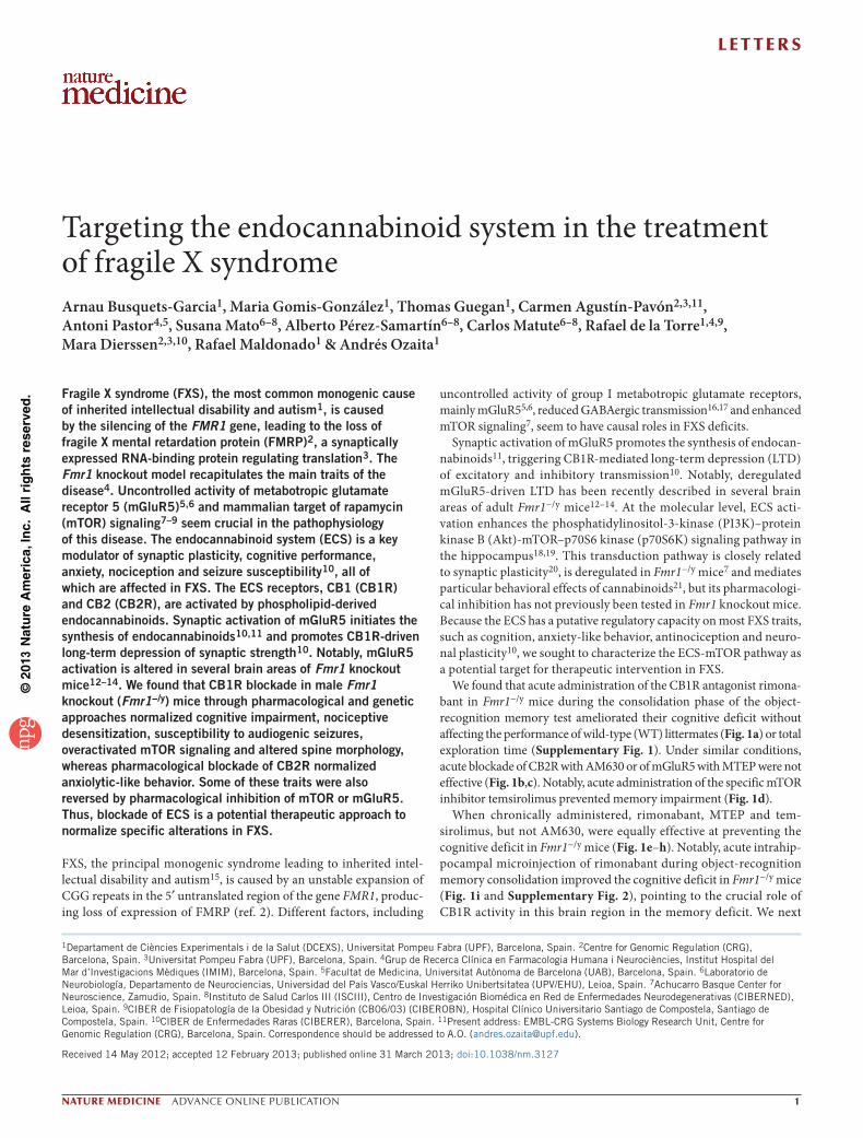
We found that acute administration of the CB1R antagonist rimona-bant in Fmr1−/y mice during the consolidation phase of the object- recognition memory test ameliorated their cognitive deficit without affecting the performance of wild-type (WT) littermates (Fig. 1a) or total exploration time (Supplementary Fig. 1). Under similar conditions, acute blockade of CB2R with AM630 or of mGluR5 with MTEP were not effective (Fig. 1b,c). Notably, acute administration of the specific mTOR inhibitor temsirolimus prevented memory impairment (Fig. 1d).
When chronically administered, rimonabant, MTEP and tem-sirolimus, but not AM630, were equally effective at preventing the cognitive deficit in Fmr1−/y mice (Fig. 1e–h). Notably, acute intrahip-pocampal microinjection of rimonabant during object-recognition memory consolidation improved the cognitive deficit in Fmr1−/y mice (Fig. 1i and Supplementary Fig. 2), pointing to the crucial role of CB1R activity in this brain region in the memory deficit. We next
Targeting the endocannabinoid system in the treatment of fragile X syndromeArnau Busquets-Garcia1, Maria Gomis-González1, Thomas Guegan1, Carmen Agustín-Pavón2,3,11, Antoni Pastor4,5, Susana Mato6–8, Alberto Pérez-Samartín6–8, Carlos Matute6–8, Rafael de la Torre1,4,9, Mara Dierssen2,3,10, Rafael Maldonado1 & Andrés Ozaita1
1Departament de Ciències Experimentals i de la Salut (DCEXS), Universitat Pompeu Fabra (UPF), Barcelona, Spain. 2Centre for Genomic Regulation (CRG), Barcelona, Spain. 3Universitat Pompeu Fabra (UPF), Barcelona, Spain. 4Grup de Recerca Clínica en Farmacologia Humana i Neurociències, Institut Hospital del Mar d’Investigacions Mèdiques (IMIM), Barcelona, Spain. 5Facultat de Medicina, Universitat Autònoma de Barcelona (UAB), Barcelona, Spain. 6Laboratorio de Neurobiología, Departamento de Neurociencias, Universidad del País Vasco/Euskal Herriko Unibertsitatea (UPV/EHU), Leioa, Spain. 7Achucarro Basque Center for Neuroscience, Zamudio, Spain. 8Instituto de Salud Carlos III (ISCIII), Centro de Investigación Biomédica en Red de Enfermedades Neurodegenerativas (CIBERNED), Leioa, Spain. 9CIBER de Fisiopatología de la Obesidad y Nutrición (CB06/03) (CIBEROBN), Hospital Clínico Universitario Santiago de Compostela, Santiago de Compostela, Spain. 10CIBER de Enfermedades Raras (CIBERER), Barcelona, Spain. 11Present address: EMBL-CRG Systems Biology Research Unit, Centre for Genomic Regulation (CRG), Barcelona, Spain. Correspondence should be addressed to A.O. ([email protected]).
Received 14 May 2012; accepted 12 February 2013; published online 31 March 2013; doi:10.1038/nm.3127
©20
13 N
atu
re A
mer
ica,
Inc.
All
rig
hts
res
erve
d.
l e t t e r s
advance online publication nature medicine
tested whether mGluR5 and CB1R inhibition converge to normalize the cognitive deficit. MTEP has a short half-life6 and was only effec-tive after chronic administration. Therefore, we treated mice chroni-cally with a suboptimal dose of MTEP (5 mg per kg body weight) and administered an acute dose of rimonabant after the training ses-sion. We found that mice that received the combined treatment had enhanced performance compared with those that received both treat-ments separately (Fig. 1j), pointing to a complementary involvement of both mechanisms.
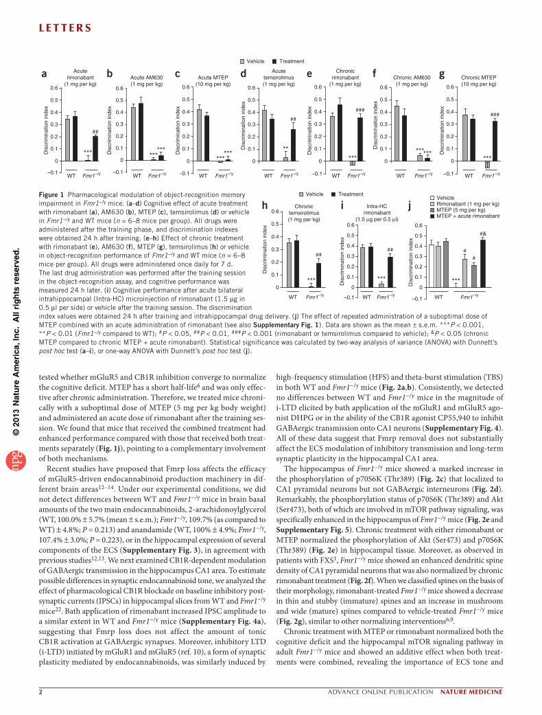
Recent studies have proposed that Fmrp loss affects the efficacy of mGluR5-driven endocannabinoid production machinery in dif-ferent brain areas12–14. Under our experimental conditions, we did not detect differences between WT and Fmr1−/y mice in brain basal amounts of the two main endocannabinoids, 2-arachidonoylglycerol (WT, 100.0% ± 5.7% (mean ± s.e.m.); Fmr1−/y, 109.7% (as compared to WT) ± 4.8%; P = 0.213) and anandamide (WT, 100% ± 4.9%; Fmr1−/y, 107.4% ± 3.0%; P = 0.223), or in the hippocampal expression of several components of the ECS (Supplementary Fig. 3), in agreement with previous studies12,13. We next examined CB1R-dependent modulation of GABAergic transmission in the hippocampus CA1 area. To estimate possible differences in synaptic endocannabinoid tone, we analyzed the effect of pharmacological CB1R blockade on baseline inhibitory post-synaptic currents (IPSCs) in hippocampal slices from WT and Fmr1−/y mice22. Bath application of rimonabant increased IPSC amplitude to a similar extent in WT and Fmr1−/y mice (Supplementary Fig. 4a), suggesting that Fmrp loss does not affect the amount of tonic CB1R activation at GABAergic synapses. Moreover, inhibitory LTD (i-LTD) initiated by mGluR1 and mGluR5 (ref. 10), a form of synaptic plasticity mediated by endocannabinoids, was similarly induced by
high-frequency stimulation (HFS) and theta-burst stimulation (TBS) in both WT and Fmr1−/y mice (Fig. 2a,b). Consistently, we detected no differences between WT and Fmr1−/y mice in the magnitude of i-LTD elicited by bath application of the mGluR1 and mGluR5 ago-nist DHPG or in the ability of the CB1R agonist CP55,940 to inhibit GABAergic transmission onto CA1 neurons (Supplementary Fig. 4). All of these data suggest that Fmrp removal does not substantially affect the ECS modulation of inhibitory transmission and long-term synaptic plasticity in the hippocampal CA1 area.
The hippocampus of Fmr1−/y mice showed a marked increase in the phosphorylation of p70S6K (Thr389) (Fig. 2c) that localized to CA1 pyramidal neurons but not GABAergic interneurons (Fig. 2d). Remarkably, the phosphorylation status of p70S6K (Thr389) and Akt (Ser473), both of which are involved in mTOR pathway signaling, was specifically enhanced in the hippocampus of Fmr1−/y mice (Fig. 2e and Supplementary Fig. 5). Chronic treatment with either rimonabant or MTEP normalized the phosphorylation of Akt (Ser473) and p70S6K (Thr389) (Fig. 2e) in hippocampal tissue. Moreover, as observed in patients with FXS1, Fmr1−/y mice showed an enhanced dendritic spine density of CA1 pyramidal neurons that was also normalized by chronic rimonabant treatment (Fig. 2f). When we classified spines on the basis of their morphology, rimonabant-treated Fmr1−/y mice showed a decrease in thin and stubby (immature) spines and an increase in mushroom and wide (mature) spines compared to vehicle-treated Fmr1−/y mice (Fig. 2g), similar to other normalizing interventions6,9.
Chronic treatment with MTEP or rimonabant normalized both the cognitive deficit and the hippocampal mTOR signaling pathway in adult Fmr1−/y mice and showed an additive effect when both treat-ments were combined, revealing the importance of ECS tone and
a Acuterimonabant
(1 mg per kg)
b Acute AM630(1 mg per kg)
d Acutetemsirolimus(1 mg per kg)
f Chronic AM630(1 mg per kg)
c Acute MTEP(10 mg per kg)
e Chronicrimonabant
(1 mg per kg)
g Chronic MTEP(10 mg per kg)
Dis
crim
inat
ion
inde
x
0.6
0.5
0.4
0.3
0.2
0.1
0
–0.1
***
##
WT Fmr1–/y
Dis
crim
inat
ion
inde
x
0.6
******
WT Fmr1–/y
0.5
0.4
0.3
0.2
0.1
0
–0.1
Dis
crim
inat
ion
inde
x
0.6
**
##
0.5
0.4
0.3
0.2
0.1
0
WT Fmr1–/y
Dis
crim
inat
ion
inde
x
0.6
******
WT Fmr1–/y
0.5
0.4
0.3
0.2
0.1
0
Dis
crim
inat
ion
inde
x
0.6
******
WT Fmr1–/y
0.5
0.4
0.3
0.2
0.1
0
–0.1
Dis
crim
inat
ion
inde
x
0.6
***
###
WT Fmr1–/y
0.5
0.4
0.3
0.2
0.1
0
–0.1
Dis
crim
inat
ion
inde
x
0.6
***
###
WT Fmr1–/y
0.5
0.4
0.3
0.2
0.1
0
–0.1
Vehicle Treatment
Vehicle Treatment
h
Dis
crim
inat
ion
inde
x
0.6Chronic
temsirolimus(1 mg per kg)
***
##
WT Fmr1–/y
0.5
0.4
0.3
0.2
0.1
0
i
Dis
crim
inat
ion
inde
x
0.6
Intra-HCrimonabant
(1.5 µg per 0.5 µl)
***
##
WT Fmr1–/y
0.5
0.4
0.3
0.2
0.1
0
–0.1
j
Dis
crim
inat
ion
inde
x
0.6
***
#&
##
WT Fmr1–/y
0.5
0.4
0.3
0.2
0.1
0
–0.1
VehicleRimonabant (1 mg per kg)MTEP (5 mg per kg)MTEP + acute rimonabant
Figure 1 Pharmacological modulation of object-recognition memory impairment in Fmr1−/y mice. (a–d) Cognitive effect of acute treatment with rimonabant (a), AM630 (b), MTEP (c), temsirolimus (d) or vehicle in Fmr1−/y and WT mice (n = 6–8 mice per group). All drugs were administered after the training phase, and discrimination indexes were obtained 24 h after training. (e–h) Effect of chronic treatment with rimonabant (e), AM630 (f), MTEP (g), temsirolimus (h) or vehicle in object-recognition performance of Fmr1−/y and WT mice (n = 6–8 mice per group). All drugs were administered once daily for 7 d. The last drug administration was performed after the training session in the object-recognition assay, and cognitive performance was measured 24 h later. (i) Cognitive performance after acute bilateral intrahippocampal (Intra-HC) microinjection of rimonabant (1.5 µg in 0.5 µl per side) or vehicle after the training session. The discrimination index values were obtained 24 h after training and intrahippocampal drug delivery. (j) The effect of repeated administration of a suboptimal dose of MTEP combined with an acute administration of rimonabant (see also Supplementary Fig. 1). Data are shown as the mean ± s.e.m. ***P < 0.001, **P < 0.01 (Fmr1−/y compared to WT); #P < 0.05, ##P < 0.01, ###P < 0.001 (rimonabant or temsirolimus compared to vehicle); &P < 0.05 (chronic MTEP compared to chronic MTEP + acute rimonabant). Statistical significance was calculated by two-way analysis of variance (ANOVA) with Dunnett’s post hoc test (a–i), or one-way ANOVA with Dunnett’s post hoc test (j).
©20
13 N
atu
re A
mer
ica,
Inc.
All
rig
hts
res
erve
d.
l e t t e r s
nature medicine advance online publication
mTOR activity in this cognitive deficit. mTOR signaling is crucial in memory consolidation23, and genetic regulation of this signaling path-way in Fmr1−/y mice prevents some of the Fmr1−/y features9, such as the elevated phosphorylation of translational control molecules, exagger-ated protein synthesis, enhanced mGluR-dependent LTD, weight gain, macro-orchidism, immature dendritic spine morphology and behav-ioral phenotypes, including social interaction deficits, impaired novel object recognition and behavioral inflexibility. We previously showed that mTOR overactivation in CA1 pyramidal neurons is directly involved in the cognitive deficit induced by ECS activation18,19,21, which can explain the efficacy of rimonabant-mediated CB1R block-ade in reversing cognitive impairment in Fmr1−/y mice. The fact that temsirolimus also prevented the object-recognition memory deficit in Fmr1−/y mice reinforces the concept that overactivation of mTOR signaling has a key role in this deficiency24 and demonstrates the pos-sibility of using these pharmacological approaches as therapy for this
disease. Given that CA1 hippocampal GABAergic presynaptic mem-branes show 10- to 20-fold higher expression of CB1R than do gluta-matergic presynaptic membranes25, rimonabant might help normalize the balance between excitatory and inhibitory input, which is altered in FXS, and lead to an improved output in the cognitive test by facilitating inhibitory transmission onto CA1 pyramidal cells26 (Supplementary Fig. 4). The balancing effect of rimonabant may also fit with the effect of other therapeutic approaches that aim to re-establish the excita-tory-inhibitory balance, such as the mGluR5 antagonist CTEP6 or the N-methyl-d-aspartate (NMDA) receptor antagonist memantine27, both of which reduce the excitatory drive, and the GABAB receptor agonist arbaclofen17, which increases the inhibitory drive.
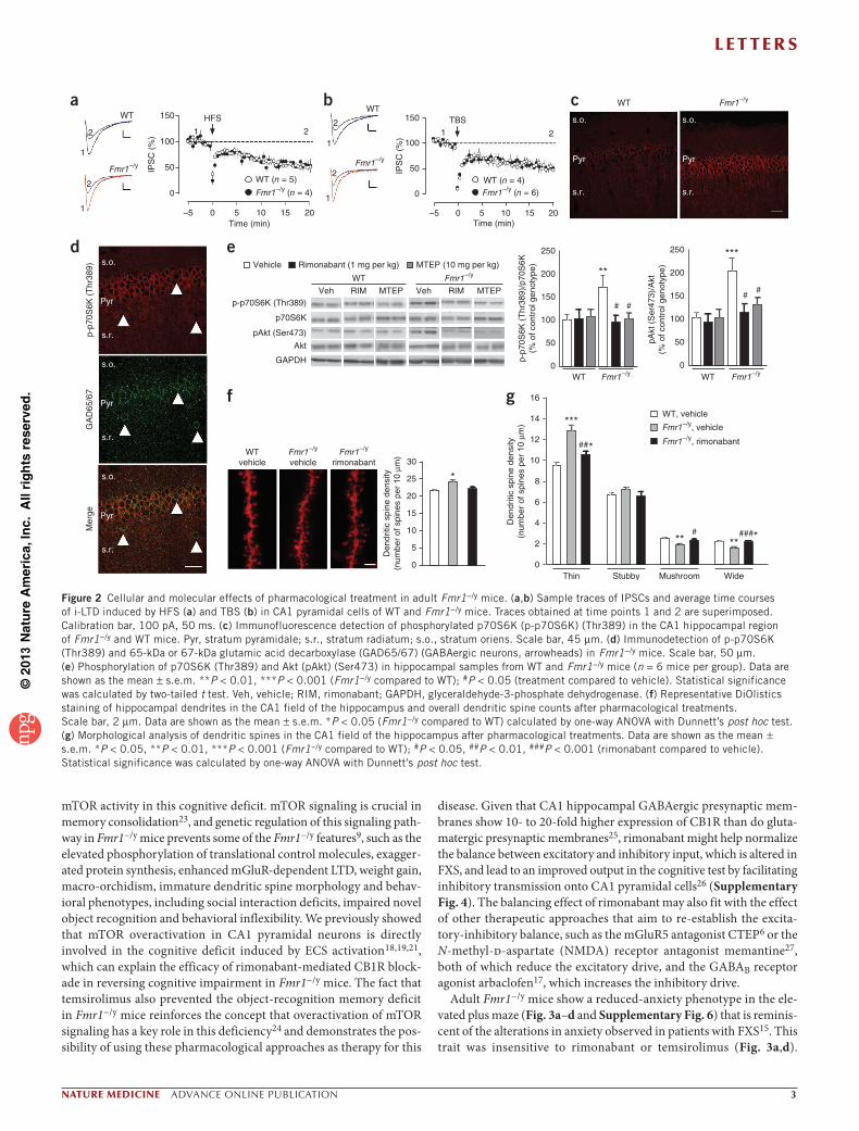
Adult Fmr1−/y mice show a reduced-anxiety phenotype in the ele-vated plus maze (Fig. 3a–d and Supplementary Fig. 6) that is reminis-cent of the alterations in anxiety observed in patients with FXS15. This trait was insensitive to rimonabant or temsirolimus (Fig. 3a,d).
b c
d e
f g
Time (min)
Vehicle Rimonabant (1 mg per kg) MTEP (10 mg per kg)
WT
WTvehicle
p-p70S6K (Thr389)
p-p7
0S6K
(T
hr38
9)
Veh Veh RIMRIM MTEP MTEP
aWT HFS
Fmr1–/y (n = 4)
WT (n = 5)
21
150
100
50
0
IPS
C (
%)
Fmr1–/y
1
22
2
1
1
2
1
Fmr1–/y (n = 6)
Fmr1–/y
vehicleFmr1–/y
rimonabant
Fmr1–/y
WT
s.o. s.o.
PyrPyr
s.r.
s.o.
Pyr
s.r.
s.o.
Pyr
s.r.
s.o.
Pyr
s.r.
s.r.
Fmr1–/y
p-p7
0S6K
(T
hr38
9)/p
70S
6K(%
of c
ontr
ol g
enot
ype)
Den
driti
c sp
ine
dens
ity(n
umbe
r of
spi
nes
per
10 µ
m)
Den
driti
c sp
ine
dens
ity(n
umbe
r of
spi
nes
per
10 µ
m)
pAkt
(S
er47
3)/A
kt(%
of c
ontr
ol g
enot
ype)
Fmr1–/y Fmr1–/yWTWT
Thin Stubby WideMushroom
Fmr1–/y, vehicle
Fmr1–/y, rimonabant
WT, vehicle
Time (min)
IPS
C (
%)
WT (n = 4)
–5 0 5 10 15 20
WT
Fmr1–/y
150
1100
50
0
TBS
2
–5
p70S6K
pAkt (Ser473)
Akt
GAPDH
GA
D65
/67
Mer
ge
0 5 10 15 20
250
200
150
100
50
0
250
30
25
20
15
10
5
0
16
14
12
10
8
6
4
2
0
***
**
# ##
#
***
** **
##*
###*#
*
200
150
100
50
0
Figure 2 Cellular and molecular effects of pharmacological treatment in adult Fmr1−/y mice. (a,b) Sample traces of IPSCs and average time courses of i-LTD induced by HFS (a) and TBS (b) in CA1 pyramidal cells of WT and Fmr1−/y mice. Traces obtained at time points 1 and 2 are superimposed. Calibration bar, 100 pA, 50 ms. (c) Immunofluorescence detection of phosphorylated p70S6K (p-p70S6K) (Thr389) in the CA1 hippocampal region of Fmr1−/y and WT mice. Pyr, stratum pyramidale; s.r., stratum radiatum; s.o., stratum oriens. Scale bar, 45 µm. (d) Immunodetection of p-p70S6K (Thr389) and 65-kDa or 67-kDa glutamic acid decarboxylase (GAD65/67) (GABAergic neurons, arrowheads) in Fmr1−/y mice. Scale bar, 50 µm. (e) Phosphorylation of p70S6K (Thr389) and Akt (pAkt) (Ser473) in hippocampal samples from WT and Fmr1−/y mice (n = 6 mice per group). Data are shown as the mean ± s.e.m. **P < 0.01, ***P < 0.001 (Fmr1−/y compared to WT); #P < 0.05 (treatment compared to vehicle). Statistical significance was calculated by two-tailed t test. Veh, vehicle; RIM, rimonabant; GAPDH, glyceraldehyde-3-phosphate dehydrogenase. (f) Representative DiOlistics staining of hippocampal dendrites in the CA1 field of the hippocampus and overall dendritic spine counts after pharmacological treatments. Scale bar, 2 µm. Data are shown as the mean ± s.e.m. *P < 0.05 (Fmr1−/y compared to WT) calculated by one-way ANOVA with Dunnett’s post hoc test. (g) Morphological analysis of dendritic spines in the CA1 field of the hippocampus after pharmacological treatments. Data are shown as the mean ± s.e.m. *P < 0.05, **P < 0.01, ***P < 0.001 (Fmr1−/y compared to WT); #P < 0.05, ##P < 0.01, ###P < 0.001 (rimonabant compared to vehicle). Statistical significance was calculated by one-way ANOVA with Dunnett’s post hoc test.
©20
13 N
atu
re A
mer
ica,
Inc.
All
rig
hts
res
erve
d.
l e t t e r s
advance online publication nature medicine
However, acute CB2R and mGluR5 blockade (Fig. 3b,c) normalized this phenotype without modifying anxiety-like behavior in WT mice (Fig. 3b,c). In this regard, CB2R has been involved in the anxiolytic-like effects produced by an enhanced amount of 2-arachidonoylg-lycerol18, whereas CB1R has been associated with the anxiolytic-like effect of anandamide accumulation18. Because we used doses of rimonabant and AM630 that did not affect the anxiety-like behav-ior in WT mice, we conclude that the reduced-anxiety phenotype in Fmr1−/y mice may specifically involve CB2R signaling.
We used the formalin test to study another characteristic of adult Fmr1−/y mice: diminished responses to inflammatory pain, which are sensitive to mGluR5 blockade28. This phenotype could be relevant to the self-injurious behavior that is often present in patients with FXS and was only normalized by rimonabant (Fig. 3e), consistent with previous findings reported in a different model of peripheral and central nocicep-tive sensitization29.
In addition, the marked sensitivity to audiogenic seizures in Fmr1−/y mice30, evoking the enhanced susceptibility of patients with FXS to epilepsy5, was bluntly decreased by pretreatment with rimonabant,
AM630 or temsirolimus (Fig. 3f), consistent with results previously reported using mGluR5 antagonism6. The involvement of CB2R and mTOR signaling in this phenotypic characteristic reveals a new cen-tral effect of these targets.
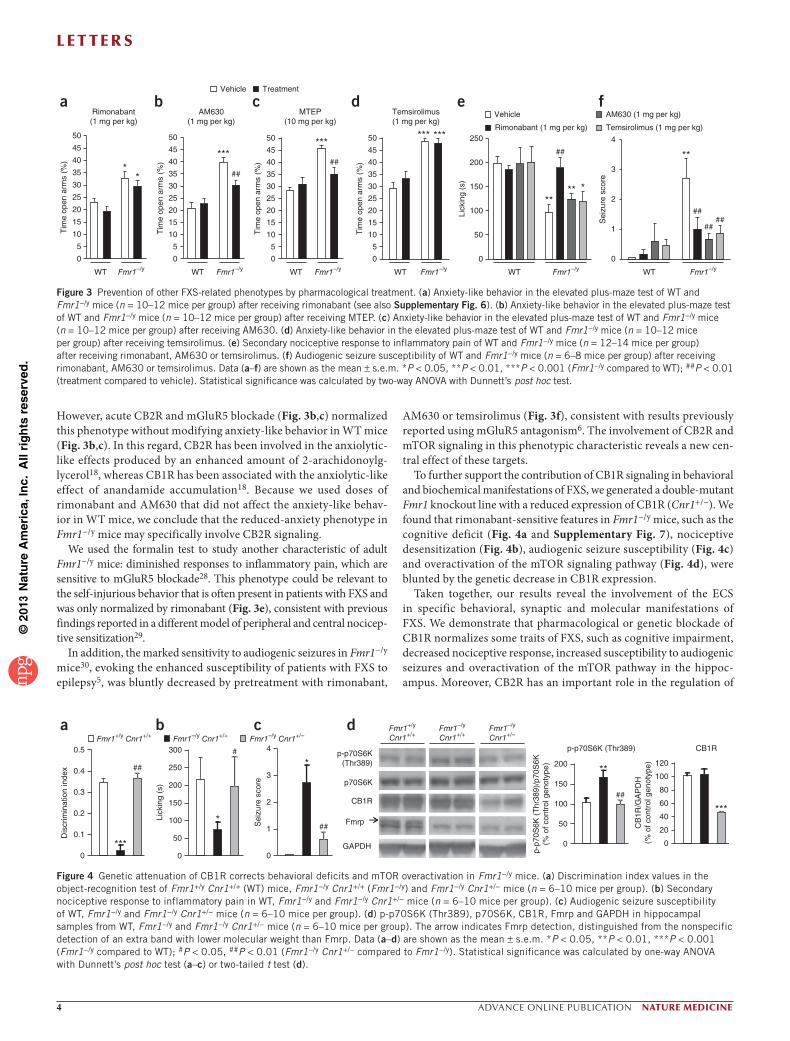
To further support the contribution of CB1R signaling in behavioral and biochemical manifestations of FXS, we generated a double-mutant Fmr1 knockout line with a reduced expression of CB1R (Cnr1+/−). We found that rimonabant-sensitive features in Fmr1−/y mice, such as the cognitive deficit (Fig. 4a and Supplementary Fig. 7), nociceptive desensitization (Fig. 4b), audiogenic seizure susceptibility (Fig. 4c) and overactivation of the mTOR signaling pathway (Fig. 4d), were blunted by the genetic decrease in CB1R expression.
Taken together, our results reveal the involvement of the ECS in specific behavioral, synaptic and molecular manifestations of FXS. We demonstrate that pharmacological or genetic blockade of CB1R normalizes some traits of FXS, such as cognitive impairment, decreased nociceptive response, increased susceptibility to audiogenic seizures and overactivation of the mTOR pathway in the hippoc-ampus. Moreover, CB2R has an important role in the regulation of
Vehicle Treatment
Temsirolimus (1 mg per kg)
Vehicle
Rimonabant (1 mg per kg)
AM630 (1 mg per kg)AM630(1 mg per kg)
b
***
##
50
45
40
35
30
25
Tim
e op
en a
rms
(%)
20
15
10
5
0
WT Fmr1–/y
MTEP(10 mg per kg)
c
***
##
50
45
40
35
30
25
Tim
e op
en a
rms
(%)
20
15
10
5
0
WT Fmr1–/y
Temsirolimus(1 mg per kg)
d
*** ***50
45
40
35
30
25
Tim
e op
en a
rms
(%)
20
15
10
5
0
WT Fmr1–/y
Rimonabant(1 mg per kg)
a
50
45
40
35
30
25
Tim
e op
en a
rms
(%)
20
15
10
5
0
WT Fmr1–/y
**
e f
WT Fmr1–/y
250
200
150
##
** *
4
3
WT Fmr1–/y
**
Lick
ing
(s)
100
50
0
**
Sei
zure
sco
re
2
1
0
##
####
Figure 3 Prevention of other FXS-related phenotypes by pharmacological treatment. (a) Anxiety-like behavior in the elevated plus-maze test of WT and Fmr1−/y mice (n = 10–12 mice per group) after receiving rimonabant (see also Supplementary Fig. 6). (b) Anxiety-like behavior in the elevated plus-maze test of WT and Fmr1−/y mice (n = 10–12 mice per group) after receiving MTEP. (c) Anxiety-like behavior in the elevated plus-maze test of WT and Fmr1−/y mice (n = 10–12 mice per group) after receiving AM630. (d) Anxiety-like behavior in the elevated plus-maze test of WT and Fmr1−/y mice (n = 10–12 mice per group) after receiving temsirolimus. (e) Secondary nociceptive response to inflammatory pain of WT and Fmr1−/y mice (n = 12–14 mice per group) after receiving rimonabant, AM630 or temsirolimus. (f) Audiogenic seizure susceptibility of WT and Fmr1−/y mice (n = 6–8 mice per group) after receiving rimonabant, AM630 or temsirolimus. Data (a–f) are shown as the mean ± s.e.m. *P < 0.05, **P < 0.01, ***P < 0.001 (Fmr1−/y compared to WT); ##P < 0.01 (treatment compared to vehicle). Statistical significance was calculated by two-way ANOVA with Dunnett’s post hoc test.
0.5
a
0.4 ##
***
0.3
Dis
crim
inat
ion
inde
x
0.2
0.1
0
b300
200
250
#
*
150
Lick
ing
(s)
100
50
0
c4
3
##
*
2
Sei
zure
sco
re
1
0
Fmr1–/y Cnr1+/–Fmr1+/y Cnr1+/+ Fmr1–/y Cnr1+/+d
200
Fmr1+/y
Cnr1+/+Fmr1–/y
Cnr1+/+Fmr1–/y
Cnr1+/–
p-p70S6K (Thr389) CB1R
150##
**
p-p7
0S6K
(T
hr38
9)/p
70S
6K(%
of c
ontr
ol g
enot
ype)
100
50
0
120
100
***
CB
1R/G
AP
DH
(% o
f con
trol
gen
otyp
e)
60
80
20
40
0
p70S6K
Fmrp
CB1R
GAPDH
p-p70S6K(Thr389)
Figure 4 Genetic attenuation of CB1R corrects behavioral deficits and mTOR overactivation in Fmr1−/y mice. (a) Discrimination index values in the object-recognition test of Fmr1+/y Cnr1+/+ (WT) mice, Fmr1−/y Cnr1+/+ (Fmr1−/y) and Fmr1−/y Cnr1+/− mice (n = 6–10 mice per group). (b) Secondary nociceptive response to inflammatory pain in WT, Fmr1−/y and Fmr1−/y Cnr1+/− mice (n = 6–10 mice per group). (c) Audiogenic seizure susceptibility of WT, Fmr1−/y and Fmr1−/y Cnr1+/− mice (n = 6–10 mice per group). (d) p-p70S6K (Thr389), p70S6K, CB1R, Fmrp and GAPDH in hippocampal samples from WT, Fmr1−/y and Fmr1−/y Cnr1+/− mice (n = 6–10 mice per group). The arrow indicates Fmrp detection, distinguished from the nonspecific detection of an extra band with lower molecular weight than Fmrp. Data (a–d) are shown as the mean ± s.e.m. *P < 0.05, **P < 0.01, ***P < 0.001 (Fmr1−/y compared to WT); #P < 0.05, ##P < 0.01 (Fmr1−/y Cnr1+/− compared to Fmr1−/y). Statistical significance was calculated by one-way ANOVA with Dunnett’s post hoc test (a–c) or two-tailed t test (d).
©20
13 N
atu
re A
mer
ica,
Inc.
All
rig
hts
res
erve
d.
l e t t e r s
nature medicine advance online publication
anxiolytic-like behavior and increased susceptibility to audiogenic seizures. In conclusion, our data point to regulation of the ECS and mTOR pathway as a potential target for the development of new thera-peutic approaches in FXS (Supplementary Fig. 8).
METhOdSMethods and any associated references are available in the online version of the paper.
Note: Supplementary information is available in the online version of the paper.
ACknOwleDGMenTSWe thank C. Fernández-Avilés, D. Real, M. Linares and H. Gómez for expert technical assistance and O.J. Manzoni for helpful comments. Fmr1 knockout mice in the C57BL/6J background were kindly provided by D. Nelson at Baylor College of Medicine. A.B.-G. is the recipient of a predoctoral fellowship (Ministerio de Educación y Cultura). S.M. is the recipient of a Ramón y Cajal contract (Ministerio de Educación y Cultura). This study was supported by grants from Fundació La Marató de TV3 (090910 to A.O.), Grants from the Ministerio de Ciencia e Innovación (SAF2009-07309 and BFU2012-33500 to A.O., SAF2011-29864 to R.M. and SAF2010-21547 to C.M.), CureFXS E-Rare EU/FIS PS09102673 to M.D., Instituto de Salud Carlos III (RD06/0001/0001 to R.M.), PLAN E (Plan Español para el Estímulo de la Economía y el Empleo), Generalitat de Catalunya (SGR-2009-00731 to R.M. and SGR-2009-00718 to R.d.l.T.) and ICREA (Institució Catalana de Recerca i Estudis Avançats) Academia to R.M.
AUTHOR COnTRIBUTIOnSA.B.-G. participated in experimental design, conducted biochemical and behavioral experiments, and wrote the manuscript. M.G.-G. conducted biochemical and behavioral experiments. T.G. performed dendritic spine morphology analysis. C.A.-P. performed stereotaxic surgeries, and M.D. provided Fmr1 knockout and WT mice, and discussed the experiments. A.P. and R.d.l.T. measured endocannabinoid levels. S.M. designed and conducted electrophysiological experiments, and wrote the manuscript. A.P.-S. and S.M. were in charge of electrophysiological equipment. C.M. funded the project. R.M. funded the project, participated in experimental design and wrote the manuscript. A.O. conceptualized, participated in experimental design, supervised, funded the project and wrote the manuscript.
COMPETING FINANCIAL INTERESTSThe authors declare no competing financial interests.
Reprints and permissions information is available online at http://www.nature.com/reprints/index.html.
1. de Vries, B.B., Halley, D.J., Oostra, B.A. & Niermeijer, M.F. The fragile X syndrome. J. Med. Genet. 35, 579–589 (1998).
2. Verkerk, A.J. et al. Identification of a gene (FMR-1) containing a CGG repeat coincident with a breakpoint cluster region exhibiting length variation in fragile X syndrome. Cell 65, 905–914 (1991).
3. Darnell, J.C. et al. FMRP stalls ribosomal translocation on mRNAs linked to synaptic function and autism. Cell 146, 247–261 (2011).
4. Bakker, C.E. et al. Fmr1 knockout mice: a model to study fragile X mental retardation. The Dutch-Belgian Fragile X Consortium. Cell 78, 23–33 (1994).
5. Bear, M.F., Huber, K.M. & Warren, S.T. The mGluR theory of fragile X mental retardation. Trends Neurosci. 27, 370–377 (2004).
6. Michalon, A. et al. Chronic pharmacological mGlu5 inhibition corrects fragile X in adult mice. Neuron 74, 49–56 (2012).
7. Sharma, A. et al. Dysregulation of mTOR signaling in fragile X syndrome. J. Neurosci. 30, 694–702 (2010).
8. Hoeffer, C.A. et al. Altered mTOR signaling and enhanced CYFIP2 expression levels in subjects with fragile X syndrome. Genes Brain Behav. 11, 332–341 (2012).
9. Bhattacharya, A. et al. Genetic removal of p70 S6 kinase 1 corrects molecular, synaptic, and behavioral phenotypes in fragile X syndrome mice. Neuron 76, 325–337 (2012).
10. Kano, M. et al. Endocannabinoid-mediated control of synaptic transmission. Physiol. Rev. 89, 309–380 (2009).
11. Varma, N., Carlson, G.C., Ledent, C. & Alger, B.E. Metabotropic glutamate receptors drive the endocannabinoid system in hippocampus. J. Neurosci. 21, RC188 (2001).
12. Zhang, L. & Alger, B.E. Enhanced endocannabinoid signaling elevates neuronal excitability in fragile X syndrome. J. Neurosci. 30, 5724–5729 (2010).
13. Maccarrone, M. et al. Abnormal mGlu 5 receptor/endocannabinoid coupling in mice lacking FMRP and BC1 RNA. Neuropsychopharmacology 35, 1500–1509 (2010).
14. Jung, K.M. et al. Uncoupling of the endocannabinoid signalling complex in a mouse model of fragile X syndrome. Nat. Commun. 3, 1080 (2012).
15. Penagarikano, O., Mulle, J.G. & Warren, S.T. The pathophysiology of fragile x syndrome. Annu. Rev. Genomics Hum. Genet. 8, 109–129 (2007).
16. D’Hulst, C. & Kooy, R.F. The GABAA receptor: a novel target for treatment of fragile X? Trends Neurosci. 30, 425–431 (2007).
17. Henderson, C. et al. Reversal of disease-related pathologies in the fragile X mouse model by selective activation of GABAB receptors with arbaclofen. Sci. Transl. Med. 4, 152ra128 (2012).
18. Busquets-Garcia, A. et al. Differential role of anandamide and 2-arachidonoylglycerol in memory and anxiety-like responses. Biol. Psychiatry 70, 479–486 (2011).
19. Puighermanal, E., Busquets-Garcia, A., Maldonado, R. & Ozaita, A. Cellular and intracellular mechanisms involved in the cognitive impairment of cannabinoids. Phil. Trans. R. Soc. Lond. B 367, 3254–3263 (2012).
20. Hoeffer, C.A. & Klann, E. mTOR signaling: at the crossroads of plasticity, memory and disease. Trends Neurosci. 33, 67–75 (2010).
21. Puighermanal, E. et al. Cannabinoid modulation of hippocampal long-term memory is mediated by mTOR signaling. Nat. Neurosci. 12, 1152–1158 (2009).
22. Lafourcade, M. et al. Nutritional omega-3 deficiency abolishes endocannabinoid-mediated neuronal functions. Nat. Neurosci. 14, 345–350 (2011).
23. Richter, J.D. & Klann, E. Making synaptic plasticity and memory last: mechanisms of translational regulation. Genes Dev. 23, 1–11 (2009).
24. Troca-Marín, J.A., Alves-Sampaio, A. & Montesinos, M.L. Deregulated mTOR-mediated translation in intellectual disability. Prog. Neurobiol. 96, 268–282 (2012).
25. Kawamura, Y. et al. The CB1 cannabinoid receptor is the major cannabinoid receptor at excitatory presynaptic sites in the hippocampus and cerebellum. J. Neurosci. 26, 2991–3001 (2006).
26. Kang-Park, M.H. et al. Differential sensitivity of GABA A receptor–mediated IPSCs to cannabinoids in hippocampal slices from adolescent and adult rats. J. Neurophysiol. 98, 1223–1230 (2007).
27. Wei, H. et al. The therapeutic effect of memantine through the stimulation of synapse formation and dendritic spine maturation in autism and fragile X syndrome. PLoS ONE 7, e36981 (2012).
28. Price, T.J. et al. Decreased nociceptive sensitization in mice lacking the fragile X mental retardation protein: role of mGluR1/5 and mTOR. J. Neurosci. 27, 13958–13967 (2007).
29. Guindon, J. & Hohmann, A.G. The endocannabinoid system and pain. CNS Neurol. Disord. Drug Targets 8, 403–421 (2009).
30. Chen, L. & Toth, M. Fragile X mice develop sensory hyperreactivity to auditory stimuli. Neuroscience 103, 1043–1050 (2001).
©20
13 N
atu
re A
mer
ica,
Inc.
All
rig
hts
res
erve
d.
nature medicine doi:10.1038/nm.3127
ONLINE METhOdSAnimals. Fmr1 knockout mice in a FVB background (FVB.129P2-Pde6b+ Tyrc−chFmr1tm1Cgr/J) and WT mice (FVB.129P2-Pde6b+ Tyrc−ch/AntJ) were purchased from The Jackson Laboratory and crossed to obtain Fmr1−/y and WT littermates. Fmr1 knockout mice in a C57BL/6J congenic background (B6.129P2-Fmr1tm1Cgr/J)4 were obtained from the Baylor College of Medicine Mouse Facility. Double-mutant mice (Fmr1−/y Cnr1+/−) in a C57BL/6J back-ground were initially generated by crossing homozygous female mice carry-ing the Fmr1 mutation (Fmr1−/−) with homozygous male mice carrying the Cnr1 mutation (Cnr1−/−). Subsequently, mice used for experimentation were derived from the crossing of female Fmr1+/− Cnr1+/+ mice with male Fmr1+/y Cnr1+/− mice. All experimental mice were bred in house at the Barcelona Biomedical Research Park (PRBB) Animal Facility. Fmr1−/y and WT mice were used at 12–16 weeks of age, except for in the study of audiogenic seizure susceptibility (where mice were 21–23 d old). Mice were housed four per cage in a temperature-controlled (21 °C ± 1 °C) (mean ± range) and humid-ity-controlled (55% ± 10%) environment. Food and water were available ad libitum. All the experiments were performed during the light phase of a 12 h light, 12 h dark cycle (lights on at 8 a.m. and off at 8 p.m.). Mice were handled for 1 week before starting the experiments. Only male mice were used. All animal procedures followed standard ethical guidelines (European Communities Directive 86/60-EEC) and were approved by the local ethical committee (Comitè Ètic d’Experimentació Animal-Parc de Recerca Biomèdica de Barcelona, CEEA-PRBB). The PRBB also has Animal Welfare Assurance (#A5388-01, Institutional Animal Care and Use Committee approval date 06/08/2009) granted by the Office of Laboratory Animal Welfare (OLAW) of the US National Institutes of Health. All behavioral tests were performed by researchers blind to the different experimental groups.
Drugs and treatments. Rimonabant was obtained from Sanofi-Aventis (Sanofi-Aventis Recherche); AM630 (6-iodo-2-methyl-1-[2- (4-morpholinyl)ethyl]-1H-indol-3-yl](4-methoxyphenyl)methanone), D-APV (DL-2-amino-5-phosphonopentanoic acid), NBQX (2,3-dioxo- 6-nitro-1,2,3,4-tetrahydrobenzo[f]quinoxaline-7-sulfonamide disodium salt) and DHPG ((S)-3,5-dihydroxyphenylglycine) were from Tocris Bioscience; MTEP (3-((2-methyl-4-thiazolyl)ethynyl)pyridine) was a kind gift from Merck Research Laboratories; and temsirolimus (CCI-779) was from LC Laboratories. CP55,940 and picrotoxin were purchased from Sigma-Aldrich. Rimonabant, AM630 and MTEP were diluted in 5% ethanol, 5% Cremophor EL and 90% saline. Temsirolimus was dissolved in 2% ethanol, 8% Cremophor EL and 90% saline31. All compounds injected intraperitoneally were administered in a volume of 10 ml per kg body weight.
Intrahippocampal administration of rimonabant. Rimonabant (1.5 µg per 0.5 µl) was administered directly into the hippocampus as described else-where32. After recovery (8 d), mice were administered rimonabant or vehi-cle bilaterally after the training session in the object-recognition test. After completion of the experimental sequence, cannula position was verified histologically (Supplementary Fig. 2), as described previously32.
Endocannabinoid quantification. Endocannabinoids, 2-AG (2-arachido-noylglycerol) and AEA (anandamide) were analyzed as described previously18 using half of the right or left brain (including the forebrain, midbrain and hindbrain) (~220–250 mg). The endogenous concentrations of 2-AG and AEA were calculated on the basis of the response of the deuterated analogs and expressed as percentage of control WT mice.
Immunoblot analysis. Tissues from different brain regions (hippocampus, frontal cortex, striatum, amygdala and cerebellum) from WT and Fmr1−/y mice were analyzed in basal conditions. Hippocampal tissues from WT and Fmr1−/y mice after chronic pharmacological treatment or from the double-mutant mouse line (Fmr1+/y Cnr1+/+, Fmr1−/y Cnr1+/+ and Fmr1−/y Cnr1+/−) were also analyzed. Tissues were processed as previously described21. The antibodies used for immunoblotting detected: Fmrp (ab17722, 1:500), monoacylglycerol lipase (MAGL) (ab24701, 1:1,000), fatty acid amide hydro-lase (FAAH) (ab54615, 1:500), (Abcam); p-p70S6K (Thr389) (9234, 1:800),
p70S6K (9202, 1:500), pAkt (Ser473) (4051, 1:200), Akt (9272, 1:2,000) (all from Cell Signaling Technology); diacylglycerol lipase α (DGL-α) (GP-Af380-1, 1:400), N-acyl phosphatidylethanolamine phospholipase D (NAPE-PLD) (GP-Af720-1, 1:1,000), mGluR5 (GP-Af270-1, 1:1,000), CB1R (Rb-Af380-1, 1:1,000) (all from Frontier Science); and GAPDH (sc-32233, 1:5,000) (Santa Cruz Biotechnology). Optical densities of relevant immunoreactive bands were quantified after acquisition on a ChemiDoc XRS System (Bio-Rad) controlled by The Quantity One software v 4.6.3 (Bio-Rad).
Immunofluorescence. Tissue was prepared as described previously21. Hippocampi-containing slices were rinsed in 0.1 M phosphate buffer, incubated in permeabilizing and blocking solution (0.2% Triton X-100, 5% BSA in phosphate buffer) for 2 h and then incubated overnight at 4 °C with antibody to p-p70S6K (Thr389) (9206, 1:100, mouse, Cell Signaling Technology) and antibody to GAD65/67 (AB1511, 1:100, rabbit, Millipore). The next day, after rinsing in phosphate buffer, sections were incubated with antibody to mouse Alexa Fluor 647 (115-165-146, 1:500, Invitrogen) or antibody to rabbit Cy3 (111-225-144, 1:500, Jackson ImmunoResearch Laboratories). After rising, slices were mounted onto gelatin-coated slides with Mowiol mounting medium. Confocal images were obtained as previously described21.
Slice preparation and electrophysiology. Fmr1−/y and WT mice were anes-thetized with isoflurane, and their brains were removed to a chilled sucrose-based solution (in mM: 215 sucrose, 2.5 KCl, 26 NaHCO3, 1.6 NaH2PO4, 1 CaCl2, 4 MgCl2, 4 MgSO4, 20 glucose and 1.3 mM ascorbic acid), and coronal brain slices (350 µm thick) were cut with a Vibratome Series 3000 Plus-Tissue Sectioning System (Ted Pella, Inc). Sections containing the hippocampus were stored for 30 min at 30–32 °C in recovery solution (in mM: 62 NaCl, 2.5 KCl, 25 NaHCO3, 1.4 NaH2PO4, 1.1 CaCl2, 3.3 MgCl2, 2 MgSO4, 15 glu-cose and 108 sucrose) and then moved into room-temperature low-calcium artificial cerebrospinal fluid (ACSF) (in mM: 124 NaCl, 2.5 KCl, 25 NaHCO3, 1.2 NaH2PO4, 1.25 CaCl2, 2.6 MgCl2 and 10 glucose) for at least 1 h. Experiments were conducted at room temperature in a submersion-type recording chamber perfused at 1.5 ml per min with ACSF (same as the low- calcium ACSF with the following exception: 2.5 mM CaCl2 and 1.3 mM MgCl2). All solutions were saturated with 95% O2 and 5% CO2, pH 7.4.
Whole-cell recordings were made on visualized CA1 pyramidal neurons voltage clamped at −70 mV with a pipette (2–4 MΩ) containing (in mM): 90 CsCH3SO3, 50 CsCl, 10 4-(2-hydroxyethyl)-1-piperazineethanesulfonic acid (HEPES), 1 MgCl2, 0.2 ethylene glycol tetraacetic acid (EGTA), 4 Mg2+-ATP, 0.3 Tris-GTP, 5 QX314, pH 7.25, and 285 mOsm). IPSCs were isolated by adding NMDA and α-amino-3-hydroxy-5-methyl-4-isoxazole propionate/kainate (AMPA/KA) receptor antagonists (25 µM D-AP5 and 10 µM NBQX) to the ACSF. To evoke monosynaptic currents, stimuli of duration 150 µs were deliv-ered every 30 s through a patch pipette filled with ACSF and placed in the stra-tum radiatum. Series resistance (typically 10–20 MΩ) was monitored through the experiments by applying a −2 mV hyperpolarizing pulse before each evoked IPSC, and cells with a more than 20% change in this parameter were excluded from the analysis. LTD was induced by HFS (2 trains of 100 pulses at 100 Hz 20 s apart) or TBS (10 bursts of 5 stimuli applied at 100 Hz with 200 ms interburst intervals repeated 4 times 5 s apart). The magnitude of LTD and the effects of the different drugs were estimated by comparing averaged responses correspond-ing to the last 5 min of the experiment with baseline-averaged responses 5 min before the induction protocol or the beginning of drug application. Recordings were performed with a MultiClamp 700B (Axon Instruments), and output sig-nals were filtered at 3 kHz. Data were digitized (10 kHz) on a DigiData 1332A (Axon Instruments). Data were collected using Clampex 9.2, and IPSC ampli-tude was analyzed using Clampfit 9.2.
Dendritic spine morphology analysis. Dendritic spine analysis was per-formed as previously described33 in mice that received chronic administra-tion of rimonabant (1 mg per kg body weight for 7 d) or vehicle. Brains were extracted after perfusion (4% paraformaldehyde (PFA) in phosphate buffer) 3 h after the last administration of rimonabant or vehicle solution on the sev-enth day of treatment. Secondary to tertiary dendrites of pyramidal neurons
©20
13 N
atu
re A
mer
ica,
Inc.
All
rig
hts
res
erve
d.
nature medicinedoi:10.1038/nm.3127
from the CA1 region of the hippocampus were chosen for spine analysis on the basis of criteria described previously33.
Object-recognition task. Object-recognition memory was assayed as described previously21. All acute treatments and the last administration of the chronic treatments were performed after the training session in the object-recognition test.
Anxiety-like response. Anxiety-like responses were evaluated as previously described18. Briefly, 5-min test sessions were performed 120 min after drug administration. The time spent in the open arms as well as the total number of entries in the arms were recorded.
Nociceptive response. The late-phase nociceptive response (corresponding to inflammatory pain)34 was recorded during 20 min starting 20 min after formalin (5%, 20 µl) injection into the right hind paw. All drugs were injected 20 min before the formalin injection. The nociceptive behavior (licking) was measured.
Audiogenic seizure sensitivity. Mice were placed individually into an observa-tion chamber, a glass cylinder 40 cm high and 16 cm in diameter, and allowed to explore for 1 min. Next, a bell (100 dB) rung for 30 s. Mice were tested only once. Seizure activity was scored as follows: no response, 0; wild running, 1;
clonic seizure, 2; tonic seizure, 3; status epilepticus, respiratory arrest or death, 4. Drugs were administered 30 min before the test.
Statistical analyses. Results are reported as the mean ± s.e.m. Most of the experiments were evaluated by one-way analysis of variance (ANOVA) followed by Dunnett’s post-hoc test when required. Two-way ANOVA (rimona-bant, AM630, MTEP, temsirolimus treatment versus WT or Fmr1−/y genotype) was used when required. Statistical comparisons of electrophysiological data were performed using Student’s t test. Comparisons were considered statistically significant when P < 0.05.
31. Puighermanal, E. et al. Dissociation of the pharmacological effects of THC by mTOR blockade. Neuropsychopharmacology advance online publication, http://dx.doi.org/10.1038/npp.2013.31 (2013).
32. Castañé, A., Maldonado, R. & Valverde, O. Role of different brain structures in the behavioural expression of WIN 55,212–2 withdrawal in mice. Br. J. Pharmacol. 142, 1309–1317 (2004).
33. Guegan, T. et al. Operant behavior to obtain palatable food modifies neuronal plasticity in the brain reward circuit. Eur. Neuropsychopharmacol. 23, 146–159 (2013).
34. Noble, F. et al. Pain-suppressive effects on various nociceptive stimuli (thermal, chemical, electrical and inflammatory) of the first orally active enkephalin-metabolizing enzyme inhibitor RB 120. Pain 73, 383–391 (1997).
Related Documents