This article was downloaded by: [Vilma Martins] On: 27 June 2015, At: 09:29 Publisher: T aylor & Francis Informa Ltd Registered in England and Wales Registered Number: 1072954 Registered office: Mortimer House, 37-41 Mortimer Street, London W1T 3JH, UK Click for updates Prion Publication details, including instructions for authors and subscription information: http://www .tandfonline.com/loi/kprn20 T argeting prion protein interactions in cancer Tiago G Santos ab , Marilene H Lopes bc & Vilma R Martins ab a International Research Center; AC Camargo Cancer Center; São Paulo, Brazil; b National Institute for T ranslational Neuroscience and National Institute of Oncogenomics (CNPq/MCT/F APESP); São Paulo, Brazil; c Department of Cell and Developmental Biology; Institute of Biomedical Sciences; University of São Paulo; São Paulo, Brazil Published online: 25 Jun 2015. T o cite this article: Tiago G Santos, Marilene H Lopes & Vilma R Martins (2015) T argeting prion protein interactions in cancer , Prion, 9:3, 165-173, DOI: 10.1080/19336896.2015.1027855 T o link to this article: http://dx.doi.org/10.1080/19336896.2015.1027855 PLEASE SCROLL DOWN FOR ARTICLE T aylor & Francis makes every effort to ensure the accuracy of all the information (the “Content”) contained in the publications on our platform. However , T aylor & Francis, our agents, and our licensors make no representations or warranties whatsoever as to the accuracy , completeness, or suitability for any purpose of the Content. Any opinions and views expressed in this publication are the opinions and views of the authors, and are not the views of or endorsed by T aylor & Francis. The accuracy of the Content should not be relied upon and should be independently verified with primary sources of information. T aylor and Francis shall not be liable for any losses, actions, claims, proceedings, demands, costs, expenses, damages, and other liabilities whatsoever or howsoever caused arising directly or indirectly in connection with, in relation to or arising out of the use of the Content. This article may be used for research, teaching, and private study purposes. Any substantial or systematic reproduction, redistribution, reselling, loan, sub-licensing, systematic supply , or distribution in any form to anyone is expressly forbidden. T erms & Conditions of access and use can be found at http:// www.tandfonline.com/page/terms-and-conditions

Welcome message from author
This document is posted to help you gain knowledge. Please leave a comment to let me know what you think about it! Share it to your friends and learn new things together.
Transcript

This article was downloaded by: [Vilma Martins]On: 27 June 2015, At: 09:29Publisher: Taylor & FrancisInforma Ltd Registered in England and Wales Registered Number: 1072954 Registered office: Mortimer House,37-41 Mortimer Street, London W1T 3JH, UK
Click for updates
PrionPublication details, including instructions for authors and subscription information:http://www.tandfonline.com/loi/kprn20
Targeting prion protein interactions in cancerTiago G Santosab, Marilene H Lopesbc & Vilma R Martinsab
a International Research Center; AC Camargo Cancer Center; São Paulo, Brazil;b National Institute for Translational Neuroscience and National Institute of Oncogenomics(CNPq/MCT/FAPESP); São Paulo, Brazil;c Department of Cell and Developmental Biology; Institute of Biomedical Sciences;University of São Paulo; São Paulo, BrazilPublished online: 25 Jun 2015.
To cite this article: Tiago G Santos, Marilene H Lopes & Vilma R Martins (2015) Targeting prion protein interactions in cancer,Prion, 9:3, 165-173, DOI: 10.1080/19336896.2015.1027855
To link to this article: http://dx.doi.org/10.1080/19336896.2015.1027855
PLEASE SCROLL DOWN FOR ARTICLE
Taylor & Francis makes every effort to ensure the accuracy of all the information (the “Content”) containedin the publications on our platform. However, Taylor & Francis, our agents, and our licensors make norepresentations or warranties whatsoever as to the accuracy, completeness, or suitability for any purpose of theContent. Any opinions and views expressed in this publication are the opinions and views of the authors, andare not the views of or endorsed by Taylor & Francis. The accuracy of the Content should not be relied upon andshould be independently verified with primary sources of information. Taylor and Francis shall not be liable forany losses, actions, claims, proceedings, demands, costs, expenses, damages, and other liabilities whatsoeveror howsoever caused arising directly or indirectly in connection with, in relation to or arising out of the use ofthe Content.
This article may be used for research, teaching, and private study purposes. Any substantial or systematicreproduction, redistribution, reselling, loan, sub-licensing, systematic supply, or distribution in anyform to anyone is expressly forbidden. Terms & Conditions of access and use can be found at http://www.tandfonline.com/page/terms-and-conditions
EXTRA VIEWS
Targeting prion protein interactions in cancer
Tiago G Santos,1,2 Marilene H Lopes,2,3,* and Vilma R Martins1,2,*
1International Research Center; AC Camargo Cancer Center; S~ao Paulo, Brazil;2National Institute for Translational Neuroscience and National Institute
of Oncogenomics (CNPq/MCT/FAPESP); S~ao Paulo, Brazil;3Department of Cell and Developmental Biology; Institute of Biomedical Sciences;
University of S~ao Paulo; S~ao Paulo, Brazil
ABSTRACT. In recent years, prion protein (PrPC) has been considered as a promising targetmolecule for cancer therapies, due its direct or indirect participation in tumor growth, metastasis, andresistance to cell death induced by chemotherapy. PrPC functions as a scaffold protein, formingmultiprotein complexes on the plasma membrane, which elicits distinct signaling pathways involvedin diverse biological phenomena and could be modulated depending on the cell type, complexcomposition, and organization. In addition, PrPC and its partners participate in self-renewal ofembryonic, tissue-specific stem cells and cancer stem cells, which are suggested to be responsible forthe origin, maintenance, relapse, and dissemination of tumors. Interference with protein–proteininteraction has been recognized as an important therapeutic strategy in cancer; indeed, the possibleinterference in PrPC engagement with specific partners is a novel strategy. Recently, our groupsuccessfully used that approach to interfere with the interaction between PrPC and HSP-90/70organizing protein (HOP, also known as stress-inducible protein 1 - STI1) to control the growth ofhuman glioblastoma in animal models. Thus, PrPC-organized multicomplexes have emerged asfeasible candidates for anti-tumor therapy, warranting further exploration.
KEYWORDS. prion protein, ligand, cancer, therapy, peptide, cell signaling, cancer stem cell
ABBREVIATIONS. HSP, heat shock proteins; GBM, glioblastoma multiforme; BBB, blood brainbarrier; CSC, cancer stem cell
*Correspondence to: Marilene H Lopes; Email: [email protected], Vilma R Martins; Email:[email protected]
Received February 2, 2015; Revised March 3, 2015; Accepted March 4, 2015.Extra View to: Lopes MH, Santos TG, Rodrigues BR, Queiroz-Hazarbassanov N, Cunha IW, Wasilew-
ska-Sampaio AP, Costa-Silva B, Marchi FA, Bleggi-Torres LF, Sanematsu PI, et al. Disruption of prionprotein-HOP engagement impairs glioblastoma growth and cognitive decline and improves overall survival.Oncogene 2014; [Epub ahead of print]; PMID:25151961; http://dx.doi.org/10.1038/onc.2014.261
Color versions of one or more figures in this article can be found online at www.tandfonline.com/kprn.
165
Prion, 9:165–173, 2015Copyright Ó Taylor & Francis Group, LLCISSN: 1933-6896 print / 1933-690X onlineDOI: 10.1080/19336896.2015.1027855
Dow
nloa
ded
by [V
ilma
Mar
tins]
at 0
9:29
27
June
201
5
INTRODUCTION
Prion protein (PrPC) has been studied thor-oughly for decades, due its involvement in trans-missible spongiform encephalopathies andconversion to infectious proteinaceous agentscalled prions.1 Despite intense discussion of thefunction of the normal protein, convincing datafrom different groups indicate that PrPC hasimportant roles in the nervous and immune sys-tems, regulating cellular processes such as celldeath and survival, proliferation, and differentia-tion.2,3 PrPC is a glycosylphosphatidylinositol-anchored protein, and many of its describedfunctions depend on specific interactions withpartners on the plasma membrane (receptors orextracellular molecules), such as laminin, vitro-nectin, NCAM, caveolin, and HSP-90/70 heatshock organizing protein, also known as stress-inducible protein 1 (HOP/STI1)4 (a list of theseligands can be found at http://www.signaling-gateway.org/molecule/query?afcsidDA003935),which can modulate cellular signaling cascades.Due to these properties, we have proposed thatPrPC plays a scaffolding role on the cell surface,recruiting diverse partners to organize signalingplatforms.5 In this review we will discuss howthese PrPC-organized complexes can beinvolved with the tumoral processes and thestrategy to target the engagement of PrPC to spe-cific ligands for therapeutic interventions.
PrPC IN TUMOR BIOLOGY:PROCESSES AND MECHANISMS
The functions of PrPC in tumor cells havebeen addressed, and evidence suggests that thisprotein is an important player in tumor biology.Several studies have demonstrated the impor-tance of PrPC in proliferation, apoptosis,invasion, metastasis, and drug resistance in dif-ferent cancer types.6 In undifferentiated gastrictumors, a worse response to chemotherapy andlower patient survival rate are associated withhigher PrPC expression levels.7 Positive corre-lations between PrPC expression and invasion,lymph node metastasis, and survival have alsobeen confirmed in patients with gastric tumors,indicating that PrPC is an independent
prognostic factor in these tumors.8 Proteomicevaluation of colorectal cancer cell lines identi-fied PrPC as a putative biomarker for adenoma–carcinoma progression, discriminating low-riskadenomas and normal colon from high-riskadenomas and colorectal cancer.9 Accordingly,a previous study showed that PrPC expressioncould be used as a prognostic factor in patientswith colorectal cancer.10 In breast cancer celllines, resistance to TNF-induced cell death isassociated with greater PrPC expression.11 Fur-thermore, lower sensitivity to neoadjuvant ther-apy has been observed in ER-negative breasttumors expressing higher (vs. lower) levels ofPrPC.12
Despite this evidence of PrPC involvementin tumor biology, the mechanisms associatedwith these functions remain largely unex-plored. In some cases, mechanistic approacheshave shown that PrPC inhibits Bax-inducedapoptosis through Bax conformational changeprevention, impairing mitochondrial translo-cation and cytochrome c release in breast can-cer cells.13 In cell lines derived from colontumors, PrPC has been shown to regulate glu-cose transporter 1 expression through the acti-vation of Fyn-HIF-2a, increasing glucoseuptake, glycolysis, and cell survival/prolifera-tion.14 PrPC activation of Fyn in breast cancercells is also involved in epithelial–mesenchy-mal transition and results in a more aggressivephenotype.15 PrPC silencing in glioma celllines causes increased autophagy due to induc-tion of LC3-II, an increase in Beclin 1, andsimultaneous decreases in p62, Bcl¡ 2, andthe phosphorylation of 4E-BP1, a target ofmTOR autophagy signaling.16 Interestingly,mTOR, a master player in cell signaling withpivotal role in tumorigenesis, is also involvedin PrPC-dependent neuronal differentiationand neuroprotection through activation ofPI3K/Akt pathways.17
PrPC ENGAGEMENT TO ITSLIGANDS: A RELIABLE TARGET FOR
THERAPEUTIC INTERVENTION
The phenotypes and mechanisms describedfor PrPC in tumoral biology are in agreement
166 Santos et al.
Dow
nloa
ded
by [V
ilma
Mar
tins]
at 0
9:29
27
June
201
5
with its roles in multicomplex protein formationand organization of diverse signaling platforms.5
PrPC priming can be induced after ligand
binding, stimulating the acquisition of specificconformations and allowing additional interac-tions in the complex.5 The upregulation of PrPC
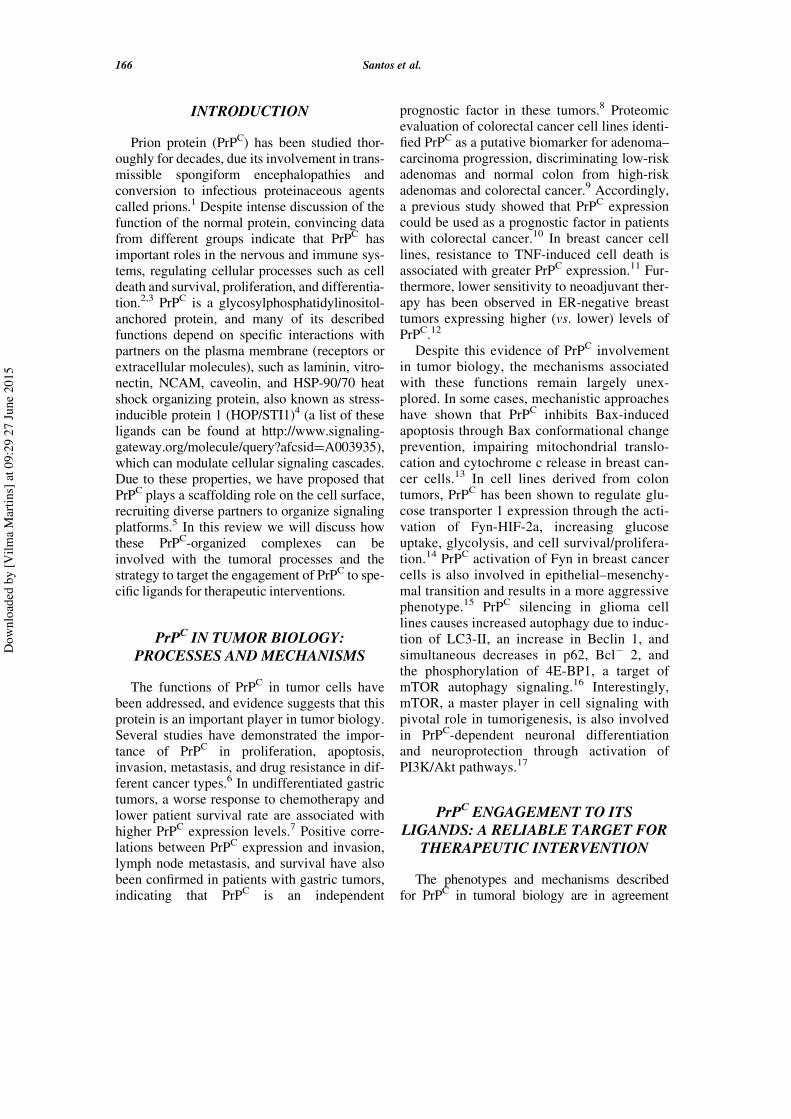
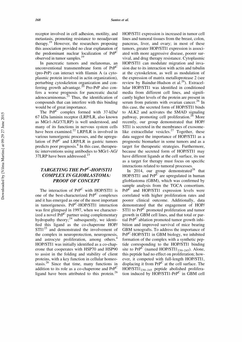
or its primary ligands in different tumor typesmay favor the assembly of tissue-specific com-plexes with different protein compositions. Thesecomplexes could alter the pattern of cellular sig-naling and, consequently, processes such as pro-liferation, adhesion, migration, differentiation,and drug resistance. Indeed, we believe that theidentification of PrPC ligands that coordinatethese processes will help researchers focus onmechanisms that can be targeted for therapeuticinterventions (as illustrated in Fig. 1).
In recent years, several independent studieshave characterized novel PrPC complexes modu-lating specific tumoral cell behaviors. In breastcancer, PrPC engagement to multidrug resistanceprotein (P-gp) and caveolin is associated withdrug resistance.18 In addition, the PrPC–P-gpcomplex may include CD44, a membrane
FIGURE 1. PrPC in signaling platforms and theirregulation. (A) PrPC (red) interaction with aprime soluble ligand (green) modulates a groupof multiple partners at the plasma membrane,organizing signaling platforms that regulate spe-cific cellular functions. Other PrPC partnersinclude integral transmembrane proteins (pur-ple, blue or orange) or peripheral membraneproteins, including cytoplasmic (beige or rose)or proteins attached to outer leaflet of theplasma membrane (light blue or light purple).Importantly, the prime ligand can be a mem-brane protein in the same domain of PrPC locali-zation (cis), in the membrane of another cell(trans), or on an extracellular vesicle surface.(B) The upregulation of PrPC allows the forma-tion of additional tissue-specific complexes withthe ability to regulate events associated totumorigenesis. (C) Higher levels of the primeligand can also improve the number of PrPC
molecules able to organize signaling platforms,thereby inducing cancer processes. (D) Con-versely, PrPC downregulation or silencing disor-ganizes these complexes, impairing PrPC-mediated signaling and, consequently, thetumoral process. (E) The inhibition of PrPC inter-action with the prime ligand impairs the organi-zation of these platforms and serves as a targetfor cancer therapy, potentially with fewer sideeffects.
FIGURE 1. (Continued.)
TARGETING PRION PROTEIN INTERACTIONS IN CANCER 167
Dow
nloa
ded
by [V
ilma
Mar
tins]
at 0
9:29
27
June
201
5
receptor involved in cell adhesion, motility, andmetastasis, promoting resistance to neoadjuvanttherapy.19 However, the researchers proposingthis association provided no clear explanation ofthe predominant nuclear localization of PrPC
observed in tumor samples.19
In pancreatic tumors and melanomas, anunconventional transmembrane form of PrPC
(pro-PrP) can interact with filamin A (a cyto-plasmic protein involved in actin organization),perturbing cytoskeleton organization and con-ferring growth advantage.20 Pro-PrP also con-fers a worse prognosis for pancreatic ductaladenocarninomas.21 Thus, the identification ofcompounds that can interfere with this bindingwould be of great importance.
The PrPC complex formed with 37-kDa/67 kDa laminin receptor (LRP/LR, also knownas MGr1-AG/37LRP) is well understood, andmany of its functions in nervous system cellshave been examined.22 LRP/LR is involved invarious tumorigenic processes, and the upregu-lation of PrPC and LRP/LR in gastric tumorspredicts poor prognosis.8 In this case, therapeu-tic interventions using antibodies to MGr1-AG/37LRP have been addresssed.23
TARGETING THE PrPC-HOP/STI1COMPLEX IN GLIOBLASTOMA:
PROOF OF CONCEPT
The interaction of PrPC with HOP/STI1 isone of the best-characterized PrPC complexesand it has emerged as one of the most importantin tumorigenesis. PrPC-HOP/STI1 interactionwas first glimpsed in 1997, when we character-ized a novel PrPC partner using complementaryhydropathy theory;24 subsequently, we identi-fied this ligand as the co-chaperone HOP/STI125 and demonstrated the involvement ofthe complex in neuroprotection, neurogenesis,and astrocyte proliferation, among others.4
HOP/STI1 was initially identified as a co-chap-erone that cooperates with HSP70 and HSP90to assist in the folding and stability of clientproteins, with a key function in cellular homeo-stasis.26 Since that time, many functions inaddition to its role as a co-chaperone and PrPC
ligand have been attributed to this protein.26
HOP/STI1 expression is increased in tumor celllines and tumoral tissues from the breast, colon,pancreas, liver, and ovary; in most of thesetumors, greater HOP/STI1 expression is associ-ated with more aggressive disease, poorer sur-vival, and drug therapy resistance. CytoplasmicHOP/STI1 can modulate migration and inva-sion due to its interaction with actin and tubulinat the cytoskeleton, as well as modulation ofthe expression of matrix metalloprotease 2 (seereview by Baindur-Hudson et al.26). Extracel-lular HOP/STI1 was identified in conditionedmedia from different cell lines, and signifi-cantly higher levels of the protein are present inserum from patients with ovarian cancer.26 Inthis case, the secreted form of HOP/STI1 bindsto ALK2 and activates the SMAD signalingpathway, promoting cell proliferation.26 Morerecently, our group demonstrated that HOP/STI1 is secreted in the membranes of exosome-like extracellular vesicles.27 Together, thesedata suggest the importance of HOP/STI1 as aprognostic biomarker in some tumors and as atarget for therapeutic strategies. Furthermore,because the secreted form of HOP/STI1 mayhave different ligands at the cell surface, its useas a target for therapy must focus on specificinteractions related to tumoral processes.
In 2014, our group demonstrated28 thatHOP/STI1 and PrPC are upregulated in humanglioblastoma (GBM), which was confirmed bysample analysis from the TGCA consortium.PrPC and HOP/STI1 expression levels werecorrelated with higher proliferation rates andpoorer clinical outcome. Additionally, datademonstrated that the engagement of HOP/STI1 to PrPC promoted proliferation and tumorgrowth in GBM cell lines, and that total or par-tial PrPC ablation promoted tumor growth inhi-bition and improved survival of mice bearingGBM xenografts. To address the importance ofPrPC-HOP/STI1 in GBM biology, we inhibitedformation of the complex with a synthetic pep-tide corresponding to the HOP/STI1 bindingsite to PrPC (named HOP/STI1230–245). Alone,this peptide had no effect on proliferation; how-ever, it competed with full-length HOP/STI1,displacing it from PrPC at the cell surface. TheHOP/STI1230–245 peptide abolished prolifera-tion induced by HOP/STI1-PrPC in GBM cell
168 Santos et al.
Dow
nloa
ded
by [V
ilma
Mar
tins]
at 0
9:29
27
June
201
5

lines. The delivery of HOP/STI1230–245 intoorthotopic xenografts hindered cell prolifera-tion and induced apoptosis, leading to tumorgrowth inhibition and increased animal sur-vival.28 Remarkably, in addition to its previ-ously demonstrated neuroprotective functionand positive effect on memory formation,29,30
the HOP/STI1230–245 peptide was able to pre-vent cognitive decline caused by tumorgrowth.28 Indeed, due its antitumoral and neu-roprotective functions, the HOP/STI1230–245peptide is a promising candidate for testing inbrain tumor treatment alone or combined withother conventional therapies.
The use of peptides for the treatment of widerange of diseases has increased recently. Manyfeatures favor the use of peptides, rather thansmall-molecule (<500 Da) or large-molecule(>5000 Da) biological drugs (e.g., monoclonalantibodies or recombinant proteins). High spec-ificity is perhaps the most important feature ofpeptides, as they can mimic structural domainsresponsible for protein–protein interactions,competing for their binding and activation.This property reduces the probability ofadverse toxic effects, and interference indrug combinations is insignificant.31 The dis-advantages of peptides as drug candidatesare being investigated intensively, and pointsfor both oral bioavailability and half-lives ofthese molecules in the organism. In the con-text of brain diseases, the blood–brain barrier(BBB) is an additional drug obstacle. Never-theless, evidence of partial BBB disruptionin GBM indicates that brain tumor cellscould be left unprotected, increasing theirvulnerability to drugs, including peptides.31
Modifications of the chemical structure ofpeptides,32 in particular the HOP/STI1230–245peptide discussed here, such as replacementof L-amino acids by unnatural D-aminoacids, cyclization, and peptidomimetics,could be performed to overcome these chal-lenging conditions and increase peptide sta-bility and half-life in the organism; theachievement of such goals would allow sys-temic administration and improve tumoraldiffusion.
Considering large-molecule biologicaldrugs, the use of antibodies could be a feasible
approach to target the interaction of extracellu-lar HOP/STI1 and PrPC in tumors. In somestudies described here, antibodies against PrPC
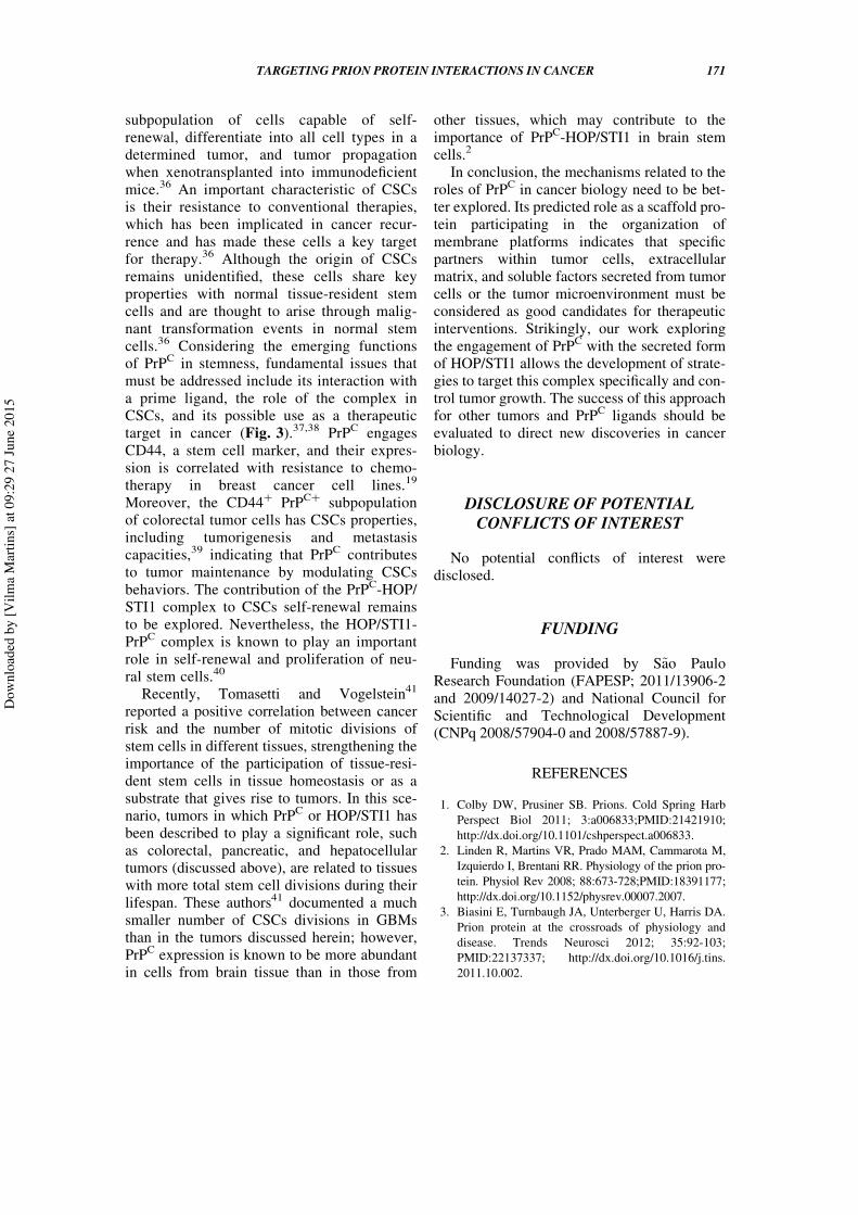
or HOP/STI1 successfully inhibited tumorgrowth in vivo in diverse organs other than thebrain. A preliminary experiment using a HOP/STI1 antibody25 has been performed. As indi-cated in Figure 2, intra-tumor delivery of anti-HOP/STI1 into orthotopic xenografts of GBMcells slightly improved animal survival.
Importantly, however, the blockage of bothPrPC and HOP/STI1 could be deleterious.Long-term,33 but not short-term,29 intracranialinfusion of antibodies against PrPC, in particu-lar those directed to the globular domain ofPrPC, can be neurotoxic.33 The short-term useof polyclonal antibodies against full-lengthHOP/STI1 or the HOP/STI1230–245 peptide hasnot caused brain toxicity.29 However, we dem-onstrated that the constitutive deletion of HOP/STI1 is embryonically lethal, and heterozygousanimals expressing half-levels of the proteinpresented higher sensitivity to brain injury,34
indicating the importance of this protein alsoin adults. Furthermore, maternally derived
FIGURE 2. Disruption of PrPC-HOP/STI1 inter-action using anti-HOP antibodies in xenograftsincreases the survival of animals with glioblas-toma. Preliminary results indicate that disruptionof the PrPC-HOP/STI1 complex using a specificantibody increases animal survival. Briefly,nude mice were injected orthotopically with aU87 cell line (5£105 cells) and treated withHOP/STI1-antibody25 for 28 d (240 ng/day)using osmotic micropumps. The complete meth-odology is described in Lopes et al.28 Kaplan–Meier survival curve of mice implanted with U87cells. Log-rank p D 0.0023, n D 3 mice pergroup.
TARGETING PRION PROTEIN INTERACTIONS IN CANCER 169
Dow
nloa
ded
by [V
ilma
Mar
tins]
at 0
9:29
27
June
201
5

HOP/STI1 autoantibodies were detected inmothers of children with autism, suggestingthat neurodevelopment is impaired by theseautoantibodies.35 Indeed, interference withPrPC-HOP/STI1 interaction in tumors, partic-ularly those in the central nervous system(CNS), using peptides that compete for theirengagement should lead to better results thanthe use of antibodies against these molecules.
TARGETING CANCER STEM CELLSBY BLOCKING PrPC INTERACTIONS
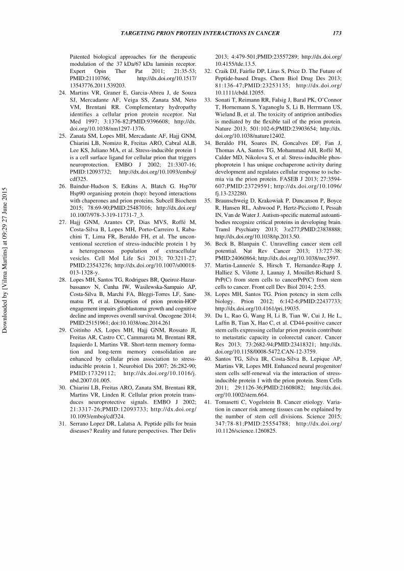
One of the most-studied recent themes inoncology is related to features that governtumor origin, and cancer stem cells (CSCs)have emerged as a pivotal component able toinitiate and maintain tumors.36 CSCs havebeen functionally defined as a small
FIGURE 3. Targeting of PrPC and its partners in CSCs for cancer therapy. Conventional therapytargets tumor cells by destroying them or decreasing their proliferation. Tumor growth is governedby multiple cellular mechanisms in which PrPC plays a role. The progression of tumor developmentis related to the presence of cancer stem cells that have a pivotal role in cell resistance, culminatingin tumor recurrence. Indeed, PrPC and its ligands in cancer stem cells could also be targeted,suggesting a promising approach for novel cancer therapies. Cells with dotted lines represent cellsundergoing cell death.
170 Santos et al.
Dow
nloa
ded
by [V
ilma
Mar
tins]
at 0
9:29
27
June
201
5
subpopulation of cells capable of self-renewal, differentiate into all cell types in adetermined tumor, and tumor propagationwhen xenotransplanted into immunodeficientmice.36 An important characteristic of CSCsis their resistance to conventional therapies,which has been implicated in cancer recur-rence and has made these cells a key targetfor therapy.36 Although the origin of CSCsremains unidentified, these cells share keyproperties with normal tissue-resident stemcells and are thought to arise through malig-nant transformation events in normal stemcells.36 Considering the emerging functionsof PrPC in stemness, fundamental issues thatmust be addressed include its interaction witha prime ligand, the role of the complex inCSCs, and its possible use as a therapeutictarget in cancer (Fig. 3).37,38 PrPC engagesCD44, a stem cell marker, and their expres-sion is correlated with resistance to chemo-therapy in breast cancer cell lines.19
Moreover, the CD44C PrPCC subpopulationof colorectal tumor cells has CSCs properties,including tumorigenesis and metastasiscapacities,39 indicating that PrPC contributesto tumor maintenance by modulating CSCsbehaviors. The contribution of the PrPC-HOP/STI1 complex to CSCs self-renewal remainsto be explored. Nevertheless, the HOP/STI1-PrPC complex is known to play an importantrole in self-renewal and proliferation of neu-ral stem cells.40
Recently, Tomasetti and Vogelstein41
reported a positive correlation between cancerrisk and the number of mitotic divisions ofstem cells in different tissues, strengthening theimportance of the participation of tissue-resi-dent stem cells in tissue homeostasis or as asubstrate that gives rise to tumors. In this sce-nario, tumors in which PrPC or HOP/STI1 hasbeen described to play a significant role, suchas colorectal, pancreatic, and hepatocellulartumors (discussed above), are related to tissueswith more total stem cell divisions during theirlifespan. These authors41 documented a muchsmaller number of CSCs divisions in GBMsthan in the tumors discussed herein; however,PrPC expression is known to be more abundantin cells from brain tissue than in those from
other tissues, which may contribute to theimportance of PrPC-HOP/STI1 in brain stemcells.2
In conclusion, the mechanisms related to theroles of PrPC in cancer biology need to be bet-ter explored. Its predicted role as a scaffold pro-tein participating in the organization ofmembrane platforms indicates that specificpartners within tumor cells, extracellularmatrix, and soluble factors secreted from tumorcells or the tumor microenvironment must beconsidered as good candidates for therapeuticinterventions. Strikingly, our work exploringthe engagement of PrPC with the secreted formof HOP/STI1 allows the development of strate-gies to target this complex specifically and con-trol tumor growth. The success of this approachfor other tumors and PrPC ligands should beevaluated to direct new discoveries in cancerbiology.
DISCLOSURE OF POTENTIALCONFLICTS OF INTEREST
No potential conflicts of interest weredisclosed.
FUNDING
Funding was provided by S~ao PauloResearch Foundation (FAPESP; 2011/13906-2and 2009/14027-2) and National Council forScientific and Technological Development(CNPq 2008/57904-0 and 2008/57887-9).
REFERENCES
1. Colby DW, Prusiner SB. Prions. Cold Spring HarbPerspect Biol 2011; 3:a006833;PMID:21421910;http://dx.doi.org/10.1101/cshperspect.a006833.
2. Linden R, Martins VR, Prado MAM, Cammarota M,Izquierdo I, Brentani RR. Physiology of the prion pro-tein. Physiol Rev 2008; 88:673-728;PMID:18391177;http://dx.doi.org/10.1152/physrev.00007.2007.
3. Biasini E, Turnbaugh JA, Unterberger U, Harris DA.Prion protein at the crossroads of physiology anddisease. Trends Neurosci 2012; 35:92-103;PMID:22137337; http://dx.doi.org/10.1016/j.tins.2011.10.002.
TARGETING PRION PROTEIN INTERACTIONS IN CANCER 171
Dow
nloa
ded
by [V
ilma
Mar
tins]
at 0
9:29
27
June
201
5

4. Martins VR, Beraldo FH, Hajj GN, Lopes MH, LeeKS, Prado MAM, Linden R. Prion protein: orches-trating neurotrophic activities. Curr Issues Mol Biol2010; 12:63-86;PMID:19767651.
5. Linden R, Cordeiro Y, Lima LMTR. Allosteric func-tion and dysfunction of the prion protein. Cell MolLife Sci 2012; 69:1105-24;PMID:21984610; http://dx.doi.org/10.1007/s00018-011-0847-7.
6. Mehrpour M, Codogno P. Prion protein: From physi-ology to cancer biology. Cancer Lett 2010; 290:1-23;PMID:19674833; http://dx.doi.org/10.1016/j.canlet.2009.07.009.
7. Wang J-H, Du J-P, Zhang Y-H, Zhao X-J, Fan R-Y,Wang Z-H, Wu Z-T, Han Y. Dynamic changes andsurveillance function of prion protein expression ingastric cancer drug resistance. World J Gastroenterol2011; 17:3986-93;PMID:22046086; http://dx.doi.org/10.3748/wjg.v17.i35.3986.
8. Zhou L, Shang Y, Liu C, Li J, Hu H, Liang C, Han Y,Zhang W, Liang J, Wu K. Overexpression of PrPc,combined with MGr1-Ag/37LRP, is predictive ofpoor prognosis in gastric cancer. Int J Cancer 2014;135:2329-37;PMID:24706505; http://dx.doi.org/10.1002/ijc.28883.
9. De Wit M, Jimenez CR, Carvalho B, Belien JAM,Delis-van Diemen PM, Mongera S, Piersma SR,Vikas M, Navani S, Ponten F, et al. Cell surface pro-teomics identifies glucose transporter type 1 andprion protein as candidate biomarkers for colorectaladenoma-to-carcinoma progression. Gut 2012;61:855-64;PMID:21890811; http://dx.doi.org/10.1136/gutjnl-2011-300511.
10. Antonacopoulou AG, Grivas PD, Skarlas L, Kalofo-nos M, Scopa CD, Kalofonos HP. POLR2F,ATP6V0A1 and PRNP Expression in Colorectal Can-cer: New Molecules with Prognostic Significance?Anticancer Res 2008; 1228:1221-7.
11. Diarra-Mehrpour M, Arrabal S, Jalil A, Pinson X,Gaudin C, Pietu G, Pitaval A, Ripoche H, Eloit M,Dormont D, et al. Prion protein prevents humanbreast carcinoma cell line from tumor necrosis factora-induced cell death. Cancer Res 2004; 64:719-27;PMID:14744790; http://dx.doi.org/10.1158/0008-5472.CAN-03-1735.
12. Meslin F, Conforti R, Mazouni C, Morel N, TomasicG, Drusch F, Yacoub M, Sabourin JC, Grassi J, Dela-loge S, et al. Efficacy of adjuvant chemotherapyaccording to Prion protein expression in patients withestrogen receptor-negative breast cancer. Ann Oncol2007; 18:1793-8;PMID:17872899; http://dx.doi.org/10.1093/annonc/mdm406.
13. Roucou X, Giannopoulos PN, Zhang Y, Jodoin J,Goodyer CG, LeBlanc A. Cellular prion protein inhib-its proapoptotic Bax conformational change in humanneurons and in breast carcinoma MCF-7 cells. Cell
Death Differ 2005; 12:783-95;PMID:15846375; http://dx.doi.org/10.1038/sj.cdd.4401629.
14. Li Q-Q, Sun Y-P, Ruan C-P, Xu X-Y, Ge J-H, He J,Xu Z-D, Wang Q, Gao W-C. Cellular prion proteinpromotes glucose uptake through the Fyn-HIF-2a-Glut1 pathway to support colorectal cancer cell sur-vival. Cancer Sci 2011; 102:400-6;PMID:21265952;http://dx.doi.org/10.1111/j.1349-7006.2010.01811.x.
15. Wang Q, Qian J, Wang F, Ma Z. Cellular prion pro-tein accelerates colorectal cancer metastasis via theFyn-SP1-SATB1 axis. Oncol Rep 2012; 28: 2029-34;PMID:22972305.
16. Barbieri G, Palumbo S, Gabrusiewicz K, Azzalin A,Marchesi N, Spedito A, Biggiogera M, Sbalchiero E,Mazzini G, Miracco C, et al. Silencing of cellular prionprotein (PrPC) expression by DNA-antisense oligonu-cleotides induces autophagy-dependent cell death in gli-oma cells. Autophagy 2011; 7:840-53;PMID:21478678;http://dx.doi.org/10.4161/auto.7.8.15615.
17. Roffe M, Beraldo FH, Bester R, Nunziante M, BachC, Mancini G, Gilch S, Vorberg I, Castilho BA, Mar-tins VR, et al. Prion protein interaction with stress-inducible protein 1 enhances neuronal protein synthe-sis via mTOR. Proc Natl Acad Sci U S A 2010;107:13147-52;PMID:20615969; http://dx.doi.org/10.1073/pnas.1000784107.
18. Li QQ, Cao XX, Xu JD, Chen Q, Wang WJ, Tang F,Chen ZQ, Liu XP, Xu ZD. The role of P-glycoprotein/cellular prion protein interaction in multidrug-resistantbreast cancer cells treated with paclitaxel. Cell MolLife Sci 2009; 66:504-15;PMID:19099191; http://dx.doi.org/10.1007/s00018-008-8548-6.
19. Cheng Y, Tao L, Xu J, Li Q, Yu J, Jin Y, Chen Q, XuZ, Zou Q, Liu X. CD44/cellular prion protein interactin multidrug resistant breast cancer cells and corre-late with responses to neoadjuvant chemotherapy inbreast cancer patients. Mol. Carcinog 2014; 53:686-97;http://dx.doi.org/10.1002/mc.22021.
20. Li C, Xin W, Sy M-S. Binding of pro-prion to filaminA: by design or an unfortunate blunder. Oncogene2010; 29:5329-45;PMID:20697352; http://dx.doi.org/10.1038/onc.2010.307.
21. Li C, Yu S, Nakamura F, Yin S, Xu J, Petrolla AA,Singh N, Tartakoff A, Abbott DW, Xin W, et al. Bind-ing of pro-prion to filamin A disrupts cytoskeleton andcorrelates with poor prognosis in pancreatic cancer. JClin Invest 2009; 119:2725-36;PMID:19690385;http://dx.doi.org/10.1172/JCI39542.
22. Mbazima V, Da Costa Dias B, Omar A, Jovanovic K,Weiss SFT. Interactions between PrP(c) and otherligands with the 37-kDa/67-kDa laminin receptor.Front Biosci 2010; 15:1150-63;http://dx.doi.org/10.2741/3667.
23. Omar A, Jovanovic K, Da Costa Dias B, Gonsalves D,Moodley K, Caveney R, Mbazima V, Weiss SFT.
172 Santos et al.
Dow
nloa
ded
by [V
ilma
Mar
tins]
at 0
9:29
27
June
201
5
Patented biological approaches for the therapeuticmodulation of the 37 kDa/67 kDa laminin receptor.Expert Opin Ther Pat 2011; 21:35-53;PMID:21110766; http://dx.doi.org/10.1517/13543776.2011.539203.
24. Martins VR, Graner E, Garcia-Abreu J, de SouzaSJ, Mercadante AF, Veiga SS, Zanata SM, NetoVM, Brentani RR. Complementary hydropathyidentifies a cellular prion protein receptor. NatMed 1997; 3:1376-82;PMID:9396608; http://dx.doi.org/10.1038/nm1297-1376.
25. Zanata SM, Lopes MH, Mercadante AF, Hajj GNM,Chiarini LB, Nomizo R, Freitas ARO, Cabral ALB,Lee KS, Juliano MA, et al. Stress-inducible protein 1is a cell surface ligand for cellular prion that triggersneuroprotection. EMBO J 2002; 21:3307-16;PMID:12093732; http://dx.doi.org/10.1093/emboj/cdf325.
26. Baindur-Hudson S, Edkins A, Blatch G. Hsp70/Hsp90 organising protein (hop): beyond interactionswith chaperones and prion proteins. Subcell Biochem2015; 78:69-90;PMID:25487016; http://dx.doi.org/10.1007/978-3-319-11731-7_3.
27. Hajj GNM, Arantes CP, Dias MVS, Roffe M,Costa-Silva B, Lopes MH, Porto-Carreiro I, Raba-chini T, Lima FR, Beraldo FH, et al. The uncon-ventional secretion of stress-inducible protein 1 bya heterogeneous population of extracellularvesicles. Cell Mol Life Sci 2013; 70:3211-27;PMID:23543276; http://dx.doi.org/10.1007/s00018-013-1328-y.
28. Lopes MH, Santos TG, Rodrigues BR, Queiroz-Hazar-bassanov N, Cunha IW, Wasilewska-Sampaio AP,Costa-Silva B, Marchi FA, Bleggi-Torres LF, Sane-matsu PI, et al. Disruption of prion protein-HOPengagement impairs glioblastoma growth and cognitivedecline and improves overall survival. Oncogene 2014;PMID:25151961; doi:10.1038/onc.2014.261
29. Coitinho AS, Lopes MH, Hajj GNM, Rossato JI,Freitas AR, Castro CC, Cammarota M, Brentani RR,Izquierdo I, Martins VR. Short-term memory forma-tion and long-term memory consolidation areenhanced by cellular prion association to stress-inducible protein 1. Neurobiol Dis 2007; 26:282-90;PMID:17329112; http://dx.doi.org/10.1016/j.nbd.2007.01.005.
30. Chiarini LB, Freitas ARO, Zanata SM, Brentani RR,Martins VR, Linden R. Cellular prion protein trans-duces neuroprotective signals. EMBO J 2002;21:3317-26;PMID:12093733; http://dx.doi.org/10.1093/emboj/cdf324.
31. Serrano Lopez DR, Lalatsa A. Peptide pills for braindiseases? Reality and future perspectives. Ther Deliv
2013; 4:479-501;PMID:23557289; http://dx.doi.org/10.4155/tde.13.5.
32. Craik DJ, Fairlie DP, Liras S, Price D. The Future ofPeptide-based Drugs. Chem Biol Drug Des 2013;81:136-47;PMID:23253135; http://dx.doi.org/10.1111/cbdd.12055.
33. Sonati T, Reimann RR, Falsig J, Baral PK, O’ConnorT, Hornemann S, Yaganoglu S, Li B, Herrmann US,Wieland B, et al. The toxicity of antiprion antibodiesis mediated by the flexible tail of the prion protein.Nature 2013; 501:102-6;PMID:23903654; http://dx.doi.org/10.1038/nature12402.
34. Beraldo FH, Soares IN, Goncalves DF, Fan J,Thomas AA, Santos TG, Mohammad AH, Roffe M,Calder MD, Nikolova S, et al. Stress-inducible phos-phoprotein 1 has unique cochaperone activity duringdevelopment and regulates cellular response to ische-mia via the prion protein. FASEB J 2013; 27:3594-607;PMID:23729591; http://dx.doi.org/10.1096/fj.13-232280.
35. Braunschweig D, Krakowiak P, Duncanson P, BoyceR, Hansen RL, Ashwood P, Hertz-Picciotto I, PessahIN, Van deWater J. Autism-specific maternal autoanti-bodies recognize critical proteins in developing brain.Transl Psychiatry 2013; 3:e277;PMID:23838888;http://dx.doi.org/10.1038/tp.2013.50.
36. Beck B, Blanpain C. Unravelling cancer stem cellpotential. Nat Rev Cancer 2013; 13:727-38;PMID:24060864; http://dx.doi.org/10.1038/nrc3597.
37. Martin-Lanneree S, Hirsch T, Hernandez-Rapp J,Halliez S, Vilotte J, Launay J, Mouillet-Richard S.PrP(C) from stem cells to cancerPrP(C) from stemcells to cancer. Front cell Dev Biol 2014; 2:55.
38. Lopes MH, Santos TG. Prion potency in stem cellsbiology. Prion 2012; 6:142-6;PMID:22437733;http://dx.doi.org/10.4161/pri.19035.
39. Du L, Rao G, Wang H, Li B, Tian W, Cui J, He L,Laffin B, Tian X, Hao C, et al. CD44-positive cancerstem cells expressing cellular prion protein contributeto metastatic capacity in colorectal cancer. CancerRes 2013; 73:2682-94;PMID:23418321; http://dx.doi.org/10.1158/0008-5472.CAN-12-3759.
40. Santos TG, Silva IR, Costa-Silva B, Lepique AP,Martins VR, Lopes MH. Enhanced neural progenitor/stem cells self-renewal via the interaction of stress-inducible protein 1 with the prion protein. Stem Cells2011; 29:1126-36;PMID:21608082; http://dx.doi.org/10.1002/stem.664.
41. Tomasetti C, Vogelstein B. Cancer etiology. Varia-tion in cancer risk among tissues can be explained bythe number of stem cell divisions. Science 2015;347:78-81;PMID:25554788; http://dx.doi.org/10.1126/science.1260825.
TARGETING PRION PROTEIN INTERACTIONS IN CANCER 173
Dow
nloa
ded
by [V
ilma
Mar
tins]
at 0
9:29
27
June
201
5
Related Documents


![Interactions between anesthetics and lipid rafts€¦ · Modifications of lipid rafts may lead to diseases like Alzheimer, Parkinson, prion diseases and cancer [10], [11], [12].](https://static.cupdf.com/doc/110x72/604dc890a58b7f65d734c520/interactions-between-anesthetics-and-lipid-rafts-modiications-of-lipid-rafts-may.jpg)