Targeting Protein-Protein Interactions: Suppression of Stat3 Dimerization with Rationally Designed Small-Molecule, Nonpeptidic SH2 Domain Binders Patrick T. Gunning [a] , Matthew P. Glenn [a] , Khandaker A. Z. Siddiquee [b] , William P. Katt [a] , Eric Masson [a] , Saïd M. Sebti [c] , James Turkson [b] , and Andrew D. Hamilton [a] [a] Dr. P. T. Gunning, Dr. M. P. Glenn, W. P. Katt, Dr. E. Masson, Prof. A. D. Hamilton, Department of Chemistry, Yale University, P.O. Box 208017, New Haven, CT 06520-8107 (USA), Fax: (+ 1) 203-432-3221 [b] Dr. K. A. Z. Siddiquee, Prof. J. Turkson, Department of Molecular Biology and Microbiology, University of Central Florida, Orlando, FL 32826 (USA) [c] Prof. S. M. Sebti, Drug Discovery Program, H. Lee Moffitt Cancer Center and Research Institute, Department of Oncology and Department of Biochemistry and Molecular Biology, University of South Florida, Tampa, FL 33612 (USA) Keywords antitumor agents; molecular recognition; protein-protein interactions; small molecules; Stat3 Protein-protein interactions remain a daunting target for disruption by small molecules due to their large interfacial areas and their often noncontiguous contact points. The prospect for inhibition increases when small interaction modules (such as SH2 domains) participate in the binding. SH2 domains are found in the family of signal transducers and activators of transcription (STAT) proteins,[1] which mediate the relay of extracellular signals from various cell-surface protein receptors to the nucleus, where they help to initiate and regulate specific gene expression.[2] In particular, the Stat3 protein is known to directly upregulate Bcl-x L , c- Myc, Mcl-1, VEGF, and cyclin D1/D2, and contributes directly to compromised cellular regulation by stimulating cell proliferation and preventing apoptosis in numerous human cancers.[2,3] Stat3 activation occurs through phosphorylation of tyrosine 705, which promotes the formation of a Stat3 dimer through reciprocal Stat3 phosphotyrosine 705-SH2 domain interactions.[4] In general, STAT dimers then translocate to the nucleus, where they regulate unique gene expression programs through interaction with specific DNA-response elements. Stat3-targeted gene expression confers resistance to apoptosis in many tumor cells and promotes cell survival, which contributes to the resistance of these cancers to currently available chemotherapeutics. [5] Successful Stat3 inhibitors might thus be used to sensitize human cancers that harbor constitutively active Stat3 to existing chemotherapeutic agents. Further, the specificity of these inhibitors toward Stat3 might potentially reduce the side effects that are associated with conventional, aggressive chemotherapy. © 2008 Wiley-VCH Verlag GmbH & Co. KGaA, Weinheim E-mail: [email protected]. NIH Public Access Author Manuscript Chembiochem. Author manuscript; available in PMC 2009 August 18. Published in final edited form as: Chembiochem. 2008 November 24; 9(17): 2800–2803. doi:10.1002/cbic.200800291. NIH-PA Author Manuscript NIH-PA Author Manuscript NIH-PA Author Manuscript

Welcome message from author
This document is posted to help you gain knowledge. Please leave a comment to let me know what you think about it! Share it to your friends and learn new things together.
Transcript
Targeting Protein-Protein Interactions:Suppression of Stat3 Dimerization with Rationally Designed Small-Molecule, Nonpeptidic
SH2 Domain Binders
Patrick T. Gunning[a], Matthew P. Glenn[a], Khandaker A. Z. Siddiquee[b], William P. Katt[a],Eric Masson[a], Saïd M. Sebti[c], James Turkson[b], and Andrew D. Hamilton[a][a]Dr. P. T. Gunning, Dr. M. P. Glenn, W. P. Katt, Dr. E. Masson, Prof. A. D. Hamilton, Departmentof Chemistry, Yale University, P.O. Box 208017, New Haven, CT 06520-8107 (USA), Fax: (+ 1)203-432-3221[b]Dr. K. A. Z. Siddiquee, Prof. J. Turkson, Department of Molecular Biology and Microbiology,University of Central Florida, Orlando, FL 32826 (USA)[c]Prof. S. M. Sebti, Drug Discovery Program, H. Lee Moffitt Cancer Center and Research Institute,Department of Oncology and Department of Biochemistry and Molecular Biology, University ofSouth Florida, Tampa, FL 33612 (USA)
Keywordsantitumor agents; molecular recognition; protein-protein interactions; small molecules; Stat3
Protein-protein interactions remain a daunting target for disruption by small molecules due totheir large interfacial areas and their often noncontiguous contact points. The prospect forinhibition increases when small interaction modules (such as SH2 domains) participate in thebinding. SH2 domains are found in the family of signal transducers and activators oftranscription (STAT) proteins,[1] which mediate the relay of extracellular signals from variouscell-surface protein receptors to the nucleus, where they help to initiate and regulate specificgene expression.[2] In particular, the Stat3 protein is known to directly upregulate Bcl-xL, c-Myc, Mcl-1, VEGF, and cyclin D1/D2, and contributes directly to compromised cellularregulation by stimulating cell proliferation and preventing apoptosis in numerous humancancers.[2,3]
Stat3 activation occurs through phosphorylation of tyrosine 705, which promotes the formationof a Stat3 dimer through reciprocal Stat3 phosphotyrosine 705-SH2 domain interactions.[4] Ingeneral, STAT dimers then translocate to the nucleus, where they regulate unique geneexpression programs through interaction with specific DNA-response elements. Stat3-targetedgene expression confers resistance to apoptosis in many tumor cells and promotes cell survival,which contributes to the resistance of these cancers to currently available chemotherapeutics.[5] Successful Stat3 inhibitors might thus be used to sensitize human cancers that harborconstitutively active Stat3 to existing chemotherapeutic agents. Further, the specificity of theseinhibitors toward Stat3 might potentially reduce the side effects that are associated withconventional, aggressive chemotherapy.
© 2008 Wiley-VCH Verlag GmbH & Co. KGaA, WeinheimE-mail: [email protected].
NIH Public AccessAuthor ManuscriptChembiochem. Author manuscript; available in PMC 2009 August 18.
Published in final edited form as:Chembiochem. 2008 November 24; 9(17): 2800–2803. doi:10.1002/cbic.200800291.
NIH
-PA Author Manuscript
NIH
-PA Author Manuscript
NIH
-PA Author Manuscript
Despite the difficulties in identifying protein surface-recognition agents, the promise of Stat3modulators warrants investigation.[6] Successful peptidic[7a-f] and nonpeptidomimetic smallmolecules[8-11] that are capable of targeting malignant cell lines with constitutively activatedStat3 protein are limited to a few examples that include Stattic,[8] STA-21,[9] and S3I-201,[10] which were all identified through high-throughput virtual or biochemical screeningapproaches. Our first-generation designs were simple peptidomimetics derived from the naturalsequence, of which ISS610 was the most potent (see Figure 1).[7f] More recently we havediscovered S3I-M2001 (15, Table 1) an oxazole-based small-molecule inhibitor that showspromising inhibition of Stat3 function,[11] and we herein report a family of rationally designedsmall-molecule, nonpeptidic Stat3 inhibitors. These agents inhibit Stat3 protein dimerizationand induce apoptosis in Stat3-transformed cells and Stat3-dependent breast oncogenic celllines.
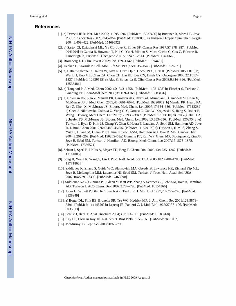
The crystal structure of the Stat3-SH2 domain reveals a shallow triangular pocket that iscomposed of two hydrophobic sites and a hydrophilic phosphate-recognition pocket. Dockingstudies on our initial lead, peptidomimetic ISS610, showed that only two of the hydrophobicpockets in the binding domain were effectively occupied (See Figure 1 A).[7f] To address thisproblem we envisaged that trisubstituted heterocyclic scaffolds, such as oxazoles and thiazoles,could effectively access all three sites (Figure 1 B). Flexible ligand-docking studies (GOLD)directed the design and assembly of oxazole and thiazole scaffolds. The oxazole inhibitorswere prepared as outlined in Scheme 1 (see the Supporting Information for thiazole synthesis).GOLD docking studies predicted a focused set of substituents that might effectively occupythe SH2 domain. Both R1 and R2 appendages need to be predominantly hydrophobic in natureto interact with the hydrophobic surface presented by residues that are present in the upper(Phe716, Met660, Pro715) and lower right (Ser636, Arg595, Lys591) pockets of the Stat3active site (Figures 1 A, B).
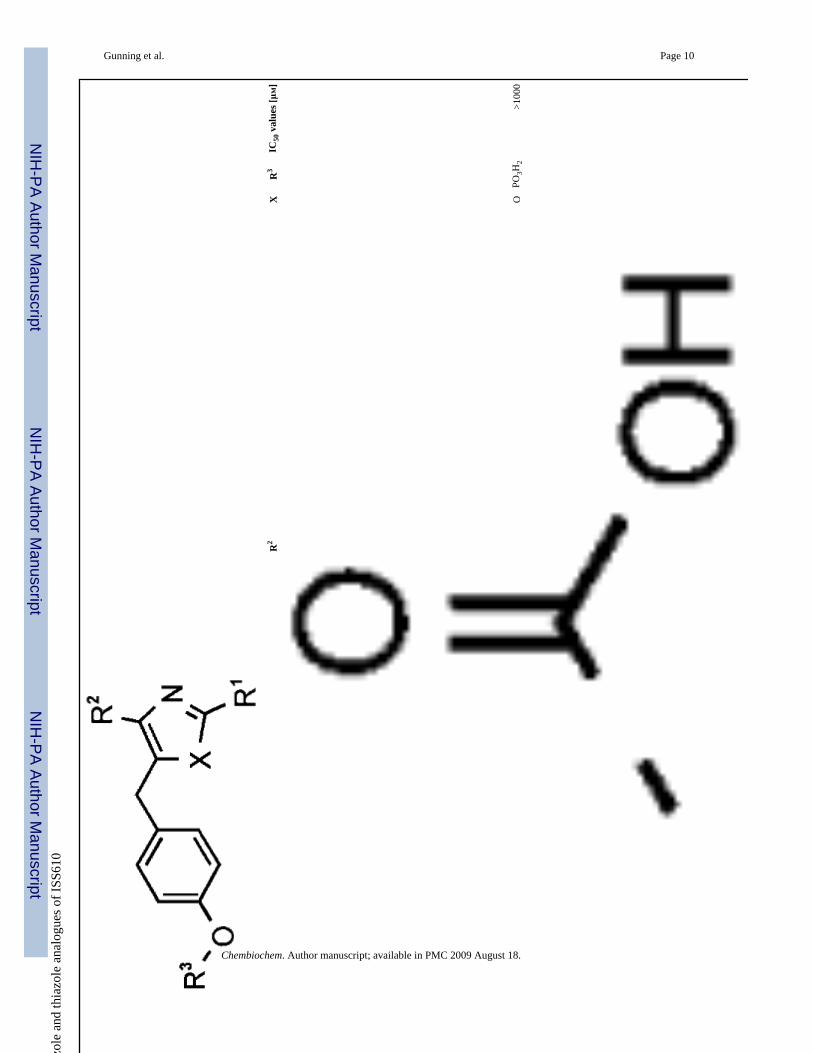
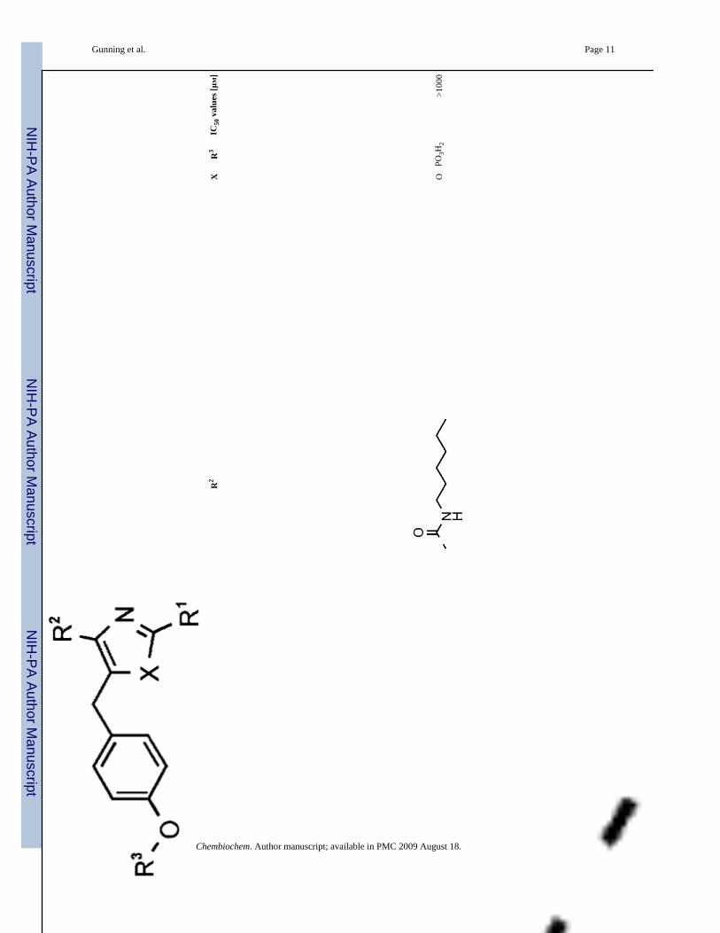
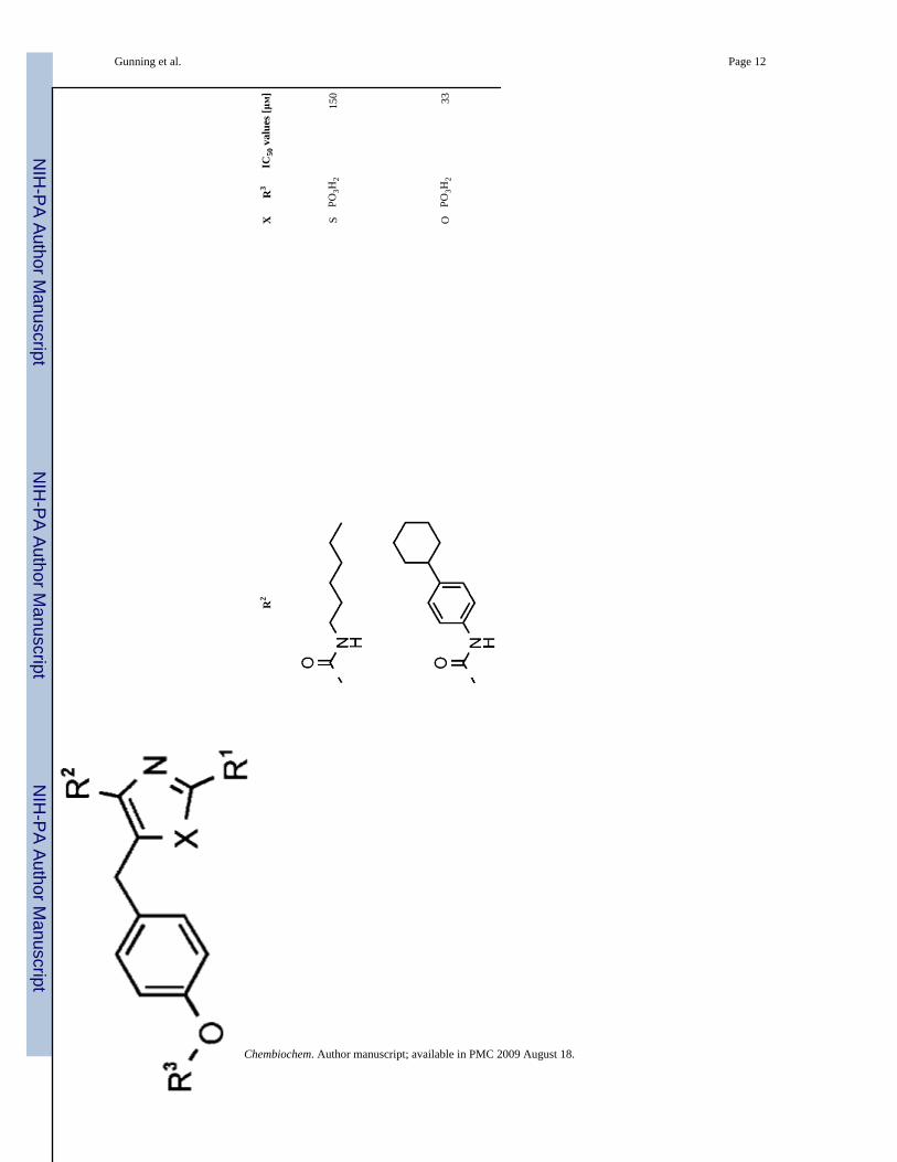
Our initial screening process sought to evaluate in vitro disruption of Stat3:Stat3-DNA complexformation through a previously published in vitro DNA-binding EMSA-based assay,[7e] whichwould give valuable insight into inhibitor action against the Stat3 in terms of disruption ofStat3:Stat3 homodimer. An initial series of Stat3 oxazole and thiazole inhibitors is shown inTable 1. Oxazole 7 was prepared as a control to explore the efficacy of compounds that do notproject sizable appendages from the R2 position. It closely resembles lead peptidomimeticISS610. Stat3-DNA binding inhibition by 7 was shown to be negligible, presumably due topoor inhibitor-protein complementarity. Substitution of ISS610 at the acidic terminus had beenshown previously to decrease potency in all cases studied; this suggests that the oxazolescaffold is able to project substituents in a substantially different orientation than could beachieved with a peptide-based inhibitor (Figure 1). Five compounds displayed IC50 valuesbelow 100 μM, with oxazole 10 (IC50 = 33 ± 16 μM), and thiazoles 14 and 16 showing significantdimer-disruption potential.
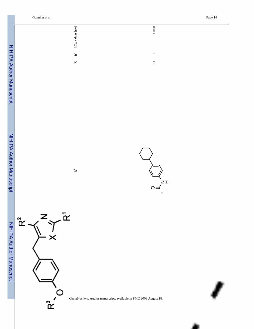
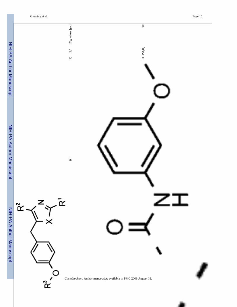
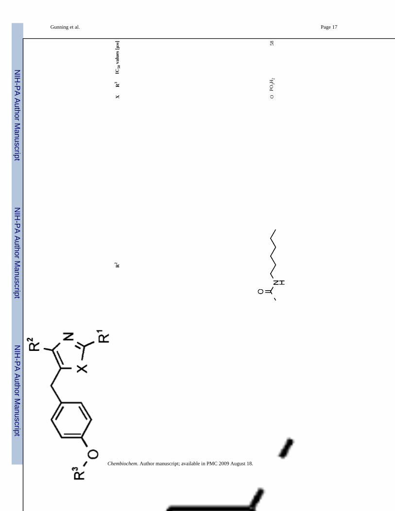
In several cases large effects were observed upon replacement of the central core. This is seenin 10 and 11, where the high potency displayed by the oxazole analogue 10 (IC50 = 33 ± 16μM; Figure 2A) is severely diminished in the analogous thiazole 11 (IC50 = 775 μM). Conversely,thiazole 16 (IC50 = 25 μM) has improved activity compared to corresponding oxazole 15(IC50 = 58 ± 10 μM). Molecular modeling studies showed that the thiazole scaffolds facilitatedimproved projection, and concomitant higher complementarity of the R1, R2, and R3 groupsto the protein surface. In almost all examples, increased potency is derived from a thiazolecore. Oxazole 12, which contains the most potent arrangement of R1 and R2 elements, but lacksa phosphate group, was shown to be devoid of activity. Further nonphosphorylated scaffoldswere prepared and found to be impotent Stat3 disruptors.
Gunning et al. Page 2
Chembiochem. Author manuscript; available in PMC 2009 August 18.
NIH
-PA Author Manuscript
NIH
-PA Author Manuscript
NIH
-PA Author Manuscript
Given the extended planar aryl-oxazole construct and the propensity of such scaffolds tointercalate with DNA, experiments were conducted to discount interaction with DNA as asource of the biological activity.[13] By using fluorescence spectroscopy it was found that theamount of DNA-bound ethidium bromide did not vary upon addition of 10 (up toconcentrations of 100 μM); this compound can thus be considered to be inert toward DNA (Seethe Supporting Information).
Work was undertaken to establish the affinity of the lead oxazole 10 for the inactive(unphosphorylated) Stat3 monomer to help determine the mode of action. By usingfluorescence spectroscopy, Ki values were calculated through displacement of an SH2 bindingfluorescein-labeled GpYLPQTV-NH2 peptide.[14] The most potent inhibitors were found tohave low affinity with the unphosphorylated Stat3 monomer (10 and 15, Ki ∼ 1mM;Figure 3).These results were attributed to the smaller size of the oxazole scaffolds relative to thephosphopeptide, which is predicted to make contacts with both the BG and EF loops thatrecognize ligand residues at the pTyr + 3 site.[15,16] Structural limitations of the inhibitorsmight preclude effective interactions with the SH2 domain and reduce their ability to displacethe extended peptide sequence.
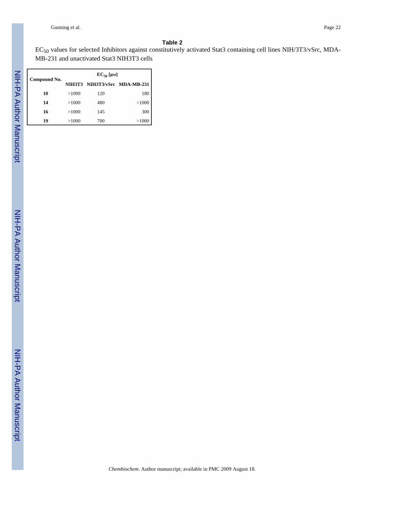
Despite the inherent difficulties associated with the cell permeability of phosphate derivatives,we observed promising whole-cell activities for several lead compounds. Initial in vitro whole-cell experiments were conducted against normal mouse NIH-3T3 fibroblasts and v-src-transformed counterparts (NIH-3T3/v-src) that harbor aberrant Stat3. Inhibitor effects uponcell viability, proliferation, and cytotoxicity were assessed through WST-1, a cell proliferationreagent that measures the metabolic activity of viable cells. The most potent in vitro inhibitor,10, displayed at least ten-fold selectivity toward malignant NIH-3T3/v-src fibroblasts withaberrant Stat3 (EC50 = 120 μM) and negligible effects toward normal NIH-3T3 fibroblasts, inwhich Stat3 pathways are tightly regulated. Conversely, the suppression by leadpeptidomimetic ISS610 in whole cells required millimolar concentrations of inhibitor. It isassumed that thepredominantly hydrophobic nature of our compounds facilitated successfulpermeation of the cell membrane despite the phosphate.
In addition, cell-based EC50 values were determined for potent inhibitors against human breast(MDA-MB-231) cancer cell lines (Table 2). Oxazole 10 had the best activity against breastcancer (ED50 = 180 μM) and NIH-3T3/v-src cells (ED50 = 120 μM) and lacked toxicity for controlNIH-3T3 cells.
In summary, we have developed the first rationally designed small-molecule inhibitors of Stat3dimerization and thereby have disrupted Stat3-mediated cell proliferation pathways. Suitablysubstituted oxazole and thiazole scaffolds, derived from peptidomimetic leads, disruptedStat3:Stat3-DNA-binding activity in vitro at low micromolar concentrations, but showed lowaffinity to the unphosphorylated Stat3 monomer. This might suggest that our compoundspreferentially bind with activated Stat3, which is further supported by the lack of activityagainst control cells (NIH-3T3); this still remains to be verified. Lead agents showed potencyand selectivity against specific human cancer cell lines and negligible toxicity towards normalNIH-3T3 fibroblasts. Our studies highlighted the widely acknowledged belief that Stat3 is apotent target for disruption by small-molecule inhibitors for novel anticancer drug developmentand that targeting of Stat3 will require a higher level of investigation.
AcknowledgementsThe authors would like to thank the National Cancer Institute for grant CA78038.
Gunning et al. Page 3
Chembiochem. Author manuscript; available in PMC 2009 August 18.
NIH
-PA Author Manuscript
NIH
-PA Author Manuscript
NIH
-PA Author Manuscript
References[1]. a) Darnell JE Jr. Nat. Med 2005;11:595–596. [PubMed: 15937466] b) Buettner R, Mora LB, Jove
R. Clin. Cancer Res 2002;8:945–954. [PubMed: 11948098] c) Turkson J. Expert Opin. Ther. Targets2004;8:409–422. [PubMed: 15469392]
[2]. a) Sartor CI, Dziubinski ML, Yu CL, Jove R, Ethier SP. Cancer Res 1997;57:978–987. [PubMed:9041204] b) Garcia R, Bowman T, Nui G, Yu H, Minton S, Muro-Cacho C, Cox C, Falcone R,Fairclough R, Parsons S. Oncogene 2001;20:2499–2513. [PubMed: 11420660]
[3]. Bromberg J. J. Clin. Invest 2002;109:1139–1142. [PubMed: 11994401][4]. Decker T, Kovarik P. Cell. Mol. Life Sci 1999;55:1535–1546. [PubMed: 10526571][5]. a) Catlett-Falcone R, Dalton W, Jove R. Curr. Opin. Oncol 1999;11:490. [PubMed: 10550013] b)
Wei LH, Kuo ML, Chen CA, Chou CH, Lai KB, Lee CN, Hsieh CY. Oncogene 2003;22:1517–1527. [PubMed: 12629515] c) Alas S, Bonavida B. Clin. Cancer Res 2003;9:316–326. [PubMed:12538484]
[6]. a) Toogood P. J. Med. Chem 2002;45:1543–1558. [PubMed: 11931608] b) Fletcher S, Turkson J,Gunning PT. ChemMedChem 2008;3:1159–1168. [PubMed: 18683176]
[7]. a) Coleman DR, Ren Z, Mandal PK, Cameron AG, Dyer GA, Muranjan S, Campbell M, Chen X,McMurray JS. J. Med. Chem 2005;48:6661–6670. [PubMed: 16220982] b) Mandal PK, Heard PA,Ren Z, Chen X, McMurray JS. Bioorg. Med. Chem. Lett 2007;17:654–656. [PubMed: 17113289]c) Chen J, Nikolovska-Coleska Z, Yang C-Y, Gomez C, Gao W, Krajewski K, Jiang S, Roller P,Wang S. Bioorg. Med. Chem. Lett 2007;17:3939–3942. [PubMed: 17513110] d) Ren Z, Cabell LA,Schaefer TS, McMurray JS. Bioorg. Med. Chem. Lett 2003;13:633–636. [PubMed: 12639546] e)Turkson J, Ryan D, Kim JS, Zhang Y, Chen Z, Haura E, Laudano A, Sebti SM, Hamilton AD, JoveR. J. Biol. Chem 2001;276:45443–45455. [PubMed: 11579100] f) Turkson J, Kim JS, Zhang S,Yuan J, Huang M, Glenn MP, Haura E, Sebti ASM, Hamilton AD, Jove R. Mol. Cancer Ther2004;3:261–269. [PubMed: 15026546] g) Gunning PT, Katt WP, Glenn MP, Siddiquee K, Kim JS,Jove R, Sebti SM, Turkson J, Hamilton AD. Bioorg. Med. Chem. Lett 2007;17:1875–1878.[PubMed: 17336521]
[8]. Schust J, Sperl B, Hollis A, Mayer TU, Berg T. Chem. Biol 2006;13:1235–1242. [PubMed:17114005]
[9]. Song H, Wang R, Wang S, Lin J. Proc. Natl. Acad. Sci. USA 2005;102:4700–4705. [PubMed:15781862]
[10]. Siddiquee K, Zhang S, Guida WC, Blaskovich MA, Greedy B, Lawrence HR, Richard Yip ML,Jove R, McLaughlin MM, Lawrence NJ, Sebti SM, Turkson J. Proc. Natl. Acad. Sci. USA2007;104:7391–7396. [PubMed: 17463090]
[11]. Siddiquee KAZ, Gunning PT, Glenn M, Katt WP, Zhang S, Schroeck C, Sebti SM, Jove R, HamiltonAD, Turkson J. ACS Chem. Biol 2007;2:787–798. [PubMed: 18154266]
[12]. Jones G, Willett P, Glen RC, Leach AR, Taylor R. J. Mol. Biol 1997;267:727–748. [PubMed:9126849]
[13]. a) Boger DL, Fink BE, Brunette SR, Tse WC, Hedrick MP. J. Am. Chem. Soc 2001;123:5878–5891. [PubMed: 11414820] b) Lepecq JB, Paoletti C. J. Mol. Biol 1967;27:87–106. [PubMed:6033613]
[14]. Schust J, Berg T. Anal. Biochem 2004;330:114–118. [PubMed: 15183768][15]. Kay LE, Forman Kay JD. Nat. Struct. Biol 1998;5:156–163. [PubMed: 9461082][16]. McMurray JS. Pept. Sci 2008;90:69–79.
Gunning et al. Page 4
Chembiochem. Author manuscript; available in PMC 2009 August 18.
NIH
-PA Author Manuscript
NIH
-PA Author Manuscript
NIH
-PA Author Manuscript
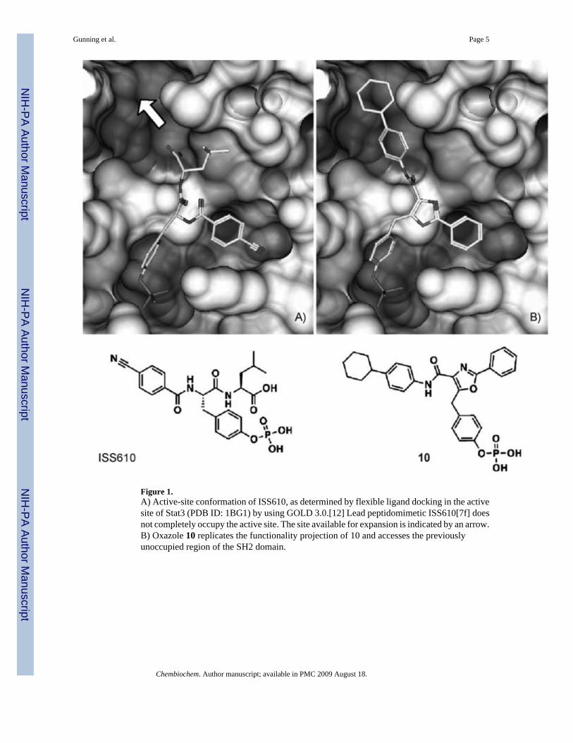
Figure 1.A) Active-site conformation of ISS610, as determined by flexible ligand docking in the activesite of Stat3 (PDB ID: 1BG1) by using GOLD 3.0.[12] Lead peptidomimetic ISS610[7f] doesnot completely occupy the active site. The site available for expansion is indicated by an arrow.B) Oxazole 10 replicates the functionality projection of 10 and accesses the previouslyunoccupied region of the SH2 domain.
Gunning et al. Page 5
Chembiochem. Author manuscript; available in PMC 2009 August 18.
NIH
-PA Author Manuscript
NIH
-PA Author Manuscript
NIH
-PA Author Manuscript
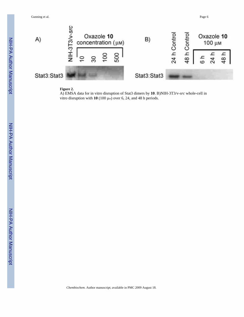
Figure 2.A) EMSA data for in vitro disruption of Stat3 dimers by 10. B)NIH-3T3/v-src whole-cell invitro disruption with 10 (100 μM) over 6, 24, and 48 h periods.
Gunning et al. Page 6
Chembiochem. Author manuscript; available in PMC 2009 August 18.
NIH
-PA Author Manuscript
NIH
-PA Author Manuscript
NIH
-PA Author Manuscript
Figure 3.Competitive binding experiment between the fluorescent probe 5-carboxyfluorescein-GpYLPQTV-NH2 and oxazoles 10, 15, 17 as well as unlabeled Ac-pYLPQTV-NH2 peptideto Stat3 protein. Whereas Ac-pYLPQTV-NH2 displays a 200 nM affinity towards Stat3,oxazoles 10, 15, and 17 do not interact significantly with Stat3.
Gunning et al. Page 7
Chembiochem. Author manuscript; available in PMC 2009 August 18.
NIH
-PA Author Manuscript
NIH
-PA Author Manuscript
NIH
-PA Author Manuscript
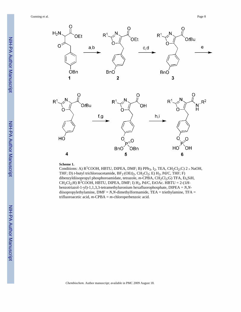
Scheme 1.Conditions: A) R1COOH, HBTU, DIPEA, DMF; B) PPh3, I2, TEA, CH2Cl2;C) 2 N NaOH,THF; D) t-butyl trichloroacetamide, BF3·(OEt)2, CH2Cl2; E) H2, Pd/C, THF; F)dibenzyldiisopropyl phosphoroamidate, tetrazole, m-CPBA, CH2Cl2;G) TFA, EtsSiH,CH2Cl2;H) R2COOH, HBTU, DIPEA, DMF; I) H2, Pd/C, EtOAc. HBTU = 2-(1H-benzotriazol-1-yl)-1,1,3,3-tetramethyluronium hexafluorophosphate, DIPEA = N,N-diisopropylethylamine, DMF = N,N-dimethylformamide, TEA = triethylamine, TFA =trifluoroacetic acid, m-CPBA = m-chloroperbenzoic acid.
Gunning et al. Page 8
Chembiochem. Author manuscript; available in PMC 2009 August 18.
NIH
-PA Author Manuscript
NIH
-PA Author Manuscript
NIH
-PA Author Manuscript

NIH
-PA Author Manuscript
NIH
-PA Author Manuscript
NIH
-PA Author Manuscript
Gunning et al. Page 9Ta
ble
1
Chembiochem. Author manuscript; available in PMC 2009 August 18.
NIH
-PA Author Manuscript
NIH
-PA Author Manuscript
NIH
-PA Author Manuscript
Gunning et al. Page 10IC
50 v
alue
s (μM
) for
the
inhi
bitio
n of
Sta
t3 D
NA
-bin
ding
act
ivity
in v
itro
via
oxaz
ole
and
thia
zole
ana
logu
es o
f ISS
610
Com
poun
d N
umbe
rR
1R
2X
R3
IC50
val
ues [μM
]
7O
PO3H
2>1
000
Chembiochem. Author manuscript; available in PMC 2009 August 18.
NIH
-PA Author Manuscript
NIH
-PA Author Manuscript
NIH
-PA Author Manuscript
Gunning et al. Page 11
Com
poun
d N
umbe
rR
1R
2X
R3
IC50
val
ues [μM
]
8O
PO3H
2>1
000
Chembiochem. Author manuscript; available in PMC 2009 August 18.
NIH
-PA Author Manuscript
NIH
-PA Author Manuscript
NIH
-PA Author Manuscript
Gunning et al. Page 12
Com
poun
d N
umbe
rR
1R
2X
R3
IC50
val
ues [μM
]
9S
PO3H
215
0
10O
PO3H
233
Chembiochem. Author manuscript; available in PMC 2009 August 18.
NIH
-PA Author Manuscript
NIH
-PA Author Manuscript
NIH
-PA Author Manuscript
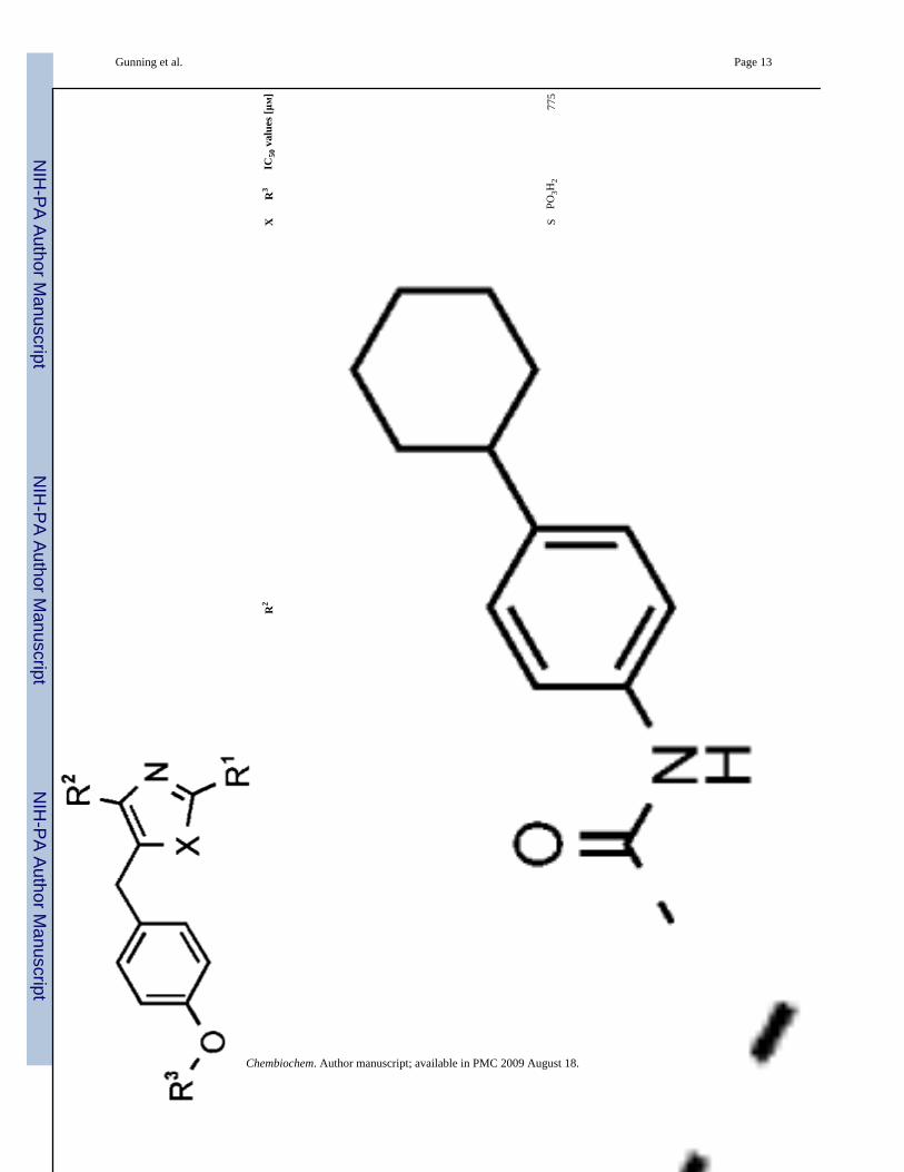
Gunning et al. Page 13
Com
poun
d N
umbe
rR
1R
2X
R3
IC50
val
ues [μM
]
11S
PO3H
277
5
Chembiochem. Author manuscript; available in PMC 2009 August 18.
NIH
-PA Author Manuscript
NIH
-PA Author Manuscript
NIH
-PA Author Manuscript
Gunning et al. Page 14
Com
poun
d N
umbe
rR
1R
2X
R3
IC50
val
ues [μM
]
12O
H>1
000
Chembiochem. Author manuscript; available in PMC 2009 August 18.
NIH
-PA Author Manuscript
NIH
-PA Author Manuscript
NIH
-PA Author Manuscript
Gunning et al. Page 15
Com
poun
d N
umbe
rR
1R
2X
R3
IC50
val
ues [μM
]
13O
PO3H
290
Chembiochem. Author manuscript; available in PMC 2009 August 18.

NIH
-PA Author Manuscript
NIH
-PA Author Manuscript
NIH
-PA Author Manuscript
Gunning et al. Page 16
Com
poun
d N
umbe
rR
1R
2X
R3
IC50
val
ues [μM
]
14S
PO3H
228
Chembiochem. Author manuscript; available in PMC 2009 August 18.
NIH
-PA Author Manuscript
NIH
-PA Author Manuscript
NIH
-PA Author Manuscript
Gunning et al. Page 17
Com
poun
d N
umbe
rR
1R
2X
R3
IC50
val
ues [μM
]
15O
PO3H
258
Chembiochem. Author manuscript; available in PMC 2009 August 18.

NIH
-PA Author Manuscript
NIH
-PA Author Manuscript
NIH
-PA Author Manuscript
Gunning et al. Page 18
Com
poun
d N
umbe
rR
1R
2X
R3
IC50
val
ues [μM
]
16S
PO3H
225
Chembiochem. Author manuscript; available in PMC 2009 August 18.
NIH
-PA Author Manuscript
NIH
-PA Author Manuscript
NIH
-PA Author Manuscript
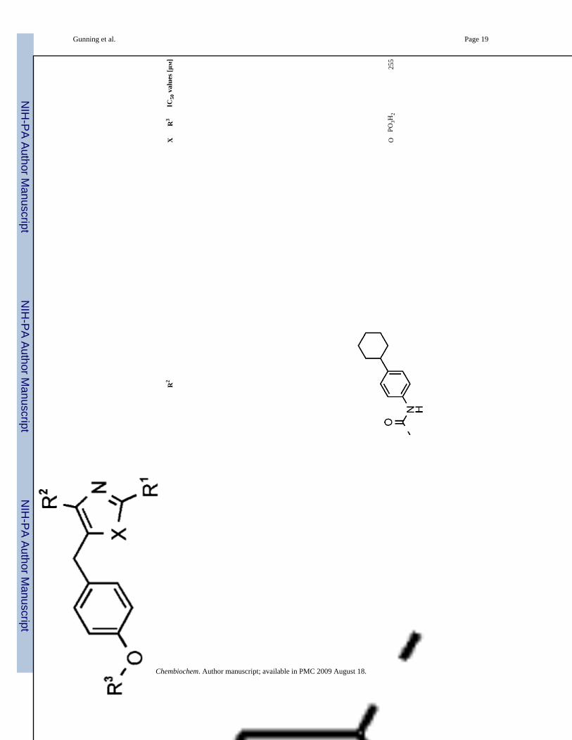
Gunning et al. Page 19
Com
poun
d N
umbe
rR
1R
2X
R3
IC50
val
ues [μM
]
17O
PO3H
225
5
Chembiochem. Author manuscript; available in PMC 2009 August 18.
NIH
-PA Author Manuscript
NIH
-PA Author Manuscript
NIH
-PA Author Manuscript
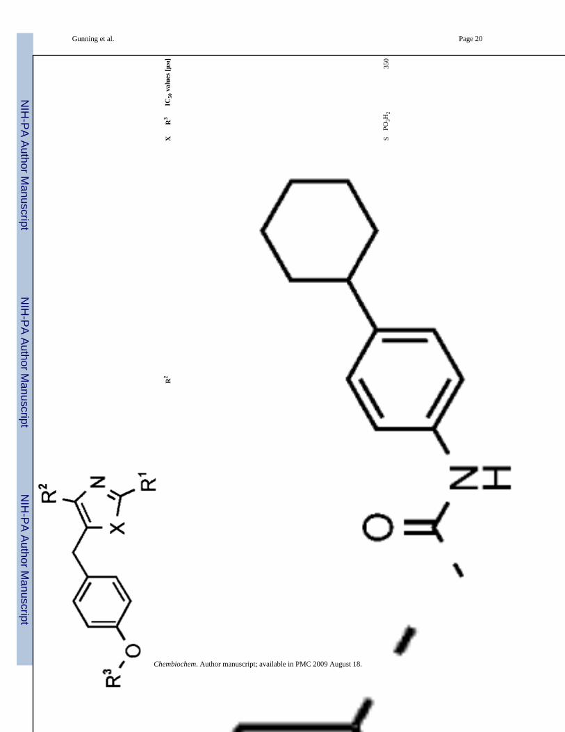
Gunning et al. Page 20
Com
poun
d N
umbe
rR
1R
2X
R3
IC50
val
ues [μM
]
18S
PO3H
235
0
Chembiochem. Author manuscript; available in PMC 2009 August 18.

NIH
-PA Author Manuscript
NIH
-PA Author Manuscript
NIH
-PA Author Manuscript
Gunning et al. Page 21
Com
poun
d N
umbe
rR
1R
2X
R3
IC50
val
ues [μM
]
19S
PO3H
212
5
[a] In
hibi
tor c
once
ntra
tion
requ
ired
to d
ecre
ase
Stat
3-D
NA
bin
ding
two-
fold
.
Chembiochem. Author manuscript; available in PMC 2009 August 18.
NIH
-PA Author Manuscript
NIH
-PA Author Manuscript
NIH
-PA Author Manuscript
Gunning et al. Page 22
Table 2EC50 values for selected Inhibitors against constitutively activated Stat3 containing cell lines NIH/3T3/vSrc, MDA-MB-231 and unactivated Stat3 NIH3T3 cells
Compound No.EC50 [μM]
NIH3T3 NIH3T3/vSrc MDA-MB-231
10 >1000 120 180
14 >1000 480 >1000
16 >1000 145 300
19 >1000 700 >1000
Chembiochem. Author manuscript; available in PMC 2009 August 18.
Related Documents


![Photonic modulation of EGFR halts receptor activation and ......cell line (Sigma-Aldrich, CLL1141) [19]. The SH2 domain protein, fused to green fluorescent protein (GFP), has high](https://static.cupdf.com/doc/110x72/60bffd1f8d5bf1052072b78f/photonic-modulation-of-egfr-halts-receptor-activation-and-cell-line-sigma-aldrich.jpg)