TARGETING AND INSERTION OF THE CHOLESTEROL-BINDING TRANSLOCATOR PROTEIN INTO THE OUTER MITOCHONDRIAL MEMBRANE Malena Rone 1,2 , Jun Liu 1 , Josip Blonder 5 , Xiaoying Ye 5 , Timothy D. Veenstra 5 , Jason C. Young 3 , and Vassilios Papadopoulos 1,2,3,4 1 Department of Biochemistry, Molecular and Cellular Biology, Georgetown University Medical Center, Washington DC 20007, USA 2 The Research Institute of the McGill University Health Centre and the Department of Medicine, McGill University, 1650 Cedar Avenue, Montreal, Quebec H3G 1A4, Canada 3 Department of Biochemistry, McGill University, 1650 Cedar Avenue, Montreal, Quebec H3G 1A4, Canada 4 Department of Pharmacology and Therapeutics, McGill University, 1650 Cedar Avenue, Montreal, Quebec H3G 1A4, Canada 5 Laboratory of Proteomics and Analytical Technologies, SAIC-Frederick Inc., National Cancer Institute at Frederick, Frederick, Maryland 21702, USA Abstract Translocator protein (18-kDa, TSPO1), previously known as the peripheral-type benzodiazepine receptor, is an outer mitochondrial membrane (OMM) protein necessary for cholesterol import and steroid production. We reconstituted the mitochondrial targeting and insertion of TSPO into the OMM to analyze the signals and mechanisms required for this process. Initial studies indicated a formation of a mitochondrial 66-kDa complex through Blue Native-PAGE analysis. The formation of this complex was found to be dependent on the presence of ATP and the cytosolic chaperone Hsp90. Through mutational analysis we identified two areas necessary for TSPO targeting, import, and function: amino acids 103−108 (Schellman motif), which provide the necessary structural orientation for import, and the cholesterol-binding C-terminus required for insertion. Although the Translocase of the Outer Mitochondria Membrane (TOM) complex proteins Tom22 and Tom40 were present in the OMM, the TOM complex did not interact with TSPO. In search of proteins involved in TSPO import, complexes known to interact with TSPO were analyzed by mass spectrometry. The 66-kDa complex formation was found to be dependent on an identified protein, Metaxin 1, for formation and TSPO import. TSPO import into steroidogenic cell mitochondria was increased following treatment of the cells with cAMP. These findings suggest that the initial targeting of TSPO to mitochondria is dependent upon the presence of cytosolic chaperones interacting with the import receptor Tom70. The C-terminus plays an important role in targeting TSPO to mitochondria whereas its import into the OMM is dependent upon the presence of the Schellman motif. Final integration 1 The abbreviations used are: ANT, anion nucleotide transporter; BN-PAGE, Blue-native polyacrylamide gel electrophoresis; FBS, fetal bovine serum; GD, geldanamycin; Hsp, heat shock protein; IMM, inner mitochondrial membrane; MPTP, mitochondria permeability transition pore; NB, Novobiocin; OMM, outer mitochondrial membrane; StAR, steroidogenesis acute regulatory protein; TOM, translocase of outer mitochondrial membrane; TSPO, translocator protein (18-kDa); VDAC, voltage-dependant anion channel Address correspondence to: V. Papadopoulos, The Research Institute of the McGill University Health Center, 1650 Cedar Avenue, C10 −148, Montreal, Quebec H3G 1A4, Canada. Tel: 514−934−1934 ext. 44580; Fax: 514−934−8439; E-mail: [email protected]. NIH Public Access Author Manuscript Biochemistry. Author manuscript; available in PMC 2010 July 28. Published in final edited form as: Biochemistry. 2009 July 28; 48(29): 6909–6920. doi:10.1021/bi900854z. NIH-PA Author Manuscript NIH-PA Author Manuscript NIH-PA Author Manuscript

Welcome message from author
This document is posted to help you gain knowledge. Please leave a comment to let me know what you think about it! Share it to your friends and learn new things together.
Transcript

TARGETING AND INSERTION OF THE CHOLESTEROL-BINDINGTRANSLOCATOR PROTEIN INTO THE OUTER MITOCHONDRIALMEMBRANE
Malena Rone1,2, Jun Liu1, Josip Blonder5, Xiaoying Ye5, Timothy D. Veenstra5, Jason C.Young3, and Vassilios Papadopoulos1,2,3,41Department of Biochemistry, Molecular and Cellular Biology, Georgetown University MedicalCenter, Washington DC 20007, USA2The Research Institute of the McGill University Health Centre and the Department of Medicine,McGill University, 1650 Cedar Avenue, Montreal, Quebec H3G 1A4, Canada3Department of Biochemistry, McGill University, 1650 Cedar Avenue, Montreal, Quebec H3G 1A4,Canada4Department of Pharmacology and Therapeutics, McGill University, 1650 Cedar Avenue, Montreal,Quebec H3G 1A4, Canada5Laboratory of Proteomics and Analytical Technologies, SAIC-Frederick Inc., National CancerInstitute at Frederick, Frederick, Maryland 21702, USA
AbstractTranslocator protein (18-kDa, TSPO1), previously known as the peripheral-type benzodiazepinereceptor, is an outer mitochondrial membrane (OMM) protein necessary for cholesterol import andsteroid production. We reconstituted the mitochondrial targeting and insertion of TSPO into theOMM to analyze the signals and mechanisms required for this process. Initial studies indicated aformation of a mitochondrial 66-kDa complex through Blue Native-PAGE analysis. The formationof this complex was found to be dependent on the presence of ATP and the cytosolic chaperoneHsp90. Through mutational analysis we identified two areas necessary for TSPO targeting, import,and function: amino acids 103−108 (Schellman motif), which provide the necessary structuralorientation for import, and the cholesterol-binding C-terminus required for insertion. Although theTranslocase of the Outer Mitochondria Membrane (TOM) complex proteins Tom22 and Tom40 werepresent in the OMM, the TOM complex did not interact with TSPO. In search of proteins involvedin TSPO import, complexes known to interact with TSPO were analyzed by mass spectrometry. The66-kDa complex formation was found to be dependent on an identified protein, Metaxin 1, forformation and TSPO import. TSPO import into steroidogenic cell mitochondria was increasedfollowing treatment of the cells with cAMP. These findings suggest that the initial targeting of TSPOto mitochondria is dependent upon the presence of cytosolic chaperones interacting with the importreceptor Tom70. The C-terminus plays an important role in targeting TSPO to mitochondria whereasits import into the OMM is dependent upon the presence of the Schellman motif. Final integration
1The abbreviations used are: ANT, anion nucleotide transporter; BN-PAGE, Blue-native polyacrylamide gel electrophoresis; FBS, fetalbovine serum; GD, geldanamycin; Hsp, heat shock protein; IMM, inner mitochondrial membrane; MPTP, mitochondria permeabilitytransition pore; NB, Novobiocin; OMM, outer mitochondrial membrane; StAR, steroidogenesis acute regulatory protein; TOM,translocase of outer mitochondrial membrane; TSPO, translocator protein (18-kDa); VDAC, voltage-dependant anion channelAddress correspondence to: V. Papadopoulos, The Research Institute of the McGill University Health Center, 1650 Cedar Avenue, C10−148, Montreal, Quebec H3G 1A4, Canada. Tel: 514−934−1934 ext. 44580; Fax: 514−934−8439; E-mail:[email protected].
NIH Public AccessAuthor ManuscriptBiochemistry. Author manuscript; available in PMC 2010 July 28.
Published in final edited form as:Biochemistry. 2009 July 28; 48(29): 6909–6920. doi:10.1021/bi900854z.
NIH
-PA Author Manuscript
NIH
-PA Author Manuscript
NIH
-PA Author Manuscript

of TSPO into the OMM occurs via its interaction with Metaxin 1. TSPO import into steroidogeniccell mitochondria is regulated by cAMP.
KeywordsTranslocator protein; mitochondria; cholesterol transport; TOM complex; Hsp90 chaperone; cAMP
Translocator protein (18-kDa, TSPO), previously known as the peripheral-type benzodiazepinereceptor, is an 18-kDa, high affinity, cholesterol- and drug-binding protein that is located inthe outer mitochondria membrane (OMM). TSPO appears to contain five α-helixes that spanthe OMM and assist with the transport of cholesterol from intracellular stores into themitochondrial matrix, the rate-limiting step in steroid biosynthesis (1-3). TSPO has been shownto interact with the voltage-dependent anion channel (VDAC) and the adenine nucleotidetranslocase (ANT) comprising the mitochondria permeability transition pore (MPTP), whichis located at the contact site between the inner and outer mitochondrial membrane (4-8). Thismitochondrial localization at the contact site influences many of the biological functions inwhich TSPO participates, including cholesterol transport, protein import, cell proliferation,and apoptosis (1;2;9-15). Due to its key role in these cellular functions, the targeting andinsertion of TSPO into the OMM is thought to be tightly regulated.
Like the majority of mitochondrial proteins, TSPO is genetically encoded in the nucleus,translated in the cytosol, and then imported into mitochondria (16-18). Unlike manymitochondrial proteins of the matrix and inner mitochondria membrane, OMM proteins suchas TSPO, do not have cleavable presequences for mitochondrial targeting. Instead, theseproteins are targeted to the OMM through internal amino acid sequences (19). To date, OMMtargeting signals have not been defined or predicted in multimembrane spanning proteins, andthe signal or signals within TSPO are unknown.
The Translocase of the Outer Mitochondria Membrane (TOM) complex is a protein complexcomposed of receptors that recognize mitochondrial proteins for import and an aqueous porefor the translocation of proteins across the membrane (20). The TOM complex includes thekey components Tom22 and Tom40, the latter of which forms the translocation pore (21).Additional import complexes further direct the protein to its correct location in the OMM, innermitochondrial membrane (IMM), intermembrane space, and mitochondrial matrix. The TOMreceptor protein, Tom70, has been shown to loosely associate with the TOM complex and isimportant for the import of IMM metabolite carriers that have internal targeting sequences aswell as larger hydrophobic proteins (22). Tom70 functions as a docking protein for both Hsc70and Hsp90 through a central tetratriacopeptide repeat (TPR) domain (23) , while its C-terminusis thought to bind mitochondrial proteins during import. Cytosolic chaperones, in particularHsc70 and Hsp90, have been shown to assist with mammalian mitochondrial import,maintaining the newly made protein in a soluble, import-competent state (23-25). Thechaperones that bind the mitochondrial protein dock onto Tom70; the mitochondrial protein isthen transferred in an ATP-dependent manner to the core TOM complex for translocation(24).
The import of OMM proteins is an active area of investigation, as these proteins appear to usea diverse array of import mechanisms. C-terminal, tail-anchored OMM proteins, such as Baxand Bcl-xL, which span the OMM once by a transmembrane α-helix, appear to require neitherchaperones nor ATP for insertion (26). β-barrel OMM proteins, such as VDAC, require anadditional OMM protein complex in addition to the TOM complex, the sorting and assemblymachinery (SAM) complex, for correct insertion (27-29). The proposed mammalian SAMcomplex proteins include Sam50, the pore forming protein of the complex, Metaxin 1, which
Rone et al. Page 2
Biochemistry. Author manuscript; available in PMC 2010 July 28.
NIH
-PA Author Manuscript
NIH
-PA Author Manuscript
NIH
-PA Author Manuscript

assists with protein integration into the OMM, and Metaxin 2, a cytosolic protein shown tobind to Metaxin 1 (30-32). The stoichiometry and stability of the SAM complex has not yetbeen firmly established. As TSPO appears to span the OMM via five α-helixes; it is unknownwhether TSPO is inserted directly into the OMM through the TOM complex or requires sortingthrough the SAM complex.
Previously, the results of Otera, et al. (33) have shown that TSPO does not use the traditionalprotein insertion pathway, but do not identify the pathway necessary for import. Our resultspresented herein further these studies by demonstrating that (i) during translocation to theOMM, TSPO interacts with cytosolic chaperones to facilitate an interaction with TOM70, (ii)there are specific amino acids necessary for the targeting of TSPO to the OMM, and (iii) oncetargeting is complete, TSPO insertion into the OMM is mediated through Metaxin 1, a memberof the SAM complex. Interestingly, TSPO import into mitochondria was increased followingtreatment of hormone-responsive steroidogenic Leydig cells with cAMP, an event that parallelsincreased cholesterol transport and steroid formation by the cells (10). These findings supportthe existence of a novel three-step integration pathway for OMM proteins and suggest thatprotein import into steroid synthesizing mitochondria might be a cAMP- and thus hormone-regulated process.
Material and MethodsCell Culture
HeLa cells (Lombardi Comprehensive Cancer Center Cell Culture Facility, GeorgetownUniversity), a well-established model used to study protein import into mitochondria, weremaintained in DMEM supplemented with 10% FBS at 37 °C and 6% CO2. MA-10 mousetumor Leydig cells, a well established model to study cholesterol transport into mitochondriaand steroidogenesis, were a gift from Mario Ascoli (University of Iowa, Ames) and weremaintained in DMEM/Ham's F12 (50:50) supplemented with 5% fetal bovine serum (FBS)and 2.5% horse serum at 37 °C and 3.7% CO2. In some experiments, confluent MA-10 cellswere used to evaluate the effect of cAMP on mitochondria import. MA-10 cells were treatedfor two hours with 1mM 8-Bromo cAMP (Biomol, Farmingdale, NY) prior to mitochondrialisolation.
Plasmid ConstructionThe mouse TSPO cDNA coding sequence (5) was subcloned into pEGFP (CLONTECHLaboratories, Inc., Otsu, Shiga, Japan) at the Sac1 and BamHI sites (pEGFP-TSPO). Forconstruction of TSPO with a truncated N-terminus, the QuickChange Site-DirectedMutagenesis Kit (Stratagene; La Jolla, CA) was used to generate a Sac1 mutation in the openreading frame of TSPO at the specified region. The vector was then digested with Sac1, gelpurified, and ligated. TSPO with a C-terminal truncation was generated by creating a sequenceverified stop codon mutation at amino acids 151 and 157. For generating TSPO constructscarrying various deletions, the appropriate regions of TSPO were amplified by PCR, gelpurified, ligated, and reinserted into the pEGFP vector. The fusion sites were verified bysequencing.
Confocal MicroscopyConfocal experiments were performed as stated previously (34) Briefly, MA-10 cells weregrown on cover glass bottom dish, (Fluorodish, WPI; Sarasota, FL) until 70% confluence.Plasmid constructs were transfected using Lipofectamine 2000 (Invitrogen; Carlsbad, CA).After 24 hours, cells were stained with 50nM Mitotracker CMX (Molecular Probes; Carlsbad,CA) for thirty minutes and viewed through an Olympus Fluoview FV1000 Laser ConfocalMicroscope.
Rone et al. Page 3
Biochemistry. Author manuscript; available in PMC 2010 July 28.
NIH
-PA Author Manuscript
NIH
-PA Author Manuscript
NIH
-PA Author Manuscript

Mitochondria IsolationMitochondria were isolated by differential centrifugation as previously described (35). Briefly,HeLa cells or confluent MA-10 cells were washed twice with PBS, harvested in Buffer A(10mM Hepes-KOH, pH 7.5, .2 M Mannitol, .07 M Sucrose, 1mM EDTA , 1x CompleteProtease Inhibitor Cocktail Tablet (Roche, Switzerland)) using a cell lifter, and centrifuged at500 × g for 10 minutes. The cell pellet was resuspended in 5 volumes of Buffer A, incubatedat 4°C for 10 minutes and then centrifuged at 500 × g for 10 minutes. The cell pellet wasresuspended in 5 volumes Buffer B (40 mM Hepes-KOH, pH 7.5, 500mM sucrose, 160 mMPotassium Acetate and 10mM Magnesium Acetate, 1x Complete Protease Inhibitor CocktailTablets) and homogenized using an electric potter (glass-Teflon) for 10 passes. Once complete,cells were centrifuged at 500 × g for 10 min. The cell pellet was resuspended in 5 volumesBuffer B with a glass-glass homogenizer (20 passes) and centrifuged at 500 × g for 10 min.The supernatant was pooled and centrifuged at 10,000 × g for 10 minutes at 4 °C to form amitochondrial pellet. The mitochondrial pellet was resuspended in 1mL Buffer B andcentrifuged at 10,000 × g for 10 minutes to enrich mitochondrial purity. Once complete, themitochondria were resuspended in mitochondria import buffer (3% BSA, 250 mM sucrose, 5mM MgCl2, 80 mM KCl, 10 mM MOPS-KOH, pH 7.2, 5 mM ADP, and 10mM succinate(Sigma, St. Louis), 1x Complete Protease Inhibitor Cocktail Tablets) to give a finalconcentration of 1 mg/mL mitochondria for BN-Page import and 5mg/mL mitochondria forsodium carbonate extraction. Mitochondria were kept on ice until use for no longer than 1 hour.
Protein ImportRadiolabeled TSPO was generated using the TNT® T7 Quick Coupled Transcription/Translation System (Promega; Madison, WI) in the presence of [35S]-methionine (AmershamBiosciences; Piscataway, NJ) as performed previously (10) for one hour at 30°C. Oncecomplete, the reaction was terminated by the addition of one volume of 2×TT buffer (20 mMHepes-KOH pH7.5, 500mM sucrose, 80 mM KOAc, 5mM MgOAc2, 1mM Methionine). 5μl of the TNT reaction was added to 50 μg isolated mitochondria in import buffer for the statedtimes. Mitochondria were centrifuged at 10,000 × g for 10 minutes, solubilized with 1%digitonin buffer (20 mM Tris-Cl, 0.1 mM EDTA, 50 mM NaCl, 10% w/v glycerol, 1% digitonin(Invitrogen) and 1 mM PMSF) for 20 minutes on ice, and centrifuged at 10,000 × g for 10minutes. One-half of each sample was digested with 250 μg/ml proteinase K (Qiagen;Dusseldorf, Germany) at 4 °C for 10 minutes while the other half of the sample remaineduntreated.
Blue Native (BN)-PAGEBN-PAGE was performed as described by Simpson (36). BN-PAGE loading dye (5% w/vCoomassie Brilliant Blue G-250, 500 mM ∈-amino-n-caproic acid, and 160 mM BisTris, pH7.0) was added to the sample supernatants, loaded onto a 4−16% native gel (Invitrogen), andrun at 130 V. Once complete, the gel was transferred using a semi-dry transfer apparatus forone hour at 25 V. The PVDF membrane was then fixed and dried. Then membrane was exposedto a multipurpose phosphor screen for 1−7 days and analyzed by phosphorimaging using theCyclone Storage Phosphor System (Packard BioScience; Waltham, MA). Once complete, themembrane was then used for immunodetection. Image analysis was performed by Multi GaugeV3.0 from FujiFilm.
Carbonate ExtractionMitochondrial protein import was analyzed as described by Fujiki et al. (37). After import,mitochondria were incubated at 0.5 mg/ml in 180 μl of 0.1 M Na2CO3 (pH 10.5) at 4 °C for30 minutes. Forty μl of sucrose buffer (500 mM sucrose and 0.1 M Na2CO3, pH 10.5) wasadded to the top of the sample and centrifuged at 180,000 × g for 30 minutes at 4 °C in a
Rone et al. Page 4
Biochemistry. Author manuscript; available in PMC 2010 July 28.
NIH
-PA Author Manuscript
NIH
-PA Author Manuscript
NIH
-PA Author Manuscript

Beckman Coulter TLA-100 rotor. Trichloroacetic acid precipitation was performed on thesupernatant, and both pellet and supernatant were analyzed by SDS-PAGE.
Nickel-Sepharose Pull Down AssayThe phosphate carrier (PiC) was also generated from cell free transcription/translation reactionsas stated previously (38). Radiolabeled TSPO and PiC were diluted 10-fold with reticulocytelysate and incubated with either geldanamycin or novobiocin. Tom70-H3 was then added tothe TNT reaction of either PiC or TSPO (23). After five minutes, apyrase was added toterminate the incubation reaction and 50 μl of GTI buffer (100mM KOAc, 20mM Hepes-KOHpH7.5, 5mM MgOAc2, 20mM imidazole, 0.1% Triton X-100) was added. Nickel-Sepharoseslurry was added to the mixture and incubated on an orbital shaker at 4°C for 30 minutes. Theresulting reactions contained 5% rectulate lysate reaction of TSPO or PiC containing the nickel-Sepharose slurry, the negative control reaction did not contain Tom70-H3, positive control,geldanamycin, and novobiocin treated samples contained 5μM Tom70-H3 and either 18μMgeldanamycin or 1mM novobiocin. The beads were then washed twice with GTI buffer andonce with GI buffer (lacking Triton X-100). The protein was eluted with 40μl LLB with EDTAand separated by SDS-PAGE.
Steroid BiosynthesisSteroid analysis was preformed as previously described (34). Briefly, MA-10 cells were platedin 24-well plates at a density of 50,000 cells/well. Cells were transfected after 24 hours withvarious GFP-TSPO constructs. Culture media were collected 24−48 hours post-transfection.In certain experiments, cells were washed with serum-free media and treated with 50 ng/mlhCG for two hours to determine the hormone-responsiveness of the cells. At the end of theincubation, media were collected. To determine maximal steroid production, the hydrosolublesubstrate 22R-hydroxycholesterol (10 μM) was added for two hours to the cells cultured inserum-free media. Progesterone production was measured by RIA in the media. Anti-progesterone antiserum was from MP-Biomedicals (Solon, OH) and [1,2,6,7-3H(N)]-progesterone (specific activity: 17.5 Ci/mmol) was from PerkinElmer Life Sciences.
In-gel Digestion and MS AnalysisSelected protein spots from HeLa extracts separated by BN-Page followed by 2D-SDS-PAGEwere subjected to an in-gel digestion procedure as described elsewhere (39). Resulting in-geldigests were desalted using C18 Zip Tips (Millipore) before analysis by nano-flow reversed-phase liquid chromatography (nanoRPLC) using an Agilent 1100 LC system (AgilentTechnologies, Inc., Paolo Alto, CA) coupled online to a linear ion trap (LIT) mass spectrometer(LTQ, Thermo Scientific, San Jose, CA). Reversed-phase separations were performed using75 μm i.d. × 360 μm o.d. × 10 cm long capillary columns (Polymicro Technologies Inc.,Phoenix, AZ) that were slurry packed in-house with a 5-μm, 300 Å pore size Jupiter C-18 silicabonded stationary phase (Phenomenex Torrance, CA). After being injected with 5 μl of sample,the column was washed for 20 minutes with 98% solvent A (0.1% formic acid in water, vol/vol) and peptides were eluted using a linear gradient of 2% solvent B (0.1% formic acid in100% acetonitrile, vol/vol) to 85% solvent B for 110 minutes at a constant flow rate of 250 nl/min. The LIT-MS was operated in a data-dependent mode in which each full MS scan wasfollowed by seven MS/MS scans where the most abundant peptide molecular ions weredynamically selected for collision-induced dissociation (CID) using a normalized collisionenergy of 36%. The temperature of the heated capillary and electrospray voltage (applied oncolumn base) was 180 °C and 1.7 kV, respectively. The CID spectra were searched against anon-redundant human protein database using SEQUEST (Thermo Scientific, San Jose, CA)and results were tabulated for each identified peptide/protein.
Rone et al. Page 5
Biochemistry. Author manuscript; available in PMC 2010 July 28.
NIH
-PA Author Manuscript
NIH
-PA Author Manuscript
NIH
-PA Author Manuscript

Transfection with siRNAMetaxin 1 siRNAs (5’-GCGCUGUCCUCAGAAUAAACCUGTT-3’; 5’-CGUAAAGAGAAGUAUAAUGCCGACT-3’;5’GGAUAGACGCCAAGAACUAUGUGGA-3’) were purchased from IDT (Coralville, IA)in a TriFecta kit. A hypoxanthine-guanine phosphoribosyltransferase (HPRT)-targetedpositive control and a scrambled negative control were obtained from the same provider.MA-10 cells cultured in 100-mm dishes were transfected with 30 μl Lipofectamine RNAiMAX(Invitrogen) and 20 nM total of either the control siRNA duplex or protein-targeted duplexesfor 48 hours. At the end of the treatment, media was changed and 3 days later cells wereharvested and mitochondria were isolated for import studies.
Real-Time qPCR1×105 cells were harvested from the control and 8-Bromo cAMP treated MA-10 cells and usedfor RNA isolation to measure stated mRNA levels. RNA was isolated with an RNeasy MiniKit (Qiagen) with optional DNase Digestion. The cDNA was then generated from AdvantageRT-for-PCR kit (Clontech), using100ng RNA incubated with oligo(dT) primer. Analysis wasperformed with 7900HT Sequence Detection Agents (Applied Biosystems), primers used arestated in Table 1, TaqMan™ was from Applied Biosystems (Foster City, CA). All sequenceswere normalized to HPRT.
StatisticsStatistical analysis was performed using Prism version 4.0 (GraphPad Software, San Diego,CA). Group means were compared using student's t test or two-way ANOVA test followed bya Bonferroni column test. Data are presented as mean ± sem, p < 0.05 was consideredsignificant.
MaterialsAntibodies specific for TOM22 (Sigma), VDAC, Cox IV, Tom40 (Abcam; Cambridge, MA),Metaxin 1 (BD Bioscience; San Jose, CA) GapDH (Trevigen, Gaithersburg, MD) werepurchased from the various vendors. StAR antibody was a generous gift from Dr. DB Hales(40). A specific rabbit polyclonal antibody was raised against the purified cytosolic fragmentof human Tom70 (amino acids 111−608; manuscript in preparation). The TSPO polyclonalantibody was developed as previously described (41).
ResultsImport of TSPO
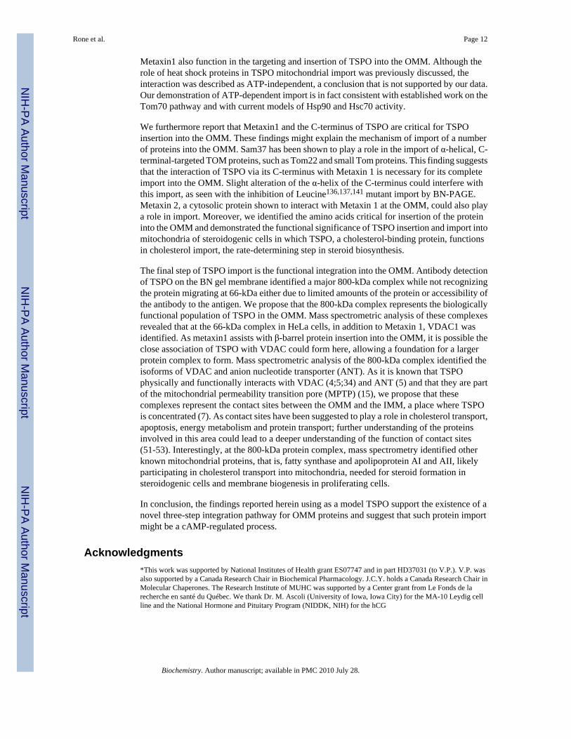
To identify the import pathway of TSPO, we incubated cell-free, radiolabeled, in vitrotranslated TSPO with isolated HeLa cell mitochondria and monitored the formation ofradiolabeled protein complexes through BN-PAGE by phosphoimaging. We have previouslyshown that the incubation of TSPO with isolated mitochondria results in import of a fullyfunctional protein, capable of binding and transporting cholesterol in isolated steroidogenicmitochondria from MA-10 cells with consequent generation of steroids (10). As the TSPOimport reaction with isolated mitochondria proceeded, we observed the presence of aradioactive band migrating at approximately 66-kDa that increased in intensity with incubationtime (Fig. 1A). A denaturing 2D SDS-PAGE of the BN-PAGE import reactions produced an18-kDa radioactive band, further confirming TSPO import into the mitochondria (data notshown). The presence of the TOM complex in the mitochondria was assessed by immunoblotanalysis of proteins separated on BN-PAGE gels, electrotransferred on membranes, and blottedusing antibodies against Tom22 and Tom40. Both of these antibodies recognizedimmunoreactive proteins that migrated at 440-kDa, thus indicating the presence of the core
Rone et al. Page 6
Biochemistry. Author manuscript; available in PMC 2010 July 28.
NIH
-PA Author Manuscript
NIH
-PA Author Manuscript
NIH
-PA Author Manuscript

TOM complex at the appropriate size though TSPO was not present in the complex. These datawere confirmed in experiments in which the major import receptor Tom20 was knocked down;its absence failed to affect TSPO import into mitochondria (data not shown). We further probednative membranes with an affinity-purified, anti-TSPO peptide-specific polyclonal antibody.Immunoreactive proteins were identified to migrate at 300-kDa and 600-kDa. It should benoted that HeLa cells are known to have low levels of endogenous TSPO. In parallel studiesusing the rich-in-TSPO MA-10 cell mitochondria we identified immunoreactive TSPO at 66-kDa, 300-kDa and 600-kDa (data not shown). As mature TSPO is known to form polymers(42) and also associate with various other IMM and OMM proteins (4;5), these large complexeslikely contain both homo- and/or hetero-polymers of TSPO. The 66-kDa protein complexformed by newly imported TSPO was not detected in HeLa cells by immunoblot analysis asseen of steady-state endogenous TSPO. This may be due to the low levels of TSPO generatedby the TNT radiolabeling kit, it must be remembered that TSPO antibodies directed towardsthe mature peptide are unlikely to recognize protein morphology altered during mitochondrialimport.
To confirm TSPO import into isolated mitochondria, we performed a proteinase K digestionto degrade non-imported proteins. Protein import was performed at 4°C and 33°C and importreactions were terminated at 0 and 30 minutes. Samples were then divided into two sets, withand without proteinase K treatment. With the 4°C import reactions, the amount of 66-kDacomplex was greatly reduced relative to reactions at 33°C. Proteinase K digestion of the 4°Creactions removed the 66-kDa protein complexes that were observable in untreated samples(Fig. 1B). In contrast, with the permissive 33°C-import reaction, the 66-kDa complex wasresistant to proteinase treatment (Fig. 1B). These data suggest that the 66-kDa complex, underpermissive conditions, is fully incorporated into mitochondrial membranes.
Further confirmation of TSPO membrane integration was obtained by carbonate extraction.Here TSPO was imported under permissive conditions and mitochondria were purified. Thesere-isolated mitochondria were treated with sodium carbonate to remove proteins that wereassociated with, but not integrated into, the OMM. Upon carbonate treatment andcentrifugation, the majority of TSPO was found in the pellet with the mitochondrial membranefraction, implying full incorporation of the protein into the OMM (Fig. 1C). As a positivecontrol, the carbonate extraction experiment was performed with radiolabeled Tom70, anintegral OMM protein, which displayed the same association profile as TSPO (Fig. 1C). Incontrast, the soluble matrix protein Hsp60 was mostly found in the extracted supernatantfraction. Thus, the HeLa mitochondria have the necessary machinery to drive authentic TSPOimport.
TSPO associates with HSP90 and Tom70Heat shock proteins (HSPs), including Hsp90, are known to play an important role in deliveringproteins to the OMM for import, interacting with Tom70 and assisting translocation in an ATPdependent manner (24). Therefore, to determine if TSPO interacts with Hsp90 for import intothe OMM we incubated the purified C-terminal fragment of Hsp90 (C90) with TSPO importreactions into isolated mitochondria. C90 has previously been shown not to interact withmitochondrial proteins before import but does bind stably to Tom70, which outcompetes theHSP90 interaction and inhibits Tom70-dependent import (23). The addition of C90, resultedin a large reduction in TSPO import compared to control (Fig 2A, C), suggesting that importis dependent upon Hsp90 interaction.
To further test this hypothesis, we performed a series of BN-PAGE experiments. Consideringthat the chaperone-Tom70 pathway requires ATP for the function of Hsp90 or Hsc70, wedepleted ATP from import reactions using apyrase. BN-PAGE analysis of the samplesindicated that the membrane-integrated 66-kDa complex was reduced dramatically in the early
Rone et al. Page 7
Biochemistry. Author manuscript; available in PMC 2010 July 28.
NIH
-PA Author Manuscript
NIH
-PA Author Manuscript
NIH
-PA Author Manuscript

stages of import (Fig. 2B). As Hsp90 seemed to function in TSPO targeting to Tom70, we nextused the specific Hsp90 inhibitors geldanamycin (GD) and novobiocin (NB) to directly confirmthe role of Hsp90 in TSPO import. GD obstructs the N-terminal ATP-binding domain of Hsp90and inhibits the ATPase activity of the protein, resulting in a stalled complex and decreasedtranslocation across the OMM (24;43). NB interferes with the targeting of proteins to themitochondria by binding near the C-terminus of Hsp90, inhibiting substrate binding(44). NBalso interferes with the docking of Hsp90 to Tom70. As shown in Figures 2B & C, NB andGD significantly reduced import of TSPO compared to control (p<0.01 by ANOVA). NBshows the greatest decrease in import; this is expected as NB would prevent TSPO from bindingto Tom70 and associating with the mitochondria. GD is decreased, though not to the sameextent as NB, caused by the prevention of cleavage of ATP, thus stalling TSPO on themitochondrial membrane.
To further confirm that TSPO interacts with Tom70 for import, radiolabeled TSPO wasincubated with the His-tagged Tom70 cytosolic fragment and then co-precipitated with nickel-sepharose, reconstituting the targeting step as previously published. TSPO was recovered fromthe nickel-sepharose only when His-tagged Tom70 was present, indicating that TSPO interactswith Tom70 for import (Fig. 2D). GD and NB were incubated separately with the His-taggedTom70 fragment and radiolabeled TSPO, and Tom70-associated material analyzed. GD didnot inhibit the binding of TSPO to Tom70 as the recovery levels were similar to the control(Fig. 2D). This finding was similar to that observed with Hsp90-dependent precursor proteins,as GD only inhibits the ATP-dependant release of TSPO from the bound chaperone-Tom70complex. In contrast, NB did prevent TSPO from associating with Tom70. Again, this findingmatched the behavior of Hsp90-dependent precursors. Our results suggest that Hsp90 functionsto target TSPO to the Tom70 import receptor by a mechanism typical of the chaperone-Tom70pathway.
N-terminus is necessary but not sufficient to target TSPO to the mitochondriaTo identify the OMM-targeting sequence(s) of TSPO, we tested the localization of GFP-fusedTSPO in MA-10 mouse Leydig cells. TSPO was tagged on either the N- or C-terminus toconfirm that the presence of GFP did not interfere with the localization of the protein tomitochondria (data not shown). We then chose a construct in which GFP was linked at the N-terminus of TSPO to ensure that the cholesterol-binding domain on the C-terminus of theprotein (41;45) would not be directly affected by the presence of the GFP moiety. A series ofdeletion constructs were generated to determine whether the N-terminus is important formitochondrial targeting (Fig. 3). Constructs in which the first 7, 28, 48, 68, and 85 amino acidswere removed showed only a slight decrease in TSPO co-localization with the mitochondria,labeled with the Mitotracker CMX dye (Fig. 3A-F). Removal of amino acids 1−110 resultedin a loss of co-localization of TSPO with mitochondria and appearance of a diffuse pattern,consistent with an import defect (Fig. 3G).
As this region appeared necessary for TSPO import, we next examined the importance of aminoacids found between residues 88−110. A Schellman motif was predicted between amino acids103−108. This motif, often found terminating α-helical secondary structures, is stabilizedthrough hydrophobic interactions between amino acids around a signature glycine residue(46). In TSPO, the Schellman motif is predicted to reside in the second intermembrane spaceloop, bridging the third & fourth predicted transmembrane helices (Fig4a). Removal of theseamino acids (Δ103−108) resulted in focal aggregates of GFP-TSPO in areas adjacent to themitochondria, as seen through a decrease in co-localization with Mitotracker (Fig. 3H). Thepoint mutation of glycine 106 to alanine (G106A), that disrupts the Schellman motif byreducing flexibility, resulted in a similar profile as seen with TSPOΔ103−108 (Fig. 3I). TheSchellman motif between amino acids 103−108 of TSPO thus appears to be critical for proper
Rone et al. Page 8
Biochemistry. Author manuscript; available in PMC 2010 July 28.
NIH
-PA Author Manuscript
NIH
-PA Author Manuscript
NIH
-PA Author Manuscript

folding and/or membrane insertion. Though its role in localization and import remain open toinvestigation, the juxtamitochonrial location of aggregates suggests that the proteins aretargeted, but not capable of import.
C-terminus is necessary for targeting to the OMMAs roughly two-thirds of the TSPO protein from the N-terminus can be removed withoutaffecting the localization of the remaining protein to mitochondria, we then questioned whetherthe removal of the C-terminus affects TSPO localization. Generation of Δ151−169 and Δ157−169 amino acid-deletion constructs caused a decrease in the localization of the protein tomitochondria with an increase in aggregation of GFP-TSPO (Fig. 3J, K). Considering that thecholesterol-binding domain of TSPO is present within the deleted region in TSPO Δ151−169,it was possible that this domain could play a role in the targeting of TSPO to the OMM.However, removal of region 148−157, which contains the cholesterol-binding domain, did notalter the TSPO/mitochondria co-localization pattern (Fig. 3L), suggesting that the domain doesnot play a role in TSPO import. Because the degree of TSPO/mitochondria co-localizationdecreases with the removal of the C-terminus and although the cholesterol binding domainplays no role, it could be that some other feature of the C-terminus, such as the overall secondarystructure, could play a role in TSPO localization with the OMM. A comparison of theSchellman motif and C-terminal sequences was made, revealing two conserved areas of highleucine content, amino acids 112−114 and 136, 137, and 141 (Fig. 4A). Point mutations ofboth sets of leucines (Leucine112−114 and Leucine136,137,141) into alanines did not alter TSPOco-localization with mitochondria (Fig. 3M, N).
Inhibition of import of TSPO mutantsIn the above-described GFP-TSPO/mitochondria co-localization experiments, severalconstructs displayed reduced degrees of co-localization. To confirm that reduced co-localization reflects reduced TSPO import into mitochondria, BN-PAGE of in vitro importreactions was performed using [35S]-TSPO mutant constructs. Notably, the Schellman motifmutant Δ103−108 could not be imported to the 66-kDa complex (Fig. 4B). The import of TSPOcarrying the G106A point mutation also resulted in the greatly decreased import of the 66-kDaprotein complex, although not to the extent seen with the Δ103−108 TSPO construct (Fig. 4B).The ability to import was then tested for the TSPO constructs in which the C-terminal domains,151−169 and 157−169, were removed. Both deletions clearly abolished the import of the 66-kDa complex in isolated mitochondrial (Fig. 4C). To determine whether the alteration of thestructure of the C-terminus results in the inhibition of TSPO import, the Leucine112−114 andLeucine136,137,141 TSPO point mutants were analyzed by BN-PAGE. Leucine112−114 did notshow an effect on TSPO import (Fig. 4D). However, the Leucine136,137,141 TSPO mutantshowed a significant decrease in the import of the 66-kDa complex (Fig. 4D), although its levelof synthesis in vitro was similar to the Leucine112−114 mutant (Fig. 4E). This result differs fromthe results of confocal microscopy in which the Leucine112−114 TSPO mutant was found to co-localize with mitochondria. This apparent discrepancy could be due to differences between thein situ and in vitro situations, including the highly concentrated intracellular environment,which favor TSPO import and alleviate partial import defects. Overall, the in vitro and livecell results support an important role of the Schellman motif and sequences within the C-terminus of TSPO for import.
Functional evaluation of TSPO import in steroid biosynthesisAs TSPO controls the rate-limiting step in the production of steroids, cholesterol tranport intomitochondria, increasing the TSPO protein concentration in the OMM by transient transfectionwith a TSPO construct was shown to increase progesterone production by MA-10 mouseLeydig cells in culture (34) . To investigate the functional role of altered import of TSPO
Rone et al. Page 9
Biochemistry. Author manuscript; available in PMC 2010 July 28.
NIH
-PA Author Manuscript
NIH
-PA Author Manuscript
NIH
-PA Author Manuscript

mutants previously evaluated by confocal microscopy and BN-PAGE analyses, we measuredprogesterone production in MA-10 cells transfected with wild-type and mutated TSPOconstructs (Fig. 4). As the constitutive expression of TSPO in MA-10 cells was not silenced,a basal level of progesterone production was observed. However, transfection of MA-10 cellswith GFP-TSPO resulted in a marked 2.5-fold increase in progesterone production (Fig. 4F;p<0.01 by Student's t-test), consistent with previous findings (34). Transfection with TSPO inwhich the Schellman motif was mutated (G106A) did not affect basal progesterone formationbut did not cause a similar increase seen by transfection with wild-type GFP-TSPO. The resultsobtained with the G106A TSPO mutant support the data obtained in situ by confocalmicroscopy and in vitro using BN-PAGE, indicating that such mutants are not imported andtherefore not functional in cholesterol transport. The Leucine136,137,141 mutant showedprogesterone levels near basal levels, suggesting that while this mutant may be imported intothe OMM, it was not functional in the transport of cholesterol into the mitochondrial matrix.C-terminal mutants also failed to increase progesterone production, consistent with an importdefect. The finding that removal of the cholesterol-binding domain in the TSPOΔ151−169construct resulted in the reduction of basal progesterone production is in agreement withprevious findings (10;45).
Metaxin 1 is necessary for TSPO importConsidering the data presented above that TSPO does not stably associate with the core TOMcomplex during import, we undertook a detailed analysis of the identification of proteinsinvolved. Using a combination of BN-PAGE and 2D-SDS-PAGE followed by massspectrometric analysis of proteins present in all spots separated from the 66-kDa complex, aswell as the stable 800-kDa complex, we identified a number of proteins of interest (Table 2).Among the identified proteins present in the 66-kDa protein complex was Metaxin 1. Metaxin1 was shown previously to be present in MA-10 cells and be induced by hormone-treatmentin a manner parallel to steroid synthesis (47). Probing BN-PAGE immunoblots with an anti-Metaxin 1 antibody showed that the protein identified migrated at a similar molecular size to[35S]-TSPO (Fig. 5A).
Metaxin 1 is the mammalian homolog of yeast Sam37 (also called Mas37/Tom37), acomponent of the SAM complex known to function in the stable integration of OMM β-barrelproteins (29;32). Metaxin 1's primary role in the OMM is to function as a receptor in the SAMcomplex (48) It was possible that Metaxin 1 could play a role in TSPO import and to addressthis, an siRNA duplex that targets Metaxin 1 was used to knock down its expression.Mitochondria were isolated from MA-10 cells that had been treated for 5 days with 20 nM ofeither a siRNA complex toward Metaxin 1 or a scrambled negative control. Figure 5C showsthat the siRNA successfully reduced Metaxin 1 protein levels compared to the scrambledsiRNA control. Moreover, the basal levels of the inner mitochondrial membrane proteinCOXIV were unaffected by Metaxin 1 silencing, though endogenous TSPO levels were slightlydecreased. BN-PAGE of import into the mitochondria from Metaxin1 knockdown cells showedthat the rate of TSPO import at the 66-kDa complex was diminished, compared to mitochondriafrom scrambled siRNA and untreated cells (Fig. 5B). This data, quantified in Figure 5D(p<0.05 by ANOVA), suggests that Metaxin 1 is involved in TSPO import into the OMM.
Cyclic-AMP stimulated mitochondria increases TSPO importSteroidogenesis is initiated upon the transfer of cholesterol from the OMM to the IMM whereit interacts with CYP11A1 to be converted into pregnenolone. This transfer of cholesterol isactivated via a cAMP-dependent pathway, resulting in an increase in protein synthesis, proteinphosphorylation and lipid synthesis (49). To determine if activation of steroidogenesis wouldalter mitochondrial protein import, MA-10 cells were incubated with 1mM cAMP for twohours, the point in which maximal rates of steroid production are seen in vitro, and the
Rone et al. Page 10
Biochemistry. Author manuscript; available in PMC 2010 July 28.
NIH
-PA Author Manuscript
NIH
-PA Author Manuscript
NIH
-PA Author Manuscript

mitochondria were isolated (50). Upon initiation of TSPO mitochondrial import, a 1.7-foldincrease of 66-kDa TSPO was observed in mitochondria isolated from cAMP-treated cellswithin the first 5 minutes followed by a 2.2- and 1.8-fold induction over basal at 15 and 30min, respectively (Fig. 6A, B). The effect of cAMP was statistically significant (p<0.05 by t-test). To determine if gene and protein expression levels were altered both qPCR and westernblot analysis were performed. qPCR results showed no increase upon cAMP treatment of theTom40 pore for OMM protein import and Tim22 for IMM protein import. Metaxin 1 showeda slight but not significant increase upon cAMP stimulation (Fig. 6C). While the 2 hour cAMPtreatment is too short for most transcriptionally-based changes in protein expression,translation- or degradation-based regulation can cause faster effects. However, uponimmunoblot analysis there were no significant increases seen in any mitochondrial proteinsincluding Metaxin1 (Fig. 6D). StAR protein expression, which is a cAMP-induced protein,was also measured to confirm the 8-bromo cAMP activity. StAR mRNA levels were increaseby 10-fold (data not shown) and there was also a pronounced induction of StAR protein levels(Fig. 6D). This suggests that while the mechanisms necessary for mitochondrial import arebeing up-regulated, the increase in TSPO import is not due to an increase in mitochondriaimport proteins. It is possible however that changes in the composition f protein complexesand protein-protein interactions needed for import could occur.
DiscussionIn the above experiments, we determined the pathway for the targeting and insertion of themulti-membrane spanning protein, TSPO, into the OMM. A schematic representation of thesteps of the process is shown in Figure 7. The initial targeting of TSPO to mitochondria hasbeen shown to be dependent on the presence of the cytosolic chaperones Hsp90 and most likelyHsc70 as well as their interactions with Tom70 upon arrival at the OMM. As this first stepshows, these early interactions of the cytosolic chaperones and Tom70 depend on the properbinding of TSPO to the chaperones. Targeting of chaperone-TSPO complexes to Tom70 is thesecond step. The Tom70 complex formed at this stage may be relatively unstable and althoughnot detected in the BN-PAGE, it can be reconstituted with purified Tom70. The third step ofTSPO import into the OMM is insertion into the membrane. This is a temperature and ATP-dependent process involving the release of TSPO from the chaperones and Tom70. Metaxin1functions at this step to boost the kinetics of insertion. After insertion, TSPO becomes resistantto proteinase K. The third step requires formation of specific secondary structures includingthe Schellman motif, which probably aids integration of the third and fourth transmembranedomains of TSPO into the OMM. In steroidogenic and hormone-responsive MA-10 Leydigcells, the rate of import into the isolated mitochondria can be accelerated following treatmentwith cAMP (Fig. 7; yellow arrow). These conformational requirements suggest that the 66-kDa complex contains TSPO in a native folded state. The native complex is most likely a homo-oligomer of TSPO as has previously been observed. The 66-kDa complex then matures intothe final complexes of up to 800-kDa which are observed in steady-state mitochondria ofendogenous TSPO. These complexes may include the known TSPO interacting proteins VDACand ANT, further identified by mass spectrometry to be present in this complex, as well asother proteins. This process of TSPO import constitutes a novel pathway for protein importinto the OMM.
As mentioned earlier, our results broadly support the conclusion of Otera et al. (33) that TSPOis inserted into mitochondria via a novel pathway. The authors identified a 66-kDa TSPOcomplex by BN-PAGE upon import into HeLa mitochondria. They also demonstrated thatTom22, Tom20, Tom40, and Sam50 were not involved in TSPO import whereas Tom70 playeda role in TSPO targeting to mitochondria, in agreement with our findings. They hypothesizedthe importance of other unidentified mitochondrial proteins for TSPO and other protein import.We now confirm these findings and extend them by providing evidence that Hsp90 and
Rone et al. Page 11
Biochemistry. Author manuscript; available in PMC 2010 July 28.
NIH
-PA Author Manuscript
NIH
-PA Author Manuscript
NIH
-PA Author Manuscript
Metaxin1 also function in the targeting and insertion of TSPO into the OMM. Although therole of heat shock proteins in TSPO mitochondrial import was previously discussed, theinteraction was described as ATP-independent, a conclusion that is not supported by our data.Our demonstration of ATP-dependent import is in fact consistent with established work on theTom70 pathway and with current models of Hsp90 and Hsc70 activity.
We furthermore report that Metaxin1 and the C-terminus of TSPO are critical for TSPOinsertion into the OMM. These findings might explain the mechanism of import of a numberof proteins into the OMM. Sam37 has been shown to play a role in the import of α-helical, C-terminal-targeted TOM proteins, such as Tom22 and small Tom proteins. This finding suggeststhat the interaction of TSPO via its C-terminus with Metaxin 1 is necessary for its completeimport into the OMM. Slight alteration of the α-helix of the C-terminus could interfere withthis import, as seen with the inhibition of Leucine136,137,141 mutant import by BN-PAGE.Metaxin 2, a cytosolic protein shown to interact with Metaxin 1 at the OMM, could also playa role in import. Moreover, we identified the amino acids critical for insertion of the proteininto the OMM and demonstrated the functional significance of TSPO insertion and import intomitochondria of steroidogenic cells in which TSPO, a cholesterol-binding protein, functionsin cholesterol import, the rate-determining step in steroid biosynthesis.
The final step of TSPO import is the functional integration into the OMM. Antibody detectionof TSPO on the BN gel membrane identified a major 800-kDa complex while not recognizingthe protein migrating at 66-kDa either due to limited amounts of the protein or accessibility ofthe antibody to the antigen. We propose that the 800-kDa complex represents the biologicallyfunctional population of TSPO in the OMM. Mass spectrometric analysis of these complexesrevealed that at the 66-kDa complex in HeLa cells, in addition to Metaxin 1, VDAC1 wasidentified. As metaxin1 assists with β-barrel protein insertion into the OMM, it is possible theclose association of TSPO with VDAC could form here, allowing a foundation for a largerprotein complex to form. Mass spectrometric analysis of the 800-kDa complex identified theisoforms of VDAC and anion nucleotide transporter (ANT). As it is known that TSPOphysically and functionally interacts with VDAC (4;5;34) and ANT (5) and that they are partof the mitochondrial permeability transition pore (MPTP) (15), we propose that thesecomplexes represent the contact sites between the OMM and the IMM, a place where TSPOis concentrated (7). As contact sites have been suggested to play a role in cholesterol transport,apoptosis, energy metabolism and protein transport; further understanding of the proteinsinvolved in this area could lead to a deeper understanding of the function of contact sites(51-53). Interestingly, at the 800-kDa protein complex, mass spectrometry identified otherknown mitochondrial proteins, that is, fatty synthase and apolipoprotein AI and AII, likelyparticipating in cholesterol transport into mitochondria, needed for steroid formation insteroidogenic cells and membrane biogenesis in proliferating cells.
In conclusion, the findings reported herein using as a model TSPO support the existence of anovel three-step integration pathway for OMM proteins and suggest that such protein importmight be a cAMP-regulated process.
Acknowledgments*This work was supported by National Institutes of Health grant ES07747 and in part HD37031 (to V.P.). V.P. wasalso supported by a Canada Research Chair in Biochemical Pharmacology. J.C.Y. holds a Canada Research Chair inMolecular Chaperones. The Research Institute of MUHC was supported by a Center grant from Le Fonds de larecherche en santé du Québec. We thank Dr. M. Ascoli (University of Iowa, Iowa City) for the MA-10 Leydig cellline and the National Hormone and Pituitary Program (NIDDK, NIH) for the hCG
Rone et al. Page 12
Biochemistry. Author manuscript; available in PMC 2010 July 28.
NIH
-PA Author Manuscript
NIH
-PA Author Manuscript
NIH
-PA Author Manuscript

References1. Papadopoulos V, Baraldi M, Guilarte TR, Knudsen TB, Lacapere JJ, Lindemann P, Norenberg MD,
Nutt D, Weizman A, Zhang MR, Gavish M. Translocator protein (18kDa): new nomenclature for theperipheral-type benzodiazepine receptor based on its structure and molecular function. TrendsPharmacol. Sci 2006;27:402–409. [PubMed: 16822554]
2. Lacapere JJ, Papadopoulos V. Peripheral-type benzodiazepine receptor: structure and function of acholesterol-binding protein in steroid and bile acid biosynthesis. Steroids 2003;68:569–585. [PubMed:12957662]
3. Murail S, Robert JC, Coic YM, Neumann JM, Ostuni MA, Yao ZX, Papadopoulos V, Jamin N,Lacapere JJ. Secondary and tertiary structures of the transmembrane domains of the translocatorprotein TSPO determined by NMR. Stabilization of the TSPO tertiary fold upon ligand binding.Biochim. Biophys. Acta 2008;1778:1375–1381. [PubMed: 18420025]
4. McEnery MW, Snowman AM, Trifiletti RR, Snyder SH. Isolation of the mitochondrial benzodiazepinereceptor: association with the voltage-dependent anion channel and the adenine nucleotide carrier.Proc. Natl. Acad. Sci. U. S. A 1992;89:3170–3174. [PubMed: 1373486]
5. Garnier M, Dimchev AB, Boujrad N, Price JM, Musto NA, Papadopoulos V. In vitro reconstitutionof a functional peripheral-type benzodiazepine receptor from mouse Leydig tumor cells. Mol.Pharmacol 1994;45:201–211. [PubMed: 8114671]
6. Veenman L, Shandalov Y, Gavish M. VDAC activation by the 18 kDa translocator protein (TSPO),implications for apoptosis. J. Bioenerg. Biomembr 2008;40:199–205. [PubMed: 18670869]
7. Culty M, Li H, Boujrad N, Amri H, Vidic B, Bernassau JM, Reversat JL, Papadopoulos V. In vitrostudies on the role of the peripheral-type benzodiazepine receptor in steroidogenesis. J. SteroidBiochem. Mol. Biol 1999;69:123–130. [PubMed: 10418986]
8. Galiegue S, Tinel N, Casellas P. The peripheral benzodiazepine receptor: a promising therapeutic drugtarget. Curr. Med. Chem 2003;10:1563–1572. [PubMed: 12871127]
9. Galiegue S, Casellas P, Kramar A, Tinel N, Simony-Lafontaine J. Immunohistochemical assessmentof the peripheral benzodiazepine receptor in breast cancer and its relationship with survival. Clin.Cancer Res 2004;10:2058–2064. [PubMed: 15041726]
10. Hauet T, Yao ZX, Bose HS, Wall CT, Han Z, Li W, Hales DB, Miller WL, Culty M, PapadopoulosV. Peripheral-type benzodiazepine receptor-mediated action of steroidogenic acute regulatoryprotein on cholesterol entry into leydig cell mitochondria. Mol. Endocrinol 2005;19:540–554.[PubMed: 15498831]
11. Wright G, Reichenbecher V. The effects of superoxide and the peripheral benzodiazepine receptorligands on the mitochondrial processing of manganese-dependent superoxide dismutase. Exp. CellRes 1999;246:443–450. [PubMed: 9925760]
12. Falchi AM, Battetta B, Sanna F, Piludu M, Sogos V, Serra M, Melis M, Putzolu M, Diaz G.Intracellular cholesterol changes induced by translocator protein (18 kDa) TSPO/PBR ligands.Neuropharmacology 2007;53:318–329. [PubMed: 17631921]
13. Levin E, Premkumar A, Veenman L, Kugler W, Leschiner S, Spanier I, Weisinger G, Lakomek M,Weizman A, Snyder SH, Pasternak GW, Gavish M. The peripheral-type benzodiazepine receptorand tumorigenicity: isoquinoline binding protein (IBP) antisense knockdown in the C6 glioma cellline. Biochemistry 2005;44:9924–9935. [PubMed: 16026165]
14. Shoukrun R, Veenman L, Shandalov Y, Leschiner S, Spanier I, Karry R, Katz Y, Weisinger G,Weizman A, Gavish M. The 18-kDa translocator protein, formerly known as the peripheral-typebenzodiazepine receptor, confers proapoptotic and antineoplastic effects in a human colorectal cancercell line. Pharmacogenet. Genomics 2008;18:977–988. [PubMed: 18806692]
15. Veenman L, Papadopoulos V, Gavish M. Channel-like functions of the 18-kDa translocator protein(TSPO): regulation of apoptosis and steroidogenesis as part of the host-defense response. Curr.Pharm. Des 2007;13:2385–2405. [PubMed: 17692008]
16. Stojanovski D, Johnston AJ, Streimann I, Hoogenraad NJ, Ryan MT. Import of nuclear-encodedproteins into mitochondria. Exp. Physiol 2003;88:57–64. [PubMed: 12525855]
17. Rehling P, Brandner K, Pfanner N. Mitochondrial import and the twin-pore translocase. Nat. Rev.Mol. Cell Biol 2004;5:519–530. [PubMed: 15232570]
Rone et al. Page 13
Biochemistry. Author manuscript; available in PMC 2010 July 28.
NIH
-PA Author Manuscript
NIH
-PA Author Manuscript
NIH
-PA Author Manuscript

18. Neupert W, Herrmann JM. Translocation of proteins into mitochondria. Annu. Rev. Biochem2007;76:723–749. [PubMed: 17263664]
19. Rapaport D. Finding the right organelle. Targeting signals in mitochondrial outer-membrane proteins.EMBO Rep 2003;4:948–952. [PubMed: 14528265]
20. Rapaport D. How does the TOM complex mediate insertion of precursor proteins into themitochondrial outer membrane? J. Cell Biol 2005;171:419–423. [PubMed: 16260501]
21. Kunkele KP, Heins S, Dembowski M, Nargang FE, Benz R, Thieffry M, Walz J, Lill R, NussbergerS, Neupert W. The preprotein translocation channel of the outer membrane of mitochondria. Cell1998;93:1009–1019. [PubMed: 9635430]
22. Chan NC, Likic VA, Waller RF, Mulhern TD, Lithgow T. The C-terminal TPR domain of Tom70defines a family of mitochondrial protein import receptors found only in animals and fungi. J. Mol.Biol 2006;358:1010–1022. [PubMed: 16566938]
23. Young JC, Hoogenraad NJ, Hartl FU. Molecular chaperones Hsp90 and Hsp70 deliver preproteinsto the mitochondrial import receptor Tom70. Cell 2003;112:41–50. [PubMed: 12526792]
24. Fan AC, Bhangoo MK, Young JC. Hsp90 functions in the targeting and outer membrane translocationsteps of Tom70-mediated mitochondrial import. J. Biol. Chem 2006;281:33313–33324. [PubMed:16968702]
25. Bhangoo MK, Tzankov S, Fan AC, Dejgaard K, Thomas DY, Young JC. Multiple 40-kDa heat-shockprotein chaperones function in Tom70-dependent mitochondrial import. Mol. Biol. Cell2007;18:3414–3428. [PubMed: 17596514]
26. Lan L, Isenmann S, Wattenberg BW. Targeting and insertion of C-terminally anchored proteins tothe mitochondrial outer membrane is specific and saturable but does not strictly require ATP ormolecular chaperones. Biochem. J 2000;349:611–621. [PubMed: 10880361]
27. Paschen SA, Neupert W, Rapaport D. Biogenesis of beta-barrel membrane proteins of mitochondria.Trends Biochem. Sci 2005;30:575–582. [PubMed: 16126389]
28. Pfanner N, Wiedemann N, Meisinger C, Lithgow T. Assembling the mitochondrial outer membrane.Nat. Struct. Mol. Biol 2004;11:1044–1048. [PubMed: 15523480]
29. Kozjak-Pavlovic V, Ross K, Benlasfer N, Kimmig S, Karlas A, Rudel T. Conserved roles of Sam50and metaxins in VDAC biogenesis. EMBO Rep 2007;8:576–582. [PubMed: 17510655]
30. Kozjak V, Wiedemann N, Milenkovic D, Lohaus C, Meyer HE, Guiard B, Meisinger C, Pfanner N.An essential role of Sam50 in the protein sorting and assembly machinery of the mitochondrial outermembrane. J. Biol. Chem 2003;278:48520–48523. [PubMed: 14570913]
31. Armstrong LC, Saenz AJ, Bornstein P. Metaxin 1 interacts with metaxin 2, a novel related proteinassociated with the mammalian mitochondrial outer membrane. J. Cell Biochem 1999;74:11–22.[PubMed: 10381257]
32. Armstrong LC, Komiya T, Bergman BE, Mihara K, Bornstein P. Metaxin is a component of apreprotein import complex in the outer membrane of the mammalian mitochondrion. J. Biol. Chem1997;272:6510–6518. [PubMed: 9045676]
33. Otera H, Taira Y, Horie C, Suzuki Y, Suzuki H, Setoguchi K, Kato H, Oka T, Mihara K. A novelinsertion pathway of mitochondrial outer membrane proteins with multiple transmembrane segments.J. Cell Biol 2007;179:1355–1363. [PubMed: 18158327]
34. Liu J, Rone MB, Papadopoulos V. Protein-protein interactions mediate mitochondrial cholesteroltransport and steroid biosynthesis. J. Biol. Chem 2006;281:38879–38893. [PubMed: 17050526]
35. Krueger KE, Papadopoulos V. Peripheral-type benzodiazepine receptors mediate translocation ofcholesterol from outer to inner mitochondrial membranes in adrenocortical cells. J. Biol. Chem1990;265:15015–15022. [PubMed: 2168398]
36. Simpson, RJ. Proteins and proteomics a laboratory manual. Cold Spring Harbor Laboratory Press;Cold Spring Harbor, NY: 2003.
37. Fujiki Y, Hubbard AL, Fowler S, Lazarow PB. Isolation of intracellular membranes by means ofsodium carbonate treatment: application to endoplasmic reticulum. J. Cell Biol 1982;93:97–102.[PubMed: 7068762]
38. Zara V, Palmieri F, Mahlke K, Pfanner N. The cleavable presequence is not essential for import andassembly of the phosphate carrier of mammalian mitochondria but enhances the specificity andefficiency of import. J. Biol. Chem 1992;267:12077–12081. [PubMed: 1534805]
Rone et al. Page 14
Biochemistry. Author manuscript; available in PMC 2010 July 28.
NIH
-PA Author Manuscript
NIH
-PA Author Manuscript
NIH
-PA Author Manuscript

39. Wilm M, Shevchenko A, Houthaeve T, Breit S, Schweigerer L, Fotsis T, Mann M. Femtomolesequencing of proteins from polyacrylamide gels by nano-electrospray mass spectrometry. Nature1996;379:466–469. [PubMed: 8559255]
40. Hales KH, Diemer T, Ginde S, Shankar BK, Roberts M, Bosmann HB, Hales DB. Diametric effectsof bacterial endotoxin lipopolysaccharide on adrenal and Leydig cell steroidogenic acute regulatoryprotein. Endocrinology 2000;141:4000–4012. [PubMed: 11089530]
41. Li H, Yao Z, Degenhardt B, Teper G, Papadopoulos V. Cholesterol binding at the cholesterolrecognition/ interaction amino acid consensus (CRAC) of the peripheral-type benzodiazepinereceptor and inhibition of steroidogenesis by an HIV TAT-CRAC peptide. Proc. Natl. Acad. Sci. U.S. A 2001;98:1267–1272. [PubMed: 11158628]
42. Delavoie F, Li H, Hardwick M, Robert JC, Giatzakis C, Peranzi G, Yao ZX, Maccario J, LacapereJJ, Papadopoulos V. In vivo and in vitro peripheral-type benzodiazepine receptor polymerization:functional significance in drug ligand and cholesterol binding. Biochemistry 2003;42:4506–4519.[PubMed: 12693947]
43. Young JC, Hartl FU. Polypeptide release by Hsp90 involves ATP hydrolysis and is enhanced by theco-chaperone p23. EMBO J 2000;19:5930–5940. [PubMed: 11060043]
44. Marcu MG, Chadli A, Bouhouche I, Catelli M, Neckers LM. The heat shock protein 90 antagonistnovobiocin interacts with a previously unrecognized ATP-binding domain in the carboxyl terminusof the chaperone. J. Biol. Chem 2000;275:37181–37186. [PubMed: 10945979]
45. Jamin N, Neumann JM, Ostuni MA, Vu TK, Yao ZX, Murail S, Robert JC, Giatzakis C, PapadopoulosV, Lacapere JJ. Characterization of the cholesterol recognition amino acid consensus sequence ofthe peripheral-type benzodiazepine receptor. Mol. Endocrinol 2005;19:588–594. [PubMed:15528269]
46. Viguera AR, Serrano L. Experimental analysis of the Schellman motif. J. Mol. Biol 1995;251:150–160. [PubMed: 7643384]
47. Li W, Amri H, Huang H, Wu C, Papadopoulos V. Gene and protein profiling of the response ofMA-10 Leydig tumor cells to human chorionic gonadotropin. J. Androl 2004;25:900–913. [PubMed:15477362]
48. Walther DM, Rapaport D. Biogenesis of mitochondrial outer membrane proteins. Biochim. Biophys.Acta 2009;1793:42–51. [PubMed: 18501716]
49. Rone MB, Fan J, Papadopoulos V. Cholesterol transport in steroid biosynthesis: Role of protein-protein interactions and implications in disease states. Biochim. Biophys. Acta. 2009
50. Liu J, Rone MB, Papadopoulos V. Protein-protein interactions mediate mitochondrial cholesteroltransport and steroid biosynthesis. J. Biol. Chem 2006;281:38879–38893. [PubMed: 17050526]
51. Thomson M. Molecular and cellular mechanisms used in the acute phase of stimulatedsteroidogenesis. Horm. Metab Res 1998;30:16–28. [PubMed: 9503034]
52. Brdiczka DG, Zorov DB, Sheu SS. Mitochondrial contact sites: their role in energy metabolism andapoptosis. Biochim. Biophys. Acta 2006;1762:148–163. [PubMed: 16324828]
53. Reichert AS, Neupert W. Contact sites between the outer and inner membrane of mitochondria-rolein protein transport. Biochim. Biophys. Acta 2002;1592:41–49. [PubMed: 12191767]
Rone et al. Page 15
Biochemistry. Author manuscript; available in PMC 2010 July 28.
NIH
-PA Author Manuscript
NIH
-PA Author Manuscript
NIH
-PA Author Manuscript
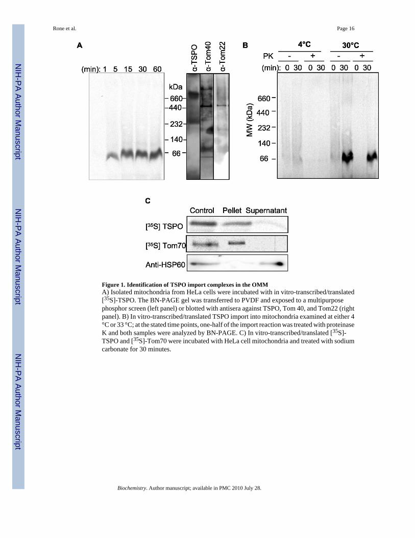
Figure 1. Identification of TSPO import complexes in the OMMA) Isolated mitochondria from HeLa cells were incubated with in vitro-transcribed/translated[35S]-TSPO. The BN-PAGE gel was transferred to PVDF and exposed to a multipurposephosphor screen (left panel) or blotted with antisera against TSPO, Tom 40, and Tom22 (rightpanel). B) In vitro-transcribed/translated TSPO import into mitochondria examined at either 4°C or 33 °C; at the stated time points, one-half of the import reaction was treated with proteinaseK and both samples were analyzed by BN-PAGE. C) In vitro-transcribed/translated [35S]-TSPO and [35S]-Tom70 were incubated with HeLa cell mitochondria and treated with sodiumcarbonate for 30 minutes.
Rone et al. Page 16
Biochemistry. Author manuscript; available in PMC 2010 July 28.
NIH
-PA Author Manuscript
NIH
-PA Author Manuscript
NIH
-PA Author Manuscript

Figure 2. TSPO import is dependent upon heat shock proteins and ATP for importA) Effects of C90 incubation with TSPO. TSPO was imported as described above (control).B) Effect of ATP depletion, 18μM geldanamycin, and 1mM novobiocin on TSPO import. C)Quantification of imported TSPO. Results shown are means±sem from three independentexperiments. D) Nickel-sepharose pull-down assay of 35S-TSPO and 35S-PIC. When indicatedgeldanamycin or novobiocin were added to the reaction mixture.
Rone et al. Page 17
Biochemistry. Author manuscript; available in PMC 2010 July 28.
NIH
-PA Author Manuscript
NIH
-PA Author Manuscript
NIH
-PA Author Manuscript

Figure 3. Identification of the TSPO amino acid sequence(s) responsible for targeting the proteinto the OMMMA-10 cells were transfected with either wild-type GFP-TSPO or various constructs of GFP-TSPO, stained with Mitotracker CMX and visualized by confocal microscopy. Each panelshows the construct used, GFP fluorescence, mitochondrial staining, and the merged image.A) Wild-type TSPO with GFP linked to the N-terminus. Cells were also transfected with GFP-Δ1−7-TSPO (B), GFP- Δ1−28-TSPO (C), GFP-Δ1−48-TSPO (D), GFP- Δ1−68-TSPO (E),GFP- Δ1−85-TSPO (F), GFP- Δ1−110-TSPO (G), GFP-Δ151−169-TSPO (J), GFP-Δ157−169-TSPO (K). The cholesterol binding domain, GFP-Δ148−157-TSPO (L), or the Schellmanmotif, GFP-Δ103−108-TSPO (H) were removed and transfected. Construct GFP-G106A-TSPO (I) point mutation altering the Schellman motif and constructs GFP-Leucine112−114-TSPO (M) and GFP-Leucine137,138,141-TSPO (N) were also transfected as described underexperimental methods.
Rone et al. Page 18
Biochemistry. Author manuscript; available in PMC 2010 July 28.
NIH
-PA Author Manuscript
NIH
-PA Author Manuscript
NIH
-PA Author Manuscript

Figure 4. TSPO protein regions and amino acids required for insertion into the OMMA) Model of the 18-kDa TSPO protein present in the OMM. Blue amino acids represent theC-terminus, red amino acids represent the mutated leucine residues, and green amino acidsshow the Schellman motif with the yellow amino acid in the middle representing the mutatedglycine. B) Schellman motif mutants of the TSPO protein were compared with wild-type TSPOfor import into HeLa cells mitochondria, followed by analysis by BN-PAGE. C) Effect ofremoval of the C-terminus with constructs Δ157−169 and Δ151−169 on inhibition of TSPOimport into isolated mitochondria. D) Effect of leucine mutations on in vitro-transcribed/translated radiolabeled TSPO import into HeLa cell mitochondria. E) SDS-PAGE ofradiolabeled constructs from import studies for confirmation of size due to mutations. F)
Rone et al. Page 19
Biochemistry. Author manuscript; available in PMC 2010 July 28.
NIH
-PA Author Manuscript
NIH
-PA Author Manuscript
NIH
-PA Author Manuscript

Measurement of progesterone production produced by MA-10 was performed as stated in themethods. Results shown are means ± SEM from three independent experiments (n = 9).
Rone et al. Page 20
Biochemistry. Author manuscript; available in PMC 2010 July 28.
NIH
-PA Author Manuscript
NIH
-PA Author Manuscript
NIH
-PA Author Manuscript
Figure 5. TSPO import is dependent on Metaxin 1A) MA-10 mitochondria incubated with in vitro-transcribed/translated TSPO analyzed by BN-PAGE, and blotted with anti-metaxin 1 antibody. B) In vitro-transcribed/translated TSPOincubated with mitochondria isolated from MA-10 cells control, Metaxin 1-depleted, andMA-10 cells treated with scrambled siRNAs. C) Immunoblot analysis for Metaxin1, COX IV,TSPO and VDAC in mitochondria isolated form MA-10 cells treated with the various siRNAs.D) Quantification of imported TSPO. Results shown are means±sem from three independentexperiments.
Rone et al. Page 21
Biochemistry. Author manuscript; available in PMC 2010 July 28.
NIH
-PA Author Manuscript
NIH
-PA Author Manuscript
NIH
-PA Author Manuscript

Figure 6. TSPO import is increased with cAMP stimulated mitochondriaA) Mitochondria isolated from MA-10 cells incubated with +/− 1mM cAMP were incubatedwith radiolabeled TSPO, analyzed via BN-PAGE. B) Quantification of imported TSPO, resultsshown are means±sem from three independent experiments. C) RT-qPCR analysis of MA-10RNA +/− 1mM cAMP. D) Immunoblot analysis of MA-10 cells incubated with and without1mM 8-bromo-AMP.
Rone et al. Page 22
Biochemistry. Author manuscript; available in PMC 2010 July 28.
NIH
-PA Author Manuscript
NIH
-PA Author Manuscript
NIH
-PA Author Manuscript

Figure 7. TSPO import into the OMMWe have outlined the steps necessary for TSPO targeting and insertion into the OMM. (1)TSPO is targeted to the OMM through its interactions with cytosolic chaperones, Hsc70 andHsp90. (2) At the OMM, TSPO interacts with Tom70 and is released from the chaperones inan ATP-dependent manner. (3) TSPO insertion into the OMM is dependent upon Metaxin 1(Mtx1) and TSPO structure, where both the C-terminus and the Schellman motif (amino acids103−108) are necessary to form a 66-kDa complex. Once import is complete, TSPO associateswith other protein complexes found in the OMM identified previously as VDAC and ANT.Yellow arrow indicates possible step accelerated by cAMP treatment.
Rone et al. Page 23
Biochemistry. Author manuscript; available in PMC 2010 July 28.
NIH
-PA Author Manuscript
NIH
-PA Author Manuscript
NIH
-PA Author Manuscript

NIH
-PA Author Manuscript
NIH
-PA Author Manuscript
NIH
-PA Author Manuscript
Rone et al. Page 24
Table 1Primers used for Q-PCR analysis:
Gene Forward Primer Reverse Primer Amplicon
Tom40(NM_001109748)
CGGTGTGGATGGCGAGTACCG CCTGAACCCACCAAGACATCTG 81
Tom22(NM_172609)
CGGGCCGAGGAATTACTCCCG CGTCTTCTTCCAGCTCCTCCTC 64
Tim 13(NM_013898)
CGATAGGCTCCTTGGATAACTCG AGAGTTGTAGGCGCGGGACAC 98
Tim22(NM_019818)
GTACCTGGTGGGCGACAAGC CGAGTCGCTCTTGGCAGGACTCG 85
Sam50(NM_178614)
GTGCATCCGCTGGTCCTATG CGGCGTAGTTCAGCTCCAGCCG 77
Metaxin 1(NM_013604)
CGGTAGAGGAGGAGCCATACCG TGGATAGAAACGATGCCACTGA 106
Metaxin 2(NM_016804)
CGTTTGCAGTGGGAAGTGAAACG CCTGGTCCAGAGTCTTGTTACCC 67
HPRT(NM_013556)
GTACCAGACCTCTCGAAGTGTTGGATAC TCCAACAACAAACTTGTCTGGAAT 77
Biochemistry. Author manuscript; available in PMC 2010 July 28.

NIH
-PA Author Manuscript
NIH
-PA Author Manuscript
NIH
-PA Author Manuscript
Rone et al. Page 25
Table 2Identification of proteins present in the 66- and 800-kDa HeLa proteincomplexes by mass spectrometryThe imported 66-kDa TSPO complex and the endogenous 800-kDa mitochondrialprotein complexes separated by BN-PAGE and 2D SDS-PAGE were analyzed bymass spectrometry; some of the major identified proteins are shown.
Protein Sequences Identified
66-kDa
Metaxin1 RSLASPGISPGPLTATIGGAVAGGGPR
Nonspecific lipid-transfer protein K.LQNLQLQPGNAKLK.IGGIFAFK.VK.ANLVFKEIEK.K
Voltage-dependent anion channel (VDAC 1) TDEFQLHTNVNDGTEFGGSIYQKR.WTEYGLTFTEK.WK.LTFDSSFSPNTGK.K
800-kDa
Adenine nucleotide translocator (ANT 1,2,3) K.LLLQVQHASK.QR.YFPTQALNFAFK.DR.AAYFGIYDTAK.GK.DFLAGGIAAAISK.T
Apolipoprotein A-I K.VSFLSALEEYTK.K
Apolipoprotein A-II K.SPELQAEAK.S
Fatty acid synthase K.VVVQVLAEEPEAVLK.GR.LQVVDQPLPVR.GK.VGDPQELNGITR.A
Mitofilin R.ELDSITPEVLPGWK.G
Voltage-dependent anion channel (VDAC 1,3) R.VTQSNFAVGYK.TK.LSQNNFALGYK.A
Annexin A2 K.SALSGHLETVILGLLK.TR.QDIAFAYQR.RK.TPAQYDASELK.AK.GVDEVTIVNILTNR.S
Biochemistry. Author manuscript; available in PMC 2010 July 28.
Related Documents