Targeted BRAF Inhibition Impacts Survival in Melanoma Patients with High Levels of Wnt/b-Catenin Signaling Andy J. Chien 1,2 * . , Lauren E. Haydu 3,4. , Travis L. Biechele 1 , Rima M. Kulikauskas 1 , Helen Rizos 3,4,5 , Richard F. Kefford 3,4,5,6 , Richard A. Scolyer 3,4,7 , Randall T. Moon 8 , Georgina V. Long 3,4,5,6 * 1 Division of Dermatology, University of Washington Department of Medicine, Seattle, Washington, United States of America, 2 The Group Health Research Institute, Seattle, Washington, United States of America, 3 Melanoma Institute of Australia, Sydney, New South Wales, Australia, 4 The University of Sydney, Sydney, New South Wales, Australia, 5 Westmead Institute for Cancer Research, Westmead Millennium Institute, Westmead, New South Wales, Australia, 6 Westmead Hospital, Sydney, New South Wales, Australia, 7 Royal Prince Alfred Hospital, Sydney, New South Wales, Australia, 8 The Howard Hughes Medical Institute, Chevy Chase, Maryland, United States of America Abstract Unprecedented clinical responses have been reported in advanced stage metastatic melanoma patients treated with targeted inhibitors of constitutively activated mutant BRAF, which is present in approximately half of all melanomas. We and others have previously observed an association of elevated nuclear b-catenin with improved survival in molecularly- unselected melanoma patients. This study sought to determine whether levels of Wnt/b-catenin signaling in melanoma tumors prior to treatment might predict patient responses to BRAF inhibitors (BRAFi). We performed automated quantification of b-catenin immunohistochemical expression in pretreatment BRAF-mutant tumors from 32 BRAFi-treated melanoma patients. Unexpectedly, patients with higher nuclear b-catenin in their tumors did not exhibit the survival advantage previously observed in molecularly-unselected melanoma patients who did not receive BRAFi. In cultured melanoma cells treated with long-term BRAFi, activation of Wnt/b-catenin signaling is markedly inhibited, coinciding with a loss of the enhancement of BRAFi-induced apoptosis by WNT3A observed in BRAFi-naı ¨ve cells. Together, these observations suggest that long-term treatment with BRAFi can impact the interaction between BRAF/MAPK and Wnt/b-catenin signaling to affect patient outcomes. Studies with larger patient cohorts are required to determine whether nuclear b-catenin expression correlates with clinical responses to BRAFi and to specific mechanisms of acquired resistance to BRAFi. Understanding these pathway interactions will be necessary to facilitate efforts to individualize therapies for melanoma patients. Citation: Chien AJ, Haydu LE, Biechele TL, Kulikauskas RM, Rizos H, et al. (2014) Targeted BRAF Inhibition Impacts Survival in Melanoma Patients with High Levels of Wnt/b-Catenin Signaling. PLoS ONE 9(4): e94748. doi:10.1371/journal.pone.0094748 Editor: Soheil S. Dadras, University of Connecticut Health Center, United States of America Received August 14, 2013; Accepted March 20, 2014; Published April 14, 2014 Copyright: ß 2014 Chien et al. This is an open-access article distributed under the terms of the Creative Commons Attribution License, which permits unrestricted use, distribution, and reproduction in any medium, provided the original author and source are credited. Funding: This work is supported by Program Grants of the National Health and Medical Research Council of Australia (NHMRC) and the Cancer Institute New South Wales (NSW). G.V.L., H.R. and R.A.S. are supported by the Cancer Institute NSW Fellowship program. H.R. and R.A.S. are also supported by the NHMRC Fellowship program. A.J.C. is supported through endowed research funds in the University of Washington Division of Dermatology. R.M.K. is supported through bridge funding from the University of Washington’s Office of the Provost. T.L.B. was supported through a NIH/NIAMS T32 training grant through the University of Washington Division of Dermatology. R.T.M. is an Investigator of the Howard Hughes Medical Institute. The funders had no role in study design, data collection and analysis, decision to publish, or preparation of the manuscript. Competing Interests: The authors have declared that no competing interests exist. * E-mail: [email protected] (GVL); [email protected] (AJC) . These authors contributed equally to this work. Introduction The incidence and mortality associated with melanoma has risen steadily since the 1970s in the USA, Europe and Australia [1], and the five-year survival rate of 5–15% for patients with advanced stage metastatic disease has remained stagnant over that time. Approximately half of all melanoma tumors harbor activating mutations in BRAF, with BRAF V600E and BRAF V600K representing approximately 70–90% and 10–30% of mutations, respectively [2–10]. Mutation-targeted BRAF inhibitors (BRAFi) such as vemurafenib (PLX4032) and dabrafenib (GSK2118436) represent a landmark development in the treatment of advanced stage BRAF V600E/K -mutant metastatic melanoma, with objective response rates of approximately 50%, and in phase III trials, a significant improvement in progression-free survival (PFS) and overall survival (OS) compared with dacarbazine chemotherapy [10–14]. In addition, almost all patients with tumors harboring activating BRAF mutations in these trials exhibit some degree of tumor reduction, even if they do not meet the criteria for an objective clinical response. Despite the promise of these targeted BRAFi, most patients develop recurrence and relapse at a median of 6–7 months. Studies utilizing patient tumor samples and preclinical models have identified several pathways to the development of BRAFi resistance, and the majority of resistance mechanisms identified to date appear to result in reactivation of the MAP kinase (MAPK) pathway as demonstrated by high levels of phosphorylated ERK1/ 2 [15–20]. Clinically, several questions remain unanswered. For example, what types of molecular and cellular determinants underlie the heterogeneity in therapeutic responses observed across patients with tumors harboring activating BRAF mutations and how can these determinants be utilized to predict clinical PLOS ONE | www.plosone.org 1 April 2014 | Volume 9 | Issue 4 | e94748

Welcome message from author
This document is posted to help you gain knowledge. Please leave a comment to let me know what you think about it! Share it to your friends and learn new things together.
Transcript
Targeted BRAF Inhibition Impacts Survival in MelanomaPatients with High Levels of Wnt/b-Catenin SignalingAndy J. Chien1,2*., Lauren E. Haydu3,4., Travis L. Biechele1, Rima M. Kulikauskas1, Helen Rizos3,4,5,
Richard F. Kefford3,4,5,6, Richard A. Scolyer3,4,7, Randall T. Moon8, Georgina V. Long3,4,5,6*
1 Division of Dermatology, University of Washington Department of Medicine, Seattle, Washington, United States of America, 2 The Group Health Research Institute,
Seattle, Washington, United States of America, 3 Melanoma Institute of Australia, Sydney, New South Wales, Australia, 4 The University of Sydney, Sydney, New South
Wales, Australia, 5 Westmead Institute for Cancer Research, Westmead Millennium Institute, Westmead, New South Wales, Australia, 6 Westmead Hospital, Sydney, New
South Wales, Australia, 7 Royal Prince Alfred Hospital, Sydney, New South Wales, Australia, 8 The Howard Hughes Medical Institute, Chevy Chase, Maryland, United States
of America
Abstract
Unprecedented clinical responses have been reported in advanced stage metastatic melanoma patients treated withtargeted inhibitors of constitutively activated mutant BRAF, which is present in approximately half of all melanomas. We andothers have previously observed an association of elevated nuclear b-catenin with improved survival in molecularly-unselected melanoma patients. This study sought to determine whether levels of Wnt/b-catenin signaling in melanomatumors prior to treatment might predict patient responses to BRAF inhibitors (BRAFi). We performed automatedquantification of b-catenin immunohistochemical expression in pretreatment BRAF-mutant tumors from 32 BRAFi-treatedmelanoma patients. Unexpectedly, patients with higher nuclear b-catenin in their tumors did not exhibit the survivaladvantage previously observed in molecularly-unselected melanoma patients who did not receive BRAFi. In culturedmelanoma cells treated with long-term BRAFi, activation of Wnt/b-catenin signaling is markedly inhibited, coinciding with aloss of the enhancement of BRAFi-induced apoptosis by WNT3A observed in BRAFi-naı̈ve cells. Together, these observationssuggest that long-term treatment with BRAFi can impact the interaction between BRAF/MAPK and Wnt/b-catenin signalingto affect patient outcomes. Studies with larger patient cohorts are required to determine whether nuclear b-cateninexpression correlates with clinical responses to BRAFi and to specific mechanisms of acquired resistance to BRAFi.Understanding these pathway interactions will be necessary to facilitate efforts to individualize therapies for melanomapatients.
Citation: Chien AJ, Haydu LE, Biechele TL, Kulikauskas RM, Rizos H, et al. (2014) Targeted BRAF Inhibition Impacts Survival in Melanoma Patients with High Levelsof Wnt/b-Catenin Signaling. PLoS ONE 9(4): e94748. doi:10.1371/journal.pone.0094748
Editor: Soheil S. Dadras, University of Connecticut Health Center, United States of America
Received August 14, 2013; Accepted March 20, 2014; Published April 14, 2014
Copyright: � 2014 Chien et al. This is an open-access article distributed under the terms of the Creative Commons Attribution License, which permitsunrestricted use, distribution, and reproduction in any medium, provided the original author and source are credited.
Funding: This work is supported by Program Grants of the National Health and Medical Research Council of Australia (NHMRC) and the Cancer Institute NewSouth Wales (NSW). G.V.L., H.R. and R.A.S. are supported by the Cancer Institute NSW Fellowship program. H.R. and R.A.S. are also supported by the NHMRCFellowship program. A.J.C. is supported through endowed research funds in the University of Washington Division of Dermatology. R.M.K. is supported throughbridge funding from the University of Washington’s Office of the Provost. T.L.B. was supported through a NIH/NIAMS T32 training grant through the University ofWashington Division of Dermatology. R.T.M. is an Investigator of the Howard Hughes Medical Institute. The funders had no role in study design, data collectionand analysis, decision to publish, or preparation of the manuscript.
Competing Interests: The authors have declared that no competing interests exist.
* E-mail: [email protected] (GVL); [email protected] (AJC)
. These authors contributed equally to this work.
Introduction
The incidence and mortality associated with melanoma has
risen steadily since the 1970s in the USA, Europe and Australia
[1], and the five-year survival rate of 5–15% for patients with
advanced stage metastatic disease has remained stagnant over that
time. Approximately half of all melanoma tumors harbor
activating mutations in BRAF, with BRAFV600E and BRAFV600K
representing approximately 70–90% and 10–30% of mutations,
respectively [2–10]. Mutation-targeted BRAF inhibitors (BRAFi)
such as vemurafenib (PLX4032) and dabrafenib (GSK2118436)
represent a landmark development in the treatment of advanced
stage BRAFV600E/K-mutant metastatic melanoma, with objective
response rates of approximately 50%, and in phase III trials, a
significant improvement in progression-free survival (PFS) and
overall survival (OS) compared with dacarbazine chemotherapy
[10–14]. In addition, almost all patients with tumors harboring
activating BRAF mutations in these trials exhibit some degree of
tumor reduction, even if they do not meet the criteria for an
objective clinical response.
Despite the promise of these targeted BRAFi, most patients
develop recurrence and relapse at a median of 6–7 months.
Studies utilizing patient tumor samples and preclinical models
have identified several pathways to the development of BRAFi
resistance, and the majority of resistance mechanisms identified to
date appear to result in reactivation of the MAP kinase (MAPK)
pathway as demonstrated by high levels of phosphorylated ERK1/
2 [15–20]. Clinically, several questions remain unanswered. For
example, what types of molecular and cellular determinants
underlie the heterogeneity in therapeutic responses observed
across patients with tumors harboring activating BRAF mutations
and how can these determinants be utilized to predict clinical
PLOS ONE | www.plosone.org 1 April 2014 | Volume 9 | Issue 4 | e94748
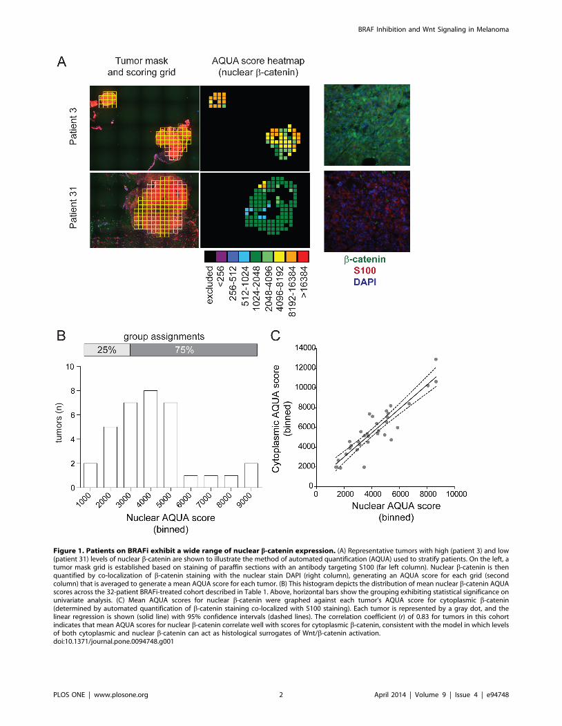
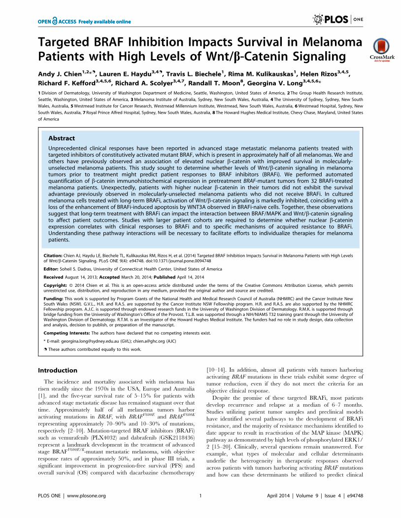
Figure 1. Patients on BRAFi exhibit a wide range of nuclear b-catenin expression. (A) Representative tumors with high (patient 3) and low(patient 31) levels of nuclear b-catenin are shown to illustrate the method of automated quantification (AQUA) used to stratify patients. On the left, atumor mask grid is established based on staining of paraffin sections with an antibody targeting S100 (far left column). Nuclear b-catenin is thenquantified by co-localization of b-catenin staining with the nuclear stain DAPI (right column), generating an AQUA score for each grid (secondcolumn) that is averaged to generate a mean AQUA score for each tumor. (B) This histogram depicts the distribution of mean nuclear b-catenin AQUAscores across the 32-patient BRAFi-treated cohort described in Table 1. Above, horizontal bars show the grouping exhibiting statistical significance onunivariate analysis. (C) Mean AQUA scores for nuclear b-catenin were graphed against each tumor’s AQUA score for cytoplasmic b-catenin(determined by automated quantification of b-catenin staining co-localized with S100 staining). Each tumor is represented by a gray dot, and thelinear regression is shown (solid line) with 95% confidence intervals (dashed lines). The correlation coefficient (r) of 0.83 for tumors in this cohortindicates that mean AQUA scores for nuclear b-catenin correlate well with scores for cytoplasmic b-catenin, consistent with the model in which levelsof both cytoplasmic and nuclear b-catenin can act as histological surrogates of Wnt/b-catenin activation.doi:10.1371/journal.pone.0094748.g001
BRAF Inhibition and Wnt Signaling in Melanoma
PLOS ONE | www.plosone.org 2 April 2014 | Volume 9 | Issue 4 | e94748

responses and tailor therapies? Such determinants may be utilized
to develop molecular assays that facilitate the identification or
selection of optimized drug combinations for patients.
The Wnt/b-catenin signaling pathway has been implicated as
an important regulator of melanoma despite the fact that
activating mutations in core pathway members appear to be rare
in this disease. This signaling pathway is activated by secreted
ligands including WNT3A, which is the WNT isoform most often
used for activating Wnt/b-catenin signaling in laboratory studies.
Frequently, the activation of Wnt/b-catenin signaling has been
detected through the measurement of endogenous downstream
target genes such as AXIN2, which encodes a core pathway protein
that promotes the degradation of b-catenin [21]. In patient tissue
samples, another surrogate marker of activated Wnt/b-catenin
signaling is the immunohistochemical detection of cytoplasmic or
nuclear b-catenin, which accumulates in cells upon activation of
the pathway [21]. Multiple studies have observed that loss of
nuclear or cytoplasmic b-catenin, the downstream effector protein
of Wnt, is associated with disease progression and decreased
survival in patients with melanoma [22–26]. Wnt/b-catenin
signaling in melanoma cells is negatively regulated by BRAFV600E
[27]. However, Wnt/b-catenin signaling also reciprocally regu-
lates BRAF-mediated signaling. In BRAF-mutant cell lines, the
activation of Wnt/b-catenin signaling in combination with BRAFi
synergistically enhanced apoptosis in vitro and increased inhibition
of tumor growth in vivo [27]. Furthermore, melanoma cell
Table 1. Characteristics of BRAFi patient cohort (n = 32).
Factor Value N %
Total Patients N 32 100%
Patient Sex Female 12 38%
Male 20 63%
Age at Trial Start (years) Mean/Median (range) 52/57 (23–73) -
Genotype V600E 30 94%
V600K 2 6%
BRAFi Dabrafenib 29 91%
Vemurafenib 3 9%
Active Brain Metastases at Trial Start No 15 44%
Yes 17 56%
Subsequent COMBI* No 31 97%
Yes 1 3%
M-stage M1a 1 3%
M1b 1 3%
M1c 30 94%
Baseline Sum of Diameters (mm) Mean/Median (range) 127/108(9–317) -
ECOG 0 15 47%
1 17 53%
LDH Normal 15 47%
Elevated 17 53%
Best CT Response PD 1 3%
SD 8 25%
PR 23 72%
CR 0 0%
Progression Status Progressed 29 94%
Not progressed 3 6%
BRAFi Status Continuing BRAFi 28 88%
Not on BRAFi 4 13%
Treatment Beyond Progression‘ No 17 53%
Yes 15 47%
Treatment Beyond Progression (days) Mean/Median (range) 116/66 (32–382) -
Last Follow-up Status Dead 22 72%
Alive 10 28%
Follow-up (weeks)* Mean/Median (range) 51/41 (8–153) -
Abbreviations: PD,progressive disease; SD, stable disease; PR, partial response; CR, complete response.*COMBI denotes subsequent enrolment in a clinical trial comparing combination BRAFi plus MEK inhibitor to placebo. Follow-up for subsequent COMBI patients (n = 1)was censored at date of cessation of mono-BRAFi.‘Treatment beyond progression classified as cessation of BRAFi greater than 30 days after date of progression.doi:10.1371/journal.pone.0094748.t001
BRAF Inhibition and Wnt Signaling in Melanoma
PLOS ONE | www.plosone.org 3 April 2014 | Volume 9 | Issue 4 | e94748
apoptosis mediated by BRAFi unexpectedly required b-catenin
and intact Wnt/b-catenin signaling [27].
Given that elevated Wnt/b-catenin signaling has been associ-
ated with improved melanoma survival outcomes in molecularly
unselected patients along with enhancement of apoptosis with
BRAFi in laboratory melanoma models, we hypothesized that
higher levels of Wnt/b-catenin signaling in pre-treatment mela-
noma tumors (as measured by increased nuclear b-catenin) might
predict a better clinical response to BRAFi. To address this
hypothesis, we performed a retrospective analysis of Wnt/b-
catenin signaling of pretreatment melanoma specimens from
patients treated clinically with BRAFi for metastatic melanoma. In
parallel, we studied the effects of long-term BRAFi treatment in
cultured melanoma cells. Our results extend the previous model
for how Wnt/b-catenin and BRAF/MAPK signaling interact in
melanoma.
Results
Patient characteristics and measurement of b-cateninPatients with metastatic melanoma carrying a BRAF mutation at
the V600 position (confirmed by DNA sequencing) who received
treatment with BRAFi (n = 32) were included in this study. Cohort
characteristics are summarized in Table 1. The response rate was
72%, median time to progression was 16.3 weeks (95% CI: 13.9–
18.6) and the median OS was 41.4 weeks (95% CI: 26.8–56.0).
Automated quantification of immunohistochemical staining was
used to measure mean nuclear b-catenin (Figure 1A-B). Mean
scores for nuclear b-catenin ranged from 1411.4 to 8668.4
(Figure 1B). The ranked scores were stratified as shown in
Figure 1B. Results using summed cytosolic and nuclear b-catenin
were the same as results with nuclear b-catenin alone (data not
shown), consistent with our observation that nuclear b-catenin
scores correlate highly with cytoplasmic b-catenin scores within
tumors (r = 0.83; Figure 1C).
Nuclear b-catenin and survival endpoints in patientstreated with BRAFi
Given the limited sample size, we performed post-hoc
exploratory analysis comparing levels of nuclear b-catenin with
the survival endpoints using different methods of patient stratifi-
cation that subdivided the cohort into two or three groups.
Comparison of patients in the lowest quartile of nuclear b-catenin
(lowest 25%) to the remaining 75% of patients within the cohort
revealed a significant difference in OS (p = 0.037; Figure 2A). A
similar trend was observed with PFS using this stratification,
although this was not statistically significant (Figure 2B
(p = 0.099)). There was no significant difference between the
groups for time to best RECIST CT response (data not shown).
Long-term BRAFi treatment leads to down-regulation ofcellular responses to WNT3A
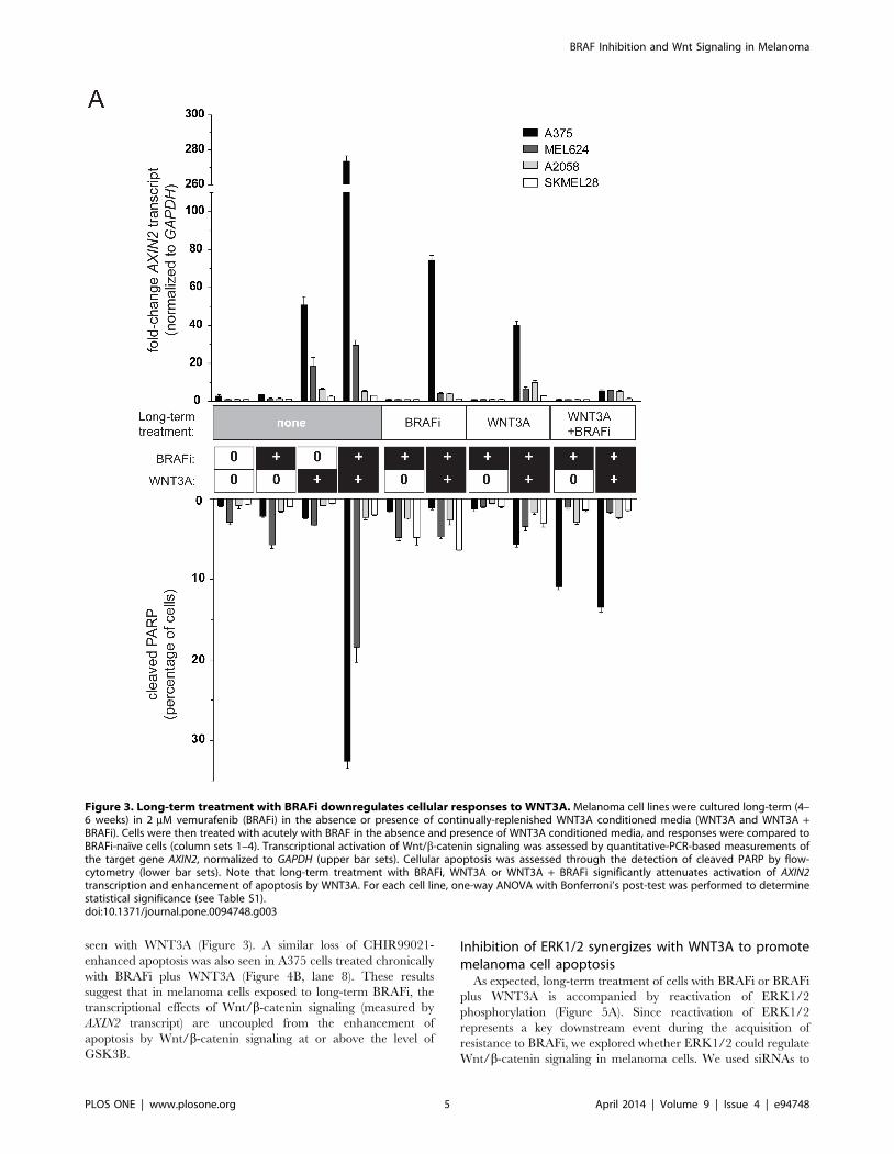
We treated a panel of melanoma cell lines for 4–6 weeks in the
continued presence of 2 mM vemurafenib (BRAFi) and in the
presence or absence of WNT3A (Figure 3). Two of these cell lines
(A375 and MEL624) were previously shown to exhibit enhanced
apoptosis in the presence of both WNT3A and BRAFi, while two
of these cell lines (A2058 and SKMEL28) do not exhibit significant
apoptosis with WNT3A and BRAFi [27]. These cells were
compared to naı̈ve cells that had not been chronically treated with
BRAFi and/or WNT3A, but only acutely exposed to BRAFi and
WNT3A overnight. Long-term treatment with BRAFi alone or
BRAFi and/or WNT3A markedly inhibited activation of Wnt/b-
catenin signaling as measured by AXIN2 transcript levels (Figure 3,
upper half). In parallel, we also measured the effects of acute
exposure to BRAFi and/or WNT3A on apoptosis measured by
cleaved PARP. Similarly, we saw that apoptosis with WNT3A and
BRAFi was inhibited in cells chronically treated with BRAFi and/
or WNT3A compared to naı̈ve cells (Figure 3, lower half).
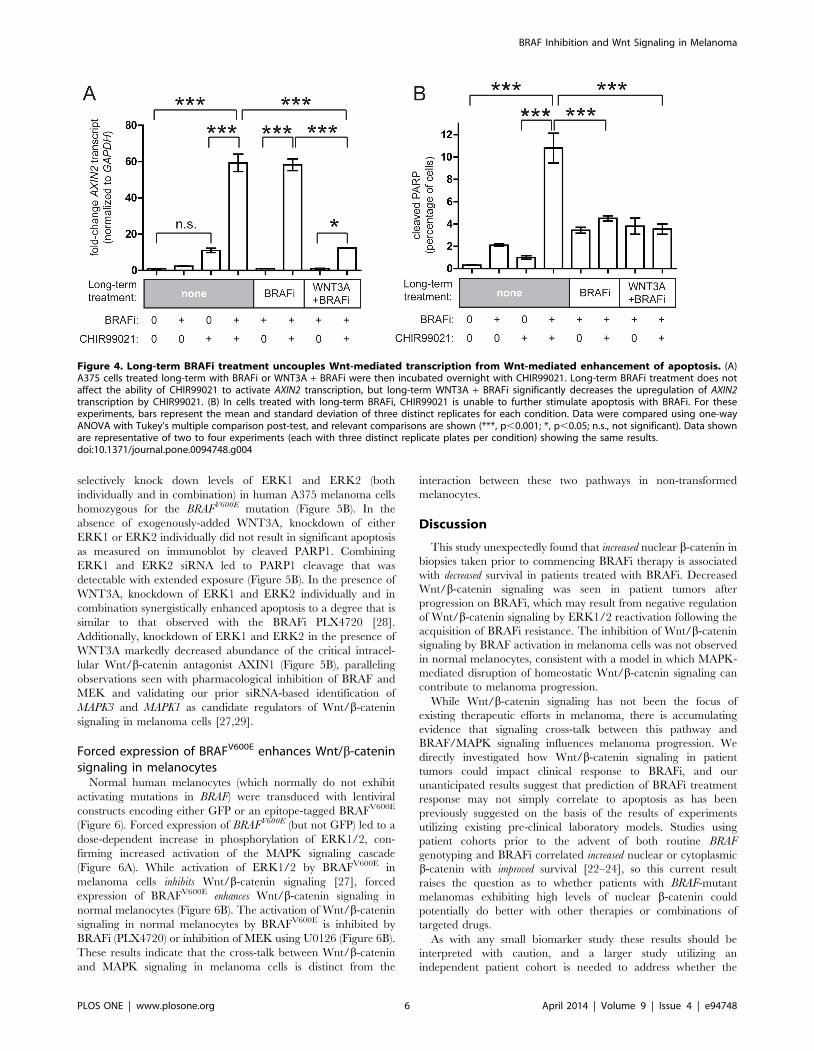
Interestingly, long-term treatment of cells with BRAFi alone did
not affect activation of Wnt/b-catenin signaling by the GSK3
inhibitor CHIR99021 (Figure 4A, compare columns 4 and 6),
suggesting that the pathway is intact at the level of GSK3B, one of
the key intracellular regulators of b-catenin abundance. However,
cells cultured with BRAFi and WNT3A exhibited a highly blunted
activation of Wnt/b-catenin signaling with CHIR99021
(Figure 4A, compare column 8 with columns 4 and 6).
Unexpectedly, while long-term BRAFi alone did not inhibit
activation of Wnt/b-catenin signaling by CHIR99021, it com-
pletely prevented any enhancement of apoptosis with CHIR99021
(Figure 4A and 4B, compare lane 4 to lane 6), similar to what was
Figure 2. Lower levels of nuclear b-catenin significantlycorrelate with improved survival on BRAFi in post-hoc analysis.(A) In exploratory post-hoc analysis, patients with nuclear b-cateninscores in the lowest quartile exhibited a statistically-significantimprovement in overall survival (p = 0.037) compared to the remainderof the cohort. (B) A similar trend towards improvement with patients inthe lowest quartile was seen with progression-free survival, but did notreach statistical significance (p = 0.099).doi:10.1371/journal.pone.0094748.g002
BRAF Inhibition and Wnt Signaling in Melanoma
PLOS ONE | www.plosone.org 4 April 2014 | Volume 9 | Issue 4 | e94748
seen with WNT3A (Figure 3). A similar loss of CHIR99021-
enhanced apoptosis was also seen in A375 cells treated chronically
with BRAFi plus WNT3A (Figure 4B, lane 8). These results
suggest that in melanoma cells exposed to long-term BRAFi, the
transcriptional effects of Wnt/b-catenin signaling (measured by
AXIN2 transcript) are uncoupled from the enhancement of
apoptosis by Wnt/b-catenin signaling at or above the level of
GSK3B.
Inhibition of ERK1/2 synergizes with WNT3A to promotemelanoma cell apoptosis
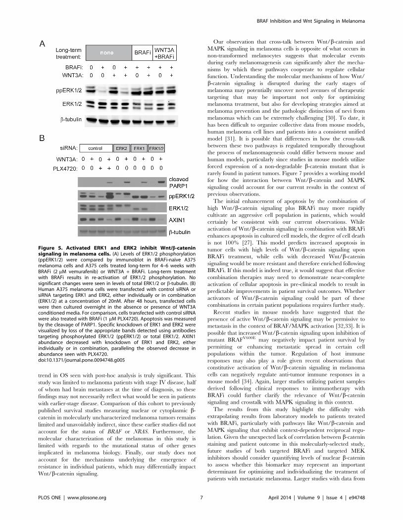
As expected, long-term treatment of cells with BRAFi or BRAFi
plus WNT3A is accompanied by reactivation of ERK1/2
phosphorylation (Figure 5A). Since reactivation of ERK1/2
represents a key downstream event during the acquisition of
resistance to BRAFi, we explored whether ERK1/2 could regulate
Wnt/b-catenin signaling in melanoma cells. We used siRNAs to
Figure 3. Long-term treatment with BRAFi downregulates cellular responses to WNT3A. Melanoma cell lines were cultured long-term (4–6 weeks) in 2 mM vemurafenib (BRAFi) in the absence or presence of continually-replenished WNT3A conditioned media (WNT3A and WNT3A +BRAFi). Cells were then treated with acutely with BRAF in the absence and presence of WNT3A conditioned media, and responses were compared toBRAFi-naı̈ve cells (column sets 1–4). Transcriptional activation of Wnt/b-catenin signaling was assessed by quantitative-PCR-based measurements ofthe target gene AXIN2, normalized to GAPDH (upper bar sets). Cellular apoptosis was assessed through the detection of cleaved PARP by flow-cytometry (lower bar sets). Note that long-term treatment with BRAFi, WNT3A or WNT3A + BRAFi significantly attenuates activation of AXIN2transcription and enhancement of apoptosis by WNT3A. For each cell line, one-way ANOVA with Bonferroni’s post-test was performed to determinestatistical significance (see Table S1).doi:10.1371/journal.pone.0094748.g003
BRAF Inhibition and Wnt Signaling in Melanoma
PLOS ONE | www.plosone.org 5 April 2014 | Volume 9 | Issue 4 | e94748
selectively knock down levels of ERK1 and ERK2 (both
individually and in combination) in human A375 melanoma cells
homozygous for the BRAFV600E mutation (Figure 5B). In the
absence of exogenously-added WNT3A, knockdown of either
ERK1 or ERK2 individually did not result in significant apoptosis
as measured on immunoblot by cleaved PARP1. Combining
ERK1 and ERK2 siRNA led to PARP1 cleavage that was
detectable with extended exposure (Figure 5B). In the presence of
WNT3A, knockdown of ERK1 and ERK2 individually and in
combination synergistically enhanced apoptosis to a degree that is
similar to that observed with the BRAFi PLX4720 [28].
Additionally, knockdown of ERK1 and ERK2 in the presence of
WNT3A markedly decreased abundance of the critical intracel-
lular Wnt/b-catenin antagonist AXIN1 (Figure 5B), paralleling
observations seen with pharmacological inhibition of BRAF and
MEK and validating our prior siRNA-based identification of
MAPK3 and MAPK1 as candidate regulators of Wnt/b-catenin
signaling in melanoma cells [27,29].
Forced expression of BRAFV600E enhances Wnt/b-cateninsignaling in melanocytes
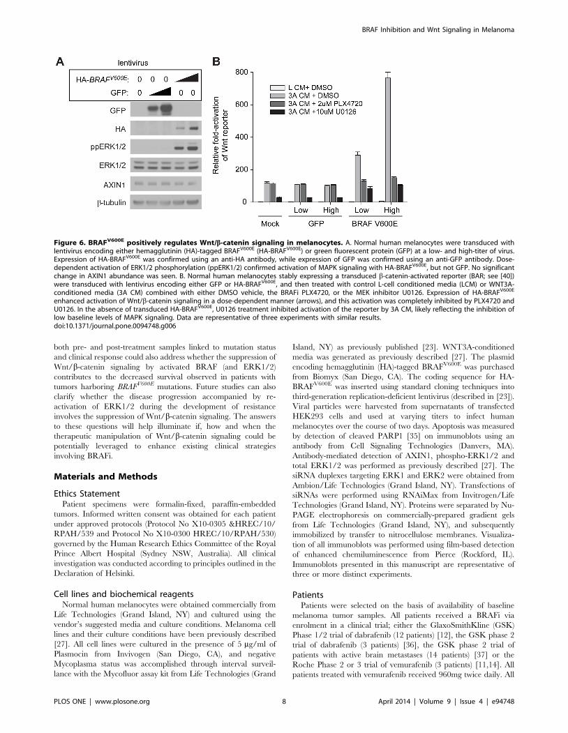
Normal human melanocytes (which normally do not exhibit
activating mutations in BRAF) were transduced with lentiviral
constructs encoding either GFP or an epitope-tagged BRAFV600E
(Figure 6). Forced expression of BRAFV600E (but not GFP) led to a
dose-dependent increase in phosphorylation of ERK1/2, con-
firming increased activation of the MAPK signaling cascade
(Figure 6A). While activation of ERK1/2 by BRAFV600E in
melanoma cells inhibits Wnt/b-catenin signaling [27], forced
expression of BRAFV600E enhances Wnt/b-catenin signaling in
normal melanocytes (Figure 6B). The activation of Wnt/b-catenin
signaling in normal melanocytes by BRAFV600E is inhibited by
BRAFi (PLX4720) or inhibition of MEK using U0126 (Figure 6B).
These results indicate that the cross-talk between Wnt/b-catenin
and MAPK signaling in melanoma cells is distinct from the
interaction between these two pathways in non-transformed
melanocytes.
Discussion
This study unexpectedly found that increased nuclear b-catenin in
biopsies taken prior to commencing BRAFi therapy is associated
with decreased survival in patients treated with BRAFi. Decreased
Wnt/b-catenin signaling was seen in patient tumors after
progression on BRAFi, which may result from negative regulation
of Wnt/b-catenin signaling by ERK1/2 reactivation following the
acquisition of BRAFi resistance. The inhibition of Wnt/b-catenin
signaling by BRAF activation in melanoma cells was not observed
in normal melanocytes, consistent with a model in which MAPK-
mediated disruption of homeostatic Wnt/b-catenin signaling can
contribute to melanoma progression.
While Wnt/b-catenin signaling has not been the focus of
existing therapeutic efforts in melanoma, there is accumulating
evidence that signaling cross-talk between this pathway and
BRAF/MAPK signaling influences melanoma progression. We
directly investigated how Wnt/b-catenin signaling in patient
tumors could impact clinical response to BRAFi, and our
unanticipated results suggest that prediction of BRAFi treatment
response may not simply correlate to apoptosis as has been
previously suggested on the basis of the results of experiments
utilizing existing pre-clinical laboratory models. Studies using
patient cohorts prior to the advent of both routine BRAF
genotyping and BRAFi correlated increased nuclear or cytoplasmic
b-catenin with improved survival [22–24], so this current result
raises the question as to whether patients with BRAF-mutant
melanomas exhibiting high levels of nuclear b-catenin could
potentially do better with other therapies or combinations of
targeted drugs.
As with any small biomarker study these results should be
interpreted with caution, and a larger study utilizing an
independent patient cohort is needed to address whether the
Figure 4. Long-term BRAFi treatment uncouples Wnt-mediated transcription from Wnt-mediated enhancement of apoptosis. (A)A375 cells treated long-term with BRAFi or WNT3A + BRAFi were then incubated overnight with CHIR99021. Long-term BRAFi treatment does notaffect the ability of CHIR99021 to activate AXIN2 transcription, but long-term WNT3A + BRAFi significantly decreases the upregulation of AXIN2transcription by CHIR99021. (B) In cells treated with long-term BRAFi, CHIR99021 is unable to further stimulate apoptosis with BRAFi. For theseexperiments, bars represent the mean and standard deviation of three distinct replicates for each condition. Data were compared using one-wayANOVA with Tukey’s multiple comparison post-test, and relevant comparisons are shown (***, p,0.001; *, p,0.05; n.s., not significant). Data shownare representative of two to four experiments (each with three distinct replicate plates per condition) showing the same results.doi:10.1371/journal.pone.0094748.g004
BRAF Inhibition and Wnt Signaling in Melanoma
PLOS ONE | www.plosone.org 6 April 2014 | Volume 9 | Issue 4 | e94748
trend in OS seen with post-hoc analysis is truly significant. This
study was limited to melanoma patients with stage IV disease, half
of whom had brain metastases at the time of diagnosis, so these
findings may not necessarily reflect what would be seen in patients
with earlier-stage disease. Comparison of this cohort to previously
published survival studies measuring nuclear or cytoplasmic b-
catenin in molecularly uncharacterized melanoma tumors remains
limited and unavoidably indirect, since these earlier studies did not
account for the status of BRAF or NRAS. Furthermore, the
molecular characterization of the melanomas in this study is
limited with regards to the mutational status of other genes
implicated in melanoma biology. Finally, our study does not
account for the mechanisms underlying the emergence of
resistance in individual patients, which may differentially impact
Wnt/b-catenin signaling.
Our observation that cross-talk between Wnt/b-catenin and
MAPK signaling in melanoma cells is opposite of what occurs in
non-transformed melanocytes suggests that molecular events
during early melanomagenesis can significantly alter the mecha-
nisms by which these pathways cooperate to regulate cellular
function. Understanding the molecular mechanisms of how Wnt/
b-catenin signaling is disrupted during the early stages of
melanoma may potentially uncover novel avenues of therapeutic
targeting that may be important not only for optimizing
melanoma treatment, but also for developing strategies aimed at
melanoma prevention and the pathologic distinction of nevi from
melanomas which can be extremely challenging [30]. To date, it
has been difficult to organize collective data from mouse models,
human melanoma cell lines and patients into a consistent unified
model [31]. It is possible that differences in how the cross-talk
between these two pathways is regulated temporally throughout
the process of melanomagenesis could differ between mouse and
human models, particularly since studies in mouse models utilize
forced expression of a non-degradable b-catenin mutant that is
rarely found in patient tumors. Figure 7 provides a working model
for how the interaction between Wnt/b-catenin and MAPK
signaling could account for our current results in the context of
previous observations.
The initial enhancement of apoptosis by the combination of
high Wnt/b-catenin signaling plus BRAFi may more rapidly
cultivate an aggressive cell population in patients, which would
certainly be consistent with our current observations. While
activation of Wnt/b-catenin signaling in combination with BRAFi
enhances apoptosis in cultured cell models, the degree of cell death
is not 100% [27]. This model predicts increased apoptosis in
tumor cells with high levels of Wnt/b-catenin signaling upon
BRAFi treatment, while cells with decreased Wnt/b-catenin
signaling would be more resistant and therefore enriched following
BRAFi. If this model is indeed true, it would suggest that effective
combination therapies may need to demonstrate near-complete
activation of cellular apoptosis in pre-clinical models to result in
predictable improvements in patient survival outcomes. Whether
activators of Wnt/b-catenin signaling could be part of these
combinations in certain patient populations requires further study.
Recent studies in mouse models have suggested that the
presence of active Wnt/b-catenin signaling may be permissive to
metastasis in the context of BRAF/MAPK activation [32,33]. It is
possible that increased Wnt/b-catenin signaling upon inhibition of
mutant BRAFV600E may negatively impact patient survival by
permitting or enhancing metastatic spread in certain cell
populations within the tumor. Regulation of host immune
responses may also play a role given recent observations that
constitutive activation of Wnt/b-catenin signaling in melanoma
cells can negatively regulate anti-tumor immune responses in a
mouse model [34]. Again, larger studies utilizing patient samples
derived following clinical responses to immunotherapy with
BRAFi could further clarify the relevance of Wnt/b-catenin
signaling and crosstalk with MAPK signaling in this context.
The results from this study highlight the difficulty with
extrapolating results from laboratory models to patients treated
with BRAFi, particularly with pathways like Wnt/b-catenin and
MAPK signaling that exhibit context-dependent reciprocal regu-
lation. Given the unexpected lack of correlation between b-catenin
staining and patient outcome in this molecularly-selected study,
future studies of both targeted BRAFi and targeted MEK
inhibitors should consider quantifying levels of nuclear b-catenin
to assess whether this biomarker may represent an important
determinant for optimizing and individualizing the treatment of
patients with metastatic melanoma. Larger studies with data from
Figure 5. Activated ERK1 and ERK2 inhibit Wnt/b-cateninsignaling in melanoma cells. (A) Levels of ERK1/2 phosphorylation(ppERK1/2) were compared by immunoblot in BRAFi-naı̈ve A375melanoma cells and A375 cells treated long-term for 4–6 weeks withBRAFi (2 mM vemurafenib) or WNT3A + BRAFi. Long-term treatmentwith BRAFi results in re-activation of ERK1/2 phosphorylation. Nosignificant changes were seen in levels of total ERK1/2 or b-tubulin. (B)Human A375 melanoma cells were transfected with control siRNA orsiRNA targeting ERK1 and ERK2, either individually or in combination(ERK1/2) at a concentration of 20nM. After 48 hours, transfected cellswere then cultured overnight in the absence or presence of WNT3Aconditioned media. For comparison, cells transfected with control siRNAwere also treated with BRAFi (1 mM PLX4720). Apoptosis was measuredby the cleavage of PARP1. Specific knockdown of ERK1 and ERK2 werevisualized by loss of the appropriate bands detected using antibodiestargeting phosphorylated ERK1/2 (ppERK1/2) or total ERK1/2. AXIN1abundance decreased with knockdown of ERK1 and ERK2, eitherindividually or in combination, paralleling the observed decrease inabundance seen with PLX4720.doi:10.1371/journal.pone.0094748.g005
BRAF Inhibition and Wnt Signaling in Melanoma
PLOS ONE | www.plosone.org 7 April 2014 | Volume 9 | Issue 4 | e94748
both pre- and post-treatment samples linked to mutation status
and clinical response could also address whether the suppression of
Wnt/b-catenin signaling by activated BRAF (and ERK1/2)
contributes to the decreased survival observed in patients with
tumors harboring BRAFV600E mutations. Future studies can also
clarify whether the disease progression accompanied by re-
activation of ERK1/2 during the development of resistance
involves the suppression of Wnt/b-catenin signaling. The answers
to these questions will help illuminate if, how and when the
therapeutic manipulation of Wnt/b-catenin signaling could be
potentially leveraged to enhance existing clinical strategies
involving BRAFi.
Materials and Methods
Ethics StatementPatient specimens were formalin-fixed, paraffin-embedded
tumors. Informed written consent was obtained for each patient
under approved protocols (Protocol No X10-0305 &HREC/10/
RPAH/539 and Protocol No X10-0300 HREC/10/RPAH/530)
governed by the Human Research Ethics Committee of the Royal
Prince Albert Hospital (Sydney NSW, Australia). All clinical
investigation was conducted according to principles outlined in the
Declaration of Helsinki.
Cell lines and biochemical reagentsNormal human melanocytes were obtained commercially from
Life Technologies (Grand Island, NY) and cultured using the
vendor’s suggested media and culture conditions. Melanoma cell
lines and their culture conditions have been previously described
[27]. All cell lines were cultured in the presence of 5 mg/ml of
Plasmocin from Invivogen (San Diego, CA), and negative
Mycoplasma status was accomplished through interval surveil-
lance with the Mycofluor assay kit from Life Technologies (Grand
Island, NY) as previously published [23]. WNT3A-conditioned
media was generated as previously described [27]. The plasmid
encoding hemagglutinin (HA)-tagged BRAFV600E was purchased
from Biomyx (San Diego, CA). The coding sequence for HA-
BRAFV600E was inserted using standard cloning techniques into
third-generation replication-deficient lentivirus (described in [23]).
Viral particles were harvested from supernatants of transfected
HEK293 cells and used at varying titers to infect human
melanocytes over the course of two days. Apoptosis was measured
by detection of cleaved PARP1 [35] on immunoblots using an
antibody from Cell Signaling Technologies (Danvers, MA).
Antibody-mediated detection of AXIN1, phospho-ERK1/2 and
total ERK1/2 was performed as previously described [27]. The
siRNA duplexes targeting ERK1 and ERK2 were obtained from
Ambion/Life Technologies (Grand Island, NY). Transfections of
siRNAs were performed using RNAiMax from Invitrogen/Life
Technologies (Grand Island, NY). Proteins were separated by Nu-
PAGE electrophoresis on commercially-prepared gradient gels
from Life Technologies (Grand Island, NY), and subsequently
immobilized by transfer to nitrocellulose membranes. Visualiza-
tion of all immunoblots was performed using film-based detection
of enhanced chemiluminescence from Pierce (Rockford, IL).
Immunoblots presented in this manuscript are representative of
three or more distinct experiments.
PatientsPatients were selected on the basis of availability of baseline
melanoma tumor samples. All patients received a BRAFi via
enrolment in a clinical trial; either the GlaxoSmithKline (GSK)
Phase 1/2 trial of dabrafenib (12 patients) [12], the GSK phase 2
trial of dabrafenib (3 patients) [36], the GSK phase 2 trial of
patients with active brain metastases (14 patients) [37] or the
Roche Phase 2 or 3 trial of vemurafenib (3 patients) [11,14]. All
patients treated with vemurafenib received 960mg twice daily. All
Figure 6. BRAFV600E positively regulates Wnt/b-catenin signaling in melanocytes. A. Normal human melanocytes were transduced withlentivirus encoding either hemagglutinin (HA)-tagged BRAFV600E (HA-BRAFV600E) or green fluorescent protein (GFP) at a low- and high-titer of virus.Expression of HA-BRAFV600E was confirmed using an anti-HA antibody, while expression of GFP was confirmed using an anti-GFP antibody. Dose-dependent activation of ERK1/2 phosphorylation (ppERK1/2) confirmed activation of MAPK signaling with HA-BRAFV600E, but not GFP. No significantchange in AXIN1 abundance was seen. B. Normal human melanocytes stably expressing a transduced b-catenin-activated reporter (BAR; see [40])were transduced with lentivirus encoding either GFP or HA-BRAFV600E, and then treated with control L-cell conditioned media (LCM) or WNT3A-conditioned media (3A CM) combined with either DMSO vehicle, the BRAFi PLX4720, or the MEK inhibitor U0126. Expression of HA-BRAFV600E
enhanced activation of Wnt/b-catenin signaling in a dose-dependent manner (arrows), and this activation was completely inhibited by PLX4720 andU0126. In the absence of transduced HA-BRAFV600E, U0126 treatment inhibited activation of the reporter by 3A CM, likely reflecting the inhibition oflow baseline levels of MAPK signaling. Data are representative of three experiments with similar results.doi:10.1371/journal.pone.0094748.g006
BRAF Inhibition and Wnt Signaling in Melanoma
PLOS ONE | www.plosone.org 8 April 2014 | Volume 9 | Issue 4 | e94748
patients treated with dabrafenib received $ the daily recom-
mended phase 2 dose of 300mg after first computed axial
tomography (CT) scan.
Response to Treatment and Clinical OutcomeObjective response to BRAFi treatment was assessed with CT
scanning 6–9 weekly, using RECIST 1.0 [38] for those on the
phase 1/2 study of dabrafenib, and RECIST 1.1 [39] for all other
patients. Three survival outcomes were tested using the Kaplan-
Meier method together with the Log Rank test; time to best
response, progression-free survival, and overall survival (Figures 2).
All time intervals were measured in relation to the commencement
of BRAFi. The primary endpoints for this study were overall
survival (OS), progression-free survival (PFS) and time to best
response. For secondary endpoints, best computerized tomogra-
phy (CT) response was assessed categorically as progressive or
stable disease versus partial response (no patients had a complete
response), and also as best percent-change in RECIST target
lesions. Follow-up for one patient taking subsequent COMBI
therapy was censored at date of cessation of BRAFi. The Mann-
Whitney U test was used to address correlations between nuclear
b-catenin and RECIST criteria.
Histological quantification of b-catenin in patient tumorsImmunohistochemical cytoplasmic and nuclear b-catenin stains
were conducted on tumor samples (either primary melanomas or
melanoma metastases) from this cohort collected prior to the
initiation of BRAFi. Five micron-thick tumor sections were labeled
immunohistochemically with a mouse monoclonal antibody to b-
catenin (BD Transduction Laboratories, San Jose CA; catalog
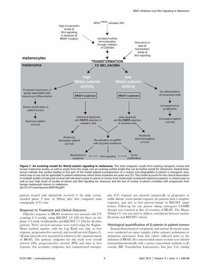
Figure 7. An evolving model for Wnt/b-catenin signaling in melanoma. This chart integrates results from existing transgenic mouse andhuman melanoma studies, as well as results from this study, into an evolving unified model that can be further tested for refinement. Dashed lines/arrows indicate that studies leading to this part of the model utilized overexpression of a mutant (non-degradable) b-catenin in transgenic mice,which may or may not be applicable in patient melanomas where these mutations are quite rare [31]. This model accounts for the clinical observationin multiple studies of improved survival with elevated nuclear b-catenin in tumors from molecularly unselected melanoma patients. In melanocytes aswell as nevi, high levels of nuclear b-catenin and Wnt signaling are observed, and the loss of nuclear b-catenin correlates with progression frombenign melanocytic lesions to melanoma.doi:10.1371/journal.pone.0094748.g007
BRAF Inhibition and Wnt Signaling in Melanoma
PLOS ONE | www.plosone.org 9 April 2014 | Volume 9 | Issue 4 | e94748
number 610154) at a dilution of 1:1000, which was experimentally
determined to be the optimal concentration using standardized
specimens. Fluorescent visualization was performed using an anti-
mouse Cy5-conjugated antibody (DAKO, Carpinteria CA). Nuclei
were visualized using Prolong Gold DAPI (Life Sciences, Grand
Island NY). Initial tumor specimens were examined, dissected,
processed and interpreted at the Melanoma Institute of Australia
prior to antigen retrieval, immunostaining, quantification and
imaging of these samples by HistoRX (Branford CT), who were
blinded to all outcomes data. S100 staining was used to identify a
tumor mask, defined as the cellular area of the tumor (Figure 1),
and the subsequent AQUA scoring grids were established by
certified pathologists. Subsequent AQUA scores were obtained for
each grid and averaged for each tumor, with nuclear and
cytoplasmic compartments defined by DAPI and S100, respec-
tively.
Statistical analysisScores for cytoplasmic and nuclear b-catenin for each patient
were ranked and stratified into five groups a priori by the team that
conducted the stains, blinded to the clinical outcome data. Scores
for nuclear b-catenin were averaged for each tumor based on an
average signal from each tumor grid (Figure 1). The GraphPad
Prism version 5.0 software suite (GraphPad Software, La Jolla CA)
and the IBM SPSS v21 software package (SPSS, Chicago IL) were
utilized for statistical analysis. All p-values less than 0.05 were
considered statistically significant. Univariate survival analyses
were conducted with the Kaplan-Meier method together with the
Log Rank test. Bivariate correlations were run using the Mann
Whitney U test or Spearman’s correlation where appropriate. All
time intervals were measured in relation to the commencement of
BRAFi. The primary endpoints for this study were overall survival
(OS), progression-free survival (PFS) and time to best response. For
secondary endpoints, best computerized tomography (CT) re-
sponse was assessed categorically as progressive or stable disease
versus partial response (no patients had a complete response), and
also as best percent-change in RECIST target lesions. Overall
survival for five patients having subsequent targeted/immune
therapy was censored at the time of cessation of BRAFi.
Supporting Information
Table S1 These tables show the results of one-wayANOVA for Figure 3, with post-test p-values indicatedfor each cell line.
(DOC)
Acknowledgments
The authors would like to recognize and thank the staff of the Melanoma
Institute of Australia and Westmead Hospital (Crown Princess Mary
Cancer Centre), including Jessica Hyman and James Wilmott, for their
technical contributions to this manuscript. The authors would also like to
thank Patrick O’Lin for helpful discussion and assistance with manuscript
preparation.
Author Contributions
Conceived and designed the experiments: AJC RTM GVL. Performed the
experiments: LEH TLB RMK HR. Analyzed the data: AJC LEH HR
RFK RAS RTM GVL. Wrote the paper: AJC RFK RAS GVL.
References
1. Lens MB, Dawes M (2004) Global perspectives of contemporary epidemiological
trends of cutaneous malignant melanoma. Br J Dermatol 150: 179–185.
2. Brose MS, Volpe P, Feldman M, Kumar M, Rishi I, et al. (2002) BRAF and
RAS mutations in human lung cancer and melanoma. Cancer Res 62: 6997–
7000.
3. Davies H, Bignell GR, Cox C, Stephens P, Edkins S, et al. (2002) Mutations of
the BRAF gene in human cancer. Nature 417: 949–954.
4. Gorden A, Osman I, Gai W, He D, Huang W, et al. (2003) Analysis of BRAF
and N-RAS mutations in metastatic melanoma tissues. Cancer Res 63: 3955–
3957.
5. Libra M, Malaponte G, Navolanic PM, Gangemi P, Bevelacqua V, et al. (2005)
Analysis of BRAF mutation in primary and metastatic melanoma. Cell Cycle 4:
1382–1384.
6. Loewe R, Kittler H, Fischer G, Fae I, Wolff K, et al. (2004) BRAF kinase gene
V599E mutation in growing melanocytic lesions. J Invest Dermatol 123: 733–
736.
7. Zalaudek I, Guelly C, Pellacani G, Hofmann-Wellenhof R, Trajanoski S, et al.
(2011) The dermoscopical and histopathological patterns of nevi correlate with
the frequency of BRAF mutations. J Invest Dermatol 131: 542–545.
8. Menzies AM, Haydu LE, Visintin L, Carlino MS, Howle JR, et al. (2012)
Distinguishing clinicopathologic features of patients with V600E and V600K
BRAF-mutant metastatic melanoma. Clin Cancer Res 18: 3242–3249.
9. Poynter JN, Elder JT, Fullen DR, Nair RP, Soengas MS, et al. (2006) BRAF and
NRAS mutations in melanoma and melanocytic nevi. Melanoma Res 16: 267–
273.
10. Yazdi AS, Palmedo G, Flaig MJ, Puchta U, Reckwerth A, et al. (2003)
Mutations of the BRAF gene in benign and malignant melanocytic lesions.
J Invest Dermatol 121: 1160–1162.
11. Chapman PB, Hauschild A, Robert C, Haanen JB, Ascierto P, et al. (2011)
Improved Survival with Vemurafenib in Melanoma with BRAF V600E
Mutation. New England Journal of Medicine 364: 9.
12. Falchook GS, Long GV, Kurzrock R, Kim KB, Arkenau TH, et al. (2012)
Dabrafenib in patients with melanoma, untreated brain metastases, and other
solid tumours: a phase 1 dose-escalation trial. Lancet 379: 1893–1901.
13. Hauschild A, Grob JJ, Demidov LV, Jouary T, Gutzmer R, et al. (2012)
Dabrafenib in BRAF-mutated metastatic melanoma: a multicentre, open-label,
phase 3 randomised controlled trial. Lancet 380: 358–365.
14. Sosman JA, Kim KB, Schuchter L, Gonzalez R, Pavlick AC, et al. (2012)
Survival in BRAF V600-mutant advanced melanoma treated with vemurafenib.
N Engl J Med 366: 707–714.
15. Johannessen CM, Boehm JS, Kim SY, Thomas SR, Wardwell L, et al. (2010)
COT drives resistance to RAF inhibition through MAP kinase pathway
reactivation. Nature 468: 968–972.
16. Nazarian R, Shi H, Wang Q, Kong X, Koya RC, et al. (2010) Melanomas
acquire resistance to B-RAF(V600E) inhibition by RTK or N-RAS upregula-
tion. Nature 468: 973–977.
17. Poulikakos PI, Zhang C, Bollag G, Shokat KM, Rosen N (2010) RAF inhibitors
transactivate RAF dimers and ERK signalling in cells with wild-type BRAF.
Nature 464: 427–430.
18. Shi H, Moriceau G, Kong X, Koya RC, Nazarian R, et al. (2012) Preexisting
MEK1 exon 3 mutations in V600E/KBRAF melanomas do not confer
resistance to BRAF inhibitors. Cancer Discov 2: 414–424.
19. Villanueva J, Vultur A, Lee JT, Somasundaram R, Fukunaga-Kalabis M, et al.
(2010) Acquired resistance to BRAF inhibitors mediated by a RAF kinase switch
in melanoma can be overcome by cotargeting MEK and IGF-1R/PI3K. Cancer
Cell 18: 683–695.
20. Wagle N, Emery C, Berger MF, Davis MJ, Sawyer A, et al. (2011) Dissecting
therapeutic resistance to RAF inhibition in melanoma by tumor genomic
profiling. J Clin Oncol 29: 3085–3096.
21. Chien AJ, Conrad WH, Moon RT (2009) A Wnt survival guide: from flies to
human disease. J Invest Dermatol 129: 1614–1627.
22. Bachmann IM, Straume O, Puntervoll HE, Kalvenes MB, Akslen LA (2005)
Importance of P-cadherin, beta-catenin, and Wnt5a/frizzled for progression of
melanocytic tumors and prognosis in cutaneous melanoma. Clin Cancer Res 11:
8606–8614.
23. Chien AJ, Moore EC, Lonsdorf AS, Kulikauskas RM, Rothberg BG, et al.
(2009) Activated Wnt/beta-catenin signaling in melanoma is associated with
decreased proliferation in patient tumors and a murine melanoma model. Proc
Natl Acad Sci U S A 106: 1193–1198.
24. Gould Rothberg BE, Berger AJ, Molinaro AM, Subtil A, Krauthammer MO, et
al. (2009) Melanoma prognostic model using tissue microarrays and genetic
algorithms. J Clin Oncol 27: 5772–5780.
25. Kageshita T, Hamby CV, Ishihara T, Matsumoto K, Saida T, et al. (2001) Loss
of beta-catenin expression associated with disease progression in malignant
melanoma. Br J Dermatol 145: 210–216.
26. Maelandsmo GM, Holm R, Nesland JM, Fodstad O, Florenes VA (2003)
Reduced beta-catenin expression in the cytoplasm of advanced-stage superficial
spreading malignant melanoma. Clin Cancer Res 9: 3383–3388.
27. Biechele TL, Kulikauskas RM, Toroni RA, Lucero OM, Swift RD, et al. (2012)
Wnt/beta-catenin signaling and AXIN1 regulate apoptosis triggered by
BRAF Inhibition and Wnt Signaling in Melanoma
PLOS ONE | www.plosone.org 10 April 2014 | Volume 9 | Issue 4 | e94748

inhibition of the mutant kinase BRAFV600E in human melanoma. Sci Signal 5:
ra3.
28. Tsai J, Lee JT, Wang W, Zhang J, Cho H, et al. (2008) Discovery of a selective
inhibitor of oncogenic B-Raf kinase with potent antimelanoma activity. Proc
Natl Acad Sci U S A 105: 3041–3046.
29. Conrad WH, Swift RD, Biechele TL, Kulikauskas RM, Moon RT, et al. (2012)
Regulating the response to targeted MEK inhibition in melanoma: Enhancing
apoptosis in NRAS- and BRAF-mutant melanoma cells with Wnt/beta-catenin
activation. Cell Cycle 11: 3724–3730.
30. Scolyer RA, Murali R, McCarthy SW, Thompson JF (2010) Histologically
ambiguous (‘‘borderline’’) primary cutaneous melanocytic tumors: approaches to
patient management including the roles of molecular testing and sentinel lymph
node biopsy. Arch Pathol Lab Med 134: 1770–1777.
31. Lucero OM, Dawson DW, Moon RT, Chien AJ (2010) A re-evaluation of the
‘‘oncogenic’’ nature of Wnt/beta-catenin signaling in melanoma and other
cancers. Curr Oncol Rep 12: 314–318.
32. Damsky WE, Curley DP, Santhanakrishnan M, Rosenbaum LE, Platt JT, et al.
(2011) beta-catenin signaling controls metastasis in Braf-activated Pten-deficient
melanomas. Cancer Cell 20: 741–754.
33. Gallagher SJ, Rambow F, Kumasaka M, Champeval D, Bellacosa A, et al.
(2012) Beta-catenin inhibits melanocyte migration but induces melanoma
metastasis. Oncogene.
34. Yaguchi T, Goto Y, Kido K, Mochimaru H, Sakurai T, et al. (2012) Immune
Suppression and Resistance Mediated by Constitutive Activation of Wnt/beta-Catenin Signaling in Human Melanoma Cells. J Immunol 189: 2110–2117.
35. Casiano CA, Ochs RL, Tan EM (1998) Distinct cleavage products of nuclear
proteins in apoptosis and necrosis revealed by autoantibody probes. Cell DeathDiffer 5: 183–190.
36. Trefzer U, Minor D, Ribas A, Lebbe C, Siegfried A, et al. (2010) BREAK-2: aphase IIA trial of the selective BRAF kinase inhibitor GSK2118436 in patients
with BRAF mutation-positive (V600E/K) metastatic melanoma. Pigment Cell &
Melanoma Research 24: 1020.37. Long GV, Trefzer U, Davies MA, Kefford RF, Ascierto PA, et al. (2012)
Dabrafenib in patients with Val600Glu or Val600Lys BRAF-mutant melanomametastatic to the brain (BREAK-MB): a multicentre, open-label, phase 2 trial.
Lancet Oncol 13: 1087–1095.38. Therasse P, Arbuck SG, Eisenhauer EA, Wanders J, Kaplan RS, et al. (2000)
New Guidelines to Evaluate the Response to Treatment in Solid Tumors.
Journal of the National Cancer Institute 92: 205–216.39. Eisenhauer EA, Therasse P, Boggaerts J, Schwartz LH, Sargent D, et al. (2009)
New response evaluation criteria in solid tumours: Revised RECIST guideline(version 1.1). Eur J Cancer 45: 228–247.
40. Biechele TL, Moon RT (2008) Assaying beta-catenin/TCF transcription with
beta-catenin/TCF transcription-based reporter constructs. Methods Mol Biol468: 99–110.
BRAF Inhibition and Wnt Signaling in Melanoma
PLOS ONE | www.plosone.org 11 April 2014 | Volume 9 | Issue 4 | e94748
Related Documents